Bionics Annual Report - Bionics Institute Annual... · BIONICS INSTITUTE ANNUAL REPORT 2014-2015 |...
36
Transcript of Bionics Annual Report - Bionics Institute Annual... · BIONICS INSTITUTE ANNUAL REPORT 2014-2015 |...


BIONICS INSTITUTE ANNUAL REPORT 2014-2015 | PAGE 1
PAGE 2 | BIONICS INSTITUTE ANNUAL REPORT 2014-2015
BIONICS INSTITUTE ANNUAL REPORT 2014-2015 | PAGE 3
We have built on the sound foundations laid by Professor Graeme Clark, the founding Director of the Institute. During my time at the Institute I have been proud to see our scientists win approximately $42 million in peer reviewed grants and our contribution to the scientifi c literature steadily increase. In the past fi ve years alone our researchers have produced nearly 200 peer-reviewed publications. As a result, the Institute has an outstanding global reputation as a leader in bionic hearing research and a growing reputation in new areas of bionic vision and neurobionics.
The Bionics Institute punches well above its weight by working with partners such as the University of Melbourne, St Vincent’s Hospital Melbourne, the Royal Victorian Eye and Ear Hospital, Bionic Vision Australia, and others. We believe our collaborative networks form the basis of an effective model for medical research, especially where we can share common facilities.
In 2007, the Board recognised the real opportunity to expand the focus of the Institute beyond its core hearing research and to apply its deep intellectual knowledge and capabilities to new areas of human ailment. As a result of this move into neurobionics and bionic vision, we rebranded the Institute to the Bionics Institute in 2011.
The Board has set a goal of improving commercialisation outcomes from the Institute’s innovations. In order to better focus resources applied to commercialisation and to be structured appropriately for injection of venture funding for commercial
projects, we set up Bionic Enterprises as our commercial arm. I believe the work being done in Bionic Enterprises on the control of Parkinson’s disease tremor is poised for success in the near future.
Funding for independent Medical Research Institutes has always been challenging and it’s very pleasing the Australian Parliament recently passed legislation to create a Future Fund for medical research which hopefully will see additional funding allocated to the sector as the fund grows.
We are extremely grateful for the support of many Trusts and Foundations that have funded specifi c research projects of the Institute during my time as Chairman. I am especially grateful for the generous support of our philanthropic donors whose fi nancial contributions are essential to the work of our scientists. Despite the impact of the global fi nancial crisis we have a very strong balance sheet and corpus which will stand the Institute in good stead for the future.
It has been a great pleasure to work with two outstanding Boards of Directors – the Bionics Institute Board and the separate Bionic Enterprises Board – as well as the very talented senior management team. I thank each for their dedication, collegiate approach, and outstanding efforts.
Mr Gerry Moriarty AMChairman
I am privileged to have been a Director of the Bionics Institute for 16 years and Chairman for 12 years. It has been especially rewarding to have been associated with such talented and dedicated staff and I’m immensely proud of their achievements.
Chairman’s report
It is a great honour to be appointed as the next Chairman of the Bionics Institute and I acknowledge the great leadership our past Chairman, Gerry Moriarty, has provided to the Institute.
Having been on the Board now for three years I have continued to be excited by the challenging research that the Bionics Institute is doing and amazed by the quality of our researchers. I have taken on this role of Chairman to continue to drive this research with what I believe to be an excellent Board and fantastic supporters.
With governments’ tight budgets, funding is increasingly diffi cult to achieve so we must continue to look to different funding models and sources.
My vision is that we keep increasing our level of self-funding through sponsors, donors, patrons and contract research, and of course the translation of our research into improved patient and commercial outcomes.
I look forward to the year ahead with great anticipation.
Mr John StanhopeIncoming Chairman
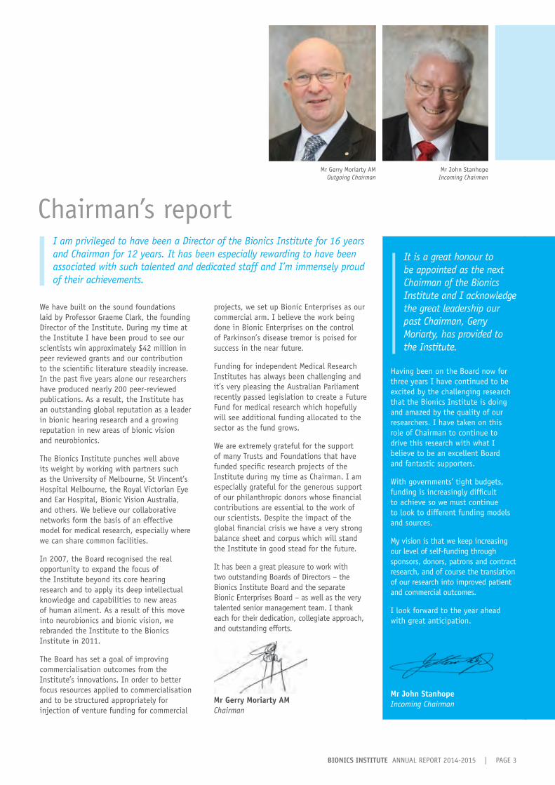
Mr Gerry Moriarty AMOutgoing Chairman
Mr John StanhopeIncoming Chairman

PAGE 4 | BIONICS INSTITUTE ANNUAL REPORT 2014-2015
One of my key roles as Director of the Bionics Institute is to represent our interests, and those of the broader Medical Research Institute community to government. Over the past year we have welcomed senators and MPs from federal and state governments to the Institute to see our translational research first hand. I highlight our capacity to deliver real outcomes for patients in the short to medium term, while also contributing to Australia’s advanced manufacturing sector.
The past year has seen the Medical Research Future Fund (MRFF) take shape and become a reality. This federal government initiative has brought hope to the medical research community that investment in our sector will improve. This is especially important in the light of Australia’s low OECD ranking in terms of funding health related research. In 2014 we saw the awarding of National Health and Medical Research (NHMRC) grants, the largest government funder of medical research in Australia, slip to its lowest point in history with only 15 percent of applications successful. Most importantly, a boost in medical research funding will reduce the burden of disease on individuals as well as reduce the burden on our health care system in the long term.
We continue to grow our core competence in hearing research and reached milestones in our bionic vision and neurobionics research programs. We received welcome news in October 2014 with the NHMRC funding a three year project grant to trial the next generation bionic eye. Together with our colleagues from the Centre for Eye Research Australia and National Information and Communications Technology Australia, this research will build on the enormous success of the clinical trial of the prototype device in three blind volunteers completed in July 2014. This new study will be a significant step forward in the development of an Australian bionic eye; the next generation device will be fully implantable, allowing recipients to benefit from its day-to-day use rather than being limited to a laboratory setting.
Our neurobionics program also secured the critical support required to progress our development of an advanced deep brain stimulation device to treat the debilitating symptoms of Parkinson’s disease and essential tremor. With the continued partnership and welcome funding provided by Colonial Foundation, we are now poised to bring together the varied aspects of our neurobionics program to achieve our goal. These include our improved understanding of the characteristics of movement disorders and device development outcomes through our
preclinical and clinical work. This year we were very pleased to welcome Dr Wesley Thevathasan (Neurologist, Royal Melbourne Hospital) to the neurobionics team. As our first Lions International Neurobionics Fellow he will work with our researchers to evaluate advanced deep brain stimulation systems to treat movement disorders.
Our bionic hearing research continues to grow and address a broad range of issues facing hearing impaired people. Our clinical and preclinical research uses state-of-the-art technologies and techniques to understand and explore ways of improving the quality of hearing provided by cochlear implants and hearing aids. This year we embarked on a major study to understand the variability in hearing outcomes in people who receive bilateral (both ears) cochlear implants. In early 2015, we were delighted to host a six-month visit by Professor Ruth Litovsky, a Fulbright Senior Scholar from the University of Wisconsin, who worked with our researchers on this project. This study will underpin future exploration of ways to ensure that bilateral users gain maximum benefit from using two devices.
Two new collaborative ventures are providing us with the opportunity to address new areas of clinical need and deliver bionic solutions to problems with visceral organs. We are part of the only Australian team taking part in GlaxoSmithKline’s global Bioelectronics Innovation Challenge – a challenge to produce an implantable device that modulates the activity of peripheral nerves to restore healthy function to organs. We are also bringing our expertise in bionic device design to a new collaborative project, recently funded by the Australian Research Council, with Neosphincter Pty Ltd and University of Melbourne researchers aimed at developing a novel treatment for severe urinary incontinence. Both of these projects represent an exciting expansion of our capabilities and collaborative networks.
In the past year, we have greatly enhanced our capacity to produce experimental devices for hearing, vision, and deep brain stimulation. We have invested significant funds in the refurbishment and equipment purchases for a new stand-alone electrode manufacturing facility at our laboratories in St Vincent’s Hospital Melbourne. This new manufacturing hub will allow us to produce sophisticated and custom-made prototype devices and deliver them rapidly. Funding for equipment for this new facility was generously provided by The Ian Potter Foundation, Lions Clubs International, and the federal Department of Health.
As Director, it is my privilege to lead the dedicated staff and students at the Bionics Institute who, every day, strive towards finding new treatments for serious medical conditions. We work closely with our clinical, research and industry partners to translate our research into real-life solutions – solutions that will transform the lives of people for years to come.
Director’s reportProfessor Robert K ShepherdDirector

BIONICS INSTITUTE ANNUAL REPORT 2014-2015 | PAGE 5
The Institute’s success in delivering practical health outcomes for people with a variety of conditions stems from our multidisciplinary and collaborative approach to research. Our widespread research partnerships remain crucial to our success and are too numerous to list here, but I particularly want to acknowledge our affi liations with two world-class hospitals – the Royal Victorian Eye and Ear Hospital and St Vincent’s Hospital Melbourne. We are a proud member of the Aikenhead Centre for Medical Discovery on the St Vincent’s campus. Our Institute forms the Medical Bionics Department within the University of Melbourne’s Faculty of Medicine, Dentistry and Health Sciences and we work closely with many colleagues within the Faculty. We also have very close ties with the School of Engineering at the University of Melbourne and the Centre for Eye Research Australia.
In the past year we have seen the recognition of worthy colleagues’ contributions to the creation and commercial development of the cochlear implant. Professor Graeme Clark AC, Director Emeritus of the Bionics Institute, was awarded the 2015 Russ prize by the US National Academy of Engineering in January. This prestigious award recognises a bioengineering achievement in widespread use that signifi cantly improves the human condition. Adjunct Professor Jim Patrick, Cochlear Limited’s Chief Scientist and an Institute Honorary Research Fellow, was awarded an Order of Australia in January and an ATSE Clunies Ross Lifetime Achievement Award in May. I congratulate Graeme and Jim on these well-deserved awards.
As a not-for-profi t organisation we are reliant on funding from multiple sources to maintain our research programs. We are very grateful for the generous commitment of the trusts and foundations that provide a crucial source of research funding. We would particularly like to acknowledge the support we receive from the Garnett Passe and Rodney Williams Memorial Foundation for hearing research, and Colonial Foundation which has been instrumental in the development of our neurobionics program. We also acknowledge the Victorian Lions Foundation which provides a Hearing Research Fellowship and, this year, supported our inaugural Neurobionics Research Fellow.
We have a dedicated group of research volunteers and Ambassadors that give generously of their time. They assist both in our clinical studies and our community involvement programs. As end-users of our research their insights greatly assist our development of new technologies and improvements to existing devices. Their presentations at community events promote our research and provide fi rst-hand accounts of their experiences with bionic devices. Your continued support and efforts are very much appreciated.
I wish to thank our talented Board and the Board of our commercial arm, Bionic Enterprises, for their contributions in building a dynamic and productive research environment. The Board’s direction and counsel throughout the year, and their enthusiasm for the Institute’s cause is unwavering. I would particularly like to acknowledge the leadership of Mr Gerry Moriarty AM who, after 12 years of dedicated service as Chairman of the Board, will step down in late 2015. We owe a great deal of thanks to Gerry for his consummate support, advice, and guidance over the past 12 years. In farewelling Gerry we welcome John Stanhope as our next Board Chairman. John brings great corporate experience following a long and distinguished career at Telstra, service on a number of corporate Boards, as well as being the Chancellor elect of Deakin University.
Finally, I take this opportunity to sincerely thank our researchers, students, support and executive staff for their unfl agging dedication. The research highlighted in the following pages is a testament to your commitment to delivering solutions to otherwise intractable medical problems. Your work is the foundation of the Institute’s international reputation as a leader in the development of innovative bionic technologies that improve human health.
Professor Rob ShepherdDirector
Professor Robert K ShepherdDirector
PAGE 6 | BIONICS INSTITUTE ANNUAL REPORT 2014-2015 ANNUAL REPORT 2014-2015BIONICS INSTITUTE
Due to the scale, scope and multidisciplinary approach needed to achieve these goals, a high level of scientifi c, operational and commercial acumen across a number of academic, hospital and industry partners is required. We have the relationships, policies, structures and right people in place to effectively manage this constant ‘innovation loop’ between researchers, clinicians, patient groups, and industry. Since 2000, this approach has led to four companies spun out and/or incubated by the Bionics Institute.
In addition to the strong partnerships with clinicians, we have forged strong relationships with patient care and advocacy community groups as well as individual patients. These strong links between research, clinicians, patients and their community groups are critical for the successful development of any medical device. In order to achieve our strategic commercial objectives, an increasing emphasis is placed on growing investment and commercial funding for research, and achieving commercial and clinical outcomes from our translational and fundamental research activities in each of the three key programs: bionic hearing, bionic vision, and neurobionics.
Our management of intellectual property (IP) enables and rewards successful translational research, innovation, publication, commercialisation, and clinical outcomes. We have 10 active patent families housing 31 individual and active patent applications across our three key programs.
Business DevelopmentWe have increased our focus on business development activities including the provision of collaborative and contract research services to industry, universities, and other R&D groups. The range of services includes medical device preclinical safety and effi cacy testing, and custom electrode prototyping, fabrication, and safety and effi cacy testing.
Bionic HearingWe continue to build further on the strong base of collaboration and partnership with a number of organisations, including Cochlear Ltd. Cochlear Ltd has delivered hearing and quality of life benefi ts to many adults and children world-wide, having sold over 300,000 cochlear implants. Several research contracts, spanning a range of hearing-related manufacturing and R&D projects have either been completed, are underway or under discussion. We entered into a signifi cant partnership with Pfi zer that aims to develop clinically translatable therapies to treat hearing disorders. The partnership will bring together Pfi zer’s experience in drug development and our preclinical research experience.
Bionic VisionTogether with our clinical partners, we have played a pivotal role in the development, regulatory approval, and clinical translation of Australia’s prototype bionic eye. This work resulted in the successful implantation, switch-on, and perceptual testing of three blind patients with the prototype device. Our current research, as a core party of Bionic Vision Australia, builds on the great success of the small scale clinical trial completed in 2014. The forthcoming clinical trial will test the safety and effectiveness of the next generation wide-view bionic eye; an exciting development in our commitment to those living with degenerative eye diseases. We will continue to provide research, intellectual property and commercial expertise in order to achieve Bionic Vision Australia’s aims to provide proof-of-concept for two bionic eye products – the wide-view and the high-acuity devices. The Institute has fi led 17 bionic eye patent applications.
Spanning the critical interface between industry and academia, we are a leader in the provision and translation of knowledge into improved health and wealth – innovative health solutions for the community and increased wealth in the form of a pipeline of new products and a skilled workforce.
Translating research into patient benefi ts and medical bionic products

BIONICS INSTITUTE ANNUAL REPORT 2014-2015 | PAGE 7 ANNUAL REPORT 2014-2015 BIONICS INSTITUTE
NeurobionicsThe goal of our neurobionics program is to leverage cochlear implant and other platform technologies into complete clinical solutions for chronic, untreatable conditions of the nervous system. Bionic Enterprises, our commercial subsidiary business, provides the commercial pathway for this program by taking neurobionic devices for the treatment of a range of poorly treated neurological conditions into early clinical trials.
To strengthen the underlying business case for funding support of our neurobionics program, an economic impact study by an expert, external group was completed late in 2014. This report underlined the ‘signifi cant economic impact’ the neurobionics program will have on the Australian medical technology industry and will form an integral part of the investment proposition to a number of potential funding bodies and investors.
We are involved in two initial clinical studies that are the critical fi rst step in demonstrating safety and preliminary effi cacy, and engaging clinicians early in the clinical and commercial pathway. The fi rst study is an externalised lead study for movement disorders that aims to identify suitable biomarkers, optimise stimulation parameters, and evaluate closed-loop stimulation in participants with movement disorders. The second is a clinical trial for devices to treat obsessive compulsive disorder. This study utilises commercial deep brain stimulation devices with outcomes being assessed by means of sophisticated functional imaging as well as conventional clinical assessments.
BIONIC ENTERPRISESBionic Enterprises (BE) was established in 2011 to provide the commercial pathway for our neurobionics research program. The Institute has invested $1.5m into BE to enable the company’s early development.
BE is focussed on neurobionics – the development of devices that monitor neural activity in the brain, spinal cord or elsewhere in the body and/or stimulate nerves electrically for therapeutic applications.
BE is committed to fast tracking the development and reducing the cost of bringing new, safe and effective medical device products to the market. The lead product is an advanced device to treat the motor symptoms of Parkinson’s disease. Signifi cant progress has been achieved with the development of customised components essential for this device, and as part of the ‘platform’ for subsequent devices. Negotiations are currently underway to secure commercial access to some key technology. Other products will follow that treat a range of disabling neurological conditions including other movement disorders, epilepsy, severe chronic pain and certain psychiatric disorders.
All devices meet a clear clinical need and address a gap in the market. Most conditions are of relatively high incidence with 10-30% of sufferers poorly treated with existing treatments.
This strategy builds on the company’s core strengths, addresses a clear clinical need, and provides a commercially attractive product for strategic partnering, licencing or acquisition via spin-out companies.
Commercialisation• Innovation and new industries• Economic development• Manufacturing and jobs• Investment
Translational research & development• Early product development• Prototyping• Proof of concept• Early clinical trials
INDUSTRYACADEMIA
Discovery Research• Knowledge (publications)• Students
PAGE 8 | BIONICS INSTITUTE ANNUAL REPORT 2014-2015

BIONICS INSTITUTE ANNUAL REPORT 2014-2015 | PAGE 9
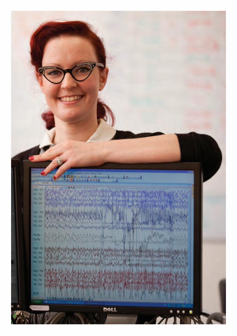
IMPROVING CLINICAL OUTCOMES FOR HEARING-IMPAIRED CHILDREN AND ADULTS
Babies as young as four months are now receiving a cochlear implant. Therefore, a method is urgently needed to be able to program the implant without asking the person when they hear a sound or how loud it is.
The current method that aims to do this is not very successful at accurately predicting the individual program levels needed for each person. We aim to use the brain’s response to sound (termed a CAEP) to measure the hearing of cochlear implant patients. We are developing ways to do this in the minimum time and by using the electrodes of the cochlear implant to make the measurements. Although this type of measurement has been used before to determine the lowest level sound a person can hear, it takes a long time, and needs the application of electrodes on the scalp. We are therefore developing the technology to use the cochlear implant itself to make the measurements.
Using the CAEP measurement, we have shown that we can accurately predict the current level at which a person begins to hear a sound. This means that it is a good measurement for automatic programming. We have tested different methods to analyse the CAEP signals to reduce the amount of repeated measurements that are needed (and hence the time for measurement), and the number of measurement electrodes needed.
We are currently testing different methods to obtain the CAEP signals from the brain using the internal electrodes of the cochlear implant as measurement electrodes. After this has been achieved and optimised, the method can be adapted into the clinical fi tting software so that a person’s program can be set ‘at the push of a button’.
The degree of benefi t gained from hearing aids and cochlear implants varies widely across users. Unfortunately, a proportion of cochlear implant users cannot understand speech with their device unless they combine it with lip reading.
While much of the variability in speech understanding is accounted for by factors such as the duration of deafness and experience with the device, a signifi cant portion of the variability amongst cochlear implant users remains unexplained.
Our goal is to understand why some people do not understand speech with a cochlear implant as well as other people. One of our projects aims to understand why this is so, to develop a way to identify these people before they get their implant, and to guide the optimal training for them after they get their implant.
In the past year we have:
• Used a variety of approaches to test the feasibility and safety of a new cochlear implant stimulation strategy aimed at conveying more precise and clearer sounds
• Examined how deafness changes the hearing brain and how it adapts to the electrical stimulation provided by cochlear implants
• Continued to develop a quick and objective method to program cochlear implants, particularly important for very young children
• Investigated gene therapy as a way to create new sensory cells and preserve the auditory nerve following hearing loss
• Investigated nanotechnologies as a means to introduce therapeutic drugs into the cochlea
Our bionic hearing program aims to improve the quality of hearing provided by cochlear implants and hearing aids. Our research explores new clinical tools and treatments, new stimulation techniques and new electrodes to improve clinical outcomes for those with hearing impairment.
Bionic hearing

PAGE 10 | BIONICS INSTITUTE ANNUAL REPORT 2014-2015 ANNUAL REPORT 2014-2015 ANNUAL REPORT 2014-2015 | BIONICS INSTITUTE
There is evidence that the brain responds to deafness by re-assigning the hearing part of the brain to other purposes such as vision, and this might be the reason for a cochlear implant to not work well in certain individuals. We are using a new brain imaging method (termed fNIRS) that uses laser light to measure the differences between people in how speech is processed in their brains. fNIRS has many advantages over standard brain imaging techniques since it is non-invasive, silent, low cost, and importantly, compatible with implanted devices.
fNIRS provides a means of observing and comparing the interconnections of different brain regions when a participant is resting in quiet conditions and when they are listening to sounds. Pilot data from a cochlear implant user has shown that we can use fNIRS to measure the brain activity resulting from listening to speech, and that listening to speech activates the visual region of the user’s brain. We have compared the brain’s resting state connectivity between cochlear implant users and normal-hearing people, and have shown differences between people that may be related to how well they understand speech with a cochlear implant.
We now have projects underway to compare the brain activity in implant users who have different abilities for understanding speech, and to compare the pre-implant imaging measurements with post-implant speech understanding. We will develop clinically useful analysis techniques that can be automated to predict implant outcomes, and also use fNIRS to evaluate the effectiveness of different types of post-implant training methods.
To understand speech a listener needs to be able to hear the different frequencies contained in the signal and to hear how they differ in strength from one frequency region to another.
This task requires both the ability to hear changes in intensity in one channel and also to be able to compare intensity differences across channels, regardless of the actual intensities. It has been suggested that these two abilities have different neural mechanisms.
We are testing this idea with the aim of developing an easy way to predict whether a new implant recipient will have diffi culty developing speech understanding. Preliminary data show that cochlear implant users with good speech perception have excellent ability to detect intensity differences on a single electrode and can also detect small changes of relative intensity differences across two electrode positions: those with poor speech understanding have more diffi culty in these tasks. We are continuing to recruit participants who have either good or poor speech perception so that we can evaluate our hypothesis reliably.
If successful in showing high predictive power for speech understanding, the test method will be developed into a clinically useful tool for prognosis in the clinic, and objective measures of the same abilities will also be developed to evaluate speech understanding development in children with a cochlear implant.
The methods currently used to detect and treat hearing damage focus on sensory impairment – an impaired ability to detect very quiet sounds.
However, this is limiting because people both with and without ‘normal’ ability to detect sounds may still suffer from disabling problems understanding speech and other complex sounds. We are developing tools that can detect ‘signatures’ of neural degeneration in brain signals recorded from the scalp. These tools focus on detecting neural activity that supports functional hearing ability, rather than only sensory impairment.
We are currently testing research participants with different hearing losses and ages. The goal of this research is to develop an objective measure of the ability of the auditory system to encode temporal (timing) fi ne structure to optimise the benefi ts that hearing devices can provide. With further development, similar measures could also be used to detect changes to functional hearing due to neuronal degradation before the onset of permanent threshold shifts and hearing loss.

BIONICS INSTITUTE ANNUAL REPORT 2014-2015 | PAGE 11
IMPROVING COCHLEAR IMPLANTS THROUGH NOVEL STIMULATION STRATEGIES
The cochlear implant has had a profound impact on the treatment of those with severe-to-profound sensorineural hearing loss, with more than 300,000 recipients world-wide benefi ting from existing devices.
While cochlear implants are highly effective at conveying speech in quiet conditions, diffi culties arise in noisy environments such as a restaurant or party. The rich texture of other complex sounds, such as music, is also not conveyed effectively. It is thought that these problems may be partly due to the broad spread of electrical current from each electrode within the implant due to the conductive nature of the fl uid-fi lled inner ear. In turn, this results in broad activation of the auditory neurons that convey information to the brain: in the process, some important features of a complex sound are lost.
New stimulation strategies are therefore needed in order to improve hearing outcomes for implant recipients. Current focussing using simultaneous stimulation via multiple electrodes is considered the most likely emerging technology to improve the resolution of the electrical stimulus and provide improved hearing for cochlear implant users.
We have explored this new technique using a number of different approaches. One project, completed this year, used a research device in fi ve patients in Melbourne to test the perceptual outcomes of the current-focussing technique. We found that using this stimulation method improved vowel recognition. In this clinical study the electrical stimuli were restricted to a range where safety was already established. In order to explore this technique further and determine its limits, we are conducting two other studies.
We are investigating the electrode-tissue interface and determining the safety limits of simultaneous current-focussing stimulation using conventional electrodes over a long period of time. In addition, we are exploring whether novel electrode coatings can be used to enhance the safety of this technique. In a complementary study, the ability of the current-focusing technique to produce more selective activation of the auditory brain is being assessed. We have found that this technique does indeed lead to narrower regions of activation in the auditory midbrain, albeit at higher threshold. Importantly, this improved selectivity is maintained in inner ears with low auditory nerve survival (as occurs in sensorineural hearing loss).
These studies have the potential to contribute to a new generation of cochlear implants that will provide a more faithful reproduction of complex sounds. If successful, this work could also provide the opportunity for current-focussing technologies to be applied to other implantable devices such as deep brain stimulation, a rapidly growing therapy where having control over the extent of activation of neural tissue is equally important.
Cochlear implantation in both ears is becoming increasingly common and while there are benefi ts to the user, they fall short of expectations.
The clue to why this is the case lies in how the brain uses the information from the two ears (termed binaural processing) and computes the location of sounds. In order to localise a sound in space the auditory brain primarily uses differences in the timing and intensity of the sound reaching each of the two ears. The brain’s ability to compare the information received by two ears also provides benefi ts when listening to speech in noisy environments.
In long-term deafness the brain’s sensitivity to the timing (temporal) differences between the two ears degrades. In addition, for technical reasons, the cochlear implant does not convey the detailed structure of ongoing timing differences contained within complex sounds such as speech. One of our projects aims to establish ways of improving the processing of binaural temporal cues and test the hypothesis that experience with high-fi delity timing information will enhance the brain’s processing of this cue and lead to improvements in performance. Our results so far suggest that the use of bilateral cochlear implants does not reverse the degradation of the ability to process timing differences that occurs following long-term deafness. Our next step is to examine whether experience with appropriate timing cues can improve this degraded ability.
The outcomes of this study will help drive the technical innovations required to maximise the benefi ts and investment of receiving two cochlear implants.
PAGE 12 | BIONICS INSTITUTE ANNUAL REPORT 2014-2015 ANNUAL REPORT 2014-2015BIONICS INSTITUTE
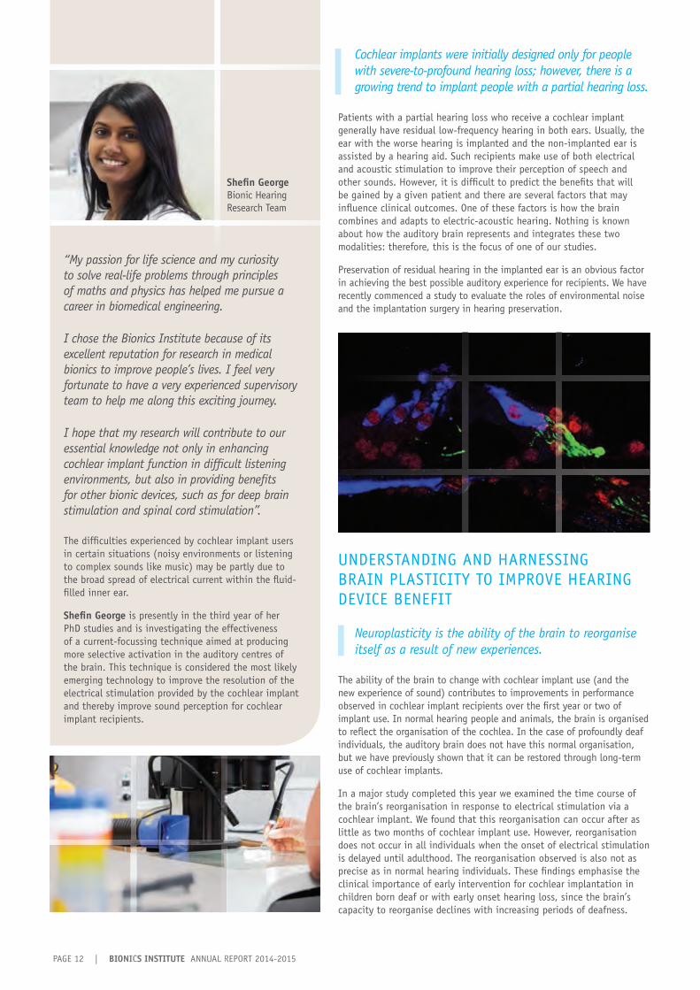
Shefi n GeorgeBionic Hearing Research Team
Cochlear implants were initially designed only for people with severe-to-profound hearing loss; however, there is a growing trend to implant people with a partial hearing loss.
Patients with a partial hearing loss who receive a cochlear implant generally have residual low-frequency hearing in both ears. Usually, the ear with the worse hearing is implanted and the non-implanted ear is assisted by a hearing aid. Such recipients make use of both electrical and acoustic stimulation to improve their perception of speech and other sounds. However, it is diffi cult to predict the benefi ts that will be gained by a given patient and there are several factors that may infl uence clinical outcomes. One of these factors is how the brain combines and adapts to electric-acoustic hearing. Nothing is known about how the auditory brain represents and integrates these two modalities: therefore, this is the focus of one of our studies.
Preservation of residual hearing in the implanted ear is an obvious factor in achieving the best possible auditory experience for recipients. We have recently commenced a study to evaluate the roles of environmental noise and the implantation surgery in hearing preservation.
UNDERSTANDING AND HARNESSING BRAIN PLASTICITY TO IMPROVE HEARING DEVICE BENEFIT
Neuroplasticity is the ability of the brain to reorganise itself as a result of new experiences.
The ability of the brain to change with cochlear implant use (and the new experience of sound) contributes to improvements in performance observed in cochlear implant recipients over the fi rst year or two of implant use. In normal hearing people and animals, the brain is organised to refl ect the organisation of the cochlea. In the case of profoundly deaf individuals, the auditory brain does not have this normal organisation, but we have previously shown that it can be restored through long-term use of cochlear implants.
In a major study completed this year we examined the time course of the brain’s reorganisation in response to electrical stimulation via a cochlear implant. We found that this reorganisation can occur after as little as two months of cochlear implant use. However, reorganisation does not occur in all individuals when the onset of electrical stimulation is delayed until adulthood. The reorganisation observed is also not as precise as in normal hearing individuals. These fi ndings emphasise the clinical importance of early intervention for cochlear implantation in children born deaf or with early onset hearing loss, since the brain’s capacity to reorganise declines with increasing periods of deafness.
“My passion for life science and my curiosity to solve real-life problems through principles of maths and physics has helped me pursue a career in biomedical engineering.
I chose the Bionics Institute because of its excellent reputation for research in medical bionics to improve people’s lives. I feel very fortunate to have a very experienced supervisory team to help me along this exciting journey.
I hope that my research will contribute to our essential knowledge not only in enhancing cochlear implant function in diffi cult listening environments, but also in providing benefi ts for other bionic devices, such as for deep brain stimulation and spinal cord stimulation”.
The diffi culties experienced by cochlear implant users in certain situations (noisy environments or listening to complex sounds like music) may be partly due to the broad spread of electrical current within the fl uid-fi lled inner ear.
Shefi n George is presently in the third year of her PhD studies and is investigating the effectiveness of a current-focussing technique aimed at producing more selective activation in the auditory centres of the brain. This technique is considered the most likely emerging technology to improve the resolution of the electrical stimulation provided by the cochlear implant and thereby improve sound perception for cochlear implant recipients.
BIONICS INSTITUTE ANNUAL REPORT 2014-2015 | PAGE 13
PREVENTING PROGRESSIVE HEARING LOSS
Sensorineural hearing loss is caused by damage to the cochlea’s sensory (hair) cells and/or the auditory neurons that convey information to the brain.
Protecting and regenerating these sensory cells is important to prevent further loss of hearing and even enable hearing loss to be reversed. One of our research projects is using gene therapy techniques to introduce genes into cells to change their function. For example, we discovered that the introduction of neurotrophin (nerve survival factors) genes causes cells to release neurotrophic factors which in turn protect auditory neurons from degeneration after hearing loss.
Recently, we discovered that the introduction of a gene called Atoh1 can make some of the supporting cells of the cochlea transform into new hair cells. We showed that new hair cells could be formed in a region of the cochlea that had been damaged by loud noise.
Furthermore, we found that the presence of new hair cells provided signifi cant protection to nearby auditory nerve fi bres and supporting cells suggesting that it may be possible to reconnect the hair cells to nerve fi bres. However, there was no evidence of connections between the two cell types (i.e., synapse formation), and hearing was not restored. We also know that Atoh1 gene therapy alone cannot fully convert supporting cells into mature hair cells after hearing loss. This suggests an additional factor is necessary for the new hair cells to fully mature.
Despite these current unknowns, gene therapy remains an exciting potential way to restore functional hearing following damage to the cochlea.
The delicate hair cells in the cochlea are very sensitive to trauma such as loud noise, some anti-cancer drugs, and a certain class of antibiotics used to fi ght infection.
When the hair cells die, the auditory neurons that make connections with them also die resulting in a permanent hearing loss. We have shown previously that administering a nerve survival factor (called brain-derived neurotrophic factor, BDNF) to the auditory neurons can prevent their degeneration and death.
We are exploring a number of approaches to introduce neurotrophins into the inner ear in order to protect and/or repair functional hearing following damaging noise exposure. One of these studies is investigating a new drug delivery system based on nanoengineering to prevent the progression of hearing loss. We have shown that nanoengineered particles loaded with BDNF can be used to release effective amounts of therapeutics into the cochlea. We found that auditory nerve survival was signifi cantly greater in cochleae that were implanted with BDNF-loaded nanoparticles compared to the control condition.
The ultimate goal of this research is to provide a treatment to prevent progressive hearing loss, especially important in cochlear implant recipients who have some residual hearing.
BIONIC HEARING RESEARCH TEAM
Dr Yuri Benovitski Prof Peter Blamey Ms Nicole Critch Mr Will Cross A/Prof James Fallon Ms Brianna Flynn Dr David Garrett Ms Melanie Gault Ms Catherine Gaunt Ms Shefi n George Dr Lisa Gillespie Dr Hamish Innes-Brown Ms Natnicha Inthavong Prof Dexter Irvine Dr Samuel Irving Dr Elisha King Ms Cara Lo Dr Mohammad Maarefvand Mr Darren Mao Dr Jeremy Marozeau
Prof Hugh McDermott Prof Colette McKay Ms Amy Morley Ms Alison Neil Ms Madeline Nicholson Dr Matt Petoe Mr Graeme Rathbone Dr Rachael Richardson Dr Natalie Rickard Mr Damian Robb Dr Philipp Senn Dr Adnan Shah Prof Rob Shepherd Dr Mohit Shivdasani Mr Nick Smale Mr Bart Steensma Ms Renee Tsongas Dr Andrew Wise Ms Xin Zhou
OUR COLLEAGUES
Dr Patrick Atkinson (Stanford University USA) Mr Robert Briggs (Royal Victorian Eye and Ear Hospital) Prof Paul Carter (Cochlear Ltd) Prof Frank Caruso (University of Melbourne) Prof Stuart Cogan (EIC Laboratories USA) Dr Jiwei Cui (University of Melbourne) Prof Wael El Deredy (University of Manchester UK) Ms Ya Lang Enke (Cochlear Ltd) Dr Tom Francart (KU Leuven Belgium) Dr Natalie James (Cochlear Ltd; University of Sydney) Mr Shaun Kumar (Cochlear Ltd) Ms Natalie Le (Cochlear Ltd) Prof Ruth Litovsky (University of Wisconsin USA) Prof Stephen Lomber (University of Western Ontario Canada) Dr Stoph Long (Cochlear Ltd) Prof Stephen O’Leary (University of Melbourne) Dr Sat Pannu (Lawrence Livermore National Laboratories USA) A/Prof Jim Patrick (Cochlear Ltd) Mr Frank Risi (Cochlear Ltd) Prof David Ryugo (Garvan Institute) Dr Karim Seghouane (University of Melbourne) Dr Zach Smith (Cochlear Ltd) Mr Daniel Smyth (Cochlear Ltd) A/Prof Paul Stoddart (Swinburne University of Technology) Dr Brett Swanson (Cochlear Ltd) Dr Helmut Thissen (CSIRO) Mr Alex Thompson (Swinburne University of Technology) Dr Angela Tooker (Lawrence Livermore National Laboratories USA) Mr Claudiu Treaba (Cochlear Ltd) Dr Kristien Verhoeven (Cochlear Ltd) Dr Scott Wade (Swinburne University of Technology) Prof Jan Wouters (KU Leuven Belgium)

PAGE 14 | BIONICS INSTITUTE ANNUAL REPORT 2014-2015
BIONICS INSTITUTE ANNUAL REPORT 2014-2015 | PAGE 15
THE NEXT GENERATION BIONIC EYE
In 2014, a clinical trial of our prototype wide-view bionic eye was successfully completed. The aim of this wide-view device is to provide patients with the ability to navigate their environment and assist in daily tasks.
Since completion of this fi rst clinical trial, we have designed and constructed an upgraded version of the prototype device. As with the original device, the next generation electrode array will be implanted between the two back layers of the eye (suprachoroidal location). This novel approach requires relatively easy surgery compared to other possible locations for electrodes within the eye and proved to be safe, stable, and reliable. Importantly, it was shown to be effective in eliciting visual perceptions in the recipients, allowing them to identify shapes, perceive movement, and undertake unassisted navigation tasks.
The next generation device will contain an array of 44 electrodes (compared to 24 in the prototype), have double the resolution, and use the vision processing strategies based on those developed for the prototype device. Before this device can be implanted in patients, we need to ensure it is safe and effective for long-term use. To do so, we have completed a number of preclinical studies, in collaboration with surgeons, clinicians, and pathologists to test that the presence of the electrode array and electrical stimulation do not cause damage to the retina.
Based on our successful clinical trial of a bionic eye prototype, the Bionics Institute and collaborators were awarded National Health and Medical Research Council (NHMRC) funding to undertake a clinical trial with an advanced ‘take home’ bionic eye.
This trial is due to commence in 2016 and in the past year we have prepared for this next cohort of bionic eye recipients. Our recent work has been concerned with designing a portable vision processor that can be used in their everyday activities.
The new clinical trial will test the safety and effectiveness of the next generation wide-view bionic eye. Whereas the prototype device used an external plug to connect the electrodes to the lab equipment, the next generation device will be fully implantable and portable. This will allow the continued testing of the recipients’ perceptions within our purpose-built laboratory but also enable the recipients to use the device in everyday life. The complete device will incorporate an external vision processing unit that will connect to a small video camera contained within spectacles. This is a signifi cant step forward in the development of an Australian bionic eye since it will allow the recipients to receive maximum benefi t from using the device and the researchers will be able to obtain far more information about the device’s capabilities.
In the past year we have:
• Conducted pre-clinical safety studies of the next generation wide-view bionic eye
• Designed and produced a portable vision processor suitable for the next cohort of bionic eye recipients to use in everyday life
• Examined ways to create ‘virtual’ electrodes in a bionic eye
• Continued to explore alternative stimulation strategies to optimise the evoked visual perceptions
• Conducted safety and design studies to position and secure a prototype high-acuity bionic eye (made from conductive diamond) on the retina’s surface
We designed, manufactured, and rigorously safety-tested Australia’s fi rst prototype bionic eye as part of Bionic Vision Australia. A clinical trial using this device was successfully completed in 2014 in three patients with retinitis pigmentosa, the most common cause of inherited blindness. We now prepare for the clinical trial of the next generation device.
Bionic vision

PAGE 16 | BIONICS INSTITUTE ANNUAL REPORT 2014-2015
Our visual psychophysics laboratory contains a suite of tests to record how visual percepts produced by bionic eye devices can be controlled by varying the parameters of electrical stimulation.
These results inform the vision processing strategies that are used to provide real-world visual information to the user. We have continued to integrate our second generation wide-view device into the psychophysics test suite and add additional test procedures to characterise and optimise bionic vision.
Over the past year, testing with a bionic eye simulator has shown that participants are able to use auditory cues to improve their performance in lab-based tasks. We also confi rmed that non-sighted participants could readily understand these same cues. With our collaborators at the Centre for Eye Research Australia, we will use these sensory augmentation techniques to improve functional outcomes for bionic eye recipients.
NEW STIMULATION STRATEGIES
In retinitis pigmentosa, the light-sensitive cells of the retina (photoreceptors) degenerate but the retinal neurons that transmit information to the brain remain.
The bionic eye electrically stimulates these surviving retinal cells to provide a sense of vision for people with degenerative eye diseases. The electrode array implanted in the retina delivers electrical stimuli in the form of current pulses that activate nearby cells. This activity is transmitted to the visual areas of the brain (visual cortex) and elicits a perception of a localised fl ash of light (termed a phosphene).
The resolution of the bionic eye is currently limited by the diffuse spread of activity that results from electrical stimulation. By optimally choosing electrode currents, this spread of activity can be altered in a predictable way. To choose these currents, we have used a computational model of the responses of the visual cortex evoked by many thousands of different patterns of electrical stimulation. The resulting model can accurately predict the cortical activity in response to any particular set of stimulation currents. This computer model will provide a basis for optimising stimulation strategies to evoke ideal patterns of activity in the visual cortex.
The bionic eye uses a train of constant, identical electrical pulses to create the perception of light. However, the visual system adapts to the constant stimulation and the light appears initially bright, but quickly fades.
This perceptual ‘fading’ can make it diffi cult for a bionic eye to produce a stable image for recipients. We are investigating the use of constantly varying sequences of stimulation to prevent perceptual fading. Pseudorandom sequences (known as maximal-length sequences) are used to change the properties of each pulse in the sequence. From a distance, the overall sequence of stimulation appears the same but, examined closely, the timing or size of each pulse is changing. It is hoped that this will reduce the adaptation that occurs in the visual system, resulting in brighter, longer lasting fl ashes of light.
While the fi rst generation device stimulated the retina with a single electrical pulse at a time, the second generation device will use pairs of pulses.
To test the long term safety of stimulating with pairs of pulses, a more advanced stimulation processor was required. The new stimulation processor was developed to be compact, lightweight, and run for days at a time on battery power, while producing twice as much stimulation as before. 3D printing technology was used to rapidly prototype and build a case to protect the sensitive electronics. In addition, wireless capabilities have been included to allow for continual measurements of eye health and the ability to check for faults in the device.
The present designs of bionic eye devices have the potential to provide patients with vision to gain orientation and mobility, and the ability to read letters and identify shapes.
The results from our prototype device were very promising; however, to achieve detailed (high resolution) vision, different stimulation techniques may be required.
At present, retinal implants have limited resolution and simply increasing the number of electrodes in the array is limited by design issues. The phosphenes evoked by stimulation of different electrodes may be too large (and therefore overlap with one another) and/or have inconsistent shapes. We are therefore investigating the use of new electrical stimulation techniques in an effort to improve the resolution of the bionic eye and provide more meaningful images to patients.
A number of new stimulation techniques are being investigated. One of these uses an approach also being explored in the bionic ear. In cochlear implants, ‘virtual’ electrodes can be successfully created by simultaneous stimulation of adjacent electrodes (a strategy called current steering). We are therefore investigating whether this stimulation strategy can be adapted for bionic eye devices. The results so far suggest that current steering can alter activation patterns in the visual brain and create ‘virtual’ responses. The results of this study will inform the development of new electrode arrays that can take advantage of current steering.
Another approach to improving the resolution of the bionic eye is to reduce the size of the individual electrodes to produce a higher density array. However, there is a safety issue associated with this approach. The solution may lie in several new electrode materials being explored by our bionic vision team. These experiments require careful characterisation of the electrical properties of novel materials, as well as studies of their safety and biocompatibility.

BIONICS INSTITUTE ANNUAL REPORT 2014-2015 | PAGE 17
“I was drawn to the Bionics Institute by the work being done on bionic eyes and was fortunate enough to be placed here through the Undergraduate Research Opportunities Program (UROP) during my undergraduate studies. With my background in biomedical science and electrical engineering this research was a perfect fi t. I have immensely enjoyed working with such intelligent and enthusiastic people, so much so that I have returned this year to start a PhD so I can continue to be part of the cutting edge research that goes on here,” Patrick said.
Current bionic eye devices use a train of constant, identical electrical pulses to create the perception of light. However, the visual system adapts to the constant stimulation and the light appears initially bright, but quickly fades. This ‘perceptual fading’ can make it diffi cult for a bionic eye to produce a stable image for recipients.
Patrick Thien commenced his PhD in 2015 and aims to explore ways of counteracting this problem. To accomplish this, he is fi rst developing and testing the safety of a new stimulation processor that has greater fl exibility in the electrical pulses it can produce.
Patrick ThienBionic Vision Research Team
“I was drawn to the Bionics Institute by the work being
THE DIAMOND BIONIC EYE As part of our involvement in Bionic Vision Australia, we have been assessing diamond as a potential new material for both retinal electrodes and implant packaging. We have found that conductive (poly-crystalline) diamond is highly biocompatible and comparable with the best materials available (e.g., medical grade silicone). Furthermore, electrodes made from diamond are capable of delivering electrical charge to retinal tissue at levels necessary to evoke neural activity. However, the great challenge facing a diamond bionic eye is the ability for these rigid devices to be securely positioned next to the delicate retina in a non-damaging yet stable manner.
To this end, our engineers have worked closely with materials scientists, clinicians and surgeons to prototype a two-part device featuring an all-diamond electrode array and electronics package. In contrast to the wide-view device which is implanted behind the retina, the diamond high-acuity device is designed to be placed on the retinal surface (termed epi-retinal). One option is to ‘tack’ the device to the retina but this can cause signifi cant damage. Therefore, we have explored the use of magnets. This epi-retinal array can be secured to the retina by a powerful magnet coupled with another magnet inserted between the two back layers of the retina (in the suprachoroidal location). Four tiny pedestals support the diamond electrode array and prevent the two magnets from damaging the delicate retina in between.
This research was supported by the Australian Research Council (ARC) through its Special Research Initiative (SRI) in Bionic Vision Science and Technology to Bionic Vision Australia (BVA). BVA is a national consortium of researchers from the Bionics Institute, the Centre for Eye Research Australia (CERA), NICTA, the University of Melbourne and the University of New South Wales. The Royal Victorian Eye and Ear Hospital, the National Vision Research Institute, and the University of Western Sydney are project partners.
BIONIC VISION RESEARCH TEAM
Mr Nick Apollo Prof Peter Blamey Mr Owen Burns Mr Darien Pardinas Diaz Ms Stephanie Epp A/Prof James Fallon Dr David Garrett Dr Lisa Gillespie Ms Kerry HalupkaMr Lachlan Hamilton Ms Madhura Korikkar Ms Vanessa Maxim Prof Hugh McDermott Ms Ceara McGowan
Mr Rodney Millard Dr David Nayagam Dr Matt Petoe Prof Peter Seligman Prof Rob Shepherd Dr Mohit Shivdasani Mr Kabir Sikder Mr Nicholas Sinclair Mr Thomas Spencer Ms Dimitra Stathopoulos Mr Patrick Thien Dr Joel Villalobos A/Prof Chris Williams
OUR COLLEAGUES
Bionic Vision Australia is an Australian wide collaboration so there are many people involved in this project. Colleagues who contributed to the research reported here include:
Dr Carla Abbott (CERA) Dr Penny Allen (CERA) Dr Lauren Ayton (CERA) A/Prof Nick Barnes (NICTA) Ms Alice Brandli (CERA) Ms Tamara Brawn (University of Melbourne) Prof Tony Burkitt (University of Melbourne) Dr Shaun Cloherty (NVRI) Dr Sue Finch (University of Melbourne) Prof Erica Fletcher (University of Melbourne) Dr David Garrett (University of Melbourne) Prof David Grayden (University of Melbourne) Dr Robyn Guymer (CERA) Mr William Kentler (University of Melbourne)
Dr Cesar Salinas LaRosa (St Vincent’s Hospital Melbourne) Mr Jason Leavens (Cochlear Ltd) A/Prof Chi Luu (CERA) Dr Chris McCarthy (NICTA, Swinburne University) Prof Penelope McKelvie (St Vincent’s Hospital Melbourne) Dr Hamish Meffi n (NVRI) Dr Bahman Tahayori (University of Melbourne) Prof Richard Williams (St Vincent’s Hospital Melbourne, University of Melbourne) Dr Yan Wong (University of Melbourne) Dr Jonathan Yeoh (CERA)

PAGE 18 | BIONICS INSTITUTE ANNUAL REPORT 2014-2015

BIONICS INSTITUTE ANNUAL REPORT 2014-2015 | PAGE 19
TREATING NERVOUS SYSTEM DISORDERS WITH DEEP BRAIN STIMULATION
Together with clinical partners and other collaborators, we are developing an innovative deep brain stimulation (DBS) system to treat disorders of the central nervous system.
Our focus is on the alleviation of the debilitating motor symptoms associated with Parkinson’s disease and essential tremor, with the longer-term aim of addressing other challenging neurological and psychiatric conditions likely to respond to DBS therapy.
DBS delivers targeted electrical pulses to the brain through surgically implanted electrodes and has been an approved treatment to control debilitating tremor in people with advanced Parkinson’s disease and essential tremor for approximately 15 years. The DBS devices presently available are effective, but they have certain limitations and there has been little advancement in the technology in the past decade. For instance, available devices provide a single level of stimulation that is determined and fixed during a patient’s visit to their neurologist. However, over a single day this fixed stimulation level may sometimes be ineffective in maintaining symptom relief while at other times produce undesirable side-effects.
Our goal is to address the limitations of currently available DBS devices by developing an ‘adaptive’ (closed-loop) stimulation strategy so that treatment is personalised and targeted in real time, as well as improve other components of current DBS technologies. We also aim to understand how and why positive therapeutic effects are obtained through DBS stimulation, and to optimise the benefit gained by patients using existing DBS devices. To accomplish this, we have designed, built, and tested several novel movement-measurement systems.
Disturbances in balance and walking commonly emerge in advanced stages of Parkinson’s disease.
Instability in a patient’s posture results in reduced mobility, increased risk of falls, and diminished quality of life. For this particular deficit, it is unclear whether stimulation of the brain area usually targeted in Parkinson’s disease (termed the subthalamic nucleus) is effective. Stimulation of alternative brain targets may better address this symptom and preliminary studies have provided promising results. The absence of reliable measures to quantify postural instability adds to the complexity of evaluating this deficit and exploring new treatments. The clinical
In the past year we have:
• Continued to develop an innovative deep brain stimulation device with many advantages over existing technologies
• Undertaken clinical studies involving people living with Parkinson’s disease and essential tremor to improve outcomes with existing DBS devices
• Developed new technologies to accurately measure motor symptoms, including tremor, gait, and balance
• Continued to develop a seizure detection device aimed at improving diagnosis and drug management for epilepsy patients
• Engaged in new projects to develop devices that can modulate peripheral nerve activity to restore function to organs
Our neurobionics research program aims to improve the diagnosis and treatment of certain neurological and psychiatric disorders. Integral to this goal is our ongoing development of an advanced deep brain stimulation (DBS) system that can be tailored to the individual and treat the intractable symptoms of a range of disabling conditions.
Neurobionics

PAGE 20 | BIONICS INSTITUTE ANNUAL REPORT 2014-2015
measure widely used to rate postural instability is subjective and insensitive in determining changes to balance.
The overall aim of this project is to understand the effects of DBS on postural instability in alternative brain targets using new and objective measures of balance and posture. A clinical test of balance is called the ‘pull’ test where the patient is pulled from behind and the clinician assesses the ability to recover balance. We have developed a new instrumented version of this test that provides real-time measures of postural response times when a person is pulled from behind. The fi rst stage of this study has been to evaluate our instrumented test in healthy volunteers and has proved to be very successful.
The long-term goal of this project is to assist clinicians in the treatment of this debilitating symptom, to improve quality of life of patients, and reduce the healthcare costs associated with falls in those living with Parkinson’s disease.
Freezing of gait is a debilitating symptom of Parkinson’s disease associated with a heightened risk of falls.
Current clinical assessment involves observation of a patient’s gait during a turning task and self-reporting questionnaires; both these measures are subjective and imprecise. We have therefore developed and tested a system for precise freezing of gait assessment using custom-designed shoe insoles that allow precise measurement of turning (duration and speed).
We have recently tested the reliability and validity of this system in people with Parkinson’s disease with freezing of gait. Participants performed 20 on-the-spot 360-degree turns which were captured using the insoles and a video camera. Our instrumented measure correlated highly with clinicians’ ratings of the turn, and the system showed high test-retest reliability.
Insoles are worn in shoes and pose no discomfort: in the laboratory setting they must be within two metres of a receiving computer. Our goal is to make this system more convenient so that the insoles can be used for in-home assessment and clinicians can monitor a patient’s treatment and disease progression via telemedicine.
Essential tremor is a progressive neurological disorder that causes involuntary shaking or trembling of particular parts of the body, usually the head and hands.
The cause of essential tremor is unknown although a strong genetic link is suggested. Although anyone of any age can develop essential tremor, older people are predisposed, with around one in fi ve people over 65 years affected. It is therefore more prevalent than Parkinson’s disease. Deep brain stimulation provides moderate relief for approximately 90 percent of people with the most severe forms of essential tremor.
Previous research has shown that an optimised selection of stimulation parameters for each patient is important to obtain the greatest benefi t with minimal side-effects (e.g., disturbed articulation of speech). However, fi nding the most effective stimulation parameters (amplitude, pulse duration, frequency) can be diffi cult due to the numerous possible parameter combinations. Furthermore, clinicians rely on subjective ratings of movement which can lack the sensitivity required to optimise DBS stimulation.
We have developed a comprehensive movement analysis system that provides objective and accurate measurements of tremor in real time. This system uses electromagnetic motion trackers to acquire absolute displacements and rotations of a body part (e.g., fi nger, hand, arm).
We recently completed a clinical study involving 12 participants with essential tremor who are receiving treatment with DBS. The aims of this study were to validate our tremor measurement system with clinical ratings of tremor severity and to determine the time taken for DBS treatment to alleviate tremor. We found the tremor measurements using our system agreed well with clinical observation. Furthermore, we discovered that the application of DBS therapy almost immediately reduced tremor. When withdrawing the therapy, however, an immediate worsening in tremor severity was followed by a slow decline to baseline over six to ten minutes. This information is useful for setting best-practice clinical standards for patient care and also for developing algorithms for our adaptive DBS system.
We are developing improved electrode arrays for both brain stimulation and recording of neural activity.
These electrodes will be a key component of our future integrated DBS implant. In preclinical studies, the properties of our thin electrodes – which have a diameter of 0.65 mm – have been compared with those of conventional electrodes, which have twice the diameter. As predicted, insertion of our electrode arrays into the brain required less force and resulted in less trauma. Furthermore, recordings of brain activity were successfully obtained from the experimental array, which incorporates specialised microelectrodes for this purpose. These initial results are promising for the development of our closed-loop DBS system which will monitor neural activity in the brain to control DBS settings automatically, leading to better outcomes for patients.
ANNUAL REPORT 2014-2015BIONICS INSTITUTE PAGE 20
BIONICS INSTITUTE ANNUAL REPORT 2014-2015 | PAGE 21
A NEW APPROACH TO EPILEPSY DIAGNOSIS
Epilepsy is a common neurological disorder but is sometimes diffi cult to diagnose and manage through medications.
For instance, unpredictable blackouts are a surprisingly common complaint: these may be due to seizures, heart irregularities or other causes. Diagnosis is diffi cult because such events are often infrequent. Although brain monitoring can be effective to rule out or confi rm epilepsy as the cause, it is only practical for short periods of time. A solution to this clinical problem is a portable, minimally-invasive brain monitoring device.
We are leading the development of a seizure monitoring device that will allow long-term brain recordings. It consists of a small, fl exible, sub-scalp electrode and implant capable of detecting seizure activity combined with a portable processing unit that analyses brain activity in real time. The long-term brain recordings can then be analysed ‘off-line’ by the treating neurologist to better inform diagnosis.
This monitoring device will also signifi cantly improve ongoing drug management in epilepsy patients. The occurrence of seizures is traditionally recorded by patients in diaries. However, these have been shown to be unreliable for a number of reasons, including if seizures occur during sleep or affect memory. In the absence of an objective measure of the frequency of seizures, the tailoring of drug dosages and/or combinations to an individual is often a time-consuming, error-prone process. Both patients and clinicians will benefi t from a device that provides this critical information. Researchers also envisage the fi nal device will include a ‘call home’ function so that carers can be alerted to a seizure event.
BEYOND THE BRAIN
We are part of the only Australian research team selected to compete for a $1 million prize as part of GlaxoSmithKline’s Bioelectronics Innovation Challenge.
Announced in December 2014, only 10 international teams were funded to work towards developing a prototype implantable device that modulates the activity of peripheral nerves to restore healthy function to organs. This approach, termed electroceutical, aims to apply electrical stimulation in order to replace the use of pharmaceutical agents for the treatment of a wide range of conditions. Electroceuticals not only have the potential to lower side-effects associated with medications, but also to manage conditions currently not treatable with traditional methods. This type of stimulation could lead to new treatments for conditions affecting the bladder, stomach, bowel and liver, to name a few.
Our research is using the bladder and urinary incontinence as the model system to investigate treatment options using electrical stimulation. In the past six months this work has progressed rapidly and, with our collaborators, we have developed an electrode array for the pelvic nerve and tested stimulation protocols to control bladder contractions and block neural refl exes. Sydney engineering fi rm EC & Associates is leading this project with the Bionics Institute and the University of Melbourne as partners.
Our pilot study has been successfully completed with promising results. It has enabled us to develop a comprehensive technology platform that includes tailored electrode arrays for visceral nerves, and a fully implantable stimulation and neural recording system. These tools will facilitate systematic studies into the underlying mechanisms of neuromodulation of visceral nerves, ultimately leading to a novel class of bionic devices that will improve the lives of people with diseases of internal organs.
PAGE 22 | BIONICS INSTITUTE ANNUAL REPORT 2014-2015
A new and complementary study, due to commence late 2015, is using a different approach to a specifi c medical problem affecting the bladder.
We are bringing our expertise in bionic device design to a new collaborative project aimed at developing a novel treatment for severe stress urinary incontinence.
This project is a collaboration between researchers from the University of Melbourne, the Institute, Charles Sturt University, and our commercial partner Neosphincter Technologies Pty Ltd. We aim to develop an electrode and implanted stimulator to control a graft (the neosphincter) to provide effective bladder control for severe urinary incontinence.
Stress urinary incontinence refers to a weakening of the muscles that support the bladder, and related structures, so that any extra muscular pressure (such as occurs when laughing, coughing or sneezing) causes urine to be released. This type of incontinence is most common in women and the elderly, and in severe cases can cause considerable distress and disruption to people’s lives. There are treatments available but they have a number of problems including infection and incomplete benefi t. Thus, there is a pressing need to develop new and effective approaches to treat those with this distressing condition.
NEUROBIONICS RESEARCH TEAM
Dr Yuri Benovitski Dr Olivier Bibari Mr Owen Burns Ms Lisa Cardamone Ms Nicole Critch A/Prof James Fallon Ms Catherine Gaunt Mr Mark Harrison Ms Cara Lo Prof Hugh McDermott Ms Ceara McGowan Prof Colette McKay Mr Rodney Millard
Ms Amy Morley Dr Thushara Perera Mr Graeme Rathbone Prof Peter Seligman Dr Philipp Senn Prof Rob Shepherd Mr Nick Sinclair Ms Joy Tan Dr Wes Thevathasan Dr Joel Villalobos Dr Amanda Walmsley A/Prof Chris Williams Mr Shivanthan Yohanandan
OUR COLLEAGUES
A/Prof Peter Bosanac (St Vincent’s Hospital Melbourne, University of Melbourne) Mr Kristian Bulluss (St Vincent’s Hospital Melbourne) Mr Edmond Capcelea (EC and Associates Pty Ltd) Prof David Castle (St Vincent’s Hospital Melbourne, University of Melbourne) Dr Michael Cole (ACU Brisbane) Prof Mark Cook (University of Melbourne, St Vincent’s Hospital Melbourne) A/Prof Andrew Danks (Monash Medical Centre) A/Prof Wendyl D’Souza (St Vincent’s Hospital Melbourne) Dr Andrew Evans (Royal Melbourne Hospital)Prof John Furness (University of Melbourne) Prof David Grayden (University of Melbourne) Ms Clare Groves (Clarity Health Care) Dr Natalie James (UNSW) Ms Mary Jones (St Vincent’s Hospital Melbourne) Prof Janet Keast (University of Melbourne) Dr Alan Lai (University of Melbourne) Dr Matthias Le Chevoir (University of Melbourne) A/Prof Sam Long (University of Melbourne) A/Prof Jennifer McGinley (University of Melbourne) Mr Peter McNeil (St Vincent’s Hospital Melbourne) Prof Michael Murphy (St Vincent’s Hospital Melbourne) Dr Kiyotada Naito (University of Melbourne) Dr James Olver (Austin Institute, University of Melbourne) Dr Sophie Payne (University of Melbourne) Dr Richard Peppard (St Vincent’s Hospital Melbourne) A/Prof Susan Rossell (Swinburne University of Technology)Prof Peter Silburn (University of Queensland) Dr Wes Thevathasan (University of Melbourne, Royal Melbourne Hospital) Dr Anneke Van Der Walt (Royal Melbourne Hospital) Dr Adam Vogel (University of Melbourne)
“The Bionics Institute is a stimulating and supportive environment to undertake my research as a PhD student. Senior researchers and other students are always approachable and helpful. Working with a team of supervisors from different professional backgrounds brings many fresh ideas and perspectives to my project. Discovering new methods for providing the best treatment outcomes for patients is extremely rewarding: it drives me to keep doing what I’m doing,” Joy said.
With her training in physiotherapy, Joy Tan knows the impact of movement problems on a person’s life. This impact is extreme and debilitating in those living with Parkinson’s disease.
Joy joined the neurobionics research team in 2014, fi rst as a research assistant and then as a PhD candidate. Her project is investigating instability in posture and balance in those with Parkinson’s disease.
Working closely with clinicians, she is examining the effectiveness of a new target for deep brain stimulation (DBS) to alleviate posture and balance defi cits. Part of her project has been to work with our engineers to develop a new instrumented system to precisely measure balance.
Joy TanNeurobionics Research Team
BIONICS INSTITUTE ANNUAL REPORT 2014-2015 | PAGE 23

PAGE 24 | BIONICS INSTITUTE ANNUAL REPORT 2014-2015

BIONICS INSTITUTE ANNUAL REPORT 2014-2015 | PAGE 25
When students graduate they leave us with a wide range of multidisciplinary skills spanning physiology, biomedical science, audiology, psychology, engineering, physics, mathematics, computer science, and other related fi elds. We believe the high standard of training they receive with us will serve them well in their current and future research endeavours.
In the past year, our dedicated senior researchers have supervised 13 PhD students, 21 Masters students, nine Honours students, and three undergraduate students, delivering active mentorship and a dynamic and supportive culture.
Postgraduate students form an integral part of our research community, comprising a quarter of our researchers. We are pleased to continually attract high-calibre people who demonstrate initiative, independence, and inventiveness.
The next generation
of innovatorsSupporting our studentsWe gratefully acknowledge the Harold Mitchell Foundation Travel Fellowships program for their support of our students and early career researchers. These fellowships allow our promising young scientists to present their research fi ndings to international audiences and greatly assist their road to success.
THE CLASS OF 2014 – 2015In the past year, four of our exceptional PhD students have graduated and moved on to the next stage of their careers. In addition, our senior staff co-supervised two other students to successful completion. These young investigators contributed signifi cantly to our research in bionic hearing, bionic vision, and neurobionics.
Dr Mohammad Maarefvand worked within the bionic hearing team and investigated music perception in bimodal cochlear implant users, that is, those with a cochlear implant in one ear and a hearing aid in the other. Hearing device users generally have poor music perception; part of the reason for this is reduced ‘auditory streaming’ – the ability to segregate and combine auditory information according to their sources, such as occurs when listening to an orchestra. Mohammad’s research investigated the effects of enhancing certain ‘streaming’ cues in bimodal listeners and found improvements in the perception of melodies and, in general, a positive impact on music appreciation.
Dr Yuri Benovitski worked within the bionic hearing team and developed a model of cochlear implantation and partial hearing loss. Cochlear implants were initially only for people without any hearing; however, there is a growing trend to implant people with some residual hearing. These patients have access to both electric hearing (via the cochlear implant) and acoustic hearing (via their remaining hearing). Yuri’s research forms the foundation for ongoing studies to understand how the brain integrates electric and acoustic hearing. Yuri is currently working within our epilepsy research team applying his engineering skills to the development of a novel system to detect seizures.
Dr Ronald Leung worked within the bionic vision team. As part of the Institute’s extensive pre-clinical studies that tested the safety and effectiveness of the prototype bionic eye, Ronald’s research investigated the feasibility of removing and replacing the electrode array implanted within the eye. This is important since implanted devices may require removal or replacement due to various reasons, e.g., infection, malfunction or device upgrade. Ronald’s work demonstrated that the prototype electrode array implanted between the two back layers of the eye (within the suprachoroidal space) was readily and safely removed and replaced. He is currently working for the Department of Defence.
Dr Philipp Senn worked within our neurobionics research program. Philipp’s focus was on chronic neuropathic pain, a range of conditions caused by damage to the nervous system that result in ongoing and severe pain. Commonly, these pain conditions are very diffi cult to treat using medications and clinicians seek alternative treatments for severe cases. It’s known that electrical stimulation of peripheral nerves close to the site of damage can provide pain relief, but it’s not fully understood how and why this works. Philipp developed a range of research tools to study the underlying principles of pain alleviation using electrical stimulation. These tools also provide the basis for the development of better stimulators and electrodes that ‘connect’ optimally with nerves. Philipp is currently working within our GSK Challenge research team and electrode development.
STUDENTS FROM THE DEPARTMENT OF MEDICAL BIONICS, UNIVERSITY OF MELBOURNE
From the beginning of 2013, students undertaking their PhD at the Institute have been able to enrol through the Department of Medical Bionics within the University of Melbourne’s Faculty of Medicine, Dentistry and Health Sciences. The department currently has seven PhD students enrolled who are working on a diverse range of projects within each of our three major research programs. We also welcome students from other faculties and universities.

PAGE 26 | BIONICS INSTITUTE ANNUAL REPORT 2014-2015
Community support of our work is critical to our ongoing success. To generate the awareness needed to drive our funding efforts we engage in a wide range of activities. These include community and educational engagements by our researchers, public lectures, hosting visits from federal and state parliamentarians, and submissions to government. Our Ambassadors give generously of their time and speak of their experiences with bionic devices at many and various events, large and small, throughout the year.
We gain support from highly motivated individuals who undertake their own fundraising to great effect and raise the profile of the work we do. A spectacular open garden hosted by an Institute volunteer in October 2014 attracted a great many people over a wonderfully sunny weekend, and raised much needed funds for our hearing and neurobionics research.
As part of their work, our researchers regularly present their findings to fellow scientists at conferences. Our researchers also enjoy the opportunity to talk about their work to secondary school and tertiary students who visit and tour our laboratories, hoping to inspire some to pursue science or biomedical engineering as a career. Our scientists also get involved in other public programs, such as National Science Week held every year in August.
In October 2014 and as part of the Melbourne Festival, one of our research fellows introduced music composer James Hullick to cochlear implant users and participated in a recording session held at the Institute. These sessions captured their thoughts, emotions, and experience of music, as well as their own voices. Through four extraordinary recorded interviews with the participants, James Hullick produced a beautiful musical piece that incorporated their words, songs, and emotions.
Our drive to improve the lives of different patient groups goes beyond the laboratory. In late August 2014, along with over 3,200 other walkers, our staff and their families enjoyed A Walk in the Park to support the annual fundraising and awareness day organised by Parkinson’s Victoria. The Bionics Institute was a Silver Sponsor of
this event, and we were pleased to raise over $5,000 towards vital support services for people living with Parkinson’s disease, their families, and carers.
Our annual public lecture is one of our key community events and aims to provide a forum to stimulate discussion and awareness of the benefits of health and medical research, and the commitment required for our sector to thrive. The keynote speaker at our 2015 annual lecture was Professor Mark Dodgson, Director of the Technology and Innovation Management Centre at the University of Queensland Business School. His lecture entitled ‘Innovation in Australia: are we losing the plot?’ highlighted the huge impact that science and innovation have on people’s lives. A world expert on innovation strategies, Professor Dodgson stressed that Australian research is world class, but we need to improve industry and research collaboration. Staged at the Melbourne Museum, the event attracted 180 people to hear about the critical challenges facing Australian innovation and the vital role of institutions such as ours.
As the funding environment tightens and the global competition in the burgeoning medical bionics industry intensifies, we must reach new communities to help us take our research and innovation to new heights. We are developing a series of engagement activities to invite business leaders, philanthropists, and political leaders to visit the Institute and witness, first hand, the outstanding work of our researchers and their contribution to health, industry, and Australian innovation.
Over the past year we have been building on our government engagement program and hosted visits from several parliamentarians. We hope to encourage support for greater investment in translational medical research and provide politicians with an appreciation of the breadth of our research and its benefits to patients, as well as its impact on reducing healthcare costs.
We invite you to visit us at the Bionics Institute and see the extraordinary work that will ensure Australia remains at the forefront of medical bionics innovation.
To fund our existing research programs, as well as our expansion into new areas of clinical need, we are more reliant than ever on philanthropic contributions and community awareness of our work.
Bionics Institute staff and families at Parkinson’s Victoria’s ‘A Walk in the Park’ (August 2014). (L-R): Prof Rob Shepherd (Bionics Institute Director), Prof Mark Dodgson (2015 keynote speaker, Professor of Innovation Studies at the University of Queensland Business School); The Hon Frank McGuire (MP for Broadmeadows and Victorian Parliamentary Secretary for Medical Research), Mr John Stanhope (Incoming Chairman, Bionics Institute Board).
Community involvement

BIONICS INSTITUTE ANNUAL REPORT 2014-2015 | PAGE 27
If you wish to make a donation to a specific research program, we would be happy to discuss this with you.
Regular GivingBy making a regular monthly commitment to the Bionics Institute you can help support long term medical bionics research. You can set up your tax deductible gift from as little as $10 per month (33 cents a day) using automatic credit card payments. The donation can be changed or cancelled at any time.
Celebration GiftsAre you planning a special celebration to mark a birthday, christening, wedding or anniversary? Why not consider asking your guests to make a celebration donation to the Bionics Institute in lieu of buying a gift.
Memorial GiftsA memorial gift is a thoughtful way to honour the memory of a loved one. Tribute donations received by the Bionics Institute help us to continue our research and develop medical bionics solutions to improve the health and quality of life of generations to come.
Bequests Leaving a bequest is a wonderful and practical way of making a real difference to people’s lives. All bequests, large and small, contribute significantly to our important medical bionics research programs, and will help many children and adults enjoy a better quality of life.
Please contact us to obtain a copy of our Bequest brochure or to discuss, in confidence, leaving a bequest in your Will.
DonationsPayment can be made by:
• Cheque or Money Order – made payable to the “Bionics Institute”
• Credit card – mail, phone 03 9667 7500 or fax 03 9667 7518• On line – via our secure website www.bionicsinstitute.org
All donations over $2.00 are tax deductible.
For more information about our fundraising activities or how you can support us, please contact our Executive Manager (Development), Michael Grigoletto, on 03 9667 7500 or email [email protected]
The bionic ear is one of Australia’s great medical success stories. A clinical trial of our next generation bionic eye will commence soon. And over the next five years we will deliver an advanced neurobionics device to provide individualised symptom relief for people living with Parkinson’s disease.
Support the Bionics Institute

PAGE 28 | BIONICS INSTITUTE ANNUAL REPORT 2014-2015
“I am now celebrating seven years since I myself received the gift of hearing again with a cochlear implant, which has transformed my life. My gratitude is boundless – my indebtedness never-ending”.
For the past two years Suzanne de Pelsenaire has opened her beautiful garden Serendipity to the public to raise funds for the Bionics Institute.

BIONICS INSTITUTE ANNUAL REPORT 2014-2015 | PAGE 29
The Bionics Institute acknowledges and thanks its generous supporters
$300-999 Mr and Mrs John & Lesley Bailey
Mr & Mrs A & R Bradey
Mrs Marion Brown
Miss Joy Buckland
Dr K S Crowley
Mr Norman Dalton
Mr Julian Fader
Dr Ergad Gold
Mr Peter Gover
Mrs Laurie Gwillim
Mr Bill Henderson
Ms Clio Hertzberg
Mr Ivor Johnson
Mr Greg Leetham
Mrs Fiona McKendry
Mrs Pam O’Connell
Mrs Pauline Powell OAM
Mr E G Renton
Mr & Mrs A & N Robinson
Professor Rob Shepherd
Mr & Mrs John and Jenny Younger
Chelsea District Lions Club Inc
Our research is supported by government and institutional grants from the Australian Research Council, Melbourne Neuroscience Institute Interdisciplinary Seed Funding, National Health and Medical Research Council, St Vincent’s Research Endowment Fund, the Federal Government’s Independent Research Institutes Infrastructure Support Scheme, and the Victorian Government’s veski Innovation Fellowship scheme.
The Bionics Institute gratefully acknowledges the support it receives from the Victorian Government through its Operational Infrastructure Support Program.
We also express our appreciation to other individuals and companies, particularly monthly givers, who donated and supported us throughout the year.
$1000-4999 Mrs Meg BentleyMrs D C BourkeMr Roy BridgesMs Siew CleelandMs Suzanne de PelsenaireMrs Pamela DeSautyMr & Mrs Wes DunnMr Baillieu Myer ACMr & Mrs Michael Robinson AOMr Campbell SinclairMr and Mrs Robert & Beverley SquireMr & Mrs P ThomasMrs Katrina TullMr Ian YoungNell & Hermon Slade TrustWoodards GroupThe William Angliss (Victoria) Charitable Fund
$5000-9999 Mr Robert Albert AO RFD RDMr and Mrs G MoriartyMr John StanhopeThe Calvert-Jones FoundationEstate of George Warren ThorntonPierce Armstrong TrustThe Eirene Lucas Foundation
$10,000-49,999 Dr Wes Thevathasan
Harold Mitchell Foundation
Hilton White Estate
Prescott Family Foundation
Gillespie Family Foundation
$50,000 & over Colonial Foundation
Estate of the Late Denise Irene Booth
Victorian Lions Foundation Inc
Garnett Passe and Rodney Williams Memorial Foundation
Community PartnersVictorian Lions Foundation Inc
AmbassadorsOur Ambassador programs would not be possible without our dedicated volunteers. Thank you for your ongoing participation.
Corporate PartnersMacquarie Group LimitedM&M Printworks
$1000-4999 Mrs Meg BentleyMrs D C BourkeMr Roy BridgesMs Siew CleelandMs Suzanne de PelsenaireMrs Pamela DeSautyMr & Mrs Wes DunnMr Baillieu Myer ACMr & Mrs Michael Robinson AOMr Campbell SinclairMr and Mrs Robert & Beverley SquireMr & Mrs P ThomasMrs Katrina TullMr Ian YoungNell & Hermon Slade TrustWoodards GroupThe William Angliss (Victoria) Charitable Fund
$5000-9999 Mr Robert Albert AO RFD RDMr and Mrs G MoriartyMr John StanhopeThe Calvert-Jones FoundationEstate of George Warren ThorntonPierce Armstrong TrustThe Eirene Lucas Foundation
We also express our appreciation to other individuals and companies, particularly monthly givers, who donated and supported us throughout the year.
$10,000-49,999 Dr Wes Thevathasan
Harold Mitchell Foundation
Hilton White Estate
Prescott Family Foundation
Gillespie Family Foundation
$50,000 & over Colonial Foundation
Estate of the Late Denise Irene Booth
Victorian Lions Foundation Inc
Garnett Passe and Rodney Williams Memorial Foundation
Community PartnersVictorian Lions Foundation Inc
AmbassadorsOur Ambassador programs would not be possible without our dedicated volunteers. Thank you for your ongoing participation.
Corporate PartnersMacquarie Group LimitedM&M Printworks
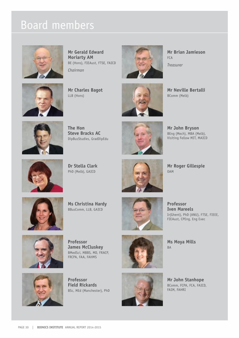
PAGE 30 | BIONICS INSTITUTE ANNUAL REPORT 2014-2015
Board members
Mr Gerald Edward Moriarty AM BE (Hons), FIEAust, FTSE, FAICD
Chairman
Mr Charles Bagot LLB (Hons)
Mr Brian Jamieson FCA
Treasurer
Mr Neville BertalliBComm (Melb)
The Hon Steve Bracks AC DipBusStudies, GradDipEdu
Dr Stella Clark PhD (Melb), GAICD
Mr John Bryson BEng (Mech), MBA (Melb), Visiting Fellow MIT, MAICD
Mr Roger GillespieOAM
Ms Christina Hardy BBusComm, LLB, GAICD
Professor James McCluskeyBMedSci, MBBS, MD, FRACP, FRCPA, FAA, FAHMS
Professor Iven MareelsIr(Ghent), PhD (ANU), FTSE, FIEEE, FIEAust, CPEng, Eng Exec
Ms Moya Mills BA
Professor Field RickardsBSc, MEd (Manchester), PhD
Mr John Stanhope BComm, FCPA, FCA, FAICD, FAIM, FAHRI

BIONICS INSTITUTE ANNUAL REPORT 2014-2015 | PAGE 31
Professor Graeme Clark ACMBBS, MS, PhD, FRCS, FRACS, FAA, FRS
Professor Robert ShepherdBSc, DipEd, PhD
Director
Professor Hugh McDermottBAppSc (Hons)(Electronics), PhD
Deputy Director
Mr Michael Grigoletto BEng, MBA
Executive Manager (Development)(from October 2014)
Mr Tim GriffithsBBus, GradCertExport, GradDip (MarLogMgt), MBT
General Manager and Company Secretary(until August 2014)
Mr Peter GoverBCompt (Hons), CA CPA ICAA
Chief Financial OfficerCompany Secretary (from August 2014)
Ms Linda PetersonBSc, GradCertBusAdm
Executive Manager (R&D, Operations, Collaborations, Projects, Organisational Capability)
Executive officers
Director Emeritus

PAGE 32 | BIONICS INSTITUTE ANNUAL REPORT 2014-2015
Summarised financial report
CONSOLIDATED INCOME STATEMENT 2015 2014
REVENUES FOR ORDINARY ACTIVITIES $ $
Federal Government grants 2,281,511 2,578,505
Victorian Government grants 496,198 503,986
Trusts & foundations 865,408 1,024,198
Public fundraising 373,401 128,665
Research contracts 559,270 297,889
Investment & interest income 813,357 908,080
Other income 816,354 927,439
TOTAL REVENUE FOR ORDINARY ACTIVITIES 6,205,499 6,368,762
Less expenditure on ordinary activities ( 7,132,544) ( 7,539,331)
DEFICIT ON ORDINARY ACTIVITIES ( 927,045) ( 1,170,569)
(Loss)/gain on sale of available-for-sale financial assets 719,987 702,450
NET DEFICIT ( 207,058) (468,119)
The financial information and statements presented in this report are based on unaudited management accounts which are used by the Directors to monitor the activities of the Institute. Directors do not believe the audited financial report prepared under the current Australian Accounting Standard AASB 1004 on contributions shows the Institute’s obligations relating to grants and other funding received, and matches the performance of the research activities between income and expenditure.
Full audited financial statements are available from the Institute’s registered office by request.
ABRIDGED FINANCIAL STATEMENT for the year ended 30 June 2015
CONSOLIDATED STATEMENT OF FINANCIAL POSITION 2015 2014
$ $
Current Assets 4,540,729 4,420,114
Non-Current Assets 12,532,166 13,986,986
TOTAL ASSETS 17,072,895 18,407,100
Current Liabilities 1,989,227 2,926,961
Non-Current Liabilities 60,959 79,552
TOTAL LIABILITIES 2,050,186 3,006,513
NET ASSETS 15,022,709 15,400,587
TOTAL INSTITUTE FUNDS 15,022,709 15,400,587
FUNDING OF OUR RESEARCH
Government funding
Private funding
Research contracts
Other income
Institute funding
2015
2014
39%
17%
8%
12%
24%
41%
15%
4%
12%
28%