Biomolecules detection based on Transmission LSPR shift reference experiment in PBS 1X: 1.2 nm....

1
G. Cappi¹, E. Accastelli¹, L. Benini², C. Guiducci¹ ¹Laboratory of Life Sciences Electronics (CLSE), EPFL, Switzerland, ²DEIS, University of Bologna, Italy 525 nm 590 nm 650 nm LEDs Sample Photodetector J. N. Anker, W. P. Hall, O. Lyandres, N. C. Shah, J. Zhao, R. P. Van Duyne, “Biosensing with Plasmonic Nanosensors”, Nature Materials, 7 (2008), 442-453 V. Cantale, F. C. Simeone, R. Gambari, M.A. Rampi, “ Gold nano-islands on FTO as plasmonic nanostructures for biosensors” , Sensors and Actuators B, 152 (2011), 206-213 450 500 550 600 650 700 750 0.55 0.6 0.65 0.7 0.75 0.8 0.85 0.9 0.95 Wavelength [nm] Transmittance 571.2 nm 578.6 nm Δλ =7.4 nm I S y P e M RTD 2010 Introduction Portable setup for real-time sensing References 10 cm 10 cm 16 cm Microfluidics Measurements The peak position is estimated from the three measured intensities by means of an algorithm that we developed. A typical surface plasmon transmission spectrum of gold NIs evaporated on a transparent slide is plotted in Figure (red line). The peak is located between 550 nm and 595 nm and red-shifts upon modification of the NIs with layers of organic molecules (green line). The measurement and extraction of the peak lo- cation has been repeated 5 times. The dispersion among the measurements is 0.2 nm. Each mea- surement has an error bar between 1.6 nm and 1.7 nm given by the standard deviation over 10000 samples collected. Peak shift of a single strand DNA layer: 7.24 nm with a dispersion of 1.65 nm over 11 samples. Peak shift reference experiment in PBS 1X: 1.2 nm. Biomolecules detection based on Transmission LSPR 5 nm gold layer evaporated on a FTO glass substrate. 5 nm gold layer evaporated on a FTO glass substrate after mild thermal annealing. Extinction spectrum of a sample after gold evaporation. Extinction spectrum of a sample after evaporation and subsequent annealing. 0 0.05 0.1 0.15 0.2 0.25 350 400 450 500 550 600 650 700 750 Wavelength [nm] Extinction 0 0.05 0.1 0.15 0.2 0.25 350 400 450 500 550 600 650 700 750 Extinction Wavelength [nm] Light Spot Gold NIs FTO Layer Photodetector Glass substrate The sensing area is made of gold NanoIslands formed on Fluorine-doped Tin Oxide (FTO) coated glass slides. FTO coated slides are characterized by excel- lent adhesion properties while showing high optical transmittance and conduc- tivity. A layer of 5 nm of gold was evaporated on FTO surface and then annealed at 200°C. Nanostructures for label-free detection NanoIslands (NIs) are suitable to probe biomolecules comparable in size Nowadays, fast detection and quantification of biomolecules is a critical issue in many sectors, included health-care, ranging from early diagnostics of diseases to personalized medicine. Among other detection techniques, Surface Plasmon Resonance proved to be a very sensitive and powerful method to detect biomolecular binding of different species, such as proteins, oligonucle- otides and viruses. We developed a device based on low-cost off- the-shelf components, namely, electronically- driven power LEDs and photodetectors, and de- veloped a novel data analysis approach that ex- tracts the peak position of the LSPR spectrum from the measurements performed with a set of three LEDs. A microfluidics system con- sisting of two parallel chan- nels has been built. One of the channels is used as a ref- erence to compensate light drift and other artifacts by a differential measurement.

Transcript of Biomolecules detection based on Transmission LSPR shift reference experiment in PBS 1X: 1.2 nm....

G. Cappi¹, E. Accastelli¹, L. Benini², C. Guiducci¹¹Laboratory of Life Sciences Electronics (CLSE), EPFL, Switzerland, ²DEIS, University of Bologna, Italy

525nm

590nm

650nm LEDs

Sample

Photodetector

J. N. Anker, W. P. Hall, O. Lyandres, N. C. Shah, J. Zhao, R. P. Van Duyne, “Biosensing with Plasmonic Nanosensors”, Nature Materials, 7 (2008), 442-453

V. Cantale, F. C. Simeone, R. Gambari, M.A. Rampi, “ Gold nano-islands on FTO as plasmonic nanostructures for biosensors” , Sensors and Actuators B, 152 (2011), 206-213

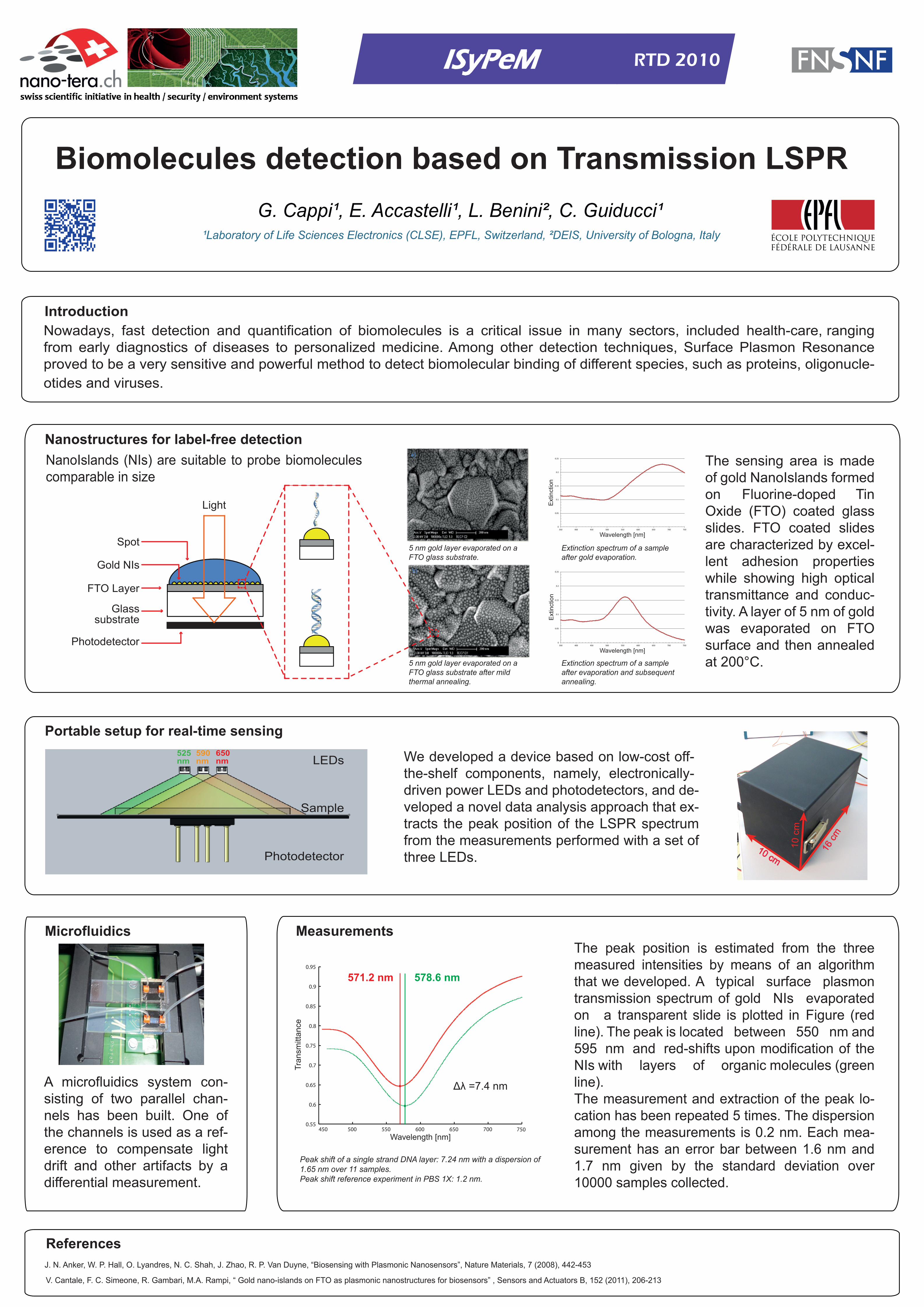
450 500 550 600 650 700 7500.55

0.6

0.65

0.7

0.75

0.8

0.85

0.9

0.95

Wavelength [nm]

Tran

smitt

ance

571.2 nm 578.6 nm

Δλ =7.4 nm

ISyPeM RTD 2010

Introduction

Portable setup for real-time sensing

References

10 cm

10 c

m

16 cm

Microfluidics MeasurementsThe peak position is estimated from the three measured intensities by means of an algorithm that we developed. A typical surface plasmon transmission spectrum of gold NIs evaporated on a transparent slide is plotted in Figure (red line). The peak is located between 550 nm and 595 nm and red-shifts upon modification of the NIs with layers of organic molecules (green line).The measurement and extraction of the peak lo-cation has been repeated 5 times. The dispersion among the measurements is 0.2 nm. Each mea-surement has an error bar between 1.6 nm and 1.7 nm given by the standard deviation over 10000 samples collected.

Peak shift of a single strand DNA layer: 7.24 nm with a dispersion of 1.65 nm over 11 samples.Peak shift reference experiment in PBS 1X: 1.2 nm.

Biomolecules detection based on Transmission LSPR

5 nm gold layer evaporated on a FTO glass substrate.

5 nm gold layer evaporated on a FTO glass substrate after mild thermal annealing.

Extinction spectrum of a sample after gold evaporation.

Extinction spectrum of a sample after evaporation and subsequent annealing.

0

0.05

0.1

0.15

0.2

0.25

350 400 450 500 550 600 650 700 750

Wavelength [nm]

Ext

inct

ion

0

0.05

0.1

0.15

0.2

0.25

350 400 450 500 550 600 650 700 750

Ext

inct

ion

Wavelength [nm]

Light

Spot

Gold NIs

FTO Layer

Photodetector

Glasssubstrate

The sensing area is made of gold NanoIslands formed on Fluorine-doped Tin Oxide (FTO) coated glass slides. FTO coated slides are characterized by excel-lent adhesion properties while showing high optical transmittance and conduc-tivity. A layer of 5 nm of gold was evaporated on FTO surface and then annealed at 200°C.

Nanostructures for label-free detectionNanoIslands (NIs) are suitable to probe biomolecules comparable in size

Nowadays, fast detection and quantification of biomolecules is a critical issue in many sectors, included health-care, ranging from early diagnostics of diseases to personalized medicine. Among other detection techniques, Surface Plasmon Resonance proved to be a very sensitive and powerful method to detect biomolecular binding of different species, such as proteins, oligonucle-otides and viruses.

We developed a device based on low-cost off-the-shelf components, namely, electronically-driven power LEDs and photodetectors, and de-veloped a novel data analysis approach that ex-tracts the peak position of the LSPR spectrum from the measurements performed with a set of three LEDs.

A microfluidics system con-sisting of two parallel chan-nels has been built. One of the channels is used as a ref-erence to compensate light drift and other artifacts by a differential measurement.