Biomimetic apatite-coated porous PVA scaffolds promote the growth of breast cancer cells
7
Biomimetic apatite-coated porous PVA scaffolds promote the growth of breast cancer cells Mao Ye, Pravansu Mohanty, Gargi Ghosh ⁎ Department of Mechanical Engineering, University of Michigan, Dearborn, 4901 Evergreen Road, Dearborn, MI 48128, United States abstract article info Article history: Received 18 January 2014 Received in revised form 17 June 2014 Accepted 5 August 2014 Available online 27 August 2014 Keywords: PVA scaffold Interconnected pores Mineralization HBSS Breast cancer cells Recapitulating the native environment of bone tissue is essential to develop in vitro models of breast cancer bone metastasis. The bone is a composite material consisting of organic matrix and inorganic mineral phase, primarily hydroxyapatite. In this study, we report the mineralization of porous poly vinyl alcohol (PVA) scaffolds upon incubation in modified Hanks' Balanced Salt Solution (HBSS) for 14 days. Scanning electron microscopy, energy dispersive X-ray analysis, and X-ray diffraction analysis revealed that the deposited minerals have composition similar to hydroxyapatite. The study demonstrated that the rate of nucleation and growth of minerals was faster on surfaces of less porous scaffolds. However, upon prolonged incubation, formation of mineral layer was observed on the surface of all the scaffolds. In addition, the study also demonstrated that 3D mineralization only occurred for scaffolds with highly interconnected porous networks. The mineralization of the scaffolds pro- moted the adsorption of serum proteins and consequently, the adhesion and proliferation of breast cancer cells. © 2014 Elsevier B.V. All rights reserved. 1. Introduction Breast cancer is the leading cause of cancer related death in American women [1]. The high mortality associated with the disease can be attrib- uted to the spread of breast cancer to different organs including the bone, lungs, brain and liver. The complex process of metastasis i.e. spread of cancer to distant organs involves migration of tumor cells from prima- ry lesion through extracellular matrix into the blood or lymph followed by extravasation and colonization at the distant sites. In the case of breast cancer patients, around 70% of them develop bone metastasis, i.e. tumor cells spread to the skeletal tissues [2]. This in turn leads to se- vere pain and pathological fractures [3,4] due to formation of secondary tumor followed by bone resorption or osteolytic remodeling. Mounting evidence corroborates that the interactions between breast cancer cells and bone tissue microenvironment are pivotal to cancer cell homing followed by the establishment of metastases. However, very little is known about how various constituents of bone tissue contribute towards attracting, harboring, and sustaining breast cancer bone metastasis. This limitation of knowledge can be attributed partly to the lack of a cell cul- ture model system which will facilitate a study of various aspects of skeletal metastasis of breast cancer. Inability to control the cell–cell and cell–matrix interactions often limits the application of animal models of cancer metastasis. Thus, polymeric scaffolds mimicking the native en- vironment of bone tissue and providing breast cancer cells with an envi- ronment which the cells encounter during homing in skeletal tissues can significantly facilitate understanding the role of bone tissue microenvi- ronment in promoting colonization. The bone is a composite material consisting of inorganic mineral phase, primarily hydroxyapatite (HA), dispersed within organic mesh- work, consisting of collagen, the most abundant protein in mammals. Around 28 types of collagen have been identified so far; type I is the most prevalent and found in the extracellular matrix of the bone [5–7]. Collagen imparts the bone with ductility and ability to absorb energy. HA is known for conferring structural and mechanical properties to the bone, excellent osteoconductivity, and osteoinductivity [8,9]. In addition, HA also modulates the cellular responses of both normal and transformed cells [10]. For example, studies have demonstrated that HA not only facilitates the formation of bone tissue but also modulates the growth and expression profiles of bone metastatic tumors [11–13]. Since, HA plays an important role in colonization of breast cancer cells, fabrication of mineralized scaffold will enable investigation of cancer cell–mineral interactions. Several studies have reported fabrication of HA containing scaffolds [12]; however, these studies utilized primarily commercially available HA particles which differ in shape, size, and chemical composition [11,14]. The inherent heterogeneity of these HA particles in turn can affect the cell–material interactions. In this study, we report the fabrication of mineralized scaffold by employing a biomimetic approach and utilization of these scaffolds to investigate the impact of mineral layers on adhesion and proliferation of breast cancer cells. Pioneering studies by Kokubo first demonstrated the ex vivo forma- tion of nanocrystalline, carbonate substituted hydroxyapatite minerals on a variety of materials [15]. Nucleation and growth of minerals on biomaterials were promoted by incubation in a defined ionic solution. Materials Science and Engineering C 44 (2014) 310–316 ⁎ Corresponding author. Tel.: +1 313 593 5013; fax: +1 313 593 3851. E-mail address: [email protected] (G. Ghosh). http://dx.doi.org/10.1016/j.msec.2014.08.044 0928-4931/© 2014 Elsevier B.V. All rights reserved. Contents lists available at ScienceDirect Materials Science and Engineering C journal homepage: www.elsevier.com/locate/msec
Transcript of Biomimetic apatite-coated porous PVA scaffolds promote the growth of breast cancer cells
Materials Science and Engineering C 44 (2014) 310–316
Contents lists available at ScienceDirect
Materials Science and Engineering C
j ourna l homepage: www.e lsev ie r .com/ locate /msec
Biomimetic apatite-coated porous PVA scaffolds promote the growth ofbreast cancer cells
Mao Ye, Pravansu Mohanty, Gargi Ghosh ⁎Department of Mechanical Engineering, University of Michigan, Dearborn, 4901 Evergreen Road, Dearborn, MI 48128, United States
⁎ Corresponding author. Tel.: +1 313 593 5013; fax: +E-mail address: [email protected] (G. Ghosh).
http://dx.doi.org/10.1016/j.msec.2014.08.0440928-4931/© 2014 Elsevier B.V. All rights reserved.
a b s t r a c t
a r t i c l e i n f oArticle history:Received 18 January 2014Received in revised form 17 June 2014Accepted 5 August 2014Available online 27 August 2014
Keywords:PVA scaffoldInterconnected poresMineralizationHBSSBreast cancer cells
Recapitulating the native environment of bone tissue is essential to develop in vitromodels of breast cancer bonemetastasis. The bone is a composite material consisting of organic matrix and inorganic mineral phase, primarilyhydroxyapatite. In this study, we report the mineralization of porous poly vinyl alcohol (PVA) scaffolds uponincubation in modified Hanks' Balanced Salt Solution (HBSS) for 14 days. Scanning electron microscopy, energydispersive X-ray analysis, and X-ray diffraction analysis revealed that the deposited minerals have compositionsimilar to hydroxyapatite. The study demonstrated that the rate of nucleation and growth of minerals was fasteron surfaces of less porous scaffolds. However, upon prolonged incubation, formation of mineral layer wasobserved on the surface of all the scaffolds. In addition, the study also demonstrated that 3D mineralizationonly occurred for scaffolds with highly interconnected porous networks. The mineralization of the scaffolds pro-moted the adsorption of serum proteins and consequently, the adhesion and proliferation of breast cancer cells.
© 2014 Elsevier B.V. All rights reserved.
1. Introduction
Breast cancer is the leading cause of cancer related death in Americanwomen [1]. The highmortality associated with the disease can be attrib-uted to the spread of breast cancer to different organs including thebone, lungs, brain and liver. The complex process ofmetastasis i.e. spreadof cancer to distant organs involvesmigration of tumor cells fromprima-ry lesion through extracellular matrix into the blood or lymph followedby extravasation and colonization at the distant sites. In the case ofbreast cancer patients, around 70% of them develop bone metastasis,i.e. tumor cells spread to the skeletal tissues [2]. This in turn leads to se-vere pain and pathological fractures [3,4] due to formation of secondarytumor followed by bone resorption or osteolytic remodeling. Mountingevidence corroborates that the interactions between breast cancer cellsand bone tissue microenvironment are pivotal to cancer cell homingfollowed by the establishment of metastases. However, very little isknown about howvarious constituents of bone tissue contribute towardsattracting, harboring, and sustaining breast cancer bone metastasis. Thislimitation of knowledge can be attributed partly to the lack of a cell cul-ture model system which will facilitate a study of various aspects ofskeletal metastasis of breast cancer. Inability to control the cell–cell andcell–matrix interactions often limits the application of animal modelsof cancer metastasis. Thus, polymeric scaffolds mimicking the native en-vironment of bone tissue and providing breast cancer cells with an envi-ronmentwhich the cells encounter during homing in skeletal tissues can
1 313 593 3851.
significantly facilitate understanding the role of bone tissue microenvi-ronment in promoting colonization.
The bone is a composite material consisting of inorganic mineralphase, primarily hydroxyapatite (HA), dispersed within organic mesh-work, consisting of collagen, the most abundant protein in mammals.Around 28 types of collagen have been identified so far; type I is themost prevalent and found in the extracellular matrix of the bone[5–7]. Collagen imparts the bone with ductility and ability to absorbenergy. HA is known for conferring structural andmechanical propertiesto the bone, excellent osteoconductivity, and osteoinductivity [8,9]. Inaddition, HA also modulates the cellular responses of both normal andtransformed cells [10]. For example, studies have demonstrated thatHA not only facilitates the formation of bone tissue but also modulatesthe growth and expression profiles of bone metastatic tumors [11–13].Since, HA plays an important role in colonization of breast cancer cells,fabrication of mineralized scaffold will enable investigation of cancercell–mineral interactions. Several studies have reported fabrication ofHA containing scaffolds [12]; however, these studies utilized primarilycommercially available HA particles which differ in shape, size, andchemical composition [11,14]. The inherent heterogeneity of theseHA particles in turn can affect the cell–material interactions. In thisstudy, we report the fabrication of mineralized scaffold by employing abiomimetic approach and utilization of these scaffolds to investigatethe impact of mineral layers on adhesion and proliferation of breastcancer cells.
Pioneering studies by Kokubo first demonstrated the ex vivo forma-tion of nanocrystalline, carbonate substituted hydroxyapatite mineralson a variety of materials [15]. Nucleation and growth of minerals onbiomaterials were promoted by incubation in a defined ionic solution.

Table 1Ionic compositions of biological fluids and media formulations.
Blood plasma SBF HBSS Modified HBSS
Na+ 142 142 141.3 141.3K+ 3.6–5.5 5 5.8 7.3Ca2+ 2.1–2.6 2.5 1.26 8Mg2+ 1 1.5 0.8 0.8HCO3
− 27 4.2 0 0SO4
2− 1 0.5 0.8 0.8PO4
3− 0.6–1.5 1 0.34 2.28Cl− 142 142.1 142.1 148.9
311M. Ye et al. / Materials Science and Engineering C 44 (2014) 310–316
This solution known as the simulated body fluid (SBF) is formulatedwith ion concentrations similar to blood plasma. This biomimetic ap-proach of inducing hydroxyapatite nucleation has been adapted laterfor a variety of materials including glasses, metals, and polymers [16].To simulate the native physiological environment more closely, recentstudies have modified HEPES free Dulbecco's Modified Eagle Medium(DMEM), a cell culture medium [17]. This modified solution combinesthe ionic constituents of SBF with the nutrients, amino acids, and vita-mins essential for cell growth. Because nucleation is induced via anaqueous solution, this biomimetic technique can be applied to scaffoldsof any complex geometry unlike techniques like plasma spraying,pulsed laser deposition, and electrophoretic depositionwhich are limitedto flat surfaces or very thin porous layers [18].
In the current study, macroporous polyvinyl alcohol (PVA) scaffoldwas utilized to promote deposition and growth of mineral layers usingthe biomimetic approach. PVA was chosen as the base material forscaffold fabrication due to the inherent advantages including watersolubility, nontoxicity, biocompatibility, and biodegradability [19–21].Additionally, PVA has been previously used in various pharmaceuticaland biomedical applications such as contact lenses [22], drug deliverydevices [23,24], wound dressings [25], orthopedic devices [26], andartificial organs [27]. Scanning electron microscopy, energy dispersiveX-ray analysis, and X-ray diffraction analysis were employed to investi-gate themorphology and composition of the accumulatedmineral layer.Finally, the ability of the mineral layer to modulate the adhesion andproliferation of breast cancer cells, MDA-MB-231 was characterized. Tothe best our knowledge, this is the first study which demonstratedthe impact of accumulated mineral layer (via biomimetic approach) onbreast cancer cell responses. Results from our studies suggest that min-eralized PVA scaffold can be utilized to develop in vitro model of breastcancer bone metastasis.
2. Materials and methods
2.1. Materials
PVA (averagemolecularweight of 146,000–186,000, 89,000–98,000,and 27,000 g/mol, hence forth referred to as 146 k PVA, 89 k PVA and27 k PVA), and Triton X-100 were obtained from Sigma (St. Louis, MO).HBSS (Hanks' Balanced Salt Solution) 1× is obtained fromMediatech Inc.,Pierce BCA Protein Assay kit is obtained from Thermo Scientific. XTTprotein reagent is obtained from American Type Cell Culture (ATCC).The polymer and surfactant were used without further purification.
2.2. Fabrication of PVA scaffolds
14% PVA solution (w/v) was prepared by dissolving PVA into dis-tilled water at 80 °C for 1 h under constant stirring. To generate thePVA foam, Triton X-100 was added to 5 mL PVA solution and the solu-tion was mechanically agitated. Stirring time was varied from 15 s to60 s. Following themechanical stirring, the generated foamswere trans-ferred to small petri dishes. The samples were then immediately placedat −80 °C for 24 h. Following freezing, the samples were dried undervacuum for 36 h to remove ice from the scaffolds. Towards this goal,the samples were placed in a small vacuum seal container which wasplaced in dry ice and connected to a vacuum pump.
The microstructures of the scaffolds were characterized by scanningelectron microscopy (SEM) (Hitachi, U.S.A.). At least 4 random imageswere obtained for each sample.
2.3. Biomineralization and analysis of mineral growth
The scaffoldswere immersed in themodifiedHBSS (mHBSS) at 37 °Cfor 14 days to elicit mineralization reaction. The basal HBSSwas supple-mented with various ionic salts (Table 1) and titrated to a pH of 7.4.mHBSS has similar composition to that of human plasma but with
higher concentration of calcium and phosphate to promote mineralgrowth. During mineralization process, mHBSS was renewed everyday to maintain a constant ionic strength. For control experiments, thescaffolds were incubated in the basal HBSS.
After the sample is incubated for selected time period, it is washedby distilled water 3 times, with each time submerged in water for15 min. Then the sample is freeze dried. The morphology and the com-position of mineral layer formed on the scaffolds were characterized bySEM and energy dispersive X-ray spectroscopy (EDS) (Hitachi). XRDpatterns of pure HA and mineralized samples were recorded usingCu Kα radiation and 2θ angles from 20° to 50°. For the purpose, thesamples were mounted flat on glass sample holder and XRD spectrumwas obtained using Rigaku.
2.4. Characterization of protein adsorption
To assess protein adsorption on the mineralized scaffolds, thescaffolds were incubated in complete RPMI medium supplementedwith 10% fetal bovine serum (FBS) and 1% Penicillin–Streptomycin(PS) for 1 h. Following incubation, the supernatant culture media werecollected. Colorimetric analysis on a plate reader (Bio-Rad) was carriedout to quantify total protein in the supernatant using BicinchoninicAcid (BCA) kit and according to themanufacturer's instructions. Proteinadsorption was calculated from the difference in protein content ofcomplete RPMI media and the supernatants.
2.5. Cellular assays
Breast cancer cell line (MDA-MB-231) was procured from AmericanType Cell Culture (ATCC) and maintained in RPMI 1640 supplementedwith 10% FBS and 1% PS in a humidified chamber at 37 °C and 5% CO2.For cell assays, MDA-MB-231 cells up to passage 6 were utilized.
To evaluate cell adhesion, 1.2 × 10⁵/mL cells were seeded onto themineralized and non-mineralized scaffolds and incubated in humidifiedchamber at 37 °C and 5% CO2 for 5 h. Following incubation, supernatantwas collected and the number of non-adhered cells was counted. Celladhesion was calculated from the difference in the number of seededand non-adhered cells.
Tumor cell proliferation on mineralized and non-mineralized scaf-folds was determined by performing XTT assay according to themanufacturer's (ATCC) instruction. Briefly, 3 days post-seeding, cellswere incubated with 100 μL of cell culture medium and 50 μL of XTT la-beling solution for 5 h at 37 °C and 5% CO2. Following which, the super-natant was collected and XTT assay performed by measuring theabsorbance of the supernatants.
2.6. Statistical analysis
For all experiments, data reported as mean ± S.E.M. of three inde-pendent experiments. Statistical analyses were carried out with oneway ANOVA. Differences between two sets of data were consideredsignificant at p-value b0.05.

Fig. 2. SEM images of (A) rhombohedral crystals observedwhen PVA scaffolds were incu-bated in mHBSS for 14 days and (B) lack of mineralization when the scaffolds were incu-bated in HBSS for 14 days.
312 M. Ye et al. / Materials Science and Engineering C 44 (2014) 310–316
3. Results and discussion
3.1. Mineral growth on PVA scaffolds
Macroporous PVA scaffoldswere incubated inHBSS andmHBSS overa span of 14 days to induce mineralization. HBSS, which providesthe cells with essential inorganic ions, was supplemented with thespecific salts to promote nucleation and growth of calcium phosphates(Table 1). As demonstrated in Fig. 1, SEM micrographs confirmed thegradual precipitation and continuous growth of mineral layer over thesurface of the scaffold with time. The smaller spheres indicate recentlynucleated crystals while larger ones represent the crystals that havebeen growingover longer period of time. As the incubation timewas ex-tended to 14 days accumulated minerals gradually grew into a nearlycontinuous laminated structure. Enhanced mineral precipitation uponprolonged incubation was probably facilitated by the interactions be-tween the surface hydroxyl groups of PVA scaffolds and the ionic con-stituents of mHBSS. Mounting evidence has suggested that exposedhydroxyl groups stimulate the calcification by extracting the availablecalcium and phosphate ions from solution and concentrating them onmaterial surface [28–32]. Previously it had been demonstrated thatcalcium and phosphorous bind to the hydrated oxide layer on titaniumimplant surfaces leading to the formation of calcium phosphate layer[33]. Similarly, calcium and phosphorus free materials had elicitedhydroxyapatite layer formation on their surface; the phenomenon hadbeen associated with the presence of surface hydroxyl groups [34–36].In addition to apatite, a certain fraction of rhombohedral crystals wasalso found on the scaffold surface (Fig. 2A). When the scaffolds wereincubated in HBSS, no evidence of surface precipitation was observed(Fig. 2B). This suggests that augmentation of the concentration ofcalcium (Ca2+) and phosphate (PO4
3−) ions inHBSS is necessary to facil-itate biomineralization.
3.2. Characterization of chemical composition of accumulated minerals
Following bio-mineralization, the composition of the depositedmineral was investigated by energy dispersive X-ray spectroscopy(EDS). The EDS analysis revealed that themineral deposited on scaffoldsincubated withmHBSS is primarily composed of Ca and P (Fig. 3A). EDS
Fig. 1. SEM images of PVA scaffolds incubated in mHBSS fo
analysis of the rhombohedral crystals revealed the absence of P, therebysuggesting that these crystals correspond to calcite minerals (Fig. 3B).These studies demonstrated that possible diffusion of carbonate result-ed in the formation of calcite (calcium carbonate) minerals; however,apatite formation predominated because the supersaturation of calciumand phosphatewith respect to apatite is higher than of themore solublecalcium carbonate. The EDS analysis (Fig. 3C) confirmed the lack ofmineralization in the control group as manifested from the absence ofcalcium and phosphorous. Quantitative analysis (Fig. 3D) revealed theelemental concentration (%) of Ca and P in the deposited mineral
r (i) 3 days, (ii) 5 days, (iii) 7 days, and (iv) 14 days.
mHBSS
HBSS
*
*
0
20
40
60
80
100
120
mHBSS HBSS Calcite
Elem
ent c
once
ntra
tion
( %)
Ca P
A B
C
E
D
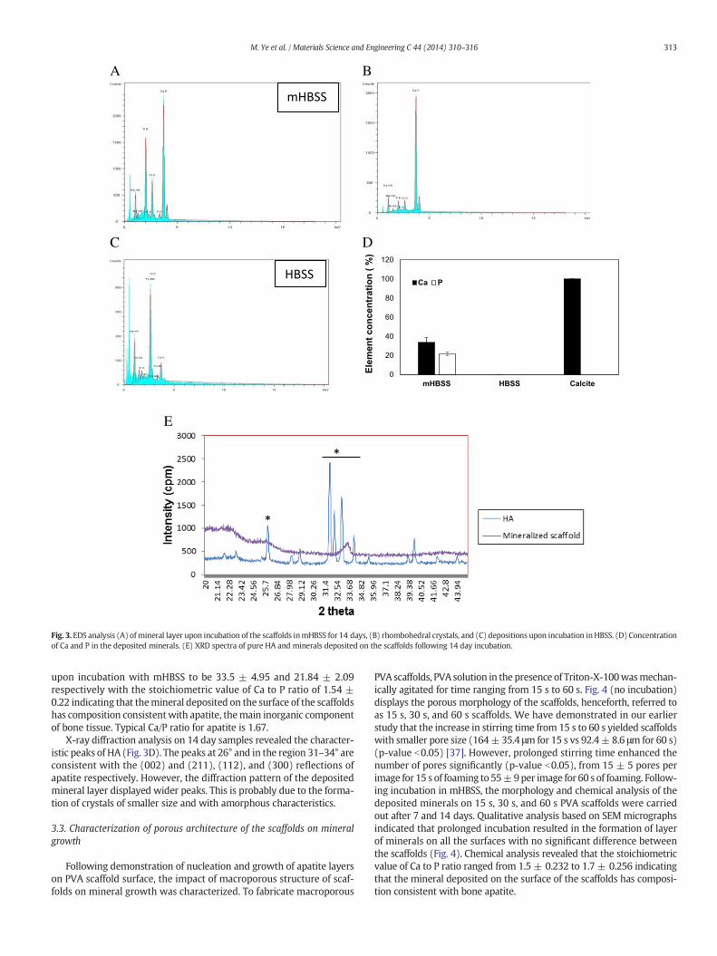
Fig. 3. EDS analysis (A) ofmineral layer upon incubation of the scaffolds inmHBSS for 14 days, (B) rhombohedral crystals, and (C) depositions upon incubation inHBSS. (D) Concentrationof Ca and P in the deposited minerals. (E) XRD spectra of pure HA and minerals deposited on the scaffolds following 14 day incubation.
313M. Ye et al. / Materials Science and Engineering C 44 (2014) 310–316
upon incubation with mHBSS to be 33.5 ± 4.95 and 21.84 ± 2.09respectively with the stoichiometric value of Ca to P ratio of 1.54 ±0.22 indicating that themineral deposited on the surface of the scaffoldshas composition consistentwith apatite, themain inorganic componentof bone tissue. Typical Ca/P ratio for apatite is 1.67.
X-ray diffraction analysis on 14 day samples revealed the character-istic peaks of HA (Fig. 3D). The peaks at 26° and in the region 31–34° areconsistent with the (002) and (211), (112), and (300) reflections ofapatite respectively. However, the diffraction pattern of the depositedmineral layer displayed wider peaks. This is probably due to the forma-tion of crystals of smaller size and with amorphous characteristics.
3.3. Characterization of porous architecture of the scaffolds on mineralgrowth
Following demonstration of nucleation and growth of apatite layerson PVA scaffold surface, the impact of macroporous structure of scaf-folds on mineral growth was characterized. To fabricate macroporous
PVA scaffolds, PVA solution in the presence of Triton-X-100wasmechan-ically agitated for time ranging from 15 s to 60 s. Fig. 4 (no incubation)displays the porous morphology of the scaffolds, henceforth, referred toas 15 s, 30 s, and 60 s scaffolds. We have demonstrated in our earlierstudy that the increase in stirring time from 15 s to 60 s yielded scaffoldswith smaller pore size (164± 35.4 μm for 15 s vs 92.4± 8.6 μm for 60 s)(p-value b0.05) [37]. However, prolonged stirring time enhanced thenumber of pores significantly (p-value b0.05), from 15 ± 5 pores perimage for 15 s of foaming to 55±9per image for 60 s of foaming. Follow-ing incubation in mHBSS, the morphology and chemical analysis of thedeposited minerals on 15 s, 30 s, and 60 s PVA scaffolds were carriedout after 7 and 14 days. Qualitative analysis based on SEM micrographsindicated that prolonged incubation resulted in the formation of layerof minerals on all the surfaces with no significant difference betweenthe scaffolds (Fig. 4). Chemical analysis revealed that the stoichiometricvalue of Ca to P ratio ranged from 1.5 ± 0.232 to 1.7 ± 0.256 indicatingthat the mineral deposited on the surface of the scaffolds has composi-tion consistent with bone apatite.
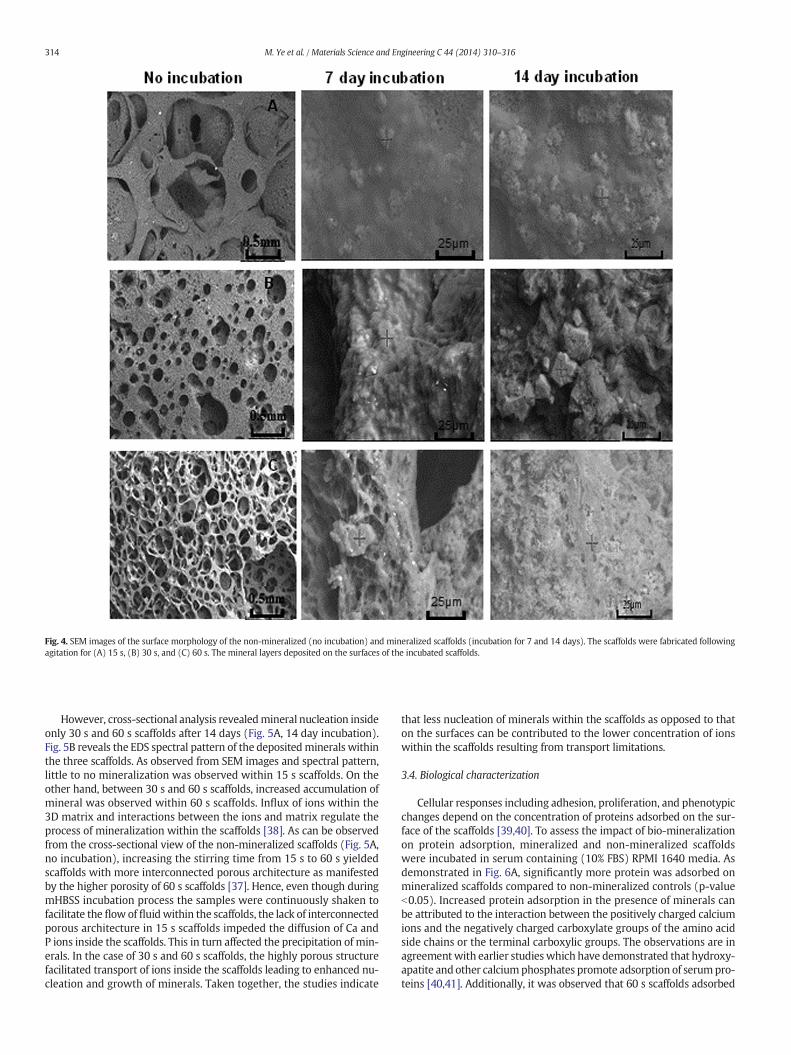
Fig. 4. SEM images of the surface morphology of the non-mineralized (no incubation) and mineralized scaffolds (incubation for 7 and 14 days). The scaffolds were fabricated followingagitation for (A) 15 s, (B) 30 s, and (C) 60 s. The mineral layers deposited on the surfaces of the incubated scaffolds.
314 M. Ye et al. / Materials Science and Engineering C 44 (2014) 310–316
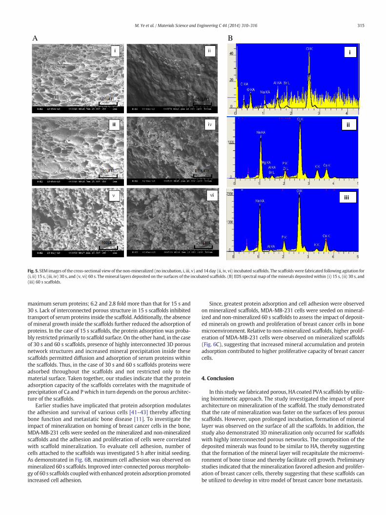
However, cross-sectional analysis revealedmineral nucleation insideonly 30 s and 60 s scaffolds after 14 days (Fig. 5A, 14 day incubation).Fig. 5B reveals the EDS spectral pattern of the depositedminerals withinthe three scaffolds. As observed from SEM images and spectral pattern,little to no mineralization was observed within 15 s scaffolds. On theother hand, between 30 s and 60 s scaffolds, increased accumulation ofmineral was observed within 60 s scaffolds. Influx of ions within the3D matrix and interactions between the ions and matrix regulate theprocess of mineralization within the scaffolds [38]. As can be observedfrom the cross-sectional view of the non-mineralized scaffolds (Fig. 5A,no incubation), increasing the stirring time from 15 s to 60 s yieldedscaffolds with more interconnected porous architecture as manifestedby the higher porosity of 60 s scaffolds [37]. Hence, even though duringmHBSS incubation process the samples were continuously shaken tofacilitate the flow of fluidwithin the scaffolds, the lack of interconnectedporous architecture in 15 s scaffolds impeded the diffusion of Ca andP ions inside the scaffolds. This in turn affected the precipitation of min-erals. In the case of 30 s and 60 s scaffolds, the highly porous structurefacilitated transport of ions inside the scaffolds leading to enhanced nu-cleation and growth of minerals. Taken together, the studies indicate
that less nucleation of minerals within the scaffolds as opposed to thaton the surfaces can be contributed to the lower concentration of ionswithin the scaffolds resulting from transport limitations.
3.4. Biological characterization
Cellular responses including adhesion, proliferation, and phenotypicchanges depend on the concentration of proteins adsorbed on the sur-face of the scaffolds [39,40]. To assess the impact of bio-mineralizationon protein adsorption, mineralized and non-mineralized scaffoldswere incubated in serum containing (10% FBS) RPMI 1640 media. Asdemonstrated in Fig. 6A, significantly more protein was adsorbed onmineralized scaffolds compared to non-mineralized controls (p-valueb0.05). Increased protein adsorption in the presence of minerals canbe attributed to the interaction between the positively charged calciumions and the negatively charged carboxylate groups of the amino acidside chains or the terminal carboxylic groups. The observations are inagreementwith earlier studieswhich have demonstrated that hydroxy-apatite andother calciumphosphates promote adsorption of serumpro-teins [40,41]. Additionally, it was observed that 60 s scaffolds adsorbed
i
ii
iii
A B
Fig. 5. SEM images of the cross-sectional view of the non-mineralized (no incubation, i, iii, v) and 14 day (ii, iv, vi) incubated scaffolds. The scaffolds were fabricated following agitation for(i, ii) 15 s, (iii, iv) 30 s, and (v, vi) 60 s. Themineral layers deposited on the surfaces of the incubated scaffolds. (B) EDS spectral map of theminerals depositedwithin (i) 15 s, (ii) 30 s, and(iii) 60 s scaffolds.
315M. Ye et al. / Materials Science and Engineering C 44 (2014) 310–316
maximum serum proteins; 6.2 and 2.8 fold more than that for 15 s and30 s. Lack of interconnected porous structure in 15 s scaffolds inhibitedtransport of serumproteins inside the scaffold. Additionally, the absenceof mineral growth inside the scaffolds further reduced the adsorption ofproteins. In the case of 15 s scaffolds, the protein adsorption was proba-bly restricted primarily to scaffold surface. On the other hand, in the caseof 30 s and 60 s scaffolds, presence of highly interconnected 3D porousnetwork structures and increased mineral precipitation inside thesescaffolds permitted diffusion and adsorption of serum proteins withinthe scaffolds. Thus, in the case of 30 s and 60 s scaffolds proteins wereadsorbed throughout the scaffolds and not restricted only to thematerial surface. Taken together, our studies indicate that the proteinadsorption capacity of the scaffolds correlates with the magnitude ofprecipitation of Ca and Pwhich in turn depends on the porous architec-ture of the scaffolds.
Earlier studies have implicated that protein adsorption modulatesthe adhesion and survival of various cells [41–43] thereby affectingbone function and metastatic bone disease [11]. To investigate theimpact of mineralization on homing of breast cancer cells in the bone,MDA-MB-231 cells were seeded on the mineralized and non-mineralizedscaffolds and the adhesion and proliferation of cells were correlatedwith scaffold mineralization. To evaluate cell adhesion, number ofcells attached to the scaffolds was investigated 5 h after initial seeding.As demonstrated in Fig. 6B, maximum cell adhesion was observed onmineralized 60 s scaffolds. Improved inter-connected porousmorpholo-gy of 60 s scaffolds coupledwith enhanced protein adsorption promotedincreased cell adhesion.
Since, greatest protein adsorption and cell adhesion were observedon mineralized scaffolds, MDA-MB-231 cells were seeded on mineral-ized and non-mineralized 60 s scaffolds to assess the impact of deposit-ed minerals on growth and proliferation of breast cancer cells in bonemicroenvironment. Relative to non-mineralized scaffolds, higher prolif-eration of MDA-MB-231 cells were observed on mineralized scaffolds(Fig. 6C), suggesting that increased mineral accumulation and proteinadsorption contributed to higher proliferative capacity of breast cancercells.
4. Conclusion
In this studywe fabricated porous, HA coated PVA scaffolds by utiliz-ing biomimetic approach. The study investigated the impact of porearchitecture on mineralization of the scaffold. The study demonstratedthat the rate of mineralization was faster on the surfaces of less porousscaffolds. However, upon prolonged incubation, formation of minerallayer was observed on the surface of all the scaffolds. In addition, thestudy also demonstrated 3D mineralization only occurred for scaffoldswith highly interconnected porous networks. The composition of thedeposited minerals was found to be similar to HA, thereby suggestingthat the formation of the mineral layer will recapitulate the microenvi-ronment of bone tissue and thereby facilitate cell growth. Preliminarystudies indicated that themineralization favored adhesion and prolifer-ation of breast cancer cells, thereby suggesting that these scaffolds canbe utilized to develop in vitro model of breast cancer bone metastasis.

Fig. 6. Comparison of (A) protein adsorption, and breast cancer cell (B) adhesion and(C) proliferation on mineralized and non-mineralized scaffolds. Error bars are S.E.M.(N = 3).
316 M. Ye et al. / Materials Science and Engineering C 44 (2014) 310–316
Acknowledgment
The authorswould like to thank theUniversity ofMichigan-Dearbornand University of Michigan-Ann Arbor (U034733): Office of the VicePresident for Research for the financial support.
References
[1] A. Jemal, R. Siegel, E.Ward, et al., Cancer statistics, CA Cancer J. Clin. 57 (2007) 43–66.[2] T.A. Guise, Antitumor effects of bisphosphonates: promising preclinical evidence,
Cancer Treat. Rev. 34 (2008) S19–S24.[3] J. Perez-Garcia, E. Munoz-Couselo, J. Cortes, Bone metastases: causes, consequences
and therapeutic opportunities, Eur. J. Cancer Suppl. 11 (2013) 254–256.[4] L.A. Kingsley, P.G.J. Fournier, J.M. Chirgwin, et al.,Molecular biologyof bonemetastasis,
Mol. Cancer Ther. 6 (2007) 2609–2617.[5] A.M. Ferreira, P. Gentile, V. Chiono, et al., Collagen for bone tissue regeneration, Acta
Biomater. 8 (2012) 3191–3200.[6] K. Gelse, E. Pöschl, T. Aigner, Collagens—structure, function, and biosynthesis, Adv.
Drug Deliv. Rev. 55 (2003) 1531–1546.[7] M.G. Patino, M.E. Neiders, S. Andreana, et al., Collagen: an overview, Implant. Dent.
11 (2002) 280–285.[8] C. Ko, W. Cheng, J. Wang, et al., Properties of osteoconductive biomaterials: calcium
phosphate cement with different ratios of platelet-rich plasma as identifiers, Mater.Sci. Eng. C 33 (2013) 3537–3544.
[9] N.Masahisa, T. Nakamura, T. Kokubo, et al., Bonebonding ability of an appetite-coatedpolymer produced using a biomimetic method: a mechanical and histological studyin vivo, J. Biomed. Mater. Res. 31 (1996) 487–494.
[10] S. Pezzatini, L.Morbidelli, R. Solito, et al., The effect of calciumphosphate nanoparticleson hormone production and apoptosis in human granulosa cells, Reprod. Biol.Endocrinol. 41 (2007) 523.
[11] S.P. Pathi, D.D.W. Lin, J.R. Dorvee, et al., Hydroxyapatite nanoparticle-containingscaffolds for the study of breast cancer bone metastasis, Biomaterials 32 (2011)5112–5122.
[12] S.S. Kim, M. Sun Park, O. Jeon, et al., Poly(lactide-co-glycolide)/hydroxyapatitecomposite scaffolds for bone tissue engineering, Biomaterials 27 (2006) 1399–1409.
[13] J.C. Reichert, V.M. Quent, L.J. Burke, et al., Mineralized human primary osteoblastmatrices as a model system to analyse interactions of prostate cancer cells withthe bone microenvironment, Biomaterials 31 (2010) 7928–7936.
[14] M.B. Conz, J.M. Granjeiro, G.A. Soares, et al., Physicochemical characterization of sixcommercial hydroxyapatites for medical–dental applications as bone graft, J. Appl.Oral Sci. 13 (2005) 21945–21970.
[15] T. Kokubo, Bioactive glass ceramics: properties and applications, Biomaterials 12 (1991)155–163.
[16] P. Habibovic, F. Barrere, C.A.V. Blitterswijk, et al., Biomimetic hydroxyapatite coatingon metal implants, J. Am. Ceram. Soc. 85 (2002) 517–522.
[17] J.T.Y. Lee, Y. Leng, K.L. Chow, et al., Cell culture medium as an alternative to conven-tional simulated body fluid, Acta Biomater. 7 (2011) 2615–2622.
[18] Y.C. Tsui, C. Doyle, T.W. Clyne, Plasma sprayed hydroxyapatite coatings on titaniumsubstrates. Part 2: optimisation of coating properties, Biomaterials 19 (1998)2031–2043.
[19] A. Khademhosseini, R. Langer, Microengineered hydrogels for tissue engineering,Biomaterials 28 (2007) 5087–5092.
[20] C. Colosi, M. Costantini, A. Barbetta, Morphological comparison of PVA scaffoldsobtained by gas foaming and microfluidic foaming techniques, Langmuir 29 (2013)82–91.
[21] C.A. Deforest, K.S. Anseth, Bioactive hydrogels for regenerative medicine, Annu. Rev.Chem. Biomol. Eng. 3 (2012) 421–444.
[22] R. Gauvin, R. Parenteau-Bareil, A. Khademhosseini, Hydrogels andmicrotechnologiesfor engineering the cellular microenvironment, Wiley Interdiscip. Rev. Nanomed.Nanobiotechnology 4 (2012) 235–246.
[23] S.H. Hyon,W.I. Cha, Y. Ikada, et al., Poly(vinyl alcohol) hydrogels as soft contact lensmaterial, J. Biomater. Sci. Polym. 5 (1994) 397–406.
[24] B. Singh, V. Sharma, Design of psyllium-PVA-acrylic acid based novel hydrogels foruse in antibiotic drug delivery, Int. J. Pharm. 389 (2010) 94–106.
[25] X. Wang, T. Yucel, D.L. Kaplan, et al., Silk nanospheres and microspheres from silk/PVA blend films for drug delivery, Biomaterials 31 (2010) 1025–1035.
[26] B. Singh, L. Pal, Sterculia crosslinked PVA and PVA-poly (AAm) hydrogel wounddressings for slow drug delivery: mechanical, mucoadhesive, biocompatible andpermeability properties, J. Mech. Behav. Biomed. Mater. 9 (2012) 9–21.
[27] T. Bhunia, A. Giri, T. Nasim, et al., Uniquely different PVA-xanthan gum irradiatedmembranes as transdermal diltiazem delivery device, Carbohydr. Polym. 95 (2013)252–261.
[28] O. Grassmann, P. Löbmann, Biomimetic nucleation and growth of CaCO3 inhydrogelsincorporating carboxylate groups, Biomaterials 25 (2004) 277–282.
[29] G.K. Toworfe, R.J. Composto, I.M. Shapiro, et al., Nucleation and growth of calciumphosphate on amine-, carboxyl- and hydroxyl-silane self-assembled monolayers,Biomaterials 27 (2006) 631–642.
[30] I. Hirata, M. Akamatsu, E. Fujii, et al., Chemical analyses of hydroxyapatite formationon SAM surfaces modified with COOH, NH2, CH3, and OH functions, Dent. Mater. J.29 (2010) 438–452.
[31] P. Zhu, Y.Masuda, K. Koumoto, The effect of surface charge on hydroxyapatite nucle-ation, Biomaterials 25 (2004) 3915–3921.
[32] G.K. Toworfe, S. Bhattacharyya, R.J. Composto, et al., Effect of functional groups ofsilane self-assembledmonolayer surface on apatite formation,fibronectin absorptionosteoblast cell function, J. Tissue Eng. Regen. Med. 3 (2009) 26–36.
[33] T. Hanawa, M. Ota, Characterization of surface film formed on titanium in electrolyteusing XPS, Appl. Surf. Sci. 55 (1992) 269–276.
[34] P. Li, K. Groot, Calcium phosphate formation within sol–gel prepared titania in vitroand in vivo, J. Biomed. Mater. Res. 27 (1993) 1495–1500.
[35] P. Li, D. Bakker, C.A. van Blitterswijk, The bone-bonding polymer Polyactive® 80/20induces hydroxycarbonate apatite formation in vitro, J. Biomed. Mater. Res. 34(1997) 79–86.
[36] P. Li, X. Ye, I. Kangasniemi, In vivo calcium phosphate formation induced by sol–gel-prepared silica, J. Biomed. Mater. Res. 29 (1995) 325–328.
[37] M. Ye, P. Mohanty, G. Ghosh, Morphology and properties of poly vinyl alcohol (PVA)scaffolds: impact of process variables, Mater. Sci. Eng. C. 42 (2014) 289–294.
[38] P. Calvert, P. Rieke, Biomimetic mineralization in and on polymers, Chem. Mater. 8(1996) 1715–1727.
[39] Y. Wu, F.I. Simonovsky, B.D. Rantner, et al., Blood protein–polymer absorption:implications for understanding complement-mediated hemoincompatibility, J.Biomed. Mater. Res. 74 (2005) 722–746.
[40] K.M. Woo, J. Seo, R. Zhang, et al., Biocomposites containing natural polymers andhydroxyapatite for bone tissue engineering, Biomaterials 28 (2007) 2622–2630.
[41] A.A. Sawyer, K.M. Hennessy, S.L. Bellis, The effect of adsorbed serum proteins, RGDand proteoglycan-binding peptides on the adhesion of mesenchymal stem cells tohydroxyapatite, Biomaterials 28 (2007) 383–392.
[42] S.P. Pathi, C. Kowalczewski, C. Fischbach, et al., A novel 3-D mineralized tumormodel to study breast cancer bone metastasis, PLoS ONE 5 (2010) 1–10.
[43] G. Balasundaram, M. Sato, T.J. Webster, Using hydroxyapatite nanoparticles anddecreased crystallinity to promote osteoblast adhesion similar to functionalizingwith RGD, Biomaterials 27 (2006) 2798–2805.