Biomechanics of the Temporomandibular Joint - InTech...
25
7 Biomechanics of the Temporomandibular Joint Shirish M. Ingawalé 1 and Tarun Goswami 1,2 1 Biomedical, Industrial and Human Factors Engineering, Wright State University, Dayton, OH 2 Orthopaedic Surgery and Sports Medicine, Wright State University, Dayton, OH U.S.A. 1. Introduction Temporomandibular joint (TMJ) connects the mandible or the lower jaw to the skull and regulates the movement of the jaw (see Figure 1). The TMJ is one of the most complex, delicate and highly used joints in a human body (Alomar et al., 2007). The most important functions of the TMJ are mastication and speech. Temporomandibular disorder (TMD) is a generic term used for any problem concerning the jaw joint. Injury to the jaw, the TMJ, or muscles of the head and neck can cause TMD. Other possible causes include grinding or clenching the teeth; dislocation of the disc; presence of osteoarthritis or rheumatoid arthritis in the TMJ; stress, which can cause a person to tighten facial and jaw muscles or clench the teeth; aging (Bakke et al., 2001; Detamore et al., 2007; Ingawalé and Goswami, 2009; Tanaka et al., 2000). The most common TMJ disorders are pain dysfunction syndrome, internal derangement, arthritis, and traumas (Breul et al., 1999; Chen et al., 1998). TMDs are seen most commonly in people between the ages of 20 and 40 years, and occur more often in women than in men (Detamore and Athanasiou, 2003; Detamore et al., 2007; Tanaka et al., 2008a). Some surveys have reported that 20-25% of the population exhibit one or more symptoms of TMD (Detamore et al., 2007; Ingawalé and Goswami, 2009). With a large part of population suffering from TMDs, it is a problem that should be looked at more fully. Relations between muscle tensions, jaw motions, bite and joint force, and craniofacial morphology are not fully understood. A large fraction of TMD causes are currently unexplained. There is a great need of better understanding of the etiology of TMDs to develop methods to prevent, diagnose, and cure joint disorders (Beek et al., 2003; Ingawalé and Goswami, 2009). This chapter provides a state-of-the-art review of TMJ anatomy, disorders, and biomechanics; and briefly discusses our approach toward three- dimensional (3D) anatomical and finite element (FE) modeling to understand the interaction between structure and function of the TMJ. 2. TMJ anatomy and function TMJ is a bi-condylar joint in which the condyles, the movable round upper ends of the mandible, function at the same time (see Figure 1). Between the condyle and the articular fossa is a disc made of fibrocartilage that acts as a cushion to absorb stress and allows the condyle to move easily when the mouth opens and closes (AAOMS, 2007; Ide et al., 1991). www.intechopen.com
Transcript of Biomechanics of the Temporomandibular Joint - InTech...

7
Biomechanics of the Temporomandibular Joint
Shirish M. Ingawalé1 and Tarun Goswami1,2 1Biomedical, Industrial and Human Factors Engineering, Wright State University,
Dayton, OH 2Orthopaedic Surgery and Sports Medicine, Wright State University, Dayton, OH
U.S.A.
1. Introduction
Temporomandibular joint (TMJ) connects the mandible or the lower jaw to the skull and
regulates the movement of the jaw (see Figure 1). The TMJ is one of the most complex,
delicate and highly used joints in a human body (Alomar et al., 2007). The most important
functions of the TMJ are mastication and speech. Temporomandibular disorder (TMD) is a
generic term used for any problem concerning the jaw joint. Injury to the jaw, the TMJ, or
muscles of the head and neck can cause TMD. Other possible causes include grinding or
clenching the teeth; dislocation of the disc; presence of osteoarthritis or rheumatoid arthritis
in the TMJ; stress, which can cause a person to tighten facial and jaw muscles or clench the
teeth; aging (Bakke et al., 2001; Detamore et al., 2007; Ingawalé and Goswami, 2009; Tanaka
et al., 2000). The most common TMJ disorders are pain dysfunction syndrome, internal
derangement, arthritis, and traumas (Breul et al., 1999; Chen et al., 1998). TMDs are seen
most commonly in people between the ages of 20 and 40 years, and occur more often in
women than in men (Detamore and Athanasiou, 2003; Detamore et al., 2007; Tanaka et al.,
2008a). Some surveys have reported that 20-25% of the population exhibit one or more
symptoms of TMD (Detamore et al., 2007; Ingawalé and Goswami, 2009).
With a large part of population suffering from TMDs, it is a problem that should be looked
at more fully. Relations between muscle tensions, jaw motions, bite and joint force, and
craniofacial morphology are not fully understood. A large fraction of TMD causes are
currently unexplained. There is a great need of better understanding of the etiology of
TMDs to develop methods to prevent, diagnose, and cure joint disorders (Beek et al., 2003;
Ingawalé and Goswami, 2009). This chapter provides a state-of-the-art review of TMJ
anatomy, disorders, and biomechanics; and briefly discusses our approach toward three-
dimensional (3D) anatomical and finite element (FE) modeling to understand the interaction
between structure and function of the TMJ.
2. TMJ anatomy and function
TMJ is a bi-condylar joint in which the condyles, the movable round upper ends of the mandible, function at the same time (see Figure 1). Between the condyle and the articular fossa is a disc made of fibrocartilage that acts as a cushion to absorb stress and allows the condyle to move easily when the mouth opens and closes (AAOMS, 2007; Ide et al., 1991).
www.intechopen.com

Human Musculoskeletal Biomechanics
160
The bony structures consist of the articular fossa; the articular eminence, which is an anterior protuberance continuous with the fossa; and the condylar process of the mandible that rests within the fossa. The articular surfaces of the condyle and the fossa are covered with cartilage (Ide et al., 1991). The disc divides the joint cavity into two compartments - superior and inferior (Ide et al., 1991; Tanaka et al., 2008b). The two compartments of the joint are filled with synovial fluid which provides lubrication and nutrition to the joint structures (Tanaka et al., 2008b). The disc distributes the joint stresses over broader area thereby reducing the chances of concentration of the contact stresses at one point in the joint. The presence of the disc in the joint capsule prevents the bone-on-bone contact and the possible higher wear of the condylar head and the articular fossa (Beek et al., 2001; Tanaka et al., 2008b). The bones are held together with ligaments. These ligaments completely surround the TMJ forming the joint capsule.
Source: American Association of Oral and Maxillofacial Surgeons (AAOMS, 2007).
Fig. 1. Anatomical structure of the temporomandibular joint (TMJ)
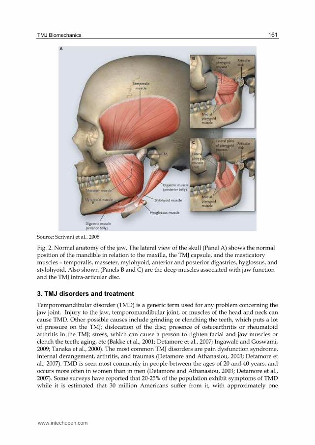
Strong muscles control the movement of the jaw and the TMJ. The temporalis muscle which attaches to the temporal bone elevates the mandible. The masseter muscle closes the mouth and is the main muscle used in mastication (see Figure 2) (Hylander, 1979). Movement is guided by the shape of the bones, muscles, ligaments, and occlusion of the teeth. The TMJ undergoes hinge and gliding motion (Alomar et al., 2007). The TMJ movements are very complex as the joint has three degrees of freedom, with each of the degrees of freedom associated with a separate axis of rotation. Rotation and anterior translation are the two primary movements. Posterior translation and mediolateral translation are the other two possible movements of TMJ (Dutton, 2004).
The Temporomandibular joint
Condyle
LigamentDisc
Articular fossa
Muscle
www.intechopen.com
TMJ Biomechanics
161
Source: Scrivani et al., 2008
Fig. 2. Normal anatomy of the jaw. The lateral view of the skull (Panel A) shows the normal position of the mandible in relation to the maxilla, the TMJ capsule, and the masticatory muscles – temporalis, masseter, mylohyoid, anterior and posterior digastrics, hyglossus, and stylohyoid. Also shown (Panels B and C) are the deep muscles associated with jaw function and the TMJ intra-articular disc.
3. TMJ disorders and treatment
Temporomandibular disorder (TMD) is a generic term used for any problem concerning the jaw joint. Injury to the jaw, temporomandibular joint, or muscles of the head and neck can cause TMD. Other possible causes include grinding or clenching the teeth, which puts a lot of pressure on the TMJ; dislocation of the disc; presence of osteoarthritis or rheumatoid arthritis in the TMJ; stress, which can cause a person to tighten facial and jaw muscles or clench the teeth; aging, etc (Bakke et al., 2001; Detamore et al., 2007; Ingawalé and Goswami, 2009; Tanaka et al., 2000). The most common TMJ disorders are pain dysfunction syndrome, internal derangement, arthritis, and traumas (Detamore and Athanasiou, 2003; Detamore et al., 2007). TMD is seen most commonly in people between the ages of 20 and 40 years, and occurs more often in women than in men (Detamore and Athanasiou, 2003; Detamore et al., 2007). Some surveys have reported that 20-25% of the population exhibit symptoms of TMD while it is estimated that 30 million Americans suffer from it, with approximately one
www.intechopen.com

Human Musculoskeletal Biomechanics
162
million new patients diagnosed yearly (Detamore and Athanasiou, 2003; Detamore et al., 2007; Tanaka et al., 2008b; Wolford, 1997).
Disc displacement is the most common TMJ arthropathy and is defined as an abnormal
relationship between the articular disc and condyle (Tanaka et al., 2000). As the disc is
forced out of the correct position, there is often bone on bone contact which creates
additional wear and tear on the joint, and often causes the TMD to worsen (Tanaka et al.,
2000). Almost 70% of TMD patients have disc displacement (Detamore and Athanasiou,
2003). Different types of functional malocclusion have been shown to be partly responsible
for signs and symptoms of TMD. The functional unilateral posterior cross-bite, habitual
body posture during sleep, juvenile chronic arthritis - a chronic arthritis in childhood with
an onset before the age of 16 years and a duration of more than three months – are also
reported as TMD risk (Bakke et al., 2001; Hibi and Ueda, 2005; Pellizoni et al., 2006).
Treatments for the various TMJ disorders range from physical therapy and nonsurgical
treatments to various surgical procedures. Usually the treatment begins with conservative,
nonsurgical therapies first, with surgery left as the last option. The majority of TMD
patients can be successfully treated by non-surgical therapies and surgical interventions
may be required for only a small part of TMD population (Ingawalé and Goswami, 2009).
The initial treatment does not always work and therefore more intense treatments such as
joint replacement may be a future option (Ingawalé and Goswami, 2009). The non-surgical
treatment options include medication; self-care; physical therapy, to keep the synovial joint
lubricated and to maintain full range of the jaw motion; wearing splints, the plastic
mouthpieces that fit over the upper and lower teeth to prevent the upper and lower teeth
from coming together, lessening the effects of clenching or grinding the teeth (Ingawalé and
Goswami, 2009). Splints are used to help control bruxism – a TMD risk factor in some cases
(Glaros et al., 2007; Kalamir et al., 2007; Tanaka et al., 2000a). However, the long-term
effectiveness of this therapy has been widely debated and remains controversial (Glaros et
al., 2007; Kalamir et al., 2007). Surgery can play an important role in the management of
TMDs. Conditions that are always treated surgically involve problems of overdevelopment
or underdevelopment of the mandible resulting from alterations of condylar growth,
mandibular ankylosis, and benign and malignant tumors of the TMJ (Laskin et al., 2006).
The surgical treatments include arthrocentesis, arthroscopy, discectomy, and joint
replacement. While more conservative treatments are preferred when possible, in severe
cases or after multiple operations, the current end stage treatment is joint replacement
(Tanaka et al., 2008b). However, before a joint replacement option is ever considered for a
patient, all non-surgical, conservative treatment options must be exhausted; and all
conservative surgical methodologies should be employed (Quinn, 199; Quinn, 2000).
4. Biomechanical behavior of the TMJ
Mandibular motions result in static and dynamic loading in the TMJ. During natural loading
of the joint, combinations of compressive, tensile, and shear loading occur on the
articulating surfaces (Tanaka et al., 2008b). The analysis of mandibular biomechanics helps
us understand the interaction of form and function, mechanism of TMDs; and aids in the
improvement of the design and the behavior of prosthetic devices, thus increasing their
treatment efficiency (Hansdottir and Bakke, 2004; Ingawalé and Goswami, 2009; Korioth
and Versluis, 1997)
www.intechopen.com

TMJ Biomechanics
163
4.1 In-vivo assessment
Very few studies which report in-vivo biomechanical assessment of the TMJ can be found in the literature. In contrast to some earlier studies which reported the TMJ to be a force-free joint, Hylander (1979) demonstrated that considerable forces were exerted on the TMJ during occlusion as well as mastication. In face of these contrary reports, Breul et al. (1999) showed that the TMJ was subjected to pressure forces during occlusion as well as during mastication and it was slightly eccentrically loaded in all positions of occlusion. Korioth and Hannam (1994) indicated that the differential static loading of the human
mandibular condyle during tooth clenching was task dependent and both the medial and
lateral condylar thirds were heavily loaded. Huddleston Slater et al. (1999) suggested that
when the condylar movement traces coincide during chewing, there is compression in the
TMJ during the closing stroke. However, when the traces do not coincide, the TMJ is not or
only slightly compressed during chewing. Naeije and Hofman (2003) used these
observations to study the loading of the TMJ during chewing and chopping tasks. Their
analysis showed that the distances traveled by the condylar kinematic centers were shorter
on the ipsilateral side than on the contralateral. The kinematic centers of all contralateral
joints showed a coincident movement pattern during chewing and chopping. The indication
that the ipsilateral joint is less heavily loaded during chewing than the contralateral joint
may explain why patients with joint pain occasionally report less pain while chewing on the
painful side.
Hansdottir and Bakke (2004) evaluated the effect of TMJ arthralgia on mandibular mobility,
chewing, and bite force in TMD patients (categorized as disc derangements, osteoarthritis,
and inflammatory disorders) compared to healthy control subjects. The pressure pain
threshold (PPT), maximum jaw opening, and bite force were significantly lower in the
patients as compared to that in controls. The patients were also found to have longer
duration of chewing cycles. The bite force and jaw opening in patients were significantly
correlated with PPT. The most severe TMJ tenderness (i.e., lowest PPT) and the most
impeded jaw function with respect to jaw opening and bite force were found to be more
severe in the patients with inflammatory disorders than the patients with disc derangement
or osteroarthritis (Hansdottir and Bakke, 2004).
4.2 In-vitro assessment – mechanical testing and finite element modeling
As the TMJ components are difficult to reach and as the applications of experimental
devices inside the TMJ cause damage to its tissue, the direct methods are not used often.
Indirect techniques utilized to evaluate mandibular biomechanics have had limited success
due to their ability to evaluate only the surface stress of the model but not its mechanical
properties (Ingawalé and Goswami, 2009). Mechanical testing and finite element modeling
(FEM) have been progressively used by TMJ researchers.
Excessive shear strain can cause degradation of the TMJ articular cartilage and collagen damage eventually resulting in joint destruction (Tanaka et al., 2008). Tanaka et al. (2008) attempted to characterize the dynamic shear properties of the articular cartilage by studying shear response of cartilage of 10 porcine mandibular condyles using an automatic dynamic viscoelastometer. The results showed that the shear behavior of the condylar cartilage is dependent on the frequency and amplitude of applied shear strain suggesting a significant role of shear strain on the interstitial fluid flow within the cartilage. Beek et al. (2001) performed sinusoidal indentation experiments and reported that the dynamic mechanical
www.intechopen.com
Human Musculoskeletal Biomechanics
164
behavior of disc was nonlinear and time-dependent. Beek et al. (2003) simulated these experiments using axisymmetric finite element model and showed that a poroelastic material model can describe the dynamic behavior of the TMJ disc. Tanaka et al. (2006) carried out a series of measurements of frictional coefficients on 10 porcine TMJs using a pendulum-type friction tester. The results showed that the presence of the disc reduces the friction in the TMJ by reducing the incongruity between the articular surfaces and by increasing synovial fluid lubrication. This study highlighted the importance of preserving the disc through alternatives to discectomy to treat internal derangement and osteoarthritis of the TMJ. The finite element modeling (FEM) has been used widely in biomechanical studies due to its
ability to simulate the geometry, forces, stresses and mechanical behavior of the TMJ
components and implants during simulated function (Beek et al., 2001; Chen et al., 1998;
Koolstra and van Eijden, 2005, 2006; Perez del Palomar and Doblare, 2006b, 2008; Reina et
al., 2007; Tanaka et al., 2000). Chen et al. (1998) performed stress analysis of human TMJ
using a two-dimensional FE model developed from magnetic resonance imaging (MRI). Due
to convex nature of the condyle, the compressive stresses were dominant in the condylar
region whereas the tensile stresses were dominant in the fossa-eminence complex owing to
its concave nature. Beek et al. (2001) developed a 3D linear FE model and analyzed the
biomechanical reactions in the mandible and in the TMJ during clenching under various
restraint conditions. Nagahara et al. (1999) developed a 3D linear FE model and analyzed
the biomechanical reactions in the mandible and in the TMJ during clenching under various
restraint conditions. All these FE simulations considered symmetrical movements of
mandible, and the models developed only considered one side of the joint. Hart et al. (1992)
generated 3D FE models of a partially edentulated human mandible to calculate the
mechanical response to simulated isometric biting and mastication loads. Vollmer et al.
(2000) conducted experimental and finite element study of human mandible to investigate
its complex biomechanical behavior. Tanaka et al. (2001, 2004) developed a 3D model to
investigate the stress distribution in the TMJ during jaw opening, analyzing the differences
in the stress distribution of the disc between subjects with and without internal
derangement. Tanaka et al. (2008c) suggested, from the results of finite element model of the
TMJ based on magnetic resonance images, that increase of the frictional coefficient between
articular surfaces may be a major cause for the onset of disc displacement. Sellers and
Crompton, (2004) used sensitivity analysis to validate the predictions of 3D FE simulations.
In 2005, Koolstra and van Eijden developed a combination of rigid-body model with a FE
model of both discs and the articulating cartilaginous surfaces to simulate the opening
movement of the jaw. Using the same model, Koolstra and van Eijden (2006) performed FEA
to study the load-bearing and maintenance capacity of the TMJ. The results indicated that
the construction of the TMJ permitted its cartilaginous structures to regulate their
mechanical properties effectively by imbibitions, exudation and redistribution of fluid.
Perez-Palomar and Doblare (2006a) used more realistic FE models of both TMJs and soft
components to study clenching of mandible. Perez del Palomar and Doblare (2006b)
developed a 3D FE model that included both discs ligaments and the three body contact
between all elements of the joints, and analyzed biomechanical behavior of the soft
components during a nonsymmetrical lateral excursion of the mandible to investigate
possible consequences of bruxism. This study suggested that a continuous lateral movement
of the jaw may lead to perforations in the lateral part of both discs, conforming to the
www.intechopen.com

TMJ Biomechanics
165
indications by Tanaka et al. (2001; 2004). Later, in 2007, Perez del Palomar and Doblare
suggested that unilateral internal derangement is a predisposing factor for alterations in the
unaffected TMJ side. However, it would be necessary to perform an exhaustive analysis of
bruxism with the inclusion of contact forces between upper and lower teeth during
grinding.
Whiplash injury is considered as a significant TMD risk factor and has been proposed to
produce internal derangements of the TMJ (Kasch et al., 2002; Perez del Palomar and
Doblare, 2008). However, this topic is still subject to debate (Detamore et al., 2007). In 2008,
Perez del Palomar and Doblare, published the results of finite element simulations of the
dynamic response of TMJ in rear-end and frontal impacts to predict the internal forces and
deformations of the joint tissues. The results, similar to suggested by Kasch et al. (2002),
indicated that neither a rear-end impact at low-velocity nor a frontal impact would produce
damage to the soft tissues of the joint suggesting that whiplash actions are not directly
related with TMDs. However; since this study has its own limitations such as analysis of
only one model, for low-velocity impacts, without any restrictions like contact with some
component of the vehicle; there is a need for more reliable finite element simulations to
obtain more accurate numerical results.
A theoretical model developed by Gallo et al. (2000) for estimating the mechanical work produced by mediolateral stress-field translation in the TMJ disc during jaw opening/closing suggested that long-term exposure of the TMJ disc to high work may result in fatigue failure of the disc. In 2001, Gross et al. proposed a predictive model of occlusal loading of the facial skeleton while May et al. (2001) developed a mathematical model of the TMJ to study the compressive loading during clenching. Effect of mandibular activity on mechanical work in the TMJ, which produces fatigue that may influence the pathomechanics of degenerative disease of the TMJ, was studied by Gallo et al. (2006). Nickel et al. (2002) validated numerical model predictions of TMJ eminence morphology and muscle forces, and demonstrated that the mechanics of the craniomandibular system are affected by the combined orthodontic and orthognathic surgical treatments. Using this validated numerical model to calculate ipsilateral and contralateral TMJ loads for a range of biting positions and angles, Iwasaki et al. (2009) demonstrated that TMJ loads during static biting are larger in subjects with TMJ disc displacement compared to subjects with normal disc position.
4.3 Post-surgery assessment
TMJ reconstruction using the partial or total TMJ prosthetics, in most cases, improves range of motion and mouth opening in the TMJ patients. However, loss of translational movements of the mandible on the operated side has been often observed, especially in anterior direction, owing to various factors like loss of pterygoid muscle function, scarring of the joint region and the muscles of mastication (Yoon et al., 2007). Komistek et al. (1998) assessed in-vivo kinematics and kinetics of the normal, partially replaced, and totally replaced TMJs. Less translation was reported in the implanted fossa and total TMJs than in the normal joints. The study suggests that total TMJ implants only rotate and do not translate; and the muscles do not apply similar forces at the joint when the subject has a total TMJ implant, compared to a subject who has a normal, healthy TMJ. In the post TMJ replacement follow-up studies, Mercuri et al. (2008) obtained the measures of mandibular interincisal opening and lateral excursions. The assessment
www.intechopen.com
Human Musculoskeletal Biomechanics
166
showed a 24% and a 30% improvement in mouth opening after 2 years and 10 years, respectively. On the other hand, at 2 years post-implantation there was a 14% decrease in left lateral excursion and a 25% decrease in right lateral excursion from the pre-implantation data. As the loss of lateral jaw movement is a great disadvantage to total TMJ prosthesis replacement, a future prosthesis must allow some lateral translation as well as the anterior movement of mandible on the operated side when the mouth is opened (van Loon et al., 1995). Yoon et al., (2007) followed a kinematic method that tracked the condylar as well as incisors path of the TMJ motion. An electromagnetic tracking device and accompanying software were used to record the kinematics of the mandible relative to temporal bone during opening-closing, protrusive, and lateral movements (Yoon et al., 2007). Mean linear distance (LD) of incisors during maximal mouth opening for the surgical patient group was 18% less than the normal subjects. Mean LD for mandibular right and left condyles was symmetrical in the normal group; however, in the surgical patient group, measurements for operated condyle and unoperated condyle were asymmetric and reduced as compared with normal subjects by 57% and 36%, respectively (Yoon et al., 2007). In protrusive movements, operated and unoperated condyles of surgical patients traveled less and significantly differently as compared with condyles of normal subjects, which moved almost identically. For the surgical patient group, the mean incisor LD away from the operated side and toward the operated side as compared with the normal group incisors were reduced by 67% and 32%, respectively (Yoon et al., 2007).
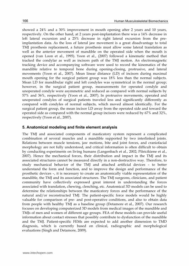
5. Anatomical modeling and finite element analysis
The TMJ and associated components of masticatory system represent a complicated combination of several muscles and a mandible supported by two interlinked joints. Relations between muscle tensions, jaw motions, bite and joint forces, and craniofacial morphology are not fully understood, and critical information is often difficult to obtain by conducting experiments on living humans (Langenbach et al., 2002; Pileicikiene et al., 2007). Hence the mechanical forces, their distribution and impact in the TMJ and its associated structures cannot be measured directly in a non-destructive way. Therefore, to study mechanical behavior of the TMJ and attached artificial devices – to better understand the form and function, and to improve the design and performance of the prosthetic devices –, it is necessary to create an anatomically viable representation of the mandible, the TMJ and its associated structures. The TMJ surgeons, clinicians, and patient community have collectively expressed great interest in understanding the forces associated with translation, chewing, clenching, etc. Anatomical 3D models can be used to determine the relationships between the masticatory forces and the performance of the natural and/or reconstructed TMJ. The patient-specific force models would be highly valuable for comparison of pre- and post-operative conditions, and also to obtain data from people with healthy TMJ as a baseline group (Detamore et al., 2007). Our research focuses on developing computerized 3D models from medical images of the mandible and TMJs of men and women of different age groups. FEA of these models can provide useful information about contact stresses that possibly contribute to dysfunction of the mandible and the TMJ. Patient-specific FEMs are expected to add another dimension to TMD diagnosis, which is currently based on clinical, radiographic and morphological evaluations (Singh and Detamore, 2009).
www.intechopen.com

TMJ Biomechanics
167
5.1 Modeling approaches
Determining the actual shape of the TMJ components through medical images greatly increases the accuracy of the model. We tried two approaches for 3D reconstruction of mandible and the TMJ from computed tomography (CT) images. In the first method, a software tool, MATLAB, was used for image processing. The MATLAB code was developed in such a way that it converts the original gray scale CT images into binary images thus separating the region of interest from rest of the data in the images (see Figure 3). The MATLAB code, then, finds the co-ordinates of the boundary pixels of the region of interest in each slice of the scan. These co-ordinates were imported into another software package, ANSYS, to plot contours corresponding to each CT slice and, subsequently, to develop a 3D model by connecting the consecutive contours to form closed areas and, subsequently, the closed volume mesh (see Figure 4). This modeling approach is very time consuming and involves a lot of manual tasks for image processing and further modeling. Accuracy of image processing is affected when the CT images have scatter due to dental implants. This requires making approximations about the actual shape of the object of interest.
Fig. 3. Processing the CT images in MATLAB. Each gray-scale image (slice) in the scan is converted into a binary image after segmentation. After performing series of morphological operations to form the skeleton of the feature of interest (i.e., mandible in this example), the code returns the co-ordinates of the boundary pixels of the skeleton. These co-ordinates are then exported to ANSYS to create a 3D representation of the object of interest.
Due to the time consuming procedures and inaccuracies in the resultant models in the first approach, later we used a 3D modeling software Mimics® (Materialise, Ann Arbor, MI). Using Mimics one can translate CT or MRI data into complete 3D models for a variety of applications. Mimics® interactively reads CT/MRI data in the DICOM format. Once an area of interest is separated, it can be visualized in 3D. The segmentation task is made easier due to the ability to see the images in three different views: axial, sagittal, and coronal. We developed several subject-specific models of the mandible and TMJ. Improper segmentation
www.intechopen.com

Human Musculoskeletal Biomechanics
168
of the medical images during reconstruction as well as less than optimal quality of medical images used for modeling hampers quality of the 3D models. Shorter the inter-slice distance in the medical images, better is the quality of resultant model. The inter-slice distance for the CT scans used to develop model 1 (see Figure 5) is 2 mm while that for the CT scans used for model 2 (see Figure 8) is 0.67mm.
Fig. 4. The co-ordinates for each CT slice are imported in ANSYS (with the z-co-ordinate = the slice thickness) and plotted manually to form a contour that represents shape of the object in the CT slice. After plotting such contours for all slices, the consecutive slices are connected to form the solid model. Such a model can be meshed and used for FEA.
5.2 Model 1 - FEA
A subject-specific 3D model of mandible was developed in Mimics using CT data (see
Figure 5). A surface mesh was formed from this solid model using 7074 triangular elements.
More the number of elements, more exact is the FEA solution. However, the large number of
elements means the model requires higher computing power and more time to run the FEA
simulations. Therefore, we try to reduce the number of elements to an appropriate extent in
such a way that the quality of the elements and the accuracy of the estimated FEA solution
are not affected by the reduction in number of elements. In this process, it is made sure that
the mesh has more elements in the areas of complex geometry.
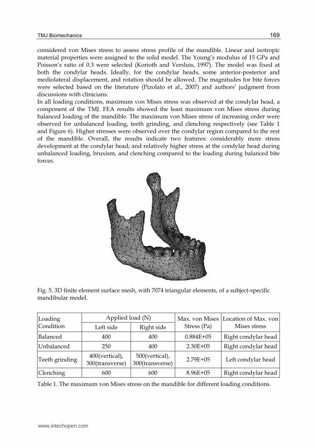
Estimating the stresses occurring over the mandible and the TMJ during different bite patterns and bite forces can be useful in understanding the function of joint and the possible mechanism of TMDs. The 3D surface mesh of mandible was imported into ANSYS to investigate comparative stress development and distribution in the mandible as a result of bite forces during four different loading conditions: normal/balanced occlusion versus three parafunctional loading conditions – which are believed to contribute to the TMDs – unbalanced loading, teeth grinding (bruxism), and teeth clenching. Since von Mises failure criterion has been widely used for mechanical testing of the ductile materials and bone, we
www.intechopen.com
TMJ Biomechanics
169
considered von Mises stress to assess stress profile of the mandible. Linear and isotropic material properties were assigned to the solid model. The Young’s modulus of 15 GPa and Poisson’s ratio of 0.3 were selected (Korioth and Versluis, 1997). The model was fixed at both the condylar heads. Ideally, for the condylar heads, some anterior-posterior and mediolateral displacement, and rotation should be allowed. The magnitudes for bite forces were selected based on the literature (Pizolato et al., 2007) and authors’ judgment from discussions with clinicians. In all loading conditions, maximum von Mises stress was observed at the condylar head, a component of the TMJ. FEA results showed the least maximum von Mises stress during balanced loading of the mandible. The maximum von Mises stress of increasing order were observed for unbalanced loading, teeth grinding, and clenching respectively (see Table 1 and Figure 6). Higher stresses were observed over the condylar region compared to the rest of the mandible. Overall, the results indicate two features: considerably more stress development at the condylar head; and relatively higher stress at the condylar head during unbalanced loading, bruxism, and clenching compared to the loading during balanced bite forces.
Fig. 5. 3D finite element surface mesh, with 7074 triangular elements, of a subject-specific mandibular model.
Loading Condition
Applied load (N) Max. von Mises Stress (Pa)
Location of Max. von Mises stress Left side Right side
Balanced 400 400 0.884E+05 Right condylar head
Unbalanced 250 400 2.30E+05 Right condylar head
Teeth grinding 400(vertical),
300(transverse) 500(vertical),
300(transverse)2.79E+05 Left condylar head
Clenching 600 600 8.96E+05 Right condylar head
Table 1. The maximum von Mises stress on the mandible for different loading conditions.
www.intechopen.com

Human Musculoskeletal Biomechanics
170
Two more FEA simulations were performed using the same 3D model with the same loading and boundary conditions; but Young’s modulus of 10 GPa and 7 GPa. This was done to see if the bone quality has any effect on the stress development in mandible and, especially, the condylar head – a TMJ component. Both of these simulations resulted in the least and highest maximum stress on the condylar head during balanced loading and teeth clenching, respectively, in accordance with the first simulation (see Figure 7). However, in contradiction with the previous simulation, the new simulations showed lower maximum von Mises stress during teeth grinding than that during unbalanced loading.
Fig. 6. Maximum von Mises stress developed over the mandibular 3D model during finite element simulation of teeth loading under four different bite conditions.
5.3 Model 2 - FEA
The second subject-specific anatomical 3D model of the mandible was developed in Mimics® from CT scan of a subject, aged 54 years, who reported moderate and intermittent pain in both TMJs. The CT images had ultra-high resolution with inter-slice thickness of 0.67mm. After importing the CT images in Mimics®; independent masks were created each for the cortical bone, cancellous bone, teeth, and articular fibrocartilage. After calculating 3D equivalent of the mandible, a volume mesh was generated using 37439 nodes and 23156 ten-node quadratic tetrahedral elements of type C3D10 (see Figures 8 and 9). Appropriate material properties were assigned to each component of the mandible using corresponding masks (see Table 2). The mandibular 3D volume mesh was, then, exported to a software package ABAQUS® (version 6.8) to perform comparative stress investigation in condylar cartilage under different loading conditions as in case of model-1.
www.intechopen.com

TMJ Biomechanics
171
Fig. 7. Maximum von Mises stress developed over the mandibular 3D model during three FE simulations – for three values of Young’s modulus (E) – under four loading conditions.
Fig. 8. Material properties were assigned to the 3D finite element volume mesh of the mandible using individual masks for each component. The cortical bone portion is indicated by yellow color, condylar cartilage by orange color, and teeth by red. As cancellous bone is covered by cortical bone, it is not visible in this figure.
www.intechopen.com

Human Musculoskeletal Biomechanics
172
Fig. 9. Three-dimensional finite element volume mesh of the mandible. The volume mesh had 37439 nodes and 23156 ten-node quadratic tetrahedral elements (C3D10).
Part Young’s Modulus (MPa)a, b Poisson’s Ratio a, b
Cortical bone 1.47E+04 0.3
Cancellous bone 4.90E+02 0.3
Teeth 1.76E+04 0.25
Cartilage 6.1 0.49
Sources: aIchim et al., 2006; bReina et al., 2007
Table 2. Material properties assigned to different components of the mandibular FE model.
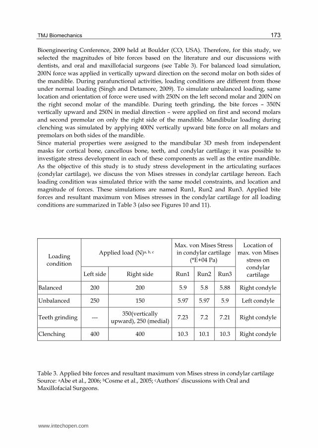
The mechanical behavior of the mandibular model was assumed to be linear-elastic, homogeneous, and isotropic. The model constraints were applied to imitate the in-vivo movements of the mandible as accurately as possible during each loading condition. Since the mastication forces are the result of the pressure in the teeth-food contact (Reina et al., 2007), the displacements were simply restrained at the nodes of the surface of the lower teeth that come in contact with the food or the upper teeth. During the balanced occlusive loading, both condyles were permitted translation of 10 mm in anterior-posterior direction and rotation of 11o along the medio-lateral axis. Same constraints were employed to simulate the unbalanced occlusive loading and bi-lateral molar clenching. During teeth grinding, the forces were applied on first and second molars and second premolar on right side only; and the right condyle was assumed free to move while the articular surface of the left condyle was constrained as during balanced loading. The magnitudes of mandibular and TMJ loading reported in the literature differ
significantly and there is currently no universally agreed upon value of TMJ loading
(Ingawalé and Goswami, 2009). Conflicting views about type, magnitude, and orientation of
masticatory forces used for FEMs were expressed by TMJ researchers at the TMJ
www.intechopen.com
TMJ Biomechanics
173
Bioengineering Conference, 2009 held at Boulder (CO, USA). Therefore, for this study, we
selected the magnitudes of bite forces based on the literature and our discussions with
dentists, and oral and maxillofacial surgeons (see Table 3). For balanced load simulation,
200N force was applied in vertically upward direction on the second molar on both sides of
the mandible. During parafunctional activities, loading conditions are different from those
under normal loading (Singh and Detamore, 2009). To simulate unbalanced loading, same
location and orientation of force were used with 250N on the left second molar and 200N on
the right second molar of the mandible. During teeth grinding, the bite forces – 350N
vertically upward and 250N in medial direction – were applied on first and second molars
and second premolar on only the right side of the mandible. Mandibular loading during
clenching was simulated by applying 400N vertically upward bite force on all molars and
premolars on both sides of the mandible.
Since material properties were assigned to the mandibular 3D mesh from independent
masks for cortical bone, cancellous bone, teeth, and condylar cartilage; it was possible to
investigate stress development in each of these components as well as the entire mandible.
As the objective of this study is to study stress development in the articulating surfaces
(condylar cartilage), we discuss the von Mises stresses in condylar cartilage hereon. Each
loading condition was simulated thrice with the same model constraints, and location and
magnitude of forces. These simulations are named Run1, Run2 and Run3. Applied bite
forces and resultant maximum von Mises stresses in the condylar cartilage for all loading
conditions are summarized in Table 3 (also see Figures 10 and 11).
Loading condition
Applied load (N)a, b, c Max. von Mises Stress in condylar cartilage
(*E+04 Pa)
Location of max. von Mises
stress on condylar cartilage Left side Right side Run1 Run2 Run3
Balanced 200 200 5.9 5.8 5.88 Right condyle
Unbalanced 250 150 5.97 5.97 5.9 Left condyle
Teeth grinding --- 350(vertically
upward), 250 (medial)7.23 7.2 7.21 Right condyle
Clenching 400 400 10.3 10.1 10.3 Right condyle
Table 3. Applied bite forces and resultant maximum von Mises stress in condylar cartilage Source: aAbe et al., 2006; bCosme et al., 2005; cAuthors’ discussions with Oral and Maxillofacial Surgeons.
www.intechopen.com
Human Musculoskeletal Biomechanics
174
(a)
(b)
(c)
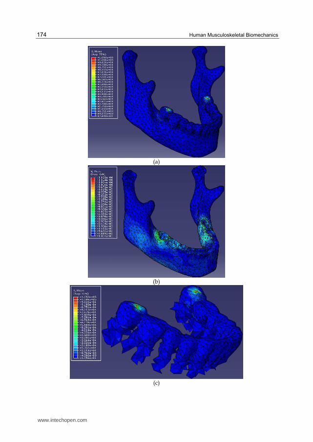
www.intechopen.com

TMJ Biomechanics
175
(d)
Fig. 10. von Mises stress [in (kg.mm/s2); (1 kg.mm/s2 = 1 kPa)] developed during balanced bilateral molar bite simulation in the entire mandible (a); and its components – cortical bone (b), teeth (c), and condylar articulating cartilage (d). (Note: The displayed sizes of components in panels c and d are not in proportion to each other and that of the components in other panels).
Fig. 11. A plot of maximum von Mises stress developed in the condylar articulating cartilage during four different occlusal static loading conditions – balanced molar bite, unbalanced molar bite, teeth grinding, and clenching – simulated thrice each. The FE simulations resulted in the highest mechanical stresses in the condylar cartilage during teeth clenching. Teeth grinding resulted in the mechanical stresses relatively less than during clenching, and higher than during unbalanced and balanced molar bites. The balanced loading produced the least stresses among all simulations.
The resultant stress data were analyzed using statistical analysis software JMP® (version 9). We employed the Tukey-Kramer HSD method to investigate the correlation between means of the peak von Mises stresses from three simulations/runs each of the four loading conditions under bite forces. From Tukey-Kramer HSD method, by comparing means of peak von Mises stresses for three runs/simulations of each loading condition, teeth grinding
www.intechopen.com

Human Musculoskeletal Biomechanics
176
and clenching were found to result in significantly different (p-value <0.0001 at α = 0.05) and higher von Mises stresses than balanced loading (see Figure 8). The von Mises stresses due to balanced and unbalanced loading were not significantly different from each other (at α = 0.05, p-value = 0.4386).
Fig. 12. The Tukey-Kramer HSD statistical analysis by comparing means of maximum von Mises stresses for three runs of each loading condition revealed that teeth grinding and clenching resulted in statistically significantly different von Mises stresses than balanced loading. The von Mises stresses due to balanced and unbalanced loading were not significantly different from each other.
The resultant maximum von Mises stresses in the condylar cartilage during balanced loading and clenching lie in the range of those reported in the literature (Hu et al., 2003; Nagahara et al., 1999). However, since most of the studies have reported stress development in bones and disc of the TMJ, we could not find any reported values of stress in the condylar cartilage under unbalanced loading and teeth grinding conditions to compare our results with. Comparatively higher mechanical stresses during clenching and teeth grinding activities suggest that these activities may lead to and exacerbate the TMDs. This indication
www.intechopen.com
TMJ Biomechanics
177
of our study conforms to the attribution that teeth grinding and clenching (as a result of physical and/or psychological stress) may be the causative factors for TMDs. Since we have applied the model constraints, material properties, and load values based on the literature, we consider the FEA results to be reliable and encouraging to advance our research efforts. We recognize that our FEA method has some limitations because we used simplified forces. We are developing subject-specific 3D models of the entire TMJ – including hard and soft tissues, and more refined FE mesh to perform biomechanical investigation under more realistic forces and model constraints. The proposed work promises to lead us to better understanding of the structural and functional aspects of natural and reconstructed TMJ. We also plan to validate the theoretical predictions of FEA through cadaver testing.
6. Summary
The TMJ literature underlines the importance of biomechanical analysis of the natural joint to better understand the structural and functional aspects; and of the reconstructed joint to assess the implant function and performance. Most of the methods reported in the literature have certain limitations due to the complex nature of the joint and also due to certain limitations of the techniques and software packages used for modeling and analysis. A more comprehensive biomechanical analysis of the natural and artificial TMJ is essential. The methodology used in this study for anatomical 3D reconstruction enables subject-specific modeling of complex structures and their constituent components. This feature can play a vital role in patient-specific anatomical modeling for diagnostic as well as therapeutic needs. Furthermore, such subject-specific anatomical models can be used to design custom prosthetic devices – which offer better fit, fixation, and efficiency – for a given anatomical structure. The FEA of such anatomical and prosthetic 3D models can be efficiently employed to better understand biomechanical behavior of the complex structures under investigation; and to improve the design, treatment efficiency, and durability of prosthetic devices. More comprehensive static and dynamic analyses of the mandible and TMJ coupled with experimental validation are necessary.
7. Acknowledgement
The authors would like to thank Dr. Deepak Krishnan (Assistant Professor, Oral and Maxillofacial Surgery, University of Cincinnati, Cincinnati, OH, USA) for sharing with us his clinical expertise and guiding our TMJ research.
8. References
Abe, M., Medina-Martinez, R. U., Itoh, K., Kohno, S., 2006. Temporomandibular joint
loading generated during bilateral static bites at molars and premolars. Medical
and Biological Engineering and Computing 44, 1017-1030.
Alomar, X., Medrano, J., Cabratosa, J., Clavero, J., Lorente, M., Serra, I., Monill, J., Salvador,
A., 2007. Anatomy of the temporomandibular joint. Seminars in Ultrasound, CT,
and MRI 28, 170-183.
www.intechopen.com

Human Musculoskeletal Biomechanics
178
American Association of Oral and Maxillofacial Surgeons (AAOMS), 2007. The
temporomandibular joint (TMJ). Retrieved on 10/14/2007, from
http://www.aaoms.org/tmj.php, 1.
Bakke, M., Zak, M., Jensen, B. L., Pedersen, F. K., Kreiborg, S., 2001. Orofacial pain, jaw
function, and temporomandibular disorders in women with a history of juvenile
chronic arthritis or persistent juvenile chronic arthritis Oral Surgery, Oral Medicine,
Oral Pathology, Oral Radiology, and Endodontics 92, 406-414.
Beek, M., Aarnts, M. P., Koolstra, J. H., Feilzer, A. J., Van Eijden, T. M. G. J., 2001. Dynamic
Properties of the Human Temporomandibular Joint Disc. Journal of Dental
Research 80, 876-880.
Beek, M., Koolstra, J. H., van Eijden, T. M. G. J., 2003. Human temporomandibular joint disc
cartilage as a poroelastic material Clinical Biomechanics 18, 69-76.
Beek, M., Koolstra, J. H., van Ruijven, L. J., van Eijden, T. M. G. J., 2001. Three-dimensional
finite element analysis of the cartilaginous structures in the human
temporomandibular joint. Journal of Dental Research 80, 1913-1918.
Breul, R., Mall, G., Landgraf, J., Scheck, R., 1999. Biomechanical analysis of stress
distribution in the human temporomandibular-joint Annals of Anatomy 181, 55-60.
Chen, J., Akyuz, U., Xu, L., Pidaparti, R. M. V., 1998. Stress analysis of the human
temporomandibular joint. Medical Engineering and Physics 20, 565-572.
Cosme, D. C., Baldisserotto, S. M., Canabarro Sde, A., Shinkai, R. S., 2005. Bruxism and
voluntary maximal bite force in young dentate adults The International Journal of
Prosthodontics 18, 328-332.
Detamore, M. S., Athanasiou, K. A., 2003. Structure and function of the temporomandibular
joint disc: implications for tissue engineering Journal of Oral and Maxillofacial
Surgery 61, 494-506.
Detamore, M. S., Athanasiou, K. A., Mao, J., 2007. A call to action for bioengineers and
dental professionals: directives for the future of TMJ bioengineering Annals of
Biomedical Engineering 35, 1301-1311.
Dutton, M., 2004. Orthopaedic Examination, Evaluation, & Intervention: A Pocket
Handbook. McGraw-Hill, New York, pp548.
Gallo, L. M., Chiaravalloti, G., Iwasaki, L. R., Nickel, J. C., Palla, S., 2006. Mechanical work
during stress-field translation in the human TMJ. Journal of Dental Research 85,
1006-1010.
Gallo, L. M., Nickel, J. C., Iwasaki, L. R., Palla, S., 2000. Stress-field translation in the healthy
human temporomandibular joint. Journal of Dental Research 79, 1740-1746.
Glaros, A. G., Owais, Z., Lausten, L., 2007. Reduction in parafunctional activity: a potential
mechanism for the effectiveness of splint therapy. Journal of Oral Rehabilitation 34,
97-104.
Gross, M. D., Arbel, G., Hershkovitz, I., 2001. Three-dimensional finite element analysis of
the facial skeleton on simulated occlusal loading. Journal of Oral Rehabilitation 28,
684-694.
Hansdottir, R., Bakke, M., 2004. Joint tenderness, jaw opening, chewing velocity, and bite
force in patients with temporomandibular joint pain and matched healthy control
subjects. Journal of Orofacial Pain 18, 108-113.
www.intechopen.com

TMJ Biomechanics
179
Hart, R. T., Hennebel, V. V., Thongpreda, N., Van Buskirk, W. C., Anderson, R. C., 1992.
Modeling the biomechanics of the mandible: a three-dimensional finite element
study. Journal of Biomechanics 25, 261-286.
Hibi, H., Ueda, M., 2005. Body posture during sleep and disc displacement in the
temporomandibular joint: a pilot study. Journal of Oral Rehabilitation 32, 85-89.
Hu, K., Qiguo, R., Fang, J., Mao, J. J., 2003. Effects of condylar fibrocartilage on the
biomechanical loading of the human temporomandibular joint in a three-
dimensional, nonlinear finite element model. 25, 107-113.
Huddleston Slater, J. J. R., Visscher, C. M., Lobbezoo, F., Naeije, M., 1999. The Intra-articular
Distance within the TMJ during Free and Loaded Closing Movements. Journal of
Dental Research 78, 1815-1820.
Hylander, W. L., 1979. An experimental analysis of temporomandibular joint reaction force
in macaques. American Journal of Physical Anthropology 51, 433-456.
Ichim, I., Swain, M., Kieser, J. A., 2006. Mandibular biomechanics and development of the
human chin. Journal of Dental Research 85, 638-642.
Ide, Y., Nakazawa, K., Garcia, L. T., 1991. Anatomical atlas of the temporomandibular joint.
Quintessence Publ. Co., Chicago, pp. 116.
Ingawalé, S., Goswami, T., 2009. Temporomandibular joint: disorders, treatments, and
biomechanics. Annals of Biomedical Engineering 37, 976-996.
Iwasaki, L. R., Crosby, M., Gonzalez, Y., McCall, W. D., Marx, D. B., Ohrbach, R., Nickel, J.
C., 2009. Temporomandibular joint loads in subjects with and without disc
displacement. Orthopedic Reviews 1, 90-93.
Kalamir, A., Pollard, H., Vitiello, A., Bonello, R., 2007. TMD and the problem of bruxism. A
review. Journal of Bodywork and Movement Therapies 11, 183-193.
Kasch, H., Hjorth, T., Svensson, P., Nyhuus, L., Jensen, T. S., 2002. Temporomandibular
disorders after whiplash injury: a controlled, prospective study Journal of Orofacial
Pain 16, 118-128.
Komistek, R. D., Dennis, D. A., Mabe, J. A., Anderson, D. T., 1998. In vivo kinematics and
kinetics of the normal and implanted TMJ. Journal of Biomechanics 31, 13.
Koolstra, J. H., van Eijden, T. M., 2006. Prediction of volumetric strain in the human
temporomandibular joint cartilage during jaw movement. Journal of Anatomy 209,
369-380.
Koolstra, J. H., van Eijden, T. M., 2005. Combined finite-element and rigid-body analysis of
human jaw joint dynamics Journal of Biomechanics 38, 2431-2439.
Korioth, T. W., Hannam, A. G., 1994. Mandibular forces during simulated tooth clenching
Journal of Orofacial Pain 8, 178-189.
Korioth, T. W. P., Versluis, A., 1997. Modeling the mechanical behavior of the jaws and their
related structures by finite element (FE) analysis. Critical Reviews in Oral Biology
& Medicine 8, 90-104.
Langenbach, G. E. J., Zhang, F., Herring, S. W., Hannam, A. G., 2002. Modelling the
masticatory biomechanics of a pig. Journal of Anatomy 201, 383-393.
Laskin, D. M., Greene, C. B., Hylander, W. L., 2006. Temporomandibular disorders an
evidence-based approach to diagnosis and treatment. Quintessence Pub., Chicago,
pp. 548.
www.intechopen.com

Human Musculoskeletal Biomechanics
180
May, B., Saha, S., Saltzan, M., 2001. A three-dimensional mathematical model of
temporomandibular joint loading. Clinical Biomechanics 16, 489-495.
Mercuri, L. G., Ali, F. A., Woolson, R., 2008. Outcomes of total alloplastic replacement with
periarticular autogenous fat grafting for management of reankylosis of the
temporomandibular joint Journal of Oral and Maxillofacial Surgery 66, 1794-1803.
Naeije, M., Hofman, N., 2003. Biomechanics of the Human Temporomandibular Joint during
Chewing. Journal of Dental Research 82, 528-531.
Nagahara, K., Murata, S., Nakamura, S., Tsuchiya, S., 1999. Displacement and stress
distribution in the temporomandibular joint during clenching. The Angle
Orthodontist 69, 372-379.
Nickel, J., Yao, P., Spalding, P. M., Iwasaki, L. R., 2002. Validated numerical modeling of the
effects of combined orthodontic and orthognathic surgical treatment on TMJ loads
and muscle forces. American Journal of Orthodontics and Dentofacial Orthopedics
121, 73-83.
Pellizoni, S. E., Salioni, M. A., Juliano, Y., Guimaraes, A. S., Alonso, L. G., 2006.
Temporomandibular joint disc position and configuration in children with
functional unilateral posterior crossbite: a magnetic resonance imaging evaluation
American Journal of Orthodontics and Dentofacial Orthopedics 129, 785-793
Perez del Palomar, A., Doblare, M., 2008. Dynamic 3D FE modelling of the human
temporomandibular joint during whiplash. 30, 700-709.
Perez del Palomar, A., Doblare, M., 2007. Influence of unilateral disc displacement on the
stress response of the temporomandibular joint discs during opening and
mastication. Journal of Anatomy 211, 453-463.
Perez del Palomar, A., Doblare, M., 2006a. The effect of collagen reinforcement in the
behaviour of the temporomandibular joint disc. Journal of Biomechanics 39, 1075-
1085.
Perez del Palomar, A., Doblare, M., 2006b. Finite element analysis of the
temporomandibular joint during lateral excursions of the mandible. Journal of
Biomechanics 39, 2153-2163.
Pileicikiene, G., Varpiotas, E., Surna, R., Surna, A., 2007. A three-dimensional model of the
human masticatory system, including the mandible, the dentition and the
temporomandibular joints. Stomatologija, Baltic Dental and Maxillofacial Journal 9,
27-32.
Pizolato, R. A., Gavião, M. B. D., Berretin-Felix, G., Sampaio, A. C. M., Trindade, A. S. J.,
2007. Maximal bite force in young adults with temporomandibular disorders and
bruxism. Brazilian Oral Research 21, 278-283.
Quinn, P. D., 2000. Lorenz Prosthesis. Oral and Maxillofacial Surgery Clinics of North
America 12, 93-104.
Quinn, P. D., 199. Alloplastic Reconstruction of the temporomandibular joint. Selected
Readings in Oral and Maxillofacial Surgery 7, 1-23.
Reina, J. M., Garcia-Aznar, J. M., Dominguez, J., Doblare, M., 2007. Numerical estimation of
bone density and elastic constants distribution in a human mandible. Journal of
Biomechanics 40, 828-836.
www.intechopen.com
TMJ Biomechanics
181
Scrivani, S. J., Keith, D. A., Kaban, L. B., 2008. Temporomandibular disorders The New
England Journal of Medicine 359, 2693-2705.
Sellers, W., Crompton, R., 2004. Using sensitivity analysis to validate the predictions of a
biomechanical model of bite forces. Annals of Anatomy 186, 89-95.
Singh, M., Detamore, M. S., 2009. Biomechanical properties of the mandibular condylar
cartilage and their relevance to the TMJ disc. Journal of Biomechanics 42, 405-417.
Tanaka, E., Dalla-Bona, D. A., Iwabe, T., Kawai, N., Yamano, E., van Eijden, T., Tanaka, M.,
Miyauchi, M., Takata, T., Tanne, K., 2006. The Effect of Removal of the Disc on the
Friction in the Temporomandibular Joint. Journal of Oral and Maxillofacial Surgery
64, 1221-1224.
Tanaka, E., Rodrigo, D. P., Miyawaki, Y., Lee, K., Yamaguchi, K., Tanne, K., 2000. Stress
distribution in the temporomandibular joint affected by anterior disc displacement:
a three-dimensional analytic approach with the finite-element method. Journal of
Oral Rehabilitation 27, 754-759.
Tanaka, E., del Pozo, R., Tanaka, M., Asai, D., Hirose, M., Iwabe, T., Tanne, K., 2004. Three-
dimensional finite element analysis of human temporomandibular joint with and
without disc displacement during jaw opening. Medical Engineering & Physics 26,
503-511.
Tanaka, E., Detamore, M. S., Mercuri, L. G., 2008a. Degenerative disorders of the
temporomandibular joint: etiology, diagnosis, and treatment. Journal of Dental
Research 87, 296-307.
Tanaka, E., Detamore, M. S., Tanimoto, K., Kawai, N., 2008b. Lubrication of the
temporomandibular joint Annals of Biomedical Engineering 36, 14-29.
Tanaka, E., Hirose, M., Koolstra, J. H., van Eijden, T. M., Iwabuchi, Y., Fujita, R., Tanaka, M.,
Tanne, K., 2008c. Modeling of the effect of friction in the temporomandibular joint
on displacement of its disc during prolonged clenching Journal of Oral and
Maxillofacial Surgery 66, 462-468.
Tanaka, E., Kikuchi, K., Sasaki, A., Tanne, K., 2000a. An adult case of TMJ osteoarthrosis
treated with splint therapy and the subsequent orthodontic occlusal reconstruction:
adaptive change of the condyle during the treatment American Journal of
Orthodontics and Dentofacial Orthopedics 118, 566-571.
Tanaka, E., Rodrigo, D. P., Tanaka, M., Kawaguchi, A., Shibazaki, T., Tanne, K., 2001. Stress
analysis in the TMJ during jaw opening by use of a three-dimensional finite
element model based on magnetic resonance images International Journal of Oral
and Maxillofacial Surgery 30, 421-430.
Tanaka, E., Rego, E. B., Iwabuchi, Y., Inubushi, T., Koolstra, J. H., van Eijden, T. M. G. J.,
Kawai, N., Kudo, Y., Takata, T., Tanne, K., 2008. Biomechanical response of
condylar cartilage-on-bone to dynamic shear. Journal of Biomedical Materials
Research 85A, 127-132.
van Loon, J. P., de Bont, L. G. M., Boering, G., 1995. Evaluation of temporomandibular
joint prostheses Review of the literature from 1946 to 1994 and implications
for future prosthesis designs. Journal of Oral and Maxillofacial Surgery 53, 984-
996.
www.intechopen.com
Human Musculoskeletal Biomechanics
182
Vollmer, D., Meyer, U., Joos, U., Vegh, A., Piffko, J., 2000. Experimental and finite element
study of a human mandible. 28, 91-96.
Wolford, L. M., 1997. Temporomandibular joint devices: Treatment factors and outcomes.
Oral Surgery, Oral Medicine, Oral Pathology, Oral Radiology, and Endodontics 83,
143-149.
Yoon, H. J., Baltali, E., Zhao, K. D., Rebellato, J., Kademani, D., An, K. N., Keller, E. E., 2007.
Kinematic study of the temporomandibular joint in normal subjects and patients
following unilateral temporomandibular joint arthrotomy with metal fossa-
eminence partial joint replacement Journal of Oral and Maxillofacial Surgery 65,
1569-1576.
www.intechopen.com

Human Musculoskeletal BiomechanicsEdited by Dr. Tarun Goswami
ISBN 978-953-307-638-6Hard cover, 244 pagesPublisher InTechPublished online 05, January, 2012Published in print edition January, 2012
InTech EuropeUniversity Campus STeP Ri Slavka Krautzeka 83/A 51000 Rijeka, Croatia Phone: +385 (51) 770 447 Fax: +385 (51) 686 166www.intechopen.com
InTech ChinaUnit 405, Office Block, Hotel Equatorial Shanghai No.65, Yan An Road (West), Shanghai, 200040, China
Phone: +86-21-62489820 Fax: +86-21-62489821
This book covers many aspects of human musculoskeletal biomechanics. As the title represents, aspects offorces, motion, kinetics, kinematics, deformation, stress, and strain are examined for a range of topics such ashuman muscles, skeleton, and vascular biomechanics independently or in the presence of devices. Topicsrange from image processing to interpret range of motion and/or diseases, to subject specifictemporomandibular joint, spinal units, braces to control scoliosis, hand functions, spine anthropometricanalyses along with finite element analyses. Therefore, this book will be valuable to students at introductorylevel to researchers at MS and PhD level searching for science of specific muscle/vascular to skeletalbiomechanics. This book will be an ideal text to keep for graduate students in biomedical engineering since it isavailable for free, students may want to make use of this opportunity. Those that are interested to participatein the future edition of this book, on the same topic, as a contributor please feel free to contact the author.
How to referenceIn order to correctly reference this scholarly work, feel free to copy and paste the following:
Shirish M. Ingawale ́ and Tarun Goswami (2012). Biomechanics of the Temporomandibular Joint, HumanMusculoskeletal Biomechanics, Dr. Tarun Goswami (Ed.), ISBN: 978-953-307-638-6, InTech, Available from:http://www.intechopen.com/books/human-musculoskeletal-biomechanics/biomechanics-of-the-temporomandibular-joint