Biomechanical Comparison of Different Surface...
12
Volume 23 Number 6 2008 Biomechanical Comparison of Different Surface Modifications for Dental Implants Stephen J. Ferguson, PhD/Jens D. Langhoff, Dr Med Vet/Katrin Voelter, Dr Med Vet/ Brigitte von Rechenberg, Dr Med Vet, PhD/Dieter Scharnweber, PhD/SusanneBierbaum, PhD/ Matthias Schnabelrauch, PhD/Armin R. Kautz, PhD/Vinzenz M. Frauchiger, PhD/ Thomas L. Mueller, MSc/G. Harry van Lenthe, PhD/Falko Schlottig, PhD Special reprint from ©2008 Quintessence Publishing Co, Inc. www.quintpub.com
Transcript of Biomechanical Comparison of Different Surface...

Volume 23 Number 6 2008
Biomechanical Comparison ofDifferent Surface Modifications
for Dental ImplantsStephen J. Ferguson, PhD/Jens D. Langhoff, Dr Med Vet/Katrin Voelter, Dr Med Vet/
Brigitte von Rechenberg, Dr Med Vet, PhD/Dieter Scharnweber, PhD/Susanne Bierbaum, PhD/Matthias Schnabelrauch, PhD/Armin R. Kautz, PhD/Vinzenz M. Frauchiger, PhD/
Thomas L. Mueller, MSc/G. Harry van Lenthe, PhD/Falko Schlottig, PhD
S p e c i a l r e p r i n t f r o m
©2008 Quintessence Publishing Co, Inc.
www.quintpub.com
The International Journal of Oral & Maxillofacial Implants 1037
Biomechanical Comparison of Different SurfaceModifications for Dental Implants
Stephen J. Ferguson, PhD1/Jens D. Langhoff, Dr Med Vet2/Katrin Voelter, Dr Med Vet2/Brigitte von Rechenberg, Dr Med Vet, PhD3/Dieter Scharnweber, PhD4/Susanne Bierbaum, PhD5/
Matthias Schnabelrauch, PhD6/Armin R. Kautz, PhD7/Vinzenz M. Frauchiger, PhD8/Thomas L. Mueller, MSc9/G. Harry van Lenthe, PhD10/Falko Schlottig, PhD11
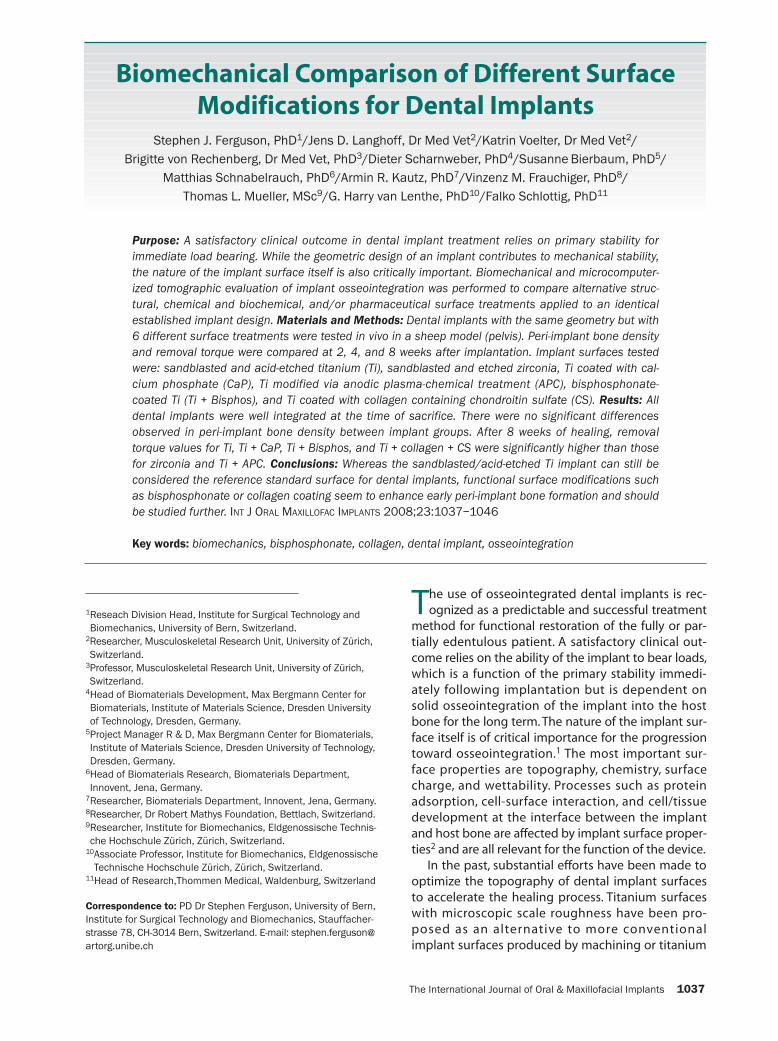
Purpose: A satisfactory clinical outcome in dental implant treatment relies on primary stability forimmediate load bearing. While the geometric design of an implant contributes to mechanical stability,the nature of the implant surface itself is also critically important. Biomechanical and microcomputer-ized tomographic evaluation of implant osseointegration was performed to compare alternative struc-tural, chemical and biochemical, and/or pharmaceutical surface treatments applied to an identicalestablished implant design. Materials and Methods: Dental implants with the same geometry but with6 different surface treatments were tested in vivo in a sheep model (pelvis). Peri-implant bone densityand removal torque were compared at 2, 4, and 8 weeks after implantation. Implant surfaces testedwere: sandblasted and acid-etched titanium (Ti), sandblasted and etched zirconia, Ti coated with cal-cium phosphate (CaP), Ti modified via anodic plasma-chemical treatment (APC), bisphosphonate-coated Ti (Ti + Bisphos), and Ti coated with collagen containing chondroitin sulfate (CS). Results: Alldental implants were well integrated at the time of sacrifice. There were no significant differencesobserved in peri-implant bone density between implant groups. After 8 weeks of healing, removaltorque values for Ti, Ti + CaP, Ti + Bisphos, and Ti + collagen + CS were significantly higher than thosefor zirconia and Ti + APC. Conclusions: Whereas the sandblasted/acid-etched Ti implant can still beconsidered the reference standard surface for dental implants, functional surface modifications suchas bisphosphonate or collagen coating seem to enhance early peri-implant bone formation and shouldbe studied further. INT J ORAL MAXILLOFAC IMPLANTS 2008;23:1037–1046
Key words: biomechanics, bisphosphonate, collagen, dental implant, osseointegration
The use of osseointegrated dental implants is rec-ognized as a predictable and successful treatment
method for functional restoration of the fully or par-tially edentulous patient. A satisfactory clinical out-come relies on the ability of the implant to bear loads,which is a function of the primary stability immedi-ately following implantation but is dependent onsolid osseointegration of the implant into the hostbone for the long term. The nature of the implant sur-face itself is of critical importance for the progressiontoward osseointegration.1 The most important sur-face properties are topography, chemistry, surfacecharge, and wettability. Processes such as proteinadsorption, cell-surface interaction, and cell/tissuedevelopment at the interface between the implantand host bone are affected by implant surface proper-ties2 and are all relevant for the function of the device.
In the past, substantial efforts have been made tooptimize the topography of dental implant surfacesto accelerate the healing process. Titanium surfaceswith microscopic scale roughness have been pro-posed as an alternative to more conventionalimplant surfaces produced by machining or titanium
1Reseach Division Head, Institute for Surgical Technology andBiomechanics, University of Bern, Switzerland.
2Researcher, Musculoskeletal Research Unit, University of Zürich,Switzerland.
3Professor, Musculoskeletal Research Unit, University of Zürich,Switzerland.
4Head of Biomaterials Development, Max Bergmann Center forBiomaterials, Institute of Materials Science, Dresden Universityof Technology, Dresden, Germany.
5Project Manager R & D, Max Bergmann Center for Biomaterials,Institute of Materials Science, Dresden University of Technology,Dresden, Germany.
6Head of Biomaterials Research, Biomaterials Department,Innovent, Jena, Germany.
7Researcher, Biomaterials Department, Innovent, Jena, Germany.8Researcher, Dr Robert Mathys Foundation, Bettlach, Switzerland.9Researcher, Institute for Biomechanics, Eldgenossische Technis-che Hochschule Zürich, Zürich, Switzerland.
10Associate Professor, Institute for Biomechanics, EldgenossischeTechnische Hochschule Zürich, Zürich, Switzerland.
11Head of Research,Thommen Medical, Waldenburg, Switzerland
Correspondence to: PD Dr Stephen Ferguson, University of Bern,Institute for Surgical Technology and Biomechanics, Stauffacher-strasse 78, CH-3014 Bern, Switzerland. E-mail: [email protected]

1038 Volume 23, Number 6, 2008
Ferguson et al
plasma spraying. Microrough titanium surfaces havebeen produced using various techniques, includingsandblasting, acid etching, and combinations ofboth, to create a modified surface topography.3 Thesandblasted and acid-etched surface has demon-strated enhanced bone apposition in histomorpho-metric studies4,5 and higher removal torque values inbiomechanical testing,6,7 indicating a direct relation-ship between the biologic and mechanical quality ofthe interface, as well as excellent mid- to long-termclinical results.8–11
Less is known about the combined effect of sur-face topography and chemistry, an important syner-gistic potential for optimizing healing time. Chemicaland biochemical surface modifications are assumedto be especially advantageous for accelerated heal-ing. A modified sandblasted/acid-etched surface hasbeen shown to enhance surface wettability and alsoimprove osseointegration and interfacial strength.12
The potential of a new generation of thin calciumphosphate (CaP) –based coatings has beendescribed by several authors.13,14 Fluoride surfacemodification seems to enhance osteoblastic differen-tiation and interfacial bone formation.15 Anotherapproach is a functionalized poly(L-lysine) andpoly(ethylene glycol) copolymer (PLL-g-PEG) coating,which inhibits in vitro bacterial growth and interactswith osteoblasts through the integrated bioli-gands.16 The modification of biochemistry using bio-logically altered surfaces has been shown. The use ofmodified titanium surfaces with extracellular matrixcomponents enhances bone remodeling in the earlystages of healing; a significant increase in osteopontin-positive osteoblasts was observed next to arginine-glycine-aspartic acid (RGD) peptide–coatedimplants.17 A newer approach is surface modificationwith bioactive molecules. Nucleic acid single strandsare fixed electrochemically via their termini(regiospecifically) by anodically growing an oxidelayer on titanium-aluminum-niobium (Ti-6Al-7Nb).18
Drug-eluting coatings are the most probable futuredevelopments. Local delivery of bisphosphonateshas resulted in increases in the mechanical fixationof implants,19 while the potential of growth factorssuch as bone morphogenetic protein 2 has beenshown in many studies.20,21
Clearly, there are many potential candidates forthe “optimal” surface of dental implants, along with alack of comparative data. Therefore, the purpose ofthis study was to perform biomechanical and micro-computerized tomographic (microCT) evaluation ofimplant osseointegration to compare alternativestructural, chemical and biochemical, and/or phar-maceutical surface treatments using a well-establishedimplant design.
MATERIALS AND METHODS
Implant DesignSix different implant surface types were evaluated:5 surface-modified, commercially pure titaniumimplants and 1 surface-modified zirconia implantwere employed. Eighteen implants were used in eachgroup. Implant shape was identical to the standardSPI Implant configuration, with a 4.2-mm diameterand 8-mm length (Thommen Medical, Waldenburg,Switzerland). Implant surface types were as follows.
• Group 1: Titanium implants were sandblasted andacid etched.
• Group 2: Zirconia implants (yttrium partially stabi-lized zirconia, medical grade) were sandblastedand etched in an alkaline bath.
• Group 3: CaP was coated via electrochemical assis-tance onto sandblasted and acid-etched titaniumimplants. The implants were coated in an aqueoussolution containing Ca and phosphate ions. Thecoating consists of the 2 CaP phases hydroxyap-atite and brushite and is commercially available.22
• Group 4: The implants were treated with theanodic plasma-chemical surface modificationmethod (APC) after sandblasting. APC is anadvanced anodization method that allows foranodic oxide layer formation and incorporation ofCaP phases in a single step. The method exploitsthe dielectric breakdown of anodic oxide films toproduce a porous oxide layer that contains signifi-cant amounts of electrolyte components. Theelectrolyte contained calcium and phosphateions, leading to a CaP-containing porous surface.23
• Group 5: The implants were coated with bisphos-phonate. Sandblasted and acid-etched titaniumimplants were spray coated with a suspension to afinal alendronate concentration of 10 µg/cm2. Thesuspension was prepared by treating an alen-dronate sodium salt with sodium dodecyl sulfate,resulting in a sparingly soluble salt.
• Group 6: Sandblasted and acid-etched implantswere coated with an artificial extracellular matrixfrom acid-soluble bovine collagen type I andchondroitin sulfate (CS). To create the latter, CS ata concentration of 30 µg/mg collagen was addedon ice to a fibrillogenesis buffer solution containing2.5 mg/mL collagen. Fibrillogenesis was allowedto take place overnight at 37°C. The resulting gelwas homogenized and fibrils were collected bycentrifugation, washed with fibrillogenesis buffer,and centrifuged again. The pellet was resus-pended in the same buffer to a concentration ofabout 5 mg/mL collagen. The implants were incu-bated in the suspension at 25°C for 5 min and air

The International Journal of Oral & Maxillofacial Implants 1039
Ferguson et al
dried. This process was performed a total of 2times. The coated implants were then washedwith distilled water and sterilized.24
Surface TopographyRoughness measurements were performed using aconfocal microscope (Nanofocus µsurf, Oberhausen,Germany) over an area of 320 3 308 µm (lateral reso-lution 0.7 µm, vertical resolution 25 nm). The rough-ness values were calculated after the subtraction ofthe third-order area regression taking the whole areainto account (not only profiles). All roughness valueswere calculated using the WinSAM 2.6 softwarepackage (Lehrstuhl für Fertigungstechnologie, Uni-versity Erlangen, Nuremberg, Germany). For electronmicroscopy, a Cambridge S360 instrument (Carl ZeissNTS, Oberkochen, Germany) was used. Secondaryelectron images were recorded using an accelerationvoltage of 20 kV and a sample current of 200 pA.
Study Design and Animal ModelThe animal experiment was conducted according toSwiss law for animal protection and welfare, and per-mission was obtained from the local authorities (Stateof Zurich, permission no. 159/2005). A previously devel-oped sheep model served as an accepted model forhuman bone healing and osseointegration.25,26 Allimplants were placed in the iliac bone of the pelvis,with 7 implants per side (one implant type was notevaluated for the present study). The iliac crest offers abroad variety of bone qualities, ranging from almostpurely cancellous (less than 0.5 mm cortical thickness)to compact cortical bone (up to 3 mm in thickness).Regarding bone quality, this allows comparison to thebone found in the mandible as classified in theLekholm and Zarb index.27 The study examined 3essential phases of implant osseointegration: the acutephase (2 weeks), the early phase (4 weeks), and the con-tinued phase (8 weeks), with 5 animals per time point.An implantation scheme was designed to achieve ahomogeneous spatial distribution of all implant types,with a sample size of 6 per implant group and healingtime for biomechanical testing (total n = 108) (Fig 1).Additional implants were placed for a concurrent histo-morphometric analysis, the topic of a separate study.
Surgical ProcedureThe access to each iliac crest was gained from thedorsolateral by a standard surgical procedure. Equallyspaced cavities for the dental implants were preparedusing a 2.0-mm pilot drill, then expanded with 2.8-mm and 3.5-mm drills (SPI VECTOdrill, ThommenMedical, Waldenburg, Switzerland). A specially con-structed drill sleeve was used to ensure a standard-ized drill depth, which was confirmed with a depth
gauge. Self-tapping implants (SPI ELEMENT, Thom-men Medical) were placed according to a randomizedimplantation scheme (randomized location of the 6implant types). Implants were covered with a healingcap and tightened with a torque wrench. After com-pletion, the muscle was repositioned and the tendi-nous insertion was resutured to its origin. The fasciaand subcutis were closed using synthetic resorbablesutures and the skin was closed with staples.
Sample CollectionAnimals were euthanized following the predefinedhealing periods. The implantation sites were har-vested with the full pelvic bone within 10 minutespostmortem. The iliac bone was sectioned with anoscillating saw into individual bone blocks, each ofwhich contained 1 implant with at least 12 mm ofadjacent bone tissue. Bone blocks for mechanicaltesting were wrapped in gauze soaked with an isotonicsodium chloride solution and tightly packed in plasticbags to prevent drying. Samples were stored at 7°Cuntil testing (maximum 36 hours).
Removal Torque TestingThe removal torque testing protocol has been previ-ously described in detail.6 To facilitate specimen han-dling and to provide adequate temperature isolation,each bone block was embedded in fast-curing dentalcement (GC Fujirock, GC Europe, Leuven, Belgium).Removal torque testing was performed on a servohy-draulic biaxial testing machine (MTS MiniBionix 358;MTS, Minneapolis, MN, USA). The implant was firstattached to the upper hydraulic actuator via a cus-tom-machined adapter, thus guaranteeing align-ment of the implant with the actuator’s rotationalaxis. The adapter for coupling the implant to thehydraulic actuator was vertically unconstrained to
Fig 1 Dorsal view of sheep pelvis, with implantation sites indi-cated. A total of 108 implants was placed for the biomechanicalstudy (6 groups 3 6 implants 3 3 healing periods).

1040 Volume 23, Number 6, 2008
Ferguson et al
eliminate any axial load on the implant. The implant-bone-cement specimen was then lowered into ametal container, and the space surrounding thespecimen was filled with a low-melting-temperaturemetal alloy (Ostalloy 117, Legierung 47° Grad FA16;Metallum, Pratteln, Switzerland). Solidification of thealloy proceeded quickly, minimizing specimen heat-ing and achieving rigid fixation of the specimen inthe machine. Removal torque testing was performedby rotating the implant counterclockwise at a rate of0.1 degree per second to a maximum angle of 30degrees while simultaneously collecting angle andtorque data at a sampling rate of 10 Hz. The speci-mens were kept moist throughout testing by sprayingwith saline solution, and the bone temperature wasmaintained at 29°C ± 0.5°C throughout.
The resulting torque-rotation curve was analyzedto determine the removal torque value and failuremode. The removal torque (N-mm) was defined asthe maximum torque recorded on the curve forspecimens that showed a clear peak and subsequentdrop in torque. For specimens with a constantlyincreasing torque-rotation curve, or a plateau in themeasured torque values, a yield point was defined byconstructing a straight line parallel to the initialslope of the torque-rotation curve, offsetting this by0.72 degrees (0.2% full rotation), and then selectingthe intersection of this offset line with the originaltorque-rotation curve.
MicroCTFor nondestructive 3-dimensional characterization ofperi-implant bone, selected samples were subse-quently scanned and analyzed by microCT.28 Onehundred samples were measured (fifty 4-week sam-ples and fifty 8-week samples) using a commercialimaging device (µCT40, Scanco Medical, Bassersdorf,Switzerland) and applying an isotropic resolution of30 µm. The analyzed sections comprised an evenlydistributed sample from both the biomechanical test-ing and the concurrent histomorphometry study. Twoglobal threshold levels were applied to the filtered
data to distinguish between implant, mineralized tissue, and background. Special image processing29
was applied to the direct vicinity of the implant toremove imaging artifacts. For quantitative analysis,2 cylindric volumes of interest (VOIs) were defined(Fig 2). The first VOI had a diameter of 6 mm centeredaround the implant (the “inner ring”); the second VOIwas a hollow cylinder with inner and outer radii of 10and 12 mm, respectively (the “outer ring”; Fig 2). Eachvolume was analyzed for bone volume density (ie, bone volume/total volume [BV/TV]). MicroCT is aprecise and validated technique that provides accu-rate measures of bone volume and architecture.30
Statistical AnalysisThe removal torque (dependent variable) was ana-lyzed, using a factorial analysis of variance (ANOVA)with interactions, for the effect of the following inde-pendent factors: implant surface type, implant posi-tion, and healing period. Post hoc comparisons weremade using the Scheffé F test. All analyses were per-formed using the ANOVA/MANOVA module of Statis-tica 7.0 (StatSoft, Tulsa, OK). The correlation betweenlocal bone density and removal torque values wasevaluated using linear regression analysis. A signifi-cance level of P = .05 was defined.
RESULTS
Characterization of SurfacesFigure 3 shows a typical sandblasted and acid-etched surface (group 1). The micropits produced bythe etching process are shown in Fig 3a, and themacrorough surface texture caused by the sand-blasting process can clearly be seen in Fig 3b. Thesandblasted and etched zirconia surface (group 2)shows a completely different topography. The micro-roughness appears cauliflowerlike (Fig 4a), and themacroroughness is less pronounced (Fig 4b). Figures5a and 5b show the typical structural elements ofthe electrochemically deposited coating (group 3).
Fig 2 Three-dimensional segmentedmicroCT images. The implant is marked inred, whereas bone is shown in grey. The 2trabecular volumes of interest are shown inorange (outer ring, “nonaffected bone”) andyellow (inner ring).
Figs 3a and 3b Scanning electron micrographs of group 1 surface (sandblasted andacid-etched titanium implant).

The International Journal of Oral & Maxillofacial Implants 1041
Ferguson et al
The figures show clearly the fine crystalline coatingstructure with fixed CaP crystallites in the shape ofplatelets or pins. The APC-modified surface structureis shown in Fig 6 (group 4). The surface layer isporous and composed of small craters with holes inthe center. In contrast, the bisphosphonate-coatedimplants (group 5, Fig 7) show no difference from theuncoated group 1 implants under the selected imag-ing conditions. A different pattern for the collagencontaining CS–coated (CCCS) (group 6) implants isapparent (Fig 8). The topographic properties of theinvestigated implant surfaces are shown in Table 1.
MicroCTBone volume density (BV/TV) at the outer ring wassimilar for all implant types, and no significantchanges were found between the 4-week and the 8-week samples. BV/TV at the inner ring was between40% and 63% higher than at the outer ring. This dif-ference in BV/TV was significant for all implant
groups as well as for both time points (Table 2).Time-dependent changes in BV/TV did not reach signifi-cance for any of the implants.
Biomechanical TestingRepresentative examples of removal torque versusrotation angle behavior are plotted in Fig 9. The char-acteristics of the bone-implant interface failure couldbe qualitatively distinguished by 3 distinctive mechan-ical responses: (1) an initial “slip” of the interface fol-lowed by a substantial increase in torque values to ayield point and torque plateau, (2) a steadily increasingtorque value until a yield point and subsequentplateau in the torque response was observed, or (3) asharply increasing torque value terminating with a sin-gle clear failure point. All failure behaviors are charac-teristic of a mechanical breakdown of the osseointe-grated interface. Failure mode 1 was predominantlyobserved with the APC surface-modified Ti implants(group 4). Failure mode 2 was characteristic of the
Figs 4a and 4b Scanning electron micro-graphs of group 2 surface (sandblasted andalkaline-etched zirconia implant).
Figs 5a and 5b Scanning electron micro-graphs of group 3 surface (sandblasted andacid-etched titanium implant with surfaceelectrochemically coated with calcium phos-phate).
Figs 6a and 6b Scanning electron micro-graphs of group 4 surface (sandblasted tita-nium implant treated with the anodicplasma-chemical surface modificationmethod).

1042 Volume 23, Number 6, 2008
Ferguson et al
Figs 7a and 7b Scanning electron micro-graphs of group 5 surface (sandblasted andacid-etched titanium implant sur facecoated with bisphosphonate).
Figs 8a and 8b Scanning electron micro-graphs of group 6 surface (sandblasted andacid-etched titanium implant, coated with amixture of type 1 collagen containing chon-droitin sulfate).
Table 1 Surface Topography of Each Implant Surface
Group Sa Ssk Sku aw
Group 1 2.002 –0.187 2.612 1.810Group 1 2.072 –0.113 2.535 1.815Group 2 1.073 –0.237 2.475 1.148Group 2 1.116 –0.273 2.700 1.151Group 3 1.054 0.023 2.240 1.224Group 3 1.383 –0.133 2.644 1.314Group 4 1.415 –0.112 2.510 1.206Group 4 1.497 –0.260 2.757 1.227Group 5 2.239 –0.128 2.492 1.843Group 5 2.176 –0.189 2.638 1.827Group 6 1.702 –0.234 2.618 1.722Group 6 1.769 –0.248 2.669 1.756
Sa = arithmetic mean deviation of the measured area; Ssk = skewness; Sku = kurtosis; aw =surface area ratio. Two samples of each surface were measured.
Table 2 Regional Bone Volume Density (BV/TV)
Bone volume density (%)
Week 4 Week 8
Inner Outer % Inner Outer %Group ring ring difference ring ring difference
Group 1 28.4 ± 7.6 18.9 ± 4.7 50.2* 30.3 ± 5.1 18.6 ± 3.3 63.3***Group 3 31.9 ± 6.2 20.4 ± 3.4 56.5** 28.5 ± 4.4 19.3 ± 4.6 47.2**Group 4 33.8 ± 9.2 21.6 ± 3.6 56.4* 30.5 ± 4.2 21.2 ± 5.4 44.0**Group 5 27.2 ± 7.3 17.8 ± 6.0 52.8* 32.1 ± 6.2 19.8 ± 4.6 62.0**Group 6 29.1 ± 4.5 20.7 ± 3.0 40.6** 28.6 ± 6.8 18.5 ± 5.1 54.5*
*P < .05; **P < .01; ***P < .001.Values reported are means ± SDs.

zirconia implants (group 2). All other implants generallydemonstrated a failure of the interface correspondingto the torque curve shown in Fig 9c.
Interface removal torque values are summarizedin Table 3. There was a substantial and significantincrease in the removal torque values with increasinghealing periods (P < .001; Fig 10). The implant surfacewas also a significant factor determining themechanical response of the bone-implant interface(P < .001; Fig 11). Post hoc analysis was performed to
evaluate specific hypotheses on implant perfor-mance. After 2 weeks and 4 weeks of healing, therewas no significant difference in removal torque val-ues for any implant surface type. Specifically, appar-ent differences in mean removal torque values forgroup 5 (Ti + bisphosphonate) and group 6 (Ti +CCCS), compared to group 4, did not reach statisticalsignificance (P = .21 and P = .16, respectively). After 8weeks of healing, removal torque values for group 1(Ti), group 3 (Ti + CaP), group 5 (Ti + bisphospho-
The International Journal of Oral & Maxillofacial Implants 1043
Ferguson et al
Fig 9 Interface failure modes during biomechanical testing.
Table 3 Removal Torque Values (Mean ± SD, in N-mm)
Group Week 2 Week 4 Week 8
Group 1 (Ti) 733 ± 240 1,413 ± 266 1,884 ± 2271,2
Group 2 (zirconia) 550 ± 112 867 ± 308 1,005 ± 2811,3,4,5
Group 3 (Ti + CaP) 660 ± 110 1,297 ± 166 1,683 ± 2143,6
Group 4 (Ti + APC) 594 ± 169 779 ± 260 919 ± 3122,6,7,8
Group 5 (Ti + bisphosphonate) 873 ± 196 1,438 ± 332 1,835 ± 3014,7
Group 6 (Ti + collagen + CS) 683 ± 115 1,462 ± 245 1,593 ± 3085,8
CS = chondroitin sulfate.4,5,6,8 P < 0.05; 1,2,3,7 P < .01 (these numbers indicate P values for individual post-hoc comparisons betweengroups).
1,7001,6001,5001,4001,3001,2001,1001,000
900800700600500400
2 4 8Time (wk)
Rem
oval
torq
ue(N
-mm
)
Fig 10 Time response of implant osseointegration, as deter-mined from removal torque testing.
2,4002,2002,0001,8001,6001,4001,2001,000
800600400200
01 4 6
Implant group
Rem
oval
torq
ue(N
-mm
)
2 3 5
2 weeks4 weeks8 weeks
Fig 11 Interaction of implant surface type and healing periodon measured removal torque values.
Angle (deg) Angle (deg) Angle (deg)
A B C
Torq
ue(N
-mm
)
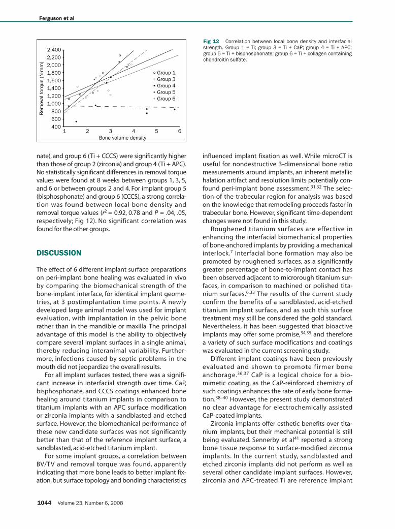
nate), and group 6 (Ti + CCCS) were significantly higherthan those of group 2 (zirconia) and group 4 (Ti + APC).No statistically significant differences in removal torquevalues were found at 8 weeks between groups 1, 3, 5,and 6 or between groups 2 and 4. For implant group 5(bisphosphonate) and group 6 (CCCS), a strong correla-tion was found between local bone density andremoval torque values (r2 = 0.92, 0.78 and P = .04, .05,respectively; Fig 12). No significant correlation wasfound for the other groups.
DISCUSSION
The effect of 6 different implant surface preparationson peri-implant bone healing was evaluated in vivoby comparing the biomechanical strength of thebone-implant interface, for identical implant geome-tries, at 3 postimplantation time points. A newlydeveloped large animal model was used for implantevaluation, with implantation in the pelvic bonerather than in the mandible or maxilla. The principaladvantage of this model is the ability to objectivelycompare several implant surfaces in a single animal,thereby reducing interanimal variability. Further-more, infections caused by septic problems in themouth did not jeopardize the overall results.
For all implant surfaces tested, there was a signifi-cant increase in interfacial strength over time. CaP,bisphosphonate, and CCCS coatings enhanced bonehealing around titanium implants in comparison totitanium implants with an APC surface modificationor zirconia implants with a sandblasted and etchedsurface. However, the biomechanical performance ofthese new candidate surfaces was not significantlybetter than that of the reference implant surface, asandblasted, acid-etched titanium implant.
For some implant groups, a correlation betweenBV/TV and removal torque was found, apparentlyindicating that more bone leads to better implant fix-ation, but surface topology and bonding characteristics
influenced implant fixation as well. While microCT isuseful for nondestructive 3-dimensional bone ratiomeasurements around implants, an inherent metallichalation artifact and resolution limits potentially con-found peri-implant bone assessment.31,32 The selec-tion of the trabecular region for analysis was basedon the knowledge that remodeling proceeds faster intrabecular bone. However, significant time-dependentchanges were not found in this study.
Roughened titanium surfaces are effective inenhancing the interfacial biomechanical propertiesof bone-anchored implants by providing a mechanicalinterlock.7 Interfacial bone formation may also bepromoted by roughened surfaces, as a significantlygreater percentage of bone-to-implant contact hasbeen observed adjacent to microrough titanium sur-faces, in comparison to machined or polished tita-nium surfaces.6,33 The results of the current studyconfirm the benefits of a sandblasted, acid-etchedtitanium implant surface, and as such this surfacetreatment may still be considered the gold standard.Nevertheless, it has been suggested that bioactiveimplants may offer some promise,34,35 and thereforea variety of such surface modifications and coatingswas evaluated in the current screening study.
Different implant coatings have been previouslyevaluated and shown to promote firmer boneanchorage.36,37 CaP is a logical choice for a bio-mimetic coating, as the CaP-reinforced chemistry ofsuch coatings enhances the rate of early bone forma-tion.38–40 However, the present study demonstratedno clear advantage for electrochemically assistedCaP-coated implants.
Zirconia implants offer esthetic benefits over tita-nium implants, but their mechanical potential is stillbeing evaluated. Sennerby et al41 reported a strongbone tissue response to surface-modified zirconiaimplants. In the current study, sandblasted andetched zirconia implants did not perform as well asseveral other candidate implant surfaces. However,zirconia and APC-treated Ti are reference implant
1044 Volume 23, Number 6, 2008
Ferguson et al
2,400
2,200
2,000
1,800
1,600
1,400
1,200
1,000
800
600
4001 4 6
Bone volume density
Rem
oval
torq
ue(N
-mm
)
2 3 5
Group 1Group 3Group 4Group 5Group 6
Fig 12 Correlation between local bone density and interfacialstrength. Group 1 = Ti; group 3 = Ti + CaP; group 4 = Ti + APC;group 5 = Ti + bisphosphonate; group 6 = Ti + collagen containingchondroitin sulfate.
surfaces with good clinical results, and therefore thelower values observed should be interpreted withcaution. Both APC-treated Ti and zirconia exhibited alower surface area ratio ( Table 1, Figs 4 and 6).Although the CaP group exhibited similarly low val-ues of aw, this was a measurement artifact caused bythe restricted lateral resolution of the confocalmicroscope, which was confirmed by the scanningelectron microscopy. The confocal microscope can-not resolve the fine crystallites of the CaP coating.The optimal surface ratio for bone growth should bearound 1.5.42 Moderate sur face roughness (Sa between 1.2 and 2.0 µm) is associated with astronger bone response than smoother or roughersurfaces.35 Because the Sa values were also rather lowfor groups 2 and 4, surface topography might be oneof the key factors for the low extraction torques measured at all time points. Both surfaces will haveto be optimized from a topographic point of view.
Biologic enhancement of bone formation remainsan elusive goal. The use of CCCS coatings to provideessential bone extracellular matrix components at theinterface is a very recent development. To date, thereare no reports of the mechanical performance of suchimplant surfaces. In the present study, a similarresponse was shown between bisphosphonate- andCCCS-coated implants at the 2-week and 4-week timepoints. Reduced torque values at week 8 for the CCCSgroup may indicate a remodeling process. Biochemicalmodification of the implant surface with CCCS has beenshown to enhance new bone formation and bone-to-implant contact—potentially by the provision of bind-ing sites for integrin receptors—and induces greaterexpression of bone-specific matrix proteins.17,43–45
The local delivery of bisphosphonates to enhancelocal peri-implant bone formation is a promisingavenue requiring further study. A higher percentageof bone contact was found around bisphosphonate-coated implants by Yoshinari et al46 and Peter etal.19,47 The potentially modest increase in the earlyand mid-term mechanical performance of bisphos-phonate-coated implants could not be statisticallyverified in this preliminary study. On the other hand,the strong correlation between bone density andinterface mechanics observed for this implant groupimplies functional osseointegration, whereby stimula-tion of bone formation may lead to improved fixation.
Whereas removal torque values represent a valu-able, objective means of appraising interfacialstrength, they do not fully and uniquely characterizethe process of bone healing. Histomorphometricanalyses are underway and will provide a more com-plete evaluation of the potential for such functionalsurface modifications to influence the rate andamount of bone-implant contact.
CONCLUSIONS
A variety of surface modifications for dental implantswere evaluated in an in vivo study, with mechanicaltesting of the bone-implant interface strength andnondestructive evaluation of bone quality bymicroCT. The results of the mechanical testing indi-cate that coatings of calcium phosphate, bisphos-phonate, and collagen containing chondroitin sulfateall have the potential to enhance peri-implant bonehealing, but a significant difference from the perfor-mance of the reference standard of a sandblasted,acid-etched titanium implant could not be shown.
ACKNOWLEDGMENTS
The authors thank Mr Philippe Gédet for his excellent technicalassistance with the mechanical testing. The study was partiallysupported by Thommen Medical, Waldenburg, Switzerland.
REFERENCES
1. Albrektsson T, Brånemark P-I, Hansson HA, Lindstrom J. Osseoin-tegrated titanium implants. Requirements for ensuring a long-lasting, direct bone-to-implant anchorage in man. Acta OrthopScand 1981;52:155–170.
2. Ratner BD, Porter SC. Surfaces in biology and biomaterials; des-cription and characterization. In: Brash JLW (ed). Interfacial Phe-nomena and Bioproducts. New York: Marcel Dekker, 1996:57–83.
3. Wieland M, Sittig C, Brunette DM,Textor M, Spencer ND. Mea-surement and evaluation of the chemical composition andtopography of titanium implant surfaces. In: Davies JE (ed). BoneEngineering.Toronto: ed squared inc, 2000:163–182.
4. Buser D, Schenk RK, Steinemann S, Fiorellini JP, Fox CH, Stich H.Influence of surface characteristics on bone integration of tita-nium implants. A histomorphometric study in miniature pigs.J Biomed Mater Res 1991;25:889–902.
5. Cochran DL, Schenk RK, Lussi A, Higginbottom FL, Buser D. Boneresponse to unloaded and loaded titanium implants with asandblasted and acid-etched surface: A histometric study in thecanine mandible. J Biomed Mater Res 1998;40:1–11.
6. Buser D, Nydegger T, Oxland T, et al. Interface shear strength oftitanium implants with a sandblasted and acid-etched surface:A biomechanical study in the maxilla of miniature pigs. J Bio-med Mater Res 1999;45:75–83.
7. Li D, Ferguson SJ, Beutler T, et al. Biomechanical comparison ofthe sandblasted and acid-etched and the machined and acid-etched titanium surface for dental implants. J Biomed Mater Res2002;60:325–332.
8. Roccuzzo M, Bunino M, Prioglio F, Bianchi SD. Early loading ofsandblasted and acid-etched (SLA) implants: A prospective split-mouth comparative study.Clin Oral Implants Res 2001;12:572–578.
9. Cochran DL, Buser D, ten Bruggenkate CM, et al.The use ofreduced healing times on ITI implants with a sandblasted andacid-etched (SLA) surface: Early results from clinical trials on ITISLA implants. Clin Oral Implants Res 2002;13:144–153.
The International Journal of Oral & Maxillofacial Implants 1045
Ferguson et al
10. Bornstein MM, Schmid B, Belser UC, Lussi A, Buser D. Early load-ing of non-submerged titanium implants with a sandblastedand acid-etched (SLA) surface: 5-year results of a prospectivestudy in partially edentulous patients. Clin Oral Implants Res2005;16:631–638.
11. Bornstein MM, Lussi A, Schmid B, Belser UC, Buser D. Early load-ing of nonsubmerged titanium implants with a sandblastedand acid-etched (SLA) surface: 3-year results of a prospectivestudy in partially edentulous patients. Int J Oral MaxillofacImplants 2003;18:659–666.
12. Ferguson SJ, Broggini N,Wieland M, et al. Biomechanical evalua-tion of the interfacial strength of a chemically modified sand-blasted and acid-etched titanium surface. J Biomed Mater Res A2006;78:291–297.
13. Schliephake H, Scharnweber D, Roesseler S, Dard M, Sewing A,Aref A. Biomimetic calcium phosphate composite coating ofdental implants. Int J Oral Maxillofac Implants 2006;21:738–746.
14. Barrere F, Layrolle P,Van Blitterswijk CA, De Groot K. Biomimeticcoatings on titanium: A crystal growth study of octacalciumphosphate. J Mater Sci Mater Med 2001;12:529–534.
15. Cooper LF, Zhou Y,Takebe J, et al.Fluoride modification effects onosteoblast behavior and bone formation at TiO2 grit-blasted c.p.titanium endosseous implants. Biomaterials 2006;27:926–936.
16. Schuler M, Owen GR, Hamilton DW, et al. Biomimetic modifica-tion of titanium dental implant model surfaces using the RGDSP-peptide sequence: A cell morphology study. Biomaterials 2006;27:4003–4015.
17. Rammelt S, Illert T, Bierbaum S, Scharnweber D, Zwipp H, Schnei-ders W. Coating of titanium implants with collagen, RGD pep-tide and chondroitin sulfate. Biomaterials 2006;27:5561–5571.
18. Michael J, Beutner R, Hempel U, Scharnweber D,Worch H,Schwenzer B. Surface modification of titanium-based alloys withbioactive molecules using electrochemically fixed nucleic acids.J Biomed Mater Res B Appl Biomater 2007;80:146–155.
19. Peter B, Gauthier O, Laib S, et al. Local delivery of bisphospho-nate from coated orthopedic implants increases implantsmechanical stability in osteoporotic rats. J Biomed Mater Res A2006;76:133–143.
20. Fiorellini JP, Buser D, Riley E, Howell TH. Effect on bone healing ofbone morphogenetic protein placed in combination withendosseous implants: A pilot study in beagle dogs. Int J Peri-odontics Restorative Dent 2001;21:41–47.
21. Jones AA, Buser D, Schenk R,Wozney J, Cochran DL.The effect ofrhBMP-2 around endosseous implants with and without mem-branes in the canine model. J Periodontol 2006;77:1184–1193.
22. Becker P, Neumann HG, Nebe B, Luthen F, Rychly J. Cellular inves-tigations on electrochemically deposited calcium phosphatecomposites. J Mater Sci Mater Med 2004;15(4):437–440.
23. Frauchiger VM, Schlottig F, Gasser B,Textor M. Anodic plasma-chemical treatment of CP titanium surfaces for biomedicalapplications. Biomaterials 2004;25:593–606.
24. Bierbaum S, Douglas T, Hanke T, et al. Collageneous matrix coat-ings on titanium implants modified with decorin and chon-droitin sulfate: Characterization and influence on osteoblasticcells. J Biomed Mater Res A 2006;77:551–562.
25. Aerssens J,Boonen S,Lowet G,Dequeker J. Interspecies differ-ences in bone composition,density,and quality:Potential implica-tions for in vivo bone research.Endocrinology 1998;139:663–670.
26. Nunamaker DM, Perren SM. A radiological and histologicalanalysis of fracture healing using prebending of compressionplates. Clin Orthop Relat Res 1979;167–174.
27. Lekholm U, Zarb GA, Albrektsson T. Patient selection and prepa-ration. In: Brånemark P-I, Zarb GA, Albrektsson T (eds).Tissue-Integrated Prostheses. Chicago: Quintessence,1985:199–209.
28. Ruegsegger P, Koller B, Muller R. A microtomographic system forthe nondestructive evaluation of bone architecture. Calcif TissueInt 1996;58:24–29.
29. van Lenthe GH, Hagenmuller H, Bohner M, Hollister SJ, Meinel L,Muller R. Nondestructive micro-computed tomography for bio-logical imaging and quantification of scaffold-bone interactionin vivo. Biomaterials 2007; 28:2479–2490.
30. Müller R, Hildebrand T, Häuselmann HJ, Rüegsegger P. In vivoreproducibility of three-dimensional structural properties ofnoninvasive bone biopsies using 3D-pQCT. J Bone Miner Res1996;11(11):1745–1750.
31. Butz F, Ogawa T, Chang TL, Nishimura I.Three-dimensional bone-implant integration profiling using micro-computed tomogra-phy. Int J Oral Maxillofac Implants 2006;21:687–695.
32. Van Oosterwyck H, Duyck J,Vander Sloten J, et al. Use of micro-focus computerized tomography as a new technique for char-acterizing bone tissue around oral implants. J Oral Implantol2000;26:5–12.
33. Wennerberg A, Albrektsson T, Andersson B, Krol JJ. A histomor-phometric and removal torque study of screw-shaped titaniumimplants with three different surface topographies. Clin OralImplants Res 1995;6:24–30.
34. Albrektsson T,Wennerberg A. Oral implant surfaces: Part 2—Review focusing on clinical knowledge of different surfaces. IntJ Prosthodont 2004;17:544–564.
35. Albrektsson T,Wennerberg A. Oral implant surfaces: Part 1—Review focusing on topographic and chemical properties of dif-ferent surfaces and in vivo responses to them. Int J Prosthodont2004;17:536–543.
36. Ellingsen JE, Johansson CB,Wennerberg A, Holmen A. Improvedretention and bone-tolmplant contact with fluoride-modifiedtitanium implants. Int J Oral Maxillofac Implants 2004;19:659–666.
37. Giavaresi G,Branda F,Causa F,et al.Poly(2-hydroxyethyl metha-crylate) biomimetic coating to improve osseointegration of aPMMA/HA/glass composite implant: In vivo mechanical and his-tomorphometric assessments. Int J Artif Organs 2004;27:674–680.
38. Sul YT, Byon ES, Jeong Y. Biomechanical measurements of cal-cium-incorporated oxidized implants in rabbit bone: Effect ofcalcium surface chemistry of a novel implant. Clin Implant DentRelat Res 2004;6:101–110.
39. Gan L,Wang J,Tache A,Valiquette N, Deporter D, Pilliar R. Cal-cium phosphate sol-gel-derived thin films on porous-surfacedimplants for enhanced osteoconductivity. Part II: Short-term invivo studies. Biomaterials 2004;25:5313–5321.
40. Tache A, Gan L, Deporter D, Pilliar RM. Effect of surface chemistryon the rate of osseointegration of sintered porous-surfaced Ti-6Al-4V implants. Int J Oral Maxillofac Implants 2004;19:19–29.
41. Sennerby L, Dasmah A, Larsson B, Iverhed M. Bone tissueresponses to surface-modified zirconia implants: A histomor-phometric and removal torque study in the rabbit. Clin ImplantDent Relat Res 2005;7(suppl 1):S13–20.
42. Wennerberg A. On Surface Roughness and Implant Incorpora-tion [thesis]. Göteborg: Göteborg University, 1996.
43. Rammelt S, Schulze E, Bernhardt R, et al. Coating of titaniumimplants with type-I collagen. J Orthop Res 2004;22:1025–1034.
44. Morra M, Cassinelli C, Cascardo G, et al. Collagen I-coated tita-nium surfaces: Mesenchymal cell adhesion and in vivo evalua-tion in trabecular bone implants. J Biomed Mater Res A 2006;78:449–458.
45. Morra M, Cassinelli C, Cascardo G, et al. Surface engineering oftitanium by collagen immobilization.Surface characterizationand in vitro and in vivo studies. Biomaterials 2003;24:4639–4654.
46. Yoshinari M, Oda Y, Inoue T, Matsuzaka K, Shimono M. Boneresponse to calcium phosphate-coated and bisphosphonate-immobilized titanium implants. Biomaterials 2002;23:2879–2885.
47. Peter B, Pioletti DP, Laib S, et al. Calcium phosphate drug deliverysystem: Influence of local zoledronate release on bone implantosteointegration. Bone 2005;36:52–60.
1046 Volume 23, Number 6, 2008
Ferguson et al

Thommen Medical, a Swiss company with its headquarters in Waldenburg is setting new standards inter-
nationally in oral implantology with its tried and tested Swiss Precision Implant System (SPI®System). A
team of expert specialists with many years of know-how is working intensely together with leading clinicians
and scientists to find fast innovative solutions. We are very well versed in the specific customer requirements
and are therefore able to offer you quality products manufactured in Switzerland, which will make your
everyday life noticeably easier.
Would you like to know more, or do you require detailed information? Give us a ring and we will be happy
to answer your questions.
Fast, competent, reliable
A SOLID PARTNER FOR SOLID IMPLANTS.Thommen Medical AG | Headquarters | Phone +41 (0)61 965 90 20www.thommenmedical.com