Biomaterials ppt
15
Wharton’s Jelly Stem Cells: A Novel Cell Source for Oral Mucosa and Skin Epithelia Regeneration -Bhagyesha Patil - Bhagyashree Bachhav - Harini Krishnan - Snehal Kolhekar 1
-
Upload
bhagyashree-bachhav -
Category
Documents
-
view
254 -
download
0
Transcript of Biomaterials ppt
7/21/2019 Biomaterials ppt
http://slidepdf.com/reader/full/biomaterials-ppt 1/15
Wharton’s Jelly Stem Cells: A Novel
Cell Source for
Oral Mucosa and Skin EpitheliaRegeneration
-Bhagyesha Patil- Bhagyashree Bachhav
- Harini Krishnan
- Snehal Kolhekar
1
7/21/2019 Biomaterials ppt
http://slidepdf.com/reader/full/biomaterials-ppt 2/15
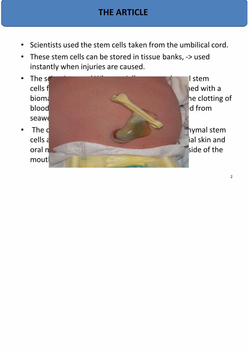
• Scientists used the stem cells taken from the umbilical cord.
• These stem cells can be stored in tissue banks, -> used
instantly when injuries are caused.
• The scientists used Wharton jelly mesenschymal stem
cells from the human umbilical cord and combined with abiomaterial made of fibrin - a protein found in the clotting of
blood - and agarose - a polymer usually extracted from
seaweed.
• The combination of the Wharton jelly mesenschymal stemcells and biomaterial led to the growth of artificial skin and
oral mucosa - a mucous membrane lining the inside of the
mouth.
THE ARTICLE
2
7/21/2019 Biomaterials ppt
http://slidepdf.com/reader/full/biomaterials-ppt 3/15
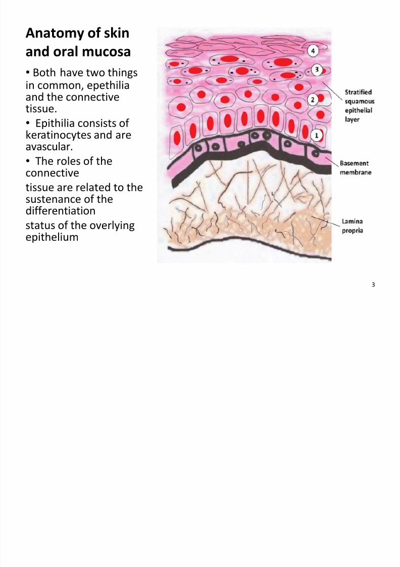
Anatomy of skin
and oral mucosa
• Both have two thingsin common, epethiliaand the connectivetissue.
• Epithilia consists of
keratinocytes and areavascular.
• The roles of theconnective
tissue are related to thesustenance of the
differentiationstatus of the overlyingepithelium
3
7/21/2019 Biomaterials ppt
http://slidepdf.com/reader/full/biomaterials-ppt 4/15
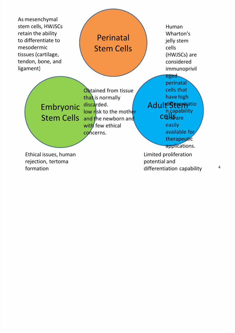
Embryonic
Stem Cells
Adult Stem
cells
PerinatalStem Cells
Ethical issues, human
rejection, tertomaformation
Limited proliferation
potential anddifferentiation capability
Obtained from tissue
that is normally
discarded.
low risk to the mother
and the newborn and
with few ethical
concerns.
Human
Wharton’s
jelly stemcells
(HWJSCs) are
considered
immunoprivil
eged
perinatalcells that
have high
differentiatio
n capability
and are
easily
available for
therapeutic
applications.
As mesenchymal
stem cells, HWJSCs
retain the ability
to differentiate tomesodermic
tissues (cartilage,
tendon, bone, and
ligament)
4
7/21/2019 Biomaterials ppt
http://slidepdf.com/reader/full/biomaterials-ppt 5/15
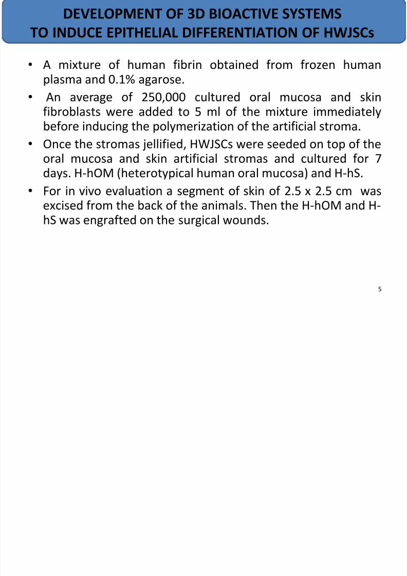
•
A mixture of human fibrin obtained from frozen humanplasma and 0.1% agarose.
• An average of 250,000 cultured oral mucosa and skinfibroblasts were added to 5 ml of the mixture immediatelybefore inducing the polymerization of the artificial stroma.
• Once the stromas jellified, HWJSCs were seeded on top of theoral mucosa and skin artificial stromas and cultured for 7days. H-hOM (heterotypical human oral mucosa) and H-hS.
• For in vivo evaluation a segment of skin of 2.5 x 2.5 cm wasexcised from the back of the animals. Then the H-hOM and H-hS was engrafted on the surgical wounds.
DEVELOPMENT OF 3D BIOACTIVE SYSTEMS
TO INDUCE EPITHELIAL DIFFERENTIATION OF HWJSCs
5
7/21/2019 Biomaterials ppt
http://slidepdf.com/reader/full/biomaterials-ppt 6/15
• Protein-based polymers have the advantage of mimicking many features ofextracellular matrix and thus have the potential to direct the migration, growthand organization of cells during tissue regeneration and wound healing and forstabilization of encapsulated and transplanted cells.
• Properties of fibrin:
Fibrin is a protein matrix produced from fibrinogen, which can be autologously
harvested from the patient providing an immunocompatible carrier for deliveryof active biomolecules.
In addition, fibrin naturally contains sites for cell binding, and therefore has beeninvestigated as a substrate for cell adhesion, spreading, migration andproliferation.
Fibrin provides a material that can be rapidly invaded, remodeled and replaced by
cell-associated proteolytic activity.
Moreover, due to its biomimetic and physical properties it is also widely used as a
cell carrier to many cell types, such as keratinocytes, tracheal epithelial cells ,murine embryonic stem cells, mesenchymal progenitor cells, etc.
But, problems of instability and degradation invivo , so combination with agaroseprovides improved properties resembling natural skin
FIBRIN-AGAROSE SCAFFOLD
6
7/21/2019 Biomaterials ppt
http://slidepdf.com/reader/full/biomaterials-ppt 7/15
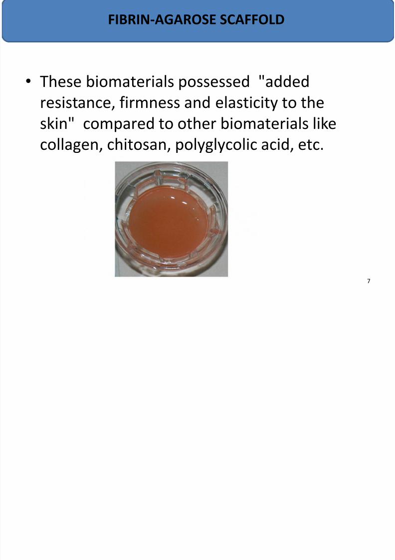
• These biomaterials possessed "added
resistance, firmness and elasticity to the
skin" compared to other biomaterials like
collagen, chitosan, polyglycolic acid, etc.
FIBRIN-AGAROSE SCAFFOLD
7
7/21/2019 Biomaterials ppt
http://slidepdf.com/reader/full/biomaterials-ppt 8/15
ANALYSIS OF THE MESENCHYMAL NATURE OF HWJSCs
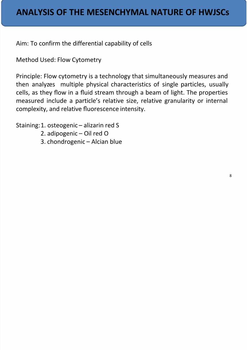
Aim: To confirm the differential capability of cells
Method Used: Flow Cytometry
Principle: Flow cytometry is a technology that simultaneously measures and
then analyzes multiple physical characteristics of single particles, usuallycells, as they flow in a fluid stream through a beam of light. The properties
measured include a particle’s relative size, relative granularity or internal
complexity, and relative fluorescence intensity.
Staining: 1. osteogenic – alizarin red S
2. adipogenic – Oil red O
3. chondrogenic – Alcian blue
8
7/21/2019 Biomaterials ppt
http://slidepdf.com/reader/full/biomaterials-ppt 9/15
IN VIVO EVALUATION OF THE EPITHELIAL
DIFFERENTIATION POTENTIAL
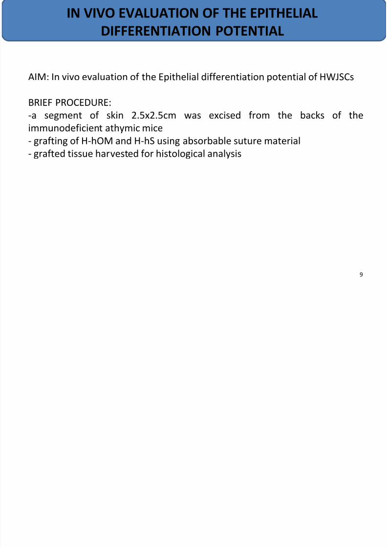
AIM: In vivo evaluation of the Epithelial differentiation potential of HWJSCs
BRIEF PROCEDURE:
-a segment of skin 2.5x2.5cm was excised from the backs of the
immunodeficient athymic mice
- grafting of H-hOM and H-hS using absorbable suture material
- grafted tissue harvested for histological analysis
9
7/21/2019 Biomaterials ppt
http://slidepdf.com/reader/full/biomaterials-ppt 10/15
HISTOLOGICAL ANALYSIS
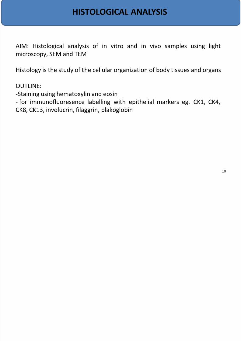
AIM: Histological analysis of in vitro and in vivo samples using light
microscopy, SEM and TEM
Histology is the study of the cellular organization of body tissues and organs
OUTLINE:-Staining using hematoxylin and eosin
- for immunofluoresence labelling with epithelial markers eg. CK1, CK4,
CK8, CK13, involucrin, filaggrin, plakoglobin
10
7/21/2019 Biomaterials ppt
http://slidepdf.com/reader/full/biomaterials-ppt 11/15
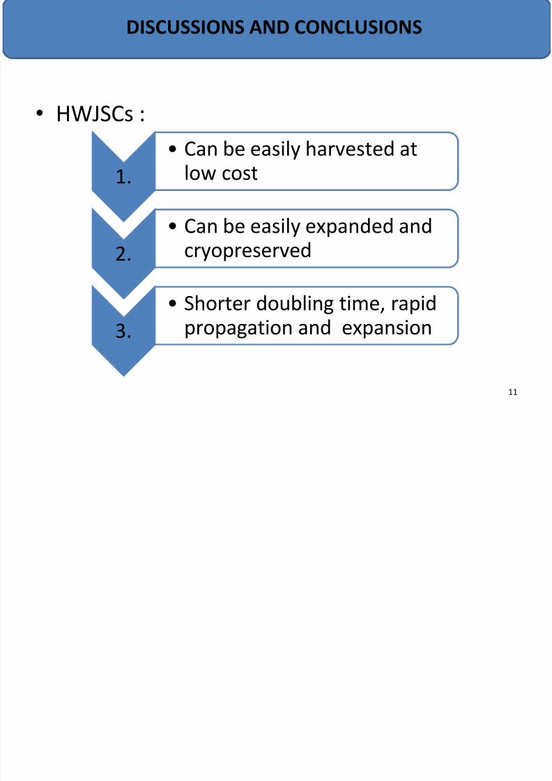
• HWJSCs :
1.
• Can be easily harvested at
low cost
2.
• Can be easily expanded and
cryopreserved
3.
• Shorter doubling time, rapid
propagation and expansion
DISCUSSIONS AND CONCLUSIONS
11
7/21/2019 Biomaterials ppt
http://slidepdf.com/reader/full/biomaterials-ppt 12/15
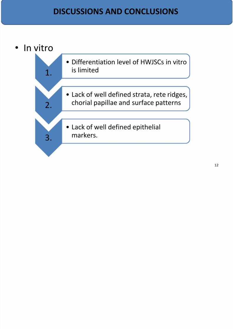
• In vitro
1.
• Differentiation level of HWJSCs in vitro
is limited
2.
• Lack of well defined strata, rete ridges,
chorial papillae and surface patterns
3.
• Lack of well defined epithelial
markers.
DISCUSSIONS AND CONCLUSIONS
12
7/21/2019 Biomaterials ppt
http://slidepdf.com/reader/full/biomaterials-ppt 13/15
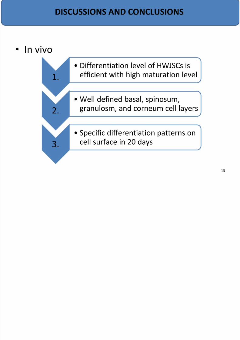
• In vivo
1.
• Differentiation level of HWJSCs is
efficient with high maturation level
2.
• Well defined basal, spinosum,
granulosm, and corneum cell layers
3.
• Specific differentiation patterns on
cell surface in 20 days
DISCUSSIONS AND CONCLUSIONS
13
7/21/2019 Biomaterials ppt
http://slidepdf.com/reader/full/biomaterials-ppt 14/15
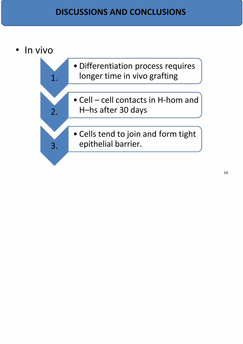
• In vivo
1.
•Differentiation process requires
longer time in vivo grafting
2.
• Cell – cell contacts in H-hom and
H –hs after 30 days
3.
• Cells tend to join and form tight
epithelial barrier.
DISCUSSIONS AND CONCLUSIONS
14
7/21/2019 Biomaterials ppt
http://slidepdf.com/reader/full/biomaterials-ppt 15/15
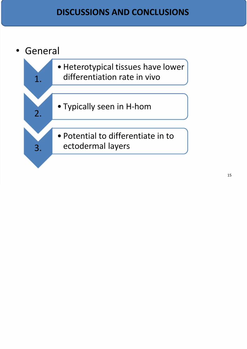
• General
1.
•Heterotypical tissues have lower
differentiation rate in vivo
2.• Typically seen in H-hom
3.
• Potential to differentiate in to
ectodermal layers
DISCUSSIONS AND CONCLUSIONS
15