Biology sem1- chap9
111
1
-
Upload
kotaknotaku -
Category
Documents
-
view
225 -
download
0
Transcript of Biology sem1- chap9

8/7/2019 Biology sem1- chap9
http://slidepdf.com/reader/full/biology-sem1-chap9 1/111
1
8/7/2019 Biology sem1- chap9
http://slidepdf.com/reader/full/biology-sem1-chap9 2/111
2
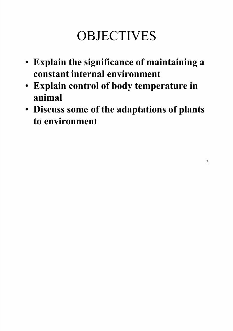
OBJECTIVES Explain the significance of maintaining a
constant internal environment
Explain control of body temperature in
animal
Discuss some of the adaptations of plants
to environment
8/7/2019 Biology sem1- chap9
http://slidepdf.com/reader/full/biology-sem1-chap9 3/111
3
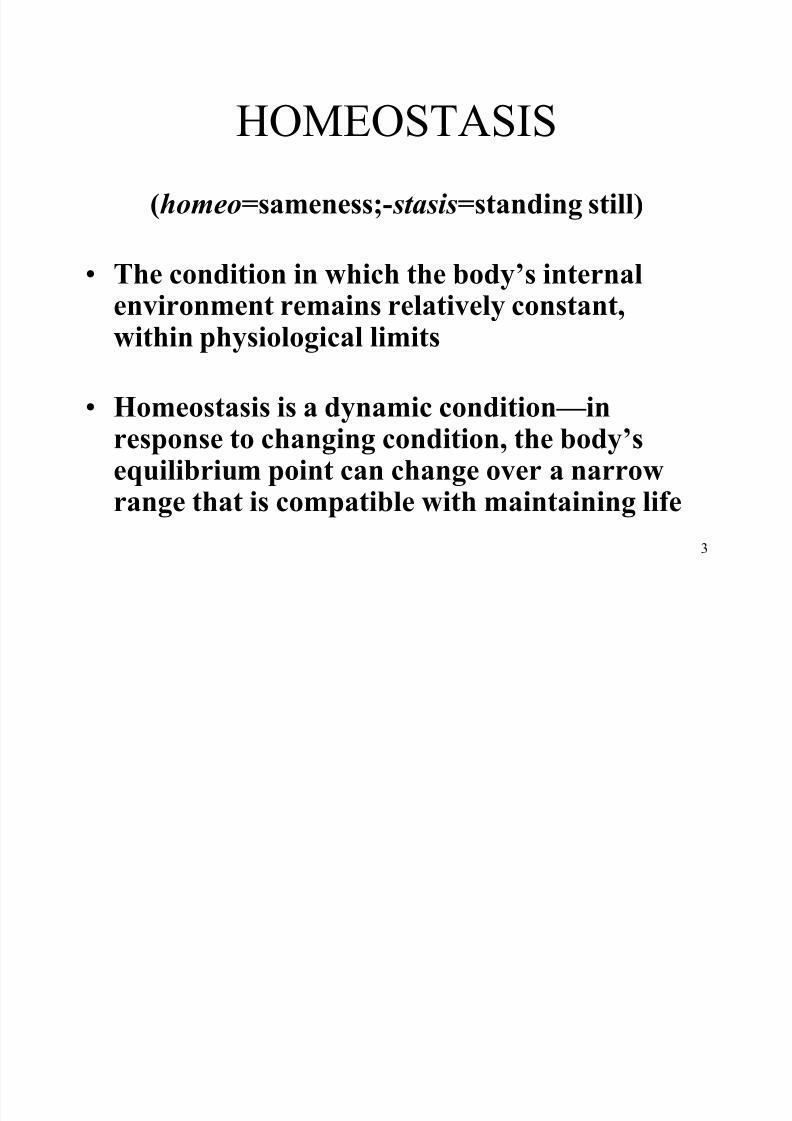
HOMEOSTASIS
(homeo=sameness;-stasis=standing still)
The condition in which the body¶s internalenvironment remains relatively constant,within physiological limits
Homeostasis is a dynamic condition²inresponse to changing condition, the body¶sequilibrium point can change over a narrowrange that is compatible with maintaining life
8/7/2019 Biology sem1- chap9
http://slidepdf.com/reader/full/biology-sem1-chap9 4/111
4
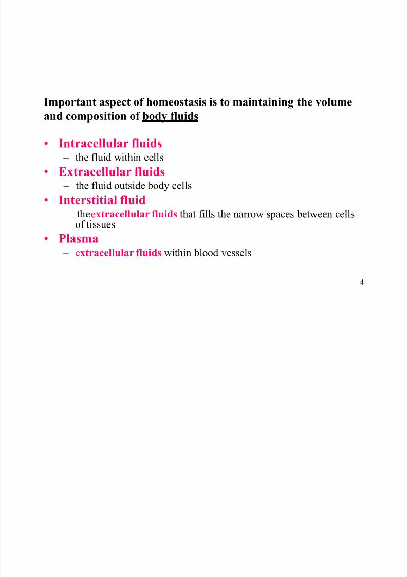
Important aspect of homeostasis is to maintaining the volume
and composition of body fluids
Intracellular fluids ± the fluid within cells
Extracellular fluids ± the fluid outside body cells
Interstitial fluid
± theextracellular fluids that fills the narrow spaces between cellsof tissues
Plasma ± extracellular fluids within blood vessels
8/7/2019 Biology sem1- chap9
http://slidepdf.com/reader/full/biology-sem1-chap9 5/111
5
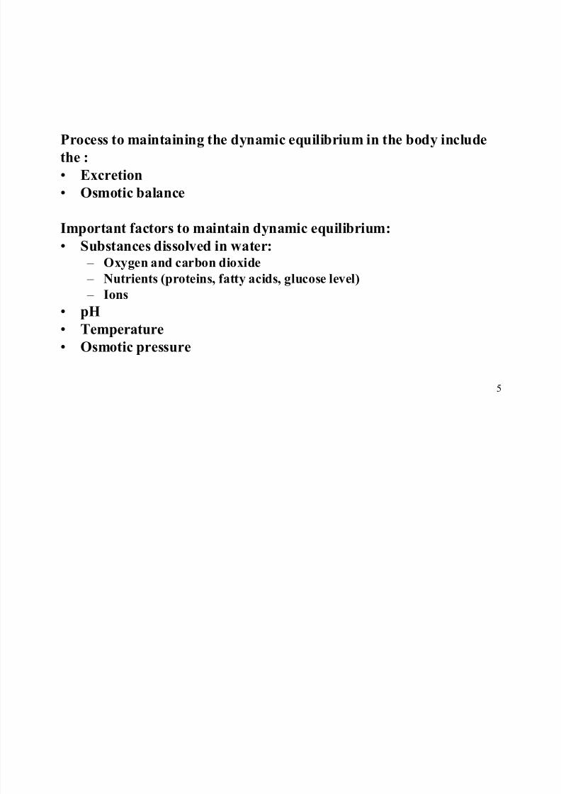
Process to maintaining the dynamic equilibrium in the body include
the :
Excretion
Osmotic balance
Important factors to maintain dynamic equilibrium:
Substances dissolved in water: ± Oxygen and carbon dioxide
± Nutrients (proteins, fatty acids, glucose level)
± Ions pH
Temperature
Osmotic pressure
8/7/2019 Biology sem1- chap9
http://slidepdf.com/reader/full/biology-sem1-chap9 6/111
6
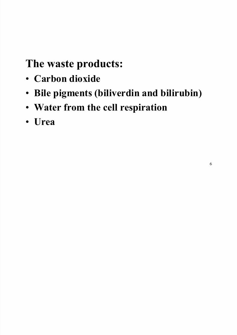
The waste products:
Carbon dioxide Bile pigments (biliverdin and bilirubin)
Water from the cell respiration
Urea
8/7/2019 Biology sem1- chap9
http://slidepdf.com/reader/full/biology-sem1-chap9 7/111
7
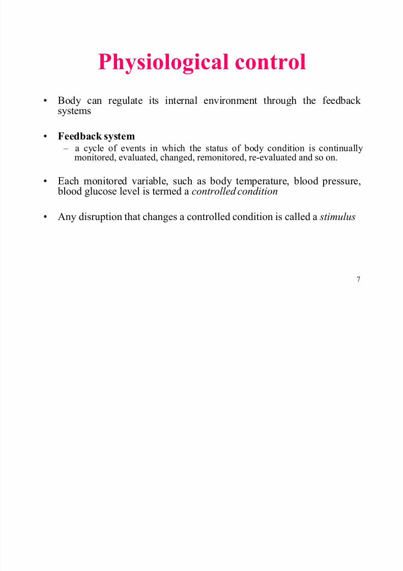
Physiological control Body can regulate its internal environment through the feedback
systems
Feedback system ± a cycle of events in which the status of body condition is continuallymonitored, evaluated, changed, remonitored, re-evaluated and so on.
Each monitored variable, such as body temperature, blood pressure, blood glucose level is termed a controlled condition
Any disruption that changes a controlled condition is called a stimulus
8/7/2019 Biology sem1- chap9
http://slidepdf.com/reader/full/biology-sem1-chap9 8/111
8
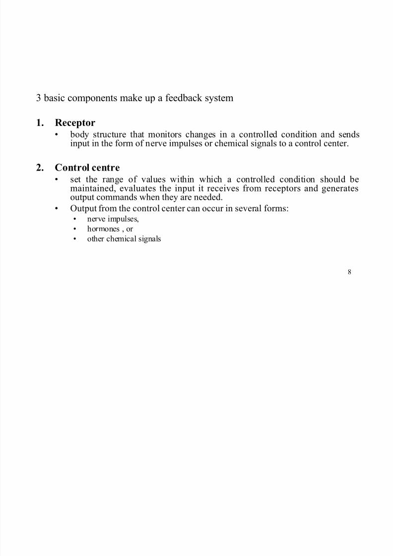
3 basic components make up a feedback system
1. R eceptor
body structure that monitors changes in a controlled condition and sendsinput in the form of nerve impulses or chemical signals to a control center.
2. Control centre set the range of values within which a controlled condition should be
maintained, evaluates the input it receives from receptors and generatesoutput commands when they are needed.
Output from the control center can occur in several forms: nerve impulses,
hormones , or
other chemical signals
8/7/2019 Biology sem1- chap9
http://slidepdf.com/reader/full/biology-sem1-chap9 9/111
9
3. Effector
body structure that receives output from the control
centre and produces a response or effect that changesthe controlled condition.
As a control system operates, the effector
response feed back and influences the magnitudeof the stimulus by either depressing it (negative
feedback) or enhancing it (positive feedback)
8/7/2019 Biology sem1- chap9
http://slidepdf.com/reader/full/biology-sem1-chap9 10/111
10
N egative feedback
Homeostatic mechanism that stops or reduces theintensity of the original stimulus and consequentlycauses a change in a variable that is opposite in directionto the initial change (The output is used to reduce input)
Positive feedback a feedback mechanism in which the response enhances
the original stimulus (The output is used to enhance theinput)
Example : The secretion of oxytocin duringchildbirth
8/7/2019 Biology sem1- chap9
http://slidepdf.com/reader/full/biology-sem1-chap9 11/111
11
Homeostatic regulation
i. Every changes in the physical or chemical factors
of the internal environment ± detected by receptor
ii. Receptor send information about the changes to a
control systemiii. Action signals are transmitted through nerve
impulses or hormone to the target organ
± Trigger the correction mechanism to return the physical
or chemical factors to normal condition
8/7/2019 Biology sem1- chap9
http://slidepdf.com/reader/full/biology-sem1-chap9 12/111
12
iv. Any increase in the value of a physical or
chemical factor in the internal environment will
trigger a correction mechanism to reduce thatvalue
Conversely, a reduction in the value of the
physical or chemical factor will trigger a
mechanism to increase the amount of that factor.
8/7/2019 Biology sem1- chap9
http://slidepdf.com/reader/full/biology-sem1-chap9 13/111
13
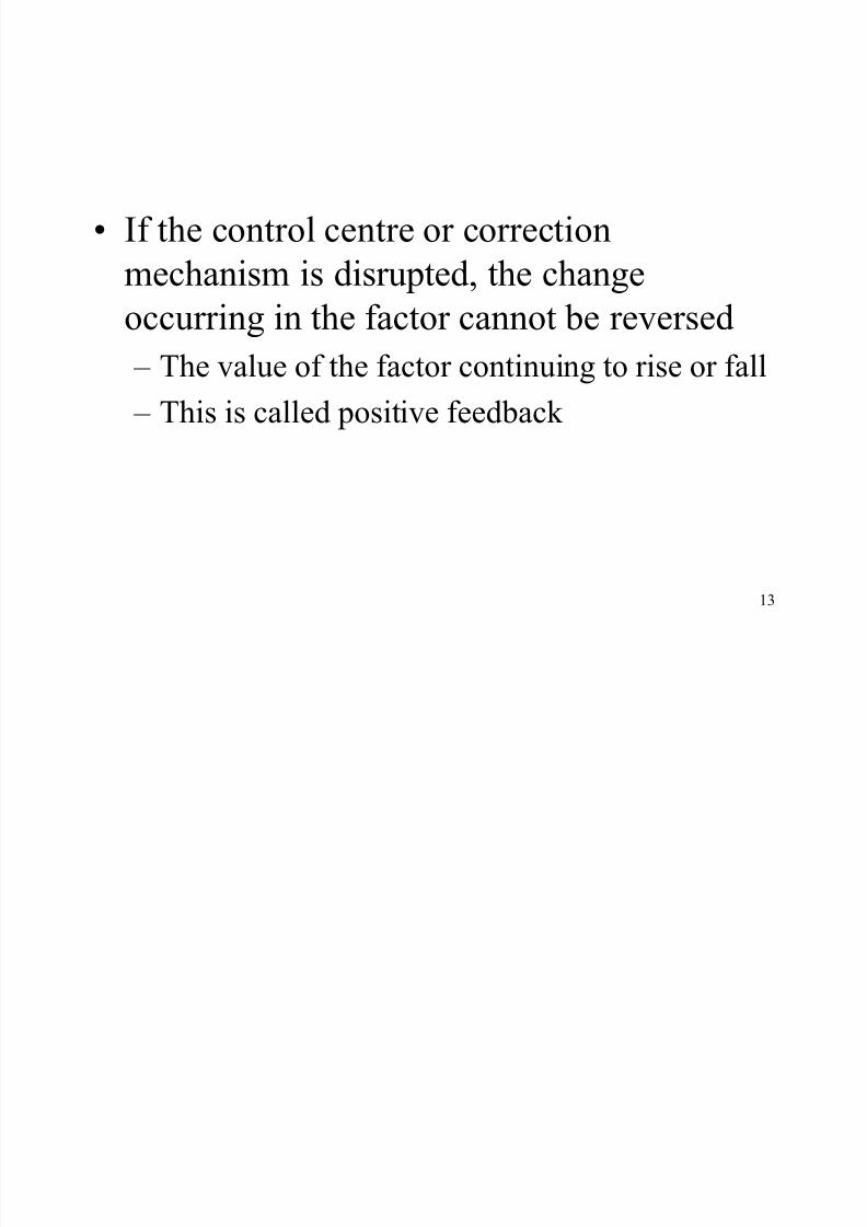
If the control centre or correction
mechanism is disrupted, the change
occurring in the factor cannot be reversed
± The value of the factor continuing to rise or fall
± This is called positive feedback
8/7/2019 Biology sem1- chap9
http://slidepdf.com/reader/full/biology-sem1-chap9 14/111
14
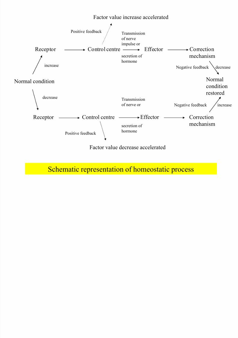
Schematic representation of homeostatic process
Normal condition
Receptor Control centre Effector Correction
mechanism
Normal
condition
restored
Receptor Control centre Effector Correction
mechanism
Factor value increase accelerated
Factor value decrease accelerated
increase
decrease
Positive feedback
Positive feedback
Transmission
of nerve
impulse or
secretion of
hormone
Negative feedback
Negative feedback
decrease
increase
Transmission
of nerve or
secretion of hormone

8/7/2019 Biology sem1- chap9
http://slidepdf.com/reader/full/biology-sem1-chap9 15/111
15
Negative feedback in
control of blood
glucose level
8/7/2019 Biology sem1- chap9
http://slidepdf.com/reader/full/biology-sem1-chap9 16/111
16
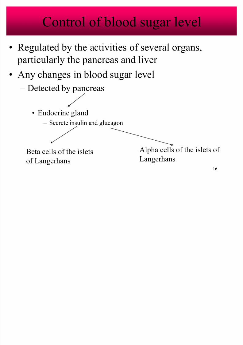
Control of blood sugar level
Regulated by the activities of several organs,
particularly the pancreas and liver
Any changes in blood sugar level
± Detected by pancreas
Endocrine gland
± Secrete insulin and glucagon
Beta cells of the islets
of Langerhans
Alpha cells of the islets of
Langerhans
8/7/2019 Biology sem1- chap9
http://slidepdf.com/reader/full/biology-sem1-chap9 17/111
17
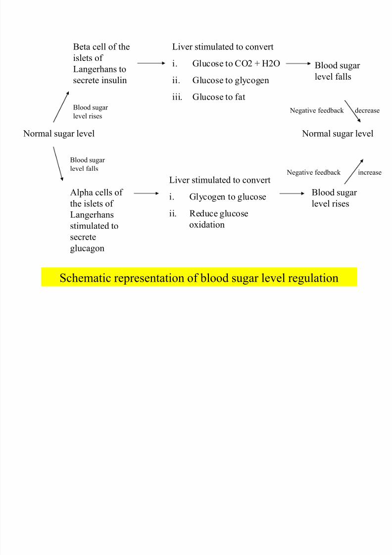
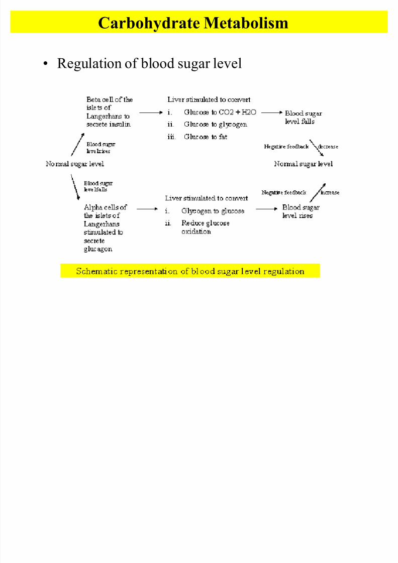
Schematic representation of blood sugar level regulation
Normal sugar level
Beta cell of the
islets of
Langerhans tosecrete insulin
Liver stimulated to convert
i. Glucose to CO2 + H2O
ii. Glucose to glycogen
iii. Glucose to fat
Blood sugar level falls
Normal sugar level
Alpha cells of
the islets of Langerhans
stimulated to
secrete
glucagon
Liver stimulated to convert
i. Glycogen to glucose
ii. Reduce glucose
oxidation
Blood sugar
level rises
Blood sugar
level rises
Blood sugar
level falls
Negative feedback
Negative feedback
decrease
increase
8/7/2019 Biology sem1- chap9
http://slidepdf.com/reader/full/biology-sem1-chap9 18/111
8/7/2019 Biology sem1- chap9
http://slidepdf.com/reader/full/biology-sem1-chap9 19/111
19
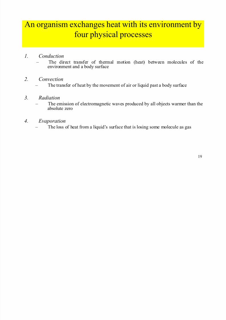
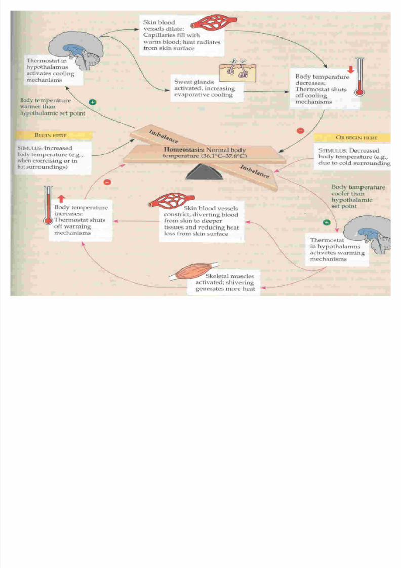
An organism exchanges heat with its environment by
four physical processes
1. C onduction ± The direct transfer of thermal motion (heat) between molecules of the
environment and a body surface
2. C onvection ± The transfer of heat by the movement of air or liquid past a body surface
3. Radiation ± The emission of electromagnetic waves produced by all objects warmer than the
absolute zero
4. E vaporation ± The loss of heat from a liquid¶s surface that is losing some molecule as gas
8/7/2019 Biology sem1- chap9
http://slidepdf.com/reader/full/biology-sem1-chap9 20/111
20
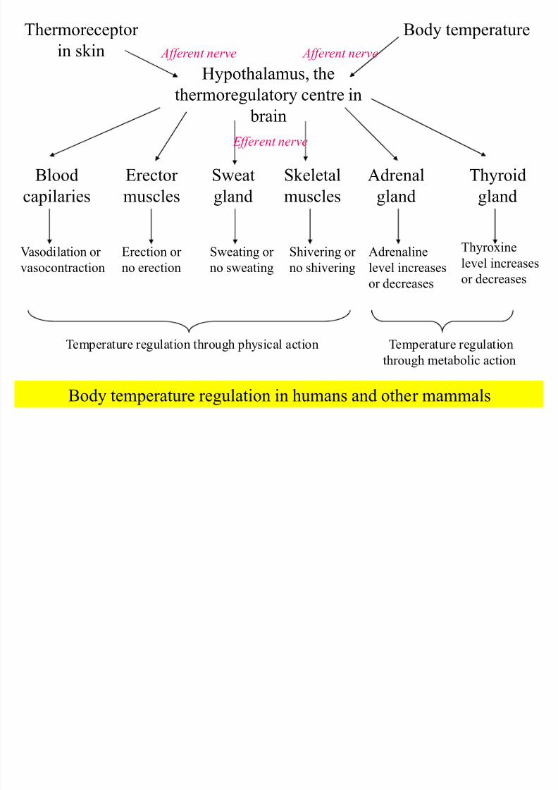
Body temperature regulation in humans and other mammals
Thermoreceptor
in skin
Body temperature
Hypothalamus, the
thermoregulatory centre in brain
Blood
capilaries
Erector
muscles
Sweat
gland
Skeletal
muscles
Adrenal
gland
Thyroid
gland
Vasodilation or
vasocontraction
Erection or
no erection
Sweating or
no sweating
Shivering or
no shivering
Adrenaline
level increases
or decreases
Thyroxine
level increases
or decreases
E fferent nerve
Afferent nerve Afferent nerve
Temperature regulation through physical action Temperature regulation
through metabolic action
8/7/2019 Biology sem1- chap9
http://slidepdf.com/reader/full/biology-sem1-chap9 21/111
21
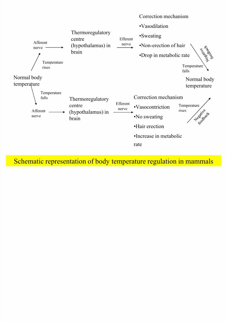
Schematic representation of body temperature regulation in mammals
Normal bodytemperature
Thermoregulatory
centre(hypothalamus) in
brain
Correction mechanism
Vasodilation
Sweating
Non-erection of hair
Drop in metabolic rate
Normal bodytemperature
Afferent
nerve
Thermoregulatory
centre
(hypothalamus) in
brain
Correction mechanism
Vasocontriction
No sweating
Hair erection
Increase in metabolic
rate
Temperature
rises
Temperature
falls
Efferentnerve
Temperature
falls
Temperature
rises
Efferent
nerve
Afferent
nerve
8/7/2019 Biology sem1- chap9
http://slidepdf.com/reader/full/biology-sem1-chap9 22/111
22
8/7/2019 Biology sem1- chap9
http://slidepdf.com/reader/full/biology-sem1-chap9 23/111
23
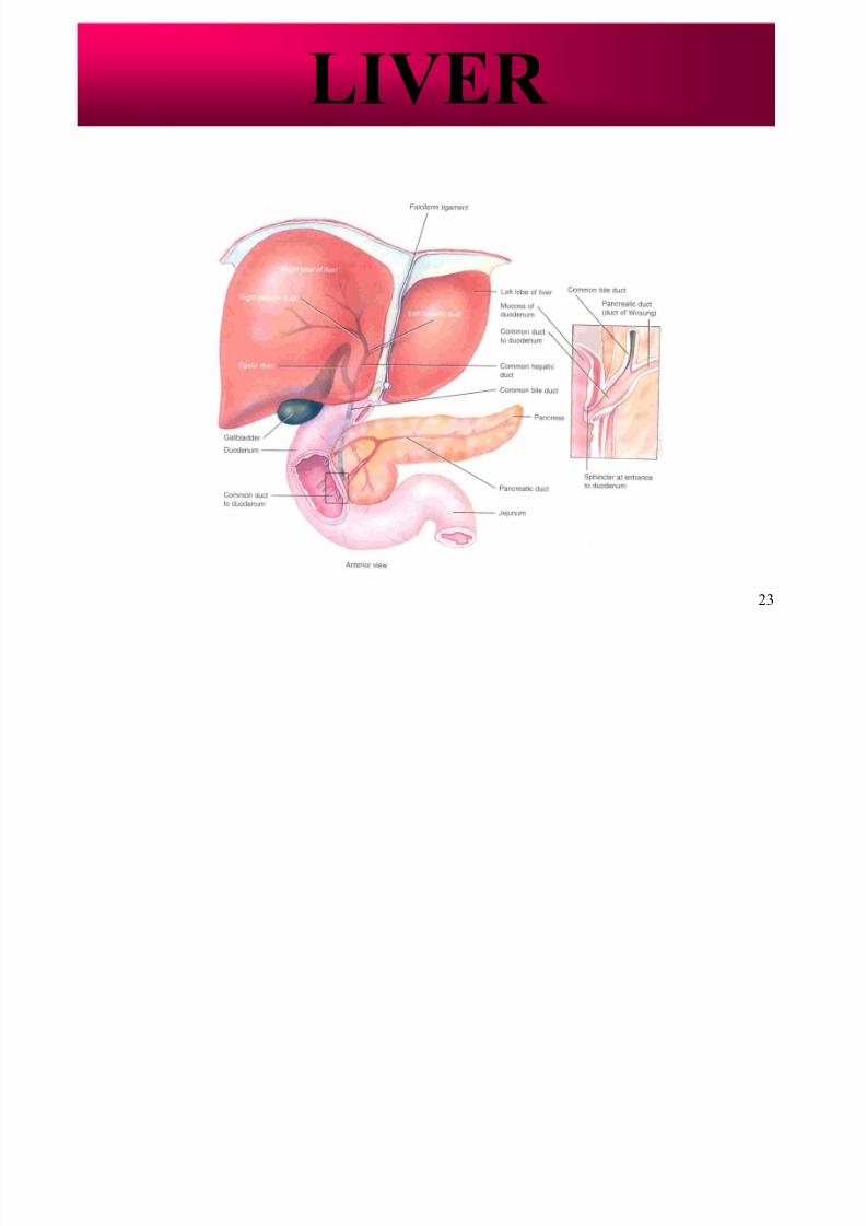
LIVER
8/7/2019 Biology sem1- chap9
http://slidepdf.com/reader/full/biology-sem1-chap9 24/111
24
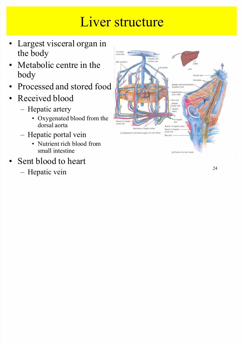
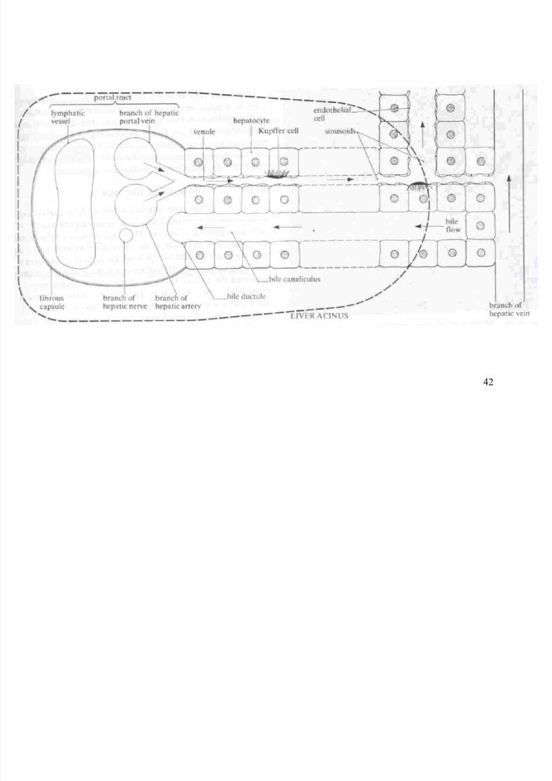
Liver structure
Largest visceral organ inthe body
Metabolic centre in the body
Processed and stored food Received blood
± Hepatic artery
Oxygenated blood from the
dorsal aorta ± Hepatic portal vein
Nutrient rich blood fromsmall intestine
Sent blood to heart
± Hepatic vein
8/7/2019 Biology sem1- chap9
http://slidepdf.com/reader/full/biology-sem1-chap9 25/111
25
Liver cell ± hepatocyte
Liver is made up of
many cylindrical lobes
Interlobular blood
vessel
± Branches of the hepatic
artery and hepatic
artery and hepatic
portal vein
± Connect to periphery
of each lobes

8/7/2019 Biology sem1- chap9
http://slidepdf.com/reader/full/biology-sem1-chap9 26/111
26
Canalikuli
± Bile duct branches into
a network of finevessels
± Pass between cells of
lobes
Sinusoid ± Capillaries which form
from hepatic arteriole
and hepatic portal
venule
± Flow to central vein
8/7/2019 Biology sem1- chap9
http://slidepdf.com/reader/full/biology-sem1-chap9 27/111
27
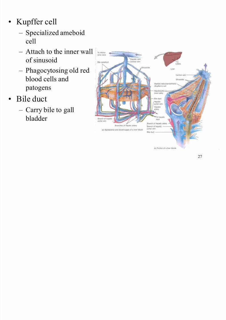
Kupffer cell
± Specialized ameboid
cell
± Attach to the inner wall
of sinusoid
± Phagocytosing old red
blood cells and patogens
Bile duct
± Carry bile to gall
bladder
8/7/2019 Biology sem1- chap9
http://slidepdf.com/reader/full/biology-sem1-chap9 28/111
28
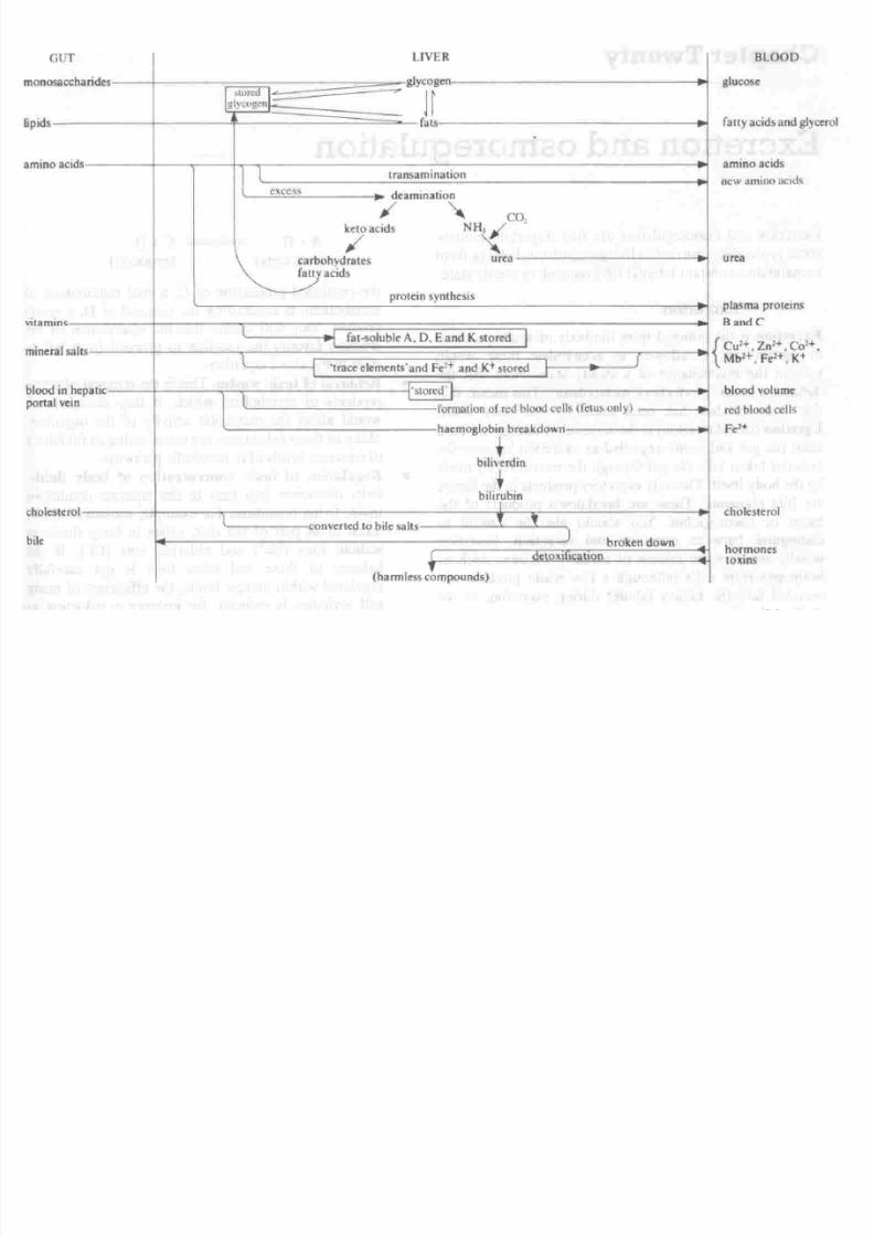
Functions of liver
1. Carbohydrate metabolism
Regulates blood sugar level
± Glycogenesis
Convert excess sugar to glycogen
± Glycogenolysis
Broken down the glycogen
± Cori cycle
Convert lactate into glucose
8/7/2019 Biology sem1- chap9
http://slidepdf.com/reader/full/biology-sem1-chap9 29/111
29
2. Lipid metabolism
± Breaks down fat
± Transport of lipid
Produces globulin to transport fat
Stored in the form of adipose tissues
8/7/2019 Biology sem1- chap9
http://slidepdf.com/reader/full/biology-sem1-chap9 30/111
30
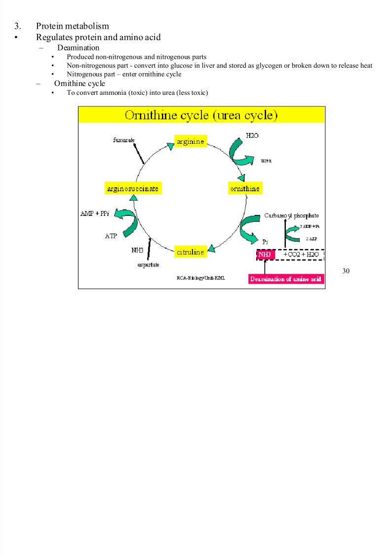
3. Protein metabolism
Regulates protein and amino acid ± Deamination
Produced non-nitrogenous and nitrogenous parts
Non-nitrogenous part - convert into glucose in liver and stored as glycogen or broken down to release heat
Nitrogenous part ± enter ornithine cycle
± Ornithine cycle To convert ammonia (toxic) into urea (less toxic)
8/7/2019 Biology sem1- chap9
http://slidepdf.com/reader/full/biology-sem1-chap9 31/111
31
8/7/2019 Biology sem1- chap9
http://slidepdf.com/reader/full/biology-sem1-chap9 32/111
32
4. Processing drugs and hormones
the liver can detoxify substances such as alcohol or excrete drugssuch as penicillin, erythromycin and sulfonamides into bile
5. Excretion of bilirubin
bilirubin derived from the heme of aged red blood cells, isabsorbed by the liver from the blood and secreted into bile
6. Synthesis of bile salts
bile salts are used in the small intestine for the emulsification andabsorption of lipids, cholesterol, phospholipids and lipoproteins
7. Storage
in addition to glycogen, the liver is a prime storage site for certainvitamins (A, cobalamin, D, E and K) and minerals (iron andcopper) which are released from the liver when needed elsewherein the body
8/7/2019 Biology sem1- chap9
http://slidepdf.com/reader/full/biology-sem1-chap9 33/111
33
8. Phagocytosis
Kupffer¶s cells phagocytize aged red blood cells
and white blood cells and some bacteria
9. Activation of vitamin D
the skin, liver and kidneys participate in
synthesizing the active form of vitamin D
8/7/2019 Biology sem1- chap9
http://slidepdf.com/reader/full/biology-sem1-chap9 34/111
34
R ole of the liver in (the
control) blood glucoselevel, fatty acids and
protein
8/7/2019 Biology sem1- chap9
http://slidepdf.com/reader/full/biology-sem1-chap9 35/111
35
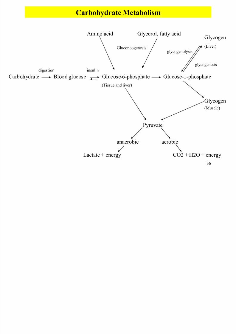
Carbohydrate Metabolism
Regulation of blood sugar level
8/7/2019 Biology sem1- chap9
http://slidepdf.com/reader/full/biology-sem1-chap9 36/111
36
Carbohydrate Blood glucose Glucose-6-phosphate
(Tissue and liver)
Glucose-1-phosphate
Glycogen
(Liver)
Glycogen
(Muscle)
Pyruvate
aerobicanaerobic
Lactate + energy CO2 + H2O + energy
Amino acid Glycerol, fatty acid
digestion insulin
glycogenesis
glycogenolysisGluconeogenesis
Carbohydrate Metabolism
8/7/2019 Biology sem1- chap9
http://slidepdf.com/reader/full/biology-sem1-chap9 37/111
37
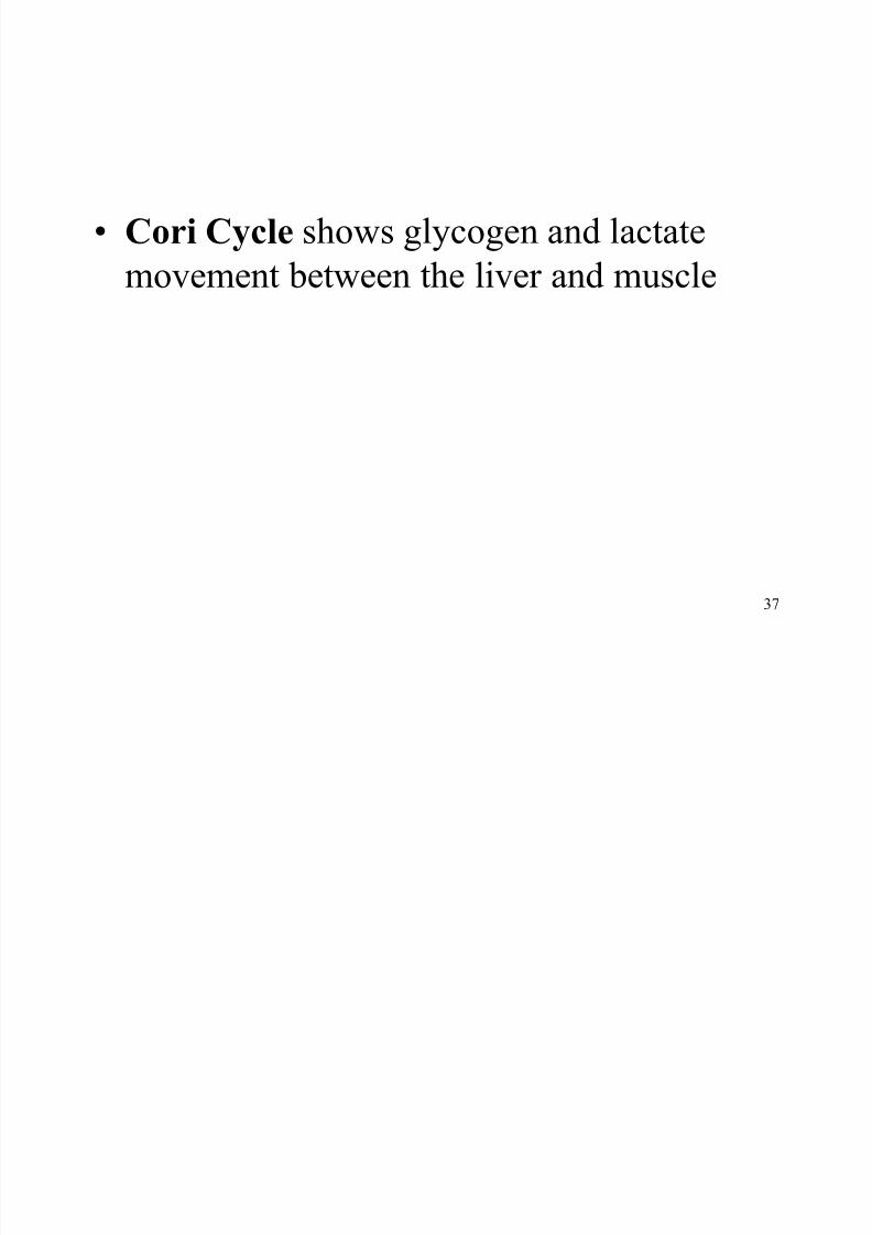
Cori Cycle shows glycogen and lactate
movement between the liver and muscle
8/7/2019 Biology sem1- chap9
http://slidepdf.com/reader/full/biology-sem1-chap9 38/111
38
Fats (lipid) metabolism
Fats in the liver can be modified for respiration and can be stored in the body
cells
Hepatocytes synthesize the cholesterolwhen the level is decreased. It also canexcreted cholesterol into bile when the levelof the cholesterol is increased
8/7/2019 Biology sem1- chap9
http://slidepdf.com/reader/full/biology-sem1-chap9 39/111
39
Protein Metabolism
Protein being recycled are first broken down into amino
acids. Hepatocytes convert amino acids to fatty acid, ketone
bodies, glucose or oxidize them to carbon dioxide andwater
There are two ways of protein metabolism ± Deamination
a conversion consists of removing the amino group from the aminoacids and converting it to ammonia
± Transamination
the transfer of an amino group from an amino acid to pyruvic acid or toan acid in the Krebs cycle-can synthesized nonessential amino acids
Ornithine Cycle shows the formation of urea
8/7/2019 Biology sem1- chap9
http://slidepdf.com/reader/full/biology-sem1-chap9 40/111
40
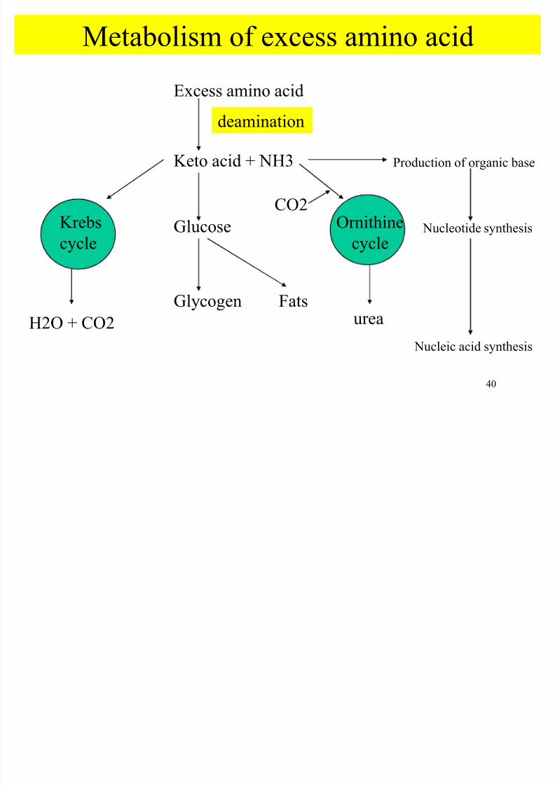
Metabolism of excess amino acid
Excess amino acid
Keto acid + NH3
Glucose
Glycogen Fats
Krebs
cycle
Ornithine
cycle
deamination
H2O + CO2 urea
CO2
Production of organic base
Nucleotide synthesis
Nucleic acid synthesis
8/7/2019 Biology sem1- chap9
http://slidepdf.com/reader/full/biology-sem1-chap9 41/111
41
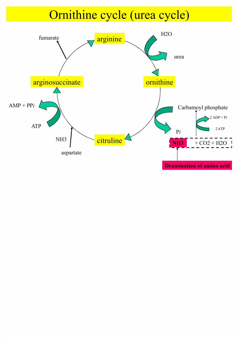
Ornithine cycle (urea cycle)
citruline
arginosuccinate
arginine
ornithine
H2O
urea
Carbamoyl phosphate
Pi
aspartate
NH3
ATP
AMP + PPi
fumarate
Deamination of amino acid
NH3 + CO2 + H2O
2 ATP
2 ADP + Pi
8/7/2019 Biology sem1- chap9
http://slidepdf.com/reader/full/biology-sem1-chap9 42/111
42
8/7/2019 Biology sem1- chap9
http://slidepdf.com/reader/full/biology-sem1-chap9 43/111
43
That¶s all for today
8/7/2019 Biology sem1- chap9
http://slidepdf.com/reader/full/biology-sem1-chap9 44/111
44
K idney
8/7/2019 Biology sem1- chap9
http://slidepdf.com/reader/full/biology-sem1-chap9 45/111
45
Objectives Kidney structure and nephron
Urine formation involving ultrafiltration and
reabsorption Urine concentration by counter current
multiplier mechanism
Water regulation by ADH
Osmoregulation of mineral ions by aldosterone
pH regulation of the tissue fluid
8/7/2019 Biology sem1- chap9
http://slidepdf.com/reader/full/biology-sem1-chap9 46/111
46
Introduction
yKidney
± main function in homeostasis
± urine formatian ; eliminate the nitrogen by products of protein metabolism and maintain
water balance
8/7/2019 Biology sem1- chap9
http://slidepdf.com/reader/full/biology-sem1-chap9 47/111
47
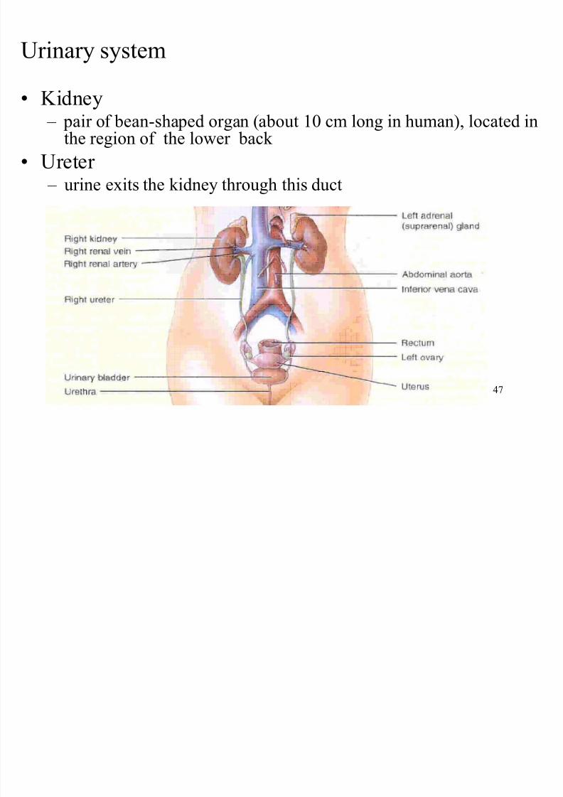
Urinary system
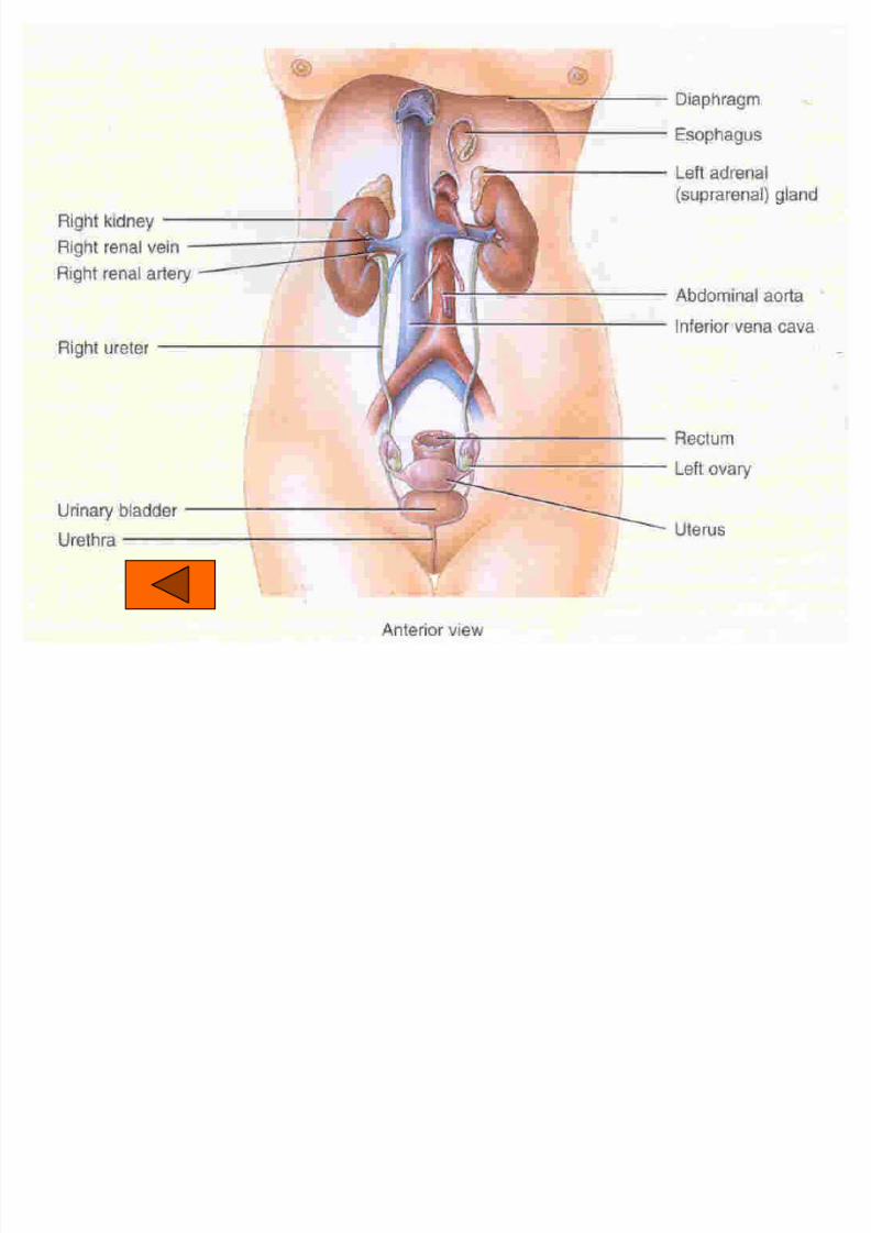
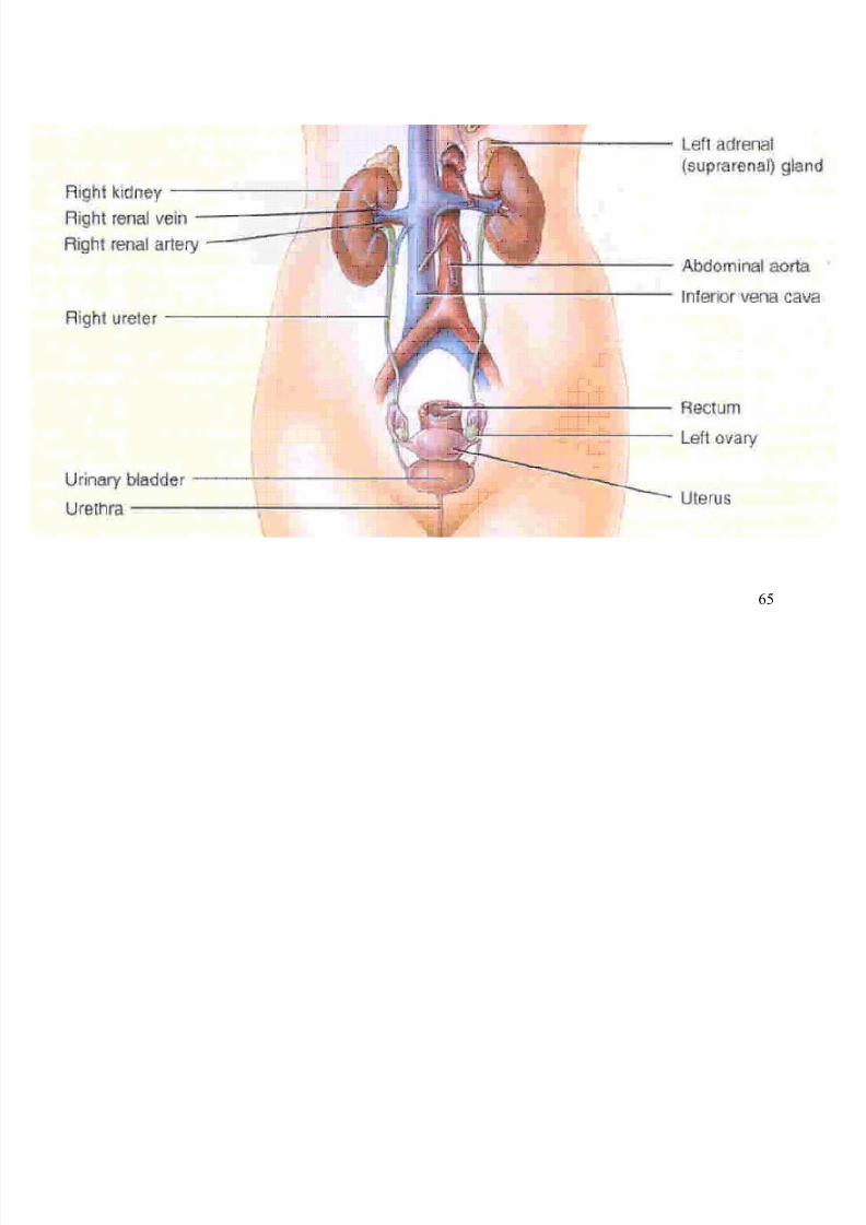
Kidney ± pair of bean-shaped organ (about 10 cm long in human), located in
the region of the lower back
Ureter ± urine exits the kidney through this duct
8/7/2019 Biology sem1- chap9
http://slidepdf.com/reader/full/biology-sem1-chap9 48/111
48
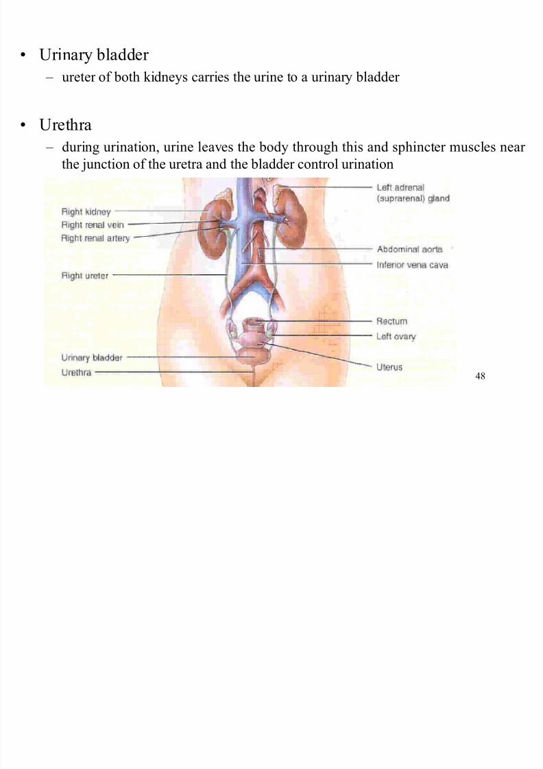
Urinary bladder
± ureter of both kidneys carries the urine to a urinary bladder
Urethra
± during urination, urine leaves the body through this and sphincter muscles near
the junction of the uretra and the bladder control urination
8/7/2019 Biology sem1- chap9
http://slidepdf.com/reader/full/biology-sem1-chap9 49/111
49
Structure of kidney
andnephron
8/7/2019 Biology sem1- chap9
http://slidepdf.com/reader/full/biology-sem1-chap9 50/111
50
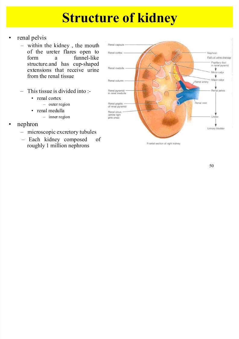
Structure of kidney
renal pelvis
± within the kidney , the mouthof the ureter flares open toform a funnel-likestructure.and has cup-shapedextensions that receive urinefrom the renal tissue
± This tissue is divided into :-
renal cortex
± outer region
renal medulla
± inner region
nephron
± microscopic excretory tubules
± Each kidney composed of roughly 1 million nephrons
8/7/2019 Biology sem1- chap9
http://slidepdf.com/reader/full/biology-sem1-chap9 51/111
51
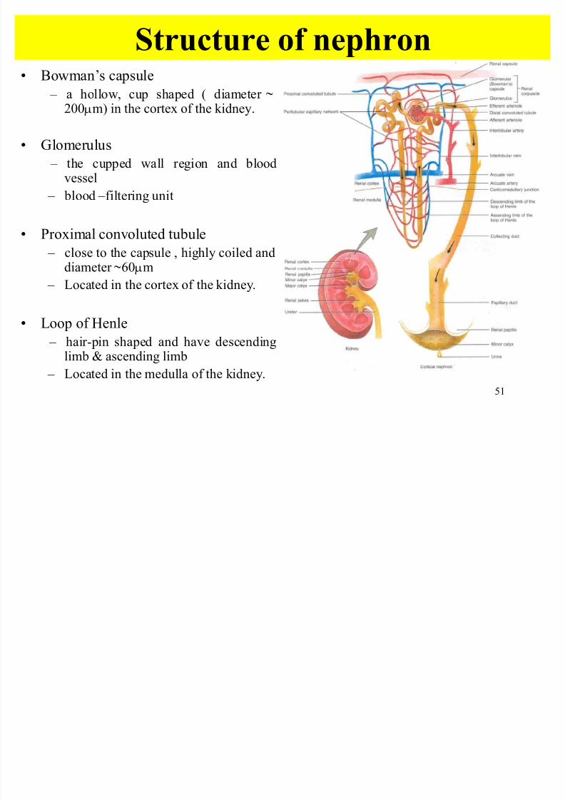
Structure of nephron Bowman¶s capsule
± a hollow, cup shaped ( diameter b
200Qm) in the cortex of the kidney.
Glomerulus
± the cupped wall region and bloodvessel
± blood ±filtering unit
Proximal convoluted tubule
± close to the capsule , highly coiled anddiameter b60Qm
± Located in the cortex of the kidney.
Loop of Henle
± hair-pin shaped and have descendinglimb & ascending limb
± Located in the medulla of the kidney.
8/7/2019 Biology sem1- chap9
http://slidepdf.com/reader/full/biology-sem1-chap9 52/111
52
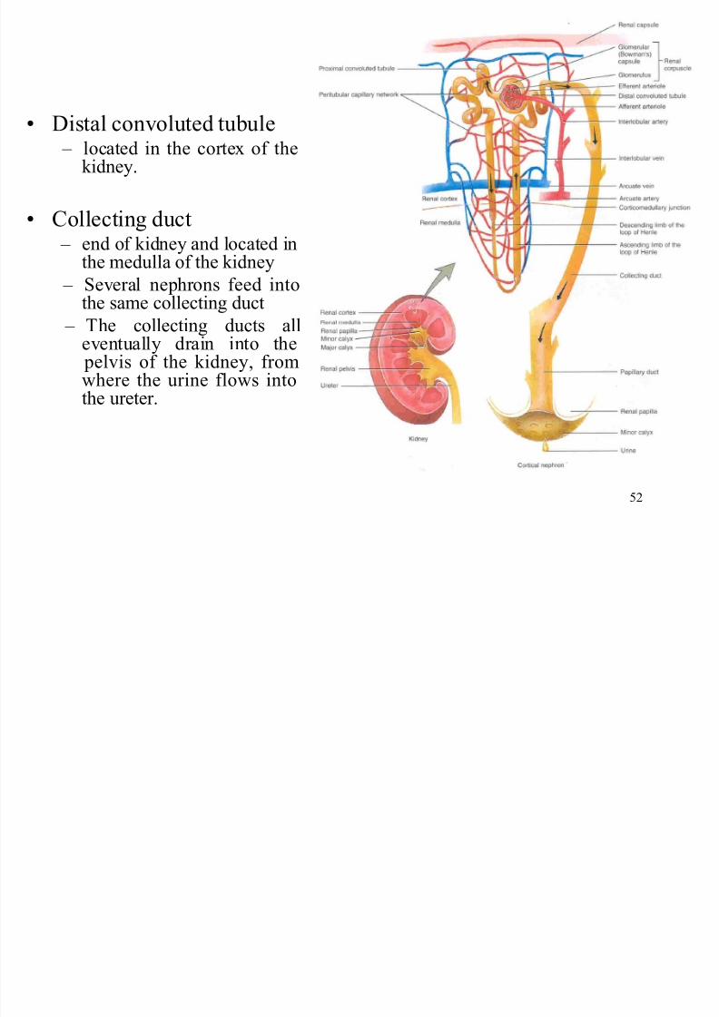
Distal convoluted tubule ± located in the cortex of thekidney.
Collecting duct ± end of kidney and located in
the medulla of the kidney ± Several nephrons feed into
the same collecting duct
± The collecting ducts alleventually drain into the pelvis of the kidney, from
where the urine flows intothe ureter.
8/7/2019 Biology sem1- chap9
http://slidepdf.com/reader/full/biology-sem1-chap9 53/111
53
Nephron and blood
circulation
8/7/2019 Biology sem1- chap9
http://slidepdf.com/reader/full/biology-sem1-chap9 54/111
54
Nephron and blood circulation
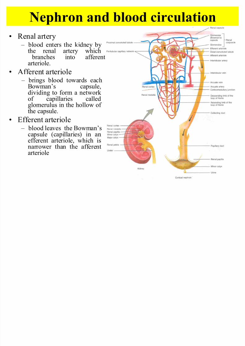
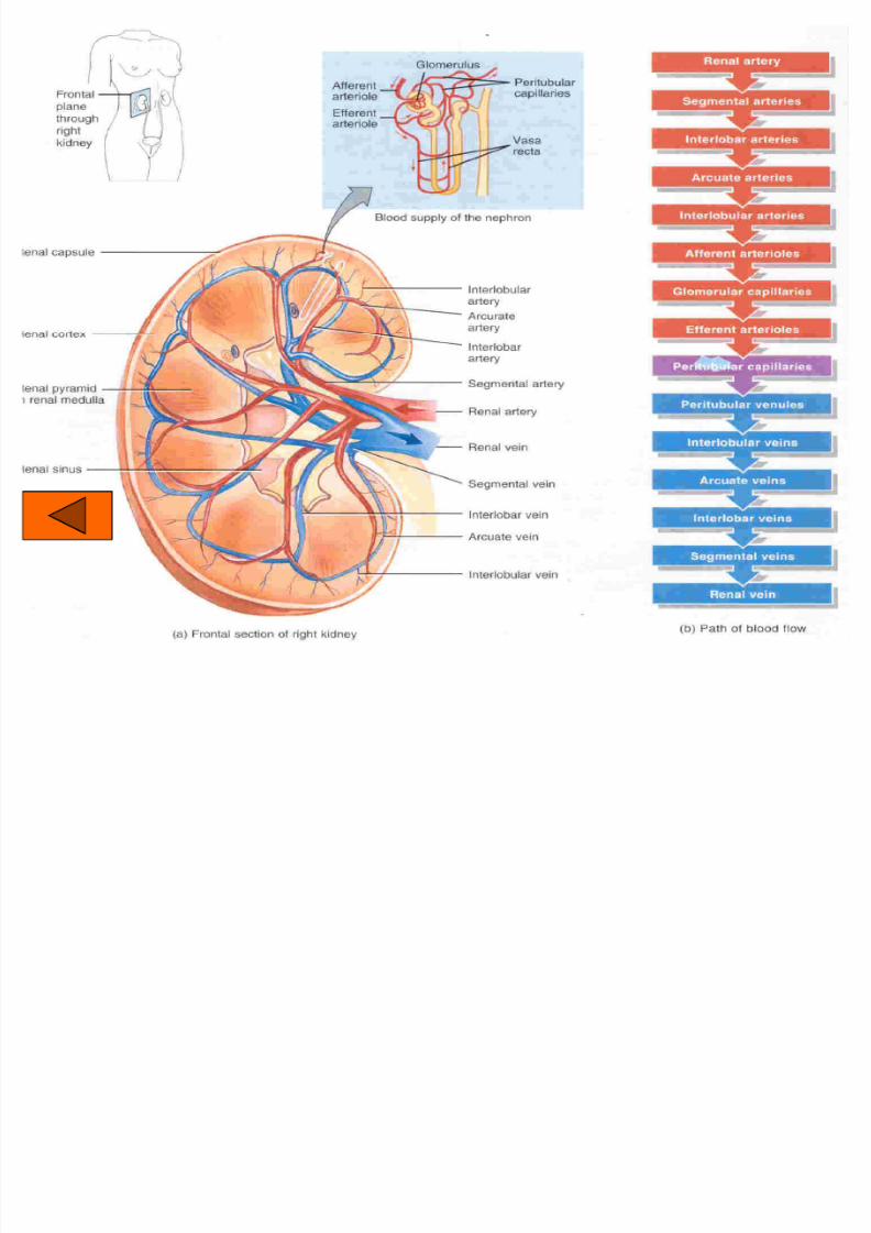
Renal artery
± blood enters the kidney bythe renal artery which branches into afferentarteriole.
Afferent arteriole ± brings blood towards each
Bowman¶s capsule,dividing to form a network of capillaries calledglomerulus in the hollow of the capsule.
Efferent arteriole
± blood leaves the Bowman¶scapsule (capillaries) in anefferent arteriole, which isnarrower than the afferentarteriole
The efferent arteriole
8/7/2019 Biology sem1- chap9
http://slidepdf.com/reader/full/biology-sem1-chap9 55/111
55
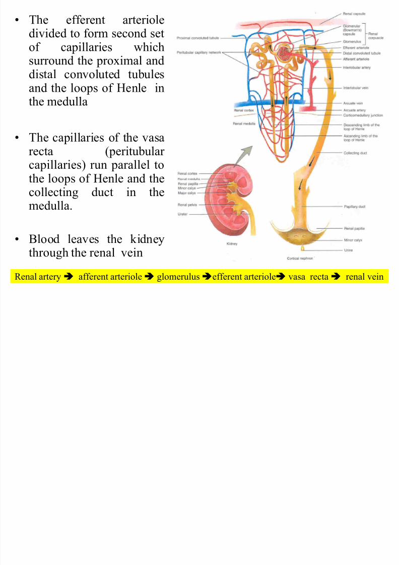
The efferent arterioledivided to form second setof capillaries whichsurround the proximal and
distal convoluted tubulesand the loops of Henle inthe medulla
The capillaries of the vasarecta (peritubular capillaries) run parallel tothe loops of Henle and thecollecting duct in themedulla.
Blood leaves the kidneythrough the renal vein
Renal arteryÄ afferent arterioleÄ glomerulusÄefferent arterioleÄ vasa rectaÄ renal vein
8/7/2019 Biology sem1- chap9
http://slidepdf.com/reader/full/biology-sem1-chap9 56/111
56
Urine Formation
U i F ti
8/7/2019 Biology sem1- chap9
http://slidepdf.com/reader/full/biology-sem1-chap9 57/111
57
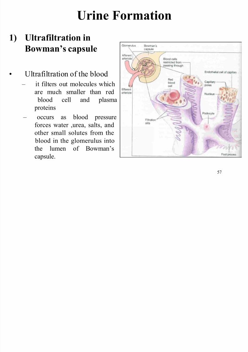
Urine Formation
1) Ultrafiltration in
Bowman¶s capsule
Ultrafiltration of the blood
± it filters out molecules which
are much smaller than red
blood cell and plasma
proteins
± occurs as blood pressure
forces water ,urea, salts, and
other small solutes from the blood in the glomerulus into
the lumen of Bowman¶s
capsule.
Factors contribute to ultrafiltration process:-
8/7/2019 Biology sem1- chap9
http://slidepdf.com/reader/full/biology-sem1-chap9 58/111
58
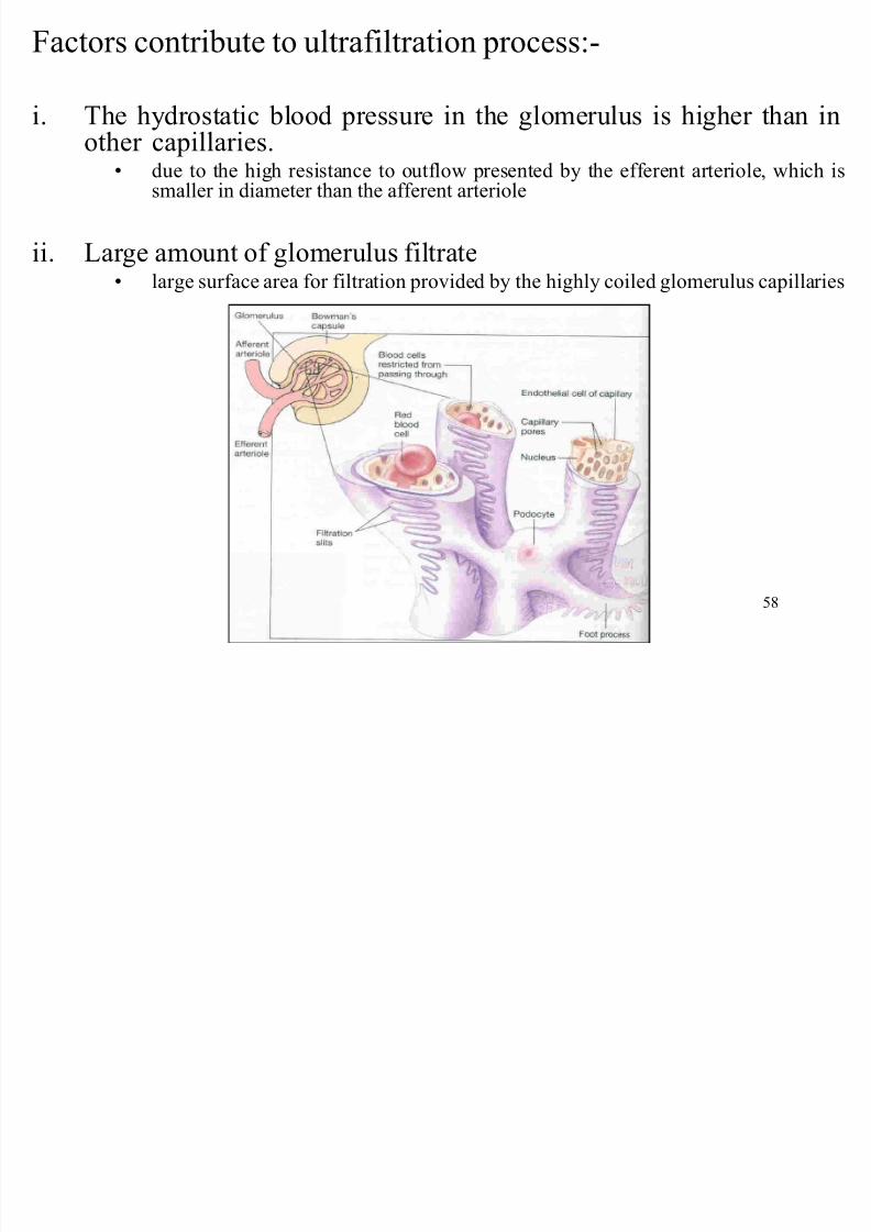
Factors contribute to ultrafiltration process:
i. The hydrostatic blood pressure in the glomerulus is higher than inother capillaries.
due to the high resistance to outflow presented by the efferent arteriole, which issmaller in diameter than the afferent arteriole
ii. Large amount of glomerulus filtrate large surface area for filtration provided by the highly coiled glomerulus capillaries
8/7/2019 Biology sem1- chap9
http://slidepdf.com/reader/full/biology-sem1-chap9 59/111
59
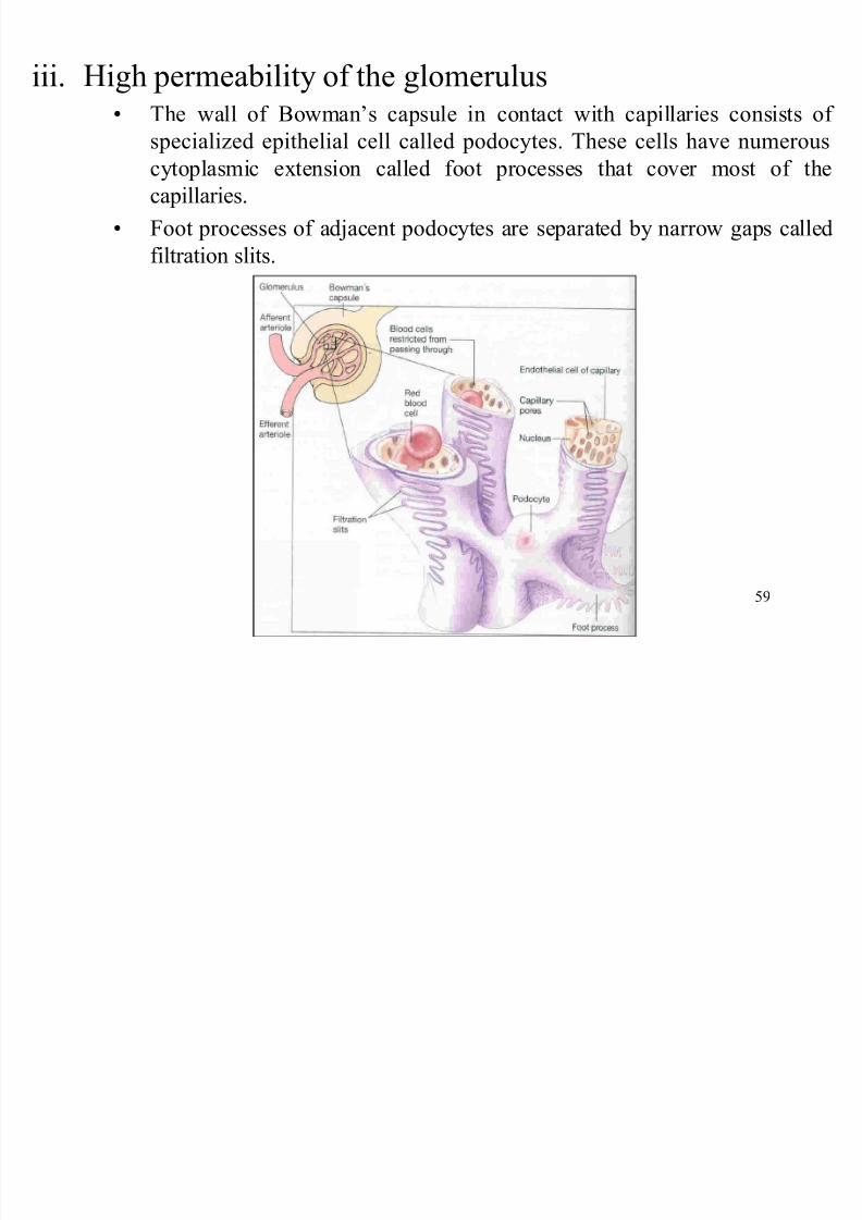
iii. High permeability of the glomerulus The wall of Bowman¶s capsule in contact with capillaries consists of
specialized epithelial cell called podocytes. These cells have numerous
cytoplasmic extension called foot processes that cover most of the
capillaries.
Foot processes of adjacent podocytes are separated by narrow gaps called
filtration slits.
8/7/2019 Biology sem1- chap9
http://slidepdf.com/reader/full/biology-sem1-chap9 60/111
60
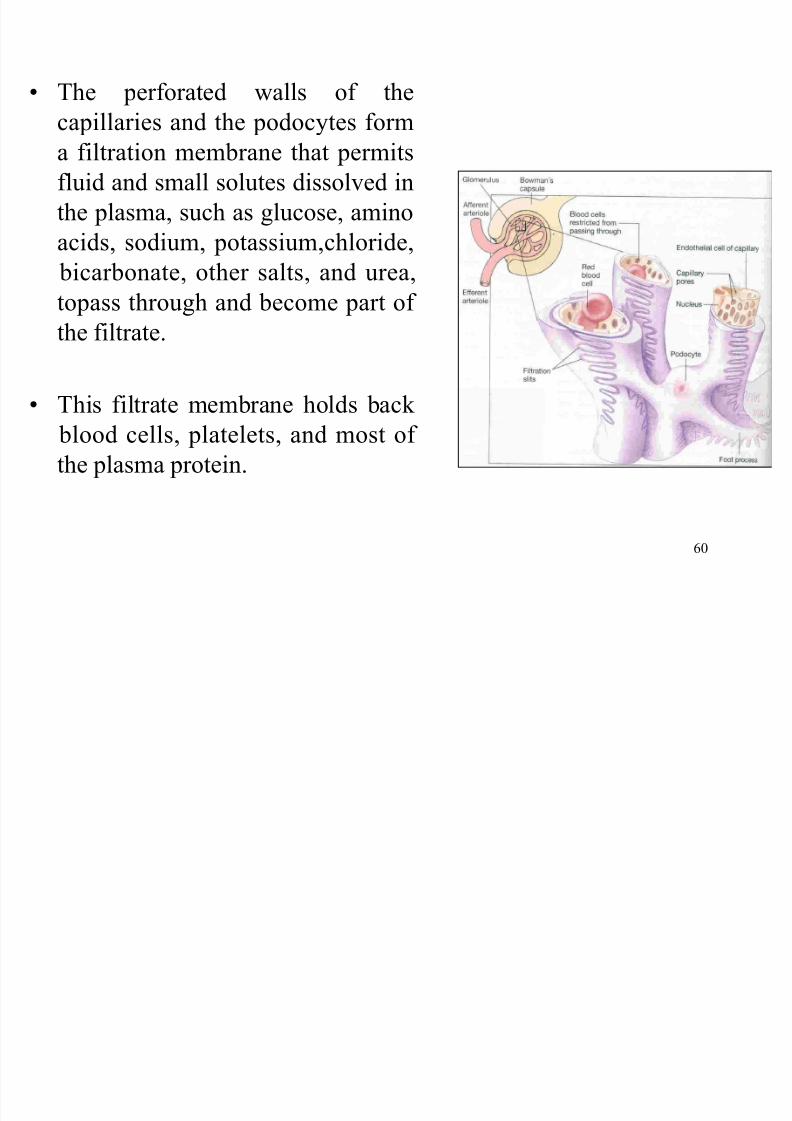
The perforated walls of the
capillaries and the podocytes form
a filtration membrane that permitsfluid and small solutes dissolved in
the plasma, such as glucose, amino
acids, sodium, potassium,chloride,
bicarbonate, other salts, and urea,
topass through and become part of
the filtrate.
This filtrate membrane holds back
blood cells, platelets, and most of
the plasma protein.
8/7/2019 Biology sem1- chap9
http://slidepdf.com/reader/full/biology-sem1-chap9 61/111
61
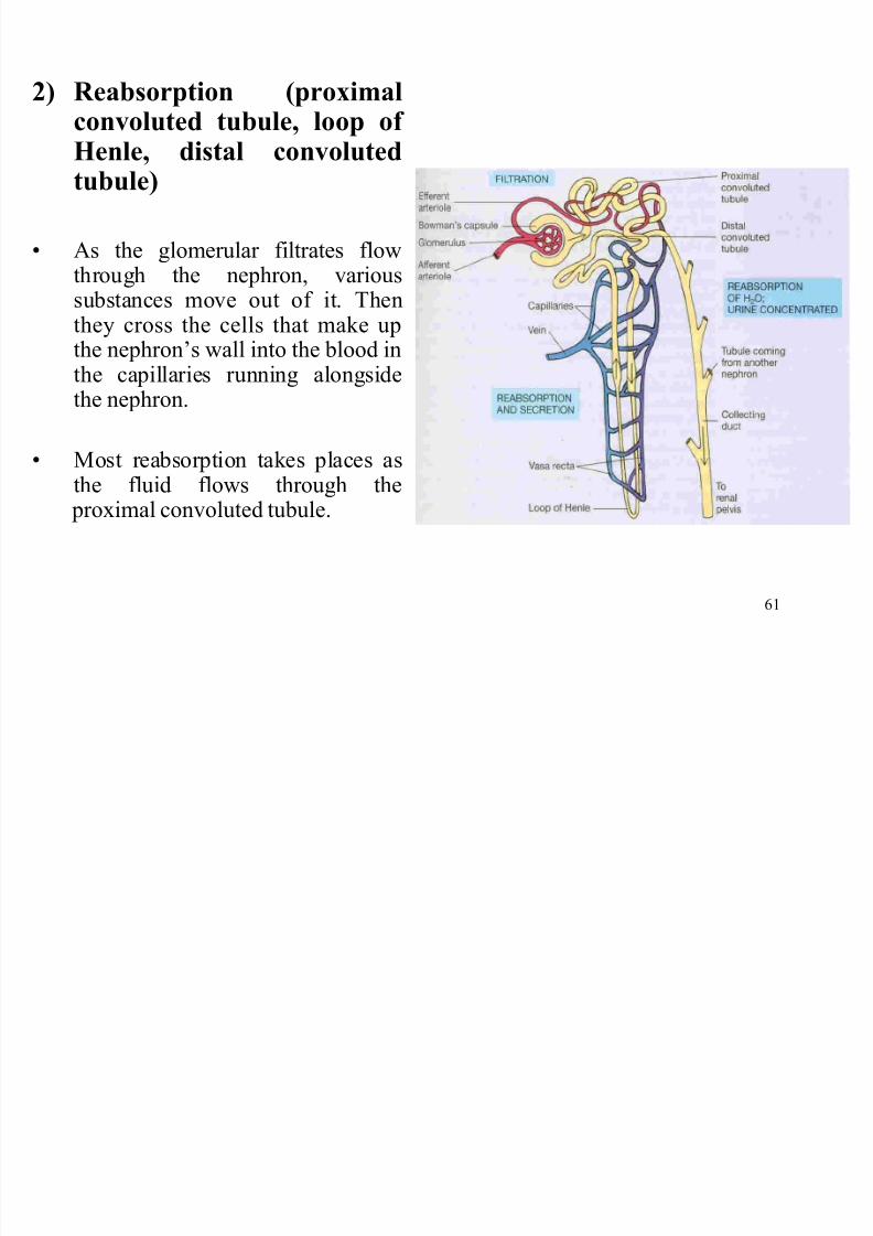
2) R eabsorption (proximalconvoluted tubule, loop of
Henle, distal convolutedtubule)
As the glomerular filtrates flowthrough the nephron, varioussubstances move out of it. Thenthey cross the cells that make upthe nephron¶s wall into the blood inthe capillaries running alongsidethe nephron.
Most reabsorption takes places asthe fluid flows through the proximal convoluted tubule.
8/7/2019 Biology sem1- chap9
http://slidepdf.com/reader/full/biology-sem1-chap9 62/111
62
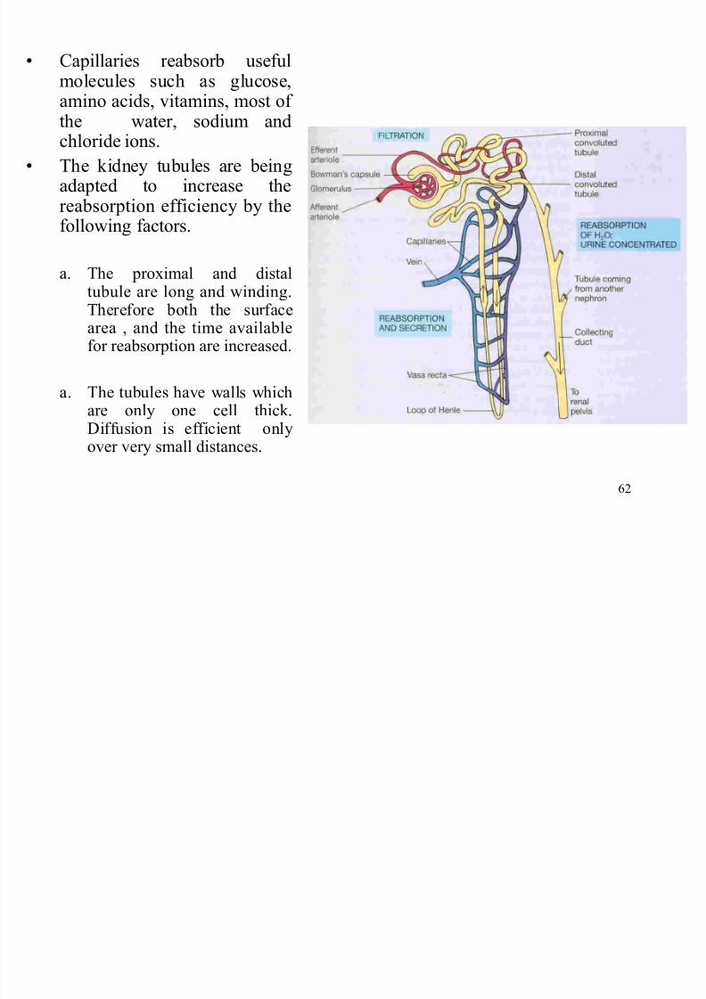
Capillaries reabsorb usefulmolecules such as glucose,amino acids, vitamins, most of the water, sodium andchloride ions.
The kidney tubules are beingadapted to increase thereabsorption efficiency by thefollowing factors.
a. The proximal and distaltubule are long and winding.Therefore both the surfacearea , and the time availablefor reabsorption are increased.
a. The tubules have walls whichare only one cell thick.Diffusion is efficient onlyover very small distances.
8/7/2019 Biology sem1- chap9
http://slidepdf.com/reader/full/biology-sem1-chap9 63/111
63
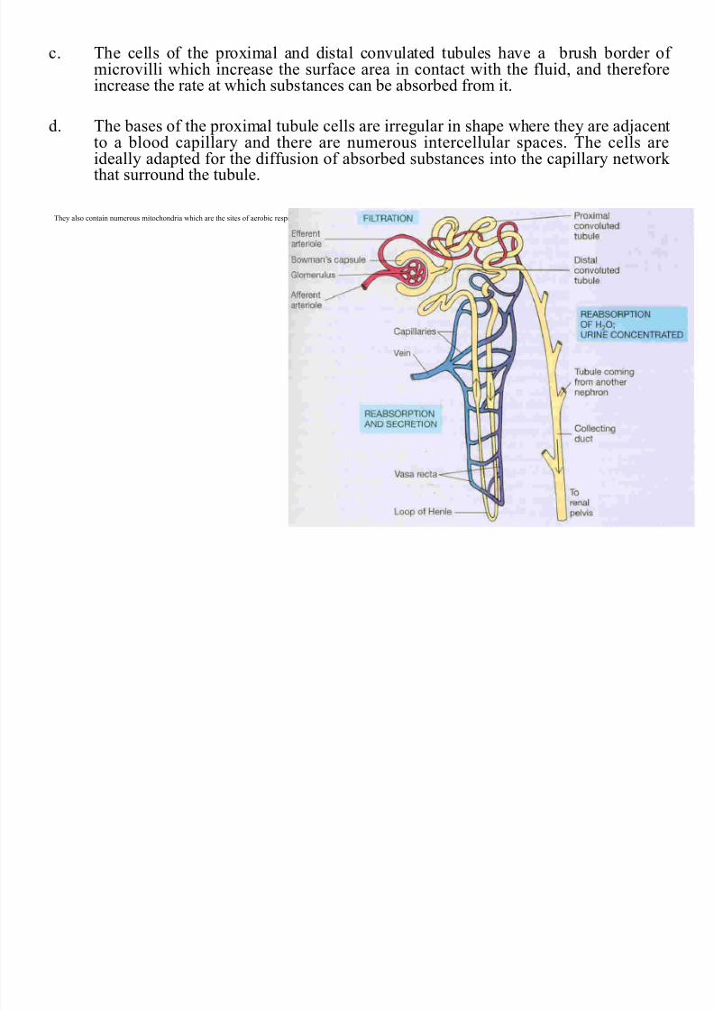
c. The cells of the proximal and distal convulated tubules have a brush border of microvilli which increase the surface area in contact with the fluid, and thereforeincrease the rate at which substances can be absorbed from it.
d. The bases of the proximal tubule cells are irregular in shape where they are adjacentto a blood capillary and there are numerous intercellular spaces. The cells areideally adapted for the diffusion of absorbed substances into the capillary network that surround the tubule.
They also contain numerous mitochondria which are the sites of aerobic respiration. The energy is required for active uptake when substances are moved across cells such as sodium ions and glucose.
8/7/2019 Biology sem1- chap9
http://slidepdf.com/reader/full/biology-sem1-chap9 64/111
64
8/7/2019 Biology sem1- chap9
http://slidepdf.com/reader/full/biology-sem1-chap9 65/111
65
8/7/2019 Biology sem1- chap9
http://slidepdf.com/reader/full/biology-sem1-chap9 66/111
66
8/7/2019 Biology sem1- chap9
http://slidepdf.com/reader/full/biology-sem1-chap9 67/111
67
8/7/2019 Biology sem1- chap9
http://slidepdf.com/reader/full/biology-sem1-chap9 68/111
68
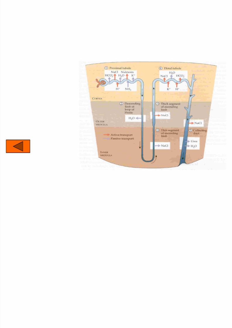
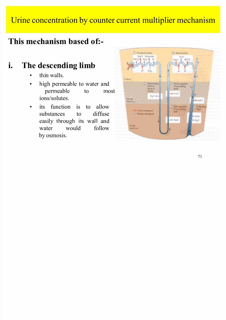
Urine concentration by counter current multiplier mechanism
8/7/2019 Biology sem1- chap9
http://slidepdf.com/reader/full/biology-sem1-chap9 69/111
69
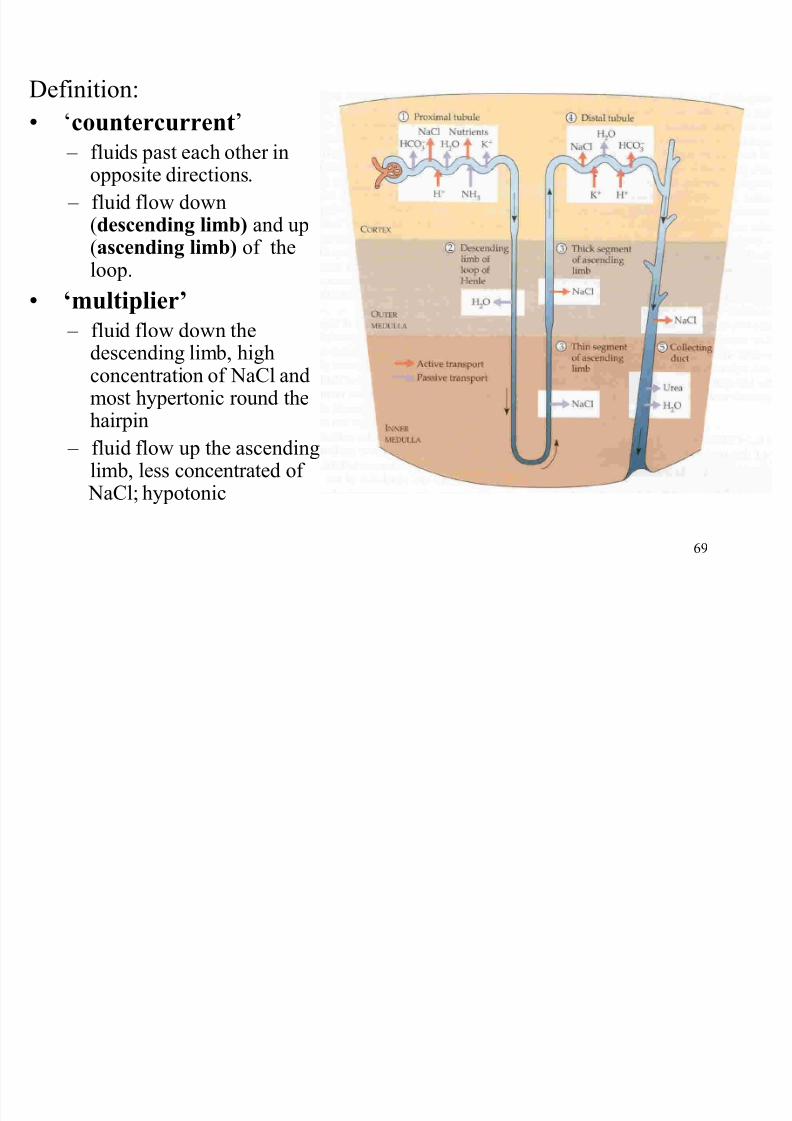
Definition:
µcountercurrent
± fluids past each other inopposite directions.
± fluid flow down(descending limb) and up(ascending limb) of theloop.
µmultiplier
± fluid flow down thedescending limb, highconcentration of NaCl andmost hypertonic round the
hairpin ± fluid flow up the ascending
limb, less concentrated of NaCl; hypotonic
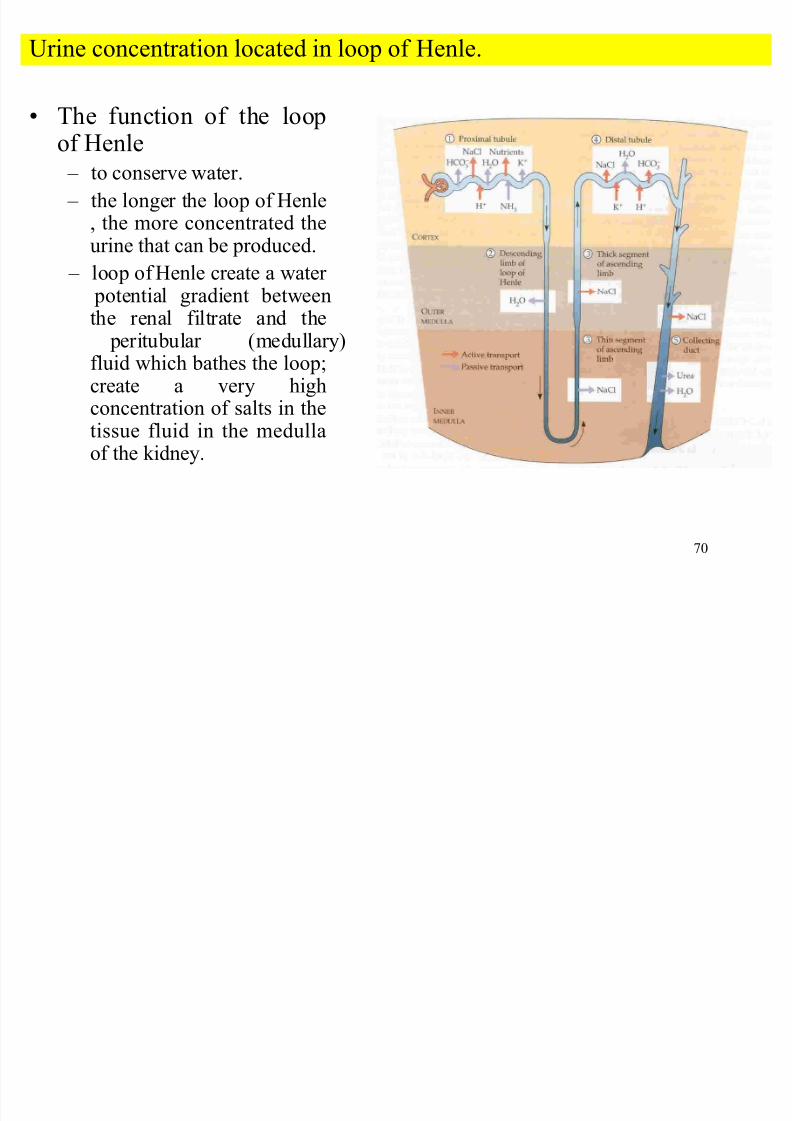
Urine concentration located in loop of Henle
8/7/2019 Biology sem1- chap9
http://slidepdf.com/reader/full/biology-sem1-chap9 70/111
70
The function of the loop
of Henle ± to conserve water.
± the longer the loop of Henle, the more concentrated theurine that can be produced.
± loop of Henle create a water potential gradient betweenthe renal filtrate and the peritubular (medullary)fluid which bathes the loop;create a very highconcentration of salts in the
tissue fluid in the medullaof the kidney.
Urine concentration located in loop of Henle.
8/7/2019 Biology sem1- chap9
http://slidepdf.com/reader/full/biology-sem1-chap9 71/111
71
Urine concentration by counter current multiplier mechanism
This mechanism based of:-
i. The descending limb thin walls.
high permeable to water and permeable to most
ions/solutes.
its function is to allow
substances to diffuse
easily through its wall andwater would follow
by osmosis.
ii Th di li b
8/7/2019 Biology sem1- chap9
http://slidepdf.com/reader/full/biology-sem1-chap9 72/111
72
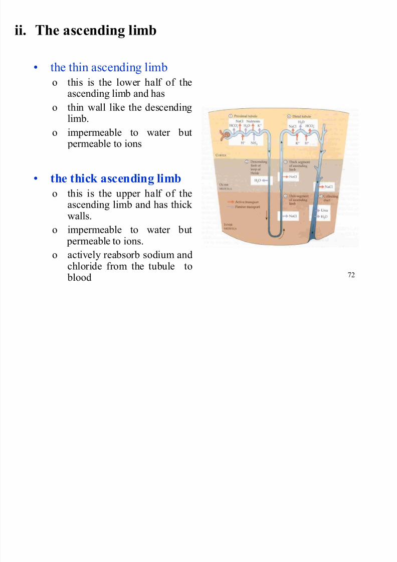
ii. The ascending limb
the thin ascending limb
o this is the lower half of theascending limb and has
o thin wall like the descendinglimb.
o impermeable to water but
permeable to ions
the thick ascending limb
o this is the upper half of theascending limb and has thick
walls.o impermeable to water but
permeable to ions.
o actively reabsorb sodium andchloride from the tubule to
blood
Urine concentration by counter current multiplier mechanism
8/7/2019 Biology sem1- chap9
http://slidepdf.com/reader/full/biology-sem1-chap9 73/111
73
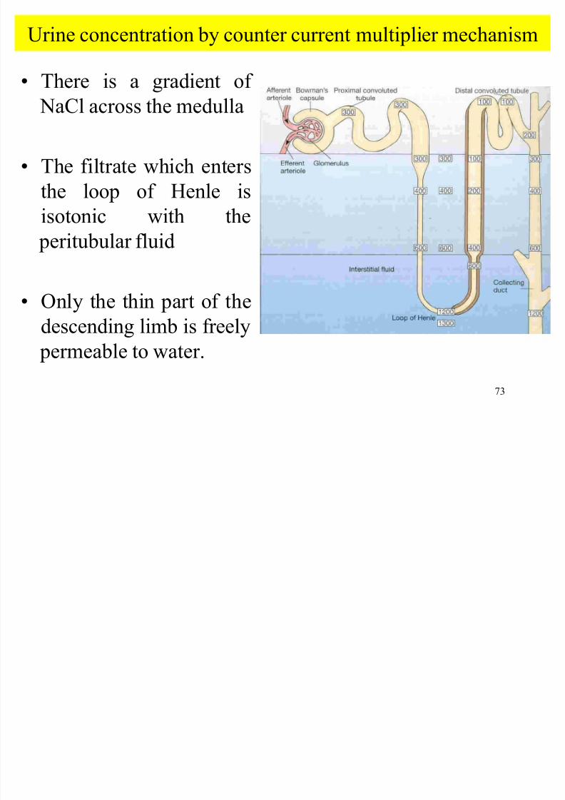
Urine concentration by counter current multiplier mechanism
There is a gradient of
NaCl across the medulla
The filtrate which enters
the loop of Henle is
isotonic with the
peritubular fluid
Only the thin part of thedescending limb is freely
permeable to water.
8/7/2019 Biology sem1- chap9
http://slidepdf.com/reader/full/biology-sem1-chap9 74/111
74
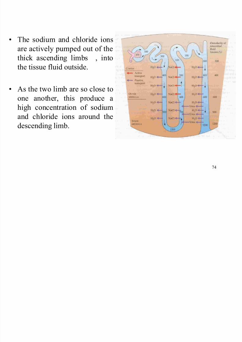
The sodium and chloride ions
are actively pumped out of the
thick ascending limbs , into
the tissue fluid outside.
As the two limb are so close toone another, this produce a
high concentration of sodium
and chloride ions around the
descending limb.
8/7/2019 Biology sem1- chap9
http://slidepdf.com/reader/full/biology-sem1-chap9 75/111
75
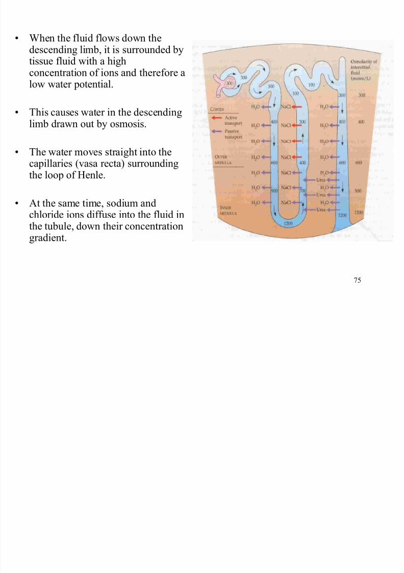
When the fluid flows down thedescending limb, it is surrounded bytissue fluid with a high
concentration of ions and therefore alow water potential.
This causes water in the descendinglimb drawn out by osmosis.
The water moves straight into thecapillaries (vasa recta) surroundingthe loop of Henle.
At the same time, sodium and
chloride ions diffuse into the fluid inthe tubule, down their concentrationgradient.
8/7/2019 Biology sem1- chap9
http://slidepdf.com/reader/full/biology-sem1-chap9 76/111
76
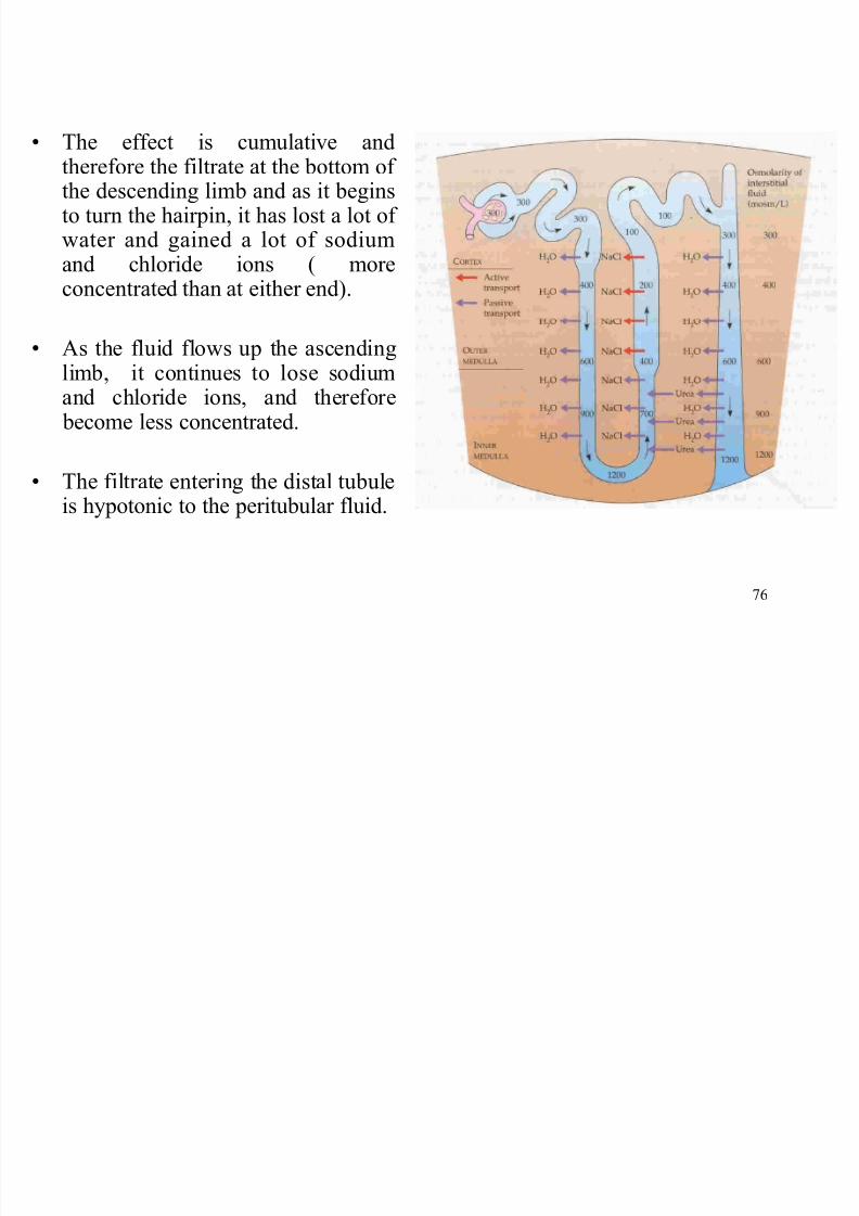
The effect is cumulative and
therefore the filtrate at the bottom of the descending limb and as it beginsto turn the hairpin, it has lost a lot of water and gained a lot of sodiumand chloride ions ( moreconcentrated than at either end).
As the fluid flows up the ascendinglimb, it continues to lose sodiumand chloride ions, and therefore become less concentrated.
The filtrate entering the distal tubuleis hypotonic to the peritubular fluid.
8/7/2019 Biology sem1- chap9
http://slidepdf.com/reader/full/biology-sem1-chap9 77/111
77
Function of complex of blood capillaries runsalongside the loop of Henle:
± supply oxygen and nutrients so that cells in the walls of the loop can produce the large amount of ATP for active transport.
± take away much of the salt and water from the tissuefluid in the medulla, helping to maintain the gradients
built up by the loop.
H d hi h i h l
8/7/2019 Biology sem1- chap9
http://slidepdf.com/reader/full/biology-sem1-chap9 78/111
78
How does this mechanism help to
conserve water?
The final part of each nephron, the collecting duct,also passes through the medulla of the kidney.
As the fluid flow through the collecting ducts,water can be drawn out of them, by osmosis, intothe concentrated tissue fluid in the medulla.
The more concentrated the tissue fluid, the morewater can be drawn out, and the moreconcentrated the urine can be.
8/7/2019 Biology sem1- chap9
http://slidepdf.com/reader/full/biology-sem1-chap9 79/111
79
Water regulation by ADH
8/7/2019 Biology sem1- chap9
http://slidepdf.com/reader/full/biology-sem1-chap9 80/111
80
Water regulation by ADH
Concepts
The kidney play central role in the regulation of the water content of the body or osmoregulation.
The water content of the blood is monitored by
osmoreceptor cells in the hypothalamus and these
cells produce antidiuretic hormone, ADH.
Summarize
8/7/2019 Biology sem1- chap9
http://slidepdf.com/reader/full/biology-sem1-chap9 81/111
81
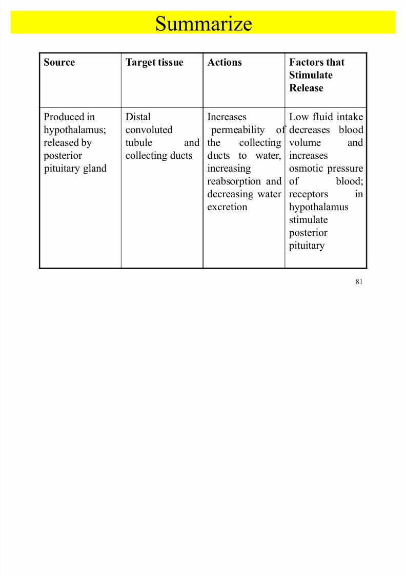
Su e
Source Target tissue Actions Factors that
StimulateR elease
Produced in
hypothalamus;
released by
posterior
pituitary gland
Distal
convoluted
tubule and
collecting ducts
Increases
permeability of
the collecting
ducts to water,
increasing
reabsorption and
decreasing water
excretion
Low fluid intake
decreases blood
volume and
increases
osmotic pressure
of blood;
receptors in
hypothalamus
stimulate posterior
pituitary
Water regulation by ADH
8/7/2019 Biology sem1- chap9
http://slidepdf.com/reader/full/biology-sem1-chap9 82/111
82
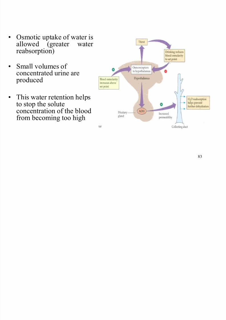
g y
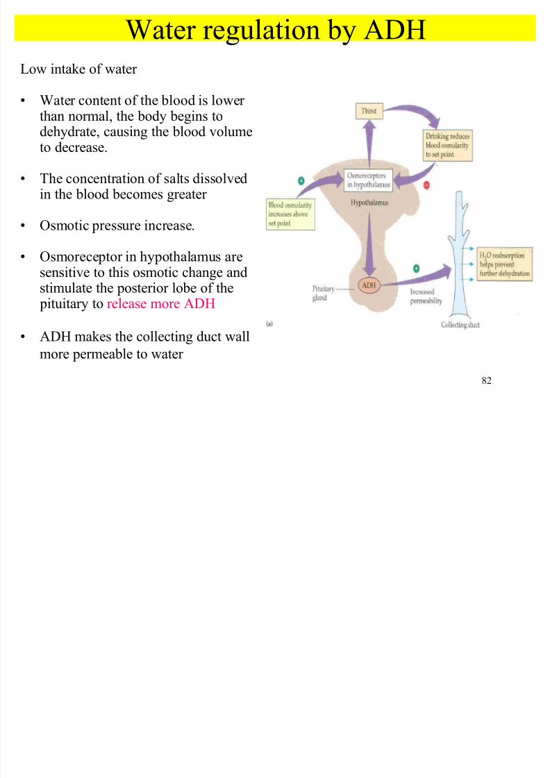
Low intake of water
Water content of the blood is lower than normal, the body begins todehydrate, causing the blood volumeto decrease.
The concentration of salts dissolved
in the blood becomes greater
Osmotic pressure increase.
Osmoreceptor in hypothalamus aresensitive to this osmotic change andstimulate the posterior lobe of the pituitary to release more ADH
ADHmakes the collecting duct wall
more permeable to water
8/7/2019 Biology sem1- chap9
http://slidepdf.com/reader/full/biology-sem1-chap9 83/111
8/7/2019 Biology sem1- chap9
http://slidepdf.com/reader/full/biology-sem1-chap9 84/111
84
High intake of water
Water content of the blood is higher than normal, the body fluids begin todilute, causing the blood volume to increase.
Concentration of salts dissolved in the blood becomes less
Osmotic pressure decrease.
Osmoreceptor in hypothalamus are sensitive to this osmotic change and
stimulate the posterior lobe of the pituitary to release less ADH
ADH makes the collecting duct wall less permeable to water
Less osmotic uptake of water is allowed (less water reabsorption)
Large volumes of diluted urine are produced
This water loss reduces the solute concentration of the blood
8/7/2019 Biology sem1- chap9
http://slidepdf.com/reader/full/biology-sem1-chap9 85/111
85
Osmoregulation of mineral ions by aldosterone
Osmoregulation of mineral ions by aldosterone
8/7/2019 Biology sem1- chap9
http://slidepdf.com/reader/full/biology-sem1-chap9 86/111
86
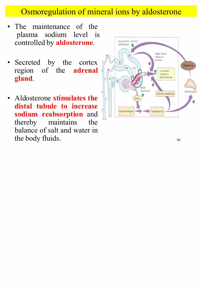
The maintenance of the plasma sodium level iscontrolled by aldosterone.
Secreted by the cortex
region of the adrenalgland.
Aldosterone stimulates the
distal tubule to increasesodium reabsorption andthereby maintains the balance of salt and water inthe body fluids.
Osmoregulation of mineral ions by aldosterone
8/7/2019 Biology sem1- chap9
http://slidepdf.com/reader/full/biology-sem1-chap9 87/111
87
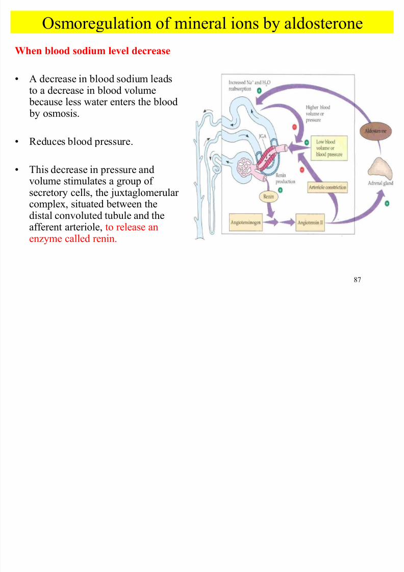
When blood sodium level decrease
A decrease in blood sodium leadsto a decrease in blood volume because less water enters the blood by osmosis.
Reduces blood pressure.
This decrease in pressure andvolume stimulates a group of secretory cells, the juxtaglomerular complex, situated between the
distal convoluted tubule and theafferent arteriole, to release anenzyme called renin.
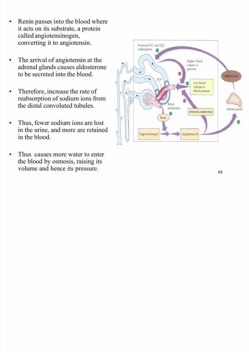
Renin passes into the blood here
8/7/2019 Biology sem1- chap9
http://slidepdf.com/reader/full/biology-sem1-chap9 88/111
88
Renin passes into the blood whereit acts on its substrate, a proteincalled angiotensinogen,converting it to angiotensin.
The arrival of angiotensin at theadrenal glands causes aldosteroneto be secreted into the blood.
Therefore, increase the rate of reabsorption of sodium ions fromthe distal convoluted tubules.
Thus, fewer sodium ions are lostin the urine, and more are retainedin the blood.
Thus causes more water to enter the blood by osmosis, raising itsvolume and hence its pressure.
8/7/2019 Biology sem1- chap9
http://slidepdf.com/reader/full/biology-sem1-chap9 89/111
89
pH regulation
of
the tissue fluid
8/7/2019 Biology sem1- chap9
http://slidepdf.com/reader/full/biology-sem1-chap9 90/111
8/7/2019 Biology sem1- chap9
http://slidepdf.com/reader/full/biology-sem1-chap9 91/111
91
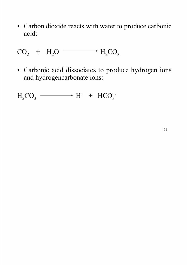
Carbon dioxide reacts with water to produce carbonic
acid:
CO2 + H2O H2CO3
Carbonic acid dissociates to produce hydrogen ionsand hydrogencarbonate ions:
H2CO3
H+ + HCO3
-
pH regulation of the tissue fluid
8/7/2019 Biology sem1- chap9
http://slidepdf.com/reader/full/biology-sem1-chap9 92/111
92
p g
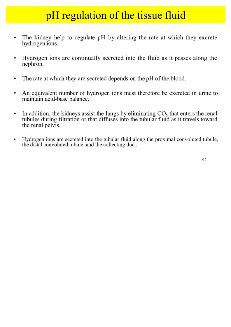
The kidney help to regulate pH by altering the rate at which they excrete
hydrogen ions.
Hydrogen ions are continually secreted into the fluid as it passes along thenephron.
The rate at which they are secreted depends on the pH of the blood.
An equivalent number of hydrogen ions must therefore be excreted in urine tomaintain acid-base balance.
In addition, the kidneys assist the lungs by eliminating CO2 that enters the renaltubules during filtration or that diffuses into the tubular fluid as it travels toward
the renal pelvis.
Hydrogen ions are secreted into the tubular fluid along the proximal convoluted tubule,the distal convoluted tubule, and the collecting duct.
8/7/2019 Biology sem1- chap9
http://slidepdf.com/reader/full/biology-sem1-chap9 93/111
93
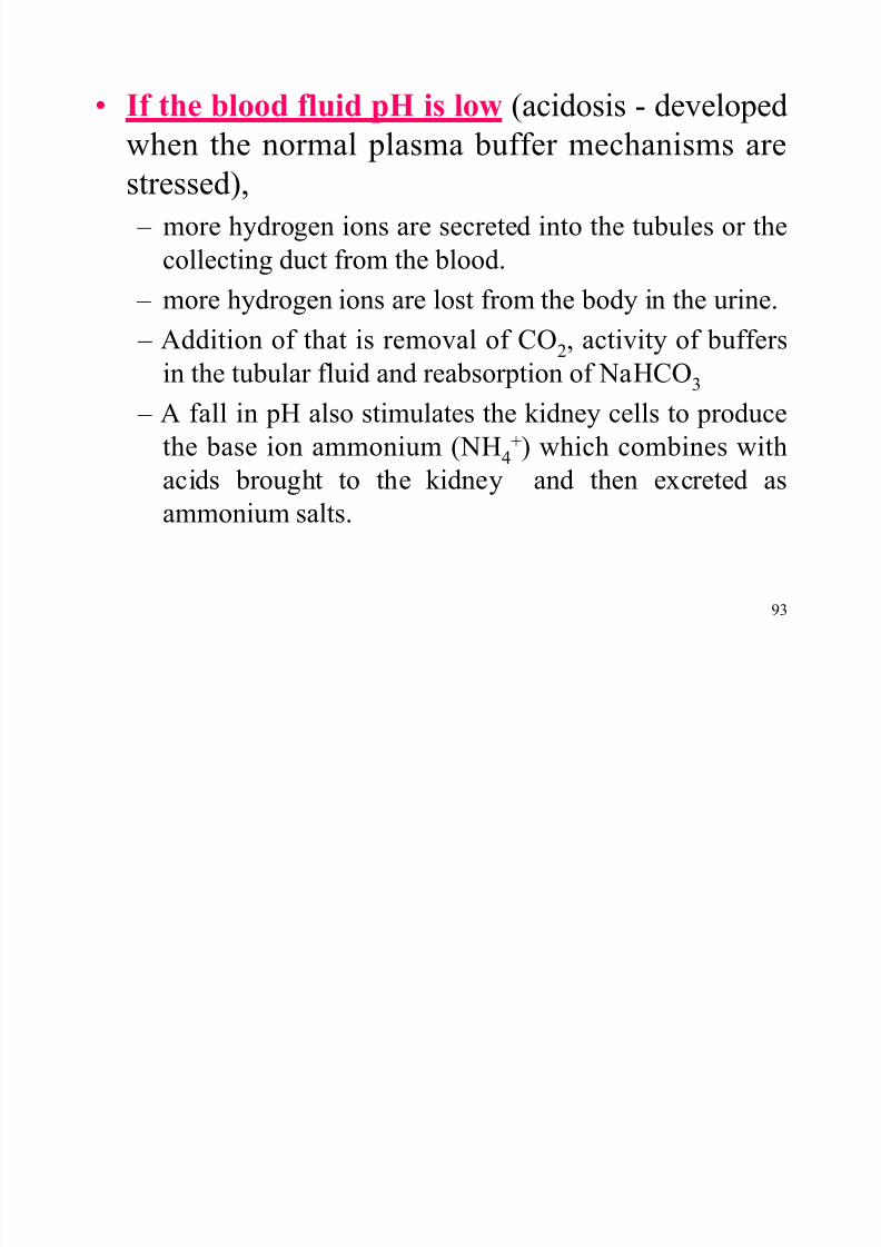
If the blood fluid pH is low (acidosis - developed
when the normal plasma buffer mechanisms are
stressed),
± more hydrogen ions are secreted into the tubules or the
collecting duct from the blood.
± more hydrogen ions are lost from the body in the urine.
± Addition of that is removal of CO2, activity of buffers
in the tubular fluid and reabsorption of NaHCO3
± A fall in pH also stimulates the kidney cells to produce
the base ion ammonium (NH4
+) which combines with
acids brought to the kidney and then excreted asammonium salts.
If blood pH is high (alkalosis)
8/7/2019 Biology sem1- chap9
http://slidepdf.com/reader/full/biology-sem1-chap9 94/111
94
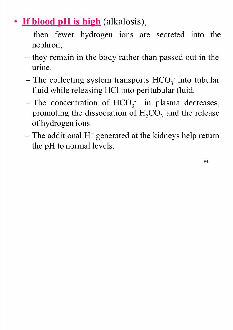
If blood pH is high (alkalosis),
± then fewer hydrogen ions are secreted into the
nephron; ± they remain in the body rather than passed out in the
urine.
± The collecting system transports HCO3
- into tubular
fluid while releasingHCl into peritubular fluid.
± The concentration of HCO3
- in plasma decreases,
promoting the dissociation of H2CO3
and the release
of hydrogen ions. ± The additional H+ generated at the kidneys help return
the pH to normal levels.
8/7/2019 Biology sem1- chap9
http://slidepdf.com/reader/full/biology-sem1-chap9 95/111
95
Water
concentration in
plant
Objectives
8/7/2019 Biology sem1- chap9
http://slidepdf.com/reader/full/biology-sem1-chap9 96/111
96
Objectives
i. The role of stomata in water loss throughtranspiration
ii. Significance of transpiration
iii. Plant adaptation to the habitats
± Xerophytes
± Hydrophytes ± Mesophytes
± Halophytes
8/7/2019 Biology sem1- chap9
http://slidepdf.com/reader/full/biology-sem1-chap9 97/111
BerpenyakitTidak berpenyakit
Pengumuman Jabatan Pertanian
Bagaimana hendak mengenali penyakit Lembu Gila.
8/7/2019 Biology sem1- chap9
http://slidepdf.com/reader/full/biology-sem1-chap9 98/111
98
The role of stomata in
water loss throughtranspiration
8/7/2019 Biology sem1- chap9
http://slidepdf.com/reader/full/biology-sem1-chap9 99/111
99
The role of stomata in water loss through
transpirationSignificance of transpiration
Loss of water from plants¶surface is called transpiration
Water normally leaves the plant as water vapour.
The change from the liquid state to the vapour state
requires the addition of energy which is provided by thesun, and it is this energy that maintains the flow of water throughout the entire plant.
T i i f h b i f
8/7/2019 Biology sem1- chap9
http://slidepdf.com/reader/full/biology-sem1-chap9 100/111
100
Transpiration occurs from the stomata by evaporation of water from cells and diffusion of the water vapour throughstomata, the pores found in the epidermis of leaves andgreen stems.
About 90% of the water is lost through transpiration.
Water in the plant is in direct contact with water in soil andwith water vapour in the air around the plant.
Water movement from higher water potential to the lower water potential is called potential gradient.
The gradient is maintained by solar energy and evaporationof water from the surface of the plant (transpiration)
Water is essential for plant metabolism, but is continuously beingl t t th t h th h th t t
8/7/2019 Biology sem1- chap9
http://slidepdf.com/reader/full/biology-sem1-chap9 101/111
101
lost to the atmosphere through the stomata.
Photosynthesis requires a supply of CO2
entering the stomatafrom the atmosphere.
Therefore, plants need to minimize the loss of water to theatmosphere and the need to allow the diffusion of CO2.
Closing the stomata can control water loss.
However, the opening of stomata at times helps CO2 to enter anddissolves in the water on the walls of the intercellular spaces
below the stomata, before entering the plant¶s cells.
A plant must responds both to the need to conserve water and therequirement of CO2
8/7/2019 Biology sem1- chap9
http://slidepdf.com/reader/full/biology-sem1-chap9 102/111
102
Plant adaptation to the habitats
Xerophytes
Hydrophytes
Mesophytes
Halophytes
The rate of transpiration depends on whether
conditions like humidity and the time of the day.
8/7/2019 Biology sem1- chap9
http://slidepdf.com/reader/full/biology-sem1-chap9 103/111
103
Xerophytes
Plants which grow in dry habitats and subjected todrought.
Example : cactus
Adaptations to reduce water loss
It has a very long, shallow, spread-out root system.
It has a swollen , succulent stems or leaves.
It has specialized leaves that may be hairy, rolled or reduced to spikes or reduced leaf size.
It has a round shape, giving it a low surface area tovolume ratio.
Adaptations to reduce water loss
8/7/2019 Biology sem1- chap9
http://slidepdf.com/reader/full/biology-sem1-chap9 104/111
104
It has a very long, shallow, spread-out root system.
It has a swollen , succulent stems or leaves.
It has specialized leaves that may be hairy, rolled or reduced to spikes or reduced leaf size.
It has a round shape, giving it a low surface area to volume ratio.
The stomata are sunken into pits and surrounded by hairs.
It has hairs over its surface which trap moisture.
It has thick layers of epidermis and heavily waxed on the cuticle of the leaves.
It has crassulacean acid metabolism (CAM) mechanism of photosynthesis; their stomatastay closed during the heat of the day and open during the cooler, more humid night.
Some spesies survive in the seed or spore stage and germinate, grow, flower and seed in ashort time following rainfall.
8/7/2019 Biology sem1- chap9
http://slidepdf.com/reader/full/biology-sem1-chap9 105/111
105
Hydrophytes
plant that lives either in very wet soil or
completely or partially submerged in
water Examples : pondweed
( El odea sp), waterlily ( Nymphaea sp).
Adaptation to survive in very wet condition;
8/7/2019 Biology sem1- chap9
http://slidepdf.com/reader/full/biology-sem1-chap9 106/111
106
Adaptation to survive in very wet condition;
It has absence or reduction of a root systems
It has specialized leaves (ribbonlike leaves) that may be either floatingor finely divided, with little or no cuticle
It has a few xylem tissues
It has many air holes/air chambers in the stem (for O2 and CO2storage which supply oxygen to the roots and enable them to float)
Epidermis :
to absorb nutrients from the water, not for protection (absorpingnutrients and water through their leaves)
contained chlorophyll
No stoma ± if there is, it will be at the upper part of a leaf
8/7/2019 Biology sem1- chap9
http://slidepdf.com/reader/full/biology-sem1-chap9 107/111
107
Mesophytes
Plants growing under conditions in which
there is normally an adequate water supply
The majority of angiosperm plant spesies
are mesophytes, and they faced with the
problem of water loss by evaporation fromall aerial parts
8/7/2019 Biology sem1- chap9
http://slidepdf.com/reader/full/biology-sem1-chap9 108/111
108
Adaptation to reduce water loss
Presence of cuticleProtected stomata whose diameters can be
regulated
Variable leaf shape
Abscission (leaf fall)
Ecological distribution based upon tolerance to
dehydration
It has stoma closed when it is very hot (daytime ±afternoon)
8/7/2019 Biology sem1- chap9
http://slidepdf.com/reader/full/biology-sem1-chap9 109/111
109
Halophytes
Plants which live in an environment where there is plenty of water,but they have difficulty obtaining it because it is salty
Conditions like this are found in estuaries and salt marshes
Plants adapted to live in salty conditions
Salinity changes according to environment
Examples : S partina sp(cord grass) and mangroves
Adaptations to water conservation :
8/7/2019 Biology sem1- chap9
http://slidepdf.com/reader/full/biology-sem1-chap9 110/111
110
It has root cells with very low water potentials (high transpiration ability ; thereforecell water potential are lower than habitat water potential).
It has hydatode at the side of the leaves or special salt glands at the margins to excreteexcessive salt by active transport onto the leaf surface.
It has roots submerged in salt water/ knee root ± above the surface of the salty muds(mangrove)
It has an extensive systems of rhizomes for propagation.
It has adventititous root for anchorage and uptake of water as well as ions.
It has root systems that are able to tolerate high salinities. Many spesies haveextensive roots which are able to store water when it is freely available.
It is capable of storing water in its succulent tissues.
It has respiratory roots/pneumatophores which are dotted with lenticels, (small vent-like openings) that take in air and channel it to the parts of the root that are buried beneath the mud (mangrove).
8/7/2019 Biology sem1- chap9
http://slidepdf.com/reader/full/biology-sem1-chap9 111/111
That all for thischapter