BIOLOGY AND IMMATURE STAGES OF THE BROMELIAD BASE …
6
Vol. 4 No. 2 1993 ANGULO and OLIVARES: Castnia psittacus Biology in Chile 133 TROPICAL LEPIDOPTERA, 4(2): 133-138 BIOLOGY AND IMMATURE STAGES OF THE BROMELIAD BASE BORER, CASTNIA PSITTACUS, IN CHILE (LEPIDOPTERA: CASTNIIDAE) ANDRES O. ANGULO ANDTANIA S. OLIVARES Departamento de Zoologia, Universidad de Concepcion, Casilla 2407-10, Concepcion, Chile ABSTRACT.- The biology and immature stages and genitalia of the bromeliad base borer, Castnia psittacus (Molina), are described. An account of other hostplant is given. KEY WORDS: Bromeliaceae. chaetotaxy. Chile, distribution, hostplants, larvae, morphology, Neotropical, pupae, South America. This species was first described as Papilio psittacus Molina (1788). It was inadvertantly redescribed as Castnia eudesmia Blanchard (1852), this last name now a junior synonym. Some reports about the general natural history of this species have been made by Gazulla and Ruiz (1928) and by Reed (1935). IMMATURES LARVAL MORPHOLOGY: Figs. 2, 3, 13-19 illustrate details of the larval morphology of Castnia psittacus (Molina). Last instar larvae are ca. 8.0-9.Ocm in length. Description. HEAD: (Fig. 16) rounded head capsule light yellow in color, with blackish area on the ocular and median top areas, with secondary setae present; frons triangular, half length of large epicranial suture; stemmata in half circle of 6 (Fig. 18): Mandibles (Fig. 19) with 3 major teeth; lateral margin with 2 setae, the basal is twice longer than the other. Spinneret (Fig. 17) short and wide with round tip. BODY: with scoli and verrucae present on all segments (Fig. 2-3), scolus yellow-brownish in color, specially those of dorsum. The scoli are situated in lateral and subventral region, verrucae are in dorsal and subdorsal area; in the prothorax all are scoli, and are projected forward and to the ventrad same to the other scoli (Figs. 2 and 3). Dorsum of meso and metathorax with large cervical shield, heavy black in color and velvet interrupted at the middle line. Anal shield (Fig. 14 ) concolor with cervical shields. Crochets (Fig. 15) are uniserial and biordinal, with ca. 80-100 in each prolegs. PUPAL MORPHOLOGY: Obtecta (Figs. 4, 5, 8-12) pupal case is made of debris of the hostplant (Fig. 6-7) that is fastened with larval silk: this pupal case is inside of hostplant. 40-50mm in length and 10- 15mm in maximun width. Tip of pterotecae reach to the posterior border of the third or fourth abdominal segment (Fig. 9); proboscis is more longer than pterotecae; tip of the metathoracic podotecae is situated posteriorly to the tip of the proboscis. Tip of ceratotecae and mesothor- acic podotecae to the level of posterior border of metanotum. Dorsal view: mesonotum 3 time more longer than pronotum; metanotum half in length than pronotum. Each abdominal segment has in its anterior third an irregularly serrate border (Fig. 10), this serrate border is from one spiraculus to another one; in the posterior third of abdominal segment there is a little serrated border similar to the anterior. There is no clear cremaster, only vestigial anal prolegs projected as 2 short spines. ADULTS As seen in Fig. 1, the wings are very colorful, with white. black, reddish, blue, and other hues. The male genitalia are shown in Fig. 21-24. The female genitalia are shown in Fig. 20. MATERIAL EXAMINATED (33 exs.): 2 exs. El Peumo, Santiago; 1 ex. 21 Dec 1949; 1 ex. Pelluhue, 13 Feb 1951; 2 exs. Dichato, 17 Jan 1950; 1 ex. Dichato, 15 Jan 1950; 1 ex. Dichato, 15 Jan 1952; 1 ex. Dichato, 5 Jan 1936; 1 ex. Concepcion, 15 Nov 56, coll. Alister, 1 ex. colleccion Wagenkneght, 1973; 1 ex. Concepcion, Desembocadura, 10 Jan 60, E. Pino coll.; 4 exs. Concepcion, D. Bio Bio, 17 Jan 1960 E. Pino coll.; 3 exs. Chiguayante, 25 Dec 1955, Silva coll.; 1 ex. Tumbes, 24 Oct 54; 1 ex. Coelemu, 5 Mar 57, Hillenns coll.; 2 exs. Recinto, 14 Jan 1950; 3 exs. Recinto, Las Trancas, Jan 1970, Ocares coll.; 1 ex. Las Trancas, 5 Jan 1975, Artigas coll. nacidas en lab.; 2 exs. Recinto, Las Trancas, Jan 1970, Ocares coll.: 1 ex. Llico, Curico, 12 Jan 1936; 1 ex. Llico, Curico. Dec 1940; 2 exs. Recinto, Las Trancas, Feb 1970, Ocares coll. REMARKS The hostplant of this species is Puya chilensis (Bromeliaceae), the larva feeds as a borer and makes its pupal case with some vegetal debris, with silk near the exterior, and at the base of the hostplant; when it emerges, it is helped by its tight exit hole and its abdominal serrated border. The time of flight is October, December, January, February and March, on the coast in central and south Chile. ACKNOWLEDGEMENTS We give our thanks to Prof. Peter D. Lewis, for the larval material. Additionally, we give thanks to Project D.I. 91.38.04-6 from Direction of Investigation of the University of Concepcion, Concepcion, Chile.
Transcript of BIOLOGY AND IMMATURE STAGES OF THE BROMELIAD BASE …

Vol. 4 No. 2 1993 ANGULO and OLIVARES: Castnia psittacus Biology in Chile 133
TROPICAL LEPIDOPTERA, 4(2): 133-138
BIOLOGY AND IMMATURE STAGESOF THE BROMELIAD BASE BORER,
CASTNIA PSITTACUS, IN CHILE(LEPIDOPTERA: CASTNIIDAE)
ANDRES O. ANGULO AND TANIA S. OLIVARES
Departamento de Zoologia, Universidad de Concepcion, Casilla 2407-10, Concepcion, Chile
ABSTRACT.- The biology and immature stages and genitalia of the bromeliad base borer, Castnia psittacus (Molina), are described. An account ofother hostplant is given.
KEY WORDS: Bromeliaceae. chaetotaxy. Chile, distribution, hostplants, larvae, morphology, Neotropical, pupae, South America.
This species was first described as Papilio psittacus Molina(1788). It was inadvertantly redescribed as Castnia eudesmiaBlanchard (1852), this last name now a junior synonym. Somereports about the general natural history of this species have beenmade by Gazulla and Ruiz (1928) and by Reed (1935).
IMMATURES
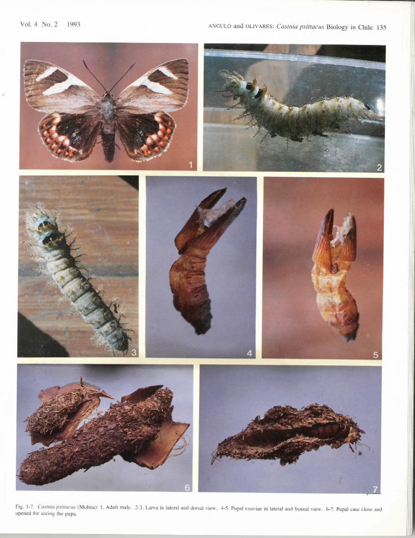
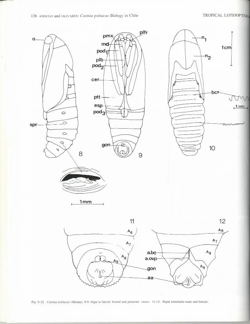
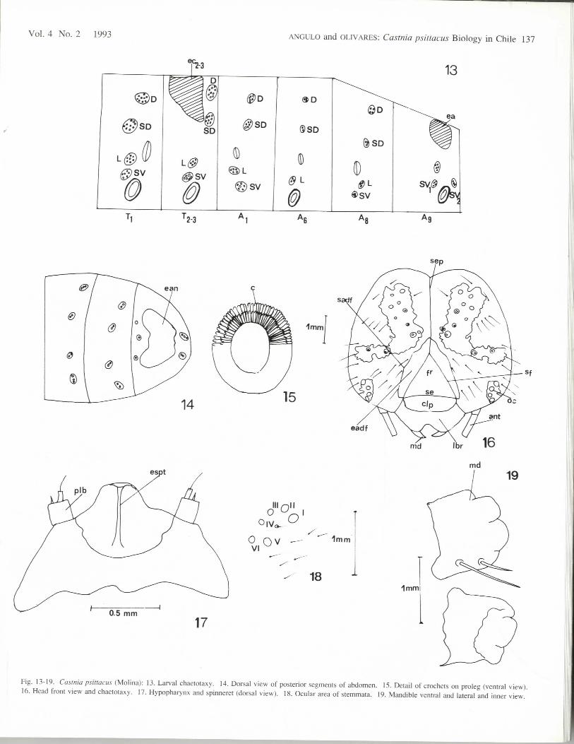
LARVAL MORPHOLOGY: Figs. 2, 3, 13-19 illustrate details of thelarval morphology of Castnia psittacus (Molina). Last instar larvae areca. 8.0-9.Ocm in length.Description. HEAD: (Fig. 16) rounded head capsule light yellow incolor, with blackish area on the ocular and median top areas, withsecondary setae present; frons triangular, half length of large epicranialsuture; stemmata in half circle of 6 (Fig. 18): Mandibles (Fig. 19) with3 major teeth; lateral margin with 2 setae, the basal is twice longer thanthe other. Spinneret (Fig. 17) short and wide with round tip.BODY: with scoli and verrucae present on all segments (Fig. 2-3), scolusyellow-brownish in color, specially those of dorsum. The scoli aresituated in lateral and subventral region, verrucae are in dorsal andsubdorsal area; in the prothorax all are scoli, and are projected forwardand to the ventrad same to the other scoli (Figs. 2 and 3). Dorsum ofmeso and metathorax with large cervical shield, heavy black in color andvelvet interrupted at the middle line. Anal shield (Fig. 14 ) concolor withcervical shields. Crochets (Fig. 15) are uniserial and biordinal, with ca.80-100 in each prolegs.PUPAL MORPHOLOGY: Obtecta (Figs. 4, 5, 8-12) pupal case ismade of debris of the hostplant (Fig. 6-7) that is fastened with larvalsilk: this pupal case is inside of hostplant. 40-50mm in length and 10-15mm in maximun width. Tip of pterotecae reach to the posterior borderof the third or fourth abdominal segment (Fig. 9); proboscis is morelonger than pterotecae; tip of the metathoracic podotecae is situatedposteriorly to the tip of the proboscis. Tip of ceratotecae and mesothor-acic podotecae to the level of posterior border of metanotum. Dorsalview: mesonotum 3 time more longer than pronotum; metanotum half inlength than pronotum. Each abdominal segment has in its anterior thirdan irregularly serrate border (Fig. 10), this serrate border is from onespiraculus to another one; in the posterior third of abdominal segment
there is a little serrated border similar to the anterior. There is no clearcremaster, only vestigial anal prolegs projected as 2 short spines.
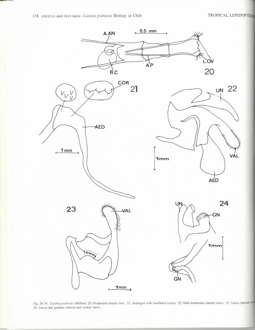
ADULTSAs seen in Fig. 1, the wings are very colorful, with white.
black, reddish, blue, and other hues. The male genitalia areshown in Fig. 21-24. The female genitalia are shown in Fig. 20.MATERIAL EXAMINATED (33 exs.): 2 exs. El Peumo, Santiago; 1ex. 21 Dec 1949; 1 ex. Pelluhue, 13 Feb 1951; 2 exs. Dichato, 17 Jan1950; 1 ex. Dichato, 15 Jan 1950; 1 ex. Dichato, 15 Jan 1952; 1 ex.Dichato, 5 Jan 1936; 1 ex. Concepcion, 15 Nov 56, coll. Alister, 1 ex.colleccion Wagenkneght, 1973; 1 ex. Concepcion, Desembocadura, 10Jan 60, E. Pino coll.; 4 exs. Concepcion, D. Bio Bio, 17 Jan 1960 E.Pino coll.; 3 exs. Chiguayante, 25 Dec 1955, Silva coll.; 1 ex. Tumbes,24 Oct 54; 1 ex. Coelemu, 5 Mar 57, Hillenns coll.; 2 exs. Recinto, 14Jan 1950; 3 exs. Recinto, Las Trancas, Jan 1970, Ocares coll.; 1 ex.Las Trancas, 5 Jan 1975, Artigas coll. nacidas en lab.; 2 exs. Recinto,Las Trancas, Jan 1970, Ocares coll.: 1 ex. Llico, Curico, 12 Jan 1936;1 ex. Llico, Curico. Dec 1940; 2 exs. Recinto, Las Trancas, Feb 1970,Ocares coll.
REMARKS
The hostplant of this species is Puya chilensis (Bromeliaceae),the larva feeds as a borer and makes its pupal case with somevegetal debris, with silk near the exterior, and at the base of thehostplant; when it emerges, it is helped by its tight exit hole andits abdominal serrated border.
The time of flight is October, December, January, February andMarch, on the coast in central and south Chile.
ACKNOWLEDGEMENTS
We give our thanks to Prof. Peter D. Lewis, for the larvalmaterial. Additionally, we give thanks to Project D.I. 91.38.04-6from Direction of Investigation of the University of Concepcion,Concepcion, Chile.
134 ANGULO and OLIVARES: Castnia psittacus Biology in Chile
REFERENCES
Blanchard, E.1852. Insectos. Lepidopteros. In Gay, C., Historic/fisica y politico de
Chile. 7:46-47. Santiago.Gazulla, P., and F. Ruiz P.
1928. Los insectos de la Hacienda de "Las Mercedes". Rev. Chil.Hist. Nat. (Santiago), 32:290-291.
Costa Lima, A. da1945. Insetos do Brasil. 59 Tomo. Capitulo XXVIII. Lepidopteros.
1s Pane, 152-159.Molina, G. I.
1788. Compendia de la historia geogrdfica. natural y civil del reynode Chile. Traduccion al espanol por Domingo Joseph. Madrid.Libra quarto: gusanos, insectos, reptiles, peces, pdxaros yquadrupedos de Chile, 213-378. Catalogo Primero nuevasespecies descritas.
Reed, E. R.1935. La Castnia eudesmia, Gray [error for Gay]. Rev. Chile. Hist.
Nat. (Santiago), 39:267-271.Tindale, N. B.
1928. Preliminary note on the life history of Synemon (Lepidoptera,fam. Castniidae). Rec. S. Aust. Mm. (Adelaide), 4:143-144.
TROPICAL LEPIDOPTERj
ABBREVIATIONS
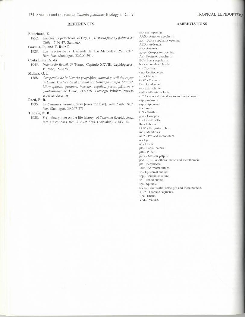
aa.- anal opening.AAN.- Anterior apophysisabc.- Bursa copulatrix opening.AED.- Aedeagus.ant.- Antenna.aovp.- Ovopositor opening.AP.- Posterior apophysis.BC.- Bursa copulatrix.bcr.- crennulated border.c.- Crochets.cer.- Ceratothecae.clp.- Clypeus.COR.- Cornutus.D.- Dorsal setae.ea.- anal sclerite.eadf.- adfrontal sclerite.ec2,3.- cervical shield meso and metathoracic.esp. proboscis.espt.- Spinneret.fr.- Frons.GN.- Gnathus.gon.- Gonopore.L.- Lateral setae.Ibr.- Labrum.LOV.- Ovopistor lobes.md.- Mandibles.nl,2.- Pro and mesonotum.o.- Eye.oc.- Ocelli.plb.- Labial palpus.plfr.- Pilifer.pmx.- Maxilar palpus.podl.2,3.- Podothecae meso and metathoracic.ptt- Pterothecae.sadf.- Adfrontal suture.se.- Epistomal suture.sep.- Epicranial suture.sf.- Frontal suture.spr.- Spiracle.SV1,2.- Subventral setae pro and mesothoracic.Tl-9.- Thoracic segments.UN.- Uncus.VAL.- Valvae.
Vol. 4 No. 2 1993 ANGULO and OLIVARES: Castnia psittacus Biology in Chile 135
Fig. 1-7. Castnia psittacus (Molina): 1. Adult male. 2-3. Larva in lateral and dorsal view. 4-5. Pupal exuviae in lateral and frontal view. 6-7. Pupal case close andopened for seeing the pupa.
I
136 ANGULO and OLIVARES: Castnia psittacus Biology in Chile TROPICAL LEPIDOPTERji
spr
,—1mm ,
Fig. 8-12. Castnia psittacus (Molina): 8-9. Pupa in lateral, frontal and posterior views. 11-12. Pupal terminalia male and female.
Vol. 4 No. 2 1993 ANGULO and OLIVARES: Castnia psittacus Biology in Chile 137
©SV
®SD
®SV
13
ea
T2-3 A1
ean
OVI
1mm
n"
sadf
1mm
Fig. 13-19. Castnia psittacus (Molina): 13. Larval chaetotaxy. 14. Dorsal view of posterior segments of abdomen. 15. Detail of crochets on proleg (ventral view).16. Head front view and chaetotaxy. 17. Hypopharynx and spinneret (dorsal view). 18. Ocular area of stemmata. 19. Mandible ventral and lateral and inner view.
I
138 ANGULO and OLIVARES: Castnia psittacus Biology in Chile TROPICAL LEPIDOPTEW
A.AN , 0.5 mm
22
Fig. 20-24. Castnia psittacus (Molina): 20. Ovopositor dorsal view. 21. Aedeagus with insuflated vesica. 22. Male terminalia (lateral view). 23. Valva (lateral viw24. Uncus and gnathos (lateral and ventral view).