Biological Control of Lettuce Drop and Host Plant Colonization by ...
12
ORIGINAL RESEARCH published: 20 May 2016 doi: 10.3389/fmicb.2016.00714 Frontiers in Microbiology | www.frontiersin.org 1 May 2016 | Volume 7 | Article 714 Edited by: Michael Thomas-Poulsen, University of Copenhagen, Denmark Reviewed by: Akifumi Sugiyama, Kyoto University, Japan Mika Tapio Tarkka, Helmholtz Centre for Environmental Research - UFZ, Germany Alessio Mengoni, Università degli Studi di Firenze, Italy *Correspondence: Paolo Cortesi [email protected] † These authors have contributed equally to this work. Specialty section: This article was submitted to Microbial Symbioses, a section of the journal Frontiers in Microbiology Received: 08 March 2016 Accepted: 29 April 2016 Published: 20 May 2016 Citation: Chen X, Pizzatti C, Bonaldi M, Saracchi M, Erlacher A, Kunova A, Berg G and Cortesi P (2016) Biological Control of Lettuce Drop and Host Plant Colonization by Rhizospheric and Endophytic Streptomycetes. Front. Microbiol. 7:714. doi: 10.3389/fmicb.2016.00714 Biological Control of Lettuce Drop and Host Plant Colonization by Rhizospheric and Endophytic Streptomycetes Xiaoyulong Chen 1† , Cristina Pizzatti 1† , Maria Bonaldi 1 , Marco Saracchi 1 , Armin Erlacher 2 , Andrea Kunova 1 , Gabriele Berg 2 and Paolo Cortesi 1 * 1 Department of Food, Environmental and Nutritional Sciences, University of Milan, Milan, Italy, 2 Institute of Environmental Biotechnology, Graz University of Technology, Graz, Austria Lettuce drop, caused by the soil borne pathogen Sclerotinia sclerotiorum, is one of the most common and serious diseases of lettuce worldwide. Increased concerns about the side effects of chemical pesticides have resulted in greater interest in developing biocontrol strategies against S. sclerotiorum. However, relatively little is known about the mechanisms of Streptomyces spp. as biological control agents against S. sclerotiorum on lettuce. Two Streptomyces isolates, S. exfoliatus FT05W and S. cyaneus ZEA17I, inhibit mycelial growth of Sclerotinia sclerotiorum by more than 75% in vitro. We evaluated their biocontrol activity against S. sclerotiorum in vivo, and compared them to Streptomyces lydicus WYEC 108, isolated from Actinovate ® . When Streptomyces spp. (10 6 CFU/mL) were applied to S. sclerotiorum inoculated substrate in a growth chamber 1 week prior lettuce sowing, they significantly reduced the risk of lettuce drop disease, compared to the inoculated control. Interestingly, under field conditions, S. exfoliatus FT05W and S. cyaneus ZEA17I protected lettuce from drop by 40 and 10% respectively, whereas S. lydicus WYEC 108 did not show any protection. We further labeled S. exfoliatus FT05W and S. cyaneus ZEA17I with the enhanced GFP (EGFP) marker to investigate their rhizosphere competence and ability to colonize lettuce roots using confocal laser scanning microscopy (CLSM). The abundant colonization of young lettuce seedlings by both strains demonstrated Streptomyces’ capability to interact with the host from early stages of seed germination and root development. Moreover, the two strains were detected also on 2-week-old roots, indicating their potential of long-term interactions with lettuce. Additionally, scanning electron microscopy (SEM) observations showed EGFP-S. exfoliatus FT05W endophytic colonization of lettuce root cortex tissues. Finally, we determined its viability and persistence in the rhizosphere and endorhiza up to 3 weeks by quantifying its concentration in these compartments. Based on these results we conclude that S. exfoliatus FT05W has high potential to be exploited in agriculture for managing soil borne diseases barely controlled by available plant protection products. Keywords: biocontrol, hazard ratio, lettuce, Sclerotinia sclerotiorum, Streptomyces, rhizosphere competence, endophytes
Transcript of Biological Control of Lettuce Drop and Host Plant Colonization by ...
ORIGINAL RESEARCHpublished: 20 May 2016
doi: 10.3389/fmicb.2016.00714
Frontiers in Microbiology | www.frontiersin.org 1 May 2016 | Volume 7 | Article 714
Edited by:
Michael Thomas-Poulsen,
University of Copenhagen, Denmark
Reviewed by:
Akifumi Sugiyama,
Kyoto University, Japan
Mika Tapio Tarkka,
Helmholtz Centre for Environmental
Research - UFZ, Germany
Alessio Mengoni,
Università degli Studi di Firenze, Italy
*Correspondence:
Paolo Cortesi
†These authors have contributed
equally to this work.
Specialty section:
This article was submitted to
Microbial Symbioses,
a section of the journal
Frontiers in Microbiology
Received: 08 March 2016
Accepted: 29 April 2016
Published: 20 May 2016
Citation:
Chen X, Pizzatti C, Bonaldi M,
Saracchi M, Erlacher A, Kunova A,
Berg G and Cortesi P (2016) Biological
Control of Lettuce Drop and Host
Plant Colonization by Rhizospheric
and Endophytic Streptomycetes.
Front. Microbiol. 7:714.
doi: 10.3389/fmicb.2016.00714
Biological Control of Lettuce Dropand Host Plant Colonization byRhizospheric and EndophyticStreptomycetesXiaoyulong Chen 1 †, Cristina Pizzatti 1 †, Maria Bonaldi 1, Marco Saracchi 1, Armin Erlacher 2,
Andrea Kunova 1, Gabriele Berg 2 and Paolo Cortesi 1*
1Department of Food, Environmental and Nutritional Sciences, University of Milan, Milan, Italy, 2 Institute of Environmental
Biotechnology, Graz University of Technology, Graz, Austria
Lettuce drop, caused by the soil borne pathogen Sclerotinia sclerotiorum, is one of the
most common and serious diseases of lettuce worldwide. Increased concerns about
the side effects of chemical pesticides have resulted in greater interest in developing
biocontrol strategies against S. sclerotiorum. However, relatively little is known about the
mechanisms of Streptomyces spp. as biological control agents against S. sclerotiorum
on lettuce. Two Streptomyces isolates, S. exfoliatus FT05W and S. cyaneus ZEA17I,
inhibit mycelial growth of Sclerotinia sclerotiorum by more than 75% in vitro. We
evaluated their biocontrol activity against S. sclerotiorum in vivo, and compared them
to Streptomyces lydicus WYEC 108, isolated from Actinovate®. When Streptomyces
spp. (106 CFU/mL) were applied to S. sclerotiorum inoculated substrate in a growth
chamber 1 week prior lettuce sowing, they significantly reduced the risk of lettuce drop
disease, compared to the inoculated control. Interestingly, under field conditions, S.
exfoliatus FT05W and S. cyaneus ZEA17I protected lettuce from drop by 40 and 10%
respectively, whereas S. lydicus WYEC 108 did not show any protection. We further
labeled S. exfoliatus FT05W and S. cyaneus ZEA17I with the enhanced GFP (EGFP)
marker to investigate their rhizosphere competence and ability to colonize lettuce roots
using confocal laser scanning microscopy (CLSM). The abundant colonization of young
lettuce seedlings by both strains demonstrated Streptomyces’ capability to interact with
the host from early stages of seed germination and root development. Moreover, the two
strains were detected also on 2-week-old roots, indicating their potential of long-term
interactions with lettuce. Additionally, scanning electron microscopy (SEM) observations
showed EGFP-S. exfoliatus FT05W endophytic colonization of lettuce root cortex tissues.
Finally, we determined its viability and persistence in the rhizosphere and endorhiza up to
3 weeks by quantifying its concentration in these compartments. Based on these results
we conclude that S. exfoliatus FT05W has high potential to be exploited in agriculture for
managing soil borne diseases barely controlled by available plant protection products.
Keywords: biocontrol, hazard ratio, lettuce, Sclerotinia sclerotiorum, Streptomyces, rhizosphere competence,
endophytes
Chen et al. Lettuce Biocontrol and Colonization by Streptomyces
INTRODUCTION
The world population will continue to grow until at least 2050,and possibly increase from 7 to 11 billion people (Van Den Berghand Rietveld, 2004). For this reason, food security has becomeone of the main challenges to human development, and thereforeany plant pathogen causing substantial crop yield losses needsto be minimized. Drop, caused by Sclerotinia species, is globallyone of the most destructive soil borne diseases of importanthorticultural crops. Three are the possible Sclerotinia speciesinvolved in lettuce drop, S. sclerotiorum, S. minor, and S. nivalis(Van Beneden et al., 2009). On lettuce, the pathogens can survivein the soil as sclerotia for years, or as mycelium on dead plants.Sclerotinia can infect the lettuce crown, roots, and leaves at anystage of plant development (Rabeendran et al., 2006). The hyphaearising from sclerotia penetrate lettuce directly through senescentleaves and root tissues, and can cause wilting and complete plantcollapse in less than 2 days (Subbarao, 1998). In Lombardy,northern Italy, commercial lettuce cultivation is threatened byS. sclerotiorum infections (Bonaldi et al., 2014) and differentstrategies and methods are being used to prevent and managelettuce drop epidemics. So far, fungicides have been extensivelyused, however, the adverse side effects of chemicals representa serious threat to living organisms including human and theenvironment (Kohler and Triebskorn, 2013; Lamberth et al.,2013). In addition, for many plant pathogens, fungicide resistantpopulations have made many molecules ineffective. Therefore,there is an increasing demand for alternative and sustainablemethods of disease management (Spadaro and Gullino, 2004;Ishii, 2006). An up-and-coming alternative to chemicals isthe use of biological control agents (BCAs). Coniothyrium,Trichodema, Bacillus, and Pseudomonas spp. have been usedfor the management of numerous diseases (Walsh et al., 2001;Howell, 2003; Jacobsen et al., 2004). In comparison to these well-known BCAs, there is only limited application of Streptomycesin agriculture, contrary to its exploitation in pharmaceuticalindustry.
Streptomyces are Gram-positive bacteria ubiquitously foundin soil, where they significantly contribute to the turnover oforganic matter. They are the largest genus of Streptomycetaceaefamily (order Actinomycetales), comprising more than 500species (Labeda et al., 2012). Very few species are pathogenicto human or plants. S. scabies and S. turgidiscabies cause scabdisease on tuber and taproot crops, such as potatoes, sweetpotatoes, carrots or beet (Lehtonen et al., 2004; Loria et al.,2006). On the contrary, many species produce a variety ofbioactive secondary metabolites and enzymes, which gives thempotential in biocontrol and plant growth promotion. It hasbeen hypothesized that high levels of antagonistic Streptomycesin naturally-occurring or induced suppressive soils significantlycontribute to disease suppression (Kinkel et al., 2012). Similarly,organic soil amendments resulted in shift and increase of thedensity of indigenous Streptomyces populations and led to diseasesuppression (Cohen et al., 2005; Mazzola and Zhao, 2010).The current research, however, focused mainly on evaluatingbiocontrol activity of individual antagonistic Streptomyces spp.:S. globisporus JK-1 inhibited Pyricularia oryzae, reducing thus
rice blast severity (Li et al., 2011); S. rochei ACTA1551 protectedtomato seeds from F. oxysporum infection (Kanini et al.,2013); the metabolites of S. bikiniensis HD-087 effectivelysuppressed F. oxysporum and induced resistance in cucumber(Zhao et al., 2012); three endophytic Streptomyces isolatessignificantly promoted tomato plant growth by producingauxins and siderophores (Verma et al., 2011). Until now, onlyfew commercial Streptomyces-based biocontrol products havebeen developed for the market, e.g., Mycostop R© based on S.griseoviridis strain K61, or Actinovate R© and Micro108 R© basedon S. lydicus strain WYEC 108 (Palaniyandi et al., 2013).They showed moderate protection of different plants againstvarious pathogens (Paulitz and Belanger, 2001; Zeng et al.,2012; Tian and Zheng, 2013). Although vast array of secondarymetabolites have been assumed to act in the biocontrol and plantgrowth promoting activity of streptomycetes (Trejo-Estradaet al., 1998; Prapagdee et al., 2008; Schrey and Tarkka, 2008;Tarkka andHampp, 2008), only in few cases the exact mechanismwas elucidated, e.g., disruption of geldanamycin production inrecombinant S. melanosporofaciens strain FP-60 resulted in theloss of its activity against S. scabies (Agbessi et al., 2003), orthe involvement of siderophores in rice growth promotion byStreptomyces sp. GMKU 3100 (Rungin et al., 2012). Moreover,priming by streptomycetes to activate plant defense responsesthrough induced and/or acquired systemic resistance pathwayscould be an additional mechanism of action involved in diseasesuppression (Conn et al., 2008; Lehr et al., 2008; Kurth et al., 2014;Salla et al., 2016).
Plant roots are colonized by vast amount of microbes,some of which contribute to biological control (Whipps, 2001;Hardoim et al., 2015). The complex community of microbesproduces a variety of compounds and develops interactions,including the competition between BCAs and plant pathogens(Raaijmakers et al., 2009). The rhizosphere—a layer of thesoil surrounding the root surface including rhizoplane—harborsan array of microorganisms, whose composition is influencedby root exudates (Hiltner, 1904; Lugtenberg and Kamilova,2009). Rhizosphere competence is a prerequisite for a BCA toestablish beneficial relationship with the host. In fact, somerhizobacteria successfully colonizing rhizosphere protected thehost from soil borne fungal pathogens (Kloepper et al., 2004;Haas and Defago, 2005; Weller, 2007). Nowadays, several geneticmarkers are available for the identification and quantification ofmicroorganisms in the rhizosphere as well as in the endorhiza—the plant inner root area. Among these, antibiotic resistance hasbeen used as a marker to quantify the colonization dynamicsof microbes in the plant root system (Gamalero et al., 2003;Adesina et al., 2009; Angelopoulou et al., 2014; Schreiter et al.,2014; Bonaldi et al., 2015). At the same time, fluorescent proteins,such as the green fluorescent protein (GFP), provide appropriatetool to monitor the colonization patterns of BCAs on plants.Enhanced GFP (EGFP), a modified version of GFP, has numeroussilent nucleotide substitutions to maximize its expression inmammalian cells (Haas et al., 1996), and is also suitable foruse in Streptomyces spp. because of a similar codon usage (Sunet al., 1999). GFP tagging was frequently used to determinecolonization of host by beneficial Bacillus and Pseudomonas
Frontiers in Microbiology | www.frontiersin.org 2 May 2016 | Volume 7 | Article 714
Chen et al. Lettuce Biocontrol and Colonization by Streptomyces
species (Krzyzanowska et al., 2012; Li et al., 2013; Subramanianet al., 2015). However, up to now, very few studies haveaddressed the plant colonization by EGFP-tagged Streptomycesspp. The strain EN 27 colonized the inner seed area of wheatat early stage of development (Coombs and Franco, 2003) andthe pathogenic strain S. turgidiscabies Car8 colonized several-day-old radish seedlings (Joshi et al., 2007). For BCAs, theviability and persistence in rhizosphere and endorhiza are pre-requisites for their application against soil borne pathogens. Infact, certain biocontrol rhizobacteria showed stable and long-term colonization of the root surface, as well as endophyticcolonization (Compant et al., 2005; Berg, 2009). Therefore,determining the rhizosphere competence and endophyticcolonization of the host by tagged Streptomyces will unravelpart of the mechanisms involved in Streptomyces-mediatedbiocontrol. Moreover, the evidence of disease suppressionby beneficial microbes in vivo encourages their developmentinto bio-products for large-scale applications. However, theinconsistency between the biocontrol performance of BCAs inlaboratory and in field occurred frequently and is considered oneof the restraining factors of the biocontrol products (Velivellilet al., 2014). In addition, the application timing and method,as well as the concentration of BCAs play crucial roles in theirbiocontrol efficacy in vivo (Bonaterra et al., 2003; Fravel, 2005;Fernando et al., 2007; Müller and Berg, 2008).
In our previous study, two Streptomyces strains, S. exfoliatusFT05W and S. cyaneus ZEA17I, showed high in vitro inhibitionof S. sclerotiorum (Bonaldi et al., 2015). The objective of thiswork was to evaluate their in vivo biological control activityagainst S. sclerotiorum on lettuce, assessing two different cellconcentrations and two application timings in growth chamber,and subsequently their activity in field. Their performance ingreenhouse and in field experiments was compared to S. lydicusWYEC 108, isolated from the commercial product Actinovate R©.Simultaneously, we determined the colonization patterns of theEGFP-tagged Streptomyces on lettuce rhizoplane, using confocallaser scanning microscopy (CLSM) and we performed scanningelectronmicroscopy (SEM) observations to verify the endophyticcolonization of lettuce roots by EGFP- S. exfoliatus FT05W, themost promising strain. Finally, we determined the colonizationdynamics by quantifying its concentration in lettuce rhizosphereand endorhiza at different times after lettuce inoculation.
MATERIALS AND METHODS
Sclerotinia Sclerotiorum InoculumPreparationSclerotinia sclerotiorum strain FW598 from the PlantPathology Laboratory fungi repository, Department of Food,Environmental and Nutritional Sciences (DeFENS), Universityof Milan, was grown for 3 days at 20◦C on Malt Extract Agar(MEA) medium (30 g/L Malt Extract, Difco, 15 g/L agar,Applichem). Then, ten agar-mycelium discs (6mm diameter)were taken from the edge of an actively growing colony andtransferred into a 300mL flask containing 25 g of sterilizedwheat kernels and 50mL distilled water (Budge and Whipps,
2001). The flask was incubated for 3 weeks at 20◦C and wasregularly shaken. Afterwards, the pathogen-colonized wheatkernels were blended with 100mL of sterilized water to obtainthe “S. sclerotiorum slurry”. One gram of S. sclerotiorum slurrywas diluted in an adequate volume of water to facilitate thedistribution and added to 100 g of non-sterile Irish and Balticpeat-based growing substrate (Vigorplant, Piacenza, Italy). Theinoculum density of S. sclerotiorum was estimated by platingserial dilutions on MEA medium. The plates were incubated at20◦C for 2 days, the number of colonies was counted and theinoculum density was calculated as CFU/g of slurry.
Streptomyces Biological Control of LettuceDrop in Growth Chamber ExperimentBiological activity of the two Streptomyces strains, S. exfoliatusFT05W and S. cyaneus ZEA17I against S. sclerotiorum was firstinvestigated in vivo in a growth chamber (24◦C, 55% relativehumidity and 15 h photoperiod) using plastic pots (Sterivent,Duchefa, Italy), 10 × 10 × 10 cm, filled with 200 g of inoculatedgrowing substrate as mentioned above. S. sclerotiorum inoculumwas ca. 3 × 10⁴ CFU/g of slurry. One mL of each Streptomycesstrain spore suspensions (104 CFU/mL or 106 CFU/mL) wassprayed on the growing substrate immediately after the pathogeninoculation. Lettuce seeds, Lactuca sativa var. capitata, “Reginadei ghiacci”, (Semeurop, Italy) were sterilized in 2mL of 0.7%sodium hypochlorite (NaOCl) for 5min and were rinsed threetimes with sterilized water. Thirty seeds were sown in threerows in each pot at two different times. In the experiment A,lettuce was sown on the same day of substrate inoculation withStreptomyces strains and the pathogen. In the experiment B,lettuce was sown 7 days after the inoculation of Streptomycesstrains and pathogen inoculation. Streptomyces lydicus WYEC108, isolated from commercial product Actinovate R© (NaturalIndustries, Inc. Houston), was used as the reference strain.The pot inoculated only with S. sclerotiorum was used asthe inoculated control. The pot inoculated neither with S.sclerotiorum nor Streptomyces was used as the non-inoculatedcontrol. For experiments A and B, eight trials were prepared inthree replicates: (1) non-inoculated control; (2) S. sclerotioruminoculated control; (3) S. exfoliatus FT05W-104 CFU/mL; (4)S. exfoliatus FT05W-106 CFU/mL; (5) S. cyaneus ZEA17I-104
CFU/mL; (6) S. cyaneus ZEA17I-106 CFU/mL; (7) S. lydicusWYEC 108-104 CFU/mL; (8) S. lydicusWYEC 108-106 CFU/mL.Dead plants were counted from the emergence up to 18 days forthe experiment A, and up to 25 days for the experiment B. Diseaseincidence was calculated as the percent of dead plants over theplants germinated in the non-inoculated control.
Streptomyces Biological Control of LettuceDrop in Field ExperimentField experiment was carried out in Travacò Siccomario (Pavia,Italy), characterized by loamy soil. Lettuce, Lactuca sativa var.capitata, “Regina dei ghiacci” was grown in polystyrene seedtrays (84 cells—48 cm3 each), filled with the non-sterile Irish andBaltic peat-based growing substrate described above. One seedwas sown in each cell added with 0.5mL of Streptomyces spore
Frontiers in Microbiology | www.frontiersin.org 3 May 2016 | Volume 7 | Article 714
Chen et al. Lettuce Biocontrol and Colonization by Streptomyces
suspension (104 CFU/mL) uniformly distributed on the growingsubstrate. Each tray was first covered with a thin layer of thegrowing substrate and then with coarse perlite. Two weeks later,the same amount of Streptomyces spore suspension was addedto each cell. Three weeks after sowing, each cell was inoculatedwith 1mL of S. sclerotiorum slurry (ca. 3 × 10⁴ CFU/g ofslurry) prepared as described above. One day after the pathogeninoculation, the lettuce plants were transplanted into the fieldunder plastic tunnel (width 1.2 m), at a density of 5.5 plants/m2.Five trials were prepared following a completely randomizedblock design in four replicates: (1) non-inoculated control; (2)S. sclerotiorum inoculated control; (3) S. exfoliatus FT05W; (4) S.cyaneus ZEA17I; (5) S. lydicusWYEC 108. Each trial consisted of20 plants. Dead plants were counted at 3-week intervals, fromthe day the disease symptoms appeared until the end of theexperiment. Disease incidence was calculated as the percent ofdead plants over the number of transplanted plants.
CLSM Observations of Lettuce RootColonization by EGFP-Streptomyces
StrainsA Leica TCS SPE Confocal Laser Scanning Microscope (LeicaMicrosystems, Mannheim, Germany) equipped with solid statelasers for excitation was used to unravel lettuce root colonizationpatterns by the two Streptomyces strains, EGFP- S. exfoliatusFT05W and EGFP- S. cyaneus ZEA17I. Plant colonization assayswere carried out at the Institute of Environmental Biotechnology,Graz University of Technology, Austria. The lettuce seeds weresterilized and bacterized with EGFP-tagged Streptomyces aspreviously described (Bonaldi et al., 2015). Subsequently, ninebacterized seeds were sown in three rows in a seed tray filled with640 g of amixture of autoclaved quartz sand (Scherf GmbH&Co.KG, Austria) and peat soil (“Gramoflor Profi-Substrat-TopfpikierM+Ton+Fe” GBC, Kalsdorf, Austria) in 1:3 ratio (w/w), and200mL of sterilized tap water were added. In two trays, noseeds were planted to monitor the soil moisture (≥25%) by amoisture analyzer (MB35 Halogen, Ohaus, USA). Nine surfacesterilized non-bacterized seeds were sown in seed trays preparedin the same way and were used as non-inoculated control. Aftersowing, the seed trays were incubated in a growth chamber(24◦C, 55% relative humidity and 14 h photoperiod). Two- andthree-day-old seedlings and 2-week-old plants were used toverify the ability of EGFP-Streptomyces to colonize lettuce. Ateach interval, the roots of three bacterized plants, taken froma different seed tray, and one non-bacterized plant (negativecontrol) were cleaned in sterile water and cut into 0.5 cm longsections for CLSM observation. Filter settings were adjusted toachieve the maximum signal from EGFP and low backgroundautofluorescence of the plant tissues. The EGFP was excited witha 488 nm laser beam and the detection window was optimizedfor every field of view, in order to gain a better discriminationbetween the signal and the noise. Plant tissues were excitedwith a 635 nm laser beam and the autofluorescence emitted inthe range 650–690 nm was recorded. The fluorescence signalsfrom EGFP and from plant tissues were acquired sequentially.For each field of view, maximum projections of an appropriate
number optical slices were acquired with a Z-step of 0.15–0.5µm(“confocal stacks”) and the software Imaris 7.3 (Bitplane, Zurich,Switzerland) was used for post-processing (Erlacher et al., 2015).
SEM Observations of Streptomyces
Endophytic ColonizationTo further verify the endophytic Streptomyces colonization oflettuce roots, we carried out SEM observations (Leo ElectronMicroscopy, Cambridge, UK) at DeFENS, University of Milan,Italy, using the representative strain EGFP- S. exfoliatus FT05W,whose wild-type strain showed promising biocontrol potential.Inoculated and non-inoculated control lettuce seeds were grownin sterile conditions as described for CLSM observations withminor modification. Each seed was sown individually in a closed200mL box containing 80 g of a mixture of autoclaved sandysubstrate (“Sabbia Vagliata” Gras Calce s.p.a., Italy) and peatsoil (Vigorplant, Piacenza, Italy) in 1:3 ratio (w/w), and 20mLsterilized tap water. Root samples from 1-, 2- and 3-week-oldplants were harvested from two inoculated plants and one controlplant. Root fragments, 1 cm long, were cut: in the proximity ofsoil surface, in the middle, and at the root apex. The fragmentswere rapidly frozen in liquid nitrogen, broken into pieces withthe aid of two forceps (cryo-fractured) in order to expose theinternal tissues, and prepared for SEM observations (Sardi et al.,1992; Rocchi et al., 2010). In total, 22 samples from 6 inoculatedplants and 11 samples from three control plants were observed.
Colonization Dynamics in LettuceRhizosphere and EndorhizaTo understand the competence of EGFP- S. exfoliatus FT05Wto colonize lettuce rhizosphere and endorhiza, we exploitedthe introduced apramycin resistance marker to quantify theamount (as colony forming units, CFU) in sterile conditionsas described by Bonaldi et al. (2015) for non-sterile conditions.Briefly: lettuce plants, obtained as described above for SEMobservations, were collected at 1, 2, and 3 weeks after sowing.Seedlings with the whole root system were carefully extractedfrom the growth substrate and the bulk soil was removed bygently shaking the plants. Excised roots were immersed in 50mLFalcon tubes containing 8–18mL (volume varying according toplant age) of sterilized washing solution containing 0.9% NaCl(Sigma-Aldrich, United States) and 0.02% Silwet L-77 (ChemturaManufacturing, Italy) and vortexed two-times for 15 s. Theroots were removed and kept for inner root tissue analysis. Therhizosphere suspension was filtered through a 100µm nylonmesh placed on the top of a Falcon tube, and centrifuged for60 s to remove any remaining washing solution from the nylonmesh. The rhizosphere soil retained on the nylon mesh wascollected and its dry weight was determined. The suspension wascentrifuged at 10,600 g for 10min and the pellet was resuspendedin 2.5mL of washing solution and plated in serial dilutions onWater Agar (WA) medium (15 g/L agar) added with 50 mg/Lapramycin, 50 mg/L cycloheximide, and 50 mg/L nystatin. Theplates were incubated at 24◦C for 7 days. Streptomyces colonieswere counted and the concentration was expressed as CFU/g ofrhizosphere dry weight. For inner root tissues analysis, the roots
Frontiers in Microbiology | www.frontiersin.org 4 May 2016 | Volume 7 | Article 714
Chen et al. Lettuce Biocontrol and Colonization by Streptomyces
were surface sterilized with propylene oxide for 1 h. Afterwards,they were washed in 2–3mL of washing solution, depending onplant age, and 0.5mL of the total volume of washing solution wasplated on WA medium to verify the absence of contaminants.Subsequently, the roots were finely homogenized in the washingsolution, left to macerate for 1 h and plated in serial dilutions onWAmedium. The Streptomyces concentration was determined asdescribed above and expressed as CFU/g of roots dry weight.
Statistical AnalysesAll analyses were done using R software, version R3.0.2.(R_Core_Team, 2013). The data of the in vivo biological controlexperiments, concerning the activity of Streptomyces strainsagainst S. sclerotiorum, were submitted to survival analysis bythe survival package (Therneau, 2014). First, the time-to-deathof lettuce plants untreated and treated with the streptomyceteswas computed using the Kaplan-Meier method. Then, theestimated survivor curve of each Streptomyces-inoculated groupwas compared to the inoculated control via log-rank test (P =
0.05). Finally, the effect of each strain was quantified using theCox proportional hazard model (Kleinbaum and Klein, 2012).This model computes the hazard h at time t, as follows: h (t,X) =
h0(t)e∑p
i=1 βiXi where Xi are the explanatory variables and ßi arethe coefficients for each variable included in the model. The effectof each treatment was quantified as Hazard Ratio (HR) expressed
as HR = exp[∑p
i=1 βi(X∗i −Xi)
]where X∗ is the covariate for
one group, generally the one with the larger hazard, and Xfor the group with the smaller hazard. The HR values equalto 1 were interpreted as no effect of Streptomyces-treated trialover Streptomyces non-treated control, HR > 1 means that theStreptomyces non-treated plants have a higher risk of lettucedrop and HR < 1 the opposite. The rhizosphere and endorhizacolonization dynamics data were submitted to ANOVA, followedby a Tukey post hoc test for multiple comparison (P = 0.05),using the TukeyC package (Faria et al., 2013).
RESULTS
Streptomyces Biological Control of LettuceDrop in Growth Chamber ExperimentThe germination rate of lettuce, calculated from the non-inoculated control, was 86.7%. When lettuce was sown the sameday of the pathogen and Streptomyces inoculation (experimentA), the number of dead plants was recorded from the 4th day aftersowing to the 18th day after sowing (Supplementary Table 1).The disease incidence of S. sclerotiorum inoculated controlat the end of the experiment was 85% and none of theStreptomyces strains showed significant protection against lettucedrop according to both log-rank test and Cox model analysis(Table 1).
When lettuce was sown 7 days after the pathogen andStreptomyces inoculation (experiment B), the dead plants wererecorded from the 4th day after sowing to the 25th dayafter sowing (Supplementary Table 2). Disease incidence ofthe S. sclerotiorum inoculated control was 74.4% at the endof the experiment. The Streptomyces strains showed 25.7–51.7% protection of lettuce against S. sclerotiorum, which was
statistically significant based on survival curves analyzed by log-rank test, except for S. lydicus WYEC 108 applied at the lowerdose (104 CFU/mL; P = 0.175). According to Cox regressionmodel, S. exfoliatus FT05W, at both spore concentrations,significantly reduced the risk of lettuce drop disease, comparedto the S. sclerotiorum inoculated control (HR = 2.078 and HR= 2.172, respectively). S. cyaneus ZEA17I was less effective thanS. exfoliatus FT05W at both spore concentrations (HR = 1.595and HR = 1.784, respectively). S. lydicus WYEC 108 applied at106 CFU/mL reduced the most the risk of lettuce drop (HR =
2.462), whereas when applied at 104 CFU/mL, it was ineffective,which was in accordance with log-rank test analysis (HR= 1.261,P = 0.24, Table 2).
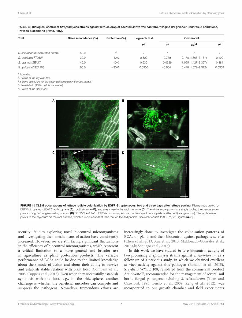
Streptomyces Biological Control of LettuceDrop in Field ExperimentUnder field conditions, the number of dead plants wasrecorded from the 10th to the 142nd day after transplanting(Supplementary Table 3). At the end of the experiment, dropincidence of the S. sclerotiorum inoculated control was 50.0%and treatments with S. exfoliatus FT05W and S. cyaneus ZEA17Ishowed respectively 40.0% and 10.0% protection against lettucedrop (Supplementary Figure 1). Survival curves of lettucetreated with S. exfoliatus FT05W and S. cyaneus ZEA17I were notsignificantly different from the S. sclerotiorum inoculated controlaccording to the log-rank test (Table 3). However, the HR usedto estimate the effect of S. exfoliatus FT05W was 2.178, thereforethe model estimated a risk of lettuce drop about two-times lowerthan that of the S. sclerotiorum inoculated control (P = 0.120).The survival curve of lettuce treated with S. lydicus WYEC 108was significantly different from the S. sclerotiorum inoculatedcontrol (P = 0.0305), but with a negative protection of 30%. TheHR of S. lydicus WYEC 108 was 0.448, confirming that plantsinoculated only with S. sclerotiorum had significantly lower riskof drop compared to those treated with the S. lydicusWYEC 108(P = 0.0309, Table 3).
CLSM Observations of Lettuce RootColonization by EGFP-Streptomyces
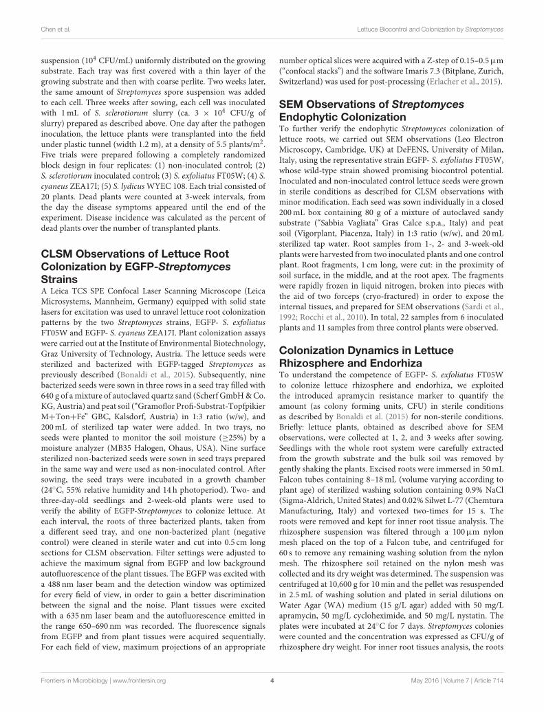
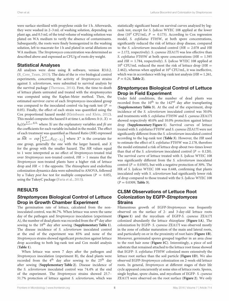
StrainsFilamentous growth of EGFP-Streptomyces was frequentlyobserved on the surface of 2- and 3-day-old lettuce roots(Figure 1) and the mycelium of EGFP-S. cyaneus ZEA17Icolonized abundantly the lettuce rhizoplane (Figure 1A). Thecolonization by EGFP- S. cyaneus ZEA17I was observed mostlyin the zone of cellular maturation of the main and lateral roots,and particularly on or in the proximity of root hairs (Figure 1B).Moreover, germinated spores grouped together in an area closeto the root hair zone (Figure 1C). Interestingly, a piece of soilsubstrate that remained attached to the lettuce root tissue showedthat EGFP- S. exfoliatus FT05W colonized more extensively thelettuce root surface than the soil particle (Figure 1D). We alsoobserved EGFP-Streptomyces colonization on 2-week-old lettuceroots. In general, Streptomyces at different stages of their lifecycle appeared concurrently at some sites of lettuce roots. Spores,single hyphae, spore chains, and mycelium of EGFP- S. cyaneusZEA17I were observed on the root surface (Figure 2). We only
Frontiers in Microbiology | www.frontiersin.org 5 May 2016 | Volume 7 | Article 714

Chen et al. Lettuce Biocontrol and Colonization by Streptomyces
TABLE 1 | Biological control of Streptomyces strains against lettuce drop, when Lactuca sativa var. capitata, “Regina dei ghiacci” was sown the same
day of S. sclerotiorum and Streptomyces co-inoculation.
Trial Disease Incidence (%) Protection (%) Log-rank test Cox model
Pb βc HRd Pe
S. sclerotiorum inoculated control 84.6 /a / / / /
S. exfoliatus FT05W (104 CFU/mL) 92.3 −9.09 0.0952 −0.273 0.761 (0.544–1.065) 0.111
S. exfoliatus FT05W (106 CFU/mL) 79.5 6.06 0.847 0.0171 1.017 (0.721–1.436) 0.923
S. cyaneus ZEA17I (104 CFU/mL) 75.6 10.6 0.336 0.142 1.152 (0.812–1.635) 0.428
S. cyaneus ZEA17I (106 CFU/mL) 83.3 1.52 0.864 0.0226 1.023 (0.727–1.439) 0.896
S. lydicus WYEC 108 (104 CFU/mL) 76.9 9.09 0.399 −0.183 0.833 (0.594–1.168) 0.290
S. lydicus WYEC 108 (106 CFU/mL) 88.5 −4.55 0.213 −0.233 0.792 (0.565–1.111) 0.177
a No value.bP-value of the log-rank test.cβ is the coefficient for the treatment covariate in the Cox model.dHazard Ratio (95% confidence interval).eP-value of the Cox model.
TABLE 2 | Biological control of Streptomyces strains against lettuce drop, when Lactuca sativa var. capitata, “Regina dei ghiacci” was sown one week
after S. sclerotiorum and Streptomyces co-inoculation.
Trial Disease Incidence (%) Protection (%) Log-rank test Cox model
Pb βc HRd Pe
S. sclerotiorum inoculated control 74.4 /a / / / /
S. exfoliatus FT05W (104 CFU/mL) 41.0 44.8 0.000942 0.731 2.078 (1.366–3.161) 0.000634
S. exfoliatus FT05W (106 CFU/mL) 42.3 43.1 0.000337 0.776 2.172 (1.427–3.307) 0.000296
S. cyaneus ZEA17I (104 CFU/mL) 53.9 27.6 0.0242 0.467 1.595 (1.072–2.372) 0.0212
S. cyaneus ZEA17I (106 CFU/mL) 53.9 27.6 0.00523 0.579 1.784 (1.200–2.653) 0.00422
S. lydicus WYEC 108 (104 CFU/mL) 55.1 25.7 0.175 0.232 1.261 (0.856–1.857) 0.24
S. lydicus WYEC 108 (106 CFU/mL) 35.9 51.7 5.08E-05 0.901 2.462 (1.589–3.812) 5.43E-05
a No value.bP-value of the log-rank test.cβ is the coefficient for the treatment covariate in the Cox model.dHazard Ratio (95% confidence interval).eP-value of the Cox model.
rarely detected colonization on the root cap and elongation zoneof the roots.
SEM Observations of Lettuce RootEndophytic Colonization by Streptomyces
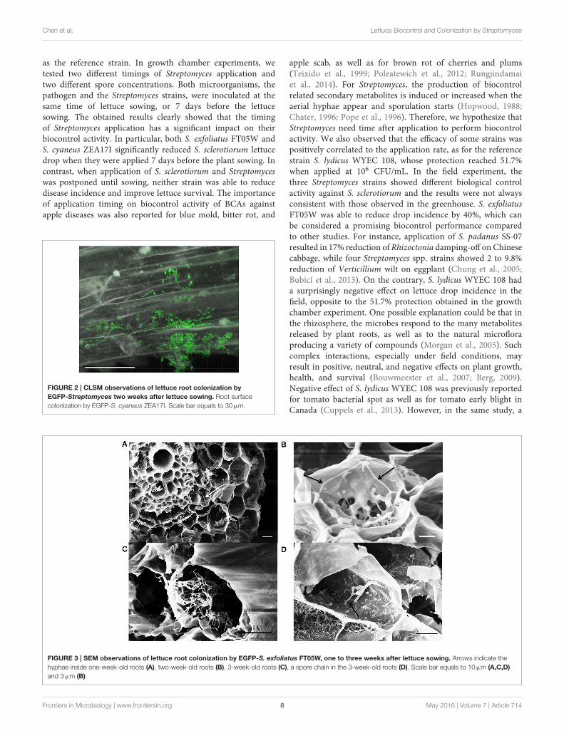
StrainsFollowing sample cryo-fracturation, 88 sections were obtainedand observed. Mycelium of EGFP- S. exfoliatus FT05W wasfrequently observed on the root surface of inoculated plants(micrograph not shown). Endophytic colonization of lettuceroots by EGFP- S. exfoliatus FT05W was observed in 99% of rootsections from all samples from 1- to 3-week-old roots. Generally,several cells were colonized in each section (Figure 3). Along theentire length of the root, both close to the collar and near theapex, single hyphae were frequently detected inside cortical cellsin 1-week-old (Figure 3A), and 2-week-old roots (Figure 3B),but not inside the vascular cylinder. In a few cases, mainly in 3-week-old roots, the hyphae grew abundantly inside cortical cellsforming a tangled structure (Figure 3C). Hyphae growing inside
cortical cells had a diameter of about 0.2µm, half the size the onesgrown on the root surface or in vitro cultures. EGFP- S. exfoliatusFT05W mainly colonized the endorhiza of lettuce as vegetativehyphae and rarely short spore chains were found (Figure 3D).
Streptomyces Colonization Dynamics inLettuce Rhizosphere and EndorhizaEGFP- S. exfoliatus FT05W showed stable concentration up tothree weeks, both in lettuce rhizosphere and endorhiza, rangingfrom 1.72× 10⁶ to 5.49× 10⁶ CFU/g rhizosphere dry weight andfrom 1.10×10⁵ to 7.36×10⁶ CFU/g root dry weight, respectively(Table 4). There were no statistically significant differences inits concentration based on plant age both in rhizosphere and inendorhiza.
DISCUSSION
Biological control strategies are gaining popularity in agricultureas a way to address some of the concerns about food
Frontiers in Microbiology | www.frontiersin.org 6 May 2016 | Volume 7 | Article 714
Chen et al. Lettuce Biocontrol and Colonization by Streptomyces
TABLE 3 | Biological control of Streptomyces strains against lettuce drop of Lactuca sativa var. capitata, “Regina dei ghiacci” under field conditions,
Travacò Siccomario (Pavia, Italy).
Trial Disease incidence (%) Protection (%) Log-rank test Cox model
Pb βc HRd Pe
S. sclerotiorum inoculated control 50.0 /a / / / /
S. exfoliatus FT05W 30.0 40.0 0.802 0.779 2.178 (1.366–3.161) 0.120
S. cyaneus ZEA17I 45.0 10.0 0.939 0.0626 1.065 (1.427–3.307) 0.884
S. lydicus WYEC 108 65.0 −30.0 0.0305 −0.804 0.448 (1.072–2.372) 0.0309
a No value.bP-value of the log-rank test.cβ is the coefficient for the treatment covariate in the Cox model.dHazard Ratio (95% confidence interval).eP-value of the Cox model.
FIGURE 1 | CLSM observations of lettuce radicle colonization by EGFP-Streptomyces, two and three days after lettuce sowing. Filamentous growth of
EGFP- S. cyaneus ZEA17I at rhizoplane (A), root hair zone (B), and area close to the root hair zone (C). The white arrow points to a single hypha, the orange arrow
points to a group of germinating spores, (D) EGFP-S. exfoliatus FT05W colonizing lettuce root tissue with a soil particle attached (orange arrow). The white arrow
points to the mycelium on the root surface, which is more abundant than that on the soil particle. Scale bar equals to 30µm, for Figures (A–D).
security. Studies exploring novel biocontrol microorganismsand investigating their mechanisms of action have consistentlyincreased. However, we are still facing significant fluctuationsin the efficiency of biocontrol microorganisms, which representa critical limitation to a more general and broader usein agriculture as plant protection products. The variableperformance of BCAs could be due to the limited knowledgeabout their mode of action and about their ability to surviveand establish stable relation with plant host (Compant et al.,2005; Cuppels et al., 2013). Even when they successfully establishsymbiosis with the host, e.g., in the rhizosphere, anotherchallenge is whether the beneficial microbes can compete andsuppress the pathogens. Nowadays, tremendous efforts are
increasingly done to investigate the colonization patterns ofBCAs on plants and their biocontrol against pathogens in vivo(Chen et al., 2013; Xue et al., 2013; Maldonado-Gonzalez et al.,2015a,b; Santiago et al., 2015).
In this work we have studied in vivo biocontrol activity oftwo promising Streptomyces strains against S. sclerotiorum as afollow up of a previous study, in which we obtained excellentin vitro activity against this pathogen (Bonaldi et al., 2015).S. lydicus WYEC 108, reisolated from the commercial productActinovate R©, recommended for the management of several soilborne fungal pathogens including S. sclerotiorum (Yuan andCrawford, 1995; Leisso et al., 2009; Zeng et al., 2012), wasincorporated to our growth chamber and field experiments
Frontiers in Microbiology | www.frontiersin.org 7 May 2016 | Volume 7 | Article 714
Chen et al. Lettuce Biocontrol and Colonization by Streptomyces
as the reference strain. In growth chamber experiments, wetested two different timings of Streptomyces application andtwo different spore concentrations. Both microorganisms, thepathogen and the Streptomyces strains, were inoculated at thesame time of lettuce sowing, or 7 days before the lettucesowing. The obtained results clearly showed that the timingof Streptomyces application has a significant impact on theirbiocontrol activity. In particular, both S. exfoliatus FT05W andS. cyaneus ZEA17I significantly reduced S. sclerotiorum lettucedrop when they were applied 7 days before the plant sowing. Incontrast, when application of S. sclerotiorum and Streptomyceswas postponed until sowing, neither strain was able to reducedisease incidence and improve lettuce survival. The importanceof application timing on biocontrol activity of BCAs againstapple diseases was also reported for blue mold, bitter rot, and
FIGURE 2 | CLSM observations of lettuce root colonization by
EGFP-Streptomyces two weeks after lettuce sowing. Root surface
colonization by EGFP-S. cyaneus ZEA17I. Scale bar equals to 30µm.
apple scab, as well as for brown rot of cherries and plums(Teixido et al., 1999; Poleatewich et al., 2012; Rungjindamaiet al., 2014). For Streptomyces, the production of biocontrolrelated secondary metabolites is induced or increased when theaerial hyphae appear and sporulation starts (Hopwood, 1988;Chater, 1996; Pope et al., 1996). Therefore, we hypothesize thatStreptomyces need time after application to perform biocontrolactivity. We also observed that the efficacy of some strains waspositively correlated to the application rate, as for the referencestrain S. lydicus WYEC 108, whose protection reached 51.7%when applied at 106 CFU/mL. In the field experiment, thethree Streptomyces strains showed different biological controlactivity against S. sclerotiorum and the results were not alwaysconsistent with those observed in the greenhouse. S. exfoliatusFT05W was able to reduce drop incidence by 40%, which canbe considered a promising biocontrol performance comparedto other studies. For instance, application of S. padanus SS-07resulted in 17% reduction of Rhizoctonia damping-off on Chinesecabbage, while four Streptomyces spp. strains showed 2 to 9.8%reduction of Verticillium wilt on eggplant (Chung et al., 2005;Bubici et al., 2013). On the contrary, S. lydicus WYEC 108 hada surprisingly negative effect on lettuce drop incidence in thefield, opposite to the 51.7% protection obtained in the growthchamber experiment. One possible explanation could be that inthe rhizosphere, the microbes respond to the many metabolitesreleased by plant roots, as well as to the natural microfloraproducing a variety of compounds (Morgan et al., 2005). Suchcomplex interactions, especially under field conditions, mayresult in positive, neutral, and negative effects on plant growth,health, and survival (Bouwmeester et al., 2007; Berg, 2009).Negative effect of S. lydicus WYEC 108 was previously reportedfor tomato bacterial spot as well as for tomato early blight inCanada (Cuppels et al., 2013). However, in the same study, a
FIGURE 3 | SEM observations of lettuce root colonization by EGFP-S. exfoliatus FT05W, one to three weeks after lettuce sowing. Arrows indicate the
hyphae inside one-week-old roots (A), two-week-old roots (B), 3-week-old roots (C), a spore chain in the 3-week-old roots (D). Scale bar equals to 10µm (A,C,D)
and 3µm (B).
Frontiers in Microbiology | www.frontiersin.org 8 May 2016 | Volume 7 | Article 714

Chen et al. Lettuce Biocontrol and Colonization by Streptomyces
TABLE 4 | Colonization dynamics of EGFP-S. exfoliatus FT05W in Lactuca
sativa var. capitata, “Regina dei ghiacci” rhizosphere and endorhiza.
EGFP-S. exfoliatus FT05W (CFU/g dry weight)
1 week 2 weeks 3 weeks
Rhizosphere 1.72× 10⁶nsa 2.45× 10⁶ns 5.49× 10⁶ns
Endorhiza 4.31× 10⁵ns 7.36× 10⁶ns 1.10× 10⁵ns
aANOVA analysis, means in a row were not significantly different (P = 0.05).
combined application of S. lydicus WYEC108 and P. fluorescensA506 resulted in a good protection against the two diseases.Similarly, S. lydicus WYEC 108 applied to control Fusariumwilt of watermelon resulted in increased disease severity inAmerican soils, whereas the combination of green manure andS. lydicus WYEC 108 mitigated the negative effect. S. lydicusinefficacy was probably due to its lack of survival on watermelonroots in those specific conditions (Himmelstein et al., 2014).Another hypothesis might be that under certain environmentalconditions S. lydicus WYEC 108 produces fungal growthpromoting secondary metabolites, which enhance the pathogengrowth and promote the infection of the host plant. Fungalgrowth promotion was shown for Streptomyces sp. strain AcH505 producing auxofuran, a molecule which improved mycelialgrowth of the ectomycorrhizal fungus, Amanita muscaria, andits interaction with spruce (Schrey et al., 2005; Riedlinger et al.,2006).
The beneficial plant-microbe interactions occurring atspecific sites usually require the microbe competence for hostcolonization (Berg et al., 2015; Hardoim et al., 2015). It hasbeen hypothesized that the Streptomyces-mediated diseasesuppression is linked to the production of active secondarymetabolites and their ability to colonize plant roots (Tokalaet al., 2002; Franco et al., 2007). In this study, we investigatedStreptomyces lettuce colonization as one of the charactersunderlying Streptomyces-mediated biocontrol. The use offluorescent proteins to study plant colonization by BCAs suchas Bacillus and Pseudomonas spp. has been widely reported(Buddrus-Schiemann et al., 2010; De-Bashan et al., 2010;Krzyzanowska et al., 2012; Sun et al., 2014). However, veryfew studies investigated Streptomyces colonization patterns onplants using fluorescent proteins in combination with CLSM.Coombs and Franco (2003) demonstrated that the EGFP-taggedendophytic Streptomyces sp. strain EN27 rapidly colonized thewheat embryo, as it was detected in developing seeds as early as24 h after inoculation, but long-term rhizosphere competenceand root colonization were not investigated. Similarly, Joshiet al. (2007) labeled a pathogenic strain of S. turgidiscabies withEGFP, and it was detected mainly on the surface of several-day-old radish seedlings, without any further monitoring. In ourstudy, both EGFP-S. exfoliatus FT05W and EGFP-S. cyaneusZEA17I were able to rapidly colonize the lettuce root system,and establish interactions with the host from early stages ofseed germination and root development. Although it is notknown if the localization of Streptomyces regulates their activityfor biological control of pathogens, it has been hypothesizedthat endophytic bacteria form more stable interactions with
plants than rhizospheric or epiphytic bacteria (Ryan et al.,2008; Compant et al., 2010; Malfanova et al., 2011). UsingCLSM, we were able to detect EGFP-Streptomyces extensivelycolonizing the rhizoplane, and the SEM analyses confirmed thepresence of EGFP-S. exfoliatus FT05W on the root surface andrevealed the endophytic colonization in the root cortex. To ourknowledge, this is the first study, which describes the observationof lettuce epiphytic and endophytic colonization by EGFP-taggedStreptomyces up to three weeks. In addition, we consistentlyrecovered high concentration of EGFP-S. exfoliatus FT05W(105-106 CFU/g dry weight) from both, lettuce rhizosphereand endorhiza, up to three weeks after seed inoculation. Thisevidence allows us to conclude that S. exfoliatus FT05W is bothrhizospheric and endophytic in lettuce roots.
The ability of microorganisms to colonize plant roots enablesthem to establish long-term beneficial interactions includingbiocontrol against plant pathogens (Adesina et al., 2009; Schreiteret al., 2014). The ability of S. exfoliatus FT05W to producechitinases, to solubilize phosphates and to synthesize IAA(Bonaldi et al., 2015) coupled with its stable rhizospherecompetence and endophytic colonization of lettuce rootsdetermined in this study, could explain its biocontrol activityagainst S. sclerotiorum. When S. exfoliatus FT05W was applied 1week before plant sowing, it showed significant protection againstlettuce drop in growth chamber, data that have been confirmedin field. Studying the colonization patterns of Streptomyces onlettuce in the presence of the pathogen will give us insightinto whether and how Streptomyces spp. compete with plantpathogens, leading to better understanding of Streptomyces-mediated biocontrol. In addition, studies evaluating S. exfoliatusFT05W activity against other soil borne fungal pathogens (e.g.,Fusarium, Pythium, Rhizoctonia, or Verticillium spp.) and itsability to establish stable interactions with other hosts are neededto make it more attractive for its development into a commercialbiocontrol product.
AUTHOR CONTRIBUTIONS
XC performed the CLSM observations, promoted the SEM andcolonization dynamics studies, and drafted the manuscript. CPevaluated the colonization dynamics data and conducted thestatistical analyses. MB performed the biocontrol experiments ingreenhouse and in field. MS performed SEM sample preparation,observations and acquired SEM pictures. AE contributed to theCLSM observations and post-processed the CLSM photos. AKparticipated to the discussions of each section of experiments,and improved the manuscript. GB hosted and supported XC inher lab to perform the CLSM observations with assistance of AE.PC designed the outline of the study and partly supported theresearch. All authors read and approved the final manuscript.
ACKNOWLEDGMENTS
The authors thank Prof. Flavia Marinelli (University of Insubria,Italy), for providing the donor strain E. coli ET12567 andthe reference strain S. coelicolor A3(2) and Prof. Mervyn Bibb
Frontiers in Microbiology | www.frontiersin.org 9 May 2016 | Volume 7 | Article 714
Chen et al. Lettuce Biocontrol and Colonization by Streptomyces
(John Innes Centre, UK), for kindly providing the plasmidpIJ8641. The authors also thank Dr. Tomislav Cernava and Mr.Tobija Glawogger, from Graz University of Technology, Austria,for their technical assistance during the acquisition of CLSMphotos, and their helpful advice and discussion regarding theexperiments done in Graz. This research was supported in part byresearch program “Dote ricerca applicata” funded by LombardyRegion and Sipcam Italia Spa.
SUPPLEMENTARY MATERIAL
The Supplementary Material for this article can be foundonline at: http://journal.frontiersin.org/article/10.3389/fmicb.2016.00714
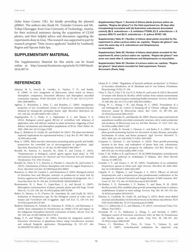
Supplementary Figure 1 | Survival of lettuce plants (Lactuca sativa var.
capitata, “Regina dei ghiacci”) in the field experiment (ca. 60 days after
transplanting) inoculated with (A) Sclerotinia sclerotiorum (inoculated
control); (B) S. sclerotiorum + S. exfoliatus FT05W; (C) S. sclerotiorum + S.
cyaneus ZEA17I; and (D) S. sclerotiorum + S. lydicus WYEC 108.
Supplementary Table S1 | Number of lettuce dead plants recorded for the
experiment A, when Lactuca sativa var. capitata, “Regina dei ghiacci” was
sown the same day of S. sclerotiorum and Streptomyces
co-inoculation.
Supplementary Table S2 | Number of lettuce dead plants recorded for the
experiment B, when Lactuca sativa var. capitata, “Regina dei ghiacci” was
sown one week after S. sclerotiorum and Streptomyces co-inoculation.
Supplementary Table S3 | Number of Lactuca sativa var. capitata, “Regina
dei ghiacci” dead plants recorded for field experiment, Travacò
Siccomario (Pavia, Italy).
REFERENCES
Adesina, M. F., Grosch, R., Lembke, A., Vatchev, T. D., and Smalla,K. (2009). In vitro antagonists of Rhizoctonia solani tested on lettuce:rhizosphere competence, biocontrol efficiency and rhizosphere microbialcommunity response. FEMS Microbiol. Ecol. 69, 62–74. doi: 10.1111/j.1574-6941.2009.00685.x
Agbessi, S., Beauséjour, J., Déry, C., and Beaulieu, C. (2003). Antagonisticproperties of two recombinant strains of Streptomyces melanosporofaciensobtained by intraspecific protoplast fusion. Appl. Microbiol. Biotechnol. 62,233–238. doi: 10.1007/s00253-003-1256-0
Angelopoulou, D. J., Naska, E. J., Paplomatas, E. J., and Tjamos, S. E.(2014). Biological control agents (BCAs) of verticillium wilt: influence ofapplication rates and delivery method on plant protection, triggering of hostdefence mechanisms and rhizosphere populations of BCAs. Plant Pathol. 63,1062–1069. doi: 10.1111/ppa.12198
Berg, G., Rybakova, D., Grube, M., and Koberl, M. (2015). The plant microbiomeexplored: implications for experimental botany. J. Exp. Bot. 67, 995–1002. doi:10.1093/jxb/erv466
Berg, G. (2009). Plant-microbe interactions promoting plant growth and health:perspectives for controlled use of microorganisms in agriculture. Appl.
Microbiol. Biotechnol. 84, 11–18. doi: 10.1007/s00253-009-2092-7Bonaldi, M., Kunova, A., Saracchi, M., Sardi, P., and Cortesi, P. (2014).
“Streptomycetes as biological control agents against basal drop,” in VIII
International Symposium on Chemical and Non-Chemical Soil and Substrate
Disinfestation, Vol. 1044 (Torino).Bonaldi, M., Chen, X. Y. L., Kunova, A., Pizzatti, C., Saracchi, M., and Cortesi, P.
(2015). Colonization of lettuce rhizosphere and roots by tagged Streptomyces.
Front. Microbiol. 6:25 doi: 10.3389/fmicb.2015.00025Bonaterra, A., Mari, M., Casalini, L., and Montesinos, E. (2003). Biological control
of Monilinia laxa and Rhizopus stolonifer in postharvest of stone fruit byPantoea agglomerans EPS125 and putative mechanisms of antagonism. Int. J.Food Microbiol. 84, 93–104. doi: 10.1016/S0168-1605(02)00403-8
Bouwmeester, H. J., Roux, C., Lopez-Raez, J. A., and Becard, G. (2007).Rhizosphere communication of plants, parasitic plants and AM fungi. TrendsPlant Sci. 12, 224–230. doi: 10.1016/j.tplants.2007.03.009
Bubici, G., Marsico, A. D., D’amico, M., Amenduni, M., and Cirulli, M. (2013).Evaluation of Streptomyces spp. for the biological control of corky root oftomato and Verticillium wilt of eggplant. Appl. Soil Ecol. 72, 128–134. doi:10.1016/j.apsoil.2013.07.001
Buddrus-Schiemann, K., Schmid, M., Schreiner, K., Welzl, G., and Hartmann, A.(2010). Root colonization by Pseudomonas sp. DSMZ 13134 and impact onthe indigenous rhizosphere bacterial community of barley. Microb. Ecol. 60,381–393. doi: 10.1007/s00248-010-9720-8
Budge, S. P., and Whipps, J. M. (2001). Potential for integrated control ofSclerotinia sclerotiorum in glasshouse lettuce using Coniothyrium minitans
and reduced fungicide application. Phytopathology 91, 221–227. doi:10.1094/PHYTO.2001.91.2.221
Chater, K. F. (1996). “Regulation of bacterial antibiotic production,” in Products
of Secondary Metabolism, ed H. Kleinkauf and H. von Dohren (Weinheim:Biotechnology VCH), 57–105.
Chen, Y., Yan, F., Chai, Y. R., Liu, H. X., Kolter, R., and Losick, R. (2013). Biocontrolof tomato wilt disease by Bacillus subtilis isolates from natural environmentsdepends on conserved genes mediating biofilm formation. Environ. Microbiol.
15, 848–864. doi: 10.1111/j.1462-2920.2012.02860.xChung, W. C., Huang, J. W., and Huang, H. C. (2005). Formulation of a
soil biofungicide for control of damping-off of Chinese cabbage (Brassicachinensis) caused by Rhizoctonia solani. Biol. Contr. 32, 287–294. doi:10.1016/j.biocontrol.2004.10.011
Cohen, M. F., Yamasaki, H., and Mazzola, M. (2005). Brassica napus seed meal soilamendment modifies microbial community structure, nitric oxide productionand incidence of Rhizoctonia root rot. Soil Biol. Biochem. 37, 1215–1227. doi:10.1016/j.soilbio.2004.11.027
Compant, S., Duffy, B., Nowak, J., Clement, C., and Barka, E. A. (2005). Use ofplant growth-promoting bacteria for biocontrol of plant diseases: principles,mechanisms of action, and future prospects. Appl. Environ. Microbiol. 71,4951–4959. doi: 10.1128/AEM.71.9.4951-4959.2005
Compant, S., Clement, C., and Sessitsch, A. (2010). Plant growth-promotingbacteria in the rhizo- and endosphere of plants: their role, colonization,mechanisms involved and prospects for utilization. Soil Biol. Biochem. 42,669–678. doi: 10.1016/j.soilbio.2009.11.024
Conn, V. M., Walker A. R., and Franco C. M. M (2008) Endophytic Actinobacteriainduce defense pathways in Arabidopsis 37 thaliana. Mol. Plant Microbe
Interact 21:208U218.Coombs, J. T., and Franco, C. M. M. (2003). Visualization of an endophytic
Streptomyces species in wheat seed. Appl. Environ. Microbiol. 69, 4260–4262.doi: 10.1128/AEM.69.7.4260-4262.2003
Cuppels, D. A., Higham, J., and Traquair, J. A. (2013). Efficacy of selectedstreptomycetes and a streptomycete plus pseudomonad combination in themanagement of selected bacterial and fungal diseases of field tomatoes. Biol.Contr. 67, 361–372. doi: 10.1016/j.biocontrol.2013.09.005
De-Bashan, L. E., Hernandez, J. P., Bashan, Y., and Maier, R. M. (2010).Bacillus pumilus ES4: candidate plant growth-promoting bacterium to enhanceestablishment of plants in mine tailings. Environ. Exp. Bot. 69, 343–352. doi:10.1016/j.envexpbot.2010.04.014
Erlacher, A., Cardinale, M., Grube, M., and Berg, G. (2015). Biotic stress shiftedstructure and abundance of Enterobacteriaceae in the lettuce microbiome. PLoSONE 10:e0118068 doi: 10.1371/journal.pone.0118068
Faria, J. C., Jelihovschi, E. G., and Allaman, I. B. (2013). Conventional Tukey Test.(Ilheus: Test.UESC).
Fernando, W. G. D., Nakkeeran, S., Zhang, Y., and Savchuk, S. (2007).Biological control of Sclerotinia sclerotiorum (Lib.) de Bary by Pseudomonas
and Bacillus species on canola petals. Crop Prot. 26, 100–107. doi:10.1016/j.cropro.2006.04.007
Franco, C., Michelsen, P., Percy, N., Conn, V., Listiana, E., andMoll, S. (2007). Actinobacterial endophytes for improved crop
Frontiers in Microbiology | www.frontiersin.org 10 May 2016 | Volume 7 | Article 714

Chen et al. Lettuce Biocontrol and Colonization by Streptomyces
performance. Australas. Plant Pathol. 36, 524–531. doi: 10.1071/AP07067
Fravel, D. R. (2005). Commercialization and implementation of biocontrol. Annu.Rev. Phytopathol. 43, 337–359. doi: 10.1146/annurev.phyto.43.032904.092924
Gamalero, E., Lingua, G., Berta, G., and Lemanceau, P. (2003). Methods forstudying root colonization by introduced beneficial bacteria. Agronomie 23,407–418. doi: 10.1051/agro:2003014
Haas, D., and Defago, G. (2005). Biological control of soil-borne pathogensby fluorescent pseudomonads. Nat. Rev. Microbiol. 3, 307–319. doi:10.1038/nrmicro1129
Haas, J., Park, E.-C., and Seed, B. (1996). Codon usage limitation in the expressionof HIV-1 envelope glycoprotein. Curr. Biol. 6, 315–324. doi: 10.1016/S0960-9822(02)00482-7
Hardoim, P. R., Van Overbeek, L. S., Berg, G., Pirttila, A. M., Compant, S.,and Campisano, A. (2015). The hidden world within plants: ecological andevolutionary considerations for defining functioning of microbial endophytes.Microbiol. Mol. Biol. Rev. 79, 293–320. doi: 10.1128/MMBR.00050-14
Hiltner (1904). Über neuere Erfahrungen und Probleme auf dem Gebiete derBodenbakteriologie unter besonderer Berücksic. Arb. Dtsch. Landwirt. Ges. 98,59–78.
Himmelstein, J. C., Maul, J. E., and Everts, K. L. (2014). Impact of five cover cropgreen manures and Actinovate on Fusarium wilt of watermelon. Plant Dis. 98,965–972. doi: 10.1094/PDIS-06-13-0585-RE
Hopwood, D. A. (1988). The Leeuwenhoek Lecture, 1987. towards anunderstanding of gene switching in Streptomyces, the basis of sporulationand antibiotic production. Proc, R. Soc. B Biol. Sci. 235, 121–138. doi:10.1098/rspb.1988.0067
Howell, C. R. (2003). Mechanisms employed by Trichoderma species in thebiological control of plant diseases: the history and evolution of currentconcepts. Plant Dis. 87, 4–10. doi: 10.1094/PDIS.2003.87.1.4
Ishii, H. (2006). Impact of fungicide resistance in plant pathogens on crop diseasecontrol and agricultural environment. Jarq Jpn. Agric. Res. Q. 40, 205–211. doi:10.6090/jarq.40.205
Jacobsen, B. J., Zidack, N. K., and Larson, B. J. (2004). The role of Bacillus-basedbiological control agents in integrated pestmanagement systems: plant diseases.Phytopathology 94, 1272–1275. doi: 10.1094/PHYTO.2004.94.11.1272
Joshi, M., Rong, X., Moll, S., Kers, J., Franco, C., and Loria, R. (2007). Streptomyces
turgidiscabies secretes a novel virulence protein, Nec1, which facilitatesinfection. Mol. Plant Microbe Interact. 20, 599–608. doi: 10.1094/MPMI-20-6-0599
Kanini, G. S., Katsifas, E. A., Savvides, A. L., and Karagouni, A. D. (2013).Streptomyces rochei ACTA1551, an indigenous Greek isolate studied as apotential biocontrol agent against Fusarium oxysporum f.sp lycopersici. Biomed
Res. Int. 2013:387230. doi: 10.1155/2013/387230Kinkel, L. L., Schlatter, D. C., Bakker, M. G., and Arenz, B. E. (2012). Streptomyces
competition and co-evolution in relation to plant disease suppression. Res.Microbiol. 163, 490–499. doi: 10.1016/j.resmic.2012.07.005
Kleinbaum, D. G., and Klein, M. (eds.). (2012). “Evaluating the proportionalhazards assumption,” in Survival Analysis: A Self-Learning Text, 3rd Edn
(New York, NY: Springer), 161–200. doi: 10.1007/978-1-4419-6646-9Kloepper, J. W., Ryu, C. M., and Zhang, S. A. (2004). Induced systemic resistance
and promotion of plant growth by Bacillus spp. Phytopathology 94, 1259–1266.doi: 10.1094/PHYTO.2004.94.11.1259
Kohler, H. R., and Triebskorn, R. (2013). Wildlife ecotoxicology of pesticides: canwe track effects to the population level and beyond? Science 341, 759–765. doi:10.1126/science.1237591
Krzyzanowska, D., Obuchowski, M., Bikowski, M., Rychlowski, M., and Jafra, S.(2012). Colonization of potato rhizosphere by GFP-Tagged Bacillus subtilis
MB73/2, Pseudomonas sp. P482 and Ochrobactrum sp. A44 shown on largesections of roots using enrichment sample preparation and confocal laserscanning microscopy. Sensors 12, 17608–17619. doi: 10.3390/s121217608
Kurth, F., Mailänder, S., Bönn, M., Feldhahn, L., Herrmann, S., Große, I., et al.(2014). Streptomyces-induced resistance against oak powdery mildew involveshost plant responses in defense, photosynthesis, and secondary metabolismpathways. Mol. Plant Microbe Interact. 27, 891–900. doi: 10.1094/MPMI-10-13-0296-R
Labeda, D. P., Goodfellow, M., Brown, R., Ward, A. C., Lanoot, B.,Vanncanneyt, M., et al. (2012). Phylogenetic study of the species within
the family Streptomycetaceae. Antonie Van Leeuwenhoek 101, 73–104. doi:10.1007/s10482-011-9656-0
Lamberth, C., Jeanmart, S., Luksch, T., and Plant, A. (2013). Current challengesand trends in the discovery of agrochemicals. Science 341, 742–746. doi:10.1126/science.1237227
Lehr, N. A., Schrey, S. D., Hampp, R., and Tarkka, M. T. (2008). Rootinoculation with a forest soil streptomycete leads to locally and systemicallyincreased resistance against phytopathogens in Norway spruce. New Phytol.
177, 965–976. doi: 10.1111/j.1469-8137.2007.02322.xLehtonen, M. J., Rantala, H., Kreuze, J. F., Bang, H., Kuisma, L., Koski, P.,
et al. (2004). Occurrence and survival of potato scab pathogens (Streptomyces
species) on tuber lesions: quick diagnosis based on a PCR-based assay. PlantPathol. 53, 280–287. doi: 10.1111/j.0032-0862.2004.01009.x
Leisso, R. S., Miller, P. R., and Burrows, M. E. (2009). The influence of biologicaland fungicidal seed treatments on chickpea (Cicer arietinum) damping off.Can.J. Plant Pathol. 31, 38–46. doi: 10.1080/07060660909507570
Li, Q., Jiang, Y., Ning, P., Zheng, L., Huang, J., Li, G., et al. (2011). Suppression ofMagnaporthe oryzae by culture filtrates of Streptomyces globisporus JK-1. Biol.Contr. 58, 139–148. doi: 10.1016/j.biocontrol.2011.04.013
Li, S. Q., Zhang, N., Zhang, Z. H., Luo, J., Shen, B., Zhang, R. F., et al.(2013). Antagonist Bacillus subtilis HJ5 controls Verticillium wilt of cotton byroot colonization and biofilm formation. Biol. Fertil. Soils 49, 295–303. doi:10.1007/s00374-012-0718-x
Loria, R., Kers, J., and Joshi, M. (2006). Evolution of plant pathogenicityin Streptomyces. Annu. Rev. Phytopathol. 44, 469–487. doi:10.1146/annurev.phyto.44.032905.091147
Lugtenberg, B., and Kamilova, F. (2009). Plant-growth-promotingrhizobacteria. Annu. Rev. Microbiol. 63, 541–556. doi:10.1146/annurev.micro.62.081307.162918
Maldonado-Gonzalez, M. M., Bakker, P. A., Prieto, P., and Mercado-Blanco, J.(2015a). Arabidopsis thaliana as a tool to identify traits involved in Verticillium
dahliae biocontrol by the olive root endophyte Pseudomonas fluorescens PICF7.Front. Microbiol. 6:266. doi: 10.3389/fmicb.2015.00266
Maldonado-Gonzalez, M. M., Schiliro, E., Prieto, P., and Mercado-Blanco, J.(2015b). Endophytic colonization and biocontrol performance of Pseudomonas
fluorescens PICF7 in olive (Olea europaea L.) are determined neither bypyoverdine production nor swimming motility. Environ. Microbiol. 17,3139–3153. doi: 10.1111/1462-2920.12725
Malfanova, N., Kamilova, F., Validov, S., Shcherbakov, A., Chebotar, V.,Tikhonovich, I., et al. (2011). Characterization of Bacillus subtilis HC8, a novelplant-beneficial endophytic strain from giant hogweed. Microb. Biotechnol. 4,523–532. doi: 10.1111/j.1751-7915.2011.00253.x
Mazzola, M., and Zhao, X. (2010). Brassica juncea seedmeal particle size influenceschemistry but not soil biology-based suppression of individual agents incitingapple replant disease. Plant Soil 337, 313–324. doi: 10.1007/s11104-010-0529-5
Morgan, J. A., Bending, G. D., andWhite, P. J. (2005). Biological costs and benefitsto plant-microbe interactions in the rhizosphere. J. Exp. Bot. 56, 1729–1739.doi: 10.1093/jxb/eri205
Müller, H., and Berg, G. (2008). Impact of formulation procedures on the effectof the biocontrol agent Serratia plymuthica HRO-C48 on Verticillium wilt inoilseed rape. Biocontrol 53, 905–916. doi: 10.1007/s10526-007-9111-3
Palaniyandi, S. A., Yang, S. H., Zhang, L., and Suh, J. W. (2013). Effects ofactinobacteria on plant disease suppression and growth promotion. Appl.Microbiol. Biotechnol. 97, 9621–9636. doi: 10.1007/s00253-013-5206-1
Paulitz, T. C., and Belanger, R. R. (2001). Biological control in greenhouse systems.Ann. Rev. Phytopathol. 39, 103–133. doi: 10.1146/annurev.phyto.39.1.103
Poleatewich, A. M., Ngugi, H. K., and Backman, P. A. (2012). Assessment ofapplication timing of Bacillus spp. to suppress pre- and postharvest diseasesof apple. Plant Dis. 96, 211–220. doi: 10.1094/PDIS-05-11-0383
Pope, M. K., Green, B. D., and Westpheling, J. (1996). The bld mutants ofStreptomyces coelicolor are defective in the regulation of carbon utilization,morphogenesis and cell-cell signalling. Mol. Microbiol. 19, 747–756. doi:10.1046/j.1365-2958.1996.414933.x
Prapagdee, B., Kuekulvong, C., and Mongkolsuk, S. (2008). Antifungal potentialof extracellular metabolites produced by Streptomyces hygroscopicus againstphytopathogenic fungi. Int. J. Biol. Sci. 4, 330–337. doi: 10.7150/ijbs.4.330
R_Core_Team (2013). R: A Language and Environment for Statistical
Computing. (Vienna, Austria: R Foundation for Statistical
Frontiers in Microbiology | www.frontiersin.org 11 May 2016 | Volume 7 | Article 714

Chen et al. Lettuce Biocontrol and Colonization by Streptomyces
Computing).htigung der Gründüngung und Brache. Arb. Dtsch. Landwirtsch.Ges 98.
Raaijmakers, J. M., Paulitz, T. C., Steinberg, C., Alabouvette, C., and Moenne-Loccoz, Y. (2009). The rhizosphere: a playground and battlefield for soilbornepathogens and beneficial microorganisms. Plant Soil 321, 341–361. doi:10.1007/s11104-008-9568-6
Rabeendran, N., Jones, E. E., Moot, D. J., and Stewart, A. (2006). Biocontrolof Sclerotinia lettuce drop by Coniothyrium minitans and Trichoderma
hamatum. Biol. Contr. 39, 352–362. doi: 10.1016/j.biocontrol.2006.06.004
Riedlinger, J., Schrey, S. D., Tarkka, M. T., Hampp, R., Kapur, M., and Fiedler, H. P.(2006). Auxofuran, a novel metabolite that stimulates the growth of fly agaric,is produced by the mycorrhiza helper bacterium Streptomyces strain AcH505. Appl. Environ. Microbiol. 72, 3550–3557. doi: 10.1128/AEM.72.5.3550-3557.2006
Rocchi, F., Quaroni, S., Sardi, P., and Saracchi, M. (2010). Studies on Anthostoma
decipiens involved in Carpinus betulus decline. J. Plant Pathol. 92, 637–644. doi:10.4454/jpp.v92i3.308
Rungin, S., Indananda, C., Suttiviriya, P., Kruasuwan, W., Jaemsaeng, R., andThamchaipenet, A. (2012). Plant growth enhancing effects by a siderophore-producing endophytic streptomycete isolated from a Thai jasmine rice plant(Oryza sativa L. cv. KDML105). Antonie van Leeuwenhoek Int. J. Gen. Mol.
Microbiol. 102, 463–472. doi: 10.1007/s10482-012-9778-zRungjindamai, N., Jeffries, P., and Xu, X. M. (2014). A novel strategy to reduce
overwintering inoculum of Monilinia laxa. Eur. J. Plant Pathol. 140, 591–596.doi: 10.1007/s10658-014-0473-y
Ryan, R. P., Germaine, K., Franks, A., Ryan, D. J., and Dowling, D. N. (2008).Bacterial endophytes: recent developments and applications. FEMS Microbiol.
Lett. 278, 1–9. doi: 10.1111/j.1574-6968.2007.00918.xSalla, T. D., Astarita, L. V., and Santarém, E. R. (2016). Defense responses in plants
of Eucalyptus elicited by Streptomyces and challenged with Botrytis cinerea.Planta 243, 1055–1070. doi: 10.1007/s00425-015-2460-8
Santiago, T. R., Grabowski, C., Rossato, M., Romeiro, R. S., and Mizubuti, E. S. G.(2015). Biological control of Eucalyptus bacterial wilt with rhizobacteria. Biol.Contr. 80, 14–22. doi: 10.1016/j.biocontrol.2014.09.007
Sardi, P., Saracchi, M., Quaroni, S., Petrolini, B., Borgonovi, G. E., and Merli,S. (1992). Isolation of endophytic Streptomyces strains from surface-sterilizedroots. Appl. Environ. Microbiol. 58, 2691–2693.
Schreiter, S., Sandmann, M., Smalla, K., and Grosch, R. (2014). Soil type dependentrhizosphere competence and biocontrol of two bacterial inoculant strains andtheir effects on the rhizosphere microbial community of field-grown lettuce.PLoS ONE 9:e103726. doi: 10.1371/journal.pone.0103726
Schrey, S. D., Schellhammer, M., Ecke, M., Hampp, R., and Tarkka, M. T. (2005).Mycorrhiza helper bacterium Streptomyces AcH 505 induces differential geneexpression in the ectomycorrhizal fungus Amanita muscaria. New Phytol. 168,205–216. doi: 10.1111/j.1469-8137.2005.01518.x
Schrey, S. D., and Tarkka, M. T. (2008). Friends and foes: streptomycetes asmodulators of plant disease and symbiosis. Antonie Van Leeuwenhoek 94,11–19. doi: 10.1007/s10482-008-9241-3
Spadaro, D., and Gullino, M. L. (2004). State of the art and future prospects ofthe biological control of postharvest fruit diseases. Int. J. Food Microbiol. 91,185–194. doi: 10.1016/S0168-1605(03)00380-5
Subbarao, K. V. (1998). Progress toward integrated management oflettuce drop. Plant Dis. 82, 1068–1078. doi: 10.1094/PDIS.1998.82.10.1068
Subramanian, P., Mageswari, A., Kim, K., Lee, Y., and Sa, T. (2015).Psychrotolerant endophytic Pseudomonas sp strains OB155 andOS261 inducedchilling resistance in tomato plants (Solanum lycopersicum Mill.) by activationof their antioxidant capacity. Mol. Plant Microbe Inter. 28, 1073–1081. doi:10.1094/MPMI-01-15-0021-R
Sun, J. H., Kelemen, G. H., Fernandez-Abalos, J. M., and Bibb, M. J. (1999).Green fluorescent protein as a reporter for spatial and temporal geneexpression in Streptomyces coelicolor A3(2). Microbiology 145, 2221–2227. doi:10.1099/00221287-145-9-2221
Sun, K., Liu, J., Gao, Y. Z., Jin, L., Gu, Y. J., and Wang, W. Q. (2014). Isolation,plant colonization potential, and phenanthrene degradation performance ofthe endophytic bacterium Pseudomonas sp. Ph6-gfp. Sci. Rep. 4:5462. doi:10.1038/srep05462
Tarkka, M., and Hampp, R. (2008). “Secondary metabolites of soil streptomycetesin biotic interactions,” in Secondary Metabolites in Soil Ecology, ed P. Karlovsky(Berlin, Heidelberg: Springer Berlin Heidelberg), 107–126.
Teixido, N., Usall, J., and Vinas, I. (1999). Efficacy of preharvest and postharvestCandida sake biocontrol treatments to prevent blue mould on applesduring cold storage. Int. J. Food Microbiol. 50, 203–210. doi: 10.1016/S0168-1605(99)00105-1
Therneau, T. (2014). A Package for Survival Analysis in S. R package version 2.37-7.Avaliable online at: http://CRAN.R-project.org/package=survival
Tian, X. L., and Zheng, Y. B. (2013). Evaluation of biological control agents forFusarium wilt in Hiemalis begonia. Can. J. Plant Pathol. 35, 363–370. doi:10.1080/07060661.2013.812580
Tokala, R. K., Strap, J. L., Jung, C. M., Crawford, D. L., Salove, M. H., Deobald,L. A., et al. (2002). Novel plant-microbe rhizosphere interaction involvingStreptomyces lydicus WYEC108 and the pea plant (Pisum sativum). Appl.Environ. Microbiol. 68, 2161–2171. doi: 10.1128/AEM.68.5.2161-2171.2002
Trejo-Estrada, S. R., Sepulveda, I. R., and Crawford, D. L. (1998). In vitro
and in vivo antagonism of Streptomyces violaceusniger YCED9 against fungalpathogens of turfgrass. World J. Microbiol. Biotechnol. 14, 865–872. doi:10.1023/A:1008877224089
Van Beneden, S., Pannecoucque, J., Debode, J., De Backer, G., andHofte, M. (2009).Characterisation of fungal pathogens causing basal rot of lettuce in Belgiangreenhouses. Eur. J. Plant Pathol. 124, 9–19. doi: 10.1007/s10658-008-9385-z
Van Den Bergh, J. C. J. M., and Rietveld, P. (2004). Reconsidering the limits toworld population: meta-analysis and meta-prediction. Bioscience 54, 195–204.doi: 10.1641/0006-3568(2004)054[0195:RTLTWP]2.0.CO;2
Velivellil, S. L. S., De Vos, P., Kromann, P., Declerck, S., and Prestwich, B.D. (2014). Biological control agents: from field to market, problems, andchallenges. Trends Biotechnol. 32, 493–496. doi: 10.1016/j.tibtech.2014.07.002
Verma, V. C., Singh, S. K., and Prakash, S. (2011). Bio-control and plantgrowth promotion potential of siderophore producing endophytic Streptomyces
from Azadirachta indica A. Juss. J. Basic Microbiol. 51, 550–556. doi:10.1002/jobm.201000155
Walsh, U. F., Morrissey, J. P., and O’gara, F. (2001). Pseudomonas for biocontrol ofphytopathogens: from functional genomics to commercial exploitation. Curr.Opin. Biotechnol. 12, 289–295. doi: 10.1016/S0958-1669(00)00212-3
Weller, D. M. (2007). Pseudomonas biocontrol agents of soilborne pathogens:looking back over 30 years. Phytopathology 97, 250–256. doi: 10.1094/PHYTO-97-2-0250
Whipps, J. M. (2001). Microbial interactions and biocontrol in the rhizosphere.J. Exp. Bot. 52, 487–511. doi: 10.1093/jexbot/52.suppl_1.487
Xue, Q. Y., Ding, G. C., Li, S. M., Yang, Y., Lan, C. Z., Guo, J. H., et al.(2013). Rhizocompetence and antagonistic activity towards genetically diverseRalstonia solanacearum strains - an improved strategy for selecting biocontrolagents. Appl. Microbiol. Biotechnol. 97, 1361–1371. doi: 10.1007/s00253-012-4021-4
Yuan, W. M., and Crawford, D. L. (1995). Characterization of Streptomyces lydicus
WYEC108 as a potential biocontrol agent against fungal root and seed rots.Appl. Environ. Microbiol. 61, 3119–3128.
Zeng, W. T., Kirk, W., and Hao, J. J. (2012). Field management of Sclerotinia stemrot of soybean using biological control agents. Biol. Contr. 60, 141–147. doi:10.1016/j.biocontrol.2011.09.012
Zhao, S., Du, C.-M., and Tian, C.-Y. (2012). Suppression of Fusarium oxysporum
and induced resistance of plants involved in the biocontrol of CucumberFusarium Wilt by Streptomyces bikiniensis HD-087. World J. Microbiol.
Biotechnol. 28, 2919–2927. doi: 10.1007/s11274-012-1102-6
Conflict of Interest Statement: The authors declare that the research wasconducted in the absence of any commercial or financial relationships that couldbe construed as a potential conflict of interest.
Copyright © 2016 Chen, Pizzatti, Bonaldi, Saracchi, Erlacher, Kunova, Berg and
Cortesi. This is an open-access article distributed under the terms of the Creative
Commons Attribution License (CC BY). The use, distribution or reproduction in
other forums is permitted, provided the original author(s) or licensor are credited
and that the original publication in this journal is cited, in accordance with accepted
academic practice. No use, distribution or reproduction is permitted which does not
comply with these terms.
Frontiers in Microbiology | www.frontiersin.org 12 May 2016 | Volume 7 | Article 714