BIOL 200 (Section 921) Lecture # 6-7; June 26/27, 2006
71
BIOL 200 (Section 921) Lecture # 6-7; June 26/27, 2006 UNIT 5: MEMBRANES • Reading : • ECB (2nd ed.) Chap 2, pp 70-74 (to review carbohydrate and lipid chemistry); Chap 11, pp 365 –386; Chap 12 pp. 389-410 (essential) 411- 421 (optional), and related questions [11-9a,b,c,d,e,f, 11-10 to 11-14, 11- 17, 11-18; 12-9abce; 12-11; 12-12; 12.13; 12-15; 12-18]
-
Upload
saleema-soroush -
Category
Documents
-
view
26 -
download
0
description
BIOL 200 (Section 921) Lecture # 6-7; June 26/27, 2006. UNIT 5: MEMBRANES Reading : - PowerPoint PPT Presentation
Transcript of BIOL 200 (Section 921) Lecture # 6-7; June 26/27, 2006

BIOL 200 (Section 921)Lecture # 6-7; June 26/27, 2006
UNIT 5: MEMBRANES• Reading: • ECB (2nd ed.) Chap 2, pp 70-74 (to review
carbohydrate and lipid chemistry); Chap 11, pp 365 –386; Chap 12 pp. 389-410 (essential) 411-421 (optional), and related questions [11-9a,b,c,d,e,f, 11-10 to 11-14, 11-17, 11-18; 12-9abce; 12-11; 12-12; 12.13; 12-15; 12-18]
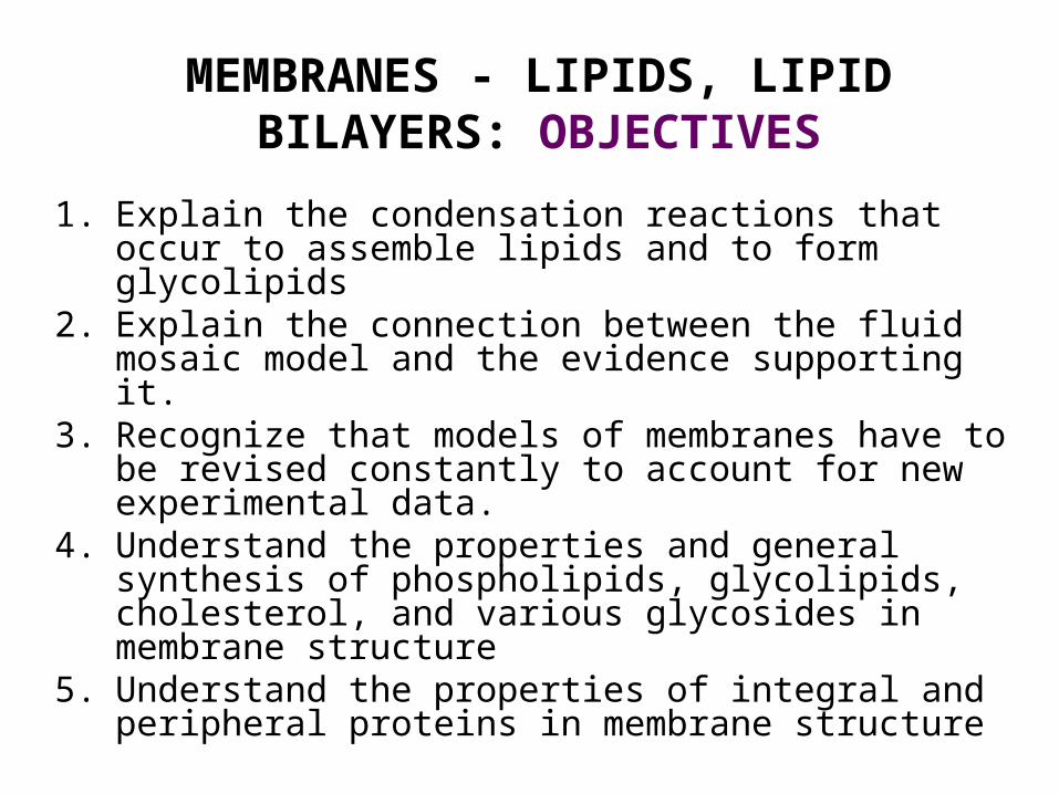
MEMBRANES - LIPIDS, LIPID BILAYERS: OBJECTIVES
1. Explain the condensation reactions that occur to assemble lipids and to form glycolipids
2. Explain the connection between the fluid mosaic model and the evidence supporting it.
3. Recognize that models of membranes have to be revised constantly to account for new experimental data.
4. Understand the properties and general synthesis of phospholipids, glycolipids, cholesterol, and various glycosides in membrane structure
5. Understand the properties of integral and peripheral proteins in membrane structure
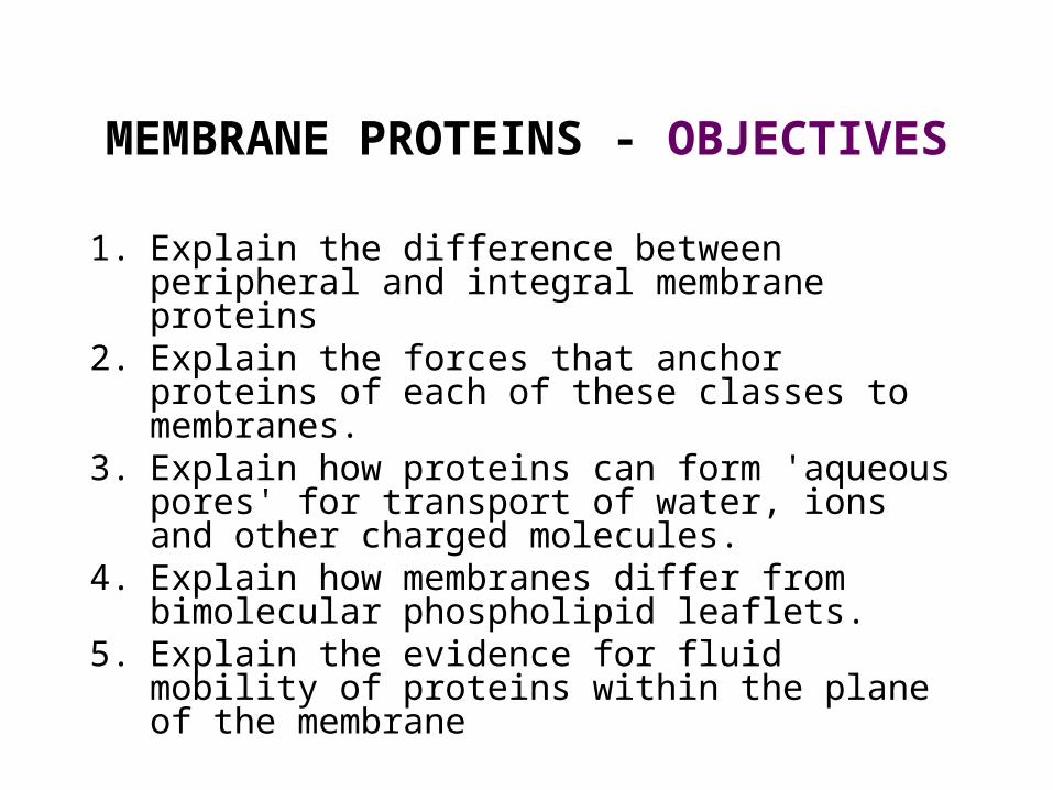
MEMBRANE PROTEINS - OBJECTIVES
1. Explain the difference between peripheral and integral membrane proteins
2. Explain the forces that anchor proteins of each of these classes to membranes.
3. Explain how proteins can form 'aqueous pores' for transport of water, ions and other charged molecules.
4. Explain how membranes differ from bimolecular phospholipid leaflets.
5. Explain the evidence for fluid mobility of proteins within the plane of the membrane
MEMBRANE FUNCTION -TRANSPORTES, CHANNELS AND MEMBRANE POTENTIAL
LEARNING OBJECTIVES1. Each membrane has specific functions which are
reflected in the functioning systems located in it. 2. To understand fundamental transport processes across
membranes and the role of membrane proteins. 3. To understand the linkages between electrical forces,
ATP hydrolysis and specific ion transport pumps. 4. To understand ion selectivity, gated channels and
membrane potential. Nernst equation5. To understand how a membrane potential is generated
and propagated. 6. To understand the link between ion channels and nerve
impulses. 7. Understand the control of directed secretion and its
relation to nerve impulse transmission

Functions of membranes [Becker et al. The World of the Cell Fig. 7-2]
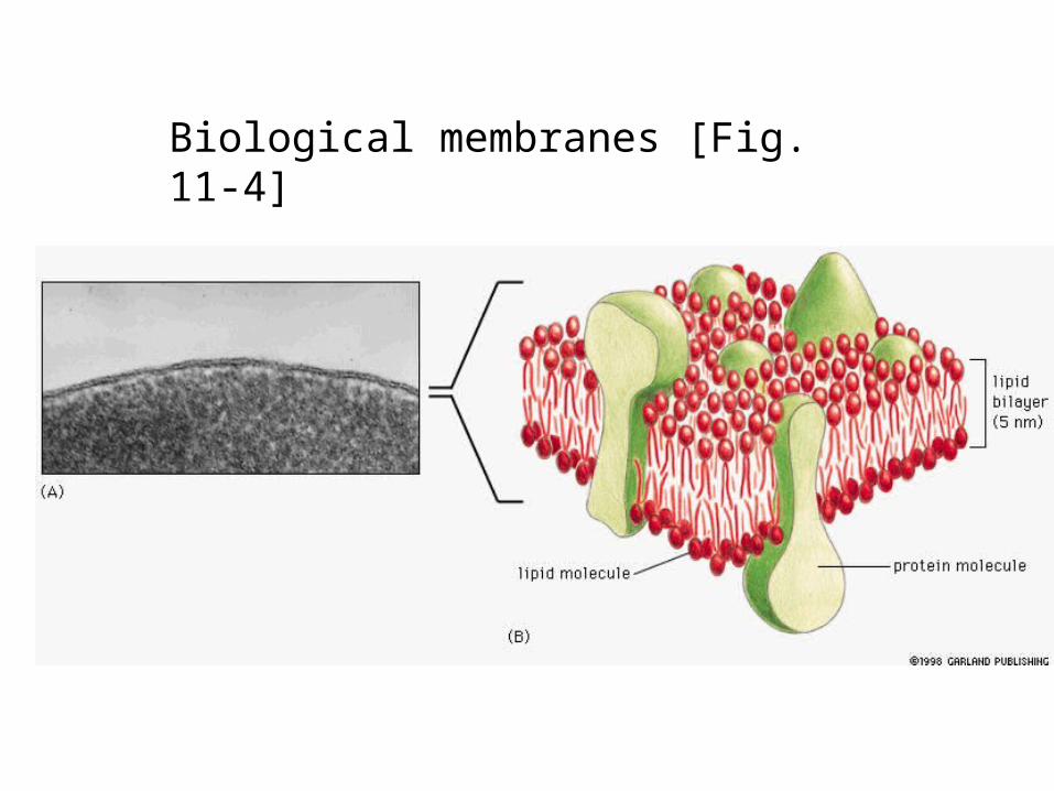
Biological membranes [Fig. 11-4]
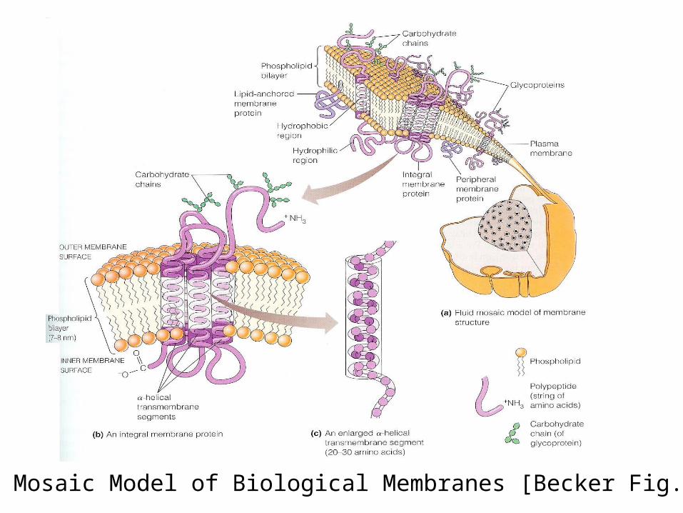
Fluid Mosaic Model of Biological Membranes [Becker Fig. 7-5]

Key features of Fluid Mosaic Model
Membrane lipids:• arranged in a bilayer • fluid (free to move in plane of bilayer) • asymmetrically arranged (different lipid components on
one face of the bilayer than the other)
Membrane proteins:• globular units with hydrophobic domains embedded in the
hydrophobic core of the membrane, • "fluid", in the lipid bilayer, unless anchored by interactions
with other proteins.
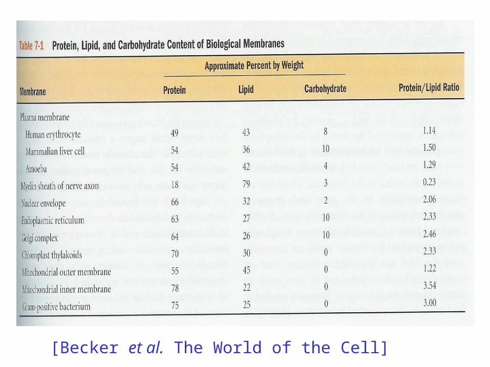
[Becker et al. The World of the Cell]

LIPID CHEMISTRY
Read pp. 73-74 for a review of chemical structures and terminology of different lipid molecules

Triacylglycerols form oil droplets
Oil seed embyro

11_10_Fat_phospholip.jpg
Fat molecules are hydrophobic, whereasPhospholipids are amphipathic [Fig. 11-10]
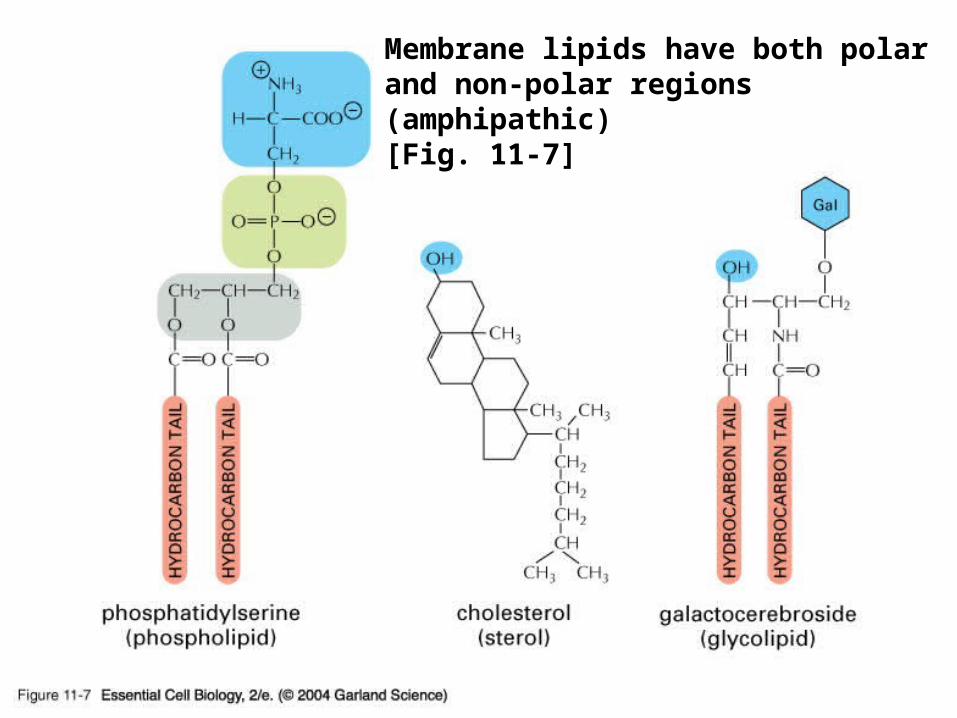
11_07_amphipathic.jpg
Membrane lipids have both polar and non-polar regions (amphipathic) [Fig. 11-7]

11_06_Phosphatidylch.jpg
Phosphatidylcholine is the most common phospholipid in cell membranes [Fig. 11-6]

Sphingolipids: serine backbone, 2 acyl tails
(Often glycolipids)

[Becker et al. The World of the Cell]

Fig. 11-8: hydrophilic, water forms H-bonds with atom carrying uneven charge distribution
Fig. 11-9: hydrophobic, water doesn’t interact with solute; forms cage-like structures
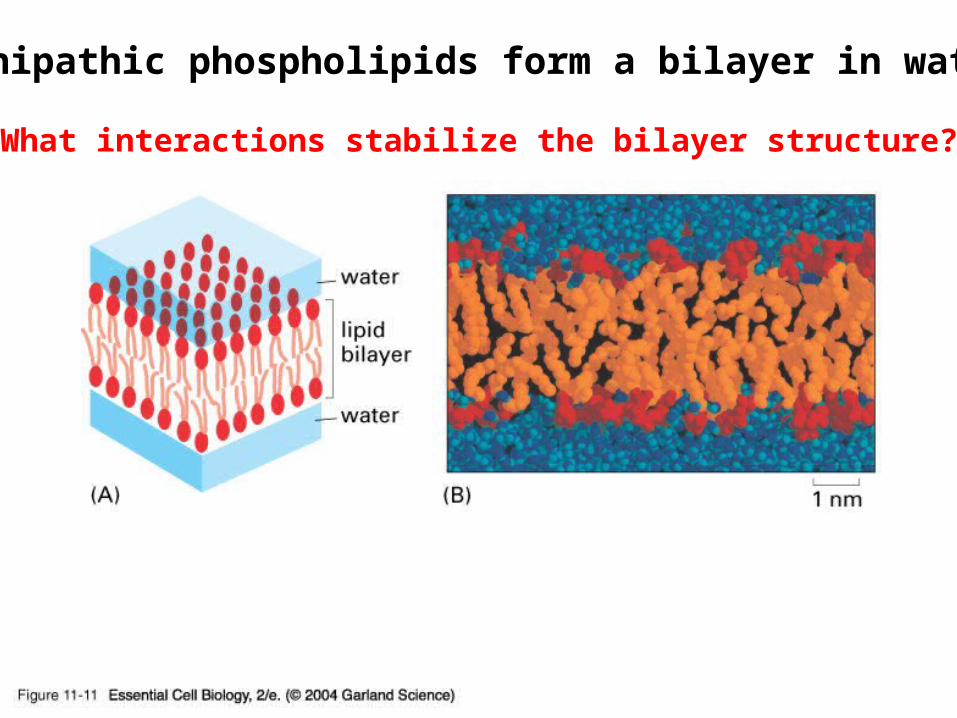
11_11_bilayer.in.H20.jpgAmphipathic phospholipids form a bilayer in water.
What interactions stabilize the bilayer structure?

Three characteristics of membrane lipids
1. Fluidity
2. Asymmetry
3. Permeability

Membrane lipid fluidity• The ease with which the lipid molecules move in
the plane of the bilayer• Depends on temperature and the phospholipid
composition [higher the temperature, lipids with longer tails and fewer double bonds are synthesized in temp.- adapting bacteria/yeast]
• Depends on the length and unsaturation of fatty acids– Shorter hydrocarbon tails = increased fluidity– Increased unsaturation (# double bonds) = increased
fluidity
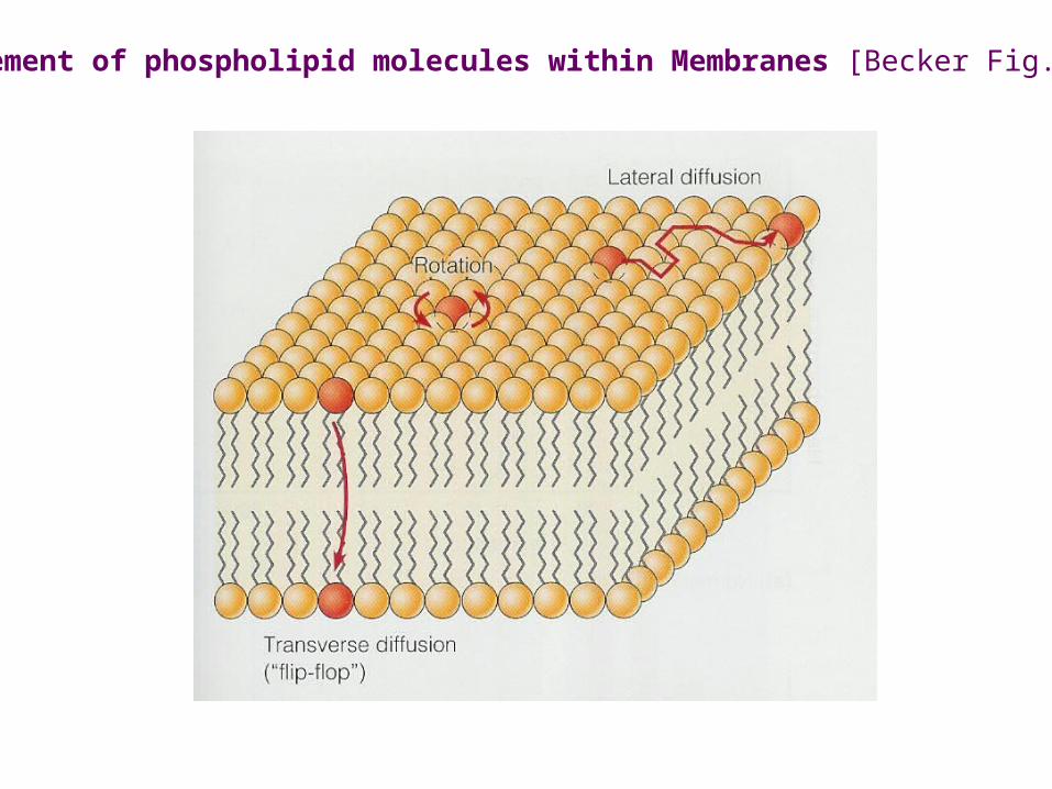
Movement of phospholipid molecules within Membranes [Becker Fig. 7-5]
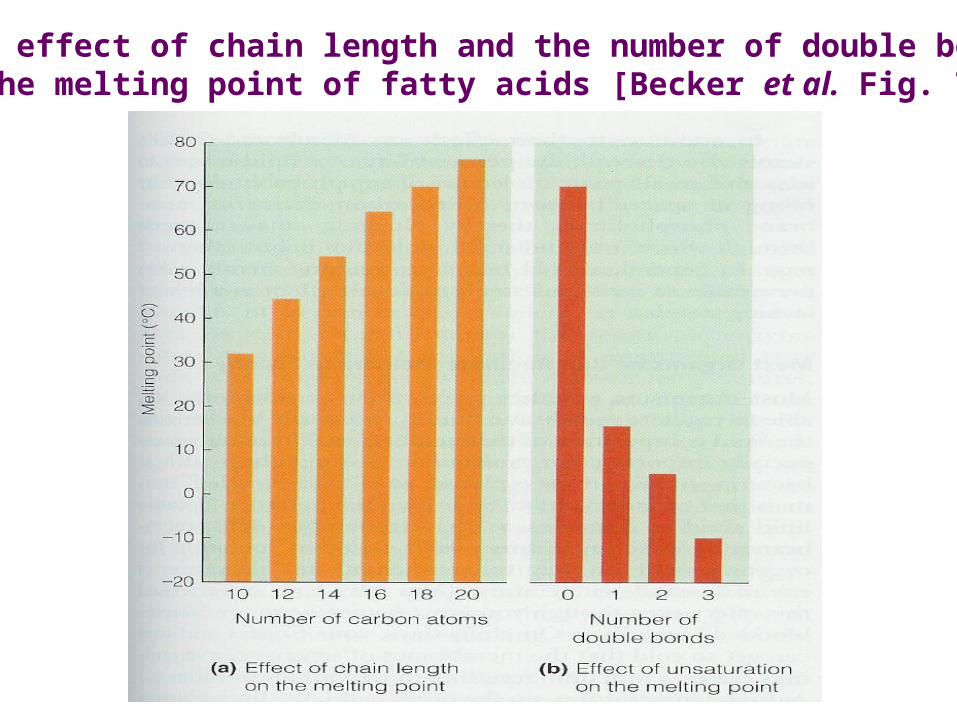
The effect of chain length and the number of double bondson the melting point of fatty acids [Becker et al. Fig. 7-13]
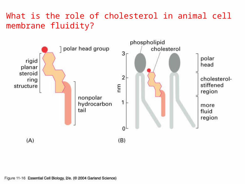
11_16_Cholesterol.jpgWhat is the role of cholesterol in animal cell membrane fluidity?

Membrane flexibility (fluidity) can be studied by either phospholipid vesicles or a synthetic
phospholipid bilayers
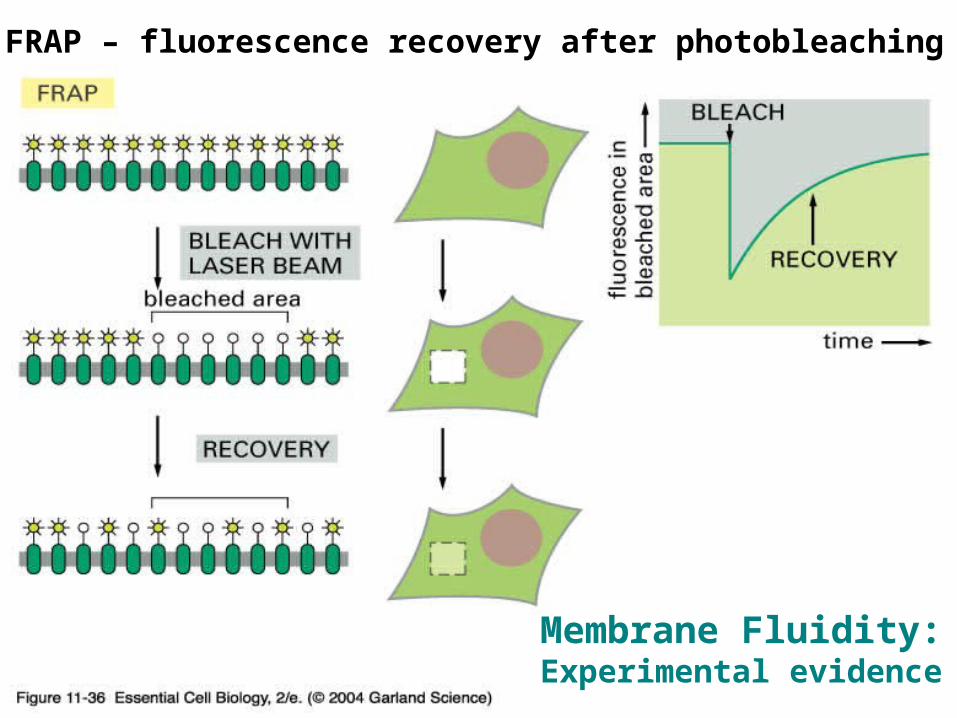
11_36_Photobleaching_techniques.jpg
FRAP – fluorescence recovery after photobleaching
Membrane Fluidity:Experimental evidence

Below are results from two FRAP experiments. What can you conclude about the membrane in cell 2 compared to cell 1? What characteristics would you predict that cell 2’s membrane lipids would have?
Cell 2Cell 1
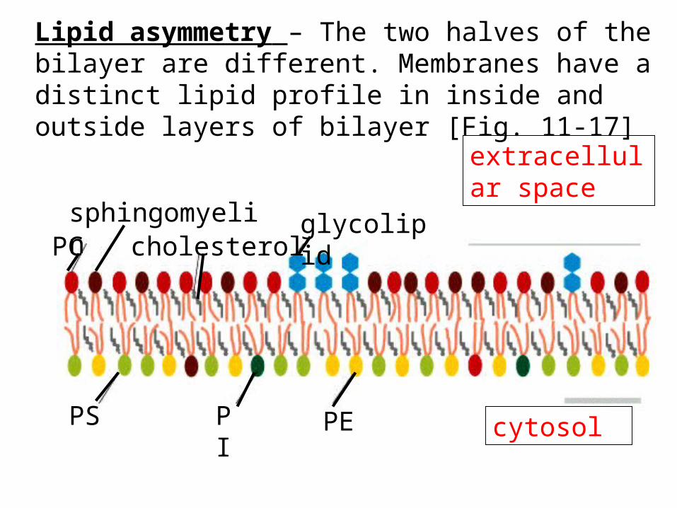
Lipid asymmetry – The two halves of the bilayer are different. Membranes have a distinct lipid profile in inside and outside layers of bilayer [Fig. 11-17]
extracellular space
cytosol
PCsphingomyelin
cholesterolglycolipid
PS PI PE

How is lipid asymmetry created?• All membrane lipids are made in the SER.• Enzymes in the SER join fatty acids and glycerol and
phosphate and head groups to make phospholipids• The phospholipid is inserted into one of the monolayers• Enzymes called FLIPPASES flip some of these lipids into
the other bilayer, so that the whole membrane will grow. They only transfer specific lipids.
• Glycolipids get their sugar groups in the Golgi only on the non-cytosolic monolayer of the membrane. It then travels as a vesicle and fuses with the plasma membrane, where it maintains the orientation of glycolipids in the non-cytosolic monolayer [see Fig. 11-15].
• It makes the membrane asymmetrical.

ER
New phospholipids added to cytoplasmic leaflet
Phospholipid translocator
Membrane protein
Both sides enlarged
Phospholipids moved between leaflets by translocator proteins
ATP

Different organelles have different lipid compositionslipid ER PMPC 40 24PE 17 7Cholesterol 6 17Sphingomyelin 5 19Glycolipids Trace 7
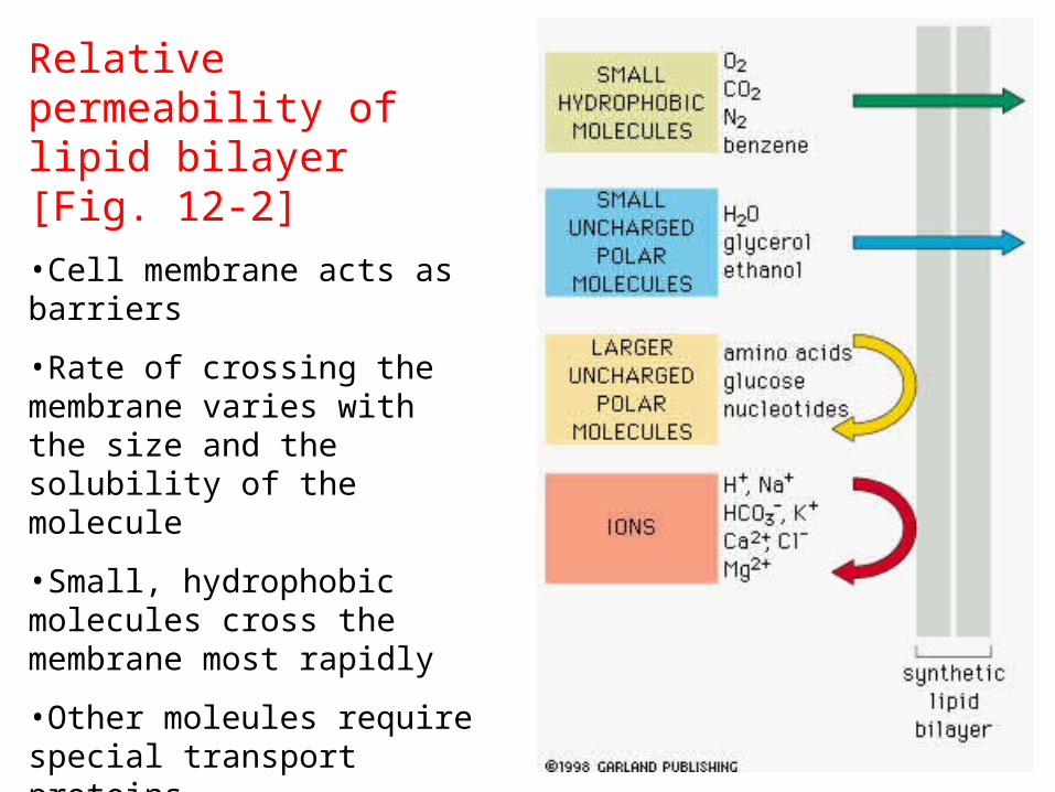
Relative permeability of lipid bilayer [Fig. 12-2]
•Cell membrane acts as barriers
•Rate of crossing the membrane varies with the size and the solubility of the molecule
•Small, hydrophobic molecules cross the membrane most rapidly
•Other moleules require special transport proteins
•Synthetic bilayers have been used to study permeability
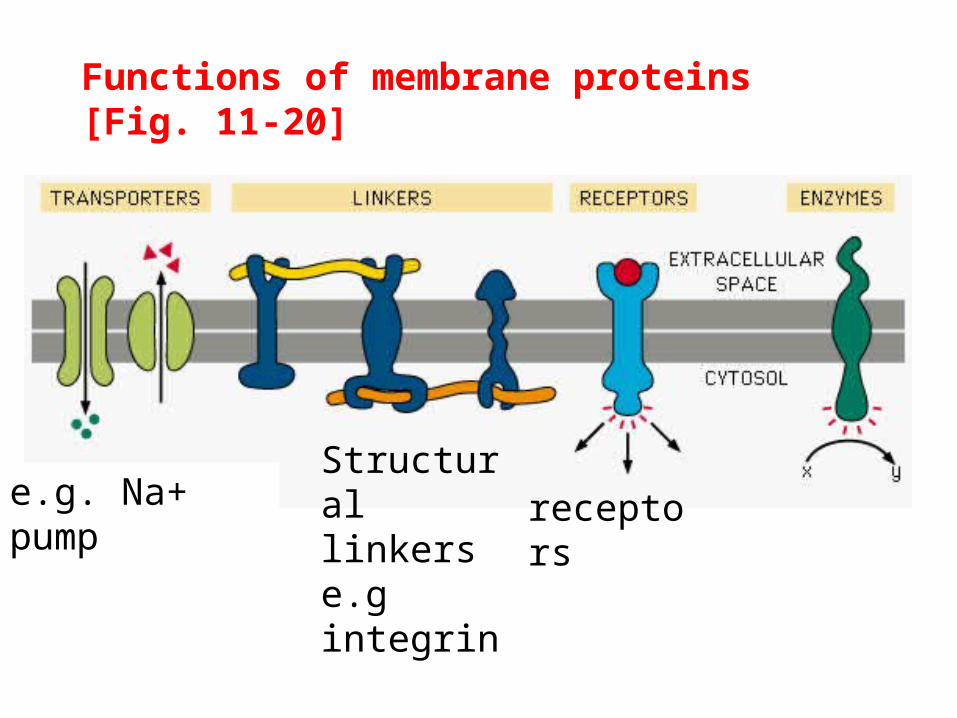
Functions of membrane proteins [Fig. 11-20]
e.g. Na+ pumpStructural linkers e.g integrin
receptors


11_21_proteins.associ.jpgMembrane proteins associate with the lipid bilayer in several different ways [Fig. 11-21]

Fig. 11-23: -helix spans the bilayer (why?)
H-bonded polypeptide chain inside,
Hydrophobic R-groups outside.
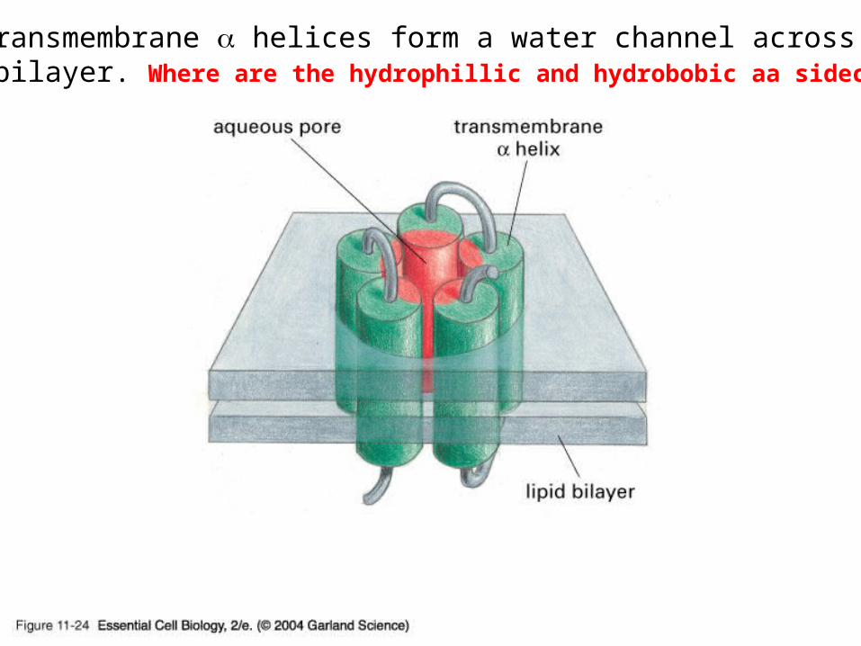
11_24_hydrophl.pore.jpg
Five transmembrane helices form a water channel across thelipid bilayer. Where are the hydrophillic and hydrobobic aa sidechains?

11_28_Bacteriorhodop.jpg
Bacteriorhodopsin acts as a proton pump. Two polar aa side chains areInvolved in proton transfer process.

Fig. 11-27: solubilizing membrane proteins with detergent like TritonX-100

Fig. 11-27: solubilizing membrane proteins -the results
Water-soluble complex of membrane protein and detergent
Water soluble complex of lipid + detergent

Panel 4-5: protein electrophoresis

[Becker et al. The World of the Cell]

11_31_spectrin_network.jpgSpectrin-based cell cortex of human red blood cells provides
mechanical strength and shape to the cell [Fig. 11-32]
Genetic abnormalities in spectrin result in abnormal, spherical and fragile red blood cells causing anemia

Glycocalyx: sugar coat
Fig. 11-32
Glycocalyx functions in cell-cell recognition, protection, lubrication and adhesion
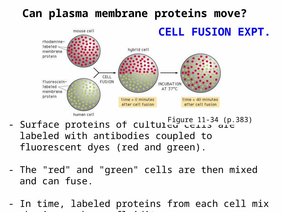
- Surface proteins of cultured cells are labeled with antibodies coupled to fluorescent dyes (red and green).
- The "red" and "green" cells are then mixed and can fuse.
- In time, labeled proteins from each cell mix showing membrane fluidity
Can plasma membrane proteins move?
Figure 11-34 (p.383)
CELL FUSION EXPT.

What restricts lateral mobility of plasma membrane proteins?
Cell cortex
Extracellularmatrix
Intercellular protein-protein interactions Diffusion barriers
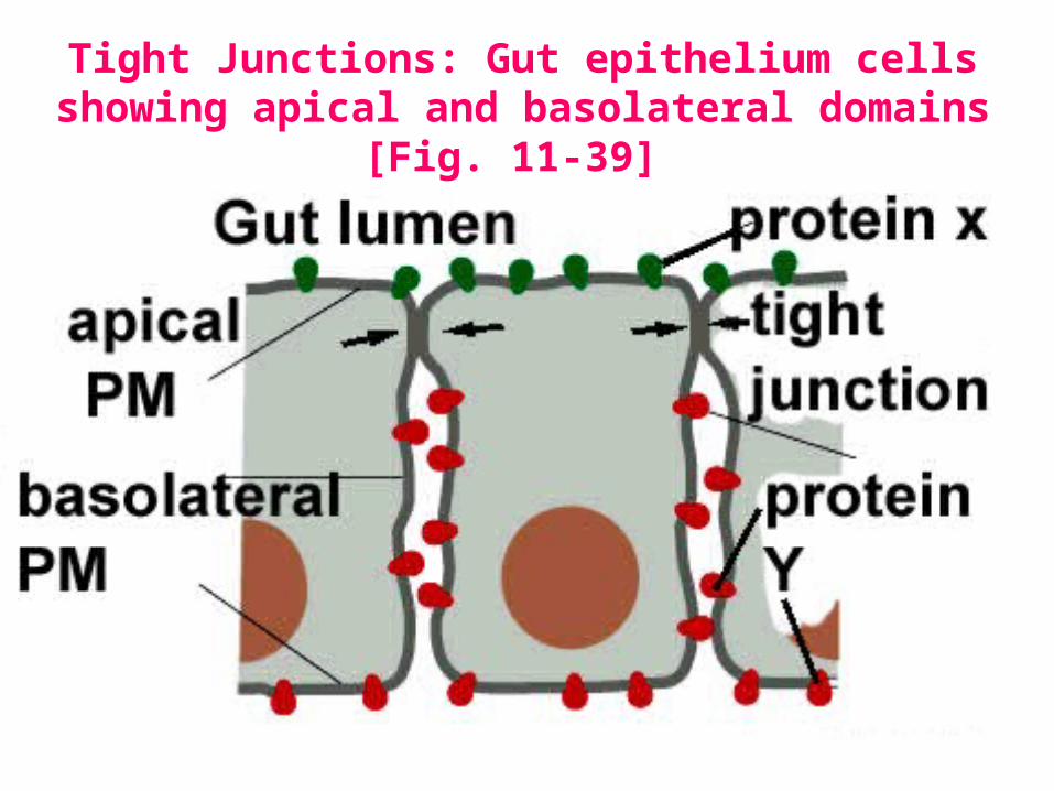
Tight Junctions: Gut epithelium cells showing apical and basolateral domains [Fig. 11-39]

Fig. 21-22: tight junctions seal epithelia
Epithelium is sheet of cells
Tight junctions wrap around the apical face of each cell
Inject dye into either apical face or baso-lateral face-can’t cross tight junction

[Becker et al.]
Membrane transport
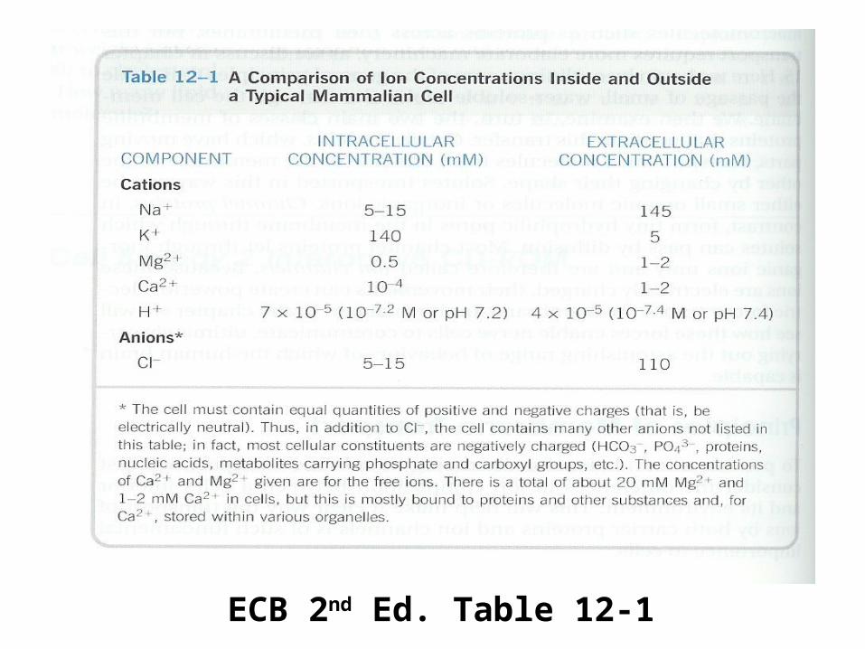
ECB 2nd Ed. Table 12-1

12_02_diffusion_rate.jpg
Membrane permeability

Carrier protein Channel protein
Carrier has conformational change, carries solute [Fig. 12-3]
channel opens, selectively allows ions in.

Passive vs. active transport [Fig. 12-4]
Passive transport Active transport

12_08_electroch_gradient .jpg
Two components of an electrochemical gradient =Concentration gradient + membrane potential

Three ways of active transport of molecules via the cell membranes [Fig. 12-9]

The Na+/K+ pump uses ATP hydrolysis to pump Na+ out and K+ in to the cell both against their
electrochemical gradient [Fig. 12-10]

12_12_Na_K_cyclic.jpg
Mechanism of the Na+/K+ pump [Fig. 12-12]

12_13_Carrier_proteins.jpgCarrier protein-mediated transport [Fig. 12-13]
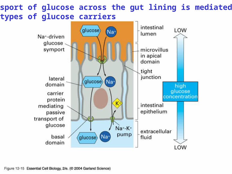
12_15_glucose_gut.jpg
Transport of glucose across the gut lining is mediated by two types of glucose carriers

Fig. 12-5: Each organelle has its own subset of unique channels and carriers
ER-
Ca2+ transporters
Ca

Fig. 12-15:
hypotonic ‘ghost’
Osmosis=movement of water molecules across a semi-permeable membrane in response to a concentration gradient

Cells use different ways to prevent osmotic swelling [Fig. 12-16]

Animal cell
Similarities and differences in carrier-mediated transport in animal and plants [Fig. 12-18]
Plant cell


12_19_selectivity_filter.jpg
A K+ channel protein
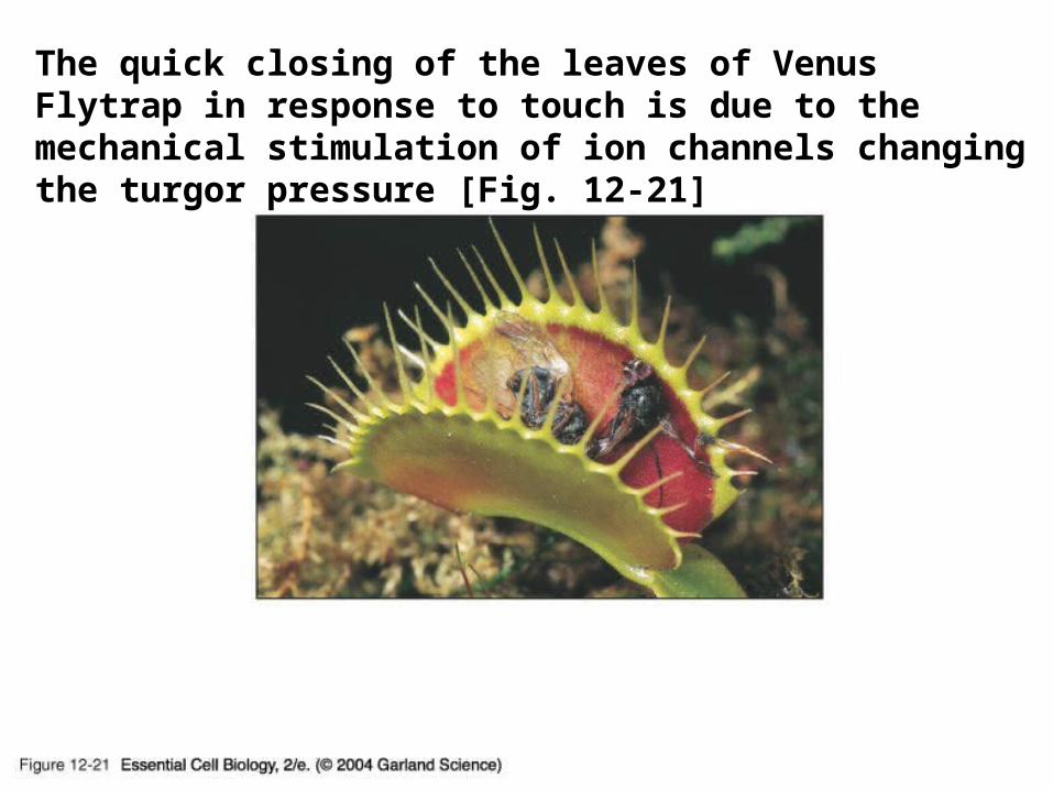
12_21_Venus _flytrap.jpgThe quick closing of the leaves of Venus Flytrap in response to touch is due to the mechanical stimulation of ion channels changing the turgor pressure [Fig. 12-21]

12_22_patch_clamp_record .jpg
The patch-clamp technique

12_23_current _measured .jpgMeasurement of ion channel activity as current by the Patch-clamp technique [Fig. 12-23]

12_24_Gated _ion_chan .jpgGated ion channels [Fig. 12-24]

Fig. 12-26: Touch-induced opening of voltage-gatedIon channels generate (1) an electric impulse followedby a rapid water loss by hinge cells, causing (3) foldingof Mimosa leaflets.

Fig. 12-27: The unequal distribution of ions across thebBilayer produces the membrane potential (electrical potential difference across the membrane).

Potassium ion channels are important in generating the membrane potential across the plasma membrane [Fig. 12-28]