Bioelectrochemistry 2013 - ise-online.org · 20/03/2013 · Enzymatic and microbial biofuel cells...
377
Program & Book of Abstracts of Bioelectrochemistry 2013 12 th Topical Meeting of the International Society of Electrochemistry & XXII International Symposium on Bioelectrochemistry and Bioenergetics of the Bioelectrochemical Society 17-21 March, 2013, Bochum, Germany Organized by: Bioelectrochemical Society ISE Division 2 Bioelectrochemistry ISE Region Germany I N T E R N A T I O N A L S O C I E T Y O F E L E C TR O C H E M I S T R Y • The Bioelectrochemical Society
Transcript of Bioelectrochemistry 2013 - ise-online.org · 20/03/2013 · Enzymatic and microbial biofuel cells...

Program & Book of Abstracts of
Bioelectrochemistry 2013
12th Topical Meeting of the International Society of Electrochemistry
&XXII International Symposium
on Bioelectrochemistry and Bioenergeticsof the Bioelectrochemical Society
17-21 March, 2013, Bochum, Germany
Organized by:Bioelectrochemical Society
ISE Division 2 Bioelectrochemistry ISE Region Germany
INTER
NATIO
NAL SO
CIETY OF ELECTR
OCHEM
ISTRY•The Bioelectrochemical Society

International Society of Electrochemistry& Bioelectrochemical Society
Rue de Sébeillon 9b1004 LausanneSwitzerland
Copyright © 2013
All rights reserved. No part of this work may be reproduced, stored in a retrieval system or transmitted in any form or by any means, electronic, mechanical, photocopying, recording or otherwise, without prior written permission of the Publisher.
No responsibility is assumed by the Publisher for any injury and/or damage to persons or property as a matter of product liability, negligence or otherwise, or from any use or operation of any methods, products, instructions or ideas contained in the material herein.
Printed in the Germany

Organizing CommitteeChair Wolfgang Schuhmann, Bochum, Germany
Members Lo Gorton, Lund, Sweden Alexander Kuhn, Pessac, France Eberhard Neumann, Bielefeld, Germany Ana Maria Oliveira-Brett, Coimbra, Portugal Woonsup Shin, Seoul, Korea Gunther Wittstock, Oldenburg, Germany
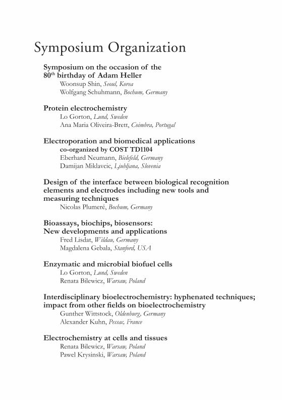
Symposium OrganizationSymposium on the occasion of the 80th birthday of Adam Heller
Woonsup Shin, Seoul, Korea Wolfgang Schuhmann, Bochum, Germany
Protein electrochemistry Lo Gorton, Lund, Sweden Ana Maria Oliveira-Brett, Coimbra, Portugal
Electroporation and biomedical applications co-organized by COST TD1104 Eberhard Neumann, Bielefeld, Germany Damijan Miklavcic, Ljubljana, Slovenia
Design of the interface between biological recognition elements and electrodes including new tools and measuring techniques
Nicolas Plumeré, Bochum, Germany
Bioassays, biochips, biosensors: New developments and applications
Fred Lisdat, Wildau, Germany Magdalena Gebala, Stanford, USA
Enzymatic and microbial biofuel cells Lo Gorton, Lund, Sweden Renata Bilewicz, Warsaw, Poland
Interdisciplinary bioelectrochemistry: hyphenated techniques; impact from other fields on bioelectrochemistry
Gunther Wittstock, Oldenburg, Germany Alexander Kuhn, Pessac, France
Electrochemistry at cells and tissues Renata Bilewicz, Warsaw, Poland Pawel Krysinski, Warsaw, Poland
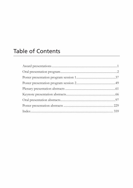
Table of Contents
Award presentations ..................................................................................1
Oral presentation program .......................................................................2
Poster presentation program session 1 .................................................37
Poster presentation program session 2 .................................................49
Plenary presentation abstracts ...............................................................61
Keynote presentation abstracts ..............................................................66
Oral presentation abstracts .....................................................................97
Poster presentation abstracts .............................................................. 229
Index ....................................................................................................... 359

Program
INTER
NATIO
NAL SO
CIETY OF ELECTR
OCHEM
ISTRY•The Bioelectrochemical Society

Special Meetings and Social Program
Sunday, 17 March 201309:00 to 12:00
BES Council
17:30 to 18:00Opening Ceremony, Conference Center Room 2a/2b
19:30 to 21:30Welcome Reception, Conference Center
Monday, 18 March 201318:15 to 19:15 in room 82
Elisabeth Renney, European CommissionSupporting creative minds - A seminar combining European Research Funding Possibilities and the experience of a grantee
18:00 to 20:30Poster Session and Reception, Conference Center Room 1
Tuesday, 19 March 2013 14:45
Excursion, Departure Bus stop Unicenter
19:00Banquet Presentation of «RUB rectorate poster awards»
Wednesday, 20 March 201312:40 to 14:00
BES General Assembly, Conference Center Room 2a
18:00 to 19:00Panel Discussion: Future Directions of Electroporation Based Approaches
18:00 to 20:30Poster Session and Reception, Conference Center Room 2

1
Prog
ram
Bioelectrochemistry 2013
Sunday, 17 March, 2013 - Afternoon
Conference Center Room 2a/2b
17:30 Opening Ceremony
Giulio Milazzo Prize LectureConference Center Room 2a/2b
Chaired by: Ana Maria Oliveira-Brett
18:00 to 18:50 James Weaver (MIT, Cambridge, USA)
An Approach to Understanding Electromagnetic Field Effects in Living Cells
Luigi Galvani Prize LectureConference Center Room 2a/2b
Chaired by: Fred Lisdat
19:00 to 19:30 Christophe Léger (Laboratoire de Bioénergétique et Ingénierie des Protéines, CNRS & Aix-Marseille Univ., Marseille, France)
Introduction to direct electrochemistry for probing molecular aspects of biological catalysis
19:30 Welcome Reception
Sund
ay, 1
7 M
arch
, 201
3
2Pr
ogra
mBioelectrochemistry 2013
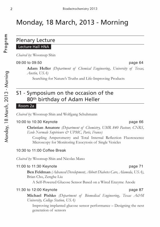
Monday, 18 March, 2013 - Morning
Plenary LectureLecture Hall HNA
Chaired by: Woonsup Shin
09:00 to 09:50 page 64 Adam Heller (Department of Chemical Engineering, University of Texas, Austin, USA)
Searching for Nature’s Truths and Life-Improving Products
S1 - Symposium on the occasion of the 80th birthday of Adam Heller
Room 2a
Chaired by: Woonsup Shin and Wolfgang Schuhmann
10:00 to 10:30 Keynote page 66Christian Amatore (Department of Chemistry, UMR 840 Pasteur, CNRS, Ecole Normale Supérieure & UPMC, Paris, France)
Coupling Amperometry and Total Internal Reflection Fluorescence Microscopy for Monitoring Exocytosis of Single Vesicles
10:30 to 11:00 Coffee Break
Chaired by: Woonsup Shin and Nicolas Mano
11:00 to 11:30 Keynote page 71Ben Feldman (Advanced Development, Abbott Diabetes Care, Alameda, USA), Brian Cho, Zenghe Liu
A Self-Powered Glucose Sensor Based on a Wired Enzyme Anode
11:30 to 12:00 Keynote page 87Michael Pishko (Department of Biomedical Engineering, Texas A&M University, College Station, USA)
Improving implanted glucose sensor performance – Designing the next generation of sensors
Mon
day,
18
Mar
ch, 2
013
- Mor
ning

3
Prog
ram
Bioelectrochemistry 2013
12:00 to 12:20 Invited page 143Ioanis Katakis (Department of Chemical Engineering, Universitat Rovira i Virgili, Tarragona, Spain)
Electrochemically Actuated, Capillarity-Driven Biodetection Devices for Food Safety and Clinical Analysis
12:20 to 12:40 page 197Woonsup Shin (Department of Chemistry, Sogang University, Seoul, Korea)
Development of Non-gassing Electroosmotic Pump for Drug Infusion System
S3 - Electroporation and biomedical applicationsRoom 82
Chaired by: Eberhard Neumann
10:00 to 10:30 Keynote page 94James Weaver (Harvard-MIT Division of Health Sciences and Technology, Massachusetts Institute of Technology, Cambridge, USA), Thiruvallur Gowrishankar, Kyle Smith, Reuben Son
Cell Electroporation Creates Complex Pore Populations
10:30 to 11:00 Coffee Break
Chaired by: Magorzata Kotulska and Richard Heller
11:00 to 11:30 Keynote page 70Ruggero Cadossi (R&D, IGEA, Carpi, Italy)
Clinical electroporation: experience in cancer treatment in Europe
11:30 to 12:00 Keynote page 86Lluis M. Mir (UMR 8203 CNRS and LEA EBAM, CNRS, Villejuif, France), Marie Breton, Isabelle Leray, Aude Silve
Cell electroporation and cell electropermeabilisation: facts and theory
Mon
day,
18
Mar
ch, 2
013
- Mor
ning
4Pr
ogra
mBioelectrochemistry 2013
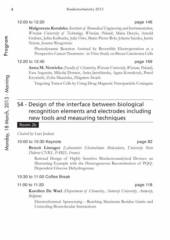
12:00 to 12:20 page 146Malgorzata Kotulska (Institute of Biomedical Engineering and Instrumentation, Wroclaw University of Technology, Wroclaw, Poland), Maria Derylo, Arnold Grabiec, Julita Kulbacka, Julie Orio, Marie-Pierre Rols, Jolanta Saczko, Justin Teissie, Joanna Wezgowiec
Photodynamic Reaction Assisted by Reversible Electroporation as a Prospective Cancer Treatment - in Vitro Study on Breast Carcinoma Cells
12:20 to 12:40 page 169Anna M. Nowicka (Faculty of Chemistry, Warsaw University, Warsaw, Poland), Ewa Augustin, Mikolaj Donten, Anita Jarzebinska, Agata Kowalczyk, Pawel Krysinski, Zofia Mazerska, Zbigniew Stojek
Targeting Tumor Cells by Using Drug-Magnetic Nanoparticle Conjugate
S4 - Design of the interface between biological recognition elements and electrodes including new tools and measuring techniques
Room 2b
Chaired by: Lars Jeuken
10:00 to 10:30 Keynote page 82Benoit Limoges (Laboratoire Electrochimie Moleculaire, University Paris Diderot CNRS, PARIS, France)
Rational Design of Highly Sensitive Bioelectroanalytical Devices: an Illustrating Example with the Heterogeneous Reconstitution of PQQ-Dependent Glucose Dehydrogenase
10:30 to 11:00 Coffee Break
11:00 to 11:20 page 118Karolien De Wael (Department of Chemistry, Antwerp University, Antwerp, Belgium)
Electrochemical Aptasensing – Reaching Maximum Residue Limits and Unraveling Biomolecular Interactions
Mon
day,
18
Mar
ch, 2
013
- Mor
ning
5
Prog
ram
Bioelectrochemistry 2013
11:20 to 11:40 page 120Thomas Doneux (Chimie Analytique et Chimie des Interfaces, Université Libre de Bruxelles, Bruxelles, Belgium), Claudine Buess-Herman, Eléonore Triffaux
Electrochemical Detection of the Protein Mdm2 by a Peptide Affinity Probe Based on the Protein p53
11:40 to 12:00 page 117Anne De Poulpiquet (Bioénergétique et Ingénierie des Protéines, CNRS - AMU, Marseille, France), Alexandre Ciaccafava, Roger Gadiou, Marie-Thérèse Giudici-Orticoni, Elisabeth Lojou, Helena Marques
Immobilisation of Aquifex aeolicus Membrane-bound Hydrogenase on carbon nanofibers for H2/O2 biofuel cells
12:00 to 12:20 page 125Artur Fandrich (Biosystems Technology, Technical University of Applied Sciences, Wildau, Germany), Jens Buller, André Laschewsky, Fred Lisdat, Erik Wischerhoff
“Smart” Polymer Interfaces at Electrodes - useful Matrix for Biorecogni-tion Reactions
12:20 to 12:40 page 109Ariadna Brotons (Química Física, University of Alicante, Alicante, Spain), Juan Miguel Feliu, Jesus Iniesta, Vicente Montiel, Jose Solla-Gullón, Francisco José Vidal-Iglesias
A First Approach to the Electrochemical Evaluation of DNA Methyla-tion on Gold Surfaces: from Single Crystal to Nanoparticles M
onda
y, 1
8 M
arch
, 201
3 - M
orni
ng

6Pr
ogra
mBioelectrochemistry 2013
Monday, 18 March, 2013 - Afternoon
S1 - Symposium on the occasion of the 80th birthday of Adam Heller
Room 2a
Chaired by: Michael Pishko and Zhiqiang Gao
14:00 to 14:30 Keynote page 76Kazuhito Hashimoto (Department of Applied Chemistry, The University of Tokyo, Tokyo, Japan)
Extracellular Electron Transfer via Conductive Minerals
14:30 to 14:50 Invited page 158Nicolas Mano (CRPP-UPR 8641, CNRS, Pessac, France)
The Evolution of the Miniature Membrane-less Biofuel cells From 2001 to 2006
14:50 to 15:10 page 198Sergey Shleev (Department of Biomedical Science, Health & Society, Malmö University, Malmö, Sweden), Plamen Atanassov
Biomedical Applications of Implantable Biofuel Cells
15:10 to 15:30 page 152Donal Leech (Department of Chemistry, National University of Ireland Galway, Galway, Ireland)
Redox complexes for mediation of electron transfer in enzymatic batte-ries and fuel cells
15:30 to 15:50 page 171Marcin Opallo (Institute of Physical Chemistry, Polish Academy of Sciences, Warszawa, Poland), Alexandre Ciaccafava, Anne De Poulpiquet, Martin Jonsson-Niedziolka, Elisabeth Lojou, Frank Marken, Helena Marques, Joanna Niedziolka-Jonsson, Katarzyna Szot
Carbon nanoparticulate films as effective scaffolds for mediatorless bio-electrocatalytic hydrogen oxidation
Mon
day,
18
Mar
ch, 2
013
- Afte
rnoo
n
7
Prog
ram
Bioelectrochemistry 2013
15:50 to 16:10 page 131Magdalena Gebala (Deparment of Biochemistry, Stanford University, School of Medicine, Stanford, USA), Gerhard Hartwich, Wolfgang Schuhmann, Andreas Zimdars
Sandwich microassay for pathogens detection related to urinary tract infections. Selective post-labeling of hybridized 16S rRNAs
16:10 to 16:40 Coffee Break
Chaired by: Ioanis Katakis and Magdalena Gebala
16:40 to 17:00 page 156Fred Lisdat (Deparment of Biosystems Technology, Technical University of Applied Sciences Wildau, Wildau, Germany)
DNA on gold – tools for the label-free analysis of hybridization and sequence specific ligand interaction
17:00 to 17:30 Keynote page 73Zhiqiang Gao (Deparment of Chemistry, National University of Singapore, Singapore, Singapore), Huimin Deng, Yuqian Ren, Wei Shen
Wired Enzyme Technology-Based Ultrasensitive Nucleic Acid Biosen-sors
17:30 to 18:00 Keynote page 74Hubert Girault (Laboratoire d’Electrochimie Physique et Analytique, Ecole Polytechnique Fédérale de Lausanne, Lausanne, Switzerland), Fernando Cortes-Salazar, Baohong Liu, Reza Pourhaghighi, Liang Qiao, Elena Tobolkina
Electrochemical methods for proteomics: From electrophoresis to mass spectrometry
S3 - Electroporation and biomedical applicationsRoom 82
Chaired by: Maja Cemacar and Veronique Preat
14:00 to 14:30 Keynote page 91Gregor Sersa (Deparment of Experimental Oncology, Institute of Oncology Ljubljana, Ljubljana, Slovenia)
Translational Research in Biomedical Applications of Electroporation
Mon
day,
18
Mar
ch, 2
013
- Afte
rnoo
n
8Pr
ogra
mBioelectrochemistry 2013
14:30 to 14:50 page 138Richard Heller (Center for Bioelectrics, Old Dominion University, Norfolk, USA), Amy Donate, Siqi Guo, Cathryn Lundberg, Shawna Shirley
Gene Electrotransfer a Versatile and Powerful Tool to Enhance Thera-peutic Applications
14:50 to 15:10 page 228Veronique Preat (Louvain Drug Research Institute, Université catholique de Louvain, Brussels, Belgium), Gaelle Vandermeulen, Paolo E. Porporato, Pierre Sonveaux, Marcus Lehnhardt, Frank Jacobsen, Veronique Preat, Lars Steinstraesser, Martin Lam
In Vivo Cutaneous Electroporation of Human Host Defense Peptide LL-37 Accelerates Wound Healing
15:10 to 15:30 page 98Franck André (UMR8203, CNRS, Villejuif, France), Léa Lesueur, Aaron Liew, Lluis M. Mir, Timothy O’Brien
Robust, efficient and practical electrogene transfer method for human mesenchymal stem cells using square electric pulses
15:30 to 15:50 page 113Maja Cemazar (Deparment of Experimental Oncology, Institute of Oncology Ljubljana, Ljubljana, Slovenia), Darja Pavlin, Gregor Sersa, Natasa Tozon
Electrogene therapy with interleukin-12 alone or combined with electro-chemotherapy for treatment of spontaneously occurring tumors in dogs
15:50 to 16:10 page 186Marie-Pierre Rols (Institute of Pharmacology and Structural Biology, CNRS and University of Toulouse, Toulouse, France), Christelle Rosazza, Andreas Zumbusch
Cellular tracking of single DNA-particles after their delivery by electro-poration
16:10 to 16:40 Coffee Break
Chaired by: Giovanna Ferrari and Wolfgang Frey
16:40 to 17:10 Keynote page 89Javier Raso (Food Technology Unit, University of Zaragoza, Zaragoza, Spain)
Applications of Pulsed Electric Fields for Food Processing
Mon
day,
18
Mar
ch, 2
013
- Afte
rnoo
n

9
Prog
ram
Bioelectrochemistry 2013
17:10 to 17:30 page 128Wolfgang Frey (Institute for Pulsed Power and Microwave Technology (IHM), Karlsruhe Institute of Technology (KIT), Eggenstein-Leopoldshafen, Germany), Christian Eing, Martina Goettel, Christian Gusbeth, Ralf Straessner
Pulsed Electric Field Treatment of Microalgae: Benefits for Downstream Processing
17:30 to 17:50 page 135Christian Gusbeth (Institute for Pulsed Power and Microwave Technology (IHM), Karlsruhe Institute of Technology, Karlsruhe, Germany), Wolfgang Frey, Annika Rieder, Thomas Schwartz
Bacterial Decontamination of Wastewater by Pulsed Electric Field Treat-ment
17:50 to 18:10 page 179Gianpiero Pataro (Industrial Engineering, University of Salerno, Fisciano, Italy)
Metal Release from Stainless Steel Electrodes of an Ohmic Heater
S4 - Design of the interface between biological recognition elements and electrodes including new tools and measuring techniques
Room 2b
Chaired by: David Waldeck
14:00 to 14:30 Keynote page 77Lars Jeuken (School of Biomedical Sciences, University of Leeds, Leeds, United Kingdom)
Electrochemistry of Fluorescently-labeled Enzymes Reveals Heteroge-neous Interfacial Electron-transfer Rates and Intramolecular Rates that Differ between Rest and Turn-over Conditions
14:30 to 14:50 Invited page 130Jose A. Garrido (Walter Schottky Institut, Technische Universität München, Garching, Germany)
Functionalization of Diamond Surfaces for Bio-applications
Mon
day,
18
Mar
ch, 2
013
- Afte
rnoo
n

10Pr
ogra
mBioelectrochemistry 2013
14:50 to 15:10 page 157Edmond Magner (Materials and Surface Science Institute, University of Limerick, Limerick, Ireland)
Spatially controlled immobilization of enzymes for use in biofuel cells and biocatalysis
15:10 to 15:30 page 200Koji Sode (Department of Biotechnology, Graduate School of Engineering, Tokyo University of Agriculture & Technology, Tokyo, Japan), Stefano Ferri, Yohei Horaguchi, Katsuhiro Kojima, Shoko Saito
Engineering FAD dependent oxidases ~development of dehydrogenases from oxidases for amperometric enzyme sensor applications~
15:30 to 15:50 page 160Joaquim T. Marquês (Centro de Química e Bioquímica, Faculdade de Ciências da Universidade de Lisboa , Lisboa, Portugal), Rodrigo F. M. de Almeida, Ana S. Viana
Building Raft-Containing Biomimetic Membranes on Bare and Modified Gold
15:50 to 16:10 page 139Ales Iglic (Faculty of Electrical Engineering, University of Ljubljana, Ljubljana, Slovenia), Ekaterina Gongazde, Peter Kramar, Alenka Macek Lebar, Sarka Perutkova, Aljaz Velikonja
Zwitterionic lipid layer in contact with monovalent ions and water dipoles in planar lipid bilayer experiments
16:10 to 16:40 Coffee Break
Chaired by: Karolien de Wael
16:40 to 17:00 Invited page 219David Waldeck (Deparment of Chemistry, University of Pittsburgh, Pittsburgh, USA)
Fundamental Studies of Charge Transport between Biomolecules and Electrodes
17:00 to 17:20 page 176Barbara Palys (Department of Chemistry, University of Warsaw, Warsaw, Poland), Piotr Olejnik, Anna Sloniewska, Agnieszka Swietlikowska
Infrared Studies of Enzymes Entrapped in Supramolecular Hydrogels and Adsorbed on Electrode Surfaces
Mon
day,
18
Mar
ch, 2
013
- Afte
rnoo
n
11
Prog
ram
Bioelectrochemistry 2013
17:20 to 17:40 page 181Aleksandra Pinczewska (School of Biological and Chemical Sciences, Queen Mary University of London, London, United Kingdom) Jessica Groppi, Jeremy Kilburn, Philip Bartlett
Towards the Control of the Surface Coverage of Carbon Electrodes with Osmium and Flavin Redox Centers
17:40 to 18:00 page 208Olga Swiech (Faculty of Chemistry, University of Warsaw, Warsaw, Poland)
New derivatives of cyclodextrins as a pH-sensitive drug carriers for anthracycline
18:15 to 19:15 in room 82Elisabeth Renney, European CommissionSupporting creative minds - A seminar combining European Research Funding Possibilities and the experience of a grantee
18:00 to 20:30 Poster Presentation Session 1
Mon
day,
18
Mar
ch, 2
013
- Afte
rnoo
n

12Pr
ogra
mBioelectrochemistry 2013
Tuesday, 19 March, 2013 - Morning
Plenary LectureLecture Hall HNA
Chaired by: Lo Gorton
09:00 to 09:50 page 62Fraser Armstrong (Department of Chemistry, Oxford University, Oxford, United Kingdom)
Fundamental Insights from Enzyme Electrocatalysis
S3 - Electroporation and biomedical applicationsRoom 82
Chaired by: P. Tom Vernier
10:00 to 10:30 Keynote page 96Ken-ichi Yano (Bioelectrics Research Center, Kumamoto University, Kumamoto, Japan), Keiko Morotomi-Yano
Adaptive Responses of Human Cells to Nanosecond Pulsed Electric Fields
10:30 to 11:00 Coffee Break
Chaired by: Lluis M. Mir and Andrei Pakhomov
11:00 to 11:20 page 212Gleb Tolstykh (RHDR, 711 HPW Air Force Research Laboratory, San Antonio, USA), Marjorie Kuipers, Hope Beier, Bennett Ibey, Caleb Roth, Gary Thompson
High-Intensity, Ultra-Short Pulsed Electric Field Exposure Initiates PIP2 Hydrolysis and Actin Cytoskeletal Cortex Remodeling
Tues
day,
19
Mar
ch, 2
013
- Mor
ning

13
Prog
ram
Bioelectrochemistry 2013
11:20 to 11:40 page 173Andrei Pakhomov (Center for Bioelectrics, Old Dominion University, Norfolk, USA), Iurii Semenov
Passive and Active Components of Intracellular Calcium Activation by Nanosecond Pulsed electric Field (nsPEF)
11:40 to 12:00 page 182Uwe Pliquett (Analysenmeßtechnik, Institut für bioprozess- und Analysenmeßtechnik, Heilbad Heiligenstadt, Germany), Nuccitelli Richard
Joule heating during treatment of solid tumor with nano-second pulsed electric field
12:00 to 12:20 page 107Marie Breton (UMR 8203 Vectorologie et Thérapeutiques Anticancéreuses, CNRS IGR Université Paris Sud, Villejuif, France), Lluis M. Mir
Electric Pulses Induce the Oxidation of the Membrane Phospholipids of Giant Unilamellar Vesicles
12:20 to 12:40 page 218P. Thomas Vernier (Frank Reidy Research Center for Bioelectrics, Old Dominion University, Norfolk, USA), Mayya Tokman
Electric Field Enhancement of the Water-Driven, Permeabilizing Reor-ganization of Phospholipid Bilayers
S5 - Bioassays, biochips, biosensors: New developments and applications
Room 2b
Chaired by: Magdalena Gebala and Fred Lisdat
10:00 to 10:30 Keynote page 69Philip Bartlett (Department of Chemistry, University of Southampton, Southampton, United Kingdom)
High Throughput Studies of Modified Electrodes for Biosensors and Biofuel Cells
10:30 to 11:00 Coffee Break
Tues
day,
19
Mar
ch, 2
013
- Mor
ning

14Pr
ogra
mBioelectrochemistry 2013
11:00 to 11:20 Invited page 213Gerald Urban (Departement of Microsystem Engineering, University Freiburg, Freiburg, Germany)
Electrochemical Lab-on-Chip Microsystems for Biomarker Analysis
11:20 to 11:40 Invited page 124Mathieu Etienne (LCPME, CNRS and Université de Lorraine, Villers-lès-Nancy, France), Wissam Ghach, Alain Walcarius
Communication Between Electrode Surface and Whole Cells with Biolo-gical Redox Shuttle for Biosensor Applications
11:40 to 12:00 page 106Harold Braustein (Department of Molecular Microbiology and Biotechnology, Tel Aviv University, Jerusalem, Israel)
Point-of-Care Biosensor Electrochemical Arrays Based on Polymeric Solid State Kit
12:00 to 12:20 page 223Maria Yakovleva (Department of Analytical Chemistry/Biochemistry and Structural Biology, Lund University, Lund, Sweden), Lo Gorton, Aniko Killyéni, Clemens K. Peterbauer, Ionel Catalin Popescu
Further insights into the catalytical properties of deglycosylated pyra-nose dehydrogenase from Agaricus meleagris recombinantly expressed in Pichia pastoris
12:20 to 12:40 page 99Stéphane Arbault (Institut of Molecular Sciences, CNRS UMR 5255, University of Bordeaux, Pessac, France), Salem Ben-Amor, Serge Bottari, Anne Devin, Michel Rigoulet, Neso Sojic
Electroanalytical Study of The Oxidative Stress/Respiration Balance in Mitochondria
Tues
day,
19
Mar
ch, 2
013
- Mor
ning
15
Prog
ram
Bioelectrochemistry 2013
S6 - Enzymatic and microbial biofuel cellsRoom 2a
Chaired by: Renata Bilewicz and Johanna Juhaniewicz
10:00 to 10:30 Keynote page 93Jens Ulstrup (Department of Chemistry, Technical University of Denmark, Kongens Lyngby, Denmark), Allan Glargaard Hansen, Kasper Kannegaard Karlsen, Princia Salvatore, Jingdong Zhang
Electrochemistry of single protein and DNA-based molecules
10:30 to 11:00 Coffee Break
11:00 to 10:20 Invited page 211Isao Taniguchi ((Former Applied Chemistry & Biochemistry), Kumamoto University, Kumamoto, Japan)
Direct Electron-Transfer Reactions of Enzymes and Recent Develop-ments on New Sugar-Air Bio-Fuel Batteries
11:20 to 11:40 page 150Pawel Kulesza (Department of Chemistry, University of Warsaw, Warsaw, Poland), Katarzyna Brzostek, Weronika Lotowska, Adrianna Raczkowska, Iwona A. Rutkowska, Ewelina Seta, Ewelina Szaniawska, Sylwia Zoladek
Specific interactions of noble metal nanoparticles with biofilms grown on electrode surfaces: from anti-bacterial properties to development of electrocatalytic systems active towards oxygen reduction
11:40 to 12:00 page 185Russell Reid (Mechanical Engineering, University of Utah, Salt Lake City, USA), Bruce Gale, Shelley Minteer
A Microfluidic Enzymatic Biofuel Cell Using a Flow-Through Bioanode and an Air-Breathing Cathode
12:00 to 12:20 page 187Raphaël Rousseau (Laboratoire de Génie Chimique (UMR 5503), Université de Toulouse-CNRS, Toulouse, France), Wafa Achouak, Alain Bergel, Marie-Line Delia, Jean-Jacques Godon, Catherine Santaella
Electrochemical, Microbiological and Morphological Characterization of Microbial Bianodes for MEC
12:20 to 12:40 page 145Ivan Kazarinov (Physical Chemistry, Saratov State University, Saratov, Russia)
Microbic Fuel Elements: Prospects and Problems
Tues
day,
19
Mar
ch, 2
013
- Mor
ning

16Pr
ogra
mBioelectrochemistry 2013
Tuesday, 19 March, 2013 - Afternoon
S3 - Electroporation and biomedical applicationsRoom 82
Chaired by: Justin Teissie
14:00 to 14:30 Keynote page 80Bruno Le Pioufle (CNRS SATIE, ENS de Cachan, Cachan, France), Claire Dalmay, Olivier Français
Design and use of microfluidic devices for the real time monitoring of micro/nanopulses effect on cells
S5 - Bioassays, biochips, biosensors: New developments and applications
Room 2b
Chaired by: Philip Bartlett
14:00 to 14:30 Keynote page 67Damien Arrigan (Department of Chemistry, Curtin University, Perth, Australia), Eva Alvarez de Eulate, Sharon Fletcher, Philip Newsholme, Shane O’Sullivan
Electrochemistry of the Polypeptide Amylin at the Interface Between Aqueous and Gelled Organic Electrolyte Phases
S6 - Enzymatic and microbial biofuel cellsRoom 2a
Chaired by: Lo Gorton
14:00 to 14:30 Keynote page 83Elisabeth Lojou (Bioénergétique et Ingenierie des Protéines, CNRS, Marseille, France), Alexandre Ciaccafava, Anne De Poulpiquet, Marie-Thérèse Giudici-Orticoni, Christophe Innocent, Sophy Tingry
An innovative H2/O2 biofuel cell based on a O2, CO and T° tolerant hydrogenase
Tues
day,
19
Mar
ch, 2
013
- Afte
rnoo
n

17
Prog
ram
Bioelectrochemistry 2013
Wednesday, 20 March, 2013 - Morning
Plenary LectureLecture Hall HNA
Chaired by: Ana Maria Oliveira-Brett
09:00 to 09:50 page 65Eberhard Neumann (Department of Physical and Biophysical Chemistry, Faculty of Chemistry, University of Bielefeld, Bielefeld, Germany)
Thirty Years of Membrane Electroporation - Evolution of a Concept for Gene Electro-Transfer up to Clinical Tumour Curing
S3 - Electroporation and biomedical applicationsRoom 82
Chaired by: Guillermo Marshall
10:00 to 10:30 Keynote page 92Mounir Tarek (SRSMC, CNRS-Universite de Lorraine, Vandoeuvre les Nancy, France), Damijan Miklavcic, Lluis M. Mir
Synergic use of molecular dynamics simulations and sophisticated expe-riments reveal key aspects of lipid membranes electroporation
10:30 to 11:00 Coffee Break
Chaired by: Marie-Pierre Rols and James Weaver
11:00 to 11:20 page 119Rumiana Dimova (Theory and Bio-Systems, Max Planck Institute of Colloids and Interfaces, Potsdam, Germany)
Giant Vesicles in Electric Fields – Approaches for Measuring Properties of Lipid Membranes
Wed
nesd
ay, 2
0 M
arch
, 201
3 - M
orni
ng

18Pr
ogra
mBioelectrochemistry 2013
11:20 to 11:40 page 191Gintautas Saulis (Department of Biology, Vytautas Magnus University, Kaunas, Lithuania), Raminta Rodaite-Riseviciene, Rita Saule, Valentinas Snitka
Release of Iron Ions from the Stainless–Steel Anode during High-Vol-tage Pulses and its Consequences for Cell Electroporation Technology
11:40 to 12:00 page 154Hao Lin (Department of Mechanical and Aerospace Engineering, Rutgers, The State University of New Jersey, Piscataway, USA), Jianbo Li, Mohamed Sadik, Jerry Shan, David Shreiber, Miao Yu
Quantification of Basic Transport Processes in Electroporation-Media-ted Molecular Delivery
12:00 to 12:20 page 114Louise Chopinet (Department of Cellular Biophysics, IPBS and LAAS-CNRS, Toulouse, France), Etienne Dague, Marie-Pierre Rols
Measuring and imaging electropermeabilization effects on cell membrane elasticity using Atomic Force Microscopy
12:20 to 12:40 page 163Damijan Miklavcic (Faculty of Electrical Engineering, Department for Biomed Eng, University of Ljubljana, Ljubljana, Slovenia), Franci Bajd, Selma Corovic, Matej Kranjc, Igor Serša
Increased Electrical Conductivity of Cells and Tissue due to Electropo-ration – Modeling and Experiments
S5 - Bioassays, biochips, biosensors: New developments and applications
Room 2b
Chaired by: Gerald Urban
10:00 to 10:30 Keynote page 78Heinz-Bernhard Kraatz (Department of Chemistry, University of Toronto Scarborough, Toronto, Canada)
New adventures in phosporylation chemistry: Using electrochemistry to probe biochemistry
10:30 to 11:00 Coffee Break
Wed
nesd
ay, 2
0 M
arch
, 201
3 - M
orni
ng
19
Prog
ram
Bioelectrochemistry 2013
11:00 to 11:20 Invited page 202Giuseppe Spoto (Department of Chemistry, University of Catania, Catania, Italy)
DNA Detection in Droplet-based Microfluidic Devices
11:20 to 11:40 Invited page 149Steffi Krause (School of Engineering and Materials Science, Queen Mary University of London, London, United Kingdom), Gleb Sukhorukov, Jian Wang, Michael Watkinson
Combined Electrochemical and Optical Imaging of Polymeric Micro-capsules using Photocurrent Measurements at Electrolyte/ Insulator/ Semiconductor Field Effect Structures
11:40 to 12:00 page 166Elizabeth Murago (School of Chemistry, University of New South Wales, Sydney, Australia), Rose Amal, Justin Gooding, D. Brynn Hibbert
Au@Fe3O4 Nano-Electrodes: Their Electroanalytical Performance as ‘Dispersible Electrodes’ and their use as Sensors
12:00 to 12:20 page 209Agnieszka Swietlikowska (Department of Chemistry, University of Warsaw, Warsaw, Poland), Barbara Palys
Electrodeposited graphene nano-stacks for biosensor applications. Sur-face groups as redox mediators
12:20 to 12:40 page 161Frank Meiners (Department of Pure and Applied Chemistry, Carl v. Ossietzky University of Oldenburg, Oldenburg, Germany)
Modification of Silicon Oxides with Oligoethylene Glycol-Terminated Perfluorinated Silanes
S6 - Enzymatic and microbial biofuel cellsRoom 2a
Chaired by: Sunil A. Patil and Arto Heiskanen
10:00 to 10:30 Keynote page 68Plamen Atanassov (Chemical & Nuclear Engineering, University of New Mexico, Albuquerque, USA), Sofia Babanova, Kristen Garcia, Jared Roy
Microbial Fuel Cells with “Artificial Biofilms”
Wed
nesd
ay, 2
0 M
arch
, 201
3 - M
orni
ng

20Pr
ogra
mBioelectrochemistry 2013
10:30 to 11:00 Coffee Break
11:00 to 11:20 page 172Roberto Ortiz (Department of Biochemistry and Structural Biology / Analytical Chemistry, Lund University, Lund, Sweden), Lo Gorton, Roland Ludwig
Highly Efficient Membrane Less Glucose/O2 Biofuel Cell Anode based on Corynascus thermophilus Cellobiose Dehydrogenase on Aryl Diazo-nium Activated Single-Walled Carbon Nanotubes
11:20 to 11:40 page 215Mieke C.A.A. van Eerten-Jansen (Department of Environmental Technology, Wageningen University, Wageningen, Netherlands), Cees J.N. Buisman, Hubertus V.M. Hamelers, Annemiek Ter Heijne
Microbial Electrolysis Cells for Production of Methane from CO2
11:40 to 12:00 page 190Sabine Sané (Imtek, Microsystems Engineering, University of Freiburg, Freiburg, Germany), Sven Kerzenmacher, Corinna Kräß, Stefanie Rubenwolf
A simple mediator-less enzymatic biofuel cell based on unpurified fungus culture supernatant
12:00 to 12:20 page 217Krishnaveni Venkidusamy (Centre for Environmental Risk Assessment and Remediation, University of South Australia, Adelaide, Australia), Robin Lockington, Mallavarapu Megharaj, Ravi Naidu
Enriched diesel fed microbial fuel cell systems for enhanced remediation of petroleum hydrocarbon contaminants
12:20 to 12:40 page 136Kamrul Hasan (Department of Analytical Chemistry/Biochemistry and Structural Biology, Lund University, Lund, Sweden), Lo Gorton, Hassan Hamidi
Electrochemical Communication between Thylakoid Membranes and Osmium Redox Polymers Modified Electrodes
Wed
nesd
ay, 2
0 M
arch
, 201
3 - M
orni
ng

21
Prog
ram
Bioelectrochemistry 2013
Wednesday, 20 March, 2013 - Afternoon
S5 - Bioassays, biochips, biosensors: New developments and applications
Room 2b
Chaired by: Heinz-Bernhard Kraatz
14:00 to 14:30 Keynote page 79Wlodzimierz Kutner (Department of Physical Chemistry of Supramolecular Complexes, Institute of Physical Chemistry, Polish Academy of Sciences, Warsaw, Poland)
A Systematic Approach to Devising of Chemical Sensors Using Conduc-ting Molecularly Imprinted Polymers
14:30 to 14:50 page 147Barbara Kowalewska (Department of Chemistry, University of Warsaw, Warsaw, Poland), Pawel Kulesza
Designing Integrated Systems with Positively Charged Carbon Nano-tubes as Platforms for the Construction of High Performance Bienzyme Biosensors
14:50 to 15:10 page 224Aysu Yarman (Department of Chemistry Biochemistry, University of Potsdam, Potsdam, Germany), Frieder Wolfram Scheller
Enzyme/MIP Architecture in a Novel Bio(mimetic)sensor
15:10 to 15:30 page 203Anca-Iulia Stoica (CEST, Centre of Electrochemical Surface Technology, Wiener Neustadt, Austria), Christoph Kleber, Francesc Teixidor, Clara Vinas
The use of Cobaltabisdicarbollide as a generator of ion-pair complexes with bioactive nitrogen containing compounds for sensors development
15:30 to 15:50 page 199Anna Sloniewska (Department of Chemistry, University of Warsaw, Warsaw, Poland), Barbara Palys
Supramolecular Hydrogels as a Substrate for Biosensors
Wed
nesd
ay, 2
0 M
arch
, 201
3 - A
ftern
oon

22Pr
ogra
mBioelectrochemistry 2013
15:50 to 16:10 page 108Christopher Brett (Department of Chemistry, University of Coimbra, Coimbra, Portugal), Madalina Barsan, M. Emilia Ghica, Somayeh Kakhki, Esmaeil Shams, A. Carolina Torres
Biosensor Architectures for Cholesterol Sensing
16:10 to 16:40 Coffee Break
Chaired by: Elena E. Ferapontova
16:40 to 17:00 page 111Holly Campbell (Department of Chemistry, University of Calgary, Calgary, Canada), Viola Birss, Hanna Elzanowska
Optimization of an IrOx-Based Glucose Biosensor
17:00 to 17:20 page 137Stanislav Hason (Department of Biophysical Chemistry and Molecular Oncology, Institute of Biophysics, ASCR, v.v.i., Kralovopolska 135, Brno, Czech Republic)
Sensitive Electrochemical Monitoring of Purine Derivatives in Real Bio-logical Matrixes at Carbon-based Materials
17:20 to 17:40 page 178Neil Pasco (Bioelectrochemistry Group, Lincoln Ventures Ltd, Christchurch, New Zealand), Claire Clark, Nick Glithero, Lo Gorton, Wolfgang Schuhmann
At-line measurement of lactose in dairy processing plants
17:40 to 18:00 page 132Mohamed Ghanem (Department of Chemistry, King Saud University, Riyadh, Saudi Arabia), Abdullah Al-Mayouf, Mansour AlHoshan, Philip Bartlett, Izzet Kocak
Electrochemical and Solid Phase Chemical Modification of Carbon Electrodes for Biosensor Applications
Wed
nesd
ay, 2
0 M
arch
, 201
3 - A
ftern
oon
23
Prog
ram
Bioelectrochemistry 2013
S6 - Enzymatic and microbial biofuel cellsRoom 2a
Chaired by: Christopher Schulz and Roberto Ortiz
14:00 to 14:30 Keynote page 85Shelley Minteer (Department of Chemistry and Materials Science and Engineering, University of Utah, Salt Lake City, USA), Michelle Rasmussen
Thylakoid Bioelectrocatalysis for Energy Conversion and Sensing
14:30 to 14:50 page 142Maciej Karaskiewicz (Department of Chemistry, University of Warsaw, Warsaw, Poland), Jan F. Biernat, Renata Bilewicz, Jerzy Rogalski, Kamila Zelechowska
AA battery- shape biofuel cell based on carbon nanotubes modified with phytochemical compounds at the biocathode
14:50 to 15:10 page 140Martin Jönsson-Niedziolka (Department of Electrode Processes, Institute of Physical Chemistry, Polish Academy of Sciences, Warsaw, Poland), Anna Celebanska, Marcin Opallo, Katarzyna Szot, Dorota Tomaszewska, Adrianna Zloczewska
An Ascorbic Acid Biofuel Cell Using Nanocarbon Electrodes for Cataly-tic Ascorbic Acid Oxidation
15:10 to 15:30 page 177Deepak Pant (Separation & Conversion Technology, VITO- Flemish Institute for Technological Research, MOL, Belgium), Yolanda Alvarez Gallego, Suman Bajracharya, Ekin Dalak, Xochitl Dominguez-Benetton, Mohita Sharma, Karolien Vanbroekhoven
Development and characterization of low-cost, gas porous electrodes based on different carbon compositions and binder types for use in bioe-lectrochemical systems
15:30 to 15:50 page 227Monika Zygowska (Department of Chemistry, Life Sciences Interface, Tyndall National Institute, University College Cork, Cork, Ireland), Veronika Urbanova, Mathieu Etienne, Gregoire Herzog, Vladimir Ogourtsov
Electrochemically-assisted deposition of chitosan and sol-gel enzyme bio-composites for microfluidic biofuel cell applications
Wed
nesd
ay, 2
0 M
arch
, 201
3 - A
ftern
oon

24Pr
ogra
mBioelectrochemistry 2013
15:50 to 16:10 page 216Miroslava Varnicic (Department of Process System Engineering, Max-Planck-Institute for Dynamics of Complex Technical Syste, Magdeburg, Germany)
Fluorescence Spectroscopy for the Visualization of the Enzyme Distri-bution on Enzymatic Fuel Cell Electrodes
16:10 to 16:40 Coffee Break
Chaired by: Jenny Emnéus and Kamrul Hassan
16:40 to 17:00 page 183Sascha Pöller (Analytische Chemie - Elektroanalytik & Sensorik, Ruhr-Universität Bochum, Bochum, Germany), Dominique Koster, Wolfgang Schuhmann
Stabilization of redox polymer films by electrochemically induced cross-linking
17:00 to 17:20 page 205Krzysztof Stolarczyk (Faculty of Chemistry, Warsaw University, Warsaw, Poland), Jan F. Biernat, Renata Bilewicz, Michal Kizling, Dominika Lyp, Jerzy Rogalski, Kamila Zelechowska
Carbon Nanotubes Covalently Modified with Glucose Oxidase and Dehydrogenase for Biofuel Cells
17:20 to 17:40 page 167Claudia Narváez Villarrubia (Department of Chemical and Nuclear Engineering, University of New Mexico, Albuquerque, USA), Plamen Atanassov, Sergio Omar Garcia, Sergey Shleev
Composite Nanomaterial-Based Air-breathing Cathode for Contact Lens-Biofuel Cell Design
17:40 to 18:00 page 116Javier Coronado (Departement de Génie Chimique, École Polytechnique Montréal, Montréal, Canada), Michel Perrier, Boris Tartakovsky
Microbial Fuel Cell Operation with Pulse-Width Modulated Connection of the External Resistor
Wed
nesd
ay, 2
0 M
arch
, 201
3 - A
ftern
oon
25
Prog
ram
Bioelectrochemistry 2013
S7 - Interdisciplinary bioelectrochemistry: hyphenated techniques; impact from other fields on bioelectrochemistry
Room 82
Chaired by: Gunther Wittstock and Magdalena Gebala
14:00 to 14:30 Keynote page 84Tomokazu Matsue (WPI-Advanced Institute of Materials Research (WPI-AIMR), Tohoku University, Sendai, Japan)
High-Resolution Bioimaging of Live Cells by Scanning Electrochemical Microscopy (SECM)
14:30 to 14:50 Invited page 148Christine Kranz (Institute of Analytical and Bioanalytical Chemistry, University of Ulm, Ulm, Germany), Elena Hecht, Peter Knittel, Charlotte Steinbach
Hyphenated analytical techniques for studying living cells
14:50 to 15:10 Invited page 164Diego Millo (Department of Physics, Vrije Universiteit, LaserLaB, Amsterdam, Netherlands)
Spectroelectrochemical Analysis of Electroactive Microbial Biofilms
15:10 to 15:30 page 141Joanna Juhaniewicz (Faculty of Chemistry, University of Warsaw, Warsaw, Poland), Jacek Lipkowski, Slawomir Sek
Influence of Antibiotic Peptide, Melittin, on Lipid Membranes of Dif-ferent Composition
15:30 to 15:50 page 194Slawomir Sek (Department of Chemistry, University of Warsaw, Warsaw, Poland)
STM and AFM Studies of Structure and Dynamics of Supported Lipid Films on Gold Electrodes
15:50 to 16:10 page 188Manuela Rueda (Department of Physiscal Chemistry, University of Seville, Seville, Spain), Julia Alvarez, Francisco Prieto, Antonio Rodes
Adenine-Thymine Coadsorption at Gold Electrodes Interfaces. An in-situ FT-IR Spectroscopy Study
Wed
nesd
ay, 2
0 M
arch
, 201
3 - A
ftern
oon

26Pr
ogra
mBioelectrochemistry 2013
16:10 to 16:40 Coffee Break
Chaired by: Uwe Schröder and Izabella Brand
16:40 to 17:00 Invited page 153Frédéric Lemaître (Chimie, Ecole Normale Supérieure, Paris, France), Christian Amatore, Stéphane Arbault, François Darchen, Rémy Fulcrand, Manon Guille Collignon, Ouardane Jouannot, Anne Meunier
Investigating Exocytosis at the Single Cell Level : Combination of Ampe-rometry and Total Internal Reflection Fluorescence Microscopy
17:00 to 17:20 Invited page 226José H. Zagal (Departamento de Química de los Materiales, Universidad de Santiago de Chile, Santiago, Chile), Miguel A. Gulppi, Gonzalo Ochoa, Maritza A. Paez, Jorge Pavez
Optimizing the reactivity of Surface Confined Cobalt N4- Macrocy-clics for the Electrocatalytic Oxdation of L-cysteine by Modulating the Co(II)/(I) formal potential of the Catalyst
17:20 to 17:40 page 196Milica Sentic (Institut des Sciences Moléculaires, University of Bordeaux, Pessac, France), Alexander Kuhn, Gabriel Loget, Dragan Manojlovic, Neso Sojic
Light-Emitting Electrochemical Swimmers
17:40 to 18:00 page 192Albert Schulte (Schools of Chemistry and Biochemistry, Suranaree University of Technology, Nakhon Ratchasima, Thailand), Somjai Theanponkrang, Helge Weingart, Mathias Winterhalter
Robotic Drug Electroanalysis in Microtiter Plates: Convenience Paired with Potential
18:00 to 20:30 Poster Presentation Session 2
Wed
nesd
ay, 2
0 M
arch
, 201
3 - A
ftern
oon
27
Prog
ram
Bioelectrochemistry 2013
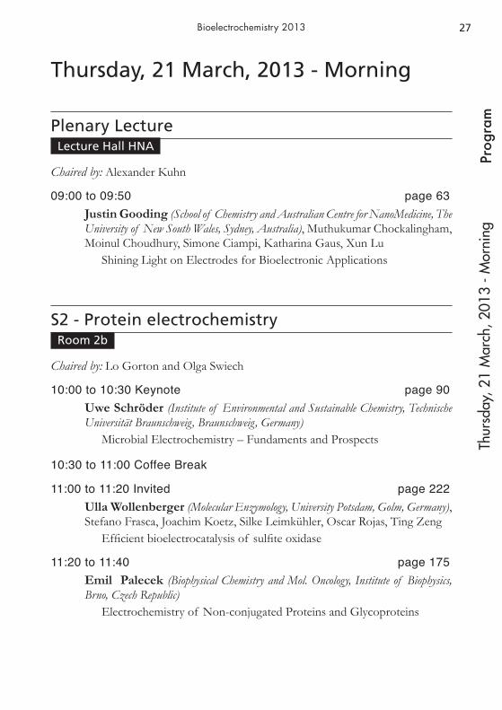
Thursday, 21 March, 2013 - Morning
Plenary LectureLecture Hall HNA
Chaired by: Alexander Kuhn
09:00 to 09:50 page 63Justin Gooding (School of Chemistry and Australian Centre for NanoMedicine, The University of New South Wales, Sydney, Australia), Muthukumar Chockalingham, Moinul Choudhury, Simone Ciampi, Katharina Gaus, Xun Lu
Shining Light on Electrodes for Bioelectronic Applications
S2 - Protein electrochemistryRoom 2b
Chaired by: Lo Gorton and Olga Swiech
10:00 to 10:30 Keynote page 90Uwe Schröder (Institute of Environmental and Sustainable Chemistry, Technische Universität Braunschweig, Braunschweig, Germany)
Microbial Electrochemistry – Fundaments and Prospects
10:30 to 11:00 Coffee Break
11:00 to 11:20 Invited page 222Ulla Wollenberger (Molecular Enzymology, University Potsdam, Golm, Germany), Stefano Frasca, Joachim Koetz, Silke Leimkühler, Oscar Rojas, Ting Zeng
Efficient bioelectrocatalysis of sulfite oxidase
11:20 to 11:40 page 175Emil Palecek (Biophysical Chemistry and Mol. Oncology, Institute of Biophysics, Brno, Czech Republic)
Electrochemistry of Non-conjugated Proteins and Glycoproteins
Thur
sday
, 21
Mar
ch, 2
013
- Mor
ning
28Pr
ogra
mBioelectrochemistry 2013
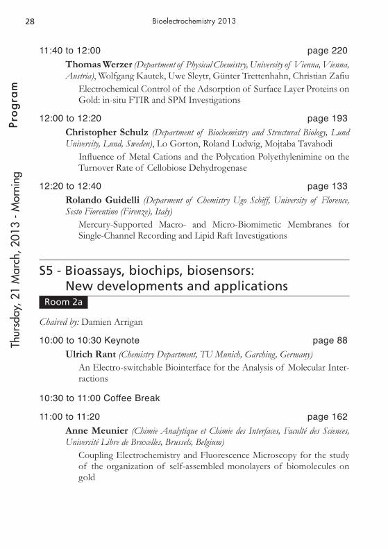
11:40 to 12:00 page 220Thomas Werzer (Department of Physical Chemistry, University of Vienna, Vienna, Austria), Wolfgang Kautek, Uwe Sleytr, Günter Trettenhahn, Christian Zafiu
Electrochemical Control of the Adsorption of Surface Layer Proteins on Gold: in-situ FTIR and SPM Investigations
12:00 to 12:20 page 193Christopher Schulz (Department of Biochemistry and Structural Biology, Lund University, Lund, Sweden), Lo Gorton, Roland Ludwig, Mojtaba Tavahodi
Influence of Metal Cations and the Polycation Polyethylenimine on the Turnover Rate of Cellobiose Dehydrogenase
12:20 to 12:40 page 133Rolando Guidelli (Deparment of Chemistry Ugo Schiff, University of Florence, Sesto Fiorentino (Firenze), Italy)
Mercury-Supported Macro- and Micro-Biomimetic Membranes for Single-Channel Recording and Lipid Raft Investigations
S5 - Bioassays, biochips, biosensors: New developments and applications
Room 2a
Chaired by: Damien Arrigan
10:00 to 10:30 Keynote page 88Ulrich Rant (Chemistry Department, TU Munich, Garching, Germany)
An Electro-switchable Biointerface for the Analysis of Molecular Inter-ractions
10:30 to 11:00 Coffee Break
11:00 to 11:20 page 162Anne Meunier (Chimie Analytique et Chimie des Interfaces, Faculté des Sciences, Université Libre de Bruxelles, Brussels, Belgium)
Coupling Electrochemistry and Fluorescence Microscopy for the study of the organization of self-assembled monolayers of biomolecules on gold
Thur
sday
, 21
Mar
ch, 2
013
- Mor
ning

29
Prog
ram
Bioelectrochemistry 2013
11:20 to 11:40 page 127Neville Freeman (R and D, NanoFlex Ltd, Daresbury, United Kingdom), Andrew Mount, Ilka Schmuser, Reshma Sultana, Jonathan Terry, Anthony Walton
Practical Implications of using Nano-Electrodes for Bioanalytical Mea-surements
11:40 to 12:00 page 165Milena Milutinovic (Institut des Sciences Moléculaires, ENSCBP-NSYSA, Pessac, France), Stéphane Arbault, Dragan Manojlovic, Milica Sentic, Neso Sojic
Electrochemiluminescence Imaging at the Single Bead Level: New Approach to Investigate the ECL Mechanism
12:00 to 12:20 page 206Lutz Stratmann (Department of Analytische Chemie - Bioanalytik und Sensorik, Ruhr-Universität, Bochum, Germany) Magdalena Gebala, Wolfgang Schuhmann
Interface design of an EBV immunoassay based on recombinant native antigens
12:20 to 12:40 page 210Shahida Syed (Division of Pathway Medicine, University of Edinburgh, Edinburgh, United Kingdom), Till. T. Bachmann, Jason Crain, Daniel MacDonald, Andrew Mount, Holger Schulze
Electrochemical Control of DNA Hybridization
S8 - Electrochemistry at cells and tissuesRoom 82
Chaired by: Renata Bilewicz and Pawel Krysinski
10:00 to 10:30 Keynote page 95Joachim Wegener (Institut fuer Analytische Chemie, Chemo- & Biosensorik, Universitaet Regensburg, Regensburg, Germany)
In Vitro Monitoring of Animal Cells by Electrochemical Impedance Ana-lysis
10:30 to 11:00 Coffee Break
Thur
sday
, 21
Mar
ch, 2
013
- Mor
ning

30Pr
ogra
mBioelectrochemistry 2013
Chaired by: Britta Sethson and Izabella Brand
11:00 to 11:20 page 134Manon Guille Collignon (Department of Chemistry UMR 8640, Ecole Normale Supérieure, Paris, France), Christian Amatore, Rémy Fulcrand, Frédéric Lemaître, Yun Li, Anne Meunier, Catherine Sella, Laurent Thouin
Microsystems for Oxidative Stress Electrochemical Detection on Murine Macrophages Population
11:20 to 11:40 page 170Ana Maria Oliveira-Brett (Departamento de Química, Universidade de Coimbra, Coimbra, Portugal)
Human Colon Adenocarcinoma HT-29 cell: Electrochemistry and Nico-tine Stimulation
11:40 to 12:00 page 121Alexander Dubinin (Department of Bioelectrochemistry, Mendeleyev University of Chemical Technology of Russia, Moscow, Russia), T.B Shvets-Teneta-Gurii, G.I. Troshin
Rythms of Wake and Sleep in the rat’s Cerebral Cortex Redox Potential
12:00 to 12:20 page 105Izabella Brand (Institute of Pure and Applied Chemistry, University of Oldenburg, Oldenburg, Germany), Sorge Kelm, Karl-Wilhelm Koch, Martina Nullmeier
Impact of a strong and weak protein-lipid interaction on the structure of a model lipid bilayer
12:20 to 12:40 page 159Stephane Marinesco (Lyon Neuroscience Research Center, Team Wake, Inserm, Universite Claude Bernard Lyon 1, Lyon, France), Natalia Vasylieva
An immobilization method to preserve enzyme specificity in microelec-trode biosensors: consequences for brain glutamate detection
Thur
sday
, 21
Mar
ch, 2
013
- Mor
ning
31
Prog
ram
Bioelectrochemistry 2013
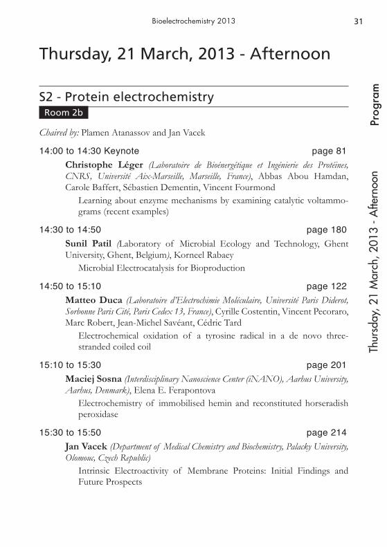
Thursday, 21 March, 2013 - Afternoon
S2 - Protein electrochemistryRoom 2b
Chaired by: Plamen Atanassov and Jan Vacek
14:00 to 14:30 Keynote page 81Christophe Léger (Laboratoire de Bioénergétique et Ingénierie des Protéines, CNRS, Université Aix-Marseille, Marseille, France), Abbas Abou Hamdan, Carole Baffert, Sébastien Dementin, Vincent Fourmond
Learning about enzyme mechanisms by examining catalytic voltammo-grams (recent examples)
14:30 to 14:50 page 180Sunil Patil (Laboratory of Microbial Ecology and Technology, Ghent University, Ghent, Belgium), Korneel Rabaey
Microbial Electrocatalysis for Bioproduction
14:50 to 15:10 page 122Matteo Duca (Laboratoire d’Electrochimie Moléculaire, Université Paris Diderot, Sorbonne Paris Cité, Paris Cedex 13, France), Cyrille Costentin, Vincent Pecoraro, Marc Robert, Jean-Michel Savéant, Cédric Tard
Electrochemical oxidation of a tyrosine radical in a de novo three-stranded coiled coil
15:10 to 15:30 page 201Maciej Sosna (Interdisciplinary Nanoscience Center (iNANO), Aarhus University, Aarhus, Denmark), Elena E. Ferapontova
Electrochemistry of immobilised hemin and reconstituted horseradish peroxidase
15:30 to 15:50 page 214Jan Vacek (Department of Medical Chemistry and Biochemistry, Palacky University, Olomouc, Czech Republic)
Intrinsic Electroactivity of Membrane Proteins: Initial Findings and Future Prospects
Thur
sday
, 21
Mar
ch, 2
013
- Afte
rnoo
n
32Pr
ogra
mBioelectrochemistry 2013
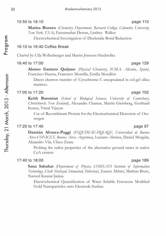
15:50 to 16:10 page 110Marisa Buzzeo (Chemistry Department, Barnard College, Columbia University, New York, USA), Faizunnahar Dewan, Lindsey Walker
Electrochemical Investigation of Diselenide Bond Reduction
16:10 to 16:40 Coffee Break
Chaired by: Ulla Wollenberger and Martin Jönsson-Niedziolka
16:40 to 17:00 page 129Alonso Gamero Quijano (Physical Chemistry, IUMA, Alicante, Spain), Francisco Huerta, Francisco Montilla, Emilia Morallón
Direct electron transfer of Cytochrome C encapsulated in sol-gel silica matrices.
17:00 to 17:20 page 102Keith Baronian (School of Biological Sciences, University of Canterbury, Christchurch, New Zealand), Alexandre Chamas, Martin Giersberg, Gotthard Kunze, Vimal Vijayan
Use of Recombinant Protein for the Electrochemical Detection of Oes-trogen
17:20 to 17:40 page 97Damián Alvarez-Paggi (INQUIMAE-DQIAQF, Universidad de Buenos Aires-CONICET, Buenos Aires, Argentina), Luciano Abriata, Daniel Murgida, Alejandro Vila, Ulises Zitare
Probing the redox properties of the alternative ground states in native CuA centers
17:40 to 18:00 page 189Sana Sabahat (Department of Physics, COMSATS Institute of Information Technology, Chak Shahzad, Islamabad, Pakistan), Zareen Akhter, Mathias Brust, Naveed Kausar Janjua
Electrochemical Quantification of Water Soluble Ferrocene Modified Gold Nanoparticles onto Electrode Surface
Thur
sday
, 21
Mar
ch, 2
013
- Afte
rnoo
n
33
Prog
ram
Bioelectrochemistry 2013
S5 - Bioassays, biochips, biosensors: New developments and applications
Room 2a
Chaired by: Ulrich Rant
14:00 to 14:30 Keynote page 72Elena E. Ferapontova (iNANO, Aarhus University, Aarhus, Denmark)
Electronic properties of the surface-tethered DNA duplex
14:30 to 14:50 Invited page 104Laurent Bouffier (Institute of Molecular Sciences, Univ. Bordeaux - CNRS, Pessac, France), Sébastien Bonhommeau, Pierre-Alexis Condon, Patrick Garrigue, Sophie Lecomte, Neso Sojic, David Talaga
Electrochemical and Raman Spectroscopic Detection of DNA Hybridi-sation with Pyridoacridine Intercalators
14:50 to 15:10 page 174Ilaria Palchetti (Dipartimento di Chimica, Università di Firenze, Sesto Fiorentino, Italy), Francesca Bettazzi, Diego Voccia
Electrochemical Biosensing Platforms for microRNA Detection
15:10 to 15:30 page 112Rui Campos (Interdisciplinary Nanoscience Center (iNANO), Aarhus University, Aarhus, Denmark), Elena E. Ferapontova, Michael R. Horsman
Electrochemical analysis of miRNA deregulated during the hypoxia conditions
15:30 to 15:50 page 126Miroslav Fojta (Department of Biophysical Chemistry and Molecular Oncology, Institute of Biophysics, ASCR, v.v.i., Brno, Czech Republic), Jana Balintová, Aleš Danhel, Ludek Havran, Michal Hocek, Petra Ménová, Veronika Raindlova, Jan Spacek, Pavlína Vidláková, Zdenka Vychodilová
Utilization of organic electroactive moieties for redox DNA labelling and electrochemical monitoring of modified DNA synthesis
15:50 to 16:10 page 168Gilbert Nöll (Department of Chemistry-Biology, Siegen University, Siegen, Germany)
Monitoring DNA Hybridization by Faradaic Impedance Spectroscopy in Combination with QCM-D Measurements
Thur
sday
, 21
Mar
ch, 2
013
- Afte
rnoo
n

34Pr
ogra
mBioelectrochemistry 2013
16:10 to 16:40 Coffee Break
Chaired by: Mathieu Etienne
16:40 to 17:00 page 151Mathieu Lazerges (UPCGI, U 1022 INSERM, UMR 8151 CNRS, ENSCP, Université Paris Descartes, Paris, France), Fethi Bedioui, Vanna.-T. Tal
Label-free electrochemical DNA-biosensor : setup for detection in microliter samples and miniaturization
17:00 to 17:20 page 207Qiang Su (Fak.IV/Dept. Chemie-Biologie, Universität Siegen, Siegen, Germany)
Designing a Reusable and Label-Free Sensing Platform for Specific Oli-gonucleotides: Optimization of Sensor Response Based On Surface Plasmon Fluorescence Spectroscopy
17:20 to 17:40 page 100 Artavazd Badalyan (Department of Molecular Enzymology, Institute for Biochemistry and Biology, University of Potsdam, Potsdam, Germany), Marlen Dierich, Silke Leimkühler, Sascha Pöller, Wolfgang Schuhmann, Ulla Wollenberger
Bioelectrocatalysis of PaoABC-aldehyde oxidoreductase from E. Coli: the ionic strength effect and biosensor for benzaldehyde
17:40 to 18:00 page 195Alina Sekretaryova (Department of Chemisrty, Lomonosov Moscow State University, Moscow, Russia), Arkady Karyakin
Unsubstituted Phenothiazine as a New Efficient Electron Transfer Mediator for OxidasesTh
ursd
ay, 2
1 M
arch
, 201
3 - A
ftern
oon
35
Prog
ram
Bioelectrochemistry 2013
S8 - Electrochemistry at cells and tissuesRoom 82
Chaired by: Ana Maria Oliveira-Brett and Cigdem Yidirim
14:00 to 14:30 Keynote page 75Lo Gorton (Department of Analytical Chemistry/Biochemistry and Structural Biology, Lund University, Lund, Sweden), Cecilia Hägerhäll, Kamrul Hasan, Donal Leech, Sunil A. Patil
Mediated and Direct Electrochemical Communication between Shewa-nella Oneidensis MR-1 and Electrodes
14:30 to 14:50 Invited page 123Jenny Emnéus (Micro and Nanotechnology, Technical University of Denmark, Kgs. Lyngby, Denmark), M. Adamovski, L. Amato, A. Boisen, M. Carminati, V. Coman, M. Dimaki, M. Dufva, G. Ferrari, A. Ghio, A. Heiskanen, S. Keller, Zs. Keresztes, M. Kokaia, E. Landini, A. Martinez Serrano, R. Raiteri, T. Ramos Moreno, D. Sabourin, M. Sampietro, W.E. Svendsen, M. Vergani, Ulla Wollenberger, K. Zor
Microfluidic Electrochemical Lab-on-a-Chip Systems
14:50 to 15:10 page 225Cigdem Yildirim (Department of Analytical Biochemistry, University of Potsdam, Potsdam, Germany), Miriam Adamovski, Dafna Benayahu, Carsten Beta, Matthias Gerhardt, Helmar Leonhardt, Ulla Wollenberger
Electrochemical Assay on Osteoblastic Cells on a Sensor Chip
15:10 to 15:30 page 101Aliaksandr Bandarenka (Center for Electrochemical Sciences, Ruhr-Universität Bochum, Bochum, Germany), Ramon Bragos, Benjamin Sanchez
Real-time Analysis of Bioimpedance Spectra
15:30 to 15:50 page 144Ritu Kataky (Department of Chemistry, Durham, Durham, United Kingdom), Rui Campos, Anne Krol
Electrochemical investigations of lipid membranes, membrane associa-ted molecules and functionalized nanoparticles
Thur
sday
, 21
Mar
ch, 2
013
- Afte
rnoo
n

36Pr
ogra
mBioelectrochemistry 2013
15:50 to 16:10 page 155Britta Lindholm-Sethson (Department of Chemistry, Umeå University, Umeå, Sweden), Mohammad Yaser Khani Meynaq
Ionic Permeability of Lipid Cubic Phases. An Investigation with Electro-chemical Impedance
16:10 to 16:40 Coffee Break
S7 - Interdisciplinary bioelectrochemistry: Hyphenated techniques; impact from other fields on bioelectrochemistry
Room
Chaired by: Alexander Kuhn and Ian Burgess
16:40 to 17:00 page 204Zbigniew Stojek (Faculty of Chemistry, University of Warsaw, Warsaw, Poland), Michal Bystrzejewski, Mikolaj Donten, Mateusz Donten, Agata Kowalczyk, Anna M. Nowicka
Carbon-Encapsulated Iron Nanoparticles as New Ferromagnetic Matrix for Oxygen Reduction in the Presence of Immobilized Laccase
17:00 to 17:20 page 221Gunther Wittstock (Department of Pure and Applied Chemistry, Carl v. Ossietzky University of Oldenburg, Oldenburg, Germany)
How Gentle Can a Soft Electrode Array Be?
17:20 to 17:40 page 103Pascal Beese (Interface Chemistry and Surface Engineering, Max-Planck-Institut für Eisenforschung GmbH, Düsseldorf, Germany), Dennis Enning, Karl J.J. Mayrhofer, Martin Stratmann, Hendrik Venzlaff, Friedrich Widdel
Monitoring anaerobic microbially influenced corrosion with electroche-mical frequency modulation
17:40 to 18:00 page 115Jan Clausmeyer (Analytische Chemie and Center for Electrochemical Sciences, Ruhr-Universität Bochum, Bochum, Germany), Jörg Henig, Nicolas Plumeré, Wolfgang Schuhmann
Scanning Droplet Cell for Chemoselective Patterning via Local Elec-troactivation of Protected Quinone Monolayers
Thur
sday
, 21
Mar
ch, 2
013
- Afte
rnoo
n

37
Poste
r Pre
sent
atio
n
Bioelectrochemistry 2013
Sess
ion
1 - M
onda
y
Poster Presentation Session 1
INTER
NATIO
NAL SO
CIETY OF ELECTR
OCHEM
ISTRY•The Bioelectrochemical Society

38Po
ster P
rese
ntat
ion
Bioelectrochemistry 2013Se
ssio
n 1
- Mon
day
s1 - Symposium on the occasion of the 80th birthday of Adam Heller
S1-001Kamrul Hasan (Department of Biochemistry and Structual Biology, Lund University, Lund, Sweden), Vera Eßmann, Kamil Górecki, Cecilia Hägerhäll, Sunil A. Patil, Wolfgang Schuhmann
Electrochemical communication of heterotrophically grown Rhodobac-ter capsulatus with graphite electrodes via various polymeric mediators
S1-002Shuji Nakanishi (RCAST, The University of Tokyo, Tokyo, Japan)
Redox responsive regulation of microbial metabolism activity in an iron reducing bacterium
S1-003Stephan Vogt (Nöll Junior Research Group, University of Siegen, Siegen, Germany), Gilbert Nöll
Spectroelectrochemical Investigation on Glucose Oxidase: pH-depen-dent Determination of the Redox Potential
s2 - Protein electrochemistry
S2-001Damián Álvarez Paggi (INQUIMAE, Universidad de Buenos Aires, Buenos Aires, Argentina), María A. Castro, Daniel Murgida, Rafael Radi, Verónica Tórtora
Electrostatically-Driven Second Sphere Ligand Switch Between High and Low Reorganization Energy Forms of Native Cytochrome c
S2-002Lucia Becucci (Department of Chemistry, University of Padova, Padova, Italy), Rolando Guidelli
Electrochemical Techniques as Versatile Tools for the Investigation of the Mechanism of Peptide-Induced Membrane Permeabilization
39
Poste
r Pre
sent
atio
n
Bioelectrochemistry 2013
Sess
ion
1 - M
onda
y
S2-003Mahdi Dargahi (Department of Chemical Engineering, McGill University, Montreal, Canada), Sasha Omanovic
The Influence of Surface Charge on the Interactive Adsorption Behavior of Fibrinogen on a Gold Surface
S2-004Mahdi Dargahi (Department of Chemical Engineering, McGill University, Montreal, Canada), Mari T. Kaartinen, Aisha Mousa, Valentin Nelea, Sasha Omanovic
Electrochemically-assisted Immobilization of Fibronectin on Metal Sur-faces: Enhancement of Cell/Surface Interactions
S2-005Victor Diculescu (Departamento de Química, Universidade de Coimbra, Coimbra, Portugal), Oana Popa
Electrochemical Study of the Bcr-abl Tyrosine Kinase Inhibitor Danu-sertib
S2-006Thomas Dietz (Institute for Biochemistry and Biology, University of Potsdam, Potsdam, Germany), Silke Leimkühler, Konstanze Stiba, Ulla Wollenberger
Is superoxide involved in human sulfite oxidase (hSOx) catalysis?
S2-007Francisco Fabregat-Santiago (Department of Physics, Universitat Jaume I, Castello de la Plana, Spain), Paulo R. Bueno, Rocío Cejudo, Jason J. Davis
The Effect of Capacitances and Resistances in the Electrochemistry of Electroactive Self-Assembled Monolayers
S2-008Artur Fandrich (Department of Biosystems Technology, Technical University of Applied Sciences, Wildau, Germany), Sven Christian Feifel, Lo Gorton, Fred Lisdat, Roland Ludwig
Nanoscaled Protein Architectures with CDH on Electrodes for Selective Analyte Detection
S2-009Aniko Killyéni (Department of Physical Chemistry, Babes-Bolyai University, Cluj-Napoca, Romania), Lo Gorton, Ionel Catalin Popescu, Maria Yakovleva
Effect of Deglycosylation on the Selectivity of Agaricus meleagris Pyra-nose Dehydrogenase Modified Electrodes
40Po
ster P
rese
ntat
ion
Bioelectrochemistry 2013Se
ssio
n 1
- Mon
day
S2-010Svenja Kochius (Department of Biochemical Engineering, DECHEMA Research Institute, Frankfurt, Germany), Dirk Holtmann, Jens Schrader
Electrochemical regeneration of oxidized nicotinamide cofactors in a scalable reactor
S2-011Nur Azura Mohd Said (Life Sciences Interface (LSI) Group, Tyndall National Institute (UCC), Cork, Ireland), Gregoire Herzog, Mingzhi Liang, Vladimir Ogourtsov, Michael Prentice
Electrochemical and Atomic Force Microscopy Studies of PduA Shell Protein Immobilisation on Gold Electrode Surface
S2-012Severino Carlos Oliveira (Química, Universidade de Coimbra, Coimbra, Portugal), Ana Maria Oliveira-Brett, Inês Santarino
Electrochemical detection of DNA-Anticancer Antibody Rituximab Interaction
41
Poste
r Pre
sent
atio
n
Bioelectrochemistry 2013
Sess
ion
1 - M
onda
y
s3 - Electroporation and biomedical applications
S3-001Mario Alberto Ascencio-Pinedo (Department of Chemical Engineering, McGill University, Montreal, Canada), Sasha Omanovic
Investigation of the corrosion mechanism of WE43 Mg-alloy for biode-gradable implant applications
S3-002Tanja Blagus (Department of Experimental Oncology, Institute of Oncology Ljubljana, Ljubljana, Slovenia), Maja Cemazar, Bostjan Markelc, Matej Rebersek, Gregor Sersa
New methodological approach for tracking systemic and local uptake of macromolecule enhanced by electroporation in vivo
S3-003Mustafa Fincan (Department of Food Engineering, Erciyes University, Kayseri, Turkey), Seyma Avcý, Fatma Gundogdu, Betul Oskaybas
Effects of Different Pulse Electric Field Parameters on Electropermea-bilization of Fresh Rose Petals
S3-004Sasa Haberl (Laboratory of Biocybernetics, University of Ljubljana, Faculty of Electrical Engineering, Ljubljana, Slovenia), Marko Jarc, Damijan Miklavcic, Ales Strancar
Optimization of electroporation protocol for extracting plasmid DNA from E. coli
S3-005Katarzyna Kilan (Jerzy Haber Institute of Catalysis and Surface Chemistry, Polish Academy of Sciences , Cracow, Poland), Krzysztof Szczepanowicz, Lilianna Szyk-Warszyñska, Piotr Warszyñski
Influence of calcium ions on the buildup and permeability of multilayer polymer films
S3-006Katarzyna Krukiewicz (Department of Physical Chemistry and Technology of Polymers, Silesian University of Technology, Gliwice, Poland), Jerzy Zak
Electrochemical and spectroscopic characterization of PEDOT matrix containing iso-butyl-propanoic phenolate

42Po
ster P
rese
ntat
ion
Bioelectrochemistry 2013Se
ssio
n 1
- Mon
day
S3-007Léa Lesueur (UMR8203, CNRS, Villejuif, France), Franck André, Lluis M. Mir
Improvement of both cell survival and efficacy for large plasmids elec-trotransfer in MSC
S3-008Bostjan Markelc (Department of Experimental Oncology, Institute of Oncology Ljubljana, Ljubljana, Slovenia), Maja Cemazar, Gregor Sersa
Mechanisms associated with vascular-disrupting action of electrochemo-therapy: intravital microscopy on a single tumor blood vessel level
S3-009Guillermo Marshall (Laboratorio de Sistemas Complejos, DC, University of Buenos Aires, Buenos Aires, Argentina), Pieranna Chiarella, Stefania De Santis, Vito M. Fazio, Nahuel Olaiz, Emanuela Signori, Alejandro Soba, Pablo Turjanski
EGT protocols: the role of electroporation based techniques, hyaluroni-dase and pH effects in the permeabilization of tissue fibers
S3-010Damijan Miklavcic (Faculty of Electrical Engineering, Department for Biomed Eng, University of Ljubljana, Ljubljana, Slovenia)
COST TD1104 Action – A Network for Development of Electropora-tion-based Technologies and Treatments
S3-011Tristan Nagy (Dept. of Physical Chemistry, University of Vienna, Vienna, Austria), Oskar Armbruster, Wolfgang Kautek, Ulrich Pacher, Günter Trettenhahn
Laser-Pulse-Induced in-situ Diagnostics of Processes at Solid-Fluid Inter-faces
S3-012Ewa Nazaruk (Chemistry Department, University of Warsaw, Warsaw, Poland), Renata Bilewicz, Ewa Górecka, Monika Szlêzak
Tailored Lipidic Cubic Phases as Novel Drug Delivery System
S3-013Gianpiero Pataro (Institute for Electromagnetic Sensing of the Environment, National Research Council of Italy, Naples, Italy), Giovanna Ferrari, Stefania Romeo, Anna Sannino, Maria Rosaria Scarfì, Olga Zeni
A Nanosecond, High-Voltage Pulse Generator for Electric Pulse Appli-cation to Low Conductivity Liquid Media
S3-014Denis Pavliha (Faculty of Electrical Engineering, Department for Biomed Eng, University of Ljubljana, Ljubljana, Slovenia), Maja M. Mušic, Damijan Miklavcic, Gregor Serša
Liver Segmentation for Electrochemotherapy Treatment Planning

43
Poste
r Pre
sent
atio
n
Bioelectrochemistry 2013
Sess
ion
1 - M
onda
y
S3-015Andraz Polak (Faculty of Electrical Engineering, University of Ljubljana, Ljubljana, Slovenia), Peter Kramar, Damijan Miklavcic, Mounir Tarek
Molecular dynamics simulation of Archaea Aeropyrum pernix membrane
S3-016Veronique Preat (Louvain drug Research Institute, Université Catholique de Louvain, Brussels, Belgium), Olivier Schakman, Cédric Szpirer, Gaelle Vandermeulen, Kevin Vanvarenberg
Electrotransfer of pStaby: A new safe and efficient DNA vaccine vector devoid of antibiotic resistance marker
S3-017Jolanta Saczko (Department of Medical Biochemistry, Medical University, Wroclaw, Poland), Katarzyna Biezunska-Kusiak, Anna Choromanska, Malgorzata Daczewska, Malgorzata Kotulska, Julita Kulbacka, Nina Rembialkowska, Joanna Rossowska
Doxorubicin delivery enhanced by electroporation to colon adenocarci-noma cells with P-gp overexpression
S3-018Gintautas Saulis (Department of Biology, Vytautas Magnus University, Kaunas, Lithuania), Saulius Balevicius, Aiste Bitinaite, Ruta Maciuleviciene, Rita Saule, E. Shatkovskis, Vitalij Stankevic, Arunas Stirke, Nerija Zurauskiene
System For Nanoporation of Biological Cells Based on Optically-Trigge-red High-Voltage Spark-Gap Switch
S3-019Emanuela Signori (Department of Biomedicine, CNR-Institute of Translational Pharmacology, Rome, Italy), Pieranna Chiarella, Mariangela De Robertis, Stefania De Santis, Vito Michele Fazio, Emanuela Massi
Alternative Therapy Protocols For Tumours Treatment: DNA Vaccina-tion Mediated By Electrotransfer
S3-020Wojciech Simka (Faculty of Chemistry, Silesian University of Technology, Gliwice, Poland), Beata Cwalina, Tadeusz Gorewoda, Marzena Kik-Jaworska, Anna Klimczyk, Agnieszka Krzakala, Artur Maciej, Joanna Michalska, Anna Osyczka, Grzegorz Tylko, Magdalena Widziolek
Plasma Electrolytic Oxidation of Ti-13Nb-13Zr Alloy - Corrosion and Bioactivity Investigations
S3-021Wojciech Simka (Faculty of Chemistry, Silesian University of Technology, Gliwice, Poland), Agnieszka Krzakala, Joanna Michalska, Robert Socha, Maciej Sowa
Anodic Oxidation of Tantalum in Silicate Solutions

44Po
ster P
rese
ntat
ion
Bioelectrochemistry 2013Se
ssio
n 1
- Mon
day
s4 - Design of the interface between biological recognition elements and electrodes including new tools and measuring techniques
S4-001Harold Braustein (Molecular Microbiology and Biotechnology, Tel Aviv University, Jerusalem, Israel)
New analytical tools for electrochemical biomarkers detection implying solid state biological recognition kits on nano-modified electrodes
S4-002Anna Koper (Chemistry, Maria Curie-Sklodowska University, Lublin, Poland), Malgorzata Grabarczyk, Agnieszka Nosal-Wiercinska, Cecylia Wardak
The Different Electrochemical Aspects of Studies of Humic Substances as a Natural Biopolymers Present in Environmental Samples
S4-003Agata Kowalczyk (Faculty of Chemistry, Warsaw University, Warsaw, Poland), Michal Fau, Anna M. Nowicka, Zbigniew Stojek, Marcin Strawski
Phenyl Layers – Matrix for Specific Immobilization of Biologically Important Compounds
S4-004Jan Pawlowski (Department of Chemistry, Univeristy of Warsaw, Warsaw, Poland), Slawomir Sek
Structural Diversity and Nanomechanical Stability of Solid-Supported Phospholipid Bilayers Formed by Vesicle Fusion
S4-005Piyanut Pinyou (Analytische Chemie - Elektroanalytik & Sensorik, Ruhr-Universität Bochum, Bochum, Germany), Natalia Guerrero Alburquerque, André Laschewsky, Nicolas Plumeré, Wolfgang Schuhmann, Jan Szeponik, Erik Wischerhoff
Control of Small Molecules Diffusion in Temperature-Responsive Poly-mers Films at Heatable Electrode
S4-006Mykola Rozhitskii (Laboratory of Analytical Optochemotronics, Kharkiv National University of Radioelectronics, Kharkiv, Ukraine), Dmytro Snizhko
Ultra Fast Cyclic Voltammetry for Bioelectrochemical Assays

45
Poste
r Pre
sent
atio
n
Bioelectrochemistry 2013
Sess
ion
1 - M
onda
y
S4-007Carlos Sanchis (Química-Física & Instituto de Materiales, University of Alicante, Alicante, Spain), Emilia Morallon, Horacio J. Salavagione, Jose Miguel Sansano
An innovative route for the functionalization of PANI: Comparative study of attached vs. adsorbed ferrocene
S4-008Sylwia Strzalkowska (Department of Chemistry, University of Warsaw, Warsaw, Poland), Andrzej Lewenstam, Magdalena Maj-Zurawska, Tomasz Sokalski, Wladyslaw Wieczorek
Ordered biomaterials in electrochemical sensors
S4-009Christoph Traunsteiner (Department of Physics, TU München, Garching, Germany), Julia Kunze
Electrochemical and Scanning Probe Microscopy Studies of Laccase on Au(111) Surfaces
S4-010Annalisa Vacca (Dipartimento di Ingegneria Meccanica Chimica e dei Materiali, Università degli Studi di Cagliari , Cagliari, Italy)
Coating of gold substrates with polyaniline through electrografting of diazonium salts
S4-011Tal Yoetz-Kopelman (Molecular Microbiology and Biotechnology; Phys. Electronics, Tel Aviv University, Tel Aviv, Israel)
Novel Design of Impedimetric Affinity Biosensor based on Metal-Pro-tein Hybrids and a New Polymeric Adhesion Layer
S4-012Yongchun Zhu (Department of Chemistry, Shenyang Normal University, Shenyang, China), Yue Dong, Hongyan Gao, Jie Lu, Chunyan Pang
An amperometric urea biosensor based on in-situ secretory antibody modified electrode from wild winter Jasmine petal cells and inductive effect of urea

46Po
ster P
rese
ntat
ion
Bioelectrochemistry 2013Se
ssio
n 1
- Mon
day
s6 - Enzymatic and microbial biofuel cells
S6-001Sidney Aquino Neto (Department of Chemistry, University of São Paulo, Ribeirão Preto, Brazil), Franciane P. Cardoso, Laís B. Crepaldi, Paula G. Fenga, Matthew T. Meredith, Shelley Minteer, Adalgisa R. De Andrade, Thiago S. Almeida
The use of MWCNTs/methylene green electrodes to enhance NADH electrocatalysis in ethanol biofuel cell
S6-002Magdalena Blicharska (Department of Chemistry, University of Warsaw, Warsaw, Poland), Anna Dobrzeniecka, Pawel Kulesza, Jadwiga Stroka, Sylwia Zoladek
Application of polyoxometallate-modified gold nanoparticles to oxida-tion of glucose at physiological pH
S6-003Tunc Catal (Department of Molecular Biology and Genetics, Uskudar University, Istanbul, Turkey)
Effect of Heavy Metals from Wastewater on Electricity Generation in Microbial Fuel Cells
S6-004Anna Dobrzeniecka (Department of Chemistry, University of Warsaw, Warsaw, Poland), Pawel Kulesza, Wolfgang Schuhmann, Aleksandar Zeradjanin
Bioelectrocatalytic and electrocatalytic oxygen and hydrogen peroxide reduction of multicomponent films at a physiological pH electrolyte
S6-005Marta Gierwatowska (Department of Chemistry, University of Warsaw, Warsaw, Poland), Barbara Kowalewska, Pawel Kulesza
Development of integrated mediating systems utilizing ultra-thin films of conducting polymers and functionalized carbon nanotubes for bio-electrocatalytic oxidation of glucose
S6-006Maria José González-Guerrero (Instituto de Microelectrónica de Barcelona, IMB-CNM (CSIC), Universidad Autónoma de Barcelona, Barcelona, Spain), F. Javier del Campo, Juan Pablo Esquivel, Shelley Minteer, Neus Sabaté
Modified Pyrolized Photoresist Bioelectrodes for Membraneless Glu-cose/O2 Enzyme Microfluidic Fuel Cells

47
Poste
r Pre
sent
atio
n
Bioelectrochemistry 2013
Sess
ion
1 - M
onda
y
S6-007Minerva Guerra-Balcázar (División de Investigación y Posgrado, Facultad de Ingeniería, Universidad Autónoma de Querétaro, Santiago de Querétaro, Mexico), Gerardo Arriaga, Francisco M. Cuevas-Muñiz, Christophe Innocent, Janet Ledesma-García, Berenice López-González, Louis Renaud, Sophie Tingry
Coupled enzymatic and inorganic oxygen electroreduction reactions to increase performances of a microfluidic biofuel cell
S6-008Jia Shin Ho (Division of Chemistry and Biological Chemistry, Nanyang Technological University, Singapore), Chee-Seng Toh
Membrane-Based UV-Powered Low Wattage Biofuel Cell Sensor System
S6-009Ivan Kazarinov (Department of Physical Chemistry, Saratov State University, Saratov, Russia), Oleg Ignatov, Anna Ignatova, Mariya Naumova
Kinetics of bioelectrocatalytic glucose oxidation by Escherichia Coli in the presence of exogenic mediators
S6-010Donal Leech (Department of Chemistry, National University of Ireland Galway, Galway, Ireland), Partha Jana, Krishna Katuri, Paul Kavanagh, Amit Kumar, Piet Lens, Raghavulu Sapireddy
Biofilm formation on graphite anode surfaces for application to micro-bial electrochemical cells
S6-011Berenice López González (Facultad de Química, Universidad Autónoma de Querétaro, Santiago de Querétaro, Mexico), Francisco M. Cuevas-Muñiz, Minerva Guerra-Balcázar, Janet Ledesma-García, Vanessa Vallejo Becerra
Biocathode based in Laccase on Vulcan XC-72 prepared by adsorption method
S6-012Dominika Lyp (Department of Chemistry, University of Warsaw, Warsaw, Poland), Renata Bilewicz, Pawel Krysinski, Krzysztof Stolarczyk
Biobattery Based on Carbon Nanotube Structured Biocathode and Pla-ted Zinc Anode
S6-013Mickaël Rimboud (Laboratoire de Génie Chimique, Université de Toulouse, Toulouse, France)
Screening Different Sludges from Sewage Treatment Led to Different Microbial Electrodes

48Po
ster P
rese
ntat
ion
Bioelectrochemistry 2013Se
ssio
n 1
- Mon
day
S6-014Zane Rutkovska (Department of Microbiolgy and Biotechnology, University of Latvia, Riga, Latvia), Ilze Dimanta, Arturs Gruduls, Janis Kleperis, Vizma Nikolajeva
Comparison of commercial and custom made microbial fuel cells, using bacterial substrates from wastewater treatment plants of Latvia as a sub-strate.
S6-015Woonsup Shin (Department of Chemistry, Sogang University, Seoul, Korea)
Electrochemical Activation of Carbon Dioxide to Formate by Moorella Thermoacetica and Clostridium formicoaceticum
S6-016Nadèje Tekaya (Institut des Sciences Analytiques, Université Claude Bernard Lyon 1, Villeurbanne, France), Hatem Ben Ouada, Hafedh Ben Ouada, Nicole Jaffrezic-Renault, Florence Lagarde, Olga Saiapina
Ultra-sensitive Conductometric Detection of Pesticides Based on Inhibi-tion of Esterase Activity from Arthrospira platensis
S6-017Mieke C.A.A. van Eerten-Jansen (Environmental Technology, Wageningen University, Wageningen, Netherlands), Cees J.N Buisman, Tim I.M Grootscholten, Hubertus V.M. Hamelers, Tom H.J.A. Sleutels, Kirsten J.J. Steinbusch, Annemiek Ter Heijne
Bioelectrochemical Production of Caproate and Caprylate from Acetate by Mixed Cultures
S6-018Jeevanthi Vivekananthan (Analytische Chemie,Elektroanalytik&Sensorik, Ruhr-Universität Bochum, Bochum, Germany), Wolfgang Schuhmann
A comparative study of carbon-based electrodes for direct electron transfer with multicopper oxidases
S6-019Stéphanie Ketep (Laboratoire de Génie Chimique de Toulouse, Institut Polytechnique de Toulouse, Toulouse, France)Alain Bergel, Wafa Achouak, Marie Bertrand, Eric Fourest
An Innovative Procedure for the Construction of Microbial Bioanodes for the Treatment of Paper Mill Effluents in Microbial Fuel Cells

49
Poste
r Pre
sent
atio
n
Bioelectrochemistry 2013
Sess
ion
2 - W
edne
sday
INTER
NATIO
NAL SO
CIETY OF ELECTR
OCHEM
ISTRY•The Bioelectrochemical Society
Poster Presentation Session 2

50Po
ster P
rese
ntat
ion
Bioelectrochemistry 2013Se
ssio
n 2
- Wed
nesd
ay
s5 - Bioassays, biochips, biosensors: New developments and applications
S5-001Stéphane Arbault (Institute of Molecular Sciences, CNRS UMR 5255, University of Bordeaux, Pessac, France), Salem Ben-Amor, Anne Devin, Michel Rigoulet, Neso Sojic, Emmanuel Suraniti, Suresh Vajrala
Fluorescence Microscopy of Isolated Mitochondria Coupled with Elec-trochemical Detection of Reactive Oxygen Species at The Single Orga-nelle Level
S5-002Martin Bartošík (RECAMO, Masaryk Memorial Cancer Hospital, Brno, Czech Republic), Roman Hrstka, Emil Palecek, Mojmír Trefulka, Boøivoj Vojtìšek
Simple label-based electrochemical assay for detection of microRNAs as potential cancer biomarkers
S5-003Maria Bosserdt (Fraunhofer Institute for Biomedical Engineering (IBMT), university Potsdam, Potsdam-Golm, Germany), Nenad Gajovic-Eichelmann, Frieder Wolfram Scheller
First Electrochemically active Protein MIPS5-004
Christopher Brett (Department of Chemistry, University of Coimbra, Coimbra, Portugal), Aziz Amine, Aisha Attar, Ricardo Carvalho, M. Emilia Ghica
Inhibitor Effect of Heavy Metal Cations at Redox-Mediated Enzyme Biosensors
S5-005Ariadna Brotons (Química Física, University of Alicante, Alicante, Spain), Craig E. Banks, Jesus Iniesta, Vicente Montiel, Jose Solla-Gullón, Francisco José Vidal-Iglesias
Voltammetric behaviour of free DNA bases, methylcytosine and oligo-nucleotides at disposable screen printed graphite electrode platforms
S5-006Bogdan Bucur (Bioanalysis Center, National Institute of R&D for Biological Sciences, Bucharest, Romania), Ana Chira, Gabriel-Lucian Radu, Maria-Cristina Radulescu
Synthesis of 4,4’-Dipyridine Derivatives for Immobilization on the Elec-trode Surface
51
Poste
r Pre
sent
atio
n
Bioelectrochemistry 2013
Sess
ion
2 - W
edne
sday
S5-007Anna Celebanska (Department of Electrode Processes, Institute of Physical Chemistry PAS, Warsaw, Poland), Marcin Filipiak, Martin Jonsson-Niedziolka, Olga Krysiak, Marcin Opallo
Biosensor based on the layer-by-layer method and a new sol-gel matrix for detection of highly toxic organophosphate pesticides
S5-008Andrea Contin (Analytische Chemie, Ruhr-Universität Bochum, Bochum, Germany), Dmitrii A. Guschin, Véronique Lapeyre, Sascha Pöller, Valerie Ravaine, Wolfgang Schuhmann
New Osmium Loaded Hybrid Microgels as Biosensors with Controlled Redox Centers Nano-Gaps
S5-009Cecilia Cristea (Department of Analytical Chemistry, Faculty of Pharmacy, Iuliu Hatieganu, University of Medicine & Pharmacy, Cluj-Napoca, Romania), Anca Florea, Giovanna Marrazza, Robert Sandulescu
Electrochemical immunoassay for the detection of MUC1 cancer bio-marker
S5-010Ales Danhel (Department of Biophysical Chemistry and Molecular Oncology, Institute of Biophysics, AS CR, v.v.i., Brno, Czech Republic), Miroslav Fojta, Ludek Havran, Michal Hocek, Hana Pivonkova, Veronika Raindlova
Novel DNA Redox Labels at Silver Solid Amalgam ElectrodeS5-011
Sema Demirci Uzun (Polymer Science and Technology Department, Middle East Technical University, Ankara, Turkey), Naime Akbasoglu Ünlü, Fulya Ekiz Kanýk, Duygu Kozanoglu, Emren Nalbant Esentürk, Suna Timur, Levent Toppare
Surface Modification of a Novel Benzimidazole Containing Polymer for Biomolecule Immobilization and Biosensing Applications
S5-012Fulya Ekiz Kanik (Biotechnology, Middle East Technical University, Ankara, Turkey), Müfit Bahadir, Marit Kolb, Suna Timur, Levent Toppare
Development of a Conducting Polymer Based Amperometric Acetyl-choline Biosensor for Pesticide Detection
52Po
ster P
rese
ntat
ion
Bioelectrochemistry 2013Se
ssio
n 2
- Wed
nesd
ay
S5-013Hanna Elzanowska (Faculty of Chemistry, University of Warsaw, Warsaw, Poland), Agnieszka Gniazdowska, Magdalena Maj-Zurawska, Adriana Palinska-Saadi
Opposing effects of riboflavin and methylene blue on DNA oxidationS5-014
Fatma Bilge Emre (Department of Elementary Education, Ýnönü University, Malatya, Turkey), Yasemin Aslan Udum, Funda Sayilkan, Levent Toppare
Carbazole Unit and TiO2 Nanoparticles Containing Biosensor and Its Applications
S5-015Fatma Bilge Emre (Ýnönü University, Malatya, Turkey), Evren Aslan Gurel, Fulya Ekiz Kanik, Melis Kesik, Levent Toppare
Benzoimidazole Based Conducting Polymer and PMMA/Clay Nano-composite Containing Biosensor and Its Applications
S5-016Fabiane Galdino (Department of Chemistry and Biotechnology, Federal University of Alagoas, Maceio, Brazil), Jonathan Caranto, Carlos Garcia, Donald Kurtz
Electrochemical Detection of Superoxide Using SOR Immobilized on Carbon Nanotubes
S5-017Isabel Patricia Garrido Fernandes (Departamento de Química, Universidade de Coimbra, Coimbra, Portugal), Carlos Severino Bezerra Oliveira, Ana Maria Oliveira-Brett, Angelo Pinto, Barbara Silva
Isatin Halogen-Derivatives Anodic BehaviourS5-018
Stefanie Grützke (Analytische Chemie - Elektroanalytik und Sensorik, Ruhr-Universität Bochum, Bochum, Germany), Magdalena Gebala, Sascha Pöller, Wolfgang Schuhmann
Detection of biomolecules at nanostructured surfaces by combining of electrochemistry and spectroscopy
S5-019Vinod Kumar Gupta (Department of Chemistry, Indian Institute of Technology Roorkee, Roorkee, India), Shilpi Agarwal, A. Dwivedi, R. Jain, R. Mishra
Electrochemical determination of antihypertensive drug irbesartan in pharmaceuticals

53
Poste
r Pre
sent
atio
n
Bioelectrochemistry 2013
Sess
ion
2 - W
edne
sday
S5-020Ludek Havran (Department of Biophysical Chemistry and Molecular Oncology, Institute of Biophysics, ASCR, v.v.i., Brno, Czech Republic), Miroslav Fojta, Iva Kejnovská, Hana Pivonkova, Pavlína Vidláková, Michaela Vorlícková
Electrochemical analysis of DNA structure transitions S5-021
Jia Shin Ho (Division of Chemistry and Biological Chemistry, Nanyang Technological University, Singapore, Singapore), Ming Soon Cheng, Vincent T. K. Chow, Chee-Seng Toh
Impedimetric Microbial Sensor for Real-Time Monitoring of Phage In-fection of Escherichia coli
S5-022Evgeniia Konishcheva (Department of Chemistry, M.V. Lomonosov Moscow State University, Moscow, Russia), Arkady Karaykin, Oleg Voronin
Development of the 3rd Generation Hydrogen BiosensorS5-023
Mie Lillethorup (Department of Chemistry, Aarhus University, Aarhus C, Denmark), Marcel Cecatto, Kim Daasbjerg, Kristian Torbensen, Steen Uttrup Pedersen
Charge Transfer Processes in Ferrocene-containing Polymer BrushesS5-024
Fred Lisdat (Department of Biosystems Technology, University of Applied Sciences Widlau, Wildau, Germany), Robert Brunner, Gero Göbel, Carolin Nietzold
Development of a voltammetric and an amperometric immunoassay for E. coli detection
S5-025Magdalena Maj-Zurawska (Faculty of Chemistry, University of Warsaw, Warsaw, Poland), Hanna Elzanowska, Piotr Kozlowski, Adriana Palinska-Saadi, Aleksandra Szajerska
Screen-printed electrodes (SPEs) in investigation of DNA interactions with Ethidium Bromide and Methylene Blue
S5-026Monika Mroczkiewicz (Faculty of Chemistry, Department of Microbioanalytics, Warsaw University of Technology, Warsaw, Poland), Lukasz Górski, Elzbieta Malinowska, Mariusz Pietrzak, Joanna Zajda
Porphyrin-based Anion Selective Electrodes and Their Application as Detectors in FIA Systems

54Po
ster P
rese
ntat
ion
Bioelectrochemistry 2013Se
ssio
n 2
- Wed
nesd
ay
S5-027Hamid Naghibi (Department of Mechanical Engineering, Sharif University of Technology, Tehran, Iran)
Electrochemical studies of the hemin modified graphite nanostructures as a new biosensor for H2O2
S5-028Piotr Olejnik (Department of Chemistry, University of Warsaw, Warsaw, Poland), Barbara Palys
Structure and stability of adsorbed laccaseS5-029
Severino Carlos Oliveira (Química, Universidade de Coimbra, Coimbra, Portugal), Ilanna Lopes, Ana Maria Oliveira-Brett
In situ Electrochemical and Gel-Electrophoresis Evaluation of Antican-cer Drug Temozolomide and its Metabolites-DNA Interaction
S5-030Ana Maria Oliveira-Brett (Departamento de Química, Universidade de Coimbra, Coimbra, Portugal)
Virgin olive oil ortho-phenols electrochemical behaviourS5-031
Sanaz Pilehvar (Department of Chemistry, University of Antwerp, Antwerp, Belgium), Jahangir Ahmad Rather, Karolien De Wael
Biosensing of Endocrine Disruptors: Two Case Studies S5-032
Heftsi Ragones (Department of Physical Electronics, Tel-Aviv University, Tel-Aviv, Israel)
Novel 3D Integration Technology for Whole Cell Bio-Electrochemical Sensor
S5-033Ana Dora Rodrigues Pontinha (Departamento de Química, Universidade de Coimbra, Coimbra, Portugal), Stephen Neidle, Ana Maria Oliveira-Brett, Silvia Sparapani
Triazole–Acridine Conjugates: Redox Mechanisms and in situ Electroche-mical Evaluation of Interaction with DNA
S5-034Mykola Rozhitskii (Laboratory of Analytical Optochemotronics, Kharkiv National University of Radioelectronics, Kharkiv, Ukraine), Olga Sushko
Sensor Based on Semiconductor Nanostructures for Polycyclic Aromatic Hydrocarbons Detection in Water Objects

55
Poste
r Pre
sent
atio
n
Bioelectrochemistry 2013
Sess
ion
2 - W
edne
sday
S5-035Saniye Soylemez (Department of Chemistry, Middle East Technical University, Ankara, Turkey), Sema Demirci, Fulya Ekiz Kanik, Levent Toppare
Application of a Novel Benzimidazole Containing Polymer to Biosen-sors
S5-036Jan Spacek (Department of Biophysical Chemistry and Molecular Oncology, Institute of Biophysics, Academy of Sciences, Brno, Czech Republic)
Utilization of Controlled Length Homopolymer Tails Synthesised by Terminal Deoxyribonucleic Transferase for Electrochemical Detection and DNA Manipulation
S5-037Milan Sýs (Department of Analytical Chemistryy, Faculty of Chemical Technology, University of Pardubice, Pardubice, Czech Republic), Kurt Kalcher, Bruna Pekec, Dai Long Vu, Karel Vytøas
Amperometric Tyrosinase Biosensor Based on Multi Wall Carbon Na-notubes Immobilized on the Surface of Carbon Paste Electrode for the Determination of Trolox Antioxidant Capacity
S5-038Sven Verguts (Department of Materials & Chemistry, Vrije Universiteit Brussel, Brussel, Belgium), Kurt Barbé, Annick Hubin, Oscar Olarte, Yves Van Ingelgem, Wendy Van Moer
Frequency Domain Identification for Non-Invasive Glucose Measure-ments
S5-039Pavlína Vidláková (Department of Biophysical Chemistry and Molecular Oncology, Institute of Biophysic v. v.i., ASCR, Brno, Czech Republic)
Electrochemical analysis of DNA multiply labeled with selected organic electroactive tags
S5-040Martina Zatloukalova (Department of Medical Chemistry and Biochemistry, Palacky University Olomouc, Olomouc, Czech Republic), Teodor Adrian Enache, Vacek Jan, Vladimír Kren, Ana Maria Oliveira-Brett, Jitka Ulrichova
Electrooxidation Chemistry of Quercetin-3-Gallate S5-041
Robert Ziólkowski (Institute of Biotechnology; Microbioanalytics, Faculty of Chemistry, Warsaw University of Technology, Warsaw, Poland), Lukasz Górski, Elzbieta Malinowska
Electrochemical DNA biosensor for Hg2+ detection

56Po
ster P
rese
ntat
ion
Bioelectrochemistry 2013Se
ssio
n 2
- Wed
nesd
ay
s7 - Interdisciplinary bioelectrochemistry: Hyphenated techniques; impact from other fields on bioelectrochemistry
S7-001Rafael Martos Buoro (Fundamental Chemistry, Institute of Chemistry from University of São Paulo, São Paulo, Brazil), Raphael Prata Bacil, Robson Pinho da Silva, Luis Carlos Cides da Silva, Antonio William Oliveira Lima, Silvia Helena Pires Serrano
Interaction between the Nitroanion Radical Derivative from Nitrofural and Guanine Immobilized at a Carbon Paste Composite Electrode
S7-002Rafael Martos Buoro (Fundamental Chemistry, Institute of Chemistry from University of São Paulo, São Paulo, Brazil), Robson Pinho da Silva, Antonio William Oliveira Lima, Silvia Helena Pires Serrano
Simultaneous Detection of Purine Derivatives at a Dopamine Pyrolytic Graphite Modified Electrode
S7-003Ana-Maria Chiorcea-Paquim (Departamento de Química, Universidade de Coimbra, Coimbra, Portugal), Ana Maria Oliveira-Brett, Paulina Viegas Santos
Atomic Force Microscopy and Voltammetric Characterization of Homo-Oligodeoxynucleotides
S7-004Balazs Endrodi (Department of Physical Chemistry and Materials Science, University of Szeged, Szeged, Hungary), Csaba Janaky, Attila Kormanyos, Csaba Visy
Laccase Enzyme Immobilization in Conducting Polymer Matrix through Magnetite Nanoparticles
S7-005Sajjad Habibzadeh (Department of Chemical Engineering, McGill University, Montreal, Canada), Grishma Hirode, Sasha Omanovic, Dominique Shum-Tim
Electrochemical Surface Treatment of 316L Stainless Steel for Biomedi-cal Applications
S7-006Dirk Holtmann (Department of Biochemical Engineering, DECHEMA-Forschungsinstitut, Frankfurt, Germany), Laura Getrey, Jens Schrader
Electrochemical reaction system to overcome enzyme instability

57
Poste
r Pre
sent
atio
n
Bioelectrochemistry 2013
Sess
ion
2 - W
edne
sday
S7-007Joanna Michalska (Department of Materials Science, Silesian University of Technology, Gliwice, Poland), Weronika Dec, Marzena Jaworska-Kik
Electrochemical Behavior of Duplex Stainless Steel in the Presence of Sulphate-reducing Bacteria Biofilms
S7-008Frank Müller (CES, Ruhr-Universität Bochum, Bochum, Germany), Nicolas Plumeré, Tarik Abdulazim, Thomas Happe, Joerg Henig, Thore Schmidt, Martin Winkler
Double intrachain histidine-tag for isotropic self-assembly of redox en-zyme on electrode surfaces
S7-009Iveta Pilarova (Department of Chemistry, Masaryk University, Brno, Czech Republic), Rudolf Navratil, Libuse Trnkova
A comparative voltammetric study of the redox behavior of 6-benzyla-minopurine and its derivatives on mercury and pencil graphite electrodes
S7-010Alexandra Revina (Department of Polymer Nanomaterials Photonic & Electronic Processes Lab., Frumkin Institute of Physical Chemistry and Electrochemistry, Moscow, Russia)
On the Role of Early Stages of Molecular Oxygen Activation in Bioelec-trochemistry and Nanotechnology
S7-011Libuse Trnkova (Department of Chemistry, Masaryk University, Brno, Czech Republic), Sylvie Dohnalikova, Libor Gurecky, Iveta Pilarova, Paula Toimil Loureiro
Hemiprotonated C-C+ base pairing investigated by electrochemical and spectral methods
S7-012Nehar Ullah (Department of Chemical Engineering, McGill University, Montreal, Canada), Sasha Omanovic
Development of Neural Stimulating Electrodes Based on Iridium-Ruthe-nium Mixed Oxides
S7-013Agnieszka Wieckowska (Faculty of Chemistry, University of Warsaw, Warsaw, Poland)
Laccase – Substrate Interaction – Surface Plasmon Resonance Study

58Po
ster P
rese
ntat
ion
Bioelectrochemistry 2013Se
ssio
n 2
- Wed
nesd
ay
S7-014Martina Zatloukalova (Department of Medical Chemistry and Biochemistry, Palacky, University Olomouc, Olomouc, Czech Republic), Barbora Papouškova, Jana Skopalova, Jan Vacek
Electrochemical Oxidation of the Isoquinoline Alkaloid Berberine in Aqueous Medium
s8 - Electrochemistry at cells and tissues
S8-001Yun Li (Department of Chemistry, UMR 8640, Ecole Normale Supérieure, Paris, France), Christian Amatore, Manon Guille Collignon, Frédéric Lemaître, Catherine Sella, Laurent Thouin
Highly Sensitive Platinum-Black Coated Platinum Electrodes For Elec-trochemical Detection of Oxidative Stress in Microchannels
S8-002Dorota Nieciecka (Department of Chemistry, University of Warsaw, Warsaw, Poland), Aleksandra Joniec, Agata Królikowska, Pawel Krysinski
Interaction of Anthracycline Drug with Model Membrane SystemS8-003
Alice Solda (Department of Chemistry, University of Bologna, Bologna, Italy), Marco Giorgio, Francesco Paolucci, Pier Giuseppe Pelicci, Stefania Rapino
Study of cancer cells altered processes by mapping both glucose uptake and lactate release
S8-004Charlotte Steinbach (Institute of Analytical and Bioanalytical Chemistry, University of Ulm, Ulm, Germany), Elena Hecht, Anita Ignatius, Christine Kranz, Astrid Liedert, Boris Mizaikoff, Stefanie Weber
A motorized stretching device in combination with scanning electroche-mical microscopy: Towards detection of ATP at bone cells

59
Poste
r Pre
sent
atio
n
Bioelectrochemistry 2013
Sess
ion
2 - W
edne
sday
S8-005Bilyana Tacheva (Department of Physics and Biophysics, Trakia University, Faculty of Medicine, Stara Zagora, Bulgaria), Ivan T. Ivanov, Miroslav Karabaliev, Boyana Paarvanova
Thermal dielectroscopy study of erythrocyte Triton-X-100 skeletonsS8-006
Bilyana Tacheva (Department of Physics and Biophysics, Trakia University, Faculty of Medicine, Stara Zagora, Bulgaria), Ivan T. Ivanov, Miroslav Karabaliev, Boyana Paarvanova
Phenothiazine drug interactions with thin lipid films modified electrodes

60Po
ster P
rese
ntat
ion
Bioelectrochemistry 2013Se
ssio
n 2
- Wed
nesd
ay

Abstracts of Plenary, Keynote, Invited Oral
and Oral contribution presentations
INTER
NATIO
NAL SO
CIETY OF ELECTR
OCHEM
ISTRY•The Bioelectrochemical Society
61
Plen
ary
Lect
ures
Bioelectrochemistry 2013
Fundamental Insights from Enzyme Electrocatalysis
Fraser Armstrong Department of Chemistry, Oxford University
Oxford OX1 3QR, UK [email protected]
Electrochemical methods have revolutionalised our understanding of some classes of enzyme, particularly hydrogenases whose complex chemistry could not have been unraveled by classical enzyme kinetics. Hydrogenases (H2ase) are inspirational catalysts for future energy technologies, along with other fuel-converting enzymes like carbon monoxide dehydrogenases (CODH).1 Protein film electrochemistry studies2 have provided unique and valuable insight into how small molecules – CO, CN−, NCO−, aldehydes, and particularly O2 – target particular redox states and catalytic intermediates of H2ase and CODH. Experiments linking H2ase and CODH with semiconductors have led to benchmark models for solar fuel production.3,4 Elucidation of the properties of O2-tolerant H2ases has led to small fuel cells able to produce electrical power from H2/air mixtures.5 Enzymes have another interesting place in electrochemistry –that of providing fundamental insight into the mechanism of molecular electrocatalysis.6,7 Some enzymes are among the very best electrocatalysts known. The seemingly daunting fact that they are very large, multi-centred molecules with buried active sites and complex structures is turned instead to our advantage: the site of catalysis is protected from the surface of the electrode, interfacial electron transfer and catalytic turnover may be treated as independent processes, and a host of structural, thermodynamic and kinetic parameters can be varied and tested with the tools of genetic engineering. 1. T. W. Woolerton, S. Sheard, Y. S. Chaudhary and F. A. Armstrong. Energy Environ. Science 5, 7470 - 7490 (2012). 2. M. J. Lukey, M. M. Roessler, A. Parkin, R. M. Evans, R. A. Davies, O. Lenz, B. Friedrich, F. Sargent and F. A. Armstrong. J. Amer. Chem. Soc. 133, 16881–16892 (2011). 3. E. Reisner, D. Powell, C. Cavazza, J. Fontecilla-Camps and F. A. Armstrong. J. Amer. Chem. Soc. 131, 18457-18466 (2009). 4. T. W. Woolerton, S. Sheard, E. Pierce, S. W. Ragsdale and F. A. Armstrong. Energy Environ. Science 4, 2393-2399 (2011). 5. S. Krishnan and F. A. Armstrong. Chemical Science 3, 1015-1023 (2012). 6. F. A. Armstrong and J. Hirst. Proc. Natl. Acad. Sci. USA 108, 14049-14054 (2011). 7. S. V. Hexter, F. Grey, T. Happe, V. Climent and F. A. Armstrong. Proc. Natl. Acad. Sci. USA 109, 11516-11521 (2012).
62Pl
enar
y Le
ctur
esBioelectrochemistry 2013
Shining Light on Electrodes for Bioelectronic Applications
J. Justin Gooding, Simone Ciampi, Moinul Choudhury, Muthukumar Chockalingham, Xun Lu, Katharina Gaus
School of Chemistry and Australian Centre for NanoMedicine The University of New South Wales, Sydney, 2052 Australia
Light and electrodes have a long history with spectroelectrochemistry, electrochemiluminescence and photovoltaics. Here we exploit light shined on electrodes in two novel approaches. The first pertaining to creating modified electrodes that can also be interrogated by fluorescence microscopy and the second pertained to using light to increase the conductivity of silicon electrodes at defined locations. To develop surfaces that can be used both for fluorescence microscopy and electrochemistry we use indium tin oxide (ITO) electrodes. Methods for modifying these electrodes using organophosphonic acid based self-assembled monolayers were developed where the optimal surface structures and solvent conditions were identified. A base monolayer of 16-phosphonohexadecanoic acid (PHDA) was then used to construct a biointerface where 1-amino tetra(ethylene oxide) (EO) was attached. To the EO species the peptide GRGDC was attached as a cell binding ligand. The density of these ligands could be determined using both classical surface characterisation techniques and fluorescence microscopy. The number and spreading of adhered HeLa cells was shown to be dependent on the coverage of these ligands by fluorescence microscopy. More importantly, changes in the electrochemical impedance measurements of these cell modified surfaces upon exposure to the neurotransmitter, histamine. Histamine causes cells to contract on the surface due to its interaction with G-protein coupled receptors which causes an increase in calcium in the cell. This increase in calcium in monitored using fluorescence microscopy simultaneously with the impedance measurements. Such dual electrochemical and microscope compatible surfaces are being developed as cell chips. In the second development involving electrochemistry and light we modify silicon electrodes with self-assembled monolayers of 1,8-nonadiyne and show these electrodes can be used to perform amperometry in aqueous solution. Using lowly doped silicon we show the electrochemistry can be modulated using light to increase the conductivity of the surfaces. This conductivity increase can be used to localise electrochemistry anywhere on a silicon surface. We demonstrate the spatial resolution available with this strategy as well as the ability to draw features on a silicon electrode surface or do multianalyte measurements.
63
Plen
ary
Lect
ures
Bioelectrochemistry 2013

Searching for Nature’s Truths and Life-Improving Products
Adam Heller
Department of Chemical Engineering, University of Texas, Austin TX 78712 USA
Uncovering a truth of nature stands on its own in beauty and elegance. It requires no justification by its utility. Yet, the truths uncovered are the basis of technologies through which humanity progressed more rapidly during my lifetime than at any other time in human history. Modest truths that I uncovered formed the basis of people-serving products. These products were built in collaboration with exceptional colleagues, from the differing worlds of science, business, management, engineering and technology, some participating in this Symposium. My colleagues were willing to work with me because we jointly defined societal needs and charted ways to meeting these. Our focus was always on the need, not on our expertise, tools or capabilities. In order to bridge knowledge gaps on the way to early lithium batteries, high density fast interconnection of silicon chips, painless blood glucose monitoring and accurate continuous glucose monitoring in diabetic people, we had to practically continuously learn fields about we knew little. Presently, we are targeting Parkinson’s disease management. Learning, collaborating with exceptional colleagues and belief in our ability to help people, gave me, and gives me now, when I am nearing my 80th birthday, much joy.
64Pl
enar
y Le
ctur
esBioelectrochemistry 2013

Thirty Years of Membrane Electroporation – Evolution of a Concept up to Clinical Tumour Curing
Eberhard Neumann
Physical and Biophysical Chemistry, Faculty of Chemistry, University of Bielefeld, P.O.Box 100131, D-33501 Bielefeld, Germany
[email protected] The digression revisits early history and some important later developments of membrane electroporation (MEP) defined as an electric pulse technique to induce, via electric polarizations, structural changes in the curved lipid parts of cell membranes, organelles and vesicles, and the concomitant membrane permeability changes for intracellular compounds and extracellular substances. The physical-chemical concept of MEP had been introduced in 1982 to describe initial reaction steps accompanying the electro-uptake of (adsorbed) foreign gene DNA into cultured mouse lyoma cells, induced by trains of short high voltage pulses. The subsequent gene expression represents the first example for the electro-reprogramming of biological cells; seminal for many subsequent applications such as the electro-transfer of biogenic and therapeutic agents into the tissue cells of organs, up to tumour curing therapies. The concept of membrane electroporation was specified as a chemical-structural hysteresis of rapid in-field pore formation followed by slower field-off pore resealing and was thermodynamically formulated in terms of van’t Hoff relationships for the field effect (electroporation), for temperature effect (thermoporation, by laser pulses) and for sound wave effect (sonoporation). The hydrophilic (electro) pore model has been instrumental for rationalizing the greatly increased rate of electro-flip flop of lipids or of the electro-facilitation of exocytosis and endocytosis processes. . The hysteresis concept was also instrumental to rationalize the delayed transport of macromolecules via interactive reactive diffusion, based on the observed longevity of the rapidly induced electropores. Structural pore longevity is also the reason for the pulse induced cell-cell fusion .Recently, kinetic analysis of pulse-induced molecular transport of green fluorescent protein (GFP) during cell-cell fusion of isolated cells of the microorganism Dictyostelium discoideum revealed that electroporation can cause massive reorgani-zation of the intracellular actins and tubulins, initiated probably via influx of Ca. The diffusion coefficients of GPF have been determined for both, the electroporation-induced leak of GFP through the electroporated membrane of the donor cell (D=10-4 μm2 /s) well as the diffusion through the fusion pores in the electroporated contact zone of the two membranes of donor and acceptor cells (D=18 μm2 /s) as compared for free diffusion in the cytosol (D= 24 μm2 /s). Early MEP references: Neumann, E., Schaefer-Ridder, M., Wang, Y. and Hofschneider, P.H., Gene transfer into mouse lyoma cells by electroporation in high electric fields. EMBO J. 1, 841-845 (1982). Wong, T.K. and Neumann, E., Electric field mediated gene transfer. Biophys. Biochem. Res. Commun. 107, 584-587 (1982). Neumann, E. and Rosenheck, K., Permeability changes induced by electric impulses in vesicular membranes. J. Memb. Biol. 10, 279-290 (1972).
65
Plen
ary
Lect
ures
Bioelectrochemistry 2013

66K
eyno
te L
ectu
res
Bioelectrochemistry 2013
Coupling Amperometry and Total Internal Reflection Fluorescence Microscopy for Monitoring Exocytosis of
Single Vesicles
Dr. Christian Amatore Ecole Normale Supérieure, UPMC & CNRS. Département de Chimie
24 rue Lhomond, 75231 Paris Cedex 05, France e-mail : [email protected]
Water-soluble hormones and neurotransmitters are packaged in secretory vesicles and secreted into the extracellular medium by exocytosis, a process involving the fusion of the vesicle membrane with the cell membrane. Transport of the secretory vesicles to the cell’s periphery, the maturation stages they undergo there to acquire fusion competence, and the factors controlling the fusion process itself (including the dynamics of the fusion pore) are important biological questions that are not fully understood. To elucidate secretory mechanisms at the single-vesicle level, currently only a few analytical methods exist, which can be grouped into electrical or optical recordings.[1] The great advantage of electrical recordings (patch-clamp membrane capacitance and electrochemical amperometry) is their exquisite time resolution (~tens of microseconds), allowing studies of the dynamics of the fusion pore itself. However, a major disadvantage is the fact that signals appear only after fusion has commenced; i.e. dynamics of the secretory vesicle itself or any labeled regulatory protein prior to the fusion event, cannot be detected. In contrast, optical recordings allow secretory vesicles or regulatory proteins to be visualized and tracked prior to their fusion, yet generally they lack the time resolution required to follow the dynamics of the fusion pore (typical time resolution is ~100 ms). Because of their complementary nature, combining electrochemical and optical measurements for investigating single cells behavior was highly warranted. To fulfill this goal, we report a micro-device based on transparent ITO (indium tin oxide) electrodes allowing simultaneous TIRFM (total internal reflection microscopy) and amperometric measurements.[2,3]
References [1] C Amatore, S. Arbault, M. Guille, F. Lemaître. Chem. Rev., 108, 2008, 2585–2621. [2] A. Meunier, O. Jouannot, R. Fulcrand, I. Fanget, M. Bretou, E. Karatekin, S. Arbault, M.
Guille, F. Darchen, F. Lemaître and C. Amatore. Angew. Chem. 50, 2011, 5081-5084. [3 C. Amatore, R. Fulcrand, François Darchen, M. Guille Collignon, F. Lemaître, A. Meunier.
Biophys. Chem., 162, 2012, 14-21. (Corrigendum: Biophys. Chem. 162, 2012, 14–21.).

67
Key
note
Lec
ture
s
Bioelectrochemistry 2013
Electrochemistry of the Polypeptide Amylin at the Interface Between Aqueous and Gelled Organic
Electrolyte Phases
Damien W.M. Arrigan,*1 Shane O’Sullivan,1 Eva Alvarez de Eulate,1 Sharon Fletcher,1,2 Philip Newsholme2
1 Nanochemistry Research Institute, Department of Chemistry, Curtin University, Perth, WA 6845, Australia
2 School of Biomedical Sciences, Curtin University, Perth, WA 6845, Australia e-mail [email protected]
The electrochemical behaviour of proteins at electrified liquid-liquid interfaces is of interest for a number of potential applications, including label-free detection in biomedical diagnostics, the stability of biopharmaceuticals and food additive characterisation. In recent years, we have explored the behaviour of proteins such as lysozyme and myoglobin liquid-gel interfaces and found that they can be detected in solution at low pH (pH < pI) and when the organic phase contains hydrophobic anions that can complex with the cationic protein. The behaviour of smaller polypeptides can be of use in elucidating aspects of the detection mechanism at the soft interfaces employed. We have undertaken a study of the behaviour of Amylin (islet amyloid polypeptide) at micro-interface arrays. Amylin is a 37–amino acid peptide co-secreted with insulin and implicated in the formation of islet amyloid deposits following secretion from islet beta-cell granules. In this study, Amylin from rat was found to undergo an interfacial transfer process, from water to gelled organic phase. Steady-state forward voltammograms and peak-shaped reverse voltammograms are consistent with diffusion-controlled transfer (from aqueous to organogel phase) and back-transfer processes, in marked contrast to the behaviour of larger protein species and indicative of different detection mechanisms at the interface. The diffusion-controlled current was greater when Amylin was present in an acidic aqueous phase than when it was present in aqueous phase close to neutral pH, reflecting the greater charge on the molecule under acidic conditions. The current obtained by cyclic voltammetry was dependent on Amylin concentration over the range of 1-10 μM. At physiological pH, Amylin was selectively detected in the presence of a protein mixture, illustrating the bioanalytical possibilities for this electrochemical behaviour.

68K
eyno
te L
ectu
res
Bioelectrochemistry 2013
Microbial Fuel Cells with “Artificial Biofilms”
Plamen Atanassov, Jared N. Roy, Kristen E. Garcia and Sofia Babanova
Center for Emerging Energy Technologies, Chemical and Nuclear Engineering Dept., University of New Mexico, Albuquerque, New Mexico, USA, 87131, [email protected]
Microbial fuel cells are bio-electrochemical devices in which carbon substrates are oxidized by microorganisms populating the surface of the anode, forming a biofilm engaged in electron transfer to the electrode interface. Biofilms formed naturally or artificially of Shewanella oneidensis are studied in this work. Three different approaches for "artificial biofilm" formation are being examined: encapsulation in silica, applying stress (carbon source limitation) and polarization of the interface. A method to encapsulate the biomass in an artificial extracellular polymeric substance (EOS) is employed. This methodology is based on the chemical vapor deposition (CVD) of the silica precursor tetramethyl orthosilicate (TMOS).1 This room temperature, biologically compatible, one-step encapsulation processes binds S. oneidensis cultures to the electrode effectively preventing a loss of biomass. This method allows encapsulating cultures grown under various conditions, different levels of oxygenation and under applied potential. Furthermore, these biofilms and others are utilized in novel anode architectures based on hierarchically structured nano-materials and built into hybrid biological fuel cells incorporating enzyme-catalyzed cathodes operated in air-breathing mode.2
This paper will discuss the mechanism of electron transfer between and electrode and S. oneidensis trapped in “artificial biofilm”. A case is made for riboflavin being an electrochemically active metabolite that is released into the surrounding media as a result of carbon starvation (stress response).3 Fig. 1 presents a summary data obtained with high optical density planktonic cultures of S. oneidensis, grown to early stationary phase, and then exposed to an electrode polarized at -300 mV (vs. Ag/AgCl) under anaerobic conditions. This experiment allows demonstrating “potential zones” of riboflavin mediated electron transfer (MET) current, direct electron transfer (DET) via surface-bound cytochromes
and current limited by the metabolic processes (ML). This paper will bring a discussion of the correlation of the metabolic state of S. oneidensis and the charge-transfer mode. 1. H.R. Luckarift et al., Chem. Comm., 46 (2010) 6048-605 2. S.R. Higgins et al., ACS Catalysis, 1 (2011) 994-997 3. J. Roy et al., RSC Advances, 2 (2012) 10020-10027
-1.0 -0.8 -0.6 -0.4 -0.2 0.0 0.2 0.4 0.6 0.8 1.0-0.4
-0.2
0.0
0.2
0.4
0.6
0.8
1.0
1.2
1.4
1.6
E (V) vs Ag/AgCl [sat'd KCl]
i (m
A)
Polariza oncurve
Cyclicvoltammogram
-1.0 -0.8 -0.6 -0.4 -0.2 0.0 0.2 0.4 0.6 0.8 1.0
0
200
400
600
800
i (A)
MET DET ML
MET -Mediatedelectrontransfer
DET -Directelectrontransfer
ML -“Metabolic”limita ons
Poten alregionsof:
Krebscycle
Cytochromes
Flavins
-Pointswithlowercurrentuncertainty
i(μA
)
Fig. 1: Polarization of S. oneidensis anodes

69
Key
note
Lec
ture
s
Bioelectrochemistry 2013
High Throughput Studies of Modified Electrodes for Biosensors and Biofuel Cells
P. N. Bartlett
University of Southampton
Southampton, SO17 1BJ, UK. [email protected]
By taking a modular approach to the construction of modified electrodes and using clean, high yielding reactions to connect the different components together it is possible to construct libraries of modified electrodes. These libraries can then be screened to identify those modified electrodes which show the best performance for a particular application. We have been working to develop this approach using carbon electrodes and attachments to the carbon surface by either diazo coupling of through amine oxidation, both of which yield stable attachment to the surface of glassy carbon [1]. Subsequent coupling of linkers and redox centres to the modified carbon electrode surface is then used to build up the library of different modified electrodes [2, 3]. This approach has been used to look at modified electrodes for the oxidation of NADH to NAD+ [4, 5] and to develop modified electrodes for the direct electrochemistry of laccase for oxygen reduction [6]. References 1. J-M. Chrétien, M. A. Ghanem, P. N. Bartlett and J. D. Kilburn, Chem. Eur. J., 14, 2008, 2548. 2. M. A. Ghanem, J-M. Chrétien, A. Pinczewska, J. D. Kilburn and P. N. Bartlett, J. Mater. Chem., 18, 2008, 4917. 3. M. A. Ghanem, J-M. Chrétien, J. D. Kilburn and P. N. Bartlett, Bioelectrochemistry, 76, 2009, 115. 4. J-M. Chrétien, M. A. Ghanem, P. N. Bartlett and J. D. Kilburn, Chem. Eur. J., 15, 2009, 11928. 5. A. Pinczewska, M. Sosna, S. Bloodworth, J. D. Kilburn and P. N. Bartlett, J. Am. Chem. Soc., 134, 2012, 18022. 6. M. Sosna, J-M. Chrétien, J. D. Kilburn, P. N. Bartlett, Phys. Chem. Chem. Phys.,12, 2010, 10018.
Redox Centre LinkerAttachment
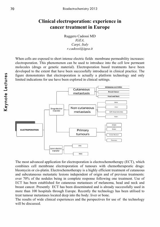
70K
eyno
te L
ectu
res
Bioelectrochemistry 2013
Clinical electroporation: experience in
cancer treatment in Europe
Ruggero Cadossi MD IGEA,
Carpi, Italy [email protected]
When cells are exposed to short intense electric fields membrane permeability increases: electroporation. This phenomenon can be used to introduce into the cell low permeant molecules (drugs or genetic material). Electroporation based treatments have been developed to the extent that have been successfully introduced in clinical practice. The figure demonstrates that electroporation is actually a platform technology and only limited indications for use have been explored in clinical settings.
The most advanced application for electroporation is electrochemotherapy (ECT), which combines cell membrane electroporation of tumours with chemotherapeutic drugs: bleomycin or cis-platin. Electrochemotherapy is a highly efficient treatment of cutaneous and subcutaneous metastatic lesions independent of origin and of previous treatments: over 70% of the nodules being in complete response following one treatment. Use of ECT has been established for cutaneous metastases of melanoma, head and neck and breast cancer. Presently ECT has been disseminated and is already successfully used in more than 100 hospitals through Europe. Recently the technology has been utilised to treat tumour metastases located deep into the body: liver or bone. The results of wide clinical experiences and the perspectives for use of the technology will be discussed.
71
Key
note
Lec
ture
s
Bioelectrochemistry 2013
A Self Powered Glucose Sensor Based on a Wired Enzyme Anode
Ben Feldman, Zenghe Liu, and Brian Cho
Abbott Diabetes Care 1360 South Loop Road, Alamda, CA USA
In-vivo glucose sensing is typically performed by externally powered transcutaneous amperometric sensors. The power consumption requirements of these sensors often present design constraints, especially in efforts to miniaturize them for fully implanted long term applications. Self-powered glucose sensors (SPGSs) have been developed which take conceptual inspiration from the bio-fuel cells of Heller et al. In this tradition, we describe here a robust SPGS that may be built from four simple components: (1) a low-potential, wired glucose oxidase anode; (2) a Pt/C cathode; (3) an overlying glucose flux–limiting membrane; and (4) a resistor bridging the anode and cathode. In-vitro evaluation showed that the SPGS output is nearly identical to that of the above anode paired with counter and reference electrodes in powered operation. Miniaturization (0.1 mm2 electrode area) of all components, including the O2-reducing cathode was accomplished. Operation is also linear down to very low O2 concentrations, especially with a sufficiently non-polar encapsulating membrane. Clinical trial data suggests that the in-vivo performance of the device was very similar to that of a conventional powered amperometric sensor. This SPGS presents an attractive alternative to conventional powered devices, especially for fully implanted long term applications.

72K
eyno
te L
ectu
res
Bioelectrochemistry 2013
Electronic properties of the surface-tethered DNA duplex
Elena Ferapontova Interdisciplinary Nanoscience Center (iNANO) and Center for DNA Nanotechnology
(CDNA) Gustav Wieds Vej 14, Aarhus University, DK-8000 Aarhus C, Denmark [email protected]
Electron transfer (ET) properties of individual DNA molecules at electrodes essentially depend on the whole structural design of the DNA-electrode systems. Generally, an electrochemical signal stemming from the reaction between the redox probe bound to the surface-tethered DNA double strand (ds) and the electrode may be ascribed to the electronic conductivity of dsDNA [1,2]. However, in the case when intercalation of the redox probe into the DNA duplex does not occur, alternative mechanisms of ET between the electrodes and DNA-conjugated redox probes should be considered [3-6].
Here, I discuss how the type of the redox probe and its conjugation to DNA affects the kinetics of ET between the redox probe and the electrode, under experimental conditions excluding the redox probe intercalation into DNA, i.e. the conditions under which DNA-mediated ET is not observed. I show that in loosely packed DNA monolayers ET between the negatively charged electrode and DNA-conjugated, positively charged redox probe is either governed by the nanoscale motion
of the probe (and thus dsDNA) towards the electrode surface (Figure 1) or absent, when the redox probe is uncharged [5]. Those movements are very likely due to the electric field effects and also depend on the way dsDNA is tethered to the electrode [6]. In contrast to that, DNA-mediated ET depends mostly on the way the redox probe interacts with DNA, i.e. on the extent of the redox probe intercalation into the DNA duplex. References: [1] Hartwich, G.; Caruana, D. J.; de Lumley-Woodyear, T.;Wu, Y.; Campbell, C. N.; Heller, A. J. Am. Chem. Soc. 1999, 121, 10803-10812. [2] Slinker, J. D.; Muren, N. B.; Renfrew, S. E.; Barton, J. K. Nature Chem. 2011, 3, 228-233 [3] Anne, A.; Demaille, C. J. Am. Chem. Soc. 2006, 128, 542-557. [4] Farjami, E.; Clima, L.; Gothelf, K. V.; Ferapontova, E. E. Anal. Chem. 2011, 83, 1594-1602 [5] Abi A, Ferapontova E.E., J. Am. Chem. Soc. 134 2012, 14499–14507 [6] Farjami, E.; Campos, R.; Ferapontova, E. Langmuir 2012, doi: 10.1021/la3032336
73
Key
note
Lec
ture
s
Bioelectrochemistry 2013
Wired EnzymeTM Technology-Based Ultrasensitive Nucleic Acid Biosensors
Huimin Deng, Yuqian Ren, Wei Shen, and Zhiqiang Gao*
Department of Chemistry, National University of Singapore 3 Science Drive 3, Singapore 117543, Republic of Singapore
*Corresponding author, Tel: +65-6516-3887, email: [email protected] Over the past decade, there have been significant advances in the development of nucleic acid biosensors. The most frequently used methods involve fluorescent materials as signal generators. However, only a few of the fluorescence-based techniques have sufficient sensitivities for direct detection of nucleic acids at subpicomolar levels. For direct detection of nucleic acids, mostly in cancer-related disease diagnosis and treatment, electrochemical techniques are among the most widely studied systems. In particular, the inherent miniaturization of electrochemical devices as well as their low cost and compatibility with advanced semiconductor technology make them excellent candidates for developing ultrasensitive nucleic acid biosensors. In addition, electrochemical technology offers a simple workflow and a user-friendly assay design process, thereby allowing rapid development of new single and/or multiplex assays for use in research or molecular diagnostics In this presentation, ultrasensitive nucleic acid biosensors based on wired enzymeTM
technology are presented. The biosensors are made of mixed monolayers of thiolated single-stranded DNA capture probes through self-assembly. It is shown that wired enzyme molecules are excellent tags/amplifiers and make the biosensors electrocatalytic. The electrocatalytic current correlates directly to the amount of nucleic acid. Nucleic acid quantifications have been performed on oligonucleotides, full-length house-keeping genes, microRNAs, as well as several cancer genes. The amount of nucleic acid that can be detected is in the vicinity of 103 copies, which is at least 100 times more sensitive than conventional fluorescent techniques. The biosensors can directly detect cancer susceptibility genes in extracted total RNA without engaging a PCR amplification step. We are currently integrating these biosensors with our sample preparation system, in order to develop a fully automated sample-to-answer molecular diagnostic platform to be used in the field or at the point-of-care.

74K
eyno
te L
ectu
res
Bioelectrochemistry 2013
Electrochemical methods for proteomics: From electrophoresis to Mass spectrometry
Hubert H. Girault, Liang Qiao, Elena Tobolkina, Reza Pourhaghighi, Fernando Cortes-
Salazar, BaoHong Liu* Laboratoire d’Electrochimie Physique et Analytique
Ecole Polytechnique Fédérale de Lausanne Station 6, CH-1015 Lausanne, Switzerland
*Department of Chemistry, Fudan University, Shanghai 200433, P.R. China [email protected]
Proteomics is making intensive use of two techniques, namely electrophoresis and mass spectrometry. This lecture will review the electrochemical aspects of both. In electrophoresis, the first fractionation technique is often IsoElectric Focusing (IEF). Different formats are used routinely, but we shall present here Offgel electrophoresis and in particular the current distribution aspects. If an electrophoresis setup is considered as an electrochemical cell, we shall show how a good control of the primary currant distribution can be used to optimize the separation. In mass spectrometry, soft ionization methods are currently employed to inject large biomolecules. First, we shall discuss Matrix Assisted Laser Desorption Ionization (MALDI) and show that a MALDI plate can be considered as a photo-electrode where it is possible to do oxidation reactions to generate a,x fragments of peptides or to do reduction to generate c,z fragments. Then, we shall consider the electrochemical aspects of Electrospray Ionization (ESI) and show how the electrode reaction can be put to good use, for example to carry out in-situ tagging reactions. Finally, we shall describe the principles of a new ionization technique, ElectroStatic Spray Ionization (ESTASI). This contactless technique offers many opportunities such as spraying from a droplet deposited on an inert polymer support or imaging a substrate. To conclude, we shall show how we can couple IEF electrophoresis with ESTASI to read directly an IEF gel with mass spectrometry.

75
Key
note
Lec
ture
s
Bioelectrochemistry 2013
Mediated and Direct Electrochemical Communication between ShewanellaoneidensisMR-1 and electrodes
Lo Gorton1, Sunil A. Patil1, Kamrul Hasan1, Donal Leech2, Cecilia Hägerhäll1
1Department of Analytical Chemistry/Biochemistry and Structural Biology, Lund University, P.O.Box 124, SE-221 00 Lund, Sweden
2School of Chemistry, National University of Ireland Galway, University Road, Galway, Ireland
Electrochemical transfer (ET) communication between bacterial cells and electrodes can usually be obtained through the use of freely diffusing monomeric redox mediators. Previously we have, however, also shown that flexible osmium redox polymers can work as efficient mediators for a number of both Gram – as well as Gram + bacteria and electrodes, clearly showing that the mediator does not need to pass the inner membrane to be able to shuttle the charge between the cells and the electrode [1]. In some restricted cases ET can be obtained directly between the bacterial cells and the electrode, e.g. between Geobacter sp. and Shewanella sp. and various carbon electrodes [2,3]. Here it will be shown that the current densityfromShewanellaoneidensisMR-1 to graphite electrodes in the presence of lactate can be increased at least four times by the use of an Os-polymer with an E°’-value of 0.221 V(vsAg|AgCl, sat. KCl) compared with the use of naked graphite [4]. Further increases in the current density for bioelectrocatalysis can be obtained by exchanging ordinary graphite electrodes for electrospun carbon fibres. For example for 18 mM lactate the current density increased from 10 to 120 μA/cm2. Currently we are also investigating the effect of feeding the cells with cis-platin, which causes the cells just to grow but not to divide. When comparing normal cells with cis-platin treated cells it is possible to get current densities up to four times higher with the treated cells. [1] K. Hasan, S. A. Patil, D. Leech, L. Gorton, Biochem. Soc. Rev., in press. [2]D. Lovley, Annu.Rev. Microbiol., 66 (2012) 391-409. [3] S. A. Patil, C. Hägerhäll, L. Gorton, Bioanal. Rev., submitted. [4] S. A. Patil, K. Hasan, D. Leech, C. Hagerhall, L. Gorton, Chem. Commun., 48 (2012) 10183–10185.

76K
eyno
te L
ectu
res
Bioelectrochemistry 2013
Extracellular Electron Transfer via Conductive Minerals
Kazuhito Hashimoto Department of Applied Chemistry, The University of Tokyo
7-3-1, Hongo Bukyo-ku Tokyo 113-8656 Japan [email protected]
Some of microorganism, for example,Genus Shewanella has a unique property of utilizing solid state metal-oxide as a terminal electron acceptor. The extracellular electron transfer (ET) occurs via cytochromes located in the outer membrane (outermembrane-cytochrome, OMC). Due to the unique property, the extracellular ET has attracted a lot of attentions from the point of view of biogeochemical cycle and microbial fuel cell. Here we show our recent results on the EET by using conductive minetrals as electron mediator. Long-distance extracellular ET in Shewanella/Iron-Oxide nanocolloid network(1,2) We found the ability of Shewanella to self-assemble into an electrically conductive network using naturally abundant semiconducting iron-oxides as an EET conduit. In the result, microbial current generation for S. loihica and S. oneidensis was significantly improved (∼50-fold) when inoculated anaerobically in the presence of the α-Fe2O3 and α-FeOOH nanocolloids. In vivo optical movie microscope, together with confocal fluorescent microscope indicated that the mobile cells (approximately 2 μm size) possessed a specific affinity for the nanocolloids, and developed the intercellular networks with thickness of ∼30 μm. Microbial Interspecies Electron Transfer via Conductive Minerals(3,4) We hypothesized that the above electron exchange proceeds in the natural environments, and microbes use them to transfer electrons between different species, too. In order to address this possibility, we established binary cultures of model soil bacteria (Geobacter and Thiobacillus) in the presence of conductive iron-oxide particles, and found that magnetite substantially (over 10 folds) accelerated their cooperative catabolism. This indicates that electrons are transferred in magnetite particles, facilitating the connection of their metabolisms. In the environment, a huge diversity of microbes inhabits, and they should establish complex interspecies interactions. However, since microbiology has been developed based on Pasteur’s isolation and pure-cultures techniques, knowledge on interspecies interactions is quite limited. Their finding will have broad impacts on our understanding of microbial interspecies interactions in nature and bases of strategies for developing more efficient bioenergy These instructions are an example of what a properly prepared meeting abstract should look like. Proper column and margin measurements are indicated.\ References 1. Nakamura, R.; Kai, F.; Okamoto, A.; Newton, G. J.; Hashimoto, K., Angew. Chem. Int. Ed. 2009, 48, 508. 2. Nakamura, R.; Okamoto, A.; Tajima, N.; Newton, G. J.; Kai, F.; Takashima, T.; Hashimoto, K. ChemBioChem. 2010, 11, 643. 3. Kato, S.; Kai, F.; Nakamura, R.; Watanabe, K.; Hashimoto, K. Environ. Microbiol. 2010, 12, 3114. 4. Kato, S.; Hashimoto, K.; Watanabe, K., Proc. Nat. Acad. Sci., 2012, 109, 10042.
77
Key
note
Lec
ture
s
Bioelectrochemistry 2013
Electrochemistry of Fluorescently-labeled Enzymes Reveals Heterogeneous Interfacial Electron-transfer Rates
and Intramolecular Rates that Differ between Rest and Turn-over Conditions
Lars J.C. Jeuken1,*, Lukasz Kzreminski1, Lionel Ndamba2, Thijs J. Aartsma2, Gerard
W. Canters2
1 School of Biomedical Sciences, University of Leeds, Leeds, United Kingdom 2 Leiden Institute of Physics, University Leiden, Leiden, the Netherlands
Fluorescently-labeled nitrite reductase (NiR) from Alcaligenes faecalis S6 on gold electrodes modified with self-assembled monolayers (SAMs) of 6-mercaptohexanol and mixtures of various octanethiols has been studied with a combination of fluorescence spectroscopy and electrochemistry. Voltammetry and quartz-crystal microbalance with dissipation (QCM-D) measurements have been performed on dye-labeled and unlabeled forms of the wild type (wt) and a cysteine-surface variant (L93C) of NiR. The results indicate that interfacial electron transfer is only possible if the negatively-charged surface patch surrounding the electron-entry site of NiR is directed towards the electrode. This can be achieved either by introducing positive charges in the SAM or, when the SAM does not carry a charge, by neutralizing the negative charges around the electron entry site of the NiR by introduction of positively charged groups like the ATTO 565 dye label. The catalytic activity of NiR is measured electrochemically by exploiting the direct electron transfer to fluorescently labeled NiR, whereas the redox state of the type-1 copper site is determined from fluorescence intensity changes caused by Förster Resonance Energy Transfer (FRET) between a fluorophore attached to NiR and its type-1 copper site. The homotrimeric structure of the enzyme is reflected in heterogeneous interfacial electron transfer kinetics with two monomers having a 25-fold slower kinetics than the third monomer. Further analysis shows that intramolecular electron-transfer rate is significantly reduced in turn-over conditions compared to the enzyme at rest, with an exception at low pH / nitrite conditions. This effect is attributed to slower reduction rate of type-2 copper centre due to an unknown rate-limiting step of residues in the enzyme’s active site, gating the intramolecular electron transfer.

78K
eyno
te L
ectu
res
Bioelectrochemistry 2013
New adventures in phosporylation chemistry: Using electrochemistry to probe biochemistry
H.-Bernie Kraatz
University of Toronto Scarborough 1265 Military Trail, Toronto, Ontario M1C 1A4, Canada
[email protected] The detection of biological analytes or of biochemical processes by electrochemical methods requires in many cases the presence of a redox-active probe as part of the detection system. Here we report some recent results on the use of ferrocene as a redox label to study enzymatic transformations. The focus will be on the study of protein kinase catalyzed transformations. During protein phosphorylation reactions, protein kinases catalyze the transfer of a phosphate group from ATP to a specific serine, threonine, or tyrosine residues of a protein. We have recently introduced an organometallic conjugate of adenosine triphosphate bearing a ferrocene group at the γ-phosphate. Our studies show that protein kinases can use this molecule as a co-substrate thus transferring a redox active Fc group to a target. Here, results are presented for a range of kinases showing that this approach can be used to assess the activity of kinases and measure their inhibition by small molecules.
S. Martin, M. K. Rains, D. Freeman, H.-B. Kraatz, Bioconjugate Chem. 2011, 22, 1663-1672. K. Kerman, H. Song, J.S. Duncan, D.W. Litchfield, H.-B. Kraatz, Anal. Chem. 2008, 80, 9395-9401; H. Song, K. Kerman, H.-B. Kraatz, Chem. Commun. 2008, 502-504.

79
Key
note
Lec
ture
s
Bioelectrochemistry 2013
A Systematic Approach to Devising of Chemical Sensors Using Conducting Molecularly Imprinted Polymers
Wlodzimierz Kutner1,2
1Institute of Physical Chemistry, Kasprzaka 44/52, 01-224 Warsaw, Poland; 2Cardinal Stefan Wyszynski University in Warsaw, Woycickiego 1/3, 01-938 Warsaw,
Poland E-mail address: [email protected]
Using a concept of molecular imprinting, we devised and fabricated chemo- and biosensors mimicking recognition of chosen biologically important analytes, like biogenic amines (including neurotransmitters), 1-5 drugs, and adenosine-5'-triphosphate (ATP).6 For that, first, we synthesized a range of bis(2,2’-bithiophen-5-yl)methane derivatives to serve as functional and cross-linking monomers bearing different recognition sites. Then, we computationally modeled structures and calculated thermodynamic parameters of formation of pre-polymerization complexes of the most suitable functional monomers with the analytes. Towards that, we selected the monomers bearing recognition sites compatible to binding sites of the analytes. At this initial step, these analytes were used as templates of imprinting. By allowing for self-assembling of the complexes in solutions in the next step, we experimentally confirmed these calculated thermodynamic parameters by determining the complex stability constants using fluorescence titration. Next, the conducting analyte-templated molecularly imprinted polymers (MIPs) were prepared by potentiodynamic polymerization of these complexes. This polymerization resulted in deposition of thin MIP films onto different conducting substrates. After subsequent template extractions, monitored by the measurements of XPS, UV-vis and FTIR spectroscopy, as well as those of DPV and EIS, molecularly imprinted cavities were generated in MIPs. These cavities, complementary in size, shape, and orientation of their binding sites to the templating molecules, selectively recognized the respective analytes. Availability of the imprinted cavities to the analyte molecules was governed by suitable adjustment of the film viscosity and porosity using proper cross-linking monomers and ionic liquids, respectively. With the direct and indirect transduction of the chemical recognition event into the useful analytical signal with piezoelectric microgravimetry (PM) at EQCM and EIS capacitive impedometry as well as DPV, respectively, we determined the analytes both under the batch-solution and flow-injection analysis (FIA) conditions with detectability at the nanomole level of concentration. Our sensors were capable of selective analyte determination in the presence of several structural and functional analogue interferents. 1 Pietrzyk, A., et al., Anal. Chem. 2009, 81, 10061-10070. 2. Pietrzyk, A., et al., Biosens. Bioelectron. 2010, 25, 2522-2529. 3. Pietrzyk, A., et al., Bioelectrochemistry 2010, 80, 62-72. 4. Pietrzyk, A., et al., Anal. Chem. 2009, 81, 2633-2643. 5. Huynh, T-P., et al., Bioelectrochemistry 2012, DOI:
10.1016/j.bioelechem.2012.07.003. 6. Huynh, T-P., et al., Biosens. Bioelectron., 2012, DOI: 10.1016/j.bios.2012.09.038.

80K
eyno
te L
ectu
res
Bioelectrochemistry 2013
Design and use of microfluidic devices for the real time monitoring of micro/nanopulses effect on cells
B. Le Pioufle, C. Dalmay, O. Français,
SATIE-BIOMIS, CNRS, ENS de Cachan, 61 av du Pdt Wilson, 94230 Cachan, France
The effects of electric field pulses on the cell membranes – like electropermeabilization – are now broadly used for transfection or drug insertion purposes (pulses having a typical amplitude of 1kV/cm and more than 1μs duration). Nevertheless the effects of ultra-short nanopulses (below 10ns duration, above 40kV/cm) on cell membranes and intracellular components (Beebe 2003, Napotnik 2012) are still under investigation within several research groups. In this context, our group is working toward the design and microfabrication of miniaturized biodevices devoted to the application of ultra short electrical pulses (nanopulses) to live cells, circulating within microfluidic channels or adherent in the exposition chamber. Extremely high electrical fields (up 280 kV.cm-1 ) were achieved within our device. Two criteria have to be observed when designing these devices i) the impedance matching which is necessary in order to avoid a destructive feedback wave caused by reflections on the pulse generator, which produces particularly high voltages (up to 9kV) ii) good homogeneity of the electric field applied to circulating cells. The first objective is held thanks to the dimensions of the exposition channels while the second objective is achieved thanks to the electrode thickness in regard to the channel width (Dalmay 2011). As adherent cells needs to be cultured for several hours prior to be exposed, a devoted structure has been proposed (Dalmay 2012). In that case the cell adhesion support is a conventional culture glass slide, that we report on the top of the device for the electrical field application. In that way the same device can be re-used successively at short time intervals while multiple culture slides are prepared separately. The slide is centered on the device thanks to thick resist frame, at the appropriate height thanks to pillars patterned in the same material.
References:
- Beebe et Al., 2003. DNA Cell Biol. 22 (12), 785–796 - T. B. Napotnik et Al., Bioelectromagnetics, 2012, 33(3), 257–264. - C. Dalmay et Al.. Sensors and Actuators B, 160, (1), 1573-1580, 2011 - C. Dalmay et Al., Lab on a Chip, 12, (22), 4709-15, 2012.
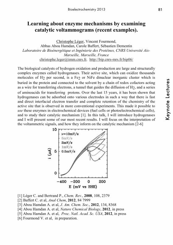
81
Key
note
Lec
ture
s
Bioelectrochemistry 2013
Learning about enzyme mechanisms by examining catalytic voltammograms (recent examples).
Christophe Léger, Vincent Fourmond,
Abbas Abou Hamdan, Carole Baffert, Sébastien Dementin Laboratoire de Bioénergétique et Ingénierie des Protéines, CNRS Université Aix-
Marseille, Marseille, France [email protected], http://bip.cnrs-mrs.fr/bip06/
The biological catalysts of hydrogen oxidation and production are large and structurally complex enzymes called hydrogenases. Their active site, which can oxidize thousands molecules of H2 per second, is a Fe2 or NiFe dinuclear inorganic cluster which is buried in the protein and connected to the solvent by a chain of redox cofactors acting as a wire for transferring electrons, a tunnel that guides the diffusion of H2, and a series of aminoacids for transferring protons. Over the last 15 years, it has been shown that hydrogenases can be adsorbed onto various electrodes in such a way that there is fast and direct interfacial electron transfer and complete retention of the chemistry of the active site that is observed in more conventional experiments. This made it possible to use these enzymes in electrochemical devices (fuel cells or photoelectrochemical cells), and to study their catalytic mechanism [1]. In this talk, I will introduce hydrogenases and I will present some of our most recent results. I will focus on the interpretation of the voltammetric signals, and how they inform on the catalytic mechanism [2-6]
[1] Léger C. and Bertrand P., Chem. Rev., 2008, 108, 2379 [2] Baffert C. et al, Anal Chem, 2012, 84 7999 [3] Abou Hamdan A. et al, J. Am. Chem. Soc., 2012, 134, 8368 [4] Abou Hamdan A. et al, Nature Chemical Biology, 2012, in press [5] Abou Hamdan A. et al, Proc. Natl. Acad. Sc. USA, 2012, in press [6] Fourmond V. et al, in preparation.
82K
eyno
te L
ectu
res
Bioelectrochemistry 2013
Rational Design of Highly Sensitive Bioelectroanalytical Devices: an Illustrating Example with the Heterogeneous
Reconstitution of PQQ-Dependent Glucose Dehydrogenase
F. Durand,1 B. Limoges,2 N. Mano,1 F. Mavré,2 R. Miranda-Castro,2 L. Zhang2. 1 Centre de Recherche Paul Pascal, UPR CNRS 8641, Av. Albert Schweitzerm, 33600
Pessac, France, 2 Laboratoire d'Electrochimie Moléculaire, Université Paris Diderot/CNRS, 15 rue Jean de Baïf, 75251, Paris, France.
Among the wide range of redox enzymes available, the pyrroloquinoline quinone-dependent glucose dehydrogenase (PQQ-GDH) is of great interest because of its high catalytic efficiency towards glucose oxidation and its ability to accept a wide range of redox mediators for enzyme electrocatalysis. Additionally, the active enzyme (holo form) can be efficiently reconstituted from its inactive apo form (overexpressed from an E. coli recombinant strain) by simply mixing the apo form enzyme with a diluted solution of the prosthetic group (PQQ) [1]. This latter property has been shown to be of great interest in the development of novel signal amplification systems coupled to bioaffinity binding assays [2]. In the present work, the heterogeneous reconstitution of apo-GDH immobilized on an electrode surface through an avidin-biotin linkage has been investigated in detail. It will be shown that under these conditions subpicomolar concentrations of PQQ in solution could be detected which is a particularly attractive performance for future analytical applications. With the aim to quantitatively understand the heterogeneous enzyme reconstitution process and to rationally predict the lowest concentrations of PQQ that can be indirectly determined from the electrocatalytic activity of the reconstituted enzyme on the electrode, a detail electrochemical analysis of the catalytic mechanism of PQQ-GDH has been carried out, first, with the enzyme in homogenous solution, and then, once immobilized on the electrode surface. From this study, it was possible to derive each individual step involved in the complex mechanism of PQQ-GDH, including substrate inhibition and subunit cooperativity [3]. After quantitative analysis of the amount of active enzyme attached to the electrode surface and comparison with the homogenous kinetics, it was possible to conclude that a nearly fully active monolayer of holo-GDH could be immobilized on the electrode through the avidin-biotin linkage. It was also possible to shown that under a saturating concentration of PQQ, the entire amount of immobilized apo-GDH on an electrode could be fully reconstituted but with a rate of reconstitution that was ca. 100-fold lower than in homogenous solution. Finally, the thermodynamics and kinetics of the heterogeneous reconstitution process were determined and used to calculate, with an appropriate model, the lowest concentrations of PQQ detectable by a monolayer of apo-GDH. The resulting theoretical values were in perfect agreement with the experimental ones.
[1] A. J. J. Olsthoorn, J. A. Duine, Biochemistry, 37, 1998, 13854. [2] D. Shen, M. E. Meyerhoff, Anal. Chem., 81, 2009, 1564. [3] F. Durand, B. Limoges, N. Mano, F. Mavré, R. Miranda-Castro, J.-M. Savéant, J. Am. Chem. Soc., 133, 2011, 12801.
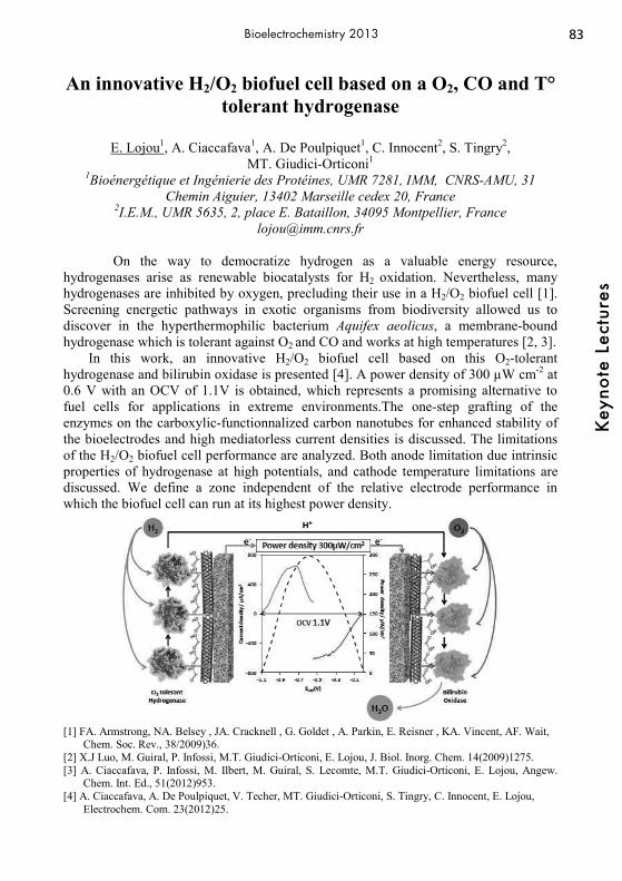
83
Key
note
Lec
ture
s
Bioelectrochemistry 2013
An innovative H2/O2 biofuel cell based on a O2, CO and T° tolerant hydrogenase
E. Lojou1, A. Ciaccafava1, A. De Poulpiquet1, C. Innocent2, S. Tingry2,
MT. Giudici-Orticoni1 1Bioénergétique et Ingénierie des Protéines, UMR 7281, IMM, CNRS-AMU, 31
Chemin Aiguier, 13402 Marseille cedex 20, France 2I.E.M., UMR 5635, 2, place E. Bataillon, 34095 Montpellier, France
[email protected] On the way to democratize hydrogen as a valuable energy resource,
hydrogenases arise as renewable biocatalysts for H2 oxidation. Nevertheless, many hydrogenases are inhibited by oxygen, precluding their use in a H2/O2 biofuel cell [1]. Screening energetic pathways in exotic organisms from biodiversity allowed us to discover in the hyperthermophilic bacterium Aquifex aeolicus, a membrane-bound hydrogenase which is tolerant against O2 and CO and works at high temperatures [2, 3].
In this work, an innovative H2/O2 biofuel cell based on this O2-tolerant hydrogenase and bilirubin oxidase is presented [4]. A power density of 300 μW cm-2 at 0.6 V with an OCV of 1.1V is obtained, which represents a promising alternative to fuel cells for applications in extreme environments.The one-step grafting of the enzymes on the carboxylic-functionnalized carbon nanotubes for enhanced stability of the bioelectrodes and high mediatorless current densities is discussed. The limitations of the H2/O2 biofuel cell performance are analyzed. Both anode limitation due intrinsic properties of hydrogenase at high potentials, and cathode temperature limitations are discussed. We define a zone independent of the relative electrode performance in which the biofuel cell can run at its highest power density.
[1] FA. Armstrong, NA. Belsey , JA. Cracknell , G. Goldet , A. Parkin, E. Reisner , KA. Vincent, AF. Wait,
Chem. Soc. Rev., 38/2009)36. [2] X.J Luo, M. Guiral, P. Infossi, M.T. Giudici-Orticoni, E. Lojou, J. Biol. Inorg. Chem. 14(2009)1275. [3] A. Ciaccafava, P. Infossi, M. Ilbert, M. Guiral, S. Lecomte, M.T. Giudici-Orticoni, E. Lojou, Angew.
Chem. Int. Ed., 51(2012)953. [4] A. Ciaccafava, A. De Poulpiquet, V. Techer, MT. Giudici-Orticoni, S. Tingry, C. Innocent, E. Lojou,
Electrochem. Com. 23(2012)25.

84K
eyno
te L
ectu
res
Bioelectrochemistry 2013
High-Resolution Bioimaging of Live Cells by Scanning Electrochemical Microscopy (SECM)
Tomokazu Matsue
WPI-Advanced Institute of Materials Research (WPI-AIMR) and Graduate School of Environmental Studies, Tohoku University, Sendai 980-8579, Japan
E-mail: [email protected]
A high temporal and spatial resolution tool working in physiological conditions is needed to evaluate the relationship of the localized topography and function of biomolecules. One of the important tools meeting the requirements is a scanning electrochemical microscope (SECM), which uses a micro/nanoelectrode as a scanning probe to provide electrochemical properties of sample surfaces under physiological conditions without physical contact. However, the distance control between the probe and sample has been an important challenge to improve the temporal resolution and sensitivity. We have incorporated ion-conductance feedback (SECM-SICM) [1,2] and voltage-switching mechanisms (VSM-SECM) [3] into the system with nanoelectrode probe for non-invasive, high resolution bioimaging. Several nanoprobes were fabricated by using fine glass capillaries including nanopipette-nano Au or Pt ring electrode and double-barrel carbon nanoelectrode. For SECM-SICM, the nanoaperture of the probe was used to detect ionic currents for distance control at nanometer levels and the nanoelectrode was used for electrochemical measurements. We also fabricated carbon nanoelectrode for VMS-SECM as the probe. The tip radii of the above probes ranged from 10 nm to 100 nm. SECM-SICM was applied to simultaneous imaging of topography and electrochemical responses of enzymes and single live cells. The SICM topographic images showed structure of live cells and the SECM images showed permeation of redox species through membranes of live cells [1]. We also demonstrated high-resolution imagines of living cells using a double-barrel carbon nanoelelctrode as a probe. The cell bodies were clearly observed in both the SICM and SECM images [2]. VSM-SECM with a nanoelectrode was also successfully applied to acquire high quality topographical and electrochemical images of living cells simultaneously [3]. References [1] Y. Takahashi et al., J. Am. Chem. Soc., 132, 10118 (2010). [2] Y. Takahashi et al., Angew. Chem. Int. Ed., 50, 9638 (2011). [3] Y. Takahashi et al., Proc. Natl. Acad. Sci. USA, 109, 11540(2012).

85
Key
note
Lec
ture
s
Bioelectrochemistry 2013
Thylakoid Bioelectrocatalysis for Energy Conversion and Sensing
Shelley D Minteer, Michelle Rasmussen
University of Utah 315 S 1400 E Rm 2020
Salt Lake City, UT 84112 [email protected]
Organelles are subcellular components that have a variety of functions in the cell, but two organelles are well known for their role in energy conversion - thylakoids and mitochondria. Mitochondria are considered the powerhouse of the living cell, because they contain the metabolic pathways of the Krebs cycle, the electron transport chain, and fatty acid oxidation. Thylakoids are the subcellular component of plant cells responsible for photosynthesis. Both thylakoids and mitochondria can be immobilized on carbon electrodes and are capable of direct electron transfer with carbon electrode structures. This presentation will discuss immobilization of organelles (specifically thylakoids), use for energy harvesting applications, and use for sensor applications, as well as strategies for understanding electron transfer mechanisms. Thylakoids were purified from spinach leaves. The thylakoids were immobilized onto Toray carbon paper using tetramethyl orthosilicate (TMOS) vapor deposition techniques. The TMOS immobilization was performed in layers – first a layer of thylakoids was pipetted on the electrode followed by vapor deposition of the TMOS. The electrodes were characterized by cyclic voltammetry. A small reversible peak was present at approximately 0.15 V vs. SCE and an irreversible oxidation peak just below 0.3 V vs. SCE. Next, amperometry was performed to test the electrodes for activity. The current was measured at 0.3 V vs. SCE in the dark until a stable value was obtained and then the cell was exposed to light. As expected, the current increased. The current increase was approximately 40 nA with TMOS. The thylakoid electrodes were then connected with a platinum cathode to make a biosolar cell and i-V curves were used to evaluate the electrochemical performance. The thylakoid solar cell gave a maximum current density of 112 nAcm-2. This solar cell has also been used for self-powered sensing of herbicides. This paper will compare self-powered organelle biofuel cell-based sensors with self-powered biosolar cell-based sensors.
86K
eyno
te L
ectu
res
Bioelectrochemistry 2013
Cell electroporation and cell electropermeabilisation: facts and theory
Lluis M. Mir1, 2, 3, Marie Breton1, 2, 3, Isabelle Leray1, 2, 3, Aude Silve1, 2, 3, 4
1Université Paris-Sud, Laboratoire de Vectorologie et Thérapeutiques Anticancéreuses, UMR 8203, Orsay F-91405, France
2CNRS, Laboratoire de Vectorologie et Thérapeutiques Anticancéreuses, UMR 8203, Orsay F-91405, France
3Institut Gustave Roussy, Laboratoire de Vectorologie et Thérapeutiques Anticancéreuses, UMR 8203, Villejuif F-94805, France
4Karlstruhe Institute of Technology [email protected]
“All we know about membrane electropermeabilisation”… Until recently, the reviews summarizing the mechanisms describing cell electropermeabilization were entitled “all what we do not know …” The membranes of cells exposed to short and intense electric pulses, from ns duration till hundreds of ms duration, under conditions avoiding thermal raise, were told “electroporated” while “electroporation” was only detected by a functional consequence, namely the “permeabilization” of the membranes. In the last years, molecular dynamics brought insights on the events occurring during very short (ns) and intense electric pulses, and even a few nanoseconds later: these numerical simulations showed the presence of “pores” rapidly generated, but also rapidly disappearing at the end of the pulse. Therefore “electroporation” seemed confirmed but was also unable to explain the long-lived “electropermeabilized” state that is maintained for minutes in almost all cells, whether they were treated by classical electric pulses (of 100 microseconds) or by pulses as short as 10 nanoseconds. We propose a new model that allows assembling all the observations made previously, for years, both in vitro and in vivo. Our Membrane Impermeability Rupture model claims that the consequences of the cells or lipids membranes exposure to electric pulses are: 1°) the membrane electroporation, during the pulse application, which allows not only the electrophoretic and diffusive transport of molecules across the pores, but also chemical reactions to occur at the level of the cell membrane; and 2°) almost immediately after the electric pulse interruption, the membrane electropermeabilization, which exists in the absence of membrane pores, which allows for diffusive transport, and which results from the lipids structure modification during the membrane electroporation.

87
Key
note
Lec
ture
s
Bioelectrochemistry 2013
Improving implanted glucose sensor performance – Designing the next generation of sensors
Michael Pishko
Stewart & Stevenson Professor II Director, National Center for Therapeutics Manufacturing
Department of Biomedical Engineering, 3122 TAMU Texas A&M University
College Station, TX 77843 979-845-3348 (o), 979-847-5850 (o)
Glucose sensors, which use an enzyme (glucose oxidase) to achieve specificity, are currently not stable or sensitive enough to meet the demands of a closed-loop delivery system. As a result, the application of glucose biosensors has been primarily limited to home glucose test meter, a few implantable sensors, and bench-top blood-gas instruments containing sensors for glucose. There are a number of reasons for this lack of commercial and technical progress. Technically, many proposed biosensors for glucose simply do not have the accuracy and stability (operational or storage) to meet the desired need. Inaccuracy and imprecision in sensor performance are frequently due to inactivation of the sensing by species present in the sensing environment. For implantable glucose sensors to be successful, the issues of reproducibility and instability must be addressed. This presentation will explore reasons behind sensor instability in vivo and potential methods to maximize sensor stability and performance, including sensor redundancy.

88K
eyno
te L
ectu
res
Bioelectrochemistry 2013
An Electro-switchable Biointerface for the Analysis of Molecular Interactions
Ulrich Rant
Technische Universität München Chemie Department, Lichtenbergstr. 4, 85748 Garching
I will introduce a novel biosensing principle, switchSENSE, which employs an electrically actuated bio-interface. Short end-tethered DNA molecules are driven to oscillate (switch their conformation) on microelectrodes by the application of ac voltages. The induced molecular motion is monitored in real-time on a sub-microsecond timescale by fluorescence energy transfer. The conformation switching amplitude and the dynamics of the actuated DNA-levers not only reveal the mere presence of biomolecular targets on the sensor surface, but also permit the simultaneous analysis of the target molecule size, shape, and charge in a single experiment. I will describe the mechanism of swaying molecules atop of DNA levers and discuss how kon, koff, KD values are quantitatively determined from the DNA’s switching behavior. The method features a very high sensitivity, which is demonstrated by the affinity ranking of monoclonal antibodies in the one-digit pM concentration range. DNA targets can be discriminated with single base mismatch specificity, which is exemplified at hand of a SNP in the p53 gene. The size of the target is inferred from a molecular dynamics measurement and analysed with a theoretical model that accounts for the molecule’s hydrodynamic friction. The analysis yields the molecule’s effective diameter with 0.2 nm accuracy. Dynamic switching measurements are also applied to the analysis of antibodies, where fragmentation states and agglomerates are readily identified. The implications of switchSENSE as a high information content (affinity, reaction rates, size, shape, charge) analytical platform technology will be highlighted for the discovery process of biological drugs.

89
Key
note
Lec
ture
s
Bioelectrochemistry 2013
Applications of Pulsed Electric Fields for Food Processing
Javier Raso Food Technology Unit, University of Zaragoza
Faculty of Veterinary C/ Miguel Servet, 155 50013 Zaragoza (Spain) [email protected]
Food industry requires a continuous adaptation of its production processes in order to improve food quality or to introduce new products in the market while reducing energy inputs. Innovative nonthermal technologies such as pulsed electric fields (PEF) offer a range of opportunities for improving conventional processing. During PEF processing, a liquid food or pumpable product is passed through a treatment chamber where it is subjected to short pulses (μs) of very high voltage. The generated external electric fields (0.5-30 kV/cm) induce the electroporation of the cytoplasmatic membrane of both eukaryotic and prokaryotic cells. The pore formation affects the permeability of the biological membranes causing microbial inactivation and enhancing the diffusion processes through cell membranes. Due to these effects, PEF has gained increasing interest in recent years for liquid food pasteurization and for improving mass transfer operations in the food industry. Processing unit operations that seek to inactivate harmful microorganisms are of primary importance in ascertaining the safety of food. The capability of PEF to inactivate vegetative cells of pathogenic and spoiling microorganisms at temperatures below those used in thermal processing makes this technology very attractive for extending the shelf-life and guarantee safety of foods that retain the characteristics of fresh. Mass transfer phenomenon through cell membranes occurs in many operations of the food industry that aim obtaining a given intracellular compound of interest, removing water from foods (drying) or introducing a given substance into the food matrix. Breakdown of the cell membranes by different techniques such as grinding, heating or enzymatic maceration is common pretreatment step to improve mass transfer rates. Electroporation of the cytoplasmatic membrane is an alternative to these pretreatments especially when the complete disintegration of cell membranes is not desired. The recent development of PEF apparatus with sufficient power for processing large quantities of products, the easy implementation of the treatment chambers into the existing processing and the low energy consumption are keys of PEF technology for becoming a commercially viable technology for the food industry In this presentation fundamental and applied aspect of application of PEF for improving current food processing systems will be reviewed including challenges for future research.

90K
eyno
te L
ectu
res
Bioelectrochemistry 2013
Microbial Electrochemistry – Fundaments and Prospects
Prof. Dr. Uwe Schröder Technische Universität Braunschweig, Institute of Environmental and Sustainable
Chemistry, Hagenring 30, 38106 Braunschweig, [email protected]
The last decade has seen tremendous progress in the development of microbial electrochemical technologies. The performance of microbial fuel cells has increased remarkably. Especially the exploitation of biofilm electrodes, i.e., electrodes based on the electrocatalytic activity of electroactive microbial biofilms, made developments towards large scale conceivable. Here, the coupling of waste water treatment and energy recovery from wastewater represents a major application target and driving force for the development of the technology. New technologies like microbial electrolysis cells or microbial desalination cells have expanded the spectrum of potential applications. Moreover, the exocellular electron transfer, which wires the microbial metabolism to an electrode, opens a fully new range of (bio)electrochemical processes, ranging from simple redox transformations to complex bioelectrosyntheses and to the transformation and decomposition of persistent chemicals like antibiotics. This lecture gives an overview about these new developments in the field of microbial electrochemistry, highlights recent trends and examples and discusses future needs..
91
Key
note
Lec
ture
s
Bioelectrochemistry 2013
Translational Research in Biomedical Applications of Electroporation
Gregor Sersa
Institute of Oncology Ljubljana, Department of Experimental Oncology Zaloska 2, SI-1000 Ljubljana, Slovenia
Electroporation based technology has recently spread over the Europe in treatment of cancer. Electrotransfer is used for delivery of drugs or nucleic acids into tissues; electrochemotherapy for delivery of bleomycin or cisplatin, and gene electrotransfer for delivery of nucleic acids, like naked plasmids, siRNA ….. Electrochemotherapy is currently used in many European countries for treatment of cutaneous and subcutaneous tumors/metastases, predominantly melanoma. The success rate of the treatment is very high, with long term complete responses of the tumors. In spite of its effectiveness on different tumor types, recent meta-analysis has demonstrated that there is a higher response rate of non-melanoma tumors, than melanomas. Furthermore, attention has to be put into treatment of bigger tumors that tend to have lower response rate than small ones. In relation to this, probably a correction of the current SOP has to be made soon. Translation of this technology has been made also to treatment of deep seated tumors. However electrochemotherapy remains local treatment, therefore combined modality treatment is needed, either to potentiate local response or to add a systemic component to target disseminated disease. One of the approaches is combined treatment of electrochemotherapy with gene electrotransfer of plasmids coding for immunomodulatory molecules. Some studies have already addressed this issue, however, our recent one has demonstrated that intramuscular gene electrotransfer of plasmid coding for IL-12 potentiated curability rate of the tumors locally treated with electrochemotherapy. Gene electrotrenafer has also proved to be effective approach. Predominantly studies on melanoma and sarcoma tumors have been done. Again, local, intratumoral and peritumoral gene electrotransfer of plasmid coding for IL-12 have proved great local tumor control, with distant effects on non-treated tumors. In addition, muscle gene electrotransfer is also effective on primary tumors and lung metastases. This approach has also proved effective in combined treatment with tumor irradiation. Currently other therapeutic plasmids are investigated also, predominantly aimed to target melanoma. For example, AMEP is in preclinical and clinical testing with the first encouraging phase I clinical profile. In pipeline are also plasmids coding for siRNA against MCAM, the molecule that is involved in melanoma angiogenesis and invasion and against endoglin, which is a marker of activated tumor endothelium. The translation of these new plasmids still needs extensive pre-clinical testing in order to get translated into the clinics. In summary both electrochemotherapy and gene electrotransfer are currently being translated into the clinics, electrochemotherapy is in a more advanced stage than gene electrotransfer, but the latter will certainly find its place in treatment of cancer, since it is simple, safe and effective gene therapy approach.

92K
eyno
te L
ectu
res
Bioelectrochemistry 2013
Synergic use of molecular dynamics simulations and sophisticated experiments reveal key aspects of lipid
membranes electroporation
Mounir Tarek1, Lluis M. Mir2, 3 and Damijan Miklavcic4 1 UMR structure et Réactivité des Systèmes Moléculaires Complexes, CNRS -
Université de Lorraine, FRANCE 2 CNRS- UMR8203, Laboratoire deVectorologie et Thérapeutiques Anticancéreuses,
Institut Gustave Roussy, Villejuif, 94805, France 3 Université Paris-Sud, UMR8203, , - Orsay-91405 FRANCE
4Laboratory of Biocybernetics, Faculty of Electrical Engineering, University of Ljubljana, Tržaška 25, SI-1000 Ljubljana, SLOVENIA
[email protected] The key features of electroporation of cell membranes are based on theories involving stochastic pore formation. The direct observation of the formation of nano-sized pores in general is not possible with conventional techniques. Furthermore, due to the complexity and heterogeneity of cell membranes, it is difficult to describe and characterize their electroporation in terms of atomically resolved processes. Atomistic simulations in general and molecular dynamics (MD) simulations in particular, have proven to be effective for providing insights into both the structure and the dynamics of model lipid membranes in general. Few years ago, pioneering MD simulations have been conducted in order to model the effects of electric fields on membranes, providing perhaps the most complete molecular model of the electroporation process of lipid bilayers [1,2]. Here we show how such “computer experiments” combined with wet lab experiments provide a significant insight into the processes affecting, at the molecular level, the integrity of various types of lipid membranes when these are subject to voltage gradients of magnitude and duration equivalent to those generated by the electric pulses delivered to cells (microsecond and millisecond or nanosecond pulses) [3]. We then investigate the effect of high magnitude ns pulses on subsequent electrotransfer of small molecules e.g. siRNA [4]. $This Research was conducted in the scope of the EBAM European Associated Laboratory (LEA). [1] Tieleman DP (2004) BMC Biochemistry 5:10 [2] Tarek M (2005) Biophys. J. 88:4045-4053 [3] Delemotte, L., and Tarek, M. (2012). J. Membr. Biol., 245, 531-543. Kramar, P., Delemotte, L., Maček Lebar, A., Kotulska, M., Tarek, M. and Miklavčič, D. (2012) J. Membr. Biol. 245, 651-659. [4] Breton, M., Delemotte, L., Silve, A., Mir, L. M., & Tarek, M. (2012). J. Amer. Chem. Soc. 134, 13938–13941.

93
Key
note
Lec
ture
s
Bioelectrochemistry 2013
Electrochemistry of Single Protein and DNA-based Molecules
J. Ulstrup1, P. Salvatore1, K.K. Karlsen2, A.G. Hansen1, J. Zhang1, R.J. Nichols3 and
1Department of Chemistry, Technical Univ. of Denmark, 2800 Kgs Lyngby, Denmark+45 45252359; Fax: +45 45883136
[email protected] 2Dept. of Physics, Chemistry and Pharmacy, Univ. of Southern Denmark, 5230 Odense,
Denmark Dept. Chemistry, University of Liverpool, L69 7ZD Liverpool, UK
The scanning probe microscopies, scanning tunnelling (STM) and atomic force microscopy (AFM) enables addressing molecules on solid surfaces with a degree of detail that even reaches the single molecule directly in aqueous chemical and biological media under electrochemical control (in situ STM and AFM). Redox molecules are of particular interest but pose greater challenges than non-redox molecules. In situ STM and AFM of biomolecules such as (metallo)proteins and DNA-based molecules pose greater single-molecule challenges but offer intriguing insight in the ways these molecules operate. Single-molecule bioelectrochemistry (metalloproteins, DNA) requires well-defined (atomically planar) electrode surfaces modified by molecular monolayers (SAMs). Such surfaces have themselves been mapped to sub-molecular resolution and disclose an intriguing variety of local environments. Structural mapping of redox metalloproteins such as blue copper, heme, and iron-sulfur proteins as well as the metalloenzymes nitrite reductase and laccase were first in focus, now followed by single-molecule electron transport and enzyme function. These efforts are now being extended to DNA-based molecules. We shall overview some of these studies and note some observations, theoretical concepts, and some outstanding “puzzles”. References 1. J. Zhang, A.M. Kuznetsov, I.G. Medvedev, Q. Chi, T. Albrecht, P.S. Jensen and J.
Ulstrup, Chem. Rev. 108 (2008) 2737-2791. 2. J. Zhang, Q. Chi, A.G. Hansen, P. S. Jensen, P. Salvatore and J. Ulstrup, FEBS
Letters 586 (2012) 526-535. 3. A.G. Hansen, P. Salvatore, K.K. Karlsen, R.J. Nichols, J. Wengel and J. Ulstrup.
Phys Chem Chem Phys (2012) in press. 4. P. Salvatore, K.K. Karlsen, A.G. Hansen1, J. Zhang, R.J. Nichols3 and J. Ulstrup,.
J. Am. Chem. Soc. (2012), in press. Dx.doi.org/10.1021/ja306877s.

94K
eyno
te L
ectu
res
Bioelectrochemistry 2013
Cell Electroporation Creates Complex Pore Populations
Weaver, J.C., Son, R.S., Gowrishankar, T.R., and Smith, K.C. Harvard-MIT Division of Health Sciences and Technology,
MIT, Cambridge, MA 02139, USA. [email protected]
Closed, curved cell membranes lead to more complicated electroporation (EP) behavior than of planar membranes, but even the latter is not simple. Here we use computer modeling at both the membrane and cell levels to explore poration behavior of a cell model in response to electric field pulses for a wide range of durations. First we consider the model’s response to a conventional EP 1 kV/cm, 100 microsecond trapezoidal pulse. After a burst of pore creation a sub-population of large pores (up to 30 nm radius) gradually emerges from a subpopulation of small pores. As the pulse ends, pores shrink within microseconds, creating a single, thermally broadened distribution of pore sizes. Pores then slowly disappear. During most of the pulse the small pore population has a peak at pore radius •1.5 nm, but the peak’s location shifts to •1 nm during the post pulse decay phase. Second, we examine the EP response to single pulses with durations ranging from 100 ns to 1 ms, but with the constraint that the pulse creates 10,000 ±1% total pores. As pulse duration is lengthened, the pore size distributions vary dramatically. The model’s poration behavior is highly relevant to direct delivery of molecules of various size during a pulse, when transmembrane voltages are elevated. Electrodiffusion is then important, with or without significant pore-molecule interactions. Other pulse waveforms can be expected to yield different pore populations, a topic of on-going investigation. These modeling results describe complex poration behavior that is highly relevant to direct transport of molecules, particularly macro- molecules, through cell membranes. Supported by NIH Grant GM063857. .

95
Key
note
Lec
ture
s
Bioelectrochemistry 2013
In Vitro Monitoring of Animal Cells by Electrochemical Impedance Analysis
Joachim Wegener
Institut fuer Analytische Chemie, Chemo- & Biosensorik Universitaetsstr. 31, 93053 Regensburg
Besides many other applications in the life sciences electrochemical impedance analysis has been identified as an emerging label-free and non-invasive technique to monitor mammalian cells in vitro. It provides a totally unbiased, time resolved and integral view on the cells in very different experimental scenarios ranging from chemical, biological to physical stimuli. In impedance-based cellular assays adherent cells are grown on film electrodes - most often made from gold. Readings of the electrode impedance report either on changes in electrode coverage (e.g. during attachment or proliferation) or on morphological changes of the cells along the experiment. Based on these two fundamental sensitivities many different assays have been designed capable of probing, for instance, acute cytotoxicity, cell proliferation, cell differentiation, chemotaxis or signal transduction, to name just a few.
This talk will address how multi-frequency electrochemical impedance readings of adherent cells grown on thin-film electrodes can be used to zoom in on cellular substructures and obtain more comprehensive information of the cells under study. Modelling of the complex impedance spectrum will be discussed. Moreover, it will present specific electrode layouts and the use of invasive electric fields as additional tools to expand the scope of impedance-based cellular assays to new areas.

96K
eyno
te L
ectu
res
Bioelectrochemistry 2013
Adaptive Responses of Human Cells to Nanosecond Pulsed Electric Fields
Ken-ichi Yano, Keiko Morotomi-Yano
Bioelectrics Research Center, Kumamoto University 2-39-1 Kurokami, Kumamoto 860-8555, Japan
Nanosecond pulsed electric fields (nsPEFs) have received considerable attentions as a novel tool in life sciences, because of their unique actions on human cells. Contrary to conventional electric fields used for electroporation, nsPEFs can directly affect intracellular components without apparent structural destruction of the cell membrane. Recently, we have shown that nsPEFs induce the activation of multiple intracellular signaling pathways. First, nsPEFs act as novel cellular stress that induces transient suppression of global protein synthesis [1]. Two stress-responsive protein kinases mediate the nsPEF-induced stress response. Second, nsPEFs induce the activation of MAP kinase pathways [2], which play important roles in the regulation of cell proliferation and differentiation. The activation of MAP kinase pathways by nsPEFs leads to the elevated expression of c-fos, c-jun, and Egr1 genes, which are critical regulators of proliferation. Furthermore, nsPEFs induce the activation of AMPK pathway [3]. Because AMPK pathway is well known to be activated in response to a decline in cellular energy conditions, the observed AMPK activation suggests that nsPEFs affect the cellular energy status. Taken together, these recent findings suggest that the activation of multiple intracellular pathways seems to serve as adaptive responses to nsPEFs. Further detailed analysis of intracellular events will provide critical clues to obtain a comprehensive view of nsPEF actions on human cells. This work was supported by the Global COE Program for pulsed power engineering from MEXT, Japan, and by a research grant from Suzuken Memorial Foundation. References [1] K. Morotomi-Yano, S. Oyadomari, H. Akiyama, K. Yano, Nanosecond pulsed electric fields act as a novel cellular stress that induces translational suppression accompanied by eIF2alpha phosphorylation and 4E-BP1 dephosphorylation. Exp. Cell Research 318 (2012) 1733-1744. [2] K. Morotomi-Yano, H. Akiyama, K. Yano, Nanosecond pulsed electric fields activate MAPK pathways in human cells. Arch. Biochem. Biophys. (2011) 515, 99-106. [3] K. Morotomi-Yano, H. Akiyama, K. Yano, Nanosecond pulsed electric fields activate AMP-activated protein kinase: Implications for calcium-mediated activation of cellular signaling. Biochem. Biophys. Res. Comm. (2012) in press.

97
Ora
l pre
sent
atio
ns
Bioelectrochemistry 2013
Probing the redox properties of the alternative ground states in native CuA centers
Álvarez-Paggi D1, Zitare UA1, Abriata L2, Vila A2, Murgida DH1
1INQUIMAE (CONICET-UBA) and 2IBR (CONICET-UNR) Facultad de Ciencias Exactas y Naturales, Universidad de Buenos Aires, Ciudad
Universitaria, Pab.2, piso1, C1428EHA-Buenos Aires, Argentina [email protected]
Cytochrome c oxidase (CcO) is a transmembrane multimeric enzyme that contains several redox sites and is a component of the electron transport chain. Electrons shuttled by a soluble cytochrome c (Cyt) are delivered to the CcO’s primary acceptor, the CuA site of subunit II and from there to the catalytic site embedded in subunit I where O2 is reduced to water. These steps involve two long, nearly perpendicular pathways through the protein milieu. Despite the low driving forces, electron transfer (ET) takes place with high rates along these two pathways. CuA is a binuclear copper site, the two copper ions being bridged by two cysteine ligands, forming a nearly planar Cu2S2 diamond core characterized by a short Cu-Cu distance. The coordination sphere of the metal site is completed by two terminal histidine residues and two weakly coordinated axial ligands provided by a methionine sulfur and a backbone carbonyl. It has been reported that the electronic ground state of the CuA site is of σu
* symmetry, while a πu state could be achieved by elongation of the Cu-Cu distance. The latter state has been deemed redox inactive. Here we present the spectroscopical, electrochemical and computational characterization of several weak axial ligand and second sphere mutants that perturb the electronic structure of the metal site while retaining its native fold. Said perturbations modify the populations between the two alternative electronic ground states of the CuA site, allowing us to probe the redox and spectroscopic features of each one individually. In the present work we show evidence that conversion between both states may be achieved by several different small structural fluctuations. In addition, we show that both states are redox active and they present distinct electronic properties that allow for efficient electron (πu) entry and exit (σu
*), respectively, by means of a fine-tuning of the reorganization energy and electronic coupling. Moreover, our results allow us to rationalize previous and new evidence that show how other perturbations like pH, Cyt-CuA complex formation and the strength of the electric field generated by the membrane potential impact on the population of both ground states, thus acting as possible modulators of the ET reaction.

98O
ral p
rese
ntat
ions
Bioelectrochemistry 2013
Robust, efficient and practical electrogene transfer method for human Mesenchymal Stem Cells using square
electric pulses
Franck M. André1,2,3* & Aaron Liew4*, Léa Lesueur1,2,3, Timothy O’Brien4, Lluis M. Mir1,2,3
1Université Paris-Sud, Laboratoire de Vectorologie et Thérapeutiques Anticancéreuses, UMR 8203, Orsay, France-91405
2CNRS, Orsay, Laboratoire de Vectorologie et Thérapeutiques Anticancéreuses, UMR 8203, France-91405
3Institut Gustave Roussy, Laboratoire de Vectorologie et Thérapeutiques Anticancéreuses, UMR 8203, Villejuif, France-94805
4Regenerative Medicine Institute (REMEDI), National Centre for Biomedical Engineering Science, Orbsen Building (NCBES), National University of Ireland
Galway (NUIG), Galway, Ireland *Both authors contributed equally in this work.
Mesenchymal stem cells (MSCs) are multipotent non-hematopoietic cells with the ability to differentiate into various specific cell types thus holding great promises for regenerative medicine. Early clinical trials have proven that MSC based therapy is safe with suggestion of efficacy in various disease states. Moreover, genetic modification of MSCs to improve their function can be safely achieved using electrogene transfer. We previously achieved transfection efficiencies of up to 32% with preserved viability in rat MSCs. In this study, we further improved the transfection efficiency and transgene expression in human MSCs, while preserving the cell viability, by increasing the plasmid concentration and altering the osmotic pressure of the electrotransfer buffer. Using a square wave electric pulse generator we achieved a transfection efficiency of more than 80% with more than 70% viability and a detectable transgene expression of up to 30 days. Moreover we demonstrate that this transfection efficiency can be reproduced reliably on two different sources of human MSCs: the bone marrow and adipose tissue. We also show that there was no significant donor variability in terms of their transfection efficiency and viability. The cell confluency prior to electrotransfer had no significant effect on the transfection efficiency and viability. Cryopreservation of transfected cells maintained their transgene expression and viability upon thawing. In summary, we are reporting a robust, safe and efficient protocol of electrotransfer for human MSC with several practical suggestions for an optimal use of genetically engineered human MSC for clinical application.
99
Ora
l pre
sent
atio
ns
Bioelectrochemistry 2013
Electroanalytcal Study of The Oxdatve Stress/Respiration Balance in Mtochondra
Stéphane Arbault1, Salem Ben-Amor1, Anne Devin2, Serge Bottari3, Michel Rigoulet2,
Neso Sojic1
1 University of Bordeaux, Institute of Molecular Sciences, CNRS UMR 5255, Analytical NanoSystems Group, ENSCBP, 33607 Pessac, FRANCE
2 University Joseph Fourier-Grenoble, Laboratory of fundamental and applied Bioenergetics, INSERM U1055, 38400 Saint Martin d'Hères, FRANCE
3 University Bordeaux 2, Institute of Cell Biochemistry and Genetics, CNRS UMR 5095, 33077 Bordeaux, FRANCE
Mitochondria are the site of metabolic transformation of organic substrates into the energetic molecule-ATP. Formation of ATP occurs following oxidative phosphorylation processes by the respiratory chain. This chain of enzymes convert breathed oxygen into water. However, it is well known that mitochondria produce side species, namely the Reactive Oxygen Species (ROS), that are involved either in regulatory signaling pathways or in oxidative stress situations. Dysregulation of mitochondria metabolism leads to many pathological processes (myopathies, neurodegenerative diseases, cardiac injuries …). We are developing electrochemical sensors and integrate them in Microsystems in order to decipher on the nature of ROS, and on the quantitative and kinetic correlation between their production and oxygen consumption by mitochondria. Electrochemical sensors are based on modified microelectrodes so as to monitor locally and non invasively some concentrations of ROS. We have developed platinum microelectrodes which surface was modified by nanostructured platinum deposits. These microsensors were calibrated by chrono-amperometry and voltammetry; they were able to detect hydrogen peroxide in a range of solution concentrations between 10 nM to 100 μM. Selectivity for these species was assessed versus the electroactive interferences present in the medium for mitochondria respiration measurements. Mitochondrial secretion of hydrogen peroxide was quantified on isolated mitochondria originating from yeasts (saccharomyces cerevisiae). Measurements were achieved in different condition of respiratory induction (ethanol, succinate) and inhibition (antimycin A, FCCP…). They showed that the ratio of H2O2 production / O2 consumption strongly depends on the respiration pathway involved, but also on the disproportionation (catalase and peroxidase) activity displayed by mitochondria. These quantitative data inform directly on the range of hydrogen peroxide concentrations involved in metabolic mechanisms of signaling and oxidative stress by mitochondria. Reference : S. Ben-Amor et al., Electroanalysis, 2012, in press
100O
ral p
rese
ntat
ions
Bioelectrochemistry 2013
Bioelectrocatalysis of PaoABC-aldehyde oxidoreductase from E. Coli: the ionic strength effect and biosensor for
benzaldehyde
Artavazd Badalyan1, Marlen Dierich1, Sascha Pöller2, Wolfgang Schuhmann2, Silke Leimkühler1, Ulla Wollenberger1
1University of Potsdam, Institute for Biochemistry and Biology, Department of Molecular Enzymology
Karl-Liebknecht-Str. 24-25, 14476 Potsdam (Golm), Germany 2Ruhr-Universität Bochum, Analytical Chemistry - Electroanalytics & Sensing
Universitätsstraße 150, 44780 Bochum, Germany [email protected]
A low potential osmium redox polymer was applied for wiring of a novel molybdoenzyme – aldehyde oxidoreductase (PaoABC). PaoABC has been recently identified in E.coli and expressed, purified and characterized [1]. The enzyme belongs to the xanthine oxidase family and comprises the catalytic molybdopterin cytosine dinucleotide (MCD), two [2Fe2S]-clusters and a FAD-cofactor. PaoABC has a broad substrate spectrum with a preference for aromatic aldehydes but the reaction does not involve NAD(P)+. In addition, PaoABC is stable at low pH-values and higher temperatures. The direct protein electrochemical studies of electrode adsorbed PaoABC [2] reveal the rather low potential of redox cofactors. The bioelectrocatalytic oxidation of aromatic aldehydes can be obtained in the presence of various low and high-molecular electron mediators. Interestingly, the pH-dependence in an osmium polymer is very different from the one using hexacyanoferrate(III). The pH-optimum shifted to basic conditions with almost no activity at low pH after the change of mediator. From a detailed study, including experiments with a soluble osmium redox complex - [Os (N, N`-dimethyl-2, 2`-biimidazole)3]2+/3+ - and estimation of a driving force at different pH-values and the insightful examination of ionic strength role, a model has been derived for the reaction mechanism of PaoABC and the reactivation PaoABC in osmium redox polymer at acidic conditions. For the application, PaoABC was immobilized in an osmium polymer onto screen-printed electrodes. The electrodes were implemented in a flow set-up as a biosensor for determination of benzaldehyde. Analytical characteristics will be discussed. [1] M. Neumann, G. Mittelstädt, C. Iobbi-Nivol, M. Saggu, F. Lendzian, P. Hildebrandt, S. Leimkühler, FEBS Journal, 276 (2009) 2762-2774. [2] A. Badalyan, M. Neumann-Schaal, S. Leimkühler, U. Wollenberger, Electroanalysis, (2012) doi: 10.1002/elan.201200362.

101
Ora
l pre
sent
atio
ns
Bioelectrochemistry 2013
Real-time Analysis of Bioimpedance Spectra
Aliaksandr S. Bandarenka1, Ramon Bragos2, Benjamín Sánchez2 1 - Center for Electrochemical Sciences, Ruhr-Universität Bochum, D-44780 Bochum,
Germany 2 - Electronic and Biomedical Instrumentation Group, Departament d’Enginyeria
Electronica, Universitat Politecnica de Catalunya (UPC), Barcelona, 08034, Spain e-mail: [email protected]
An approach for the real-time analysis of bioimpedance spectra measured in-vivo has been developed. The approach assumes that a well-known Fricke and Morse model of living tissues (see Figure) is meaningful and valid within at least a narrow frequency range. The parameters of this model are estimated in the whole frequency range using so-called differential analysis of impedance data. The developed approach has been evaluated using time-varying bioimpedance data obtained during in-vivo measurements in human lung tissues (see schematics in the Figure). The developed approach uses simple algorithms; so the spectrum can be fitted in real time (0.08 msec, 20 frequencies), even using a built-in microcontroller. The developed method opens up a promising route to the real-time bioimpedance analysis of the physiological parameters describing human tissue status.
Figure. Schematics showing (left) the origin of the equivalent circuit describing bio-impedance data, which are acquired using 4-electrode measurement scheme, and (right) time variation of parameter values (averaged within the selected frequency region) of the equivalent circuit during in-vivo measurements.

102O
ral p
rese
ntat
ions
Bioelectrochemistry 2013
Use of recombinant protein for the electrochemical detection of oestrogen
Keith Baronian1, Vimal Vijayan1, Alexandre Chamas2, Martin Giersberg2, Gotthard
Kunze2 1 School of Biological Sciences, University of Canterbury,
Private Bag 4800, Christchurch, New Zealand 2Leibniz Institute of Plant Genetics and Crop Plant Research (IPK), Corrensstr. 3,
D-06466 Gatersleben, Germany [email protected]
We have previously established the detection of oestrogen using both Candida albicans and Saccharomyces cerevisiae1 cells as the detection element in an electrochemical system. The same linear dose response seen in whole cells was demonstrated with cell lysate from C. albicans with a significant increase in the linear response range and a large decrease in the incubation period required2. The gene (accession number P43084) encoding the protein responsible for the response was sequenced in 1991 by researchers interested in the pathogenicity of C. albicans3. The next step in the development of a portable rapid oestrogen biosensor was to transfer the gene encoding the protein to non-pathogenic industrial yeast so that large quantities of the protein could be made with out the health risks associated with C. albicans. The yeast selected for this task was Arxula adeninivorans. The preferred codon usage in A. adeninvorans is different from that in C. albicans and a synthetic gene with an added polyhistidine-tag (his tag) was constructed to ensure expression and to facilitate purification. The Arxula transformation/expression platform, Xplor® 2, was used to transform A. adeninvorans G1212 with the gene construct and the resulting transformants were incubated in rich growth medium. The protein was extracted by mechanically disrupting the cells and using a Ni sepharose column to extract the his tagged protein from the cell lysate. The identity of the purified protein was confirmed by amino acid sequencing and western blot. Electrochemistry was used to determine the response of the protein to oestrogen. Electron transfer from EBP to an electrode was explored with the following: NADH and TMPD, TMPD only, direct electron transfer. Experiments were conducted in this order to determine the simplest configuration that would give responses consistent with the concept of a rapid portable oestrogen biosensor. References: 1. K.H.R. Baronian, S. Gurazada, Biosens. Bioelectron., 22 (2007) 2493-2499. 2. V. Chelikani, F.J. Rawson, A.J. Downard, R. Gooneratne, G. Kunze, N. Pasco, K.H.R. Baronian, Biosens. Bioelectron., 26 (2011) 3737-3741. 3. N.D. Madani, P.J. Malloy, P. Rodriguez-Pombo, A.V. Krishnan, D. Feldman, PNAS, 91 (1994) 922.
103
Ora
l pre
sent
atio
ns
Bioelectrochemistry 2013
Monitoring anaerobic microbially influenced corrosion with electrochemical frequency modulation.
Pascal Beese 1, Hendrik Venzlaff 1, Dennis Enning 2, Karl J.J. Mayrhofer 1,
Friedrich Widdel 2 & Martin Stratmann 1 1Max-Planck-Institut für Eisenforschung GmbH,
Max-Planck-Straße 1, 40237 Düsseldorf 2Max-Planck-Institut für Marine Mikrobiologie Celsiusstraße 1, 28359 Bremen
[email protected], [email protected], [email protected], [email protected], [email protected], [email protected]
Interdisciplinary bioelectrochemistry
Corrosion in anoxic environments has for a long time been recognized to proceed much faster than theoretically possible by a pure chemical process, causing serious problems in the oil and gas industry [1,2]. Microbially influenced corrosion (MIC) contributes largely to corrosion processes under anoxic conditions, with sulphate-reducing bacteria (SRB) being the major contributors [3]. In industries the assessment of MIC is of great importance [2,4] but is most often based only on cell counting [5], which however dismisses the decisive number of cells attached on a metal surface and on weight loss measurements which result in averaged corrosion rates without indicating the trend of corrosion [6]. Since corrosion is an electrochemical process, several electrochemical methods have been applied [7–9], but acquisition of Tafel slopes, needed for the calculation of corrosion rates possess major drawbacks. Therefore electrochemical frequency modulation (EFM) with its instantaneous display of Tafel slopes [10] is a promising technique to monitor corrosion rates in conjunction with living microorganisms online, avoiding the need for large Tafel polarizations. Unfortunately EFM has rarely been tested in biological systems and reference values especially for corrosion measurements during incubations of SRB in anoxic artificial seawater hardly exist. In 2004 Dinh et al. isolated SRB strains (Desulfopila corrodens strain IS4 and Desulfovibrio ferrophilus strain IS5) with high corrosion activities that were capable of utilizing pure iron as the sole electron donor for their metabolism [11,12]. In our work we used EFM to continuously monitor corrosion rates of these highly corrosive SRB strains that were grown on iron electrodes, operated in small-scale bench top bioreactors under anoxic conditions. Results of EFM were compared to the linear polarization resistance method and advantages and disadvantages of EFM for corrosion rate monitoring will be discussed. [1] F. Widdel, Jahrbuch Biotechnologie 3, C. Hanser Verlag, München, (1990) 293–317. [2] W.A. Hamilton, Trends in Biotechnology 1 (1983) 36–40. [3] W.A. Hamilton, Annu. Rev. Microbiol. 39 (1985) 195–217. [4] R. Bryant, E. Laishley, Applied Microbiology and Biotechnology 38 (1993) 824–827. [5] R. Javaherdashti, Applied Microbiology and Biotechnology 91 (2011) 1507–1517. [6] L. Han, S. Song, Corrosion Science 50 (2008) 1551–1557. [7] F. Mansfeld, Materials and Corrosion 54 (2003) 489–502. [8] B. Little, P. Wagner, F. Mansfeld, Materials Science Forum 111-112 (1992) 1–24. [9] G.H. Booth, A.K. Tiller, Transactions of the Faraday Society 56 (1960) 1689. [10] R.W. Bosch, J. Hubrecht, W.F. Bogaerts, B.C. Syrett, Corrosion 57 (2001) 60–70. [12] D. Enning, H. Venzlaff, J. Garrelfs, H.T. Dinh, V. Meyer, K. Mayrhofer, A.W. Hassel, M. Stratmann, F. Widdel, Environmental Microbiology 14 (2012) 1772–1787.
104O
ral p
rese
ntat
ions
Bioelectrochemistry 2013
Electrochemical and Raman Spectroscopic Detection of DNA Hybridisation with Pyridoacridine intercalators
Laurent Bouffier, Pierre-Alexis Condon, Patrick Garrigue, David Talaga, Sébastien
Bonhommeau, Sophie Lecomte, Neso Sojic Institute of Molecular Sciences, UMR-5255, Univ. Bordeaux, 33400 Talence, France
Pyridoacridines constitute a large family of natural alkaloids isolated from marine sources. These compounds exhibit an extended aromatic structure responsible for strong DNA binding (so-called intercalation) which is responsible for several biological properties including antitumor activity [1-3]. In this contribution, we will illustrate how this class of biomolecules could also be a useful tool in analytical sciences. Indeed, the use of DNA intercalators is particularly attractive to design indirect DNA biosensors due to their selectivity toward dsDNA enabling an efficient labelling without the chemical modification of the complementary DNA target. We have synthesized a series of redox-active derivatives (RAD) dedicated to DNA hybridisation sensing which present good electrochemical reversibility and electroactivity at mild potentials. We will compare several approaches to design DNA biosensors including DNA immobilization and indirect detection thanks to RAD labelling [4]. Another approach consists in preparing a modified electrode capable of anchoring DNA duplex after the covalent grafting of the RAD and eventually transduction through enzymatic amplification [5]. Finally, an alternative strategy to redox detection will be illustrated with a spectroscopic detection because these molecules are also Raman-active. For that, we used an ordered optoelectrochemical array of SERS substrates based on etched optical fiber bundles [6].
dsDNA
N
N
Cl
O
HNR
Redox andSERS-ActiveIntercalatorEtching and
coating steps
Optical fiber array
[1] L. Bouffier, B. Baldeyrou, M.-P. Hildebrant, A. Lansiaux, M.-H. David-Cordonnier, D. Carrez, A. Croisy, O. Renaudet, P. Dumy, M. Demeunynck, Bioorg. Med. Chem., 2006, 14, 7520.
[2] L. Bouffier, R. Dinica, J. Debray, P. Dumy, M. Demeunynck, Bioorg. Med. Chem. Lett., 2009, 19, 4836.
[3] L. Bouffier, M. Demeunynck, P. Mailley, Bioelectrochemistry, 2012, 88, 103. [4] B. Wang, L. Bouffier, M. Demeunynck, P. Mailley, A. Roget, T. Livache, P.
Dumy, Bioelectrochemistry, 2004, 63, 233. [5] S. Cosnier, R. E. Ionescu, S. Herrmann, L. Bouffier, M. Demeunynck, R. S.
Marks, Anal. Chem., 2006, 78, 7054. [6] V. Guieu, F. Lagugné-Labarthet, L. Servant, D. Talaga, N. Sojic, Small, 2008, 4, 96.

105
Ora
l pre
sent
atio
ns
Bioelectrochemistry 2013
Impact of a strong and weak protein-lipid interaction on the structure of a model lipid bilayer
Izabella Brand1, Martina Nullmeier1,2, Sorge Kelm3 and Karl-Wilhelm Koch4
1 Carl von Ossietzky University of Oldenburg, Center of Interface Science (CIS), Department of Pure and Applied Chemistry, D-26111 Oldenburg, Germany
E-mail: [email protected] 2 Present address: Forschungszentrum Jülich, Institute of Energy and Climate
Research, Fuel Cells (IEK-3),D-52425 Jülich, Germany 3 University of Bremen, Centre for Biomolecular Interactions Bremen, Department of
Biology and Chemistry, Leobener Str. NWZ 132, D-28334 Bremen, Germany 4 Carl von Ossietzky University of Oldenburg, Department of Biology and
Environmental Sciences, D-26111 Oldenburg, Germany
Exoplasmic and endoplasmic side of a natural cell membrane interacts constantly with various proteins. Moreover, a natural cell membrane is exposed to high electric fields determining the orientation, conformation and hydration of lipid and protein molecules at this biological interface. A lipid bilayer deposited on an electrode surface is an interesting model to investigate these interactions in the presence of physiological electric fields. Recoverin and myelin-associated glycoprotein (MAG) were used to study the impact of a strong and weak protein – lipid interaction, respectively on the structure of model lipid bilayers. Recoverin contains a myristoyl group that anchors in the hydrophobic part of a cell membrane. Insertion of the protein into the 1,2-dimyristoyl-sn-glycero-3-phosphatidylcholine (DMPC) : cholesterol lipid bilayer leads to an increase in the capacitance of the lipid film adsorbed directly on the gold electrode surface. The stability and kinetics of electric field driven adsorption-desorption process are not affected by the interaction with recoverin. The structural changes in lipid bilayers adsorbed on the gold surface were followed using electrochemical polarization modulation infrared reflection-absorption spectroscopy (PM IRRAS). At the molecular scale this interaction causes easily detectable changes in the structure of the lipid bilayer. The hydrophobic hydrocarbon chains, directly involved in this interaction become less ordered, their average tilt angle increases with respect to the surface normal. The phosphate group at the polar head group region changes its orientation and hydration. MAG interacts with glycolipids present on the surface of a cell membrane. The interaction between the DMPC : cholesterol : glycolipid bilayer and MAG causes slight decrease in the capacity of the adsorbed lipid film. Simultaneously, the stability of the lipid bilayer increased towards negative potentials. At the molecular scale this interaction results in minor changes in the structure of the lipid bilayer. MAG causes small ordering in hydrocarbon chains region and an increase in the hydration of polar head group region both the at the phospholipid and glycolipid molecules. Concluding, a combination of electrochemistry to a structure sensitive technique, such as PM IRRAS, is a powerful tool to follow minor and thus, difficult to detect, changes in the structure of an assembly.

106O
ral p
rese
ntat
ions
Bioelectrochemistry 2013
Point-of-Care Biosensor Electrochemical Arrays Based on Polymeric Solid State Kit
1Harold E Braustein, 1Clementiy Levkov, 2Isabella E Braustein, 1Judith Rishpon
Tel Aviv University, 2 Israeli Health Ministry Heaquarters Ramat Aviv, Jerusalem
[email protected] Point-of-care biosensor systems can potentially improve patient care through real-time and remote health monitoring. Over the past decade, our laboratory research has been conducted in the field of biosensors to detect patterns of biomarkers and provide information on their concentration in biological samples for robust diagnosis. Our point-of-care applications are based on rapid amperometrical detection, with miniaturized sensor size, and portability, expanding the limits of the types of biosensors that can be used. This presentation describes the achievements of last ten years, revealing analytes from blod serum such as Rubella, Hormones-Steroids and Viruses using Biological MicroElectroFlow System and Solid State Polymeric kit as a potential candidate for point-of-care biosensing applications. Furthermore, detailed surveys have been carried out on both the wireless networking schemes applicable for a point-of-care environment and prognostic techniques that will enable decision-support services. Protein-based bioelectrochemical interfaces offer great potential for rapid detection, continuous use, and miniaturized sensor arrays. This presentation introduces a microsystem platform that enables multiple bioamperometricalical interfaces to be integrated simultaneously by an on-chip amperometric readout system, with a picogram sensitivity and concludes by providing a list of challenges that must be resolved before realizing biosensor systems for next-generation point-of-care applications.

107
Ora
l pre
sent
atio
ns
Bioelectrochemistry 2013
Electric Pulses Induce the Oxidation of the Membrane Phospholipids of Giant Unilamellar Vesicles
Marie Breton1,2,3, Lluis M. Mir1,2,3
1Université Paris-Sud, Laboratoire de Vectorologie et Thérapeutiques Anticancéreuses, UMR8203, Orsay, F-91405, France
2CNRS, Orsay, Laboratoire de Vectorologie et Thérapeutiques Anticancéreuses, UMR 8203, F-91405, France
3Institut Gustave Roussy, Laboratoire de Vectorologie et Thérapeutiques Anticancéreuses, UMR8203, Villejuif, F-94805, France
e-mail: [email protected] Electroporation or electropermeabilization of cells is defined as the permeabilization of the cell membrane by intense electric pulses of short duration. The use of micro- and millipulses has generated many biomedical applications such as electrogenetransfer (EGT) or electrochemotherapy (ECT). Nanopulses have also recently emerged as a very promising tool for biomedecine as they are able to provoke the reversible electroporation of the membrane of internal organelles.1 However, many questions aroused by the use of electrical pulses remain unanswered. Indeed, during the application of electric fields, many chemical species can be formed both in the pulsed organisms as well as in the medium itself. The study of the effect of nanosecond pulses is also still incomplete even in model membranes such as lipid vesicles. We therefore have investigated the effect of electric pulses on the chemical composition of the cell membrane. In particular, our study focused on the detection of the oxidation of the lipids of the membrane by an electric pulse. Indeed, it was detected that, after an electric pulse, the level of reactive oxygen species increases at the periphery of the cell.2 In addition, theoretical studies have shown that the inclusion of oxidized lipids in lipid bilayers made these model membranes more permeable.3 Finally, recent studies from our laboratory have also demonstrated that permeabilization of cells appeared to depend on the transmembrane electrical potential of the cell membrane and is only effective if this potential remains long enough above a threshold value. This may correlate with a chemical reaction that is only possible if the energy input can exceed the level of activation of the reaction for a long time. Thanks to transmission and fluorescence microscopy experiments as well as mass spectrometry analyses conducted on the membrane phospholipids of giant unilamellar vesicles submitted to various types of electric pulses, we demonstrated that electric pulses can induce an oxidation of the unsaturated phospholipids. These experiments give crucial new insight on the mechanisms of electroporation or electropermeabilization. 1 Beebe, S. J.; White, J.; et al. DNA Cell. Biol. 2003, 22, 785-796. 2 Gabriel, B.; Teissié, J. Eur. J. Biochem. 1994, 223, 25-33. 3 Vernier, P. T., Levine, Z. A.; et al. PLoS One 2009, 4, e7966. 4 Silve, A.; Leray, I.; Mir L. M. Bioelectrochem. 2012, epub ahead of print.

108O
ral p
rese
ntat
ions
Bioelectrochemistry 2013
Biosensor Architectures for Cholesterol Sensing
Christopher M.A. Brett1, A. Carolina Torres1, Somayeh Kakhki1,2, Esmaeil Shams2, M. Emilia Ghica1, Madalina M. Barsan1
1Departamento de Química, Faculdade de Ciências e Tecnologia, Universidade de Coimbra, 3004-535 Coimbra, Portugal
2Chemistry Department, University of Isfahan, 81746-73441 Isfahan, Iran [email protected]
Cholesterol is an important component of mammalian cell membranes, establishing membrane fluidity and permeability, also being important and necessary for many biological processes, among which production of bile acids, vitamin D etc. However, its high level in blood has been linked to cardiovascular diseases and, therefore, determination of cholesterol is of great importance for food and clinical applications [1]. The sensitivity and selectivity of analytical methods used for cholesterol assays can be improved by using enzyme biosensors, usually based on cholesterol oxidase (ChOx) as the biosensing element, which catalyzes the oxidation of cholesterol to cholest-4-en-3-one and H2O2 in the presence of oxygen. Different enzyme biosensor architectures for cholesterol detection have been prepared and evaluated, based on a transduction platform comprising glassy carbon or carbon film electrodes modified with one of four types of redox mediator: two phenazine mediators, poly(neutral red) (PNR) [2] and poly(methylene blue) (PMB) [3], and two metal hexacyanoferrates, iron (Prussian Blue, PB) and cobalt (CoHCF) [2]. PMB/GCE electrodes were further modified with the conducting polymer poly(3,4-ethylenedioxythiophene) (PEDOT) to increase their stability [3]. The immobilization of enzyme on the electrode surfaces, a critical step which dictates substrate diffusion to the catalytic site of the enzyme, was done by simple adsorption, a method which leads to the preparation of highly reproducible biosensors. Amperometric studies at fixed potential enabled the evaluation and comparison of the analytical performance of all four different biosensors, which will be discussed in detail. Two biosensors with superior properties, namely ChOx/PNR and ChOx/PEDOT/PMB, were evaluated in more detail, and recovery and storage stability studies will be presented together with results obtained for cholesterol detection in food products. References [1] A.K. Basu, P. Chattopadhyay, U. Roychoudhuri, R. Chakraborty,
Bioelectrochemistry, 70 (2007) 375. [2] A.C. Torres, M.E. Ghica, C.M.A. Brett, Electroanalysis, 24 (2012) 1547. [3] S. Kakhki, M.M. Barsan, E. Shams, C.M.A. Brett, Synth. Met., 161 (2011) 2718.
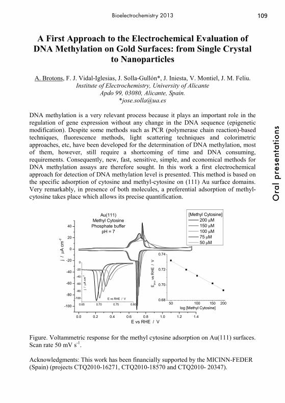
109
Ora
l pre
sent
atio
ns
Bioelectrochemistry 2013
A First Approach to the Electrochemical Evaluation of DNA Methylation on Gold Surfaces: from Single Crystal
to Nanoparticles
A. Brotons, F. J. Vidal-Iglesias, J. Solla-Gullón*, J. Iniesta, V. Montiel, J. M. Feliu. Institute of Electrochemistry, University of Alicante
Apdo 99, 03080, Alicante, Spain. *[email protected]
DNA methylation is a very relevant process because it plays an important role in the regulation of gene expression without any change in the DNA sequence (epigenetic modification). Despite some methods such as PCR (polymerase chain reaction)-based techniques, fluorescence methods, light scattering techniques and colorimetric approaches, etc, have been developed for the determination of DNA methylation, most of them, however, still require a shortcoming of time and DNA consuming, requirements. Consequently, new, fast, sensitive, simple, and economical methods for DNA methylation assays are therefore sought. In this work a first electrochemical approach for detection of DNA methylation level is presented. This method is based on the specific adsorption of cytosine and methyl-cytosine on (111) Au surface domains. Very remarkably, in presence of both molecules, a preferential adsorption of methyl-cytosine takes place which allows its precise quantification.
0.0 0.2 0.4 0.6 0.8 1.0 1.2 1.4
-100
-80
-60
-40
-20
0
20
40
j /
μA c
m-2
E vs RHE / V
[Methyl Cytosine] 200 μM 150 μM 100 μM 75 μM 50 μM
Au(111)Methyl CytosinePhosphate buffer
pH = 7
0.65 0.70 0.75 0.80
-100
-80
-60
-40
-20
0
log [Methyl Cytosine]
j /
μA c
m-2
E vs RHE / V50 100 150 200
0.68
0.70
0.72
0.74
Epi
co v
s R
HE
/ V
Figure. Voltammetric response for the methyl cytosine adsorption on Au(111) surfaces. Scan rate 50 mV s-1. Acknowledgments: This work has been financially supported by the MICINN-FEDER (Spain) (projects CTQ2010-16271, CTQ2010-18570 and CTQ2010- 20347).

110O
ral p
rese
ntat
ions
Bioelectrochemistry 2013
Electrochemical Investigation of Diselenide Bond Reduction
Marisa C. Buzzeo, Lindsey M. Walker, Faizunnahar Dewan
Barnard College, Columbia University 3009 Broadway, New York, NY 10027
[email protected] Selenium is an essential element, known to play an important role in biology. Proteins that contain selenocysteine or selenomethionone residues are powered with redox activity that allows them to take part in critical cellular pathways and processes.1 Interestingly, only one native diselenide-containing protein has been isolated to date,2
and reports on synthetic selenoproteins indicate that thioredoxin, the predominant reductant present in the cell, can only partially reduce the Se-Se bridge.3 Using a combination of electrochemical techniques, we aim to identify the conditions under which diselenide bond formation is most favorable and decipher the mechanistic details of its reduction. Ultimately, we seek to determine the role that these rare and stable bonds may play in the physiological setting. Our initial studies have focused on the redox behavior of selenocystine. Voltammetric and spectroscopic data for diselenide reduction will be presented and preliminary efforts toward electrochemical characterization of selenoproteins discussed. [1] (a) Stadtman, T. C., Annu. Rev. Biochem. (1996) 65, 83; (b) Jacob, C.; Giles, G. I.;
Giles, N. M.; Sies, H. Ang. Chem. Int. Ed. (2003) 42, 4742-4758; (c) Papp, L. V.; Lu, J.; Holmgren, A.; Khanna, K. K. Antiox. Redox Sign. (2007) 9, 775-806; (d) Wessjohann, L. A; Schneider, A.; Abba, M.; Brandt, W. Biol. Chem. (2007) 388, 997-1006 (e) Reeves, M. A.; Hoffman, P. R. Cell. Mol. Life Sci. (2009) 66, 2457-2478.
[2] Shchedrina, V. A.; Novoselov, S. V.; Malinouski, M. Y.; Gladyshev, V. M. Proc. Nat. Acad. Sci. (2007) 104, 13919-13924.
[3] Metanis, N.; Keinan, E.; Dawson, P. E. J. Am. Chem. Soc. (2006) 128, 16684-16691.

111
Ora
l pre
sent
atio
ns
Bioelectrochemistry 2013
Optimization of an IrOx-Based Glucose Biosensor
Bri Campbell, Hanna Elzanowska*, and Viola Birss Department of Chemistry, University of Calgary, Calgary, AB T2N 1N4, Canada
*Department of Chemistry, University of Warsaw, Warsaw, Poland [email protected]
We are developing a very promising IrOx-based biosensor, aimed at the accurate determination of blood glucose levels. These sensors are fabricated by depositing an aliquot of an ink, containing an EtOH-based Ir sol, glucose oxidase (GOx), and Nafion®, onto a Au electrode, followed by the electrochemical oxidation of the metallic Ir nanoparticles (NPs) to Ir oxide (IrOx). IrOx is an excellent matrix for the immobilization of redox-active enzymes such as GOx, due to its high porosity, chemical stability, rapid redox kinetics, and biocompatibility [1]. The ultimate goal of this work is to achieve O2-independent glucose sensing, with GOx regeneration (re-oxidation) occurring solely via reaction with the IrOx NPs, giving reliable and reproducible glucose readings despite the fluctuating blood O2 levels commonly observed in clinical practise. As part of this work, we examine the effect of varying the ratio of water to ethanol in the Ir sol. The glucose signal was found to increase as the amount of water in the ink increased, as would be expected with increased enzymatic activity. Detrimentally, the metallic Ir concentration in the sol was found to be diminished with increased water content and the subsequent Ir(III)/Ir(IV) oxide electrochemistry became more sluggish. These unfavourable changes may be the result of spontaneous oxidation of the metallic Ir NPs by water to form a hydrous and poorly interconnected form of IrOx, in comparison to the electrochemically formed IrOx in a neat ethanolic sol [2]. Nafion® is another component of these sensing layers, used because of its biocompatibility, ability to overcome interference effects, and its excellent binding capabilities. The addition of Nafion® (1 wt.%) to the Ir-containing ethanolic/aqueous inks was found to increase the IrOx electron transfer kinetics at all water contents, while also maintaining good GOx activity and a high glucose signal. However, too much Nafion® in the films was found to block electron transfer between IrOx NPs, leading to low glucose signals in the absence of O2, poor IrOx redox kinetics, and a more pronounced dependence on O2. Overall, it will be shown that there is an optimal amount of Nafion® (1 wt. %) in the films, as too much interferes with electron transfer between GOx and the IrOx nanoparticles, thus negatively affecting the ability of GOx to regenerate via the IrOx NPs alone. References 1. A. Jhas, H. Elzanowska, B. Sebastian, and V. Birss. (2010). Electrochimica Acta,
55: 7683-7689. 2. H.B. Campbell, H. Elzanowska, and V. Birss (2012). Biosens. And Bioelec.
(submitted Oct 2012).

112O
ral p
rese
ntat
ions
Bioelectrochemistry 2013
Electrochemical analysis of miRNA deregulated during the hypoxia conditions
Rui Camposa, Michael R. Horsmanb, Elena E. Ferapontovaa
aInterdisciplinary Nanoscience Center (iNANO Gustav Wieds Vej 14, Aarhus University, DK-8000 Aarhus C
bDept. Experimental Clinical Oncology, Aarhus University Hospital, Nørrebrogade 44, DK-8000 Aarhus C
Hypoxia is a cancer condition difficult both to diagnose and to treat; many solid tumors contain hypoxic regions due to impaired oxygen and nutrient supply. These tumors exhibit poor prognosis and high resistance to conventional therapies.1 In this context, electrochemical biosensor technologies, combining electrochemistry and DNA nanotechnology for sensitive, accurate, yet simple, inexpensive and robust nucleic acid analysis, may represent a powerful tool for medical diagnosis, both for prognostics of cancer and postcytotoxic therapy.2 Here, we exploited the electronic beacons approach3,4 to develop a genosensor able of selective simultaneous analysis of a number of miRNA deregulated during the hypoxia conditions. Redox-labeled hairpin DNA probes specific to 7 different miRNA were integrated within the 8-microelectrode electronic chip. Binding of miRNAs to DNA probes resulted in the electrochemical signal variation, reflecting the DNA probe conformational switching upon hybridization to target miRNA. Electroanalysis of synthetic miRNA mixtures was performed and used for a genosensor signal calibration and first trials of the correspondingly treated tumor cellular samples were performed. Advantages and disadvantages of the used approach for miRNA analysis are discussed. References (1) Busk, M.; Toustrup, K.; Sørensen, B. S.; Alsner, J.; Horsman, M. R.; Jakobsen,
S.; Overgaard, J. BMC Cancer 2011, 11, 63. (2) Drummond, T. G.; Hill, M. G.; Barton, J. K. Nat. Biotechnol. 2003, 21, 1192–
1199. (3) Fan, C.; Plaxco, K. W.; Heeger, A. J. PNAS 2003, 100, 9134–9137. (4) Farjami, E.; Clima, L.; Gothelf, K.; Ferapontova, E. E. Anal. Chem. 2011, 83,
1594–1602.

113
Ora
l pre
sent
atio
ns
Bioelectrochemistry 2013
Electrogene therapy with interleukin-12 alone or combined with electrochemotherapy for treatment of
spontaneously occurring tumors in dogs
Maja Cemazar1,2, Darja Pavlin3, Gregor Sersa1, Natasa Tozon3 1Institute of Oncology Ljubljana, Department of Experimental Oncology
Zaloska 2, SI-1000 Ljubljana, Slovenia 2University of Primorska, Faculty of Health Sciences, Polje 42, SI-6310 Izola; Slovenia
3University of Ljubljana, Veterinary faculty, Clinic for surgery and Small animals, Gerbiceva 60, SI-1000 Ljubljana
Electroporation has many biomedical applications. In human and veterinary oncology, its most advanced use is electrochemotherapy (ECT); in which electroporation is combined with injection of the chemotherapeutic drugs bleomycin or cisplatin to locally potentiate their antitumor effectiveness. Another application of electroporation is gene electrotransfer – electrogene therapy (EGT). It can be used either for vaccination or treatment of various diseases, including cancer, where therapies are targeted either directly to tumor cells or aim to increase the immune response of the organism against cancer. In our on-going clinical research, the effect of systemic and local EGT with interleukin -12 (IL-12) alone or in combination with ECT was evaluated in different canine tumors. 22 dogs of different breeds were treated. Prior to inclusion, written consent for participation was obtained from the owners and the study was approved by the Ethical Committee at the Ministry of Agriculture and Environment of the RS. 8 dogs were treated with intratumoral EGT and 6 dogs were treated with systemic EGT alone or as adjuvant therapy to surgery and/or chemotherapy, Additional 6 dogs were treated with combination of EGT and ECT. EGT was performed by intratumoral or intramuscular injection of the plasmid DNA encoding human IL-12 followed by application of electric pulses. Local response to the therapy was evaluated by measurements of tumors' size and histological examination. Systemic response was assessed by determination of IL-12 and IFN-γ in patients' sera. Side effects were determined by weekly clinical examinations and basic bloodwork. Intratumoral EGT IL-12 elicited good local antitumor effect with significant reduction of treated tumors' size. In 3 dogs, long lasting complete response was obtained, while in additional 3 dogs stabilization of disease for more than 12 months. Systemic release of IL-12 was detected in four dogs, without any noticeable local or systemic side effects. Intramuscular EGT IL-12 resulted in increased IFN-γ blood concentrations in 4 dogs. In these 4 dogs prolonged survival was obtained, however without effect on the tumor size. One of the other two dogs was euthanized due to progressive disease 2 months after EGT, while the other was euthanized 5.5 months after EGT due to the progression of pain, but was without radiologically evident distant metastases. Combined treatment of EGT and ECT resulted in complete response of all treated tumors. Collectively, intratumoral as well as intramuscular EGT IL-12 alone or combined with ECT are safe procedures that can result in systemic shedding of IL-12, which possibly triggers IFN-γ response, leading to prolonged disease free period and survival.

114O
ral p
rese
ntat
ions
Bioelectrochemistry 2013
Measuring and imaging electropermeabilization effects on cell membrane elasticity using Atomic Force Microscopy
Louise Chopinet,abd Marie-Pierre Rols bd and Etienne Dague *acd a CNRS ; LAAS ; 7 avenue du colonel Roche, F-31077 Toulouse Cedex 4, France. E-
mail : [email protected] b CNRS ; IPBS - UMR 5089 ; BP64182, 205 route de Narbonne, F-31077 Toulouse
Cedex 4, France. E-mail : [email protected] c CNRS ; ITAV-UMS3039 ; F31106 Toulouse, France
d Université de Toulouse ; UPS, INSA, INP, ISAE ; UT1, UTM, LAAS, ITAV ; F-31077 Toulouse Cedex 4, France
Electroporation is a physical method using pulsed electric field to deliver molecules into cells and tissues. Clinical applications have been successfully developed for antitumoral drug delivery and clinical trials for gene electrotransfer are underway [1]. However little is known about the mechanisms involved in membrane destabilization process during electropermeabilization and the direct effect on membrane struture. Mathematical models tend to prove that there is pore creation, but electropore have never been observed. However, lipid disorganization is happening, as shown by the formation of tubules and vesicles on Giant Unilamellar Vesicle, and flip flop is occurring on cells. In order to go further we exploit Atomic Force Microscopy (AFM) force spectroscopy abilities to access electric field effect on plasma membrane [2]. Chinese Hamster Ovary Cells are cultured on glass cover slip with complemented medium (MEM 0111, Eurobio, France). Classical gene transfer parameters are used (500 V/cm, 8 pulses of 5 ms), on fixed and living cells. Measurements are carried out using JPK Nanowizard 3 coupled to a fluorescence microscope (Zeiss). Si3N4 AFM probes (MLCT model manufactured by Bruker Instruments) are used for all the experiments. Force curve and image analysis are done with the OpenFovea Software [3]. We took advantage of Atomic Force Microscope to visualize direct consequences of electropermeabilization in terms of membrane organization by imaging and to locally measure the membrane elasticity on both fixed and living CHO cells. For the first time, we managed to measure a rapid propagation of membrane perturbation around the cell by visualizing transient rippling of membrane surface and measuring a decrease in membrane elasticity by 40%. We give first assumption of membrane permeabilization resealing that seems slower than ever seen by fluorescent microscopy. Thus, AFM is a useful tool to investigate basic process of electroporation in vitro and enlarge the understanding of the process at the nanoscale.
[1] Daud, A. I., R. C. DeConti, et al., J Clin Oncol (2008) 26(36): 5896-903.
[2] Philippe Carl & Hermann Schillers, Pflugers Arch - Eur J Physiol (2008) 457:551–
559.
[3].Roduit, C. et al. OpenFovea: open-source AFM data processing software. Nat.
Meth. 9, 774-775, 2012.

115
Ora
l pre
sent
atio
ns
Bioelectrochemistry 2013
Scanning Droplet Cell for Chemoselective Patterning via Local Electroactivation of Protected Quinone Monolayers
Jan Clausmeyer, Jörg Henig, Wolfgang Schuhmann and Nicolas Plumeré Ruhr-Universität Bochum, Analytische Chemie and Center for Electrochemical
Sciences - CES, D-44780 Bochum, (Germany) [email protected]
Electrochemical strategies are promising tools for local surface activation to control the spatial distribution of bio-molecules on surfaces. Procedures based on scanning probe lithography and direct mode scanning electrochemical microscopy (SECM) may be applied for surface patterning. However, these approaches rely on high applied poten-tials resulting in undefined surface chemistries. We demonstrate the selective electrochemical patterning method starting from uni-formly modified electrode surfaces and provide direct evidence for the formation of the target functional group. The electrodes are functionalized with a layer of p-hydro-quinone protected by an electrochemically cleavable group. Upon anodic treatment, deprotection and activation of the quinone moieties allow for covalent attachment of Michael donors. The patterning was achieved by exposing the region to be activated to the electrolyte with a scanning droplet cell. The key advantage of the droplet cell is the possibility to use a large counter electrode which limits formation of reactive products and undesired side reactions. Bio-molecules bearing cysteine residues on their surface can be immobilized by taking advantage of the Michael addition.
Anodic cleavage of protecting group from p-hydroquinone monolayers within a scanning droplet cell (top) and SECM image of a spot of locally deprotected p-hydroquinone on a glassy carbon electrode globally modified with the protected quinone (bottom). Acknowledgement: financial support by the EU and the state NRW in the frame work of the HighTech.NRW programme is gratefully acknowledged.

116O
ral p
rese
ntat
ions
Bioelectrochemistry 2013
0
1
2
3
4
5
0% 25% 50% 75% 100%
Pow
er (m
W)
Duty Cycle
Rext = 13.5ΩRext = 21ΩRext = 6Ω
Microbial Fuel Cell Operation with Pulse-Width Modulated Connection of the External Resistor
J. Coronado1, M. Perrier1, B. Tartakovsky1,2
1- Departement de Génie Chimique, École Polytechnique Montréal, C.P.6079 Succ., Centre-Ville Montréal, QC, Canada H3C 3A7
2- National Research Council of Canada,6100 Royalmount Ave., Montréal, QC, Canada H4P 2R2
Address e-mail : [email protected]
The concept of Microbial Fuel Cell (MFC) operation with intermittent connection of the external resistance has been already demonstrated [1]. That study demonstrated that an MFC can be operated at electrical loads below the MFC internal resistance without significant energy losses. Our new study presents results of MFC operation with pulse-width modulated connection of the external resistor in a broad range of operating frequencies (0.1 Hz to 3000 Hz). Analysis of output voltages obtained during low and high frequency tests showed process dynamics, which can be adequately described by a simple equivalent circuit model accounting for two resistors and a capacitor (Randles cell model). In agreement with this model, the power output was observed to improve during MFC operation at high frequencies (e.g. above 100 Hz). Furthermore, we demonstrate that MFC performance can be optimized and a sharp decline in MFC performance can be averted by current control through duty cycle adjustments, even with a significant mismatch between the external and internal resistances, as illustrated in the graph below (Rint estimation 14Ω). Overall, a comparison of MFCs operated with a constantly connected optimal external resistor (e.g. Rext ~ Rint) and with an equal pulse-width modulated external resistor showed a power output increase of 22-43%. This led to sustained volumetric power densities of 70 - 80 mW/L observed in long-term (over six months) MFC tests. References [1] F. Grondin, M. Perrier, B. Tartakovsky, J. Power Sources 208 (2012) 18-23.

117
Ora
l pre
sent
atio
ns
Bioelectrochemistry 2013
Immobilisation of Aquifex aeolicus Membrane-bound Hydrogenase on carbon nanofibers for H2/O2 biofuel cells.
A. de Poulpiquet1, H. Marques 2, M.T. Giudici-Orticoni1, R. Gadiou2, E. Lojou1 1Unité de Bioénergétique et Ingénierie des Protéines, UMR 7281, Institut de
Microbiologie de la Méditerranée, CNRS-AMU, 31 Chemin Aiguier, 13402 Marseille cedex 20, France
[email protected] 2Institut des Sciences des Matériaux de Mulhouse, LRC 7228, CNRS-UHA, 15 Rue
Jean Starcky, BP 2488, 68057 Mulhouse cedex, France
Fuel cells using renewable resources may be an interesting possibility in taking up the energy challenges launched by fossil fuels exhaustion. However they need conventional catalysts based on platinum that are available only in limited amounts, and also are scarcely selective. An attractive alternative in this context might be the use of enzyme,.Recently, a new kind of biofuel cells based on the oxidation of dihydrogen catalysed by a hydrogenase at the anode has emerged1. The [Ni-Fe] membrane-bound hydrogenase (Hase) from the hyperthermophilic organism Aquifex aeolicus, which oxidizes hydrogen into protons with high efficiency while presenting outstanding O2, CO and temperature resistances, has proved to be a valuable candidate in a H2/O2 biofuel cell2. To raise the power and then the current densities at the electrodes, the amounts of connected enzymes must be maximized. To that purpose conductive supports with high surface to volume ratios like porous or high surface carbon materials might be used. We already proved modifications of graphite electrodes with carbon nanotubes to be efficient in a H2/O2 biofuel cell whose power densities reached 300 μW.cm-2 3,4. Fishbone carbon nanofibers (CNFs) are another promising carbonated material as they are highly graphitic, highly electrically conductive, and bear lots of reactive edge sites that allow the entrapment of proteins and facilitate modifications of the surface chemistry. We report in this work the use of such CNFs for modification of electrodes leading to high current densities as well as interesting and intriguing catalytic signal properties. The CNF growth and intrinsic properties are the key point for an understanding of the interfacial electron transfer. Different treatments had been applied after synthesis, leading to different surface properties of the fibers. The best protocol to get the highest enzyme coverage and highest catalytic efficiency has been determined. The role of several redox dyes in enabling the mediated electron transfer, or possibly anchoring the hydrogenase has also been explored and will be discussed in this work. 1 Vincent et al., Proceedings of the national academy of sciences of the USA (2005) 2 Ciaccafava et al., Angewandte Chemie (2011) 3 Ciaccafava et al., Electrochemical communications (2012) 4 de Poulpiquet et al., Electroanalysis (submitted)

118O
ral p
rese
ntat
ions
Bioelectrochemistry 2013
Electrochemical Aptasensing – Reaching Maximum Residue Limits and Unraveling Biomolecular Interactions
Karolien De Wael
1 University of Antwerp, Department of Chemistry, Environmental Analysis Research Group, Groenenborgerlaan 171, B-2020 Antwerp, Belgium
[email protected] A highly competitive and hot topic in developing biosensors is the use of aptamers, also known as ‘chemical antibodies’, as bio-recognition compound. Aptamers (single strand (ss)DNA or RNA) are synthetic oligonucleic acid sequences which can bind to their targets with high affinity and specificity due to their flexibility. In addition, they are stable and can be employed in extreme conditions. Moreover, these oligonucleic acids can be easily modified by attachment of functional groups without affecting their affinity. Electrochemical sensors with immobilized aptamers as sensing elements are called electrochemical aptasensors. The high selectivity of these sensors is a result of the unique properties of aptamers. Many strategies are suggested for the immobilization of aptamers on transducers surface but they are mostly restricted by covalent attachment or chemisorption. Despite the fact that aptamers are chemically more stable compared to proteins, they have to be protected from nucleases. For this aim, entrapment in a protective matrix is suggested to overcome this problem. Selecting an appropriate host matrix for aptamers is one of the main challenges for the immobilization of aptamers in order to improve the analytical characteristics of the aptasensors. In this work, different immobilization strategies will be discussed aiming the development of aptasensing devices for the detection at MRL (maximum residue limit) level of environmentally important molecules such as antibiotics, PCBs, …

119
Ora
l pre
sent
atio
ns
Bioelectrochemistry 2013
Giant Vesicles in Electric Fields – Approaches for Measuring Properties of Lipid Membranes
Rumiana Dimova
Max Planck Institute of Colloids and Interfaces Science park Golm, 14424 Potsdam, Germany
Giant vesicles provide exceptional biomembrane models for systematic studies on the effect of electric fields on lipid bilayers. The main advantage of these systems is that the membrane response to AC and DC fields can be directly visualized under the optical microscope (1-3). In this talk, we will present several experimental approaches for deducing the membrane properties based on exposing giant vesicles to AC fields and DC pulses. In AC fields, the dependence of the vesicle morphology on both field frequency and media conductivity has been characterized recently (3, 4). When the conductivity of the external solution is higher than the internal conductivity, the vesicles deform into prolates at low field frequencies (few kHz); Fig. 1a. At intermediate frequencies (several kHz), prolate-oblate transitions are observed; Fig. 1b. With increasing field frequency, the vesicles undergo a prolate-to-sphere transition; Fig. 1c. Recently reported theoretical descriptions of these transitions (5, 6) predict the dependence of the frequency of the prolate-oblate transition on the vesicle size. We used this prediction to develop a method for measuring the membrane capacitance (7). At a fixed field frequency in the low frequency regime and increasing field strength, the degree of vesicle deformation increases and can be used to deduce the bending rigidity of the membrane (8). When exposed to strong DC pulses, giant vesicles porate. Using the dynamics of the pore closure we established an approach for measuring the edge tension and evaluate the membrane stability (9). The response of both fluid- and gel-phase membranes will be discussed.
Fig. 1 Deformation of a giant vesicle subjected to AC field of 0.2 kV/cm and various field frequencies. 1. R. Dimova, in Advances in Planar Lipid Bilayers and Liposomes, p. 1, Academic Press (2012). 2. R. Dimova et al. Soft Matter, 3, 817 (2007). 3. R. Dimova et al., Soft Matter, 5, 3201 (2009). 4. S. Aranda et al., Biophys. J., 95, L19 (2008). 5. P. M. Vlahovska et al., Biophys. J., 96, 4789 (2009). 6. T. Yamamoto et al., Langmuir, 26, 12390 (2010). 7. P. F. Salipante et al., Soft Matter, 8, 3810 (2012). 8. R. S. Gracià et al., Soft Matter, 6, 1472 (2010). 9. T. Portet and R. Dimova, Biophys. J., 99, 3264 (2010).
120O
ral p
rese
ntat
ions
Bioelectrochemistry 2013
Electrochemical Detection of the Protein Mdm2 by a Peptide Affinity Probe Based on the Protein p53
Thomas Doneux, Eléonore Triffaux, Claudine Buess-Herman
Chimie Analytique et Chimie des Interfaces, Faculté des Sciences, Université Libre de Bruxelles
Boulevard du Triomphe, 2, CP 255, B-1050 Bruxelles, BELGIUM [email protected]
Aptamers and peptide aptamers belong to a new class of synthetic affinity probes having a high selectivity against a given target protein. In the field of electrochemical biosensing, their use as specific recognition elements offers many advantages as compared to antibodies, essentially because of their synthesis in vitro and their small size. The present contribution is focused on the electrochemical detection of the protein Mdm2 using a peptide aptamer sequence based on the recognition domain of the tumor suppressor protein p53 against Mdm2. This latter interacts specifically with a short α-helix composed of the residues 19-26 of p53. The design of our peptide aptamer sequence is thus based on this interaction. The sequence is made of the residues 12-26 of p53, to ensure a sufficient flexibility, with an additional cysteine amino acid at the N-terminal. The thiol group on the side chain of the cysteine is used to self-assemble the peptide aptamer at gold electrodes, through the formation of Au-S bonds. The design of peptide affinity probes, illustrated here, can be extended to numerous target proteins. Moreover, redox labelling of the probes can be performed by rather conventional organic and inorganic chemistries. The formation and characterisation of various monolayers is presented, the layer consisting of the aptamer alone or co-adsorbed with a mercaptobutanol spacer. Electrochemical results were obtained by cyclic voltammetry, impedance spectroscopy in the presence of the redox markers [Fe(CN)6]3-/4- and chronocoulometry in the presence of the electroactive [Ru(NH3)6]3+ complex, and completed by in situ (i.e. under electrochemical control) fluorescence microscopy measurements. It is shown that the Mdm2 protein is succesfully detected with the peptide aptamer monolayer, down to subnanomolar concentrations. Control experiments performed with other proteins (bovine serum albumin, fibrinogen, cytochrome c) demonstrated the specificity of the sensor against the Mdm2 target.

121
Ora
l pre
sent
atio
ns
Bioelectrochemistry 2013
Rythms of Wake and Sleep in the rat`s Cerebral Cortex Redox Potential
1Dubinin A.G., 2Shvets-Teneta-Gurii T.B., 3Troshin G.I.
1Mendeleev University of Chemical Technology of Russia, Miusskaya sq. 9, Moscow, 125047, Russia;
2Institute of Higher Nervous Activity and Neurophysiology RAS ,Butlerov str., 5 a, GSP 7, Moscow, 117865, Russia;
3Scientific-Industrial Center of Federal State Unitary Enterprise “VIGSTAR”, 1st Road passage, 8, Moscow, 117545, Russia
[email protected] The brain tissue redox potential (E) depends on the ratio of the rates of the processes occurred in two compartments of energy metabolism – in the glycolysis compartment, in which glucose is degraded without oxygen utilization, and the oxidative metabolism compartment. The E changes are a valid indicator of the balance on current glycolysis and oxidative metabolism in the brain tissue energy metabolism. This work has been fulfilled to advance our pioneer studies of the cerebral cortex energy metabolism (EM) in behaving animals. The gist of this line consists of the simultaneous multipoint ceaseless recording local changes in the cerebral cortex E in behaving animals. Freely moving rats were implanted with platinum electrodes into three symmetrical cortical sites of frontal, parietal and occipital areas. Electrocorticogram, electromyogram of neck muscles and general motor activity were recorded in parallel. DC amplifiers (input resistance 4 GΩ, bandwidth 0-20 Hz). The common reference platinum electrode was implanted into the nasal bone. Episodes of wake (W) and paradoxical sleep (PS - dream time) were accompanied by the E rising and by the formation in the E of a rather regular quasi sinusoidal oscillations (the periods - near 0,3 Hz, the amplitudes - near several millvolts). Transitions of rats to slow wave sleep (SWS) were accompanied by the E decrease and by arising a complex of a rather regular E variations, in which next oscillations (the periods of near 5 s and the amplitudes of several millvolts; the periods of near 10 s and the amplitudes of several dozens millvolts) and irregular variation (the periods of 5-25 s, the amplitudes of several dozens millvolts) were the most pronounced. In sum, the given facts indicate that the increase in the cerebral cortex activity coupled to W and PS are mainly fueled by oxidative compartment, for which biochemical oscillatory mode is near 0,3 Hz. Brain function in SWS are mainly fueled by glycolytic compartment in which several biochemical oscillators are present. SWS is known to promote memory consolidation. We think the power cerebral cortex E oscillations complex in SWS to show both a tremendous energy costs and a multiformity of biochemical systems coupled to brain work in SWS.
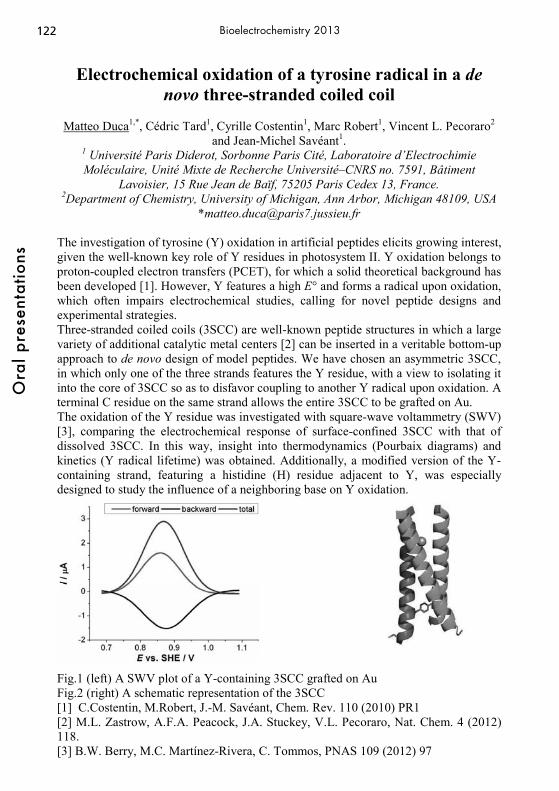
122O
ral p
rese
ntat
ions
Bioelectrochemistry 2013
Electrochemical oxidation of a tyrosine radical in a de novo three-stranded coiled coil
Matteo Duca1,*, Cédric Tard1, Cyrille Costentin1, Marc Robert1, Vincent L. Pecoraro2
and Jean-Michel Savéant1. 1 Université Paris Diderot, Sorbonne Paris Cité, Laboratoire d’Electrochimie Moléculaire, Unité Mixte de Recherche Université–CNRS no. 7591, Bâtiment
Lavoisier, 15 Rue Jean de Baïf, 75205 Paris Cedex 13, France. 2Department of Chemistry, University of Michigan, Ann Arbor, Michigan 48109, USA
*[email protected] The investigation of tyrosine (Y) oxidation in artificial peptides elicits growing interest, given the well-known key role of Y residues in photosystem II. Y oxidation belongs to proton-coupled electron transfers (PCET), for which a solid theoretical background has been developed [1]. However, Y features a high E° and forms a radical upon oxidation, which often impairs electrochemical studies, calling for novel peptide designs and experimental strategies. Three-stranded coiled coils (3SCC) are well-known peptide structures in which a large variety of additional catalytic metal centers [2] can be inserted in a veritable bottom-up approach to de novo design of model peptides. We have chosen an asymmetric 3SCC, in which only one of the three strands features the Y residue, with a view to isolating it into the core of 3SCC so as to disfavor coupling to another Y radical upon oxidation. A terminal C residue on the same strand allows the entire 3SCC to be grafted on Au. The oxidation of the Y residue was investigated with square-wave voltammetry (SWV) [3], comparing the electrochemical response of surface-confined 3SCC with that of dissolved 3SCC. In this way, insight into thermodynamics (Pourbaix diagrams) and kinetics (Y radical lifetime) was obtained. Additionally, a modified version of the Y-containing strand, featuring a histidine (H) residue adjacent to Y, was especially designed to study the influence of a neighboring base on Y oxidation.
Fig.1 (left) A SWV plot of a Y-containing 3SCC grafted on Au Fig.2 (right) A schematic representation of the 3SCC [1] C.Costentin, M.Robert, J.-M. Savéant, Chem. Rev. 110 (2010) PR1 [2] M.L. Zastrow, A.F.A. Peacock, J.A. Stuckey, V.L. Pecoraro, Nat. Chem. 4 (2012) 118. [3] B.W. Berry, M.C. Martínez-Rivera, C. Tommos, PNAS 109 (2012) 97

123
Ora
l pre
sent
atio
ns
Bioelectrochemistry 2013
Microfluidic Electrochemical Lab-on-a-Chip Systems
J. Emnéus1, K. Zόr1, M. Vergani2, E. Landini3, M. Carminati2, V. Coman1, A. Martinez Serrano4, L. Amato1, S. Keller1, A. Boisen1, M. Kokaia5, T. Ramos Moreno4, A. Ghio3,
W.E. Svendsen1, M. Dimaki1, Zs. Keresztes6, M. Adamovski7, U. Wollenberger7, D. Sabourin1, G. Ferrari2, R. Raiteri3, M. Sampietro2, M. Dufva1, and A. Heiskanen1
1Technical University of Denmark, Denmark, 2Politecnico di Milano, Italy, 3University of Genova, Italy, 4University Autonomous of Madrid, Spain, 5Wallenberg Neuroscience
Center, Lund University, Sweden, 6Hungarian Academy of Sciences, Hungary, and 7University of Potsdam, Germany.
This talk presents the course of developments towards multi-channel microfluidic electrochemical Lab-on-a-Chip (LOC) systems designed for real-time monitoring of cellular dynamics, easily adaptable for many purposes. The idea is based on a modular component based platform in which the various components (pumps, valves, liquid containers, microfluidic electrode chips, potentiostat etc.) can be combined into in principle any analytical platform desired1,2 (Fig, 1). The systems are equipped to handle a range of different plug-in microfluidic chips (Fig. 1, inset), using a combination of optical and electrochemical detection strategies. The microfluidic chips can be integrated with different types of electrode array chips. Electrochemical behavior of novel 2 and 3 dimensional (2D and 3D) carbon based electrode arrays, fabricated by patterning SU8 following pyrolysis, will be presented3 (Fig. 2).
Fig. 1. A microfluidic LOC system for optical and electrochemical detection.
Fig. 2. 3D carbon based pillar electrode array.
[1] D. Sabourin et al. The MainSTREAM component platform: A holistic approach to microfluidic system design. JALA 2012. In press.
[2] M. Vergani et al. Multichannel Bipotentiostat Integrated with a Microfluidic Platform for Electrochemcial Real-Time Monitoring of Cell Cultures, IEEE Transactions on Biomedical Circuits and Systems. In press.
[3] Amato, L. et al. Fabrication of high-aspect ratio SU-8 micropillar arrays. Microelectronic Engineering 98, 483–487 (2012).

124O
ral p
rese
ntat
ions
Bioelectrochemistry 2013
Communication between Electrode Surface and Whole Cells with Biological Redox Shuttle for Biosensor
Applications
Mathieu Etienne, Wissam Ghach, Alain Walcarius CNRS and Université de Lorraine, LCPME, UMR 7564
405, rue de Vandoeuvre, F-54600 Villers-lès-Nancy, France [email protected]
Recently, whole cells have received considerable attention in fabricating biosensors, bioreactors and biofuel cells, due to the easy and fast communication with their surrounding environment. In this regard, the utilization of a whole cell as host of intracellular enzymes provides a system with high efficiency and stability [1]. Its ability to oxidise different substrates and to transfer electrons either directly [2] or mediated by chemical redox shuttles [3,4], has improved the development of electrochemical cell-based biosensors. The limitation point for these biosensors is the toxicity of the chemical shuttles (e.g., FeCN)6
3-) used to improve the bioelectrochemical communication between bacteria and the electrode which can cause damages in cell envelope [3]. In addition, the electrochemical biosensors require the immobilization of whole cells to retain the microorganisms in close proximity to the electrode surface for higher reactivity and stability of the electrochemical communications [5]. In this work, we are studying the electrochemical communication of pseudomonas fluorescens CIP 69.13 mediated with biological redox shuttle and encapsulated in sol-gel films for construction of environmental biosensing systems.
[1] F. Lagarde, N. Jeffrezic-Renault, Anal. Bioanal. Chem., 400, 947-964 (2011) [2] L. Penga, S. J. You, J. Y. Wanga, Biosens. Bioelectron., 25, 1248–1251 (2010) [3] C. Liua, T. Suna, Y. Zhaib, S. Dong, Talanta, 78, 613–617 (2009) [4] V. Coman, T. Gustavsson, A. Finkelsteinas, C. von Wachenfeldt, C. Hägerhäll, L. Gorton, J. Am. Chem. Soc., 131, 16171–16176 (2009). [5] S.F. D’Souza, Biosens. Bioelectron., 16, 337–353 (2001)
125
Ora
l pre
sent
atio
ns
Bioelectrochemistry 2013
“Smart” Polymer Interfaces at Electrodes – useful Matrix for Biorecognition Reactions
Artur Fandrich1, Jens Buller2, Erik Wischerhoff3, André Laschewsky2, Fred Lisdat1
1 Technical University of Applied Sciences Wildau Bahnhofstraße 1
15745 Wildau (Germany) [email protected] 2 University of Potsdam
Karl-Liebknecht-Straße 24-25 14476 Potsdam (Germany)
3 Fraunhofer Institute for Applied Polymer Research Geiselbergstraße 69
14476 Potsdam (Germany)
Various organic polymers have already been established as modifiers for special applications according to their chemical and physical characteristics. A special class of compounds, the “smart” polymers, allows construction of eminently sophisticated interface systems. Such polymers undergo significant phase transitions in response to environmental triggers like temperature or pH changes. Immobilized on solid electrodes these compounds enable variations of interface properties “on demand”. In this electrochemical study a thermoresponsive polymer film covalently bound on gold surface is investigated with regard to its application as matrix for biorecognition processes. For this purpose a thermoresponsive polymer with a lower critical solution temperature (LCST) around 38 °C was synthesized and afterwards immobilized on gold electrodes. Cyclic voltammetric and impedimetric measurements with modified electrodes were realized in potassium ferro-/ferricyanide solutions at different temperatures. The results of these investigations show successful immobilization of the polymer on the surface of gold, since the interfacial impedance is clearly dominated by the polymer film on the surface. Significant changes in the voltammetric peak current and peak separation values around LCST clearly demonstrate the thermally induced phase transition. These results are also verified by surface plasmon resonance measurements where a change in the slope of sensorgram at LCST in case of a modified polymer electrode can be observed [1]. Further experiments are related to the idea of using this polymer interface as carrier for biorecognition elements and its utility for the analysis of biochemical processes. The switchable system is combined with biological components (peptide, antibody) and the effect on the responsive behavior under recognition reaction is tested. It was found that a sufficient thickness of the layer is required in order to allow structural switching of polymer fused with a biorecognition complex. [1] A. Fandrich, J. Buller, E. Wischerhoff, A. Laschewsky, F. Lisdat, Electrochemical Detection of the Thermally Induced Phase Transition of a Thin Stimuli-Responsive Polymer Film, ChemPhysChem 13 (2012) 2020-2023
126O
ral p
rese
ntat
ions
Bioelectrochemistry 2013
Utilization of organic electroactive moieties for redox DNA labelling and electrochemical monitoring of modified DNA
synthesis
M. Fojta1, P. Vidlaková1, J. Balintová2, V. Raindlová2, P. Ménová2, Z. Vychodilová1, J. Špaček1, A. Daňhel1, L. Havran1 and M. Hocek2
1 Institute of Biophysics, v.v.i. Academy of Sciences of the Czech Republic Královopolská 135, 61265 Brno (Czech Republic)
2 Institute of Organic Chemistry and Biochemistry Academy of Sciences of the Czech Republic Flemingovo nám. 2, 16610 Prague 6 (Czech Republic)
[email protected] Intrinsic electrochemical activity of nucleic acids [1] has been utilized in many label-free analytical applications. Moreover, labelling of DNA with electroactive moieties has been introduced to improve sensitivity and selectivity of electrochemical detection typically in sequence-specific DNA sensing (such as DNA hybridization, detection of +electrochemical properties of transition metals which are introduced into DNA in form of metalloorganics (ferrocene) or organic metal chelates (tris-bipyridine complexes of e.g., Co, Ru or Os, oxoosmium complexes [2]). Such labels usually exhibit reversible electrochemistry involving exchange of one or two electrons. In addition, several oxygenous and/or nitrogenous organic moieties were used as electroactive DNA markers as well, including nitrocompounds, anthraquinone (AQ) or benzofurazane (BF). With the exception of AQ, these species give irreversible electrochemistry and thus they are not suited for reusable detection systems. However, in other applications the irreversibility may help to distinguish between different redox labels as demonstrated for a combination of AQ and nitrophenyl [4]. Moreover, some of these compounds are reduced with multiple (4-6) electrons (nitrogroup, BF) which facilitates their sensitive detection, and/or are convertible into other electroactive forms which can be further utilized for selectivity improvement [4]. Last but not least, conversion of some reactive groups (e.g., aldehyde), incorporated in DNA for subsequent attachment of further labels into the respective products (e.g., hydrazone), can easily be monitored voltammetrically [5], allowing an easy monitoring of the DNA modification. References [1] E. Paleček, M. Bartošík (2012): Chem. Rev. 112, 3427-3481. [2] M. Fojta, L. Havran, H. Pivoňková, P. Horáková, M. Hocek (2011) Curr. Org. Chem., 15, 2936-2949 [3] M. Hocek, M. Fojta (2011): Chem. Soc. Rev, 40, 5802-5814. [4] J. Balintova, R. Pohl, P. Horakova, P. Vidlakova, L. Havran, M. Fojta, M. Hocek, (2011) Chem-Eur J, 17 14063-14073 [5] V. Raindlova, R. Pohl, B. Klepetarova, L. Havran, E. Simkova, P. Horakova, H. Pivonkova, M. Fojta, M. Hocek (2012), Chempluschem, 77 652-662
127
Ora
l pre
sent
atio
ns
Bioelectrochemistry 2013
Practical Implications of using Nano-Electrodes for Bioanalytical Measurements
Neville J Freeman1, Reshma Sultana1, Andrew R Mount2, Anthony J Walton3,
Jonanthan Terry3, Ilka Schmuser2 1.NanoFlex Ltd
Daresbury Innovation Centre, Keckwick Lane, Daresbury, UK 2. School of Chemistry, The University of Edinburgh, Joseph Black Building, Kings
Buildings, Edinburgh, Scotland EH9 3JJ, UK 3. Institute for Integrated Micro and Nano Systems, School of Engineering, The
University of Edinburgh, King’s Buildings, Edinburgh, EH9 3JF, UK [email protected]
Nanoelectrode structures have been of intense interest for the past decade or more. The benefits of nanoscale electrodes are well understood from a theoretical perspective which has been broadly borne out empirically. Such benefits include low capacitance, enhanced kinetics, high signal to noise ratios and immunity to hydrodynamic perturbations to name but a few. In this paper we examine a model nano-electrode system and assess some key performance aspects which are readily accessible from an experimental bioanalytical perspective. We have focused on the following characteristics using a ubiquitous electrochemical reporter: 1) Realizable limits of detection; 2) Potential modes of interrogation, both to enhance electrode lifetimes and assess temporal characteristics; 3) Susceptibility to stirring to determine the breadth of application scenarios which could potentially be addressed. We consider these characteristics are critical when considering the use of nano-electrodes in bioelectrochemical settings and discuss such deployment in the light of our results.

128O
ral p
rese
ntat
ions
Bioelectrochemistry 2013
Pulsed Electric Field Treatment of Microalgae: Benefits for Downstream Processing
Wolfgang Frey, Christian Eing, Martina Goettel, Christian Gusbeth, Ralf Straessner
Karlsruhe Institute of Technology (KIT) Institute for Pulsed Power and Microwave Technology (IHM)
Hermann-v-Helmholtz-Platz 1 76344 Eggenstein-Leopoldshafen
Compared to agriculturally produced biomass, microalgae exhibit a higher percentage of value-added intracellular components which can provide a remarkable feedstock for bio-fuel production and for bio-based fine chemicals industry. Up to now, conventional microalgae processing exhibits a poor energy balance with regard to an energetic use of algal lipids which are stored intracellular. PEF-treatment of microalgae suspensions considerably increases the yield of water-soluble cell components and improves intracellular solvent access for subsequent lipid extraction. Thus, the amount of lipids extracted from PEF-treated microalgae by Ethanol increases by a factor of 3-4. This gain in extraction efficiency was obtained for dry and wet microalgae biomass as well. The fact, that lipid droplets remain intracellular after PEF treatment and during the separation of water-soluble components, opens new downstream processing pathways for subsequent selective processing of the lipid-containing residual biomass. The required PEF treatment energy amounts to 1 MJ per kg (dry weight) biomass, which is low, compared to conventional processing. Especially the PEF-assisted lipid extraction of from wet biomass can save more than 7 MJ/kg of drying energy and can improve the energy balance for microalgae processing for energetic use. PEF-treatment related issues, like reproducible cultivation of microalgae biomass at IHM, being an important condition for proximate experimental investigations, treatment procedures and diagnostic methods used for cell content monitoring during cultivation and after extraction will be addressed.

129
Ora
l pre
sent
atio
ns
Bioelectrochemistry 2013
Direct electron transfer of Cytochrome C encapsulated in sol-gel silica matrices.
Alonso Gamero-Quijano1, Francisco Montilla1, Francisco Huerta2, Emilia Morallón1.
1Dept. Química Física e Instituto Universitario de Materiales, Universidad de Alicante, Ap. 99, E-03080, Alicante, Spain
2Dept. Ingenieria Textil y Papelera, Universitat Politecnica de Valencia, Plaza Ferrandiz y Carbonell, 1. E-03801, Alcoy, Spain
e-mail address: [email protected]
Most chemical sensors consist of two main components. First, a receptor that is able to recognize a specific analyte. This element usually produces a binding event with the analyte. The second component is a transducer, where the binding event is translated to a measurable physical change. In the particular case of electrochemical sensors, the transducer will change its redox state and, consequently, a method for quantifying that transformation must be found. Obviously, the use of suitable electrodes that convert the redox change into a quantifiable signal, such as electric current, is unavoidable. Biosensors are a class of sensors that exploit biological materials as active recognition elements with enhanced selectivity. A wide variety of biological species has been used in electrochemical biosensors, including proteins, enzymes, antibodies, etc. The development of the so-called third generation of amperometric biosensors is based on the electronic transfer between redox proteins and conventional electrodes. Redox proteins usually offer a complex behavior, so we have employed the well-studied cytochrome c as a conventional model redox protein. Cytochrome c was encapsulated in a silica matrix and then supported onto ITO electrodes. We have prepared different kind of matrices by modifying the silica precursors: TEOS for conventional silica or organic-modified silica (ormosil). The electrochemical performance of cytochrome c inside the silica-based matrix was determined by cyclic voltammetry and in situ UV-vis spectroscopy. We have observed that Cyt c encapsulated in conventional silica does not provide any electrochemical response. Oppositely, a clear electrochemical response is obtained when Cyt c is encapsulated in ormosils. To optimize the electrochemical transduction process, we have inserted a conducting polymer, such as PEDOT, within the silica matrix. Such composites were prepared by reactive insertion, following a methodology proposed in previous studies [1,2]. The presence of PEDOT facilitates the direct electron transfer between the encapsulated Cyt c and the ITO surface and, as a result, enhanced electrochemical response of Cytc was obtained. [1] F. Montilla, M. Cotarelo; J. Mater. Chem., 19 (2009) 305 [2] D. Salinas-Torres, F. Montilla; Electrochimica Acta 56 (2011)

130O
ral p
rese
ntat
ions
Bioelectrochemistry 2013
Functionalization of Diamond Surfaces for Bio-applications
Roberta Caterino, Andreas Reitinger, and Jose A. Garrido Walter Schottky Institut, Technische Universität München
Am Coulombwall 4, Garching [email protected]
The modification of the diamond surface with organic molecules is crucial for the development of bio-applications of this material. In order to introduce different functional groups and to tailor the surface properties, there is a great interest in broadening the range of linker molecules which can be covalently bound to the diamond surface. An effective surface modification can promote cell adhesion or enable the controlled grafting of functional biomolecules such as proteins or DNA, often used in biosensors. When it comes to protein immobilization, the hydropathicity of the surface has a major influence on the protein conformation and on the possibility to preserve its functionality onto the electrode surface. For bioelectrochemical applications, particular care is necessary to enhance charge transfer between the electrode and the redox center embedded in the protein, across the linker-layer. In this contribution, we will summarize our recent work on the functionalization of diamond surfaces with bio/organic molecules for the development of diamond-based biochemical sensors. Different functionalization routes will be reported depending on the surface termination of diamond. It will be shown how proteins and enzymes can be immobilized on activated diamond surfaces and still maintain their enzymatic functionality. Various examples of diamond-based bio-hybrid electronic systems will be discussed as proof of concepts, including enzyme-modified field effect transistors, enzymatic amperometric biosensors, as well as energy harvesting systems based on immobilized photoreaction centers.

131
Ora
l pre
sent
atio
ns
Bioelectrochemistry 2013
Sandwich microassay for pathogens detection related to urinary tract infections. Selective post-labeling of
hybridized 16S rRNAs
Magdalena Gebalaa*, Andreas Zimdarsb, Gerhard Hartwichb, Wolfgang Schuhmanna
a) Analytische Chemie - Elektroanalytik & Sensorik, Ruhr-Universität Bochum, Universitätsstr. 150, D-44780 Bochum, Germany, fax: ++49 234 3214683,
b) Gesellschaft für Bioanalytik mbH; Floriansbogen 2-4, 82061 Neuried, Germany
[email protected] Urinary tract infections (UTI) are the second most common type of infection in the body. Most UTIs are not serious, but some infections may cause serious problems, such as kidney infections, high blood pressure, and other problems [1]. UTIs are related in 80–85% to infections evoked by E. coli and in 5–10% to Staphylococcus saprophyticus. Other bacterial causes include: Klebsiella, Proteus, Pseudomonas, and Enterobacter. Nowadays, UTIs is diagnosed by investigating urine samples towards the presence of bacteria and white blood cells which are produced by the body to fight infection. In some cases, it can be advised to culture the urine sample to grow bacteria and then to identify them. This usually takes 1 to 3 days. In addition to that, antibiotics given to a patient have a broad spectrum against bacteria which mostly results from the fact that often diagnoses are based on the knowledge that urine probe is infected by bacteria but not knowing which type of bacteria causes UTI. Therefore there is an increasing demand for early diagnosis of UTI, which can be accomplished by detection of 16S rRNA as a molecular marker for pathogens. The 16S rRNA gene sequences, in addition to highly conserved primer binding sites, contain hyper-variable regions which can provide species-specific signature sequences useful for bacterial identification. This variability is a base for building up an assay protocol for simultaneous detection of various pathogens which may be present in urine. We report here a microarray designed to identify cause of UTIs based on hybridization event of complementary short DNA molecules characteristic for bacteria species (E. coli, Klebsiella, Pseudomonas, Proteus, Enterobacter and Entertococcus). In the pro-posed assay the hybrids are at first non-covalently labeled by intercalation of a profla-vine derivative which is functionalized via a flexible spacer with biotin moieties. Subsequent to this post-labeling a reporter system such as streptavidin/enzyme conjugates is bound. The results concerning detection of test samples (artificial oligo-nucleotides) and as well isolated 16S rRNA from bacterial cells will be presented. Reference:
1. National Kidney & Urologic Diseases Information Clearinghouse (NKUDIC): http://kidney.niddk.nih.gov/
2. Gebala M.; Hartwich G.; Schuhmann W. Faraday Discussions (2011) 149, 11-22 *present address: Stanford University, School of Medicine Dept of Biochemistry,
Beckman Center, B471 279 W. Campus Dr. Stanford, CA 94305
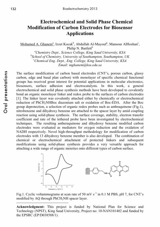
132O
ral p
rese
ntat
ions
Bioelectrochemistry 2013
Electrochemical and Solid Phase Chemical Modification of Carbon Electrodes for Biosensor
Applications
Mohamed A. Ghanema, Izzet Kocakb, Abdullah Al-Mayoufa, Mansour AlHoshanc, Philip N. Bartlettb
aChemistry Dept., Science College, King Saud University, KSA bSchool of Chemistry, University of Southampton, Southampton, UK
cChemical Eng. Dept., Eng. College, King Saud University, KSA Email: [email protected]
The surface modification of carbon based electrodes (CNT’s, porous carbon, glassy carbon, edge and basal plan carbon) with monolayer of specific chemical functional groups has received great interest for potential applications in molecular electronics, biosensors, surface adhesion and electrocatalysts. In this work, a general electrochemical and solid phase synthesis methods have been developed to covalently bond an organic monolayer linker and redox probe to the surfaces of carbon electrodes [1]. The linker layer was covalently attached either by chemically or electrochemical reduction of PhCH2NHBoc diazonium salt or oxidation of Boc-EDA. After the Boc group deprotection, a selection of organic redox probes such as anthraquinone (Fig.1), nitrobenzene and dihydroxy benzene are attached to the spacer layer by amid coupling reaction using solid-phase synthesis. The surface coverage, stability, electron transfer coefficient and rate of the tethered probe have been investigated by electrochemical techniques. The resulting antheraquinone and dihydroxy benzene modified carbon electrodes were evaluated as mediators for oxygen reduction and the oxidation of NADH respectively. Novel high-throughput methodology for modification of carbon electrodes with 13 dihydroxy benzene member is also developed. The combination of chemical or electrochemical attachment of protected linkers and subsequent modifications using solid-phase synthesis provides a very versatile approach for attaching a wide range of organic moieties onto different types of carbon surface. Fig.1. Cyclic voltammograms at scan rate of 50 mV s–1 in 0.1 M PBS, pH 7, for CNT’s modified by AQ through PhCH2NH spacer layer. Acknowledgment: This project is funded by National Plan for Science and Technology (NPST), King Saud University, Project no: 10-NAN101402 and funded by the EPSRC (EP/D038588/1). [1] M. A. Ghanem, J.M. Chrétien, J. D. Kilburn, P. N. Bartlett, J. Mater. Chem., 18 (2008) 4917; Chemistry Eur. J., 14 (2008) 2548; Bioelectrochemistry, 76, (2009) 115; Chemistry Eur. J., 15, (2009) 11928.
-1.0 -0.8 -0.6 -0.4 -0.2 0.0
-40
-20
0
20
40
Cur
rent
/ μA
Potential vs. SCE/ V
NHO
O
O
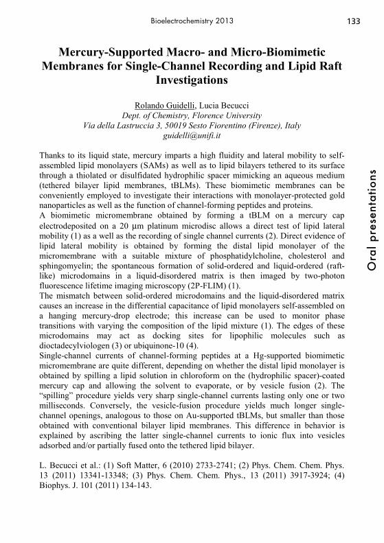
133
Ora
l pre
sent
atio
ns
Bioelectrochemistry 2013
Mercury-Supported Macro- and Micro-Biomimetic Membranes for Single-Channel Recording and Lipid Raft
Investigations
Rolando Guidelli, Lucia Becucci Dept. of Chemistry, Florence University
Via della Lastruccia 3, 50019 Sesto Fiorentino (Firenze), Italy [email protected]
Thanks to its liquid state, mercury imparts a high fluidity and lateral mobility to self-assembled lipid monolayers (SAMs) as well as to lipid bilayers tethered to its surface through a thiolated or disulfidated hydrophilic spacer mimicking an aqueous medium (tethered bilayer lipid membranes, tBLMs). These biomimetic membranes can be conveniently employed to investigate their interactions with monolayer-protected gold nanoparticles as well as the function of channel-forming peptides and proteins. A biomimetic micromembrane obtained by forming a tBLM on a mercury cap electrodeposited on a 20 μm platinum microdisc allows a direct test of lipid lateral mobility (1) as a well as the recording of single channel currents (2). Direct evidence of lipid lateral mobility is obtained by forming the distal lipid monolayer of the micromembrane with a suitable mixture of phosphatidylcholine, cholesterol and sphingomyelin; the spontaneous formation of solid-ordered and liquid-ordered (raft-like) microdomains in a liquid-disordered matrix is then imaged by two-photon fluorescence lifetime imaging microscopy (2P-FLIM) (1). The mismatch between solid-ordered microdomains and the liquid-disordered matrix causes an increase in the differential capacitance of lipid monolayers self-assembled on a hanging mercury-drop electrode; this increase can be used to monitor phase transitions with varying the composition of the lipid mixture (1). The edges of these microdomains may act as docking sites for lipophilic molecules such as dioctadecylviologen (3) or ubiquinone-10 (4). Single-channel currents of channel-forming peptides at a Hg-supported biomimetic micromembrane are quite different, depending on whether the distal lipid monolayer is obtained by spilling a lipid solution in chloroform on the (hydrophilic spacer)-coated mercury cap and allowing the solvent to evaporate, or by vesicle fusion (2). The “spilling” procedure yields very sharp single-channel currents lasting only one or two milliseconds. Conversely, the vesicle-fusion procedure yields much longer single-channel openings, analogous to those on Au-supported tBLMs, but smaller than those obtained with conventional bilayer lipid membranes. This difference in behavior is explained by ascribing the latter single-channel currents to ionic flux into vesicles adsorbed and/or partially fused onto the tethered lipid bilayer. L. Becucci et al.: (1) Soft Matter, 6 (2010) 2733-2741; (2) Phys. Chem. Chem. Phys. 13 (2011) 13341-13348; (3) Phys. Chem. Chem. Phys., 13 (2011) 3917-3924; (4) Biophys. J. 101 (2011) 134-143.

134O
ral p
rese
ntat
ions
Bioelectrochemistry 2013
Microsystems for Oxidative Stress Electrochemical Detection on Murine Macrophages Population
Manon Guille Collignon, Anne Meunier, Yun Li, Catherine Sella, Rémy Fulcrand,
Laurent Thouin, Frédéric Lemaître, Christian Amatore UMR CNRS-UPMC 8640 PASTEUR, Ecole Normale Supérieure, Département de
Chimie 24 rue Lhomond, 75231 Paris Cedex 05, France
We developed an electroanalytical method funded on the direct detection at the surface of a platinized carbon fiber ultramicroelectrode (UME) of four superoxide and nitric oxide derivatives (H2O2, ONOO-, NO2
-,..), released at minute amounts (atto- to femto-moles) by a single living cell [1]. Selectivity was offered by the choice of the adequate oxidation potential for each species. Despite adequate analytical properties (excellent time response, access to the four different species flux measurements) of our method, this approach is time-consuming. Microsystems have shown increasing interests in chemistry and biology for bioelectroanalytical applications due to numerous advantages (easily implemented, low cost device, multiplex array, versatility). Indeed they become efficient instruments for single-cell studies, especially for real-time monitoring of stimulated release from single cells [2]. In this work, we thus propose two different strategies based on electrochemical measurements in microsystems to provide kinetics, quantitative and real-time information on the emitted fluxes at the level of a cell population of murine macrophages. Firstly, a microsystem was conceived, made of four parallel and independent measurement chambers containing the working Pt microband UME, owing simultaneous monitoring of oxidative stress at four different potentials. We thus offer a proper and exhaustive characterization of this new methodology with a set of appropriate potential values to identify the contribution of each ROS and RNS species. We believe that this microsystem will provide a great chance in the future to lead comparative studies of different molecules (e.g., activators, antitumoral components) on oxidative stress behaviours or to monitor inflammatory responses in cells population. Secondly, we investigated performances of Pt-black microband UME in terms of selectivity, sensitivity and catalytic activity to offer a viable approach for monitoring single-cell releases in microfluidic devices. We thus studied the electrochemical detection of hydrogen peroxide and nitrites on such system [3]. Optimization and characterization of Pt-black deposits were performed according to the active surface and roughness factor [4]. A discussion about advantages and drawbacks of both strategies will be presented. [1] C. Amatore et al., Chem Rev, 2008, 108, 2585. [2] Y. Wang et al., Microchim. Acta, 2010, 168, 177. [3] C. Amatore et al., Anal. Chem., 2008, 80, 4976. [4] Y. Li et al., Electroanalysis, 2012, in press.

135
Ora
l pre
sent
atio
ns
Bioelectrochemistry 2013
Decontamination of Wastewater by Pulsed Electric Field Treatment
Christian Gusbeth, Wolfgang Frey, Annika Rieder* and Thomas Schartz*
Karlsruhe Institute of Technology Institute for Pulsed Power and Microwave Technology (IHM)
76344 Eggenstein-Leopoldshafen, Germany *Karlsruhe Institute of Technology
Institute of Functional Interfaces (IFG) 76344 Eggenstein-Leopoldshafen, Germany
Conventional methods of bacterial decontamination of wastewater, e.g. chlorination or ozonization can produce toxic by-products, originated from organic substances. UV-disinfection is not effective in turbid water. Thus, the pulsed electric field (PEF) treatment of wastewater as a non-chemical disinfection method seems to be a suitable alternative method for reducing this bacterial load. This was demonstrated for the decontamination of hospital wastewater loaded with pathogenic and increasingly antibiotic resistant bacteria. Besides the inactivation efficiency, the economic efficiency is an important aspect in the case of treating wastewater from municipal purification plants. In this study we investigated the efficiency of the PEF method for wastewater decontamination and the electro-sensitivity of wastewater bacteria to PEF treatment. For this purpose wastewater was sampled at the effluent of 4 different wastewater purification plants and at the wastewater outlet of two hospitals. To investigate the electro-sensitivity of wastewater bacteria, filtered wastewater samples were inoculated with representative wastewater bacteria, e.g. E. faecalis, E. faecium, E. casseliflavus and exposed to different PEF treatments. A transmission line pulse generator was used for the PEF treatments. It delivers square pulses with a voltage amplitude between 8 and 20 kV. The pulse duration ranges from 0.6 to 2 μs. In order to achieve a satisfactory bacterial inactivation (3.5 logs), a specific electric treatment energy between 120 J/ml and 240 J/ml is necessary. The high energy consumption is the limiting factor for an industrial application of this method. Combined treatments of wastewater with PEF and heat (50-60 °C) could reduce the costs for bacterial contamination considerably.
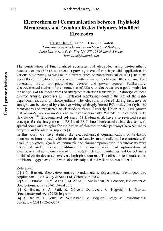
136O
ral p
rese
ntat
ions
Bioelectrochemistry 2013
Electrochemical Communication between Thylakoid Membranes and Osmium Redox Polymers Modified
Electrodes
Hassan Hamidi, Kamrul Hasan, Lo Gorton Department of Biochemistry and Structural Biology,
Lund University, P. O. Box 124, SE-22100 Lund, Sweden [email protected]
The construction of functionalized substrates and electrodes using photosynthetic reaction centers (RCs) has attracted a growing interest for their possible applications in various bio-devices, as well as in different types of photochemical cells [1]. RCs are very efficient in light energy conversion with a quantum yield near 100% making them potentially useful for photovoltaic devices and power sources. Furthermore, electrochemical studies of the interaction of RCs with electrodes are a good model for the analysis of the mechanisms of interprotein electron transfer (ET) pathways of these electron transfer processes [2]. Thylakoid membranes contain the site of the light-dependent reactions of photosynthesis. The electrons produced during incidence of sunlight can be trapped by effective wiring of deeply buried RCs inside the thylakoid membranes and transferred to electrode surfaces. Recently, Hasan et al. have proven that Rhodobacter capsulatus can be electrochemically “wired” to electrodes with flexible Os2+/3+ functionalized polymers [3]. Badura et al. have also reviewed recent concepts for the integration of PS I and PS II into bioelectrochemical devices with special focus on strategies for the design of electron transfer pathways between redox enzymes and conductive supports [4]. In this work we have studied the electrochemical communication of thylakoid membranes from spinach with electrode surfaces by functionalizing the electrode with osmium polymers. Cyclic voltammetric and chronoamperometric measurements were performed under anoxic conditions for characterization and optimization of electrochemical communication of illuminated thylakoid membranes and Os polymers modified electrodes to achieve very high photocurrents. The effect of temperature and inhibitors, oxygen evolution were also investigated and will be shown in detail. References [1] P.N. Bartlett, Bioelectrochemistry: Fundamentals, Experimental Techniques and Applications, John Wiley & Sons Ltd, Chichester, 2008. [2] S.A. Trammell, L.Y. Wang, J.M. Zullo, R. Shashidhar, N. Lebedev, Biosensors & Bioelectronics, 19 (2004) 1649-1655. [3] K. Hasan, S. A. Patil, K. Górecki, D. Leech, C. Hägerhäll, L. Gorton, Bioelectrochemistry, (2012) in press. [4] A. Badura, T. Kothe, W. Schuhmann, M. Rogner, Energy & Environmental Science, 4 (2011) 3263-3274.

137
Ora
l pre
sent
atio
ns
Bioelectrochemistry 2013
Sensitive Electrochemical Monitoring of Purine Derivatives in Real Biological Matrixes at Carbon-based
Materials
Stanislav Hason Institute of Biophysics, AS CR, v.v.i
Kralovopolska 135, CZ–612 65 Brno, Czech Republic [email protected]
Since more than 20 defects in purine metabolism are currently registered, there is a growing need to introduce directly into the doctor´s office routine, inexpensive, fast and accurate detection method for monitoring the concentration of purine metabolites in cells or body fluids as soon as possible to find out metabolic defects, which are characterized by abnormal levels of purine catabolites such as uric acid (UA), xanthine (Xan), or hypoxanthine (Hyp), resulting in indications of pathological conditions such as hyperuricemia, gout or renal failure. In addition, detection of purine nucleobases, adenine (Ade) and guanine (Gua) is frequently used in electrochemical analysis of nucleic acids. This contribution summarizes the most important electrochemical approaches to improve the sensitivity and specificity of the voltammetric sensing of purine derivatives, which can be used for: (i) early detection of diseases such as gout (UA, Xan, and Hyp), (ii) optimizing allopurinol therapy in patients with chronic gout (UA, Xan, Hyp, Oxypurinol-Oxy), and (iii) for label-free sequence-specific DNA sensing (Gua and Ade) on various carbon-based materials. Detection of purine derivatives, in all proposed protocols, is based on direct electrochemical measurement of oxidation peaks for each of the monitored substances in a three-electrode microcell during a single detection step in connection with: (i) mechanically treated compact carbon/graphite materials, (ii) electrochemically activated carbon-based materials, and (iii) anodic stripping of the electrochemically accumulated purine-Cu(I) complexes from carbon-based materials in the presence of copper ions. The above methods allow the simultaneous detection of either purine metabolites involved in the xanthine oxidase catalyzed catabolic pathway in real body fluids or purine nucleobases in acid-hydrolyzed DNA samples, respectively, at a picomolar level. Acknowledgments: This work was supported by the Grant Agency of the Czech Republic (P205/10/2378) and institutional research plan (AV0Z50040702).

138O
ral p
rese
ntat
ions
Bioelectrochemistry 2013
Gene Electrotransfer a Versatile and Powerful Tool to Enhance Therapeutic Applications.
Richard Heller1, Amy Donate1, Shawna Shirley1, Siqi Guo1, Cathryn Lundberg1 and
Loree Heller1 Frank Reidy Research Center for Bioelectrics, Old Dominion University
4211 Monarch Way, Suite 300, Noroflk, VA 23508 [email protected]
Gene medicine has held great promise for effective treatment of a variety of disorders including cancer, metabolic disorders, cardiovascular diseases and genetic disorders as well as a prophylactic approach for infectious diseases and cancer. One of the critical aspects of gene transfer is effective delivery to the appropriate target. Electro transfer of plasmid DNA is quickly being accepted as a viable approach to achieve effective delivery and has been tested in a variety of tissues. Sets of electroporation parameters, which includes electric field strength, pulse duration, number of pulses, electrode geometry and configuration, can be chosen to deliver plasmid DNA in such a way as to manipulate the onset, level, and duration of protein expression. Electrotransfer has been utilized to effectively deliver DNA to many tissue targets. One tissue target that has the potential for multiple therapeutic applications is the skin. This approach has been tested in several animal models with varying skin thickness and delivery of plasmid DNA with electroporation resulted in significantly increased expression levels. Gene electrotransfer to the skin is currently being evaluated for its potential for accelerating wound healing, treating ischemic tissue, protein replacement therapy, treatment of skin cancers and for delivering DNA vaccines. To evaluate the effectiveness of accelerating wound healing a random flap model in a Sprague Dawley rat was used. To facilitate healing, we delivered a plasmid encoding vascular endothelial growth factor (VEGF) that can induce cell proliferation, promote cell migration and differentiation as well as induce angiogenesis and inhibit apoptosis. The healing following gene electro transfer of VEGF was essentially complete within 2 weeks. Recent studies have demonstrated that the accelerated healing could be obtained by treating only 2 areas within the flap provided one was near the pedicle end and one was near the distal end. To evaluate the effectiveness of DNA vaccines, two agents were tested. A plasmid encoding Hepatitis B surface antigen was delivered tot eh skin of guinea pigs and resulted in significantly elevated antibody titers. To determine if this could also be accomplished with a bacterial antigen. A plasmid encoding protective antigen of bacillus anthracis was delivered to the flank of balbC mice. Again significant antibody levels were induced. In both cases, the level of antibody production was influenced by the delivery parameters. Gene electrotransfer is a powerful tool that allows for manipulation of expression levels and kinetics. Adjusting the delivery parameters enables maximum control of the expression profile enhancing the potential for a successful therapeutic outcome.
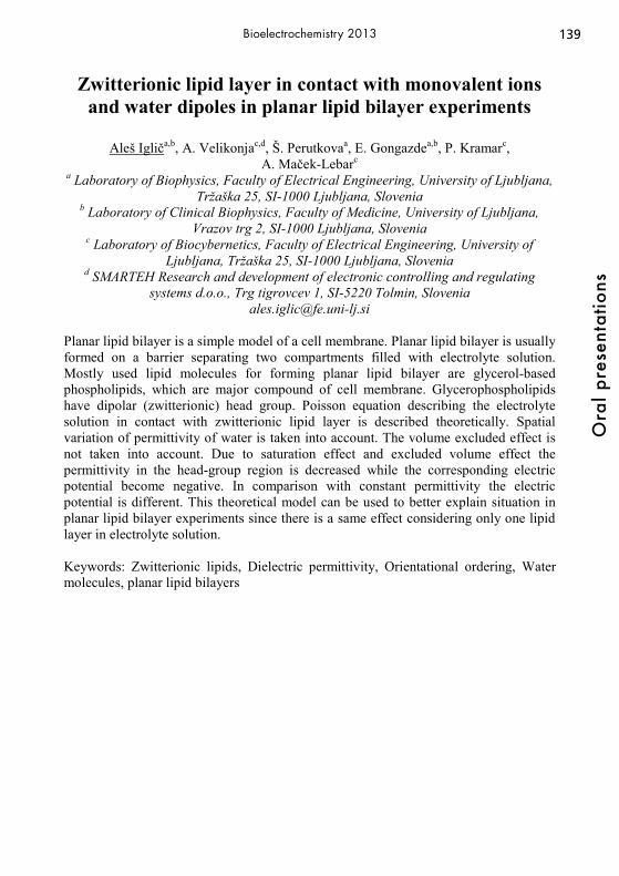
139
Ora
l pre
sent
atio
ns
Bioelectrochemistry 2013
Zwitterionic lipid layer in contact with monovalent ions and water dipoles in planar lipid bilayer experiments
Aleš Igliča,b, A. Velikonjac,d, Š. Perutkovaa, E. Gongazdea,b, P. Kramarc,
A. Maček-Lebarc
a Laboratory of Biophysics, Faculty of Electrical Engineering, University of Ljubljana, Tržaška 25, SI-1000 Ljubljana, Slovenia
b Laboratory of Clinical Biophysics, Faculty of Medicine, University of Ljubljana, Vrazov trg 2, SI-1000 Ljubljana, Slovenia
c Laboratory of Biocybernetics, Faculty of Electrical Engineering, University of Ljubljana, Tržaška 25, SI-1000 Ljubljana, Slovenia
d SMARTEH Research and development of electronic controlling and regulating systems d.o.o., Trg tigrovcev 1, SI-5220 Tolmin, Slovenia
[email protected] Planar lipid bilayer is a simple model of a cell membrane. Planar lipid bilayer is usually formed on a barrier separating two compartments filled with electrolyte solution. Mostly used lipid molecules for forming planar lipid bilayer are glycerol-based phospholipids, which are major compound of cell membrane. Glycerophospholipids have dipolar (zwitterionic) head group. Poisson equation describing the electrolyte solution in contact with zwitterionic lipid layer is described theoretically. Spatial variation of permittivity of water is taken into account. The volume excluded effect is not taken into account. Due to saturation effect and excluded volume effect the permittivity in the head-group region is decreased while the corresponding electric potential become negative. In comparison with constant permittivity the electric potential is different. This theoretical model can be used to better explain situation in planar lipid bilayer experiments since there is a same effect considering only one lipid layer in electrolyte solution. Keywords: Zwitterionic lipids, Dielectric permittivity, Orientational ordering, Water molecules, planar lipid bilayers

140O
ral p
rese
ntat
ions
Bioelectrochemistry 2013
An Ascorbic Acid Biofuel Cell Using Nanocarbon Electrodes for Catalytic Ascorbic Acid Oxidation
Martin Jönsson-Niedziolka, Adrianna Zloczewska, Anna Celebanska, Katarzyna Szot,
Dorota Tomaszewska, Marcin Opallo Institute of Physical Chemistry, Polish Academy of Sciences
Kasprzaka 44/52, Warsaw, Poland. [email protected]
The electrocatalytic properties of modified electrodes towards oxidation of ascorbic acid (AA) have been shown in numerous articles. Often these electrodes are used for determination of AA, but another common use is as a mean for separating the AA oxidation peak from that of dopamine. The reason for this is to determine minute amounts of dopamine in samples with higher concentrations of interfering AA. We show that electrodes with immobilised carbon nanoparticles (CNPs) or nanotubes (CNTs) both display efficient electrocatalytic activity towards AA oxidation. The CNP
electrodes were prepared by layer-by-layer deposition of the negatively charged CNPs with positively charged tetraalkyl-ammonium modified silica, either in the form of particles or film. Electrodes with vertically aligned CNTs were made by gluing CNT forests to ITO-coated glass. Here we demonstrate that these electrodes can be used as sensitive amperometric sensors for AA. However, more interestingly, we show that the AA oxidation is shifted to so
low potential that the nanocarbon electrodes can be used as anodes in an AA/O2-fuel cell. The same types of CNP and CNT electrodes have also been used to create efficient cathodes for bioelectrocatalytic reduction of O2 to water by immobilisation of bilirubin oxidase on the electrodes. Together the nanocarbon anode and cathode are assembled to an ascorbic acid-oxygen biofuel cell. The assembled biofuel cell gives a maximum power of 12 μW cm-2 in 1 mM ascorbic acid. This biofuel cell can be used as a self-powered sensor for determination of AA.
Figure1: Oxidation of AA on a CNP electrode as compared to clean ITO. Scan rate 20 mV s-1.
141
Ora
l pre
sent
atio
ns
Bioelectrochemistry 2013
Influence of Antibiotic Peptide, Melittin, on Lipid Membranes of Different Composition
Joanna Juhaniewicz1, Slawomir Sek1, Jacek Lipkowski2
1 Faculty of Chemistry, University of Warsaw Pasteura 1, 02093 Warsaw, Poland
2 Department of Chemistry, University of Guelph Guelph, Ontario, Canada N1G 2W1
Seeking for new compounds with a broad spectrum of activity against bacteria and pathogens is nowadays one of the main topics in the field of biochemistry and medicine[1,2]. One of the most intensively investigated groups includes the helical, cationic peptides that exhibit a strong antimicrobial properties [3]. Melittin, belonging to the antimicrobial peptides (AMPs), is a cationic, amphipathic peptide composed of 26 amino acid residues and exhibits a strong action on biological membranes [4,5]. Here we present the results of our studies on interactions of melittin with four model lipid membranes of different composition: i) 1,2-dimyristoyl-sn-glycero-3-phosphoholine (DMPC), ii) 1,2-dimyristoyl-sn-glycero-3-phosphoserine (DMPS), iii) 1,2-dimyristoyl-sn-glycero-3-phosphoglycerol (DMPG) and iv) the mixture of DMPC and cholesterol (7:3). In the first step we verified the action of melittin on the model lipid membranes at the air-water interface using Langmuir trough. Next, lipids were transferred onto gold substrate using the combination of Langmuir-Blodgett and Langmuir-Schaefer techniques and the action of melittin was investigated by means of Electrochemical Impedance Spectroscopy (EIS) and Atomic Force Microscopy (AFM). The strength of interactions of melittin with lipid membranes is significantly influenced by physical state of lipid but the crucial factor seems to be the charge of lipid polar group. In case of zwitterionic lipids melittin inserts easily into the membrane, whereas the presence of negatively charged polar group causes the electrostatic adsorption of peptide on the membrane surface that, in consequence, slows down or even inhibits the insertion of melittin into the lipid membrane and pore formation. The presence of cholesterol, one of the importants components of biological membranes, does not inhibit the disrupting action of melittin. References: [1] K. Radek, R. Gallo, Semin. Immunopathol., 2007, 29, 27. [2] G. Kokot, M. Mally, S. Svetina, Eur. Biophys. J, 2012, 41, 461. [3] S. Sek, T. Laredo, J.R. Dutcher, J. Lipkowksi, J. Am. Chem. Soc., 2009, 131, 6439. [4] T.C. Terwilliger, D. Eisenberg, J. Biol .Chem., 1982, 257, 6010. [5] K. Hall, T-H. Lee, M-I. Aguilar, J. Mol. Recognit. 2011, 24, 108.
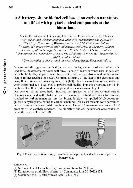
142O
ral p
rese
ntat
ions
Bioelectrochemistry 2013
AA battery- shape biofuel cell based on carbon nanotubes modified with phytochemical compounds at the
biocathode
Maciej Karaskiewicz, J. Rogalski, J. F. Biernat, K. Zelechowska, R. Bilewicz a College of Inter-Faculty Individual Studies in Mathematics and Faculty of
Chemistry, University of Warsaw, Pasteura 1, 02-093 Warsaw, Poland b Faculty of Applied Physics and Mathematics. and Dept. of Chemistry Gdansk
University of Technology, Narutowicza St. 11-12, 80-233 Gdansk, Poland d Department of Biochemistry, Maria Curie Sklodowska University, Akademicka 19,
20-033 Lublin, Poland *Corresponding author’s email address: [email protected]
Glucose and dioxygen are gradually consumed during the work of the biofuel cells leading to the decrease of power with time. In case of many enzymes used as catalysts in the biofuel cells, the products of the catalytic reactions are also natural inhibitors and lead to further decrease of power. Continuous supply of the fuel to the electrodes and using flow systems becomes very important [1-3]. Flow systems have to be considered when the biofuel cell is designed to power small medical implants or sensing devices in the body. The flow system used in the present paper is shown on Fig. 1 Our concept of the biocathode involves the application of nanostructured carbon electrodes modified with phytochemical compounds – natural substrates for laccase, attached to carbon nanotubes. At the bioanode side we applied NAD-dependent glucose dehydrogenase bound to carbon nanotubes. All measurements were performed in AA battery-shape cell with continuous exchange of substrates and removal of products of the catalytic reactions. The biobattery and cell parameters were evaluated under the external load of 1 MΩ.
Fig. 1 The cross-section of single AA battery-shaped cell and scheme of triple AA battery box.
References [1] Nazaruk et. al, Electrochemistry Communications 14 (2012) 67. [2] Karaskiewicz et. al, Electrochemistry Communications 20 (2012) 124 [3] Stolarczyk et. al, Electrochimica Acta 79 (2012) 74
143
Ora
l pre
sent
atio
ns
Bioelectrochemistry 2013
Electrochemically Actuated, Capillarity-Driven Biodetection Devices for Food Safety and Clinical Analysis
Ioanis Katakis, Alemayehu P. Washe, Luis Carlos Rosales-Rivera, Pablo Lozano-
Sánchez, Diego Bejarano-Nosas, Bruno Teixeira-Dias Bioengineering and Bioelectrochemistry Group, Department of Chemical Engineering,
Universitat Rovira i Virgili Avinguda Països Catalans 26, 43007 Tarragona, Spain
Lab-on-Chip (LOC) concepts are usually realized as microsystems fabricated by microfabrication technologies of varying degrees of complexity and operated by control equipment that commonly require external power sources, fluid movement devices, and detection systems. Investment in purpose-built manufacturing lines, for micron or sub-micron featured microsystems and sophisticated control apparatus is justified for high throughput analytical tasks based on limited-volume samples. Most analytical needs in food safety and decentralized diagnostics/theranostics are not limited by the available sample volume and are not high throughput in nature; rather it is cost and ease of use that eventually decide their large scale adoption. A convenient alternative for the realization of such application-oriented LOC concepts is to manufacture simple, basic microsystems by 3-D screen printing. Such elemental microsystems can be operated almost autonomously: fluid movement can be achieved through capillary action, and both fluidic control and detection by electrochemistry. Screen printing manufacturing requires a simple and relatively low cost production line and provides the flexibility to incorporate different materials in the 3-D design accommodating both structural and actuation or detection elements. We demonstrate that basic unit operations such as dissolution, separation, mixing, reaction, flow manipulation and detection can be satisfactorily realized and controlled for most detection applications. We applied such simple architectures in integrated devices that can detect pathogens in food and activated sludge. In a particular product developed, Salmonella could be detected in poultry meat extracts with limit of detection of 10-20 CFUs within 15 hours of sampling. When proteins need to be detected by immunochemical methods in lateral flow-type devices rendered by the 3-D screen printing method, we demonstrate that flow control is crucial for signal development and successful immunorecognition. We provide such flow control with electrochemically activated stop/go printed microvalves that modulate the hydrophilicity of the device walls. We thus achieve successful detection of β-lactoglobulin (a potential food allergen) or HCG (a pregnancy indicator). We therefore present a simple to manufacture, generic. low cost, and easy to use technology platform that can tackle a variety of analytical problems. This work was made possible through support by the Spanish Ministry of Science and Innovation (BIO2010-20359 MICROCAP and IPT 2011 11 44 90 000 00 DINRAPUC) and by the Catalan agency of support of University research (AGAUR grant 2010VALOR00063 SALMONELLA TRUST). APW and LCR acknowledge scholarships from DIUE of the Catalan government.
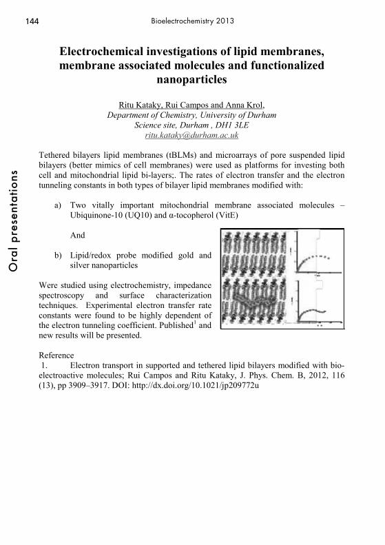
144O
ral p
rese
ntat
ions
Bioelectrochemistry 2013
Electrochemical investigations of lipid membranes, membrane associated molecules and functionalized
nanoparticles
Ritu Kataky, Rui Campos and Anna Krol, Department of Chemistry, University of Durham
Science site, Durham , DH1 3LE [email protected]
Tethered bilayers lipid membranes (tBLMs) and microarrays of pore suspended lipid bilayers (better mimics of cell membranes) were used as platforms for investing both cell and mitochondrial lipid bi-layers;. The rates of electron transfer and the electron tunneling constants in both types of bilayer lipid membranes modified with:
a) Two vitally important mitochondrial membrane associated molecules – Ubiquinone-10 (UQ10) and α-tocopherol (VitE) And
b) Lipid/redox probe modified gold and silver nanoparticles
Were studied using electrochemistry, impedance spectroscopy and surface characterization techniques. Experimental electron transfer rate constants were found to be highly dependent of the electron tunneling coefficient. Published1 and new results will be presented. Reference 1. Electron transport in supported and tethered lipid bilayers modified with bio- electroactive molecules; Rui Campos and Ritu Kataky, J. Phys. Chem. B, 2012, 116 (13), pp 3909–3917. DOI: http://dx.doi.org/10.1021/jp209772u

145
Ora
l pre
sent
atio
ns
Bioelectrochemistry 2013
Microbic Fuel Elements: Prospects and Problems
I. A. Kazarinov Saratov State University named after N.G. Chernyshevsky
83 Astrakhanskaya Str., Saratov 410012, Russian Federation e-mail: [email protected]
Biological fuel elements (BFE) are devices to use biological components as catalysts for electrical power generation. Either whole microorganisms or enzymatic preparations are applied in BFE as catalysts. In this regard, BFE are subdivided into enzymatic fuel elements (EFE) and microbic fuel elements (MFE). Besides, unlike chemical fuel elements utilizing hydrogen, ethanol, and methanol as fuel, BFE can use energy-rich but electrochemically passive substances (carbohydrates, organic acids, alcohols, and many organic wastes) as fuel. This opens wide possibilities of simultaneous solution of environmental and energetical problems. The usage of microorganisms in biofuel elements avoids the need to isolate individual enzymes, thus giving cheaper catalysts for biofuel elements. The main problem of microbic fuel elements is to implement charge extraction from the cell to the electrode since microorganisms are electrochemical inactive in most cases. The report discusses some mechanisms of electron transfer between cells and the electrode: - electron transfer by means of exogenic redox mediators; - electron transfer by means of endogenic redox mediators (primary and secondary metabolites); - direct electron transfer. Selection criteria of mediator systems are formulated:
1) the oxidized mediator must easily get through the bacterial membrane to the reduced particles inside bacteria;
2) the redox potential of the mediator should correspond to that of the reduced metabolite;
3) in no oxidation degree the mediator should interfere other metabolic processes; 4) the reduced mediator should easily leave the cell through the bacterial membrane; 5) the mediators in both oxidized and reduced states should be chemically stable in the
electrolyte solution, easily dissolved, and should not be adsorbed on bacterial cells and on the electrode surface;
6) the electrochemical kinetics of oxidation of the mediator’s reduced state on the electrode should be fast.
The overall efficiency of electron transfer mediators depends on many parameters, especially on the rate constant of electrochemical reoxidation of the mediator, which depends on the electrode material. Electronic transfer efficiency can be raised by the usage of a mixture of two redox mediators. The report will consider particular examples of implementation of some bioelectrochemical systems in microbic fuel elements, their discharging characteristics are analyzed, the scopes of MFE and the outlook of development of this bioelectrocatalytic lead are shown.

146O
ral p
rese
ntat
ions
Bioelectrochemistry 2013
Photodynamic Reaction Assisted by Reversible Electroporation as a Prospective Cancer Treatment - in
Vitro Study on Breast Carcinoma Cells
Joanna Wezgowieca, Malgorzata Kotulskaa*, Julita Kulbackab Jolanta Saczkob, Maria B. Deryloa, Justin Teissiec, Marie-Pierre Rolsc, Julie Orioc, Arnold Garbiecd
a Institute of Biomedical Engineering and Instrumentation, Wroclaw University of Technology,
Wybrzeze Wyspianskiego 27, 50-370 Wroclaw, Poland b Department of Medical Biochemistry, Wroclaw Medical University,
Chalubinskiego 10, 50-368 Wroclaw, Poland c CNRS, IPBS (Institut de Pharmacologie et de Biologie Structurale),
205 route de Narbonne, F-31077 Toulouse, France and Université de Toulouse, UPS, IPBS, F-31077 Toulouse, France
d Department of Animal Developmental Biology, Zoological Institute, University of Wroclaw,
Sienkiewicza 21, 50-335 Wroclaw, Poland *e-mail: [email protected]
We evaluated the effectiveness of a photodynamic reaction (PDR), with Photofrin and several cyanines, on breast adenocarcinoma cells (MCF-7/WT) and normal Chinese hamster ovary cells (CHO) lacking voltage-dependent channels. The study was conducted alone and supported with electropermeabilization (EP), including tests of cellular viability and intracellular localization of the photosensitizers. Among six cyanines tested, two compounds could be indicated as good therapeutic candidates: IR-775 and IR-786. Cellular effects were assessed with MTT assay reporting cell mitochondrial activity and with SRB assay based on the measurement of cellular protein content. Photodynamic reaction of MCF-7/WT cells with IR-775 and IR-786 did not result in cellular dysfunction. Significant improvement in PDR effectiveness was observed when electropermeabilization was additionally applied. Selected values of electric field intensities and pulse duration, non-toxic for cells, increased photocytotoxicity of the cyanines. Much shorter exposure times were then efficient for cyanines (10 min instead of 24 hours). We also observed significant increase of PDR effectiveness after electropermeabilization with IR-775. Similarly, electropermeabilization of cells in the presence of Photofrin increased the uptake of the photosensitizer. A combination of photodynamic reaction with electroporation improved the effectiveness of treatment even at the lowest electric field intensity. Our results indicate that EP of cancerous cells, in the presence of certain photosensitive dyes, increase the uptake of the photosensitizer. This correlates with a higher cytotoxicity in the breast adenocarcinoma cell line and helps to discover the drug uptake pathways.

147
Ora
l pre
sent
atio
ns
Bioelectrochemistry 2013
Designing Integrated Systems with Positively Charged Carbon Nanotubes as Platforms for the Construction of
High Performance Bienzyme Biosensors
Barbara Kowalewska, Pawel J. Kulesza Department of Chemistry, University of Warsaw
Pasteura 1, PL-02-093 Warsaw, Poland [email protected]
In our research, we have developed the integrated, structured and multifunctional bioelectrocatalytic system for effective oxidation of ethanol. The concept is based on the layer-by-layer (LbL) assembly through electrostatic attraction of positively charged multi-walled carbon nanotubes [1] and the controlled combination of dehydrogenase enzymes. More specifically, the LbL technique was employed for sequential immobilization of two dehydrogenase enzymes and poly(diallyldimethylammonium chloride)-covered multiwalled carbon nanotubes (CNTs/PDDA) onto glassy carbon electrode substrate [2]. Both monoenzymatic (utilizing a single enzyme, alcohol dehydrogenase, ADH) and bienzymatic (anchoring sequentially both ADH and aldehyde dehydrogenase, AldDH) systems were tested. Multilayers were characterized using scanning electron microscopy (SEM), infrared spectroscopy (FTIR) and cyclic voltammetry. The results are consistent with the view that our approach enables good control of distribution and efficient utilization of both enzymes within the bio-composite film and leads to sizeable enhancement of the oxidation of ethanol through significant (more than two-fold) increase of bioelectrocatalytic currents and by shifting the ethanol oxidation potential to 0.1 V (vs. Ag/AgCl) or decreasing the overvoltage by ca. 200 mV in comparison to the mono-enzymatic electrode system. This simple bio-composite (enzyme-cascade) system permits fabrication of highly sensitive ethanol biosensors based on nicotinamide adenine dinucleotide (NAD+) coenzyme-dependent dehydrogenases. Our ethanol biosensor exhibited a good linearity ranging from 50 to 300 μM, and it was characterized by high sensitivity of 118.8 μA mM-1 cm-2 as well as a low detection limit of 24 μM. References [1] Kowalewska B., Kulesza P.J., Electroanalysis, 24, 2012, 254-263. [2] Kowalewska B., Kulesza P.J., Analytical Chemistry, DOI: 10.1021/ac3021328.
148O
ral p
rese
ntat
ions
Bioelectrochemistry 2013
Hyphenated analytical techniques for studying living cells
Elena Hecht, Charlotte Steinbach, Peter Knittel, Christine Kranz
Institute of Analytical and Bioanalytical Chemistry, University of Ulm, Albert-Einstein-Allee 11, 89081 Ulm [email protected]
The development and application of orchestrated experimental tools and analytical methods is required for systematic analysis of biological entities and their response to external stimuli such as chemicals or forces. Ideally, a multi-parametric quantitative determination and imaging of molecular events, molecular pathways, and molecular signals is obtained. One strategy to achieve these goals is the development of hyphenated surface analytical techniques based on the combination of atomic force microscopy with electrochemical [1] and optical techniques [2,3]. Controlled mechanical or chemical stimulus by e.g., physically stretching cells during in-vitro experiments with precisely known force while simultaneously providing a localized or spatially resolved determination the concentration profiles of signaling molecules such as ATP and/or mapping changing in cell morphology are a first step towards understanding fundamental relationships in cell signaling. As examples, an uniaxial motorized cell stretching device integrated into a combined fluorescence microscopy (FM) - atomic force microscopy (AFM) system [4] and integrated in a scanning electrochemical microscopy (SECM) setup is demonstrated enabling high-resolution topographic and fluorescent imaging and mapping of concentration profiles of signaling molecules at living cells. In addition, in therapy microelectrodes are frequently used to stimulate cells in respect to sensory functions when administration of drugs is not feasible. The interaction of organic nanoelectrodes based on conducting polymers can be used for deliver highly localized electrical stimulation, while simultaneously AFM force measurements can be performed to assess the effect of physical and biomolecular forces occurring at the cell surface. The fabrication, characterization and requirements of such novel polymer-based AFM-SECM probes will be presented. [1] A. Kueng, C. Kranz, A. Lugstein, E. Bertagnolli, B. Mizaikoff, Angew. Chem. Int. Ed. 2005, 44, 3419. [2] E. Hecht, K. Thompson, M. Frick O. Wittekindt, P. Dietl, B. Mizaikoff, C. Kranz, Anal. Chem. 2012, 84, 5716. [3] L. Wang, J. Kowalik, B. Mizaikoff, C. Kranz, Anal. Chem. 2010, 82, 3139. [4] E. Hecht, P. Knittel, E. Felder, P. Dietl, B. Mizaikoff, C. Kranz, Analyst. 2012, 137, 5208.

149
Ora
l pre
sent
atio
ns
Bioelectrochemistry 2013
Combined Electrochemical and Optical Imaging of Polymeric Microcapsules using Photocurrent
Measurements at Electrolyte/ Insulator/ Semiconductor Field Effect Structures
Steffi Krause, Jian Wang, Michael Watkinson1, Gleb Sukhorukov
School of Engineering and Materials Science, Queen Mary University of London 1School of Chemical and Biological Sciences, Queen Mary University of London
Photocurrent measurements at electrolyte/ insulator/ silicon structures can be used to produce images of local impedance and potentials in Scanning Photo-induced Impedance Microscopy (SPIM) and Light-Addressable Potentiometric Sensors (LAPS) with good lateral resolution. In this work, it will be shown, that the same structures can also double as sensitive fluorescence detectors and detectors of thermal radiation generated by gold nanoparticles. An undecylenic acid monolayer was bound to hydrogen terminated silicon on a silicon-on-sapphire substrate. Hollow microcapsules labelled with gold nanoparticles or fluorescence red 633 were prepared by layer-by-layer (LBL) coating of oppositely charged polyelectrolytes (PAH/PSS) on a sacrificial template (PMMA). The capsules were attached onto the insulator surface through the electrostatic interaction between the PAH polycation and COO- terminal group. Electrochemical images were measured using single-photon excitation of photocurrents at 405 nm, while optical images were measured using two-photon excitation at 1250 nm focusing both lasers through the same microscope objective. At 405 nm, all light is absorbed by the silicon resulting in a pure impedance image (Figure 1a). At 1250 nm, two photon absorption resulted in a significant increase in the local photocurrent when
microcapsules labelled with gold nanoparticles or fluorescent dye were present on the insulator (Figure 1b). This technique has potential applications in cell imaging. Figure 1 photocurrent images and photocurrent-voltage curves of a capsule labelled with gold Capsule surface gold nanoparticles (a) 405 nm laser (b) 1250 nm laser
capsule surface
capsule surface
150O
ral p
rese
ntat
ions
Bioelectrochemistry 2013
Specific interactions of noble metal nanoparticles with biofilms grown on electrode surfaces: from anti-bacterial
properties to development of electrocatalytic systems active towards oxygen reduction
P. J. Kulesza1, W. Lotowska1, E. Szaniawska1, E. Seta1, S. Zoladek1, I. A. Rutkowska1,
K. Brzostek2, A. Raczkowska2 Department of Chemistry1 and Department of Biology,2, University of Warsaw,
KrakowskiePrzedmiescie 26/28, PL-00-927 Warsaw, Poland, [email protected]
In our research, we have exploited unique interactions of gold, silver and related bimetallic nanoparticles with biofilms formed by Pseudomonas aeruginosa, Staphylococcus aureus and Yersinia enterocolitic bacteria on glassy carbon electrodes. We consider here noble metal nanoparticles modified and stabilized with ultra-thin films of inorganic species (e.g. polyanions). Regardless the general tendency of gold and silver nanoparticles (suspended in aqueous solutions) to minimize formation of biofilms on solid surfaces, once immobilized within porous conducting polymer (e.g. poly(3,4-ethylenodioxythiophene) or PEDOT) layers, they tended to facilitate growth of robust and mature bacterial biofilms on their surfaces. Independent diagnostic electroanalytical experiments showed that biofilms grown by the following bacteria, P. aeruginosa ATCC 9027, Y. enterocolitica Ye9, Y. enterocolitica AR4, L. monocytogenes 10403S and L. monocytogenes 1115, on inert carbon substrates exhibited by themselves electrocatalytic properties towards oxygen and hydrogen peroxide reductions in neutral media. The processes were found to be further enhanced by introduction of certain metallic (Au, Ag, Pd) and bi-metallic (Au-Pt) nanoparticles both unsupported and supported on such inert metal oxide nanostructures as TiO2 and ZrO2. Coexistence of the above components leads to synergistic effect that is evident from some positive shift of the oxygen reduction voltammetric potentials and significant increase of voltammetric currents. Further, the proposed hybrid films exhibited relatively higher activities towards reduction of hydrogen peroxide. Comparative measurements were performed aiming at better understanding of electrocatalytic efficiencies of various systems including those utilizing metal nanoparticles (e.g. Au-Pt), conventional enzymes (e.g. laccase), molecular systems (e.g. metalloporphyrins) in the presence and absence of selected bacterial biofilms.

151
Ora
l pre
sent
atio
ns
Bioelectrochemistry 2013
Label-free electrochemical DNA-biosensor : setup for detection in microliter samples and miniaturization
Mathieu Lazerges, Vanna.-T. Tal, Fethi Bedioui
ENSCP*, Université Paris Descartes§, UPCGI, U 1022 INSERM, UMR 8151 CNRS * 11, rue Pierre et Marie Curie, 75005 Paris § 4, avenue de l’observatoire, 75006 Paris
[email protected] An ergonomic label-free DNA-biosensor based on a two-electrode electrochemical setup and using a microelectrode as working electrode was designed herein. A 23-base DNA-probe self-assembled monolayer was formed onto microelectrode gold surface. The microelectrode extremity was then immersed in a 50 microliterDNA-target solution drop. Transduction occurs via long-range electron transfer, which is enhanced subsequently to hybridization, due to DNA-base pi-stacking. Single mismatch detection of this first prototype was matched at room temperature and a detection limit in the nanomolar range was reached.
The detection scheme presented herein will be useful to simplify detection protocols or adapted analytic-tools to micro-litre samples involving in label-free electrochemical DNA biosensing and high-density screening platforms.
152O
ral p
rese
ntat
ions
Bioelectrochemistry 2013
Redox complexes for mediation of electron transfer in enzymatic batteries and fuel cells
D. Leech
National University of Ireland Galway School of Chemistry, University Road, Galway, Ireland
We report here on synthesis and immobilization of osmium-based redox mediator complexes. The redox complexes are designed to possess redox potentials suitable for mediation of electron transfer between glucose-oxidising, or oxygen reducing, enzymes and electrodes, and suitable functional groups to permit chemical immobilization. The redox complexes can be coupled, using electrochemically induced reactions, to conducting surfaces, or incorporated into polymeric supports or by coupling chemistries. Co-immobilization with redox enzymes results in redox hydrogels with application to biosensor and biopower devices. Devices with improved signal intensity, and lifetimes can be produced by variation in redox complex potential and coupling chemistry, variation in enzyme choice, and by addition of nanomaterials. Screening of combinations of enzyme electrodes as anodes and cathodes provides glucose/oxygen enzymatic biofuel devices with operational half-lives, in pseudo-physiological conditions, exceeding 1 week. Acknowledgments The research was supported by the EU (FP6 Biomednano, FP7 3-DNanobiodevice), ERA-Chemistry and Science Foundation Ireland.

153
Ora
l pre
sent
atio
ns
Bioelectrochemistry 2013
Investigating Exocytosis at the Single Cell Level : Combination of Amperometry and Total Internal
Reflection Fluorescence Microscopy
Anne Meunier,1 Ouardane Jouannot,2 Rémy Fulcrand,1 François Darchen,2 Stéphane Arbault,3 Manon Guille Collignon,1 Frédéric Lemaître,1 Christian Amatore1
1) UMR CNRS-UPMC UMR 8640, Département de Chimie, Ecole Normale Supérieure, 24 rue Lhomond, 75005 Paris, France
2) UMR 8192, Université Descartes, 45 rue des Saints-Pères, 75270 Paris cedex 06, France
3) UMR 5255, Université de Bordeaux 1, Institut des Sciences Moléculaires, 16 avenue Pey Berland, 33607 Pessac, France
Vesicular exocytosis is an essential biological mechanism used by cellular organisms to release bioactive molecules (hormones, neurotransmitters…) in their environment. During this process, secretory vesicles that initially stored the (bio)chemical messengers dock to the cell membrane. The subsequent fusion of vesicle and cell membranes induces the formation of a fusion pore that initiates the first exchanges between the intravesicular and extracellular media. Its following expansion thus favours a larger release of the vesicular content into the external medium. For the last twenty years, several analytical methods have been developed in order to study this biological mechanism at the single living cell level in real time. Among those techniques, amperometry with a carbon-fiber ultramicroelectrode and fluorescence microscopy appear as particularly adapted.[1] On the one hand, physico-chemical properties of ultramicroelectrodes induce a high detection sensitivity and temporal resolution, thus being particularly suitable to the real time detection of the electroactive vesicular content once the release onsets. On the other hand, total internal reflection fluorescence microscopy (TIRFM) allows visualizing the status of vesicles near the plasma membrane, and especially their behaviour before and during the fusion as well as the location of the event. In this regard, combination of these two techniques would give access to entire exocytotic events, from the vesicular final displacements to the release stage itself. We will present here an amperometry-TIRFM combination based on the development of specific ITO devices. Thus, semiconducting and transparent ITO (Indium Tin Oxide) can be used as an electrode without hindering the optic detection of marked vesicle by a fluorescent probe. The first results obtained in these conditions on single cell will be described and discussed.[2, 3] [1] C. Amatore et al., Chem Rev, 2008, 108, 2585. [2] A. Meunier et al., Angew. Chem. Int. Ed., 2011, 50, 5081. [3] A. Meunier et al., Biophys. Chem., 2012, 162, 14.
154O
ral p
rese
ntat
ions
Bioelectrochemistry 2013
Quantification of Basic Transport Processes in Electroporation-Mediated Molecular Delivery
Hao Lin, Mohamed Sadik, Miao Yu, Jianbo Li, and Jerry Shan
Department of Mechanical and Aerospace Engineering Rutgers, The State University of New Jersey
98 Brett Rd, Piscataway, NJ 08854, USA Email: [email protected]
& David Shreiber
Department of Biomedical Engineering Rutgers, The State University of New Jersey 599 Taylor Rd, Piscataway, NJ 08854, USA
This work focuses on understanding the basic processes in electroporation. Both experimental and modeling techniques are combined to investigate and quantify the contributions of the various transport mechanisms during electroporation-mediated molecular delivery. In the experimental study, the uptake of Propidium Iodide (PI) into single cells is systematically investigated with time- and space-resolved fluorescence microscopy, and as a function of extracellular buffer conductivity. The spatial and temporal analysis of the fluorescent signal demonstrates that PI stacked in the cytoplasm, and its rate of accumulation increased with decreasing conductivity. These results indicate that electrophoresis is the dominant mode of transport during the pulse. More importantly, the inverse correlation trend can be explained by an electrokinetic phenomenon known as Field-Amplified Sample Stacking (FASS). This mechanism stems from a gradient of electrophoretic velocity generated when an electric field is applied across an interface with asymmetric electrical conductivities. Furthermore, the respective contributions from electrophoresis and diffusion have been quantified; the former is shown to be consistently higher than the latter. In the modeling study, a full model is systematically established which can simulate coupled membrane permeabilization and multi-ion transport on the whole-cell level. The temporal and spatial evolution of ion concentrations is tracked, and the results are extensively and directly compared with the experimental data. Good agreements are found between the two, which reveals significant insight with respect to the basic processes governing molecular delivery efficiency. The problem of cell swelling is also tackled, and is interpreted by the overload of ions into the cell during the pulse. The experimental quantification and modeling tools are important contributions toward designing optimized protocols for a wide range of applications utilizing electroporation.

155
Ora
l pre
sent
atio
ns
Bioelectrochemistry 2013
Ionic Permeability of Lipid Cubic Phases. An Investigation with Electrochemical Impedance
Spectroscopy
Mohammad Yaser Khani Meynaq and Britta Lindholm-Sethson Dep of Chemistry, Umeå University, 90187 Umeå, Sweden
The Lipid Cubic phase has attained an increasing interest as holder for membrane proteins in for instance biofuel cells and biosensors. The lipid cubic phase is preformed in a test tube and then smeared out on an electrode surface. [1,2] In the present work a lipid cubic phase was used to fill a cylindric aperture in a 3 mm thick Teflon brick. Two sizes of apertures were used; i.e.: one with 3 mm diameter and one with 1 mm. The Teflon brick was used to separate two cell compartments in a specially designed electrochemical cell, Fig 1. Each cell contains one platinum electrode. The two compartments were then filled with electrolyte solution and electrochemical impedance was employed to probe the ionic permeability of the cubic phase. Several types of electrolyte solution at different ionic strengths were used to investigate whether cationic or anionic transport is the dominating charge transport through the cubic phase. The stability and ionic permeability of monoloein and pyanthriol cubic phases will be discussed.
Figure 1: The two compartment cell for probing ionic permeability of lipid cubic phases References [1] Anal Bioanal Chem (2008) 391:1569–1578 [2] J. Power Sources, 2008, 183, 533.

156O
ral p
rese
ntat
ions
Bioelectrochemistry 2013
DNA on gold – tools for the label-free analysis of hybridization and sequence specific ligand interaction
C. Tersch1, C. Witte1, V. Schoeppler1, J. Glöckler2, F. Lisdat1
1Biosystems Technology, Wildau Technical University of Applied Sciences Bahnhofstr. 1, 15745 Wildau, Germany,
2Alacris Theranostics GmbH, Ihnestr. 63, 14195 Berlin, Germany e-mail [email protected]
The detection of small DNA fragments and low molecular weight compounds but also proteins interacting specifically with nucleic acids has become an interesting research target. These reactions are important for many biochemical processes inside cells but also in drug development. Label-free techniques have become increasingly popular since they allow a direct detection with non-modified biomolecules. Impedimetric analysis of DNA hybridization has been successfully studied [1,2] and thus we want to investigate the potential use of this method for analytical detection of biomolecular interactions. For this purpose DNA strands of different length are immobilized on the gold surface (via thiol chemistry). Impedimetric analysis in the presence of a redox couple show that the charge transfer resistance RCT is a sensitive parameter to follow the DNA immobilization, hybridization and binding of molecules to DNA [3]. The surface concentration can be adjusted during the immobilization and controlled by a redox label. The influence of the length of the capture probe but also of the target DNA on the hybridization analysis has been evaluated. Besides the analysis of groove binding and intercalation, the electrode system is used to follow an enzyme reaction on the surface electrochemically. The specific enzymatic cleavage of a double-stranded DNA by BamHI restriction endonuclease could be followed impedimetrically. For verification it is also analyzed by cyclic voltammetry with labeled DNA. Furthermore, the sequence specific binding of the transcription factor NF-κB p50-homodimer on dsDNA with the recognition sequence is found to cause a decrease in RCT. This signal change occurs due to a neutralization effect on the negatively charged DNA backbone. Specific protein binding can be detected in the concentration range of 4-23μg/ml. This work represents some basic experiments for the label free impedimetric detection of protein-DNA interactions and shows the potential and limits for a simple, rapid and low-cost analysis. The presentation will also show the possibilities for the analysis of conformational changes in a label-free format. For this purpose very long, repetitive DNA sequences capable for G-quadruplex formation are prepared by rolling circle amplification [4]. Immobilized on gold these DNA strands can cause strong conformational changes upon analyte interaction, which can be directly detected e.g. by SPR or QCM technique. [1] R S. Pan, L. Rothberg, Langmuir 21(3) (2005) 1022 [2] J. Kafka, O. Pänke, B. Abendroth, F. Lisdat, Electrochim. Acta 53(25) (2008) 7467 [3] C. Witte, F. Lisdat, Electroanal. 23(2) (2011) 339 [4] C. Schlachter et al., Biosensors Bioelectronics 31 (2012) 571.
157
Ora
l pre
sent
atio
ns
Bioelectrochemistry 2013
Spatially controlled immobilization of enzymes for use in biofuel cells and biocatalysis
Edmond Magner, Urszula Salaj-Kosla, Alessandro Serletti, Rafal Tulodziecki
University of Limerick Materials and Surface Science Institute and Department of Chemical and
Environmental Sciences, University of Limerick, Limerick, Ireland [email protected]
Single cells have the ability to generate highly complex materials and structures while utilizing only simple nutrients as substrates and sources of fuel. A particular advantage of cells is their ability to organize the structure and function of a wide array of components which enable sequestration and stabilization of reactive and unstable systems, facilitating and controlling the sequence of reactions at the rate required by the cell. A key aspect of cellular control is the immobilization of enzymes at precise locations in a manner which facilitates and regulates the activity of the enzyme, with the cell being capable of switching on and off a particular pathway depending on its needs. The ability to achieve such spatial control is not feasible with current immobilization strategies. While high surface concentrations of enzymes on electrodes has been achieved (1, 2) such approaches do not enable the sequential, patterned immobilization that is required in cascade reactions. Electrochemical methods of immobilizing enzymes can be used to sequentially pattern enzymes with spatial resolution of μm. The immobilization of redox enzymes at this level of resolution and in a defined sequence will be demonstrated. Such controlled deposition is of interest for applications in biocatalysis and biofuel cells. 1. Salaj-Kosla, U., Poller, S, Beyl, Y., Scanlon, M.D., Beloshapkin, S., Shleev, S.
Schuhmann, W., Magner, E. Direct Electron transfer of bilirubin oxidase at an unmodified nanoporous gold electrode, Electrochem. Commun., 2012, 16, 92-85.
2. Scanlon, M.D., Salaj-Kosla, U., Belochapkine, S., MacAodha, D., Leech, D., Ding, Y., Magner, E. Fabrication and characterisation of nanoporous gold electrodes for biosensor and biofuel cell applications, Langmuir, 2012, 28, 2251-2261.

158O
ral p
rese
ntat
ions
Bioelectrochemistry 2013
The Evolution of the Miniature Membrane-less Biofuel cells From 2001 to 2006
Nicolas Mano
CRPP-UPR 8641 Avenue Albert Schweitzer, 33600 Pessac- France
I had the privilege to join Adam Heller’s research group in November 2001. I only left in November 2006. It has been one of the most enjoyable experiences I lived so far both scientifically and humanly. Here, I will discuss our work on the miniature glucose/O2 biofuel cell.
159
Ora
l pre
sent
atio
ns
Bioelectrochemistry 2013
An immobilization method to preserve enzyme specificity in microelectrode biosensors: consequences for brain
glutamate detection
Stéphane Marinesco, and Natalia Vasylieva Inserm U 1028, CNRS UMR 5292, Lyon Neuroscience research Center, Team Wake
8, avenue Rockefeller, 69373 Lyon cedex 08, France [email protected]
Microelectrode biosensors are widely used to monitor metabolites and neurotransmitters in vivo. Their selectivity is largely based on the high substrate-specificity of the enzymes immobilized on the microelectrode. However, the effect of immobilization on substrate specificity is poorly understood, and may impact the detection of neurotransmitters like glutamate, that are present at low concentrations in the brain interstitial fluid. Moreover, the accuracy of biosensor measurements in brain biological extracts has never been thoroughly established in comparison to conventional analytical techniques. In this study, microelectrode biosensors were prepared using different enzyme immobilization methods, including glutaraldehyde, a conventional cross-linker, and poly(ethylene glycol) diglycidyl ether (PEGDE), a milder immobilization reagent. Glutaraldehyde, but not PEGDE, significantly decreased the apparent substrate specificity of glutamate and glucose oxidase. Glutaraldehyde increased the detection of non-specific secondary substrates like 2-deoxy-glucose and mannose for glucose oxidase, or glutamine and asparagine for glutamate oxidase. This effect dramatically affected glutamate detection in the brain. When PEGDE was used, basal concentrations of glutamate in vivo were estimated to be 10-fold lower than with glutaraldehyde (e.g., 1.2 μM vs. 16 μM, respectively). Moreover, when analyzing brain microdialysates, PEGDE-based biosensors delivered glutamate concentration estimates similar to capillary electrophoresis, whereas glutaraldehyde-based biosensors significantly overestimated glutamate levels. Enzyme immobilization using PEGDE is therefore well-suited for the preparation of stable and sensitive biosensors that maintain substrate specificity. This development questions some of the previous biosensor studies aimed at detecting glutamate in the brain and opens new possibilities for specific neurotransmitter detection.

160O
ral p
rese
ntat
ions
Bioelectrochemistry 2013
Building Raft-Containing Biomimetic Membranes on Bare and Modified Gold
Joaquim T. Marquês, Rodrigo F. M. de Almeida, Ana S. Viana
Centro de Química e Bioquímica, Faculdade de Ciências, Universidade de Lisboa Campo Grande, Ed. C8
The relevance of supported lipid bilayers (SLB) on gold, containing specialized domains such as lipid rafts, is due to the possibility of developing promising lipid-based biosensor interfaces. Most of the reported studies address single-phase bilayers, a situation not encountered in biological membranes. We have shown, using a ternary lipid mixture (DOPC/DPPC/Cholesterol 2:2:1), that the formation of a raft-containing lipid bilayer directly on gold is only possible in very strict bilayer assembling conditions due to the hydrophobic nature of gold [1]. However, previous modification of the metal surface maybe required to maintain the bioactivity of a membrane-inserted protein. We used the same lipid mixture to find the suitable conditions for SLB formation and its properties on modified gold surfaces with hydrophilic self-assembled monolayers (SAM) of either 11-mercaptoundecanoic acid (MUA) or cysteine (Cys). MUA SAMs are commonly used for lipid deposition but due to their tight packing tend to impair the access of electroactive molecules to the substrate, whereas shorter monolayers such as Cys might be suitable for the development of lipid-based biosensor interfaces with amperometric signal transduction. Because SLBs were prepared by the method of vesicle fusion, the interaction of ternary lipid vesicles with the two SAMs was evaluated by surface plasmon resonance and quartz crystal microbalance. Both techniques revealed an identical extent of vesicle adsorption with similar kinetics. The topography of the lipid films was studied by atomic force microscopy in buffer solution. For both SAMs the presence of a planar lipid bilayer was confirmed by the direct observation of domains with nanometer differences, typical of lipid rafts. Also, relying on the gold optical and electrical properties, coverage and continuity of the SLBs were addressed by ellipsometry and cyclic voltammetry. The estimated thicknesses are, in both cases, 4-5nm attributed to a compact lipid bilayer film. This was further confirmed by force spectroscopy measurements. For the MUA-SLB assembly a total suppression of the ferricyanide redox signal was obtained, as expected, but not with Cys. Therefore we will apply Cys-modified electrodes to form raft-containing SLBs as a system to study redox properties of electroactive biomolecules in a biomimetic membrane environment. [1] J. T. Marquês, R. F. M. de Almeida, A. S. Viana, Soft Matter, 8 (2012) 2007. The authors acknowledge FCT for funding: Ph.D. fellowship: SFRH/BD/64442/2009, PEst-OE/QUI/UI0612/2011 and Ciência2007.

161
Ora
l pre
sent
atio
ns
Bioelectrochemistry 2013
Modification of Silicon Oxides with Oligoethylene Glycol-Terminated Perfluorinated Silanes
Frank Meiners1, Britta Vaske1, Jan H. Ross1, Jens Christoffers1, Anna Buling2, Manfred
Neumann2, Jessica Schröder3, Oliver Klink3, Philipp J. Köster3, Gunther Wittstock1 1Faculty of Mathematics and Science, University of Oldenburg, 26111 Oldenburg
Carl von Ossietzky-Strasse. 9-11, Germany, Tel.:(+49441)7983971 [email protected]
2Department of Physics, University of Osnabrück, 49069 Osnabrück Barbara-Strasse 7, Germany, Tel.:(+49541)9692672, [email protected]
3Department of Biophysics, University of Rostock, 18057 Rostock Gertruden-Strasse. 11A, Germany, Tel.:(+49381)4986023, philipp.koester@uni-
rostock.de
Control of the non-specific cell adhesion and protein adsorption on silicon oxide surfaces plays an important role in a range of biotechnological and medical applications like biochips or implants. Coatings of oligoethylene glycol (OEG) and polyethylene glycol (PEG) are known to prevent cell growth and non-specific protein adsorption [1]. In this work, the development of the new OEG-terminated perfluorinated silane 1 is demonstrated. This silane prevents the non-specific cell adhesion on silicon oxide surfaces like SiO2 or Si3N4 surfaces. Another interesting property of OEG-terminated self-assembled monolayers (SAM) is the possibility to "switch-off" the cell-repellent properties by oxidation with bromine formed electrochemically or provided in solution [2]. After the reaction with bromine, the OEG-units are removed from the surface and cell adsorption and protein adhesion are possible [3]. In case of silane 1, a very hydrophobic surface is exposed. In order to investigate the removal of OEG-units, silicon oxide-covered gold surfaces were modified with the silane 1 and the PEG-terminated silane 2. The self-assembled monolayers were characterized by polarization modulation infrared reflection absorption spectroscopy (PMIRRAS) and x-ray photoelectron spectroscopy (XPS) before and after the reaction with Br2. The SECM-induced removal of OEG can also be investigated locally by pulsed force mode scanning force microscopy (PFM-SFM) [4]. Surface-modified Au tips were used to generate a chemical contrast between modified and unmodified regions of OEG SAMs.
References [1] K.L. Prime, G.M. Whitesides, J. Am. Chem. Soc. 115 (1993) 10714. [2] C. Zhao, I. Witte, G. Wittstock, Angew. Chem. Int. Ed. 118 (2006) 5595. [3] I. Brand, M. Nullmeier, T. Klüner, R. Jogireddy, J. Christoffers, G. Wittstock,
Langmuir 26 (2010) 362. [4] A. Lesch, B. Vaske, F. Meiners, D. Momotenko, F. Cortes-Salazar, H.H.
Girault, G. Wittstock, Angew. Chem. Int. Ed. 51 (2012) 10413.
162O
ral p
rese
ntat
ions
Bioelectrochemistry 2013
Coupling Electrochemistry and Fluorescence Microscopy for the study of the organization of self-assembled
monolayers of biomolecules on gold
A. Meunier1, Th. Doneux1, E. Triffaux1, D. Bizzotto2, C. Buess-Herman1 1 Chimie Analytique et Chimie des Interfaces, Faculté des Sciences, Université Libre de
Bruxelles, Boulevard du Triomphe, 2, CP 255, B-1050 Bruxelles, Belgium, 2 Department of Chemistry, Advanced Materials and Process Engineering Laboratory
(AMPEL), University of British Columbia, Vancouver, British Columbia, Canada [email protected]
Self-assembled monolayers of oligonucleotides on solid substrates are of considerable importance for their applications in biosensing. The achievement of well-ordered and homogeneous monolayers is thus of major interest to improve the performances of those sensors. Although numerous contributions make use of SAMs of biomolecules for analytical purposes, relatively little attention is paid to their characterization, especially regarding the spatial distribution of the probe. In this contribution, we present a unique in situ electrofluorescence microscopy technique, which is a combination of epifluorescence microscopy and electrochemical methods, performed in a specially designed spectroelectrochemical setup. This technique allows us to monitor fluorescence intensity variations associated with the modulation of applied potential in real time. It provides spatial information on the homogeneous or heterogeneous nature of the SAM, highly complementary to the surface-averaged electrochemical data. The interest of such coupling is illustrated by our recent work on the behavior of a DNA probe self-assembled on gold surfaces. The DNA sequence is an 18-mer oligonucleotide modified by an alkylthiol chain in 5’ for the formation of a covalent bond with the gold surface, and by a fluorescent BODIPY dye in 3’ for spectroelectrochemical measurements. Like in typical biosensor configurations, a 4-mercaptobutanol (MCB) spacer is used, resulting in the formation of a mixed SAM. A systematic spectroelectrochemical investigation of the monolayer preparation is presented. Various immobilization procedures have been considered. While in the one-step adsorption, the DNA probe and MCB spacer are assembled together, in the two-step adsorption, the two molecules are immobilized successively: either DNA or MCB is immobilized first. This characterization step will allow us to optimize the conditions of formation of the mixed layer in order to obtain a homogeneous distribution of the probe molecules and thus improve the effectiveness of the recognition process. The combination of electrochemical techniques and fluorescence microscopy has proven to be a powerful method for monitoring the potential-induced changes in adsorbed DNA monolayers. The method described here can be applied to other systems such as peptide aptamers in order to improve the detection of proteins.

163
Ora
l pre
sent
atio
ns
Bioelectrochemistry 2013
Increased Electrical Conductivity of Cells and Tissue due to Electroporation – Modeling and Experiments
Damijan Miklavčič1, Matej Kranjc1, Selma Čorović1, Franci Bajd2, Igor Serša2 1University of Ljubljana, Faculty of Electrical Engineering, Tržaška 25, 1000
Ljubljana, Slovenia 2 Jozef Stefan Institute, Jamova 39, 1000 Ljubljana, Slovenia
Exposure of cells in suspension or tissue to sufficiently high electric fields leads to membrane electroporation. As a consequence of this, membrane electroporation increased mass flow across the membrane is observed in the areas where the induced transmembrane voltage exceeds a critical value. The increase of transmembrane transport depends on duration and number of pulses used for electroporation. Membrane electroporation is usually accompanied by an ionic flux through the membrane, which is normally expressed as an increase in membrane conductivity. The increase also reduces the amplitude of induced transmembrane voltage. This has been described before on single cells as well as in cells in suspension [1], however, the effect was not observed in tissue. The increased membrane conductivity in tissue translates into a tissue bulk conductivity increase. The increase of tissue conductivity leads to a considerably different local electric field distribution in comparison to a constant tissue conductivity. We have previously introduced in our electric field calculations nonlinear conductivity changes of tissue due to the local electric field, which was based on an assumption that the conductivity increase is a consequence of membrane electroporation. We have now been able to demonstrate that the tissue conductivity increase is a result of membrane electroporation by using numerical analysis of multicellular environment employing a multi-scale modeling [2]. We have also demonstrated ex vivo on a liver tissue that the induced increase of the tissue conductivity is anisotropic due to the anisotropic nature of electroporation. Tissue anisotropy induced by electroporation was further confirmed by current density imaging and magnetic resonance electric impedance tomography [3]. Acknowledgements: The work was supported through various grants from the Slovenian Research Agency. Research was conducted in collaboration with the EBAM European Associated Laboratory (LEA). References [1] Pavlin M, Leben V, Miklavčič D. Electroporation in dense cell suspension – Theoretical and experimental analysis of ion diffusion and cell permeabilization. Biochim. Biophys. Acta 1770: 12-23, 2007. [2] Essone Mezeme M, Pucihar G, Pavlin M, Brosseau C, Miklavčič D. A numerical analysis of multicellular environment for modeling tissue electroporation. Appl. Phys. Lett. 100: 143701, 2012. [3] Essone Mezeme M, Kranjc M, Bajd F, Sersa I, Brosseau C, I, Miklavcic D. Assessing how electroporation affects the effective conductivity tensor of biological tissues. Appl. Phys. Lett. In press.
164O
ral p
rese
ntat
ions
Bioelectrochemistry 2013
Spectroelectrochemical Analysis of Electroactive Microbial Biofilms
Diego Millo
Vrije Universiteit, LaserLaB Amsterdam De Boelelaan 1083, 1081 HV Amsterdam, The Netherlands
d.millou.nl
Electroactive microbial biofilms are living aggregates of cells exchanging electrons with a solid electron conductor, e.g. an electrode. This peculiarity has triggered the application of microbial biofilms as catalysts in bioelectrochemical systems (BESs), an emerging technology coupling bioremediation with sustainable energy production. The performances of BESs rely upon the complex molecular machinery providing the electrical communication between the bacterial cells and the electrode. Electrochemical techniques have revealed many aspects of the electron transfer (ET) in BESs but suffer from the lack of structural sensitivity. This shortcoming may limit the understanding of the ET process in microbial biofilms. To overcome this restriction, spectroelectro-chemical techniques have been designed consisting of a spectroscopic technique performed in combination with electrochemical methods on the same electrode sample. These analytical approaches allow performing in vivo measurements of microbial biofilms under physiologically relevant conditions and controlled applied potential. This presentation will describe these spectroelectrochemical methodologies and will critically address their impact on the understanding of the ET through biofilms.

165
Ora
l pre
sent
atio
ns
Bioelectrochemistry 2013
Electrochemiluminescence Imaging at the Single Bead Level: New Approach to Investigate the ECL Mechanism
M. Sentic,1,2 M. Milutinovic,1,2 D. Manojlovic,2 S. Arbault,1 N. Sojic1
1 University of Bordeaux, Institut des Sciences Moléculaires, Pessac, France 2 Faculty of Chemistry, University of Belgrade, 11000, Belgrade, Serbia
E-mail: [email protected] Electrogenerated ChemiLuminescence (ECL) is a controllable form of luminescence where light emission is initiated by an electron-transfer reaction occurring at an electrode surface.[1] The most common system used for analytical purposes consists of the luminophore label Ru(bpy)3
2+, or one of its derivatives, with a co-reactant such as tri-n-propylamine (TPrA) or 2-(Dibutylamino)ethanol (DBAE).[2-3] A microbead-based ECL system is successfully commercialized by Roche Diagnostics Corp. for the diagnostic market, mainly immunoassays for various diseases and for clinical biomarkers. This assay uses magnetic microbeads decorated with probe molecules, such as antibodies or nucleic acids, to capture analyte molecules in a sample. Analyte presence is measured by attaching a sensing molecule, such as an antibody conjugated to a Ru(bpy)3
2+ analog, and then generating ECL in a TPrA solution. The ECL mechanism involving TPrA with dissolved Ru(bpy)3
2+ or with the ruthenium complex immobilized onto a bead is an active area of investigation.[4]
Figure 1. PL (A) and ECL (B) images recorded on a single 12-μm modified bead.
In the present work, we imaged the distribution of the ECL intensity at the level of single beads. Beads were functionalized with ruthenium labels and imaged by photoluminescence (PL) and by ECL under different experimental conditions. The combination of both PL and ECL imaging gives insight into the ECL mechanism involved in numerous bioanalytical applications. [1] W. Miao, Chem. Rev., 2008, 108, 2506 [2] W. Miao, J.-P. Choi and A. J. Bard, J. Am. Chem. Soc., 2002, 124, 14478. [3] X. Liu, L. Shi, W. Niu, H. Li, G. Xu Angew. Chem. Int. Ed. 2007, 46,421-424 [4] F. Deiss, C. N. LaFratta, M. Symer, T. M. Blicharz, N. Sojic, D. R. Walt, J. Am. Chem. Soc., 2009, 131, 6088

166O
ral p
rese
ntat
ions
Bioelectrochemistry 2013
Au@Fe3O4 Nano-Electrodes: Their Electroanalytical Performance as ‘Dispersible Electrodes’ and their use as
Sensors
E. Murago1*, R. Amal2, J. J. Gooding1, D. B. Hibbert1
1School of Chemistry, University of New South Wales, Sydney NSW 2052, Australia 2School of Chemical sciences and Engineering, University of New South Wales, Sydney
NSW 2052, Australia [email protected]
Au@Fe3O4 nano-electrodes, which we refer to as ‘dispersible electrodes’, involves using modified gold-coated magnetic nanoparticles as the active elements in scavenging and then detecting ultra-trace amounts of analytes in solution. The fact that the dispersible electrodes are magnetic provides a means by which the analyte can be captured and brought back to the sensing surface for detection. [1] In conventional electrochemical analysis with a single monolithic electrode the analyte takes a long time to reach the sensing surface resulting, in long response times for low concentrations of analyte. Nano-sized dispersible electrodes solve this problem as they are released in high number concentration in the test solution to capture the analytes of interest in a reasonable time frame and then brought to an electrode surface using a magnet. Previous research in our group has demonstrated that 50-180 nm peptide-modified gold coated magnetite nanoparticles with 3-mercaptopropionic acid as the thiol have been used for the capture and detection of Cu2+. [2]
This current research focuses on analysis of other metals such as Pb2+ and Cd2+ as single analytes leading to the development of a multiple analyte sensor. Multiple-array sensors involve functionalizing the nano-sized dispersible electrodes with different analyte seeking molecules. We report the method of peptide modification of the dispersible electrodes using thioctic acid and detection of metal Pb2+ and Cd2+. XPS results obtained showed successful attachment of the thiol by the emergence of peaks in the C 1s spectra. Peptide modification of angiotensin and hexapeptide was confirmed by emergence of peaks in the N 1s spectra. SWV results showed a reduction potential of Pb2+ and Cd2+ at -0.37 V and 0.40 V. Current studies involve use of a primary analytical method to detect the single analytes leading to multiple element analysis and detection of non electroactive analytes such as protein 1. Goon, I.Y., et al., Fabrication and Dispersion of Gold-Shell-Protected
Magnetite Nanoparticles: Systematic Control Using Polyethyleneimine. Chemistry of Materials, 2009. 21(4): p. 673-681.
2. Goon, I.Y., et al., 'Dispersible electrodes': a solution to slow response times of sensitive sensors. Chemical Communications, 2010. 46(46): p. 8821-8823.

167
Ora
l pre
sent
atio
ns
Bioelectrochemistry 2013
Composite Nanomaterial-Based Air-breathing Cathode for Contact Lens-Biofuel Cell Design
Claudia NarváezVillarrubia1, Sergio Omar García1, Sergey Shleev2, Plamen Atanassov1
1 Center for Emerging Energy Technologies, Chemical and Nuclear Engineering Dept., University of New Mexico, Albuquerque, New Mexico, USA, 87131, [email protected]
2 Biomedical Science, Health & Society, Malmö University,20506 Malmö, Sweden
A biofuel cell integrated into contact lenses (CL) design or smart electronic contact lens (SECL), employed ex vivo, could be used for non-invasive biomedical applications in biosensors and human enhanced vision among others1. SECL will oxidize organic compounds found in lachrymal fluids such as glucose and reduce oxygen from air. The air-breathing cathode to be employed in an SECL device should overcome the criteria of fuel mass transfer, enhanced biocatalyst activity and stability over time at eye-temperature (~37ºC)2, electron transfer for minimum resistivity losses, material chemical and mechanical stability, and materials and products biocompatibility. This paper introduces the design of an air-breathing cathode integrated in a CL material for an SECL biofuel cell. This design employs composite nanomaterials, enzymatic catalytic systems and biopolymers currently in use in contact lense application for vision correction, such as poly(hydroxyethyl methacrylate) (PHEMA)3, while addressing the design criteria above. The biocatalyst used is bilirrubin oxidase (BOx), a multi-copper oxidase that reduces oxygen by a 4-electron4 direct electron transfer mechanism (DET). The electrode material consists of high conductive carbon nanotubes (CNTs)15 paper, bucky paper (BP). On the catalytic layer, BOx is tethered by pyrene butanoic acid succinimidyl ester (PBSE) on BP, due to pi-stacking interaction with the CNTs6. Additionally, a silica-gel matrix formed by chemical vapor deposition (CVD) of tetramethoxysilane (TMOS) is used as immobilization procedure and support to the 3D active BOx structure7, 8. Use of similar to-natural BOx substrate, immobilized on the electrode surface, would orient its active site, enhancing reaction rate and electron transfer. The hydrophobic layer, exposed to air consisting of a compacted teflonized carbon powder layer, initially, (preliminary results in Figure1) and teflonized films, later, enhances oxygen flow to the catalytic layer. Later, the cathode is assembled into the CL polymer by CVD of HEMA and its reaction initiator, di-tert-amyl peroxide (TAPO)3, where its performance is characterized. The CVD allows for the control of PHEMA thickness formation.
An air-breathing cathode in an SECL design to be employed in ex vivo for biomedical applications is introduced in this paper. Enzymatic activity, stability and materials stability for enhanced fuel and electron transfer of the system are addressed.
0 50 100 150 200 250 300
0.0
0.1
0.2
0.3
0.4
0.5
0.6
BOx in TMOS on TefC-BP-ContinuosTest
Pote
ntia
l (V)
Current (uA/cm2)
Day 1 Day 3 Day 4 Day 5
Figure 1
1. M. Falk, et al., Biosens. and Bioelect., 37 (2012) 38–45.
2. Å. Holmberg, Acta Ophthalm. , 30 (1952) 347–364. 3. R.K. Bose and K.K.S. Lau, Biomacromolc. 11(2010)
2116-2122. 4. S. Brocato, et al., Electrochim. Acta, 61 (2012) 44–49. 5. H. Seunghun and S Myung, Nature Nanotech. 2(2007)
207. 6. R. J. Che, et al. , Am. Chem. Soc.123 (2001) 3838. 7. D. Ivnitski, et al., Small, 4 (2008) 357-364 8. G. Gupta, et al., Langmuir, 25 (2009) 13322-13327
25ºC

168O
ral p
rese
ntat
ions
Bioelectrochemistry 2013
Monitoring DNA Hybridization by Faradaic Impedance Spectroscopy in Combination with QCM-D Measurements
Gilbert Nöll*
University of Siegen, Organic Chemistry, Nöll Junior Research Group Adolf-Reichwein-Straße 2, 57068 Siegen, Germany, phone: +49 271 740 4360
fax: +49 271 740 4362 *[email protected]
Faradaic impedance spectroscopy using ferri-/ferrocyanide as a redox probe is an important bioanalytical technique, which is frequently used to detect DNA hybridization events. However, the experimental realization leading to repeatable results is sometimes tricky and depends strongly on the experimental conditions. In order to gain insight into the experimental parameters, which have to be optimized in order to use faradaic imedance spectroscopy for the detection of DNA hybridization, we have performed combined impedance and quartz crystal microbalance with dissipation mode (QCM-D) measurements. Our investigations will show the influence of individual experimental parameters. In order to confirm our results, combined impedimetric and surface plasmon resonance (SPR) measurements were carried out as complementary approach. Furthermore, we will compare faradaic impedance spectroscopy with SPR and QCM-D as stand-alone technique for the detection of specific oligonucleotides. .
169
Ora
l pre
sent
atio
ns
Bioelectrochemistry 2013
Targeting Tumor Cells by Using Drug-Magnetic Nanoparticle Conjugate
Anna M. Nowicka1, Agata Kowalczyk1, Anita Jarzebinska1, Mikolaj Donten1,
Pawel Krysinski1, Zbigniew Stojek1 and
Ewa Augustin2, Zofia Mazerska2
1Faculty of Chemistry, University of Warsaw, ul. Pasteura 1, PL-02-093 Warsaw, Poland
2Department of Pharmaceutical Technology and Biochemistry, Gdansk University of Technology, Narutowicza 11/12, 80-233 Gdansk, Poland
e-mail: [email protected]
Chemotherapeutic agents are widely used in cancer treatment.1,2 However, they are also very toxic for healthy cells. Doxorubicin (DOX) is especially toxic to the heart and the kidneys, which limits its therapeutic applications.3 It is delivered by intravenous shots, which promotes the toxic action. Hence, novel drug delivery systems are urgently needed. The binding of a magnetic nanoparticle to doxorubicin might be a good alternative to such traditional forms of the drug like liposomes and micelles.4-6 This carrier can deliver the drug to the tumor tissue only if a properly directed external magnetic field is applied. The modified by us synthesis of the conjugate of doxorubicin with iron-oxide magnetic nanoparticles resulted in a substantial depression of the aggregation process of the nanoparticles and therefore allowed the correct examination of cytotoxicity of the modified drug. It has been shown, by performing the electrochemical microbalance measurements, that the use of magnetic field guaranteed the efficient delivery of the drug to the desired place. The change in the synthesis procedure also led to an increase in the number of DOX molecules attached to one magnetic nanoparticle. The release of the drug took place at pH 5.8 (and below it), which pH characterizes the cancer cells. It has also been found that while the iron-oxide magnetic nanoparticles were not cytotoxic toward human urinary bladder carcinoma cells UM-UC-3, the tumor cell sensitivity of the DOX-Np complex was slightly higher in comparison to the identical concentration of doxorubicin alone. (1) O’Shaughnessy, J. Oncologist 2003, 8, 1. (2) Collins, Y.; Lele, S. J. Nat. Med. Assoc. 2005, 97, 1414. (3) Lee, S.-M.; O’Halloran T. V.; Nguyen S. T. J. Am. Chem. Soc. 2010, 132, 17130. (4) Greco, F.; Vicent, M. J.; Gee, S.; Jones, A. T.; Gee, J.; Nicholson, R. J.; Duncan,
R. J. Control Release 2007, 117, 28. (5) Ku, T.-H.; Chien, M.-P.; Thompson, M.P.; Sinkovits, R.S.; Olson, N.H.; Baker,
T.S.; Gianneschi, N.C. J. Am. Chem. Soc. 2011, 133, 8392. (6) Du, J.-Z.; Du, X-J.; Mao, Ch.-Q.; Wang J. J. Am. Chem. Soc. 2011, 133, 17560.
170O
ral p
rese
ntat
ions
Bioelectrochemistry 2013
Human Colon Adenocarcinoma HT-29 cell: Electrochemistry and Nicotine Stimulation
A.M. Oliveira–Brett 1, I.B. Santarino 1, T.A. Enache 1, C. Nunes 2,
J. Laranjinha 2, R.M. Barbosa 2, S.C.B. Oliveira 1 1 Departamento de Química, Faculdade de Ciências e Tecnologia,
2 Faculdade de Farmácia, Universidade de Coimbra, 3004-535 Coimbra, Portugal [email protected]
Cancer is a leading cause of death worldwide, and one of the most incident cancers in the occidental world is the colorectal cancer and the risk is increased among smokers, due to the transport of carcinogens to the colon from inhaled or swallowed tobacco smoke, but the subjacent exact molecular mechanisms remain unknown. Recently, it was demonstrated that colorectal cancer HT-29 cells can secret epinephrine by hydroxylase tyrosine to auto-stimulate cellular growth by ß-adrenoreceptors and that this secretion is enhanced by nicotine, showing an indirect relation between colorectal cancer and tobacco. Electrochemical methodologies are a non-morphological method for following cell health state and to evaluate the effectiveness of drugs or chemical compounds. Human colon adenocarcinoma HT-29 cells from a colorectal adenocarcinoma cell line, the hormone and neurotransmitter epinephrine, and nicotine, were investigated using indium tin oxide (ITO) and screen printed carbon (SPC) electrodes by cyclic and square wave voltammetry. The oxidation of the HT-29 cells, previously grown onto ITO or SPC surfaces, followed an irreversible oxidation process that involved the formation of a main oxidation product that undergoes irreversible reduction, very similar to epinephrine oxidation. The epinephrine oxidation mechanism was investigated and involved the oxidation of phenols (OH group) and the formation of a quinone that is irreversibly reduced. A strong agreement between epinephrine oxidation and the results obtained for the HT-29 cells electrochemistry enabled the confirmation that cancer HT-29 cells can secret epinephrine by hydroxylase tyrosine by ß-adrenoreceptors to auto-stimulate its growth. In view of these results nicotine and the effect of nicotine stimulation of the HT-29 cells were also investigated. First, cyclic and square wave voltammetry were recorded for nicotine alone. Subsequently, the effect of nicotine stimulation of the HT-29 cells was investigated electrochemically following the changes in the electrochemical behaviour of HT-29 cells after a long incubation time with nicotine introduced in the cell medium. An increase of the HT-29 cells oxidation and reduction peaks with the incubation time was observed. Nicotine interaction with HT-29 cells stimulated the epinephrine secretion by hydroxylase tyrosine causing an increase in epinephrine release concentration through the HT-29 cells membrane, enabling the conclusion that epinephrine and nicotine play an important role in colorectal tumour growth.
171
Ora
l pre
sent
atio
ns
Bioelectrochemistry 2013
Carbon nanoparticulate films as effective scaffolds for mediatorless bioelectrocatalytic hydrogen oxidation
Katarzyna Szota, Anne de Poulpiquetb, Alexandre Ciaccafavab, Helena Marquesc,
Martin Jönsson-Niedziolkaa, Joanna Niedziolka-Jönssona, Frank Markend, Elisabeth Lojoub* and Marcin Opalloa*
a Institute of Physical Chemistry, Polish Academy of Sciences, ul. Kasprzaka 44/52, 01-224 Warszawa, Poland
b Unité de Bioénergétique et Ingénierie des Protéines, Institut de Microbiologie de la Méditerranée – CNRS – Aix-Marseille-University, 31 Chemin Joseph Aiguier,
13402 Marseille Cedex 20, France c Institut des Sciences des Matériaux de Mulhouse, CNRS, 15, rue Jean Starcky,
68057 Mulhouse cedex, France d Department of Chemistry, University of Bath, Bath, BA2 7AY, UK
With the advent of the hydrogen economy, it is important to fabricate highly developed surface area electrodes for entrapment of the metalloenzyme – hydrogenase – that catalyzes the hydrogen oxidation. These electrodes can be applied as bioanodes in hydrogen-dioxygen fuel cell. Membrane-bound [NiFe] hydrogenase obtained from Aquifex aeolicus is a promising enzyme for such electrode. Here, the hydrophilic carbon nanoparticles were utilized as an electroconductive, and hydrogenase adsorbent material. Hitherto, the bottom up approach was utilized for the preparation of carbon nanoparticulate film ITO electrodes. The films were made of the carbon nanoparticles of opposite charge using a layer-by-layer method, or from negatively charged carbon nanoparticles by their encapsulation in sol-gel processed silicate matrix. The enzyme was adsorbed on the carbon nanoparticulate film and its catalytic activity towards hydrogen oxidation was studied by cyclic voltammetry and chronoamperometry. Adsorbed hydrogenase exhibits mediatorless efficient electrocatalysis with maximum activity at temperatures ranging from 60 to 85 °C. The activation energies of bioelectrocatalytic reaction on both materials were estimated ca. 30 kJ mol-1 which is comparable with hydrogenase enzymatic reaction. The effect of the mediator presented in the solution as well as immobilized on carbon nanoparticulate film electrodes on bioelectrocatalytic hydrogen oxidation with adsorbed hydrogenase was also studied.

172O
ral p
rese
ntat
ions
Bioelectrochemistry 2013
Highly Efficient Membrane Less Glucose/O2 Biofuel Cell Anode based on Corynascus thermophilus Cellobiose Dehydrogenase on Aryl Diazonium Activated Single-
Walled Carbon Nanotubes
Roberto Ortiz, 1 Roland Ludwig, 2 Lo Gorton1 1 Department of Analytical Chemistry/Biochemistry and Structural Biology, Lund
University, SE-22100 Lund, Sweden 2 Food Biotechnology Laboratory, Department of Food Sciences and Technology,
BOKU-University of Natural Resources and Applied Life Sciences, Vienna, Muthgasse 18, A-1190 Vienna, Austria
We present an approach for electrode modification using the oxidoreductase cellobiose dehydrogenase from the ascomycetous fungus Corynascus thermophilus (CtCDH). CtCDH is a two domain enzyme,1 the catalytic domain hosts flavin adenine dinucleotide (FAD) as cofactor and is connected through a flexible linker to a small cytochrome domain with a heme b as cofactor (CYTCDH). This domain is responsible for the electron transfer to macromolecular electron acceptors and is able of direct electron transfer (DET) with electrode surfaces. CtCDH in contrast to the majority of CDHs shows an optimal pH at around 7.5 and one of the lowest apparent KM for glucose (2.1x10⁴ μM ).This ability to oxidase carbohydrates by CDHs has been exploited in various lactose and glucose biosensors and biofuel cells at physiological pH.1a In this work glassy carbon (GC) electrodes were modified by drop-casting single-walled carbon nanotubes (SWCTs) and further with aryl diazonium salts (DS).2 Without any other modification CYTCDH showed efficient DET with the SWCT-DS-GC electrode. Seven different functional groups for the diazonium salts were tested, displaying positive, negative and uncharged groups at pH 7.4 and compared in terms of Jmax and OCV. The up to date highest DET J obtained for CDH using glucose at physiological pH was obtained. In cyclic voltammetry measurements (sweep rate 2 mV s-1) using 5 mM glucose J was 10 μA cm-2 at the plateau region. The operational stability of the electrode was greatly increased compared with unmodified SWCNTs electrodes and lost only 10% of the initial current after 2 weeks of operation. (1) (a) R. Ludwig, W. Harreither, F. Tasca, L. Gorton, ChemPhysChem 2010, 11. 2674-2697; (b) W. Harreither, P. Nicholls, C. Sygmund, L. Gorton, R. Ludwig, Langmuir 2012, 28. 6714-6723. (2) F. Tasca, W. Harreither, R. Ludwig, J. J. Gooding, L. Gorton, Analytical Chemistry 2011, 83. 3042-3049.

173
Ora
l pre
sent
atio
ns
Bioelectrochemistry 2013
Passive and Active Components of Intracellular Calcium Activation by Nanosecond Pulsed electric Field (nsPEF)
Andrei G. Pakhomov, Iurii Semenov
Frank Reidy Research Center for Bioelectrics, Old Dominion University, Norfolk, VA 4211 Monarch Way, Suite 318, Norfolk, VA 23508
In contrast to passive electroporative uptake of dyes such as propidium, Ca2+ responses combine both passive fluxes and active components, and therefore are far more complex. Conventional fluorescent methods are only capable of detecting relative changes in cytosolic calcium concentration ([Ca2+]i), which are expressed either in arbitrary units (a. u.) or as a fold change to the control. However, bioeffects of Ca2+ are critically dependent on its actual concentration. To measure the actual changes in [Ca2+]i, we employed fast fluorescent microscopy with ratiometric Ca2+ indicator Fura-2. This method reliably measures nanomolar changes in [Ca2+]i over the tens of milliseconds time intervals; combined with pharmacological testing, the ratiometric Ca2+ imaging constitutes a powerful and quantitative tool in the analysis of intracellular Ca2+ response. To simplify the interpretation of nsPEF effects, we used CHO cells which express few endogenous channels and typically lack any voltage-gated channels. We found that a single 60-ns pulse caused fast [Ca2+]i increase by Ca2+ influx from the outside and Ca2+efflux from the ER, with the E-field thresholds of about 9 and 19 kV/cm, respectively. Once [Ca2+]i reached 200-300 nM, and regardless of how this critical level was reached, the response was biologically amplified via the calcium-induced calcium release (CICR) positive feedback mechanism. Amplification of nsPEF-induced calcium transients by CICR was efficiently blocked by either depletion of the ER store with thapsigargin or by inhibition of IP3 receptors with a blocker 2-APB. The active, CICR-mediated response exceeded severalfold the sum of calcium efflux from the ER and its influx through the PM. Remarkably, the nsPEF-induced changes in [Ca2+]i closely resembled cell responses to stimulation of Ca2+ pathway-coupled receptors, but could be elicited without any chemical agonists. The study was supported by R01CA125482 from the National Cancer Institute and R01GM088303 from the National Institute of General Medical Sciences.

174O
ral p
rese
ntat
ions
Bioelectrochemistry 2013
Electrochemical Biosensing Platforms for microRNA Detection
IlariaPalchetti, Diego Voccia, Francesca Bettazzi
Dipartimento di Chimica Ugo Shiff, Universià di Firenze Via della Lastruccia 3, Sesto Fiorentino (Fi), Italia
MicroRNAs (miRNAs) are naturally occurring small RNAs (approximately 22 nucleotides in length) that act as regulators of protein translation. Because many diseases are caused by the misregulated activity of proteins, miRNAs have been implicated in a number of diseases including a broad range of cancers, heart disease, immunological and neurological diseases. Consequently, miRNAs are intensely studied as candidates for diagnostic and prognostic biomarkers. The proposed research is focused on the determination of miRNAs specific of lung tumors (hsa-mir-221, hsa-mir-222). Electrochemical techniques, such as faradic impedance spectroscopy, scanning electrochemistry microscopy and differential pulse voltammetry, were used for the development and characterization of biosensors using screen printed electrodes and enzyme amplification for multiplexing miRNAs detection. The proposed method is based on DNA capture probes immobilized onto electrode surfaces. Total RNA is extracted from the sample, biotinylated, and then hybridized with the specific capture probes. The biosensing platform was then incubated with streptavidin alkaline phosphatase and exposed to a proper substrate. The product of the enzymatic reaction was electrochemically monitored. Biotin labeled liposomes were employed as nanointerfaces that amplify the primary miRNA-sensing events by their association to the probe-miRNA complex generated on the transducer. Biotin labeled liposomes, were used as a functional tether for the enzyme and owing to their large surface area, are capable of carrying a large number enzyme molecules. Similarly, biotin modified silica nanoparticles were tested as nanoplatforms to amplify the biocatalytic reaction. Moreover, dendritic-type amplification of a target miRNA was accomplished by the use of streptavidin and biotinylated alkaline phosphatase, which can be simply and conveniently self-assembled to build nanoarchitectures rich in enzyme label. The detection of has-miR-222 in Non Small Cell Lung Cancer (NSLC) and Glioblastoma cells was performed and results reported.

175
Ora
l pre
sent
atio
ns
Bioelectrochemistry 2013
Electrochemistry of Non-conjugated Proteins and Glycoproteins
Emil Paleček1,2, M. Trefulka1, V. Ostatna1, H. Černocká1, M. Bartošík2, V. Vargová1
1Institute of Biophysics AS CR, v.v.i., Královopolská 135, 61265 Brno, Czech Republic
2Masaryk Memorial Cancer Institute, 656 53 Brno, Czech Republic e-mail address: [email protected]
Reversible electrochemistry of conjugated proteins containing non-protein redox centers reached in recent decades a highly sophisticated level. On the other hand, little attention was paid to thousands of proteins important in proteomics and biomedicine. Recently we have shown that using constant current chronopotentiometric stripping (CPS) practically all proteins produce electrocatalytic peak H (1) at mercury and solid amalgam electrodes (SAEs). Using peak H at low current densities, proteins can be determined down to nM and subnanomolar concentrations. We showed that proteins do not denature when adsorbed close to p.z.c. but can be denatured at negative potentials (2). At higher current densities (where the rate of potential changes is extremely fast) CPS protein structure-sensitive analysis was developed. At thiol-modified electrodes, changes in properties of mutant proteins resulting from single amino acid exchange could be detected (3,4). Enzymatic activity of urease attached to bare or thiol-modified SAEs at open circuit potential was retained while prolonged exposition of the enzyme to negative potentials resulted in the enzyme denaturation. Using thiol-modified electrodes, detection of sequence-specific and nonspecific DNA-protein binding (5) was possible. For decades poly- and oligosaccharides were considered as electroinactive biopolymers not interesting for electrochemists. Recently it was shown that sulfated polysaccharides (PS) produce peak HPS(6), similar to peak H of proteins. It will be shown that peak HPS is not limited only to sulfated PS. Moreover we showed that facile modification of PSs and oligosaccharides with osmium(VI) complexes transformed the electroinactive PSs and oligosacharides in electroactive Os(VI) adducts, detectable down to pM concentrations (7,8). Direct glycan detection in a glycoprotein without any deglycosylation will be shown. Our results open the door to electrochemical analysis of glycans and glycoproteins, staying at present in the center of interest because of their important roles in health and diseases. 1. Palecek, E. and V. Ostatna, Electroanalysis, 2007, 19, 2383. 2. Palecek E. et al. Chem Rec. 2012, 12, 27 3. Ostatna V, H. Cernocka, E. Palecek, J. Am. Chem. Soc. 2010, 132, 9408. 4. Palecek E, V. et al, J. Am. Chem. Soc. 2011, 133, 7190. 5. Palecek E., M. Bartosik, Chem. Rev. 2012, 112, 3427. 6. Strmecki S., et al., Electrochem. Commun. 2009, 11, 2032. 7. Palecek E., M. Trefulka, Analyst 2011, 136, 321. 8. Trefulka M, E. Palecek, Bioelectrochemistry 2012, 88, 8.

176O
ral p
rese
ntat
ions
Bioelectrochemistry 2013
Infrared Studies of Enzymes Entrapped in Supramolecular Hydrogels and Adsorbed on Electrode
Surfaces
Barbara Pałys, Anna Słoniewska, Agnieszka Świetlikowska, Piotr Olejnik Department of Chemistry, University of Warsaw, Pasteur Street 1,
02-093 Warsaw, Poland [email protected]
Since decades a lot of research has been carried out to optimize the enzyme activity and the stability outside the living organism. Infrared spectroscopy is a valuable tool to trace fine changes in the enzyme tertiary structure and possible hydrogen bonds formed between the enzyme and the immobilizing matrix. In this contribution we investigate laccase and horse radish peroxidase entrapped in hydrogels prepared by supramolecular self-assembly of positively-charged polyaniline chains and negatively-charged poly(styrene sulfonate) chains. The positions and intensities of amide bands are analyzed. To investigated the stability of the enzyme tertiary structure the kinetics of the H-D exchange has been studied. The tertiary structure of entrapped enzymes is compared with spectra of laccase in the native form, as well as immobilized on gold by the physical adsorption. The enzyme activities were evaluated by colorimetric and electrochemical methods. The correlation between infrared results and the enzyme activity is discussed.
177
Ora
l pre
sent
atio
ns
Bioelectrochemistry 2013
Development and characterization of low-cost, gas porous electrodes based on different carbon compositions and
binder types for use in bioelectrochemical systems
Deepak Pant1, Yolanda Alvarez Gallego1, Xochitl Dominguez-Benetton1, Suman Bajracharya1,Ekin Dalak1, Mohita Sharma1, Karolien Vanbroekhoven1
1Separation & Conversion Technologies, VITO - Flemish Institute for Technological Research, Boeretang 200, 2400 Mol, Belgium
E-mail: [email protected]
Bioelectrochemical systems (BESs) are devices capable of converting organic waste fraction present in wastewaters into useful energy vectors such as electricity through microbial fuel cells (MFCs) or hydrogen through microbial electrolysis cells (MECs). Electrode materials play an important role in the performance (power output) and cost of BESs since these devises basically rely on the use of platinum (Pt) as a catalyst in the electrode. However, use of Pt as a catalyst is not feasible due to the high cost and future (un)availability and thus there is an urgent need for some low cost alternatives. Different electrode materials have been tried in BESs and these vary in their physical and chemical properties (e.g., surface area, electric conductivity, and chemical stability), thus, also varying in their impact on microbial attachment, electron transfer, resistance and rate of surface reaction. Previously, we reported on development of non-platinized Teflon coated carbon electrodes to overcome some of these severe limitations. Here, further optimization of these electrodes is reported by varying the concentration of activated carbon (AC) catalyst. Besides, polytetrafluoroethylene (PTFE) was used in both powder and suspension form as binder to make the catalyst layer. Electrodes were constructed by cold pressing at 150 bar a mixture of AC (70–90 wt%; Norit SX plus, Norit Americas Inc., TX) and PTFE binder on top of a stainless steel (SS) mesh current collector (#316L mesh) to form cathodes 600-800 μm thickness. The catalysts with different carbon ratios and binder types were characterized for density, electrical resistance, water porosity, iso-propanol porosity and BET surface area. The final electrodes were tested in electrochemical half cells (3-electrode configuration) for evaluating their oxygen reduction ability under different conditions of air flow, presence or absence of acetate and microorganisms. The best performing electrodes were then tested in a MFC as gas porous air cathodes. The electrodes with only the carbon catalyst layer and metal current collector (no third gas diffusion PTFE layer) were tested as anode for bacterial biofilm development. Significant difference in current densities for O2 reduction was observed at -200mV vs. Ag/AgCl (at pH 7) and it was higher (0.6 mA/cm²) for electrodes with PTFE suspension as binder material but same composition of carbon catalyst. The water porosity was also higher for these electrodes though there was no significant difference in the BET surface area. It is expected that use of these low cost electrodes as air cathodes in BESs will overcome several of the current limitations related to costs and performance in the development of real-world applications for BESs.
178O
ral p
rese
ntat
ions
Bioelectrochemistry 2013
At-line measurement of lactose in dairy processing plants
Neil Pascoa, Nick Glitheroa, Claire Clarka, Lo Gortonb, Wolfgang Schuhmannc aLincoln Ventures Limited
PO Box 133 Lincoln, Christchurch 7640, New Zealand [email protected]
bDepartment of Analytical Chemistry/Biochemistry and Structural Biology, Lund University, PO Box 124, SE-22100 Lund,, Sweden
cAnalytical Chemistry Department, Bochum University, Germany Environmental and process control applications have needs for sensors that operate continuously or repeatedly, making them applicable to batch measurement and flowing product stream measurement. Additionally, for lactose monitoring in dairy processing plants, the sensors must have sufficient flexibly to handle a wide range of substrate concentration and be resilient to withstand wide pH excursions brought about by frequent exposure to clean-in-place (CIP) chemicals that happen without any warning. This paper describes the development and trialing of an at-line lactose biosensor that meets the needs of the dairy industry for loss monitoring of lactose in dairy processing plants by the combination of a 3rd generation enzyme biosensor with a sequential injection analyser (SIA). Results, both from grab sample analysis and an at-line factory prototype, are shown from their operation when installed at a Fonterra dairy factory (New Zealand) during the 2011-2012 season. Previous sensor fabrication methods were converted to a single step process and the flow-through cell was adapted to bubble-free operation. The lactose concentration in waste water processing streams was successfully monitored by taking and analysing samples every 2-3 minutes, semi-continuously, for 3 months by an unskilled operator. The Fonterra site flushes approximately 100 –300,000 litres of waste water per hour from its lactose plant. In the 2011-2012 season the daily mean lactose content of this waste water varied significantly, from 0.0 % to 8.0 % w/v (0 – 233,712 μM), and equated to substantial total losses of lactose over a 6 month period. These lactose losses represent lost saleable or useable product.

179
Ora
l pre
sent
atio
ns
Bioelectrochemistry 2013
Metal Release from Stainless Steel Electrodes of an Ohmic Heater
Gianpiero Pataro(a), Giuseppe Barca(a), Antonio A. Vicente(c), José A. Teixeira(c), Ricardo
N. Pereira(c), Giovanna Ferrari(a,b) (a)Department of Industrial Engineering, University of Salerno via Ponte don Melillo,
84084, Fisciano (SA), Italy. Tel.: +39 089 969439; Fax: +39 089 964168; e-mail: [email protected]
(b)ProdAl scarl, University of Salerno (Italy), via Ponte don Melillo, 84084, Fisciano (SA), Italy
(c)IBB – Institute for Biotechnology and Bioengineering, Centre for Biological Engineering,, Universidade do Minho, Campus de Gualtar, 4710-057 Braga, Portugal
Ohmic heating, a well-known electroheating technique, has been extensively studied in the past two decades and today commercial scale equipment are operated to process a number of food products, especially those containing particulates. The technique involves the passage of electrical current (usually alternating) through an electrically conductive food material placed between two electrodes, with the primary purpose of heating the sample due to Joule effect. However, the passage of alternating current (AC) through the electrodes of an ohmic heater can potentially trigger electrochemical reactions at the electrode–solution interface, which in turn may cause the contamination of the food matrices as well as electrodes fouling and corrosion, with a substantial decrease of their life time. In the present work the effect of the frequency of the applied AC signal (50 Hz and 25 kHz), of the electric field strength (20-40 V/cm) and product factors (type, electrical conductivity and pH) on the metal release from the stainless steel (type AISI 316L) electrodes of a batch ohmic heater was investigated. The experiments were carried out utilizing two buffer solutions as model food (McIlvaine and Trizma-HCl) with different pH (3.6, 5 and 7) and electrical conductivity (1, 3 and 5 mS/cm). In each experiment, the concentrations of the main constituents of stainless steel (iron, chromium and nickel) were detected by Inductively Coupled Plasma Mass Spectrometry (ICP-MS) and Electronic Absorption Spectroscopy (EAS). The results are expressed in terms of net average mass flux of each metal at the electrode-solution interface. Results showed that the flux of metal ions from the electrodes to the treatment medium depends on the frequency and the applied field strength. In particular, for each frequency investigated, the flux of metal ions increases with increasing field strength values applied. Moreover, if a constant field strength is applied, the flux of metal ions markedly increases when the frequency of the AC signals decreases. In fact, the time available for charging the double layer capacitor at the electrode/solution interface reduces for higher frequencies, thus avoiding or reducing the extent of electrochemical reactions at the interface. It was also demonstrated that the electrochemical phenomena occurring at the electrode-solution interface strongly depend on the composition, pH and electrical conductivity of the treatment medium.
180O
ral p
rese
ntat
ions
Bioelectrochemistry 2013
Microbial Electrocatalysis for Bioproduction
Korneel Rabaey Laboratory of Microbial Ecology and Technology (LabMET), Ghent University
Coupure Links 653, 9000 Ghent, Belgium [email protected]
Whole bacterial cells can be used as catalysts for electrode reactions. In the past decade, a plethora of processes has been developed based on this principle. Most famous in this context are the microbial fuel cells, where power is produced through microbial organics conversion at an anode. This has now lead to practical implementations in the context of wastewater treatment and deep see power generation. Next to power production, many other processes relying on anodic waste organics have been developed leading to the production of hydrogen gas, sodium hydroxide, hydrogen peroxide to name a few. Very recently this approach has been used for the production of biochemicals starting from CO2 or substrate organics. The biocatalysis enables the targeted production of a wide range of chemicals even when involving multiple catalytic steps. This emerging field, termed microbial electrosynthesis, will be the focus of this presentation. I will provide an overview of the present knowledge on these bioconversions and outline the perspectives for this field.

181
Ora
l pre
sent
atio
ns
Bioelectrochemistry 2013
Towards the Control of the Surface Coverage of Carbon Electrodes with Osmium and Flavin Redox Centers.
Aleksandra Pinczewska,* Jessica Groppi,* Philip N. Bartlett,† Jeremy D. Kilburn.*
*School of Biological and Chemical Sciences, Queen Mary, University of London, Mile End Road, London, E1 4NS, UK.
†Chemistry, Faculty of Natural and Environmental Sciences, University of Southampton, Southampton, SO17 1BJ, UK.
[email protected], [email protected] Control over the surrounding environment of redox active species at an electrode surface is a powerful tool to affect their electrochemical and catalytic properties [1, 2]. The aim of this project is to achieve a high level of surface control for modified carbon electrodes with osmium (II) complexes or flavin derivatives, using electrochemical and solid-phase synthesis methods [3-5]. Subsequent variation of the surface surrounding the redox centers should allow their electrochemical properties and/or interactions with biomolecules to be precisely tuned. Partial coverage of electrodes was explored starting with chronoamperometric oxidation of bare glassy carbon (GC) with mixtures of primary mono-N-protected diamines in different ratios. The choice of amines was tailored to allow orthogonal removal of one of the protecting groups followed by covalent grafting of osmium or flavin redox centers bearing carboxylic functionality using solid-phase peptide coupling conditions. Variations in the partial coverages obtained for different amine ratios were monitored by cyclic voltammetry. Subsequent removal of the remaining protecting group allows the possibility of introducing additional structures and observing their effects on electrochemical behaviour of the flavins or osmium complexes.
[1] A. Pinczewska, M. Sosna, S. Bloodworth, J. D. Kilburn, P. N. Bartlett, Journal of American Chemical Society 2012 dx.doi.org/10.1021/ja307390x. [2] A. M. Ricci, M. Tagliazucchi, E. J. Calvo, Physical Chemistry Chemical Physics, 14 (2012) 9988. [3] J.-M. Chrétien, M. A. Ghanem, P. N. Bartlett, and J. D. Kilburn, Chemistry - A European Journal (14) 2008 2548. [4] M. A. Ghanem, J.-M. Chrétien, A. Pinczewska, J. D. Kilburn, P. N. Bartlett; Journal of Material Chemistry (18) 2008 4917. [5] J.-M. Chrétien, M. A. Ghanem, P. N. Bartlett, and J. D. Kilburn, Chemistry - A European Journal (15) 2009 11928.
182O
ral p
rese
ntat
ions
Bioelectrochemistry 2013
Joule heating during treatment of solid tumor with nano-second pulsed electric field
Uwe Pliquett1, Richard Nuccitelli2
1Institut für Bioprozess- und Analysenmesstechnik e.V., Heilbad Heiligenstadt, Germany
2BioElectroMed Corp., 849 Mitten Rd., Suite 104, Burlingame, CA 94010, USA
Nanosecond pulsed electric fields (nsPEF) trigger apoptosis in skin tumors. It can be shown, that joule heating, other than during treatment with longer lasting pulses with lower amplitude, does not account for the cellular manipulation. Both, measurement and calculation of the Joule heating were employed in order to validate the results. For the temperature measurement thermo sensitive liquid crystals were used as method to access the surface temperature while a micro-thermocouple (60 μm in diameter) was used for temperature measurement inside the tissue. The calculation of the temperature distribution used an asymptotic approach with repeated calculation of the electric field, calculation of the Joule heating and heat transfer, and subsequent readjustment of the tissue conductivity. This yields a temperature distribution both in space and time. It can be shown that for the measured increase in temperature an unexpected high conductivity of the tissue would be required, which was indeed found by using voltage and current monitoring during the experiment. Using impedance measurements within 50 μs after the pulse revealed a fast decline of the high conductivity state when the electric field ceases. Since the temperature rise does usually not exceed 2 K at all parts of the object between the electrodes, a hyperthermic effect on the tissue can be excluded. .
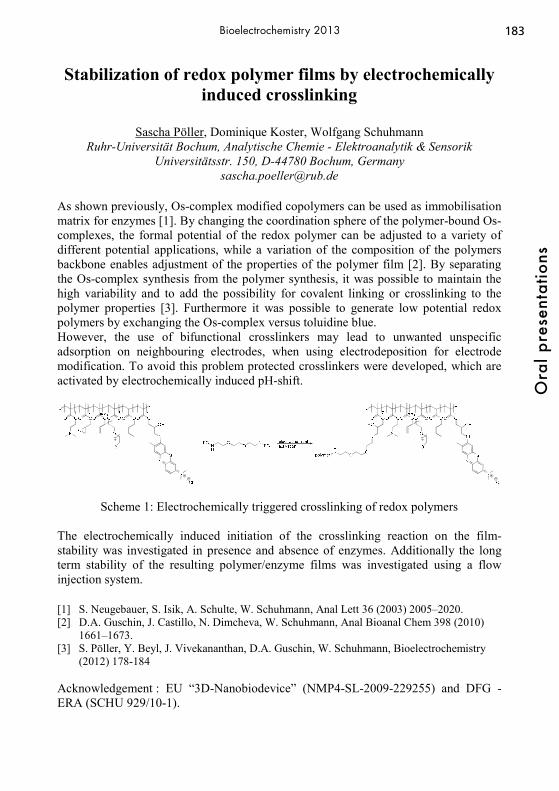
183
Ora
l pre
sent
atio
ns
Bioelectrochemistry 2013
Stabilization of redox polymer films by electrochemically induced crosslinking
Sascha Pöller, Dominique Koster, Wolfgang Schuhmann
Ruhr-Universität Bochum, Analytische Chemie - Elektroanalytik & Sensorik Universitätsstr. 150, D-44780 Bochum, Germany
As shown previously, Os-complex modified copolymers can be used as immobilisation matrix for enzymes [1]. By changing the coordination sphere of the polymer-bound Os-complexes, the formal potential of the redox polymer can be adjusted to a variety of different potential applications, while a variation of the composition of the polymers backbone enables adjustment of the properties of the polymer film [2]. By separating the Os-complex synthesis from the polymer synthesis, it was possible to maintain the high variability and to add the possibility for covalent linking or crosslinking to the polymer properties [3]. Furthermore it was possible to generate low potential redox polymers by exchanging the Os-complex versus toluidine blue. However, the use of bifunctional crosslinkers may lead to unwanted unspecific adsorption on neighbouring electrodes, when using electrodeposition for electrode modification. To avoid this problem protected crosslinkers were developed, which are activated by electrochemically induced pH-shift.
a a
Scheme 1: Electrochemically triggered crosslinking of redox polymers The electrochemically induced initiation of the crosslinking reaction on the film-stability was investigated in presence and absence of enzymes. Additionally the long term stability of the resulting polymer/enzyme films was investigated using a flow injection system. [1] S. Neugebauer, S. Isik, A. Schulte, W. Schuhmann, Anal Lett 36 (2003) 2005–2020. [2] D.A. Guschin, J. Castillo, N. Dimcheva, W. Schuhmann, Anal Bioanal Chem 398 (2010)
1661–1673. [3] S. Pöller, Y. Beyl, J. Vivekananthan, D.A. Guschin, W. Schuhmann, Bioelectrochemistry
(2012) 178-184 Acknowledgement : EU “3D-Nanobiodevice” (NMP4-SL-2009-229255) and DFG - ERA (SCHU 929/10-1).

184O
ral p
rese
ntat
ions
Bioelectrochemistry 2013
Electrotransfer of pStaby: A new safe and efficient DNA vaccine vector devoid of antibiotic resistance marker
G. Vandermeulen1, K. Vanvarenberg1, O. Schakman2, C. Szpirer3 and V. Préat1
1Université catholique de Louvain, Louvain Drug Research Institute, Pharmaceutics and Drug Delivery, 1200 Brussels, Belgium; 2 Université catholique de Louvain, Institut de Recherche Expérimentale et Clinique, Pole of Endocrinology, Diabetes, and Nutrition, 1200 Brussels, Belgium; 3Delphi Genetics, 6041 Gosselies, Belgium
The development of DNA vaccines requires both an adequate plasmid vector and a potent delivery method. During plasmid production, the selection of bacteria which contain the plasmid of interest is necessary. If the use of an antibiotic resistance gene as selection marker is efficient, it raises several concerns that are often pointed out by the regulatory authorities. Among the emerging technologies for plasmid selection, the use of a toxin-antitoxin system is particularly attractive. This study aimed to demonstrate the lack of toxicity of a new toxin-antitoxin based plasmid vector delivered by electrotransfer and its efficiency for inducing immune response to gp160. Here, the antidote gene (ccdA) is introduced in the pStaby plasmid under the control of a constitutive promoter. Expression of the poison gene (ccdB) by the bacteria is strongly repressed in the presence of the plasmid but, when the plasmid is lost, the antidote is degraded and the production of the toxin is induced, causing cell death. First, toxicity assays were performed in vitro on B16F10 and 293T cells. Death to live cell ratio was obtained from results of LDH and MTT assays and no in vitro toxicity was observed for the pStaby plasmid. we also evaluated in vivo the toxicity of pStaby administered by electrotransfer. We injected 100 μg of plasmid in 30 μl of PBS into each tibial cranial muscle. Then, we placed the leg between plate electrodes and we delivered 8 square-wave electric pulses (200 V/cm 20 ms 2 Hz). The open-field test was used to assess a nonforced ambulation and we noticed an accommodation effect when measures were performed daily but no difference was observed between treated or untreated mice. The grip strength test was used to measure the strength of combined fore limb-hindlimb muscles. No effect of the electrotransfer treatment was observed. The creatine kinase level in mice sera measured 15 days after electrotransfer were not modified. Put together, data from this in vivo experiment showed that muscle electrotransfer of pStaby did not influence the behavior of mice. Finally, the gp160 sequence was subcloned in pStaby. gp160 envelope glycoprotein is known to be an important target for HIV-1 vaccines. Mice received one priming and two boosts by muscle electrotransfer, with two weeks between each administration. Cytokine assays on splenocytes from immunized animals showed that γ-IFN and IL-2 were produced after stimulation with the antigen but no IL-10 and IL-4 were detected. This suggests that mice were efficiently immunized by electrotransfer of pStabygp160 plasmids with Th1-orientation of the immune response. In conclusion, electrotransfer of pStaby appears as a safe and efficient method for DNA vaccination. The authors acknowledge the financial support from the Biowin Project (DNAVAC) of the Walloon Region, Belgium.

185
Ora
l pre
sent
atio
ns
Bioelectrochemistry 2013
A Microfluidic Enzymatic Biofuel Cell Using a Flow-Through Bioanode and an Air-Breathing Cathode
Russell Reid, Dr. Shelley Minteer, Dr. Bruce Gale
University of Utah 50 S. Central Campus Drive, Salt Lake City, UT 84112
There are relatively few examples of enzymatic biofuel cells with flow-through electrodes in the literature even though there are potential advantages to this approach. In this work, a microfluidic enzymatic biofuel cell was designed and its performance was demonstrated. The flow-through anode was made by curing a mixture of carbon nanotubes, NAD-dependent glucose dehydrogenase, octyl-modified linearly polyethylenimine (C8-LPEI), and EGDGE on Toray carbon paper. The air-breathing cathode consisted of platinized carbon cloth that was heat-pressed onto a Nafion membrane. The housing and microfluidic channels were made from a lamination of poly (methyl methacrylate), silicone, and double-sided tape, and were all cut using a laser cutter. This fabrication method was very quick compared to other microfluidic biofuel cell fabrication methods. The electrodes were characterized in a non-microfluidic cell to ensure that the enzyme immobilization strategy was effective. Then the biofuel cell flow rate was optimized. The anode and cathode were tested at the optimal flow rate in separate half cells as well as together in the biofuel cell (Fig. 1). Finally, the biofuel cell fuel utilization was measured and compared with the theoretical value.
Fig. 1. Exploded model view of the biofuel cell showing the location of the flow-through bioanode and the air-breathing cathode.
Outlet
Toray paper bioanode
Platinized carbon cloth/Nafion cathode
Inlet
186O
ral p
rese
ntat
ions
Bioelectrochemistry 2013
Cellular tracking of single DNA-particles after their delivery by electroporation
Christelle Rosazza1,2,3, Andreas Zumbusch1 and Marie-Pierre Rols2,3
1 Department of Chemistry, University of Konstanz, Universitätstrasse, 78457 Konstanz, Germany
2 CNRS, Institut de Pharmacologie et de Biologie Structurale, 205 route de Narbonne, 31077 Toulouse, France
3 University of Toulouse III Paul Sabatier, 118 route de Narbonne, 31062 Toulouse, France
[email protected] Electroporation is a physical method of transferring molecules into cells and tissues. It takes advantage of the transient permeabilization of the cell membrane induced by electric fields which gives hydrophilic molecules access to the cytoplasm. This method offers high transfer efficiency for small molecules (below 4 kDa) which most likely originates from their free diffusion though electrically created pores. Heavier molecules, such as plasmid DNA, face physical barriers (plasma membrane, cytoplasm crowding, nuclear envelope) which reduce transfection efficiency and engender a complex mechanism of transfer. This work provides insight into the way DNA crosses the cytoplasm to reach the nucleus after electroporation. For this purpose, single particle tracking experiments of fluorescently labeled DNA were performed. Investigations were focused on the possible involvement of the cytoskeleton using drugs disrupting or stabilizing the two relevant cellular networks for particle transportation: the actin and the tubulin filaments. The analysis of 315 movies representing almost 4000 trajectories reveals that DNA is actively transported via the cytoskeleton and that the main responsible cellular network for this transport are the microtubules. The large number of analyzed events allows a statistical quantification of the DNA motion kinetics inside the cell. Disruption of both the actin and the tubulin filaments reduces the number of active transport events, the velocities, and the displacements of DNA particles. Interestingly, stabilization of both networks does not enhance DNA transport. Related publications: Chabot S, Rosazza C, Golzio M, Zumbush A, Teissié J, Rols MP. Nucleic Acids Electro-transfer: From Bench to Bedside. Curr Drug Metab. 2012 Oct 24 Rosazza C, Escoffre JM, Zumbusch A, Rols MP. The actin cytoskeleton has an active role in the electrotransfer of plasmid DNA in mammalian cells. Mol Ther. 2011; 19(5):913-21
187
Ora
l pre
sent
atio
ns
Bioelectrochemistry 2013
Electrochemical, Microbiological and Morphological Characterization of Microbial Bianodes for MEC.
ROUSSEAU Raphaël1, SANTAELLA Catherine2, GODON Jean-Jacques3,
ACHOUAK Wafa2, BERGEL Alain1,, DELIA Marie-Line1 1 Laboratoire de Génie Chimique, Université de Toulouse – CNRS, 4 allée Emile
Monso, BP84234 F-31432 Toulouse, France 2 CEA, DSV, IBEB, SBVME, Laboratoire d’ Ecologie Microbienne de la Rhizosphère et
des Environnements Extrêmes (LEMiRE), Saint-Paul-lez-Durance, France 3 INRA, UR050, Laboratoire de Biotechnologie de l’Environnement, Avenue des
Etangs, Narbonne F-11100, France [email protected]
Microbial biofilms that develop spontaneously on the surface of graphite anodes immerged in microbially-rich media can be efficient electro-catalysts for the oxidation of various organic compounds. The so-called microbial bioanodes have been commonly implemented in Microbial Electrolysis Cells (MECs) in association with a conventional abiotic cathode that achieves hydrogen evolution. Hydrogen is produced at the cathode, while organic compounds are oxidized at the anode. The power to be provided to a MEC is divided by a factor of around five with respect to a conventional water electrolysis cell. The purpose of the work was to design an optimal microbial anode by combining electrochemical, microbiological and morphological analyses. Bioanodes were formed under constant polarization in a nutritive solution inoculated with micro-organisms from salt marshes sediments. Sodium acetate 40 mM was used as substrate. Four types of electrode materials were tested and the highest currents were recorded with biofilm formed on carbon felt anode, which was kept for further study. Varying the polarization potential in the range -0.4 to +0.1 V/SCE demonstrated that the highest value led to the highest currents. Solution conductivity was increased by adding NaCl. An optimal NaCl concentration was determined of the order 45 g.L-1, which corresponded to a conductivity of 103.6 mS.cm-1, i.e. 1.5 times the seawater conductivity. The temperature was found to be a key parameter with an optimal value of 40 °C, which led to current densities up to 70 A/m2, the highest value reported so far in the literature with two-dimensional electrodes. Single-strand conformation polymorphism (SSCP) fingerprint profiles based on 16S rDNA sequence showed that neither the potential nor the salinity really influenced the structure of microbial communities that made up the bioanodes. Moreover, SSCP profiles revealed that only few bacteria belonging to the microbial community from salt marshes sediments used as inoculum was able to colonize the anode. We used laser scanning confocal microscopy to characterize the impact of polarization potential and NaCl concentration on the architecture of the biofilms. For the highest currents, biofilms were flat, spread, and discontinuous around the wires of the felt and formed a matrix between the fibers. Biofilms forming large continuous sheathes around the wires and no matrix between fibers proved less efficient in producing current. This work was part of the DefiH12 project (ANR-09-BioE-010).

188O
ral p
rese
ntat
ions
Bioelectrochemistry 2013
Adenine-Thymine Coadsorption at Gold Electrodes Interfaces. An in-situ FT-IR Spectroscopy Study
Manuela Rueda(a), Francisco Prieto(a), Julia Álvarez(a), Antonio Rodes(b)
(a)Department of Physical Chemistry. University of Seville. C/ Profesor García González nº 2, 41012 Seville (Spain)
tfn. +34954556733, e-mail: [email protected] (b) Department of Physical Chemistry and Institute of Electrochemistry.
University of Alicante. Ap. 99, E-03080, Alicante (Spain)
Adenine-Thymine interactions are essential in DNA stabilization and in replication and transcription of genetic codes in living cells. This biological relevance stimulates the studies of organised structures of these complementary bases from the molecular level. In this respect, it is known that both bases adsorb on metal substrates forming organised films [1-3], which are also interesting in many biotechnology and nanotechnology applications. The coadsorption of thymine and adenine at Au(111) electrodes has been previously studied by cyclic voltammetry [4]. Modern in-situ FTIR techniques that provide chemical specificity together with high sensitivity can be combined with electrochemical methods in order to characterise the interactions at electrode interfaces. In this work in-situ FT-IR external reflection experiments (IRRAS) and internal reflection experiments (ATR-SEIRAS) are combined with cyclic voltammetry, using either Au(111) or gold thin-film electrodes. Experiments have been performed in 0.1M HClO4 solutions with different adenine and thymine concentrations. H2O and D2O have been used as solvents. The observed adenine-thymine interactions greatly depend on the applied potential. At low potentials at which thymine is physically adsorb, chemically adsorbed adenine replaces thymine from the surface even when thymine concentration is about hundredfold the adenine concentration. However, chemical coadsorption of the two bases is detected at higher potentials inducing significant changes in the voltammetric adsorption/desorption peaks and in the signals of the representative vibration modes of the adsorbed molecules as compared to the situation when only one of the bases is adsorbed. From the observed changes in the infrared spectra it is concluded that both chemically adsorbed bases undergo reorientations after addition of the complementary base. Thymine reorients from an up-right to a flat orientation while adenine changes the coordination sites with the metal and adopts a more up-right disposition in order to facilitate the interaction between the complementary bases in the same way than in DNA. 1. - B. Roellfs, E. Bunge, C. Schroeter, T. Solomun, R.J. Nichols, J. Phys. Chem. 101 (1997) 54 2.- A. Rodes, M. Rueda, F. Prieto, C. Prado, J.M. Feliu, A. Aldaz, J. Phys. Chem. C 113 (2009) 18784. 3.- M. Rueda, F. Prieto, A. Rodes, J.M. Delgado, Electrochim. Acta. 82 (2012) 534. 4.- A. P. M. Camargo, H. Baumgärtel, C. Donner, Phys. Chem. Comm. 5(8) (2003) 1657.
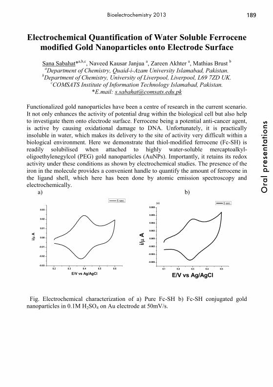
189
Ora
l pre
sent
atio
ns
Bioelectrochemistry 2013
Electrochemical Quantification of Water Soluble Ferrocene modified Gold Nanoparticles onto Electrode Surface
Sana Sabahat*a,b,c, Naveed Kausar Janjua a, Zareen Akhter a, Mathias Brust b
aDepartment of Chemistry, Quaid-i-Azam University Islamabad, Pakistan. bDepartment of Chemistry, University of Liverpool, Liverpool, L69 7ZD UK.
cCOMSATS Institute of Information Technology Islamabad, Pakistan. *E.mail: [email protected]
Functionalized gold nanoparticles have been a centre of research in the current scenario. It not only enhances the activity of potential drug within the biological cell but also help to investigate them onto electrode surface. Ferrocene being a potential anti-cancer agent, is active by causing oxidational damage to DNA. Unfortunately, it is practically insoluble in water, which makes its delivery to the site of activity very difficult within a biological environment. Here we demonstrate that thiol-modified ferrocene (Fc-SH) is readily solubilised when attached to highly water-soluble mercaptoalkyl-oligoethylenegylcol (PEG) gold nanoparticles (AuNPs). Importantly, it retains its redox activity under these conditions as shown by electrochemical studies. The presence of the iron in the molecule provides a convenient handle to quantify the amount of ferrocene in the ligand shell, which here has been done by atomic emission spectroscopy and electrochemically.
a) b)
0.2 0.3 0.4 0.5 0.6-0.03
-0.02
-0.01
0.00
0.01
0.02
0.03
i/μ A
E/V vs Ag/AgCl
5 sec.
0.1 0.2 0.3 0.4 0.5
-0.006
-0.004
-0.002
0.000
0.002
0.004
0.006
0.008
i/μ A
E/V vs Ag/AgCl
5 sec.(a)
Fig. Electrochemical characterization of a) Pure Fc-SH b) Fc-SH conjugated gold nanoparticles in 0.1M H2SO4 on Au electrode at 50mV/s.

190O
ral p
rese
ntat
ions
Bioelectrochemistry 2013
A simple mediator-less enzymatic biofuel cell based on unpurified fungus culture supernatant
Sabine Sané, C. Kräß, S. Rubenwolf, S. Kerzenmacher
University of Freiburg, IMTEK - Department of Microsystems Engineering, Georges-Koehler-Allee 103, 79110 Freiburg, Germany
We present for the first time the use of crude culture supernatant from enzyme-secreting microorganisms to supply biocatalysts to the electrodes of a mediator-less enzymatic biofuel cell. By using crude culture supernatant, we overcome the need for time consuming and expensive enzyme purification. Suitable enzymes that are secreted by fungi are laccase for cathodic oxygen reduction and cellobiose dehydrogenase (CDH) for anodic lactose oxidation. Both enzymes are capable of direct electron transfer (DET) and thus do not require a mediator, which can also be a cost factor. The cathode was supplied with culture supernatant of the fungus Trametes versicolor, grown in modified SC medium, and containing 2.20 Uml-1 secreted laccase. The anode was supplied with culture supernatant of the fungus Phanerochaete chrysosporium grown in modified SC medium and ATM medium, yielding CDH activities of 0.07 Uml-1 and 0.05 Uml-1 respectively. Furthermore, 30 mM ß-lactose was added to the anode. The two compartments were separated with a proton exchange membrane. To record the fuel cell polarization curve, the load current density was incrementally increased (steps of 2.2 μAcm-2h-1) and the potential was measured against a saturated calomel electrode (SCE). By using supernatant of P. chrysosoprium at the anode, we achieved a power density of 2.7 ± 0.7 μWcm-2 when the fungus was grown in SC medium and of 4.9 ± 0.3 μWcm-2 when it was grown in ATM medium. No significant power could be generated in control experiments using the culture medium alone, clearly demonstrating the catalytic activity of the supernatants for biofuel cell operation. In comparison, power densities of 1.9 μWcm-2 [1] and 5 μWcm-2 [2] have been reported in literature for BFCs based on purified laccase and CDH. Polarization curves of anode and cathode potentials indicate that the anode limits the power density. In summary, our results show that even with unpurified enzymes, we can achieve a power output which is in the same range as biofuel cells using purified enzymes. This opens the opportunity for simple, low-cost enzymatic biofuel cells. Future work will focus on enhancing the anode performance. Furthermore, we want to explore whether the fuel cell lifetime can be extended by resupplying fresh enzymes to the electrodes. The feasibility of this approach has already been demonstrated for the laccase cathode using a crude culture supernatant of T. versicolor [3]. [1] Stoica et al., Fuel Cells 9 (2009) 53 [2] Coman et al., Phys.Chem.Chem.Phys. 10 (2008) 6093 [3] Sané et al. Proc. 63rd Annual Meeting of the ISE (2012) 40
191
Ora
l pre
sent
atio
ns
Bioelectrochemistry 2013
Release of Iron Ions from the Stainless–Steel Anode during High-Voltage Pulses and its Consequences
for Cell Electroporation Technology
Raminta Rodaite-Riseviciene1, Rita Saule1, Valentinas Snitka2 and Gintautas Saulis1,3* 1Laboratory of Biophysics for Bionanotechnology and Medicine,
Department of Biology, Vytautas Magnus University, Kaunas, 44248, Lithuania 2Research Center for Microsystems and Nanotechnology,
Kaunas University of Technology, Kaunas, 51369, Lithuania 3Laboratory of Bio-nanotechnology, Semiconductor Physics Institute,
Center for Physical Sciences and Technology, Vilnius, 01108, Lithuania *E-mail: [email protected]
Cell electroporation – a temporal increase of the cell membrane permeability occurring due to the action of the pulses of strong electric field (up to 300 kV/cm) – is widely used in cell biology, biotechnology, and medicine. However, when a high-voltage is applied to the electrolyte solution, besides membrane permeabilization, various electrolysis reactions occur at the electrode-solution interfaces. One of these electrochemical reactions is the oxidation of the metal ions of the anode. As a result of this, the dissolution of the anode occurs. One of the most popular materials utilized for electrodes, which are used to electroporate the cells, is stainless–steel. In such a case, iron ions (Fe2+ and Fe3+) are released from the anode under the action of high-voltage electric pulses. In the present work, this process and its consequences have been studied. Due to the action of a single square-wave electric pulse with the duration of 2 ms and the amplitude of 1.2 kV/cm, the concentration of iron ions exceeded 0.5 mM. Using atomic force microscopy, we established that the roughness of the stainless-steel anode increased progressively, in proportion the total amount of the electric charge that had passed through the unit area of the electrode. Iron ions released from the electrodes behave as a Lewis acid and hydrolyze the water molecules in the solution, reducing the pH of a solution. The reduction of the viability of various cancer and non-cancerous cells by iron ions has been demonstrated. The iron ions quench the fluorescence of anticancer drugs, which are used when photodynamic tumour therapy is combined with electroporation, such as porphyrin sulphonate and Adriamycin, as well as calcein dye that is used in cell electroporation studies. The results of this work can be useful for optimizing the electroporation methods used in biotechnology, medicine, and food industry.

192O
ral p
rese
ntat
ions
Bioelectrochemistry 2013
Robotic Drug Electroanalysis in Microtiter Plates: Convenience Paired with Potential
Albert Schulte1, Mathias Winterhalter2, Helge Weingart2 and Somjai Theanponkrang1
1Biochemistry - Electrochemistry Research Unit, Schools of Chemistry and Bio-chemistry, Suranaree University of Technology, Nakhon Ratchasima, Thailand; 2Life Sciences, School of Engineering and Science, Jacobs University, Bremen, Germany
Efficient drug testing and analysis is a central task in medicine, pharmacy, forensic science and toxicology. Controlled drug release, physiological uptake, body distribution and metabolic degradation studies, for instance, are the keys for an intelligent approach of new therapeutic developments. Trace drug screening in samples from addict’s, athletes and the environment, on the other hand, help uncovering drug dependencies, performance-enhancing misuse and water, plant and soil drug pollution. For electroactive drugs voltammetric detection is often choice and not spectroscopy as electrochemical devices are cheaper, easier to use and more compact than optical ones. A computerization/automation of drug voltammetry was achieved by a combination with flow-based analyzers designed for non-manual sample and electrochemical flow-cell handling. Benefits of the merger were a better sample throughput as compared to normal trials in beakers and the exclusion of manual operation errors but not yet optimal was the ease of use because a complex flow stream management is involved. Here we report on microtiter plate-based electrochemical drug analysis as novel option with automation power. By addressing electronic micropositioners the software unit of the computerized device guides a movable reference, counter and working electrode assembly in order through the 24 wells of a standard microtiter plate. During halts in a well solution, the system can trigger either electrochemical electrode cleaning and pretreatment procedures or voltammetric trials. The functionality of the novel robotic drug assay will be demonstrated through the presentation of successful microtiter plate calibration runs for Norfloxacin (NF) and Ciprofloxacin (CF), two antibiotics of the fluoroquinolone class, and for Paracetamol (PC), one of the main fever-reducing analgesics. The linear range in the robotic differential pulse voltammetry (DPV) mode extended to 10, 100 and 150 μM for NF, CF and PC, respectively; and for all three analytes the recovery rates for test trials with model samples made of commercial drug powder or tablets deviated in worst case about ± 5% from the ideal value of 100%. Robotic NF-DPV well quantified in the more challenging medium of blood serum μM analyte levels of spiked samples. And robotic PC-DPV successfully measured in urine the painkillers absence, appearance and incomplete clearance when samples were taken from a test person before and 45 minutes and 4 hours after drug uptake, respectively. The developed robotic electrochemical drug screening is convenient, releases laboratory staff to other duties and works with a reduced risk of operator errors. These assets relate to good cost effectiveness and the method thus is suggested for routine drug testing and analysis in medical research and the pharmacy industry.

193
Ora
l pre
sent
atio
ns
Bioelectrochemistry 2013
Influence of Metal Cations and the Polycation Polyethylenimine on the Turnover Rate of Cellobiose
Dehydrogenase
Christopher Schulz1, Mostaba Tavahodi1, Roland Ludwig2, Lo Gorton1
1 Lund University, Department of Biochemistry and Structural Biology, P. O. Box 124, SE-22100 Lund, Sweden, [email protected],
[email protected], [email protected] 2BOKU-University of Natural Resources and Life Sciences, Institute of Food Technology, Muthgasse 18, 1190 Vienna, Austria, [email protected]
Cellobiose dehydrogenase (EC 1.1.99.18) is an extracellular fungal redox enzyme, which has recently shown promising properties for applications in both biosensors and biofuel cells [1]. It is a two domain enzyme composed of a catalytic, FAD containing domain, DHCDH, connected through a polypeptide linker region with a cytochrome b domain, CYTCDH. In the catalytic reaction, the substrate is oxidised at the DHCDH, which in turn is reoxidised through an intramolecular and sequential electron transfer process donating the electrons to the CYTCDH, from which the electrons can be donated directly to an electrode. The mechanism with which the electrons are transferred between the two domains is unknown and pH dependent. However, it is believed that the surface exposed heme of the CYTCDH enters the substrate channel of the DHCDH allowing the electrons to be transferred between the two domains. We have now found that when increasing the concentration of metal cations the rate of the intramolecular electron transfer reaction of CDH immobilised on an electrode surface could be increased substantially up to 23 times. Increases were higher with divalent metal cations compared to monovalent metal cations, but were also dependent on the type of cation, confirmed also by enzymatic assays of CDH in solution [2]. Beside free diffusing cations also the influence of the polycation polyethylenimine (PEI) on the performance of CDH was investigated. It was found that the pre-modification of the electrode with PEI can enhance the current response of CDH modified electrodes to up to 140x. Additionally the modification with PEI shifted the pH optimum of CDH to the human physiological range. Similar results were also obtained for the pre-modification with gold nanoparticles capped with Polyethylenimine. Polarisation curves of CDH modified electrodes revealed the presence of an additional redox wave, enhanced by PEI, originating from the dehydrogenase domain of CDH but present at a potential ca. 600 mV more positive than expected. These findings are of interest both for a deeper understanding of the electron transfer pathway in CDH and for increased bioelectrocatalytic current densities. Recent results and a proposed mechanism to explain the observed effects will be shown and discussed. [1] R. Ludwig, W. Harreither, F. Tasca, L. Gorton; ChemPhysChem, 2010, 11, 2674 [2] C. Schulz, R. Ludwig, P. O. Micheelsen, M. Silow, M. D. Toscano, L Gorton, Electrochem Comm, 2012, 17, 71-74

194O
ral p
rese
ntat
ions
Bioelectrochemistry 2013
STM and AFM Studies of Structure and Dynamics of Supported Lipid Films on Gold Electrodes
Slawomir Sek Department of Chemistry, University of Warsaw
Pasteura 1, 02-093 Warsaw, Poland [email protected]
Lipids are fundamental components of the biological membranes which play an important role in numerous processes including ion and protein transport, membrane fusion or cell signaling. In many cases, both the composition and the distribution of the lipid components within the membranes determine their properties. Therefore the knowledge about the molecular structure, permeability, hydration and thickness of the lipid films is crucial for understanding their particular functions. Unfortunately, biological systems are quite complex, therefore, simple biomimetic models involving lipid monolayers and bilayers supported on solid substrates are often used for examination of membrane properties. Numerous techniques can be used to resolve the structure, orientation, and packing of lipid molecular films. However, the most direct way to address this problem is to probe these systems using scanning tunneling microscopy (STM) and atomic force microscopy (AFM). These techniques are known to be excellent tools, which provide detailed information about the structure and the properties of the molecular films immobilized on solid substrates down to molecular scale.[1] It will be demonstrated that using in situ STM and AFM it is possible to observe adsorption behavior of the lipid molecules on Au(111) and evaluate their structure and the properties.[2] Formation of the films can be visualized at open circuit potential as well as under full electrochemical control. The latter allows one to monitor electric field driven changes in a film structure. It will be demonstrated that the structure of the lipid assembly depends strongly on numerous factors including chemical nature of the film-forming molecules, their concentration and the presence of the electric field.
References [1] S. Sęk, T. Laredo, J. Dutcher, J. Lipkowski, J. Am. Chem. Soc., 2009, 131, 6439-6444. [2] D. Matyszewska, S. Sęk, R. Bilewicz, Langmuir, 2012, 28, 5182-5189.

195
Ora
l pre
sent
atio
ns
Bioelectrochemistry 2013
Unsubstituted Phenothiazine as a New Efficient Electron Transfer Mediator for Oxidases
Alina N. Sekretaryova, Arkady A. Karyakin
Chemistry Department, Lomonosov Moscow State University Leninskie Gory 1, Moscow, Russia
Biosensors using immobilized oxidases as the bio-recognition element are among the most widely investigated devices for both fundamental research and applications. The electrical communication between the redox enzymes and the electrodes can be established by using biologically active or synthetic charge carriers as mediators. However, sensors based on electron-shuttling redox couples suffer from an inherent drawback: the soluble mediating species can diffuse away from the electrode surface into the bulk solution, which for instance would preclude their use as implantable probes. With this in mind, we have investigated systems based on oxidases immobilized in a gel of siloxanes from water-organic mixtures with the high content of organic solvent using water insoluble azines as mediators [1]. Water insolubility of a mediator prevents it from diffusing away from the electrode surface. We tested unsubstituted phenothiazine, phenoxazine and their oligomers as possible mediators for glucose, lactate and cholesterol oxidases. Among them only the enzyme containing membrane with phenothiazine as the mediator displayed catalytic-type cyclic voltammograms in the presence of the enzyme substrate. We note that being water-insoluble phenothiazine was unknown as the mediator for oxidases. The redox mechanism of phenothiazine is a two electron process. Dependence of the common peak potentials on solution pH points to the two electron – two proton reaction. Discussing diffusion peculiarities of the mediator we made an attempt to evaluate the diffusion coefficient of phenothiazine in siloxane gel. The diffusion coefficient was calculated from the cyclic voltammetry and chronoamperometry data using equations for reversible electrochemistry. The evaluated diffusion coefficient is 10-7 cm2 s−1. The results for the gel membranes with the different oxidases coincide. Kinetic parameters were obtained from the double potential step chronoamperometry and RDE mesuarements. The results obtained from the two different methods are in good agreement. The catalytic rate constants vary in the range 101 to 103 M-1 s-1 order of magnitude for the different oxidases. The values of the constants are comparable to results reported for advanced mediators of oxidases. Hence, using of the water insoluble mediator opens up a new approach to construct reagentless enzyme biosensors, owing to the multiformity of available oxidases. [1] Alina N. Sekretaryova, Darya V. Vokhmyanina, Tatyana O. Chulanova, Elena E. Karyakina, Arkady A. Karyakin. Anal. Chem., 2012, 84 (3), P. 1220-1223. Financial support through Ministry of Education and Science Contract 14.740.11.1374 is greatly acknowledged.

196O
ral p
rese
ntat
ions
Bioelectrochemistry 2013
Light-Emitting Electrochemical Swimmers
M. Sentic,1,2 G. Loget,1 D. Manojlovic,2 A. Kuhn,1 N. Sojic1 1 University of Bordeaux, Institut des Sciences Moléculaires, 33607 Pessac, France
2 Faculty of Chemistry, University of Belgrade, 11000, Belgrade, Serbia E-mail: [email protected]
Electrogenerated chemiluminescence (ECL) is a widely used electrochemical process, where light emission is initiated at an electrode surface. Commercial ECL applications are available for ECL immunoassays.[1] The combination of ECL and bipolar electrochemistry (BE) has been already employed for analytical purposes.[2] In this work we present the synergetic action of BE[3] in terms of simultaneous bubble production and ECL generation, leading in fine to the first example of a propulsion mechanism for a swimmer, which is intrinsically coupled with a chemical light source. As depicted in Figure 1, reduction of H2O at the cathodic pole and oxidation of the ECL reagents at the anodic pole induces simultaneous motion and light emission of the glassy carbon bead in a capillary.[4]
Fig. 1 Asymmetric ECL swimmer. The versatility of BE coupled to ECL allows to imagine the use of the same principle with other types of swimmers in a new class of dynamic experiments with multifunctional objects. For instance, dynamic multiplexed immunoassays or DNA assays may be developed in solution phase with biofunctionalized swimmers using ECL as a readout mechanism. [1] W. Miao, Chem. Rev., 2008, 108, 2506 ; L. Hu, G. Xu ; Chem. Soc. Rev., 2010,39, 3275 [2] F. Mavré, R. K. Anand, D. R. Laws, K.-F. Chow, B.-Y. Chang, J. A. Crooks, R. M. Crooks, Anal. Chem. 2010, 82, 8766 [3] G. Loget, A. Kuhn, Lab Chip 2012, 12, 1967; G. Loget, A. Kuhn, Nature Commun. 2011, 2, 535 [4] M. Sentic, G. Loget, D. Manojlovic, A. Kuhn, N. Sojic, Angew. Chem. Int. Ed., 2012, 51, 11284–11288. DOI 10.1002/anie.201206227

197
Ora
l pre
sent
atio
ns
Bioelectrochemistry 2013
Development of Non-gassing Electroosmotic Pump for Drug Infusion System
Woonsup Shin
Department of Chemistry and Interdisciplinary Program of Integrated Biotechnology, Sogang University, Seoul 121-742, Korea
During my second sabbatical visit to Adam’s lab started from 2009 summer, I had a chance to deal with small miniaturized pumps. We had no intention to develop the pumps and searched for available ones in the market for building up a drug infusion system, particularly for insulin. Electroosmotic pump from NanoFusion Technology was one of them. The structure of the pump is MEA (membrane electrode assembly) based on porous Pt/porous SiO2/porous Pt. We started testing it and immediately found that the gas evolution upon pumping is the main problem in pump’s stability and safety. Most of electroosmotic pumps use platinum electrodes and the pumped solution keeps being electrolyzed to produce O2 from anode and H2 from cathode. The trapped bubbles in the MEA block the fluid movement, making the flow irregular. The co-production of O2 and H2 makes the system unsafe in the closed loop. By several trials and errors we found that the substitution of the platinum electrode to Ag/Ag2O electrode enabled the operation without gassing. The 8 mm diameter, 2 mm thick pump operates at 5 - 40 μL min-1 upon applying 0.3 - 2.0 V. The resulting efficiency of the pump is 14,000 water molecules pumped per reacted electron. It is made of only 2.3 coulombic amount of Ag/Ag2O deposited consuming electrodes and provides continuous operation for 3 hours at 20 μL min-1 flow rate at 1 V; or pulsed operation for 30 sec every 30 min for 70 hours ; or daily operation for 5 min for 1 month. The pump was applied in a miniature (36 x 30 x 8 mm), single-use, subcutaneous drug infusion system. The development stroy of the non-gassing electroosmotic pump and the current status will be presented and discussed.

198O
ral p
rese
ntat
ions
Bioelectrochemistry 2013
Biomedical Applications of Implantable Biofuel Cells
Sergey Shleev Biomedical Science, Health & Society, Malmö University, 20506 Malmö, Sweden
Plamen Atanassov
Center for Emerging Energy Technologies, Chemical and Nuclear Engineering Dept., University of New Mexico, Albuquerque, New Mexico, USA, 87131
This paper overviews the critical advances in the biofuel cell technology that enabled the development of implantable devices or extracorporeal devices for biomedical application. Such applications include monitoring health indicators by a biosensor, or powering of actuators such as artificial organs or drug delivery systems. Biofuel cells are viewed as advantageous as they are been considered as devices capable of harvesting energy in vivo from organic compounds, physiologically available in the host organism. The limitations of this widely accepted concept will be discussed in the context of the site of implantation and the alternative use of extracorporeal alternatives such as patches or ocular lenses. The paper will provide a biomedical outlook on the implantation of biofuel cells as it contrast the in vivo operation of such device for demonstration purposes. In vitro and in vivo studies, animal trails, showed the feasibility of implantable power devices to work by using glucose in the blood or interstitial fluid. Much effort was placed earlier on studies that rely of noble metal (usually Pt or its allows) catalysis of glucose oxidation. These implantable fuel cells in general provide power output in the early stages of implantation and are subject to severe fouling. That limits the apparent robustness of the “abiotic” implantable fuel cells. Biological (enzymatic) fuel cells on their end suffer from durability limitations that are mostly associated with the stability of their enzyme component. New advances in immobilization strategies, developed in the two collaborating sited of this paper allow for overcoming most, if not all of these limitations. Biofuel cells allow for a great diversity of design solutions: from layered fabrication and incorporation of capillary flow to ultimate miniaturization limited only by the size of the enzymes and nano-structured materials involved. The paper will discuss the strategies that University of New Mexico and Malmö University are currently developing to deploy a multi-enzyme immobilization on nano-structured substrates (Au nanoparticles and CNT) integrated into hierarchically structured materials. The overview of the technologies for integration of those technologies into devices includes methods of nano-fabrication by self-assembly and nano-lithography. Much of this research has been influenced by the seminal work led by Prof. Adam Heller. Authors will provide a personal perspective on how the ideas first introduced in his original papers or discussed in his most influential technology notes are being brought to a new level by the confluence of technological advances in micro/nano-fabrication and integration of biological component (enzymes) with nano-structures with advanced understanding of the mechanism of electron transferred in bio-electrocatalytic systems.

199
Ora
l pre
sent
atio
ns
Bioelectrochemistry 2013
Supramolecular Hydrogels as a Substrate for Biosensors
Anna Słoniewska, Barbara Pałys Department of Chemistry, University of Warsaw, Pasteur Street 1,
02-093 Warsaw, Poland [email protected]
Supramolecular hydrogels of conducting polymers have been successfully used in bioelectrochemistry because of their mechanical and swelling properties of gels added to the specific transport properties of conducting polymers. They are defined as three-dimensional network that mainly consist of crosslinked polymers and water, which is formed by directed non-covalent interactions, such as hydrogen bonding, electrostatic interactions, π-π stacking. We have studied polyaniline-poly(styrene sulfonate) (PANI-PSS) hydrogel as a substrate for enzymes. PANI-PSS hydrogel is a supramolecular self-assembly material consisting of positively-charged PANI chains and negatively-charged PSS chains. Compared to conventional polymers, PANI-PSS hydrogels characterize high energy density, high power density and improved electrochemical stability. Therefore, we have tested the compatibility of the substrate with enzymes, such as the urease, laccase and horseradish peroxidase. Hydrogels were studied by cyclic voltammetry, infrared spectra, optical microscope and AFM. Results prove the suitability of such material for enzymes immobilization.
200O
ral p
rese
ntat
ions
Bioelectrochemistry 2013
Engineering FAD dependent oxidases ~development of dehydrogenases from oxidases for
amperometric enzyme sensor applications~
S.Saito, Y.Horaguchi, K.Kojima, S. Ferri, W.Tsugawa, K.Sode*, Department of Biotechnology, Graduate School of Engineering
Tokyo University of Agriculture & Technology 2-24-16 Naka-cho, Koganei, Tokyo, Japan
Since the report on the first enzyme sensor employing glucose oxidase (GOx) and an oxygen electrode for glucose monitoring, extensive studies have been reported to develop variety of enzyme-based electrochemical sensors. Instead of the first-generation of enzyme sensor employing oxygen as the electron acceptor, the second-generation sensors employing artificial electron acceptors are currently dominating the products in the industrial applications. However, electron mediator-type enzyme sensors employing oxidases are inherently influenced by the amount of oxygen dissolved in the sample. The high reactivity of oxidases with oxygen limits their potential applications for biosensors employing artificial electron acceptors. Therefore, oxidases that are relatively less oxygen-sensitive would be greatly advantageous for the development of amperometric enzyme sensors. The authors have been intensively studying the engineering of FAD dependent oxidoreductases. We previously reported on the mutagenesis studies toward fructosyl amino acid oxidases (FAOxs)1 and frutosyl peptide oxidases (FPOxs)2. The engineered FAOxs and FPOxs showed decreased oxidase activity whereas their dehydrogenase activity remained or even increased, consequently, the “dehydrogenases” were developed from “oxidases”. These successes inspired us to extend our concept to the enzymes belong to major FAD dependent oxidoreductases, glucose methanol choline (GMC) oxidoreductase family. GMC oxidoreductase family contains various oxidases which have been reported their applications in the mediator type enzyme sensors. We have introduced mutations in GOxs and cholesterol oxidase, to construct engineered oxidases with decrease their oxidase activities with increased dye-mediated dehydrogenase activities. In this paper, we present our strategic approaches in the engineering oxidases and their application in the enzyme sensor construction. References 1. Kim, S.; Nibe, E.; Ferri, S.; Tsugawa, W.; Sode, K. Engineering of dye-mediated
dehydrogenase property of fructosyl amino acid oxidases by site-directed mutagenesis studies of its putative proton relay system. Biotechnol. Lett. 2010, 32, 1123–1129.
2. Kim, S.; Nibe, E.; Tsugawa, W.; Kojima, K.; Ferri, S.; Sode, K. Construction of engineered fructosyl peptidyl oxidase for enzyme sensor applications under normal atmospheric conditions. Biotechnol. Lett. 2012, 34, 491–497.
201
Ora
l pre
sent
atio
ns
Bioelectrochemistry 2013
Electrochemistry of immobilised hemin and reconstituted horseradish peroxidase
Maciej Sosna and Elena E. Ferapontova
Interdisciplinary Nanoscience Center (iNANO), Aarhus University Gustav Wieds vej 14, DK-8000, Aarhus C, Denmark
[email protected] The continuous development of biosensors is the driving force behind fundamental investigations of redox modified electrodes. In particular, the immobilisation of small redox centers and redox enzymes capable of direct electron transfer (DET) is of great importance in the construction of reagentless biosensors. In order for the electron transfer to be efficient the distance between the redox center and the electrode has to be minimised. In case of redox enzymes the molecule is thus required to have appropriate orientation so that its redox center can be efficiently wired to the electrode surface. One of the methods to achieve this is the immobilisation of the redox cofactor followed by reconstitution of an apoenzyme.1 Among others, this approach was successfully employed to construct electrodes modified with heme containing peroxidases 2, which are known to be capable of DET 3,4. In this communication we present the electrochemical characterisation of hemin covalently attached to gold electrodes via self-assembled monolayers (SAMs) of alkane thiols. The surface coverage and electron transfer between the heme iron and the electrodes are quantified and compared for varying compositions of underlying SAMs. The immobilised hemin molecules are subsequently used to reconstitute an apo-peroxidase from horseradish. The efficiency of the formation of holoenzyme, its electrochemistry and bioelectrocatalysis of hydrogen peroxide reduction are discussed. (1) Fruk, L.; Kuo, C.-H.; Torres, E.; Niemeyer, C. M. Angew. Chem. Int. Ed. 2009, 48, 1550. (2) Zimmermann, H.; Lindgren, A.; Schuhmann, W.; Gorton, L. Chem.-Eur. J. 2000, 6, 592. (3) Ferapontova, E. E. Electroanalysis 2004, 16, 1101. (4) Kartashov, A. V.; Serafini, G.; Dong, M. D.; Shipovskov, S.; Gazaryan, I.; Besenbacher, F.; Ferapontova, E. E. PCCP 2010, 12, 10098.
202O
ral p
rese
ntat
ions
Bioelectrochemistry 2013
DNA Detection in Droplet-based Microfluidic Devices
Giuseppe Spoto Dipartimento di Scienze Chimiche, Università di Catania, V.le A. Doria 6, Catania,
Italy. [email protected]
The development of molecular diagnostic devices able to identify specific DNA or RNA sequences represents a crucial step towards the widespread use of genetic analyses in clinical diagnostics, food-quality control and environmental monitoring [1]. These devices are expected to perform sensitive nucleic acid detection [2,3] on the basis of simple, cheap and reliable protocols able to operate with small sample. Droplet-based microfluidics define new and powerful approaches for biomolecular diagnostics as a consequence of the capability to miniaturize standard laboratory operations. It exploits a segmented flow of a liquid in which reagents of interest are compartmentalized within nanoliter or femtoliter sized droplets that reside within a continuous and immiscible fluid. Droplet compartmentalization of reacting species greatly reduces the importance of issues related to sample contamination and reagent dispersion. Assays can be performed with high throughput and reduced sample consumption and, by using appropriate microfluidic device architectures, the rapid mixing of reagents inside droplets can be obtained, thus providing faster DNA hybridization rates than at solid-liquid interfaces. Possibilities offered by droplet-based microfluidics in DNA detection will be presented by referring to both the capability of peptide nucleic acid-molecular beacons to detect DNA target sequences in oligonucleotides and PCR amplicons [4] as well as advantages offered by the integration of the isothermal amplification of DNA in the droplet miniaturized environment. The majority of miniaturized systems used for nucleic acid analysis are based on the polymerase chain reaction (PCR) amplification of the sequence to be targeted. This method needs the thermal cycling of the sample solution at three different temperatures during the amplification of the target sequence. Droplet-microfluidic systems using isothermal amplification reactions will be presented as an alternative to PCR amplification. In particular, the detection of interacting events in nanoliter droplets based on the isothermal strand displacement polymerization reaction will be discussed. The method allows a cyclic probe/target hybridization, polymerization reaction and target displacement to occur while an increasing fluorescent signal is generated after each reaction cycle is completed. 1. G. Spoto, R. Corradini (Eds.) Detection of Non- Amplified Genomic DNA, Springer, 2012. 2. R. D’Agata, R. Corradini, C. Ferretti, L. Zanoli, M. Gatti, R. Marchelli, G. Spoto, Biosens. Bioelectron. 25 (2010) 2095-2100. 3. R. D'Agata, G. Breveglieri, L. M. Zanoli, M. Borgatti, G. Spoto, R. Gambari, Anal. Chem. 83(22), (2011) 8711-8717. 4. L. M. Zanoli, M. Licciardello, R. D’Agata, C. Lantano, A. Calabretta, R. Corradini, R. Marchelli, G. Spoto, Anal. Bioanal. Chem. 2012, DOI: 10.1007/s00216-011-5638-3.

203
Ora
l pre
sent
atio
ns
Bioelectrochemistry 2013
The use of Cobaltabisdicarbollide as a generator of ion-pair complexes with bioactive nitrogen containing
compounds for sensors development
Anca-Iulia Stoica1, Christoph Kleber1, Clara Vinas2 and Francesc Teixidor2
1Centre of Electrochemical Surface Technology, Viktor-Kaplan-Straße 2, Wiener Neustadt, Austria
2 Institute of Material Science, Campus UAB, 08193 Bellaterra, Spain [email protected]
The aim of our studies is focused on the cobaltabisdicarbollide anion ([3,3´-Co(1,2-C2B9H11)2]-) ([1]-) that is used as a new material able to generate ion-pair complexes with nitrogen containing compounds, such as chemotherapeutic drugs, antibiotics, amino acids, neurotransmitters, antidiabetic drugs, vitamins, among others. The complexes obtained between [1]– and the protonated bioactive compounds represent the electro-active part for PVC membrane based sensors. [1]– has some advantages comparing with another anions used as ion-pair generators such as: lipophilicity, low charge density, a small volume, formation of B–H…..H–N dihydrogen bonds with the electro-active cation. These properties assure a good stability of the membrane with regard to stability and operating life time. The chemical compositions of the compounds obtained were elucidated by NMR (1H, 13C and 11B), MALDI-TOF and FTIR. The analytical parameters of the prepared sensors, such as: slope, concentration range, detection limit, lifetime, and selectivity are studied and optimized. Also, we demonstrated that [1]– is a good material for the development of new ion-pair complexes as a membrane component for clinical and pharmaceutical analysis and also it can be very attractive for ISEs miniaturization. References Stoica A.I., Vinas C., Teixidor F., Chem. Commun., 2008, 6492. Stoica A.I., Vinas C., Teixidor F., Chem. Commun., 2009, 4988.
204O
ral p
rese
ntat
ions
Bioelectrochemistry 2013
Carbon-Encapsulated Iron Nanoparticles as New Ferromagnetic Matrix for Oxygen Reduction
in the Presence of Immobilized Laccase
Anna M. Nowicka, Agata Kowalczyk, Michal Bystrzejewski, Mikolaj Donten, Mateusz Donten, Zbigniew Stojek
Faculty of Chemistry, University of Warsaw, ul. Pasteura 1, PL-02-093 Warsaw
In the search for new electrochemical cells the investigation of electrocatalytic and bioelectrocatalytic systems that are active in the process of reduction of oxygen is getting in importance. Particularly useful are the systems that support the four-electron electroreduction of oxygen. The formation of H2O2 is harmful for enzymes and other biocatalysts. So far, for the electrocatalytic reduction of oxygen in an acidic medium, the best catalyst is platinum. However, platinum still exhibits some substantial disadvantages, i.e. a considerable reduction overpotential of oxygen. Therefore it is justified to work on new catalytically active matrices. We have turned to a new material: carbon nanocapsules containing iron (C-Fe). They appeared to be a promising material. The usefulness of carbon nanocapsules as modifiers of glassy carbon surface was examined by measuring the changes in the height of the electroreduction voltammetric peak in PBS buffer of pH 4.5. This pH is optimal for laccase. It has been found that the presence of nanocapsules on the electrode surface already resulted in a shift of the voltammetric peak potential by circa 250 mV towards more positive potentials. The addition of external magnetic field made the peaks not only shift by an extra 100 mV but increase considerably also. The nanocapsules appeared to be a good substrate for immobilization of laccase that can catalyze the electroreduction of oxygen. The immobilization of laccase through its adsorption on the C-Fe nanocapsules caused a further significant shift of the oxygen peak potential towards more positive potentials. The application of external magnetic field again strongly magnified the current. The discovered phenomena were characterized in function of the magnetic field strength and the angle between the electrode surface and the magnetic field direction. Some simulations of the magnetic field over the electrode surface have been done too.
205
Ora
l pre
sent
atio
ns
Bioelectrochemistry 2013
Carbon Nanotubes Covalently Modified with Glucose Oxidase and Dehydrogenase for Biofuel Cells
Krzysztof Stolarczyka, Michał, Kizlinga, Dominka Łypa, Kamila Żelechowskab, Jan F.
Biernatc, Jerzy Rogalskid, Renata Bilewicza a Faculty of Chemistry, Warsaw University, Pasteura 1, 02-093 Warsaw, Poland
b Faculty of Applied Physics and Mathematics. Gdansk University of Technology, Narutowicza St. 11-12, 80-233 Gdansk, Poland
c Gdansk University of Technology, Dept. of Chemistry; Narutowicza St. 11/12; 80-952 Gdańsk, Poland
d Department of Biochemistry, Maria Curie Sklodowska University, Akademicka 19, 20-033 Lublin, Poland
*Corresponding author’s email address: [email protected] Construction of an efficient electrode for a biofuel cell powered by external fuel is the aim of our study. We focus on the development of efficient bioelectrode using modified carbon nanotubes1,2 conjugated with different enzymes. Single-walled carbon nanotubes covalently modified with three enzymes: glucose oxidase, catalase and dehydrogenase were used for the construction of bioanode. Arylated single-walled carbon nanotubes and laccase were employed for the biocatalytic reduction of dioxygen at the cathode. The cell parameters and the potentials of each of the bioelectrodes vs. the Ag/AgCl reference electrode were measured under cell working conditions. This allowed to evaluate the changes of the potentials of the electrodes under conditions of different external resistances and working time of the biofuel cell. References 1. K. Stolarczyk, M. Sepełowska, D. Łyp, K. Żelechowska, J.F. Biernat, J. Rogalski, K.D. Farmer, K.N. Roberts, R. Bilewicz, Bioelectrochem. 87 (2012) 154. 2. K. Stolarczyk, D. Łyp, K. Żelechowska, J. F. Biernat, J. Rogalski, R. Bilewicz, Electrochim. Acta 79 (2012) 74.

206O
ral p
rese
ntat
ions
Bioelectrochemistry 2013
Interface design of an EBV immunoassay based on recombinant native antigens
Lutz Stratmann, Magdalena Gebala, Wolfgang Schuhmann
Elektroanalytik & Sensorik, Analytische Chemie, Ruhr-Universität Bochum Universitätsstraße 150, 44780 Bochum, Germany
Around 90 % of the adult world population is infected with the Epstein-Barr virus (EBV). A multi-antigen immunoassay is essential to clearly determine the status of the virus infection. Detecting different antibodies against a variety of EBV antigens in human blood serum could reveal the status of the virus infection and consequently allow for an adequate medical treatment. Thus far a test based on antigens immobilized on nitrocellulose stripes which is read out by colour indication is the standard assessment method. An electrochemical alternative was introduced by Bandilla et al. based on recombinant antigens, which have been modified with a thiol group, thus enabling an immobilization on gold electrodes.(1) Although the antigens are recom-binant the modification with a thiol group is a tedious process. As a consequence there is need for an electrochemical EBV immunoassay, which allows the use of multiple native recombinant antigens, because a single procedure revealing all possible anti-bodies against EBV implies a high efficiency and throughput. As a proof of feasibility for an immunoassay based on native recombinant antigens a mono antigen immunoassay for the antigen p23 will be presented. For this, synthetic antigens against the EBV are immobilized on a carboxy-terminated self-assembled monolayer (SAM) via an amide bond using the native amino groups of the protein p23. In case of an EBV positive sample, incubation with the human blood serum leads to the specific recognition of the surface bound antigens. In a second step, an anti-human IGG labelled with alkaline phosphatase recognizes sites on the surface where the primary antibody was bound. Addition of p-aminophenylphosphate as the enzyme substrate causes formation of p-aminophenol which can be electrochemically detected. Investigation of the properties of thiol modified DNA on gold surfaces showed that interface design for SAMs containing bio-molecules is a crucial step in their development.(2) To create a surface that allows only the components of the immunoassay to specifically adsorb, a series of mixed SAMs was screened with the generator collector mode of the scanning electrochemical microscope (SECM). Reducing the non-specific adsorption in total is not the only functionality of such a surface. Because the SAMs contain ester groups that are hydrolyzed by acid treatment before further reactions, the reduced non-specific adsorption on the untreated surface areas might open pathways to local structuring of the SAM and lateral confined immobilisation of antigens. [1] Bandilla, M.; Zimdars, A.; Neugebauer, S.; Motz, M.; Schuhmann, W.; Anal.
Bioanal. Chem. 398 (2010); p. 2617 [2] Gebala, M.; Schuhmann, W.; ChemPhysChem; 11 (2010); p. 2887

207
Ora
l pre
sent
atio
ns
Bioelectrochemistry 2013
Designing a Reusable and Label-Free Sensing Platform for Specific Oligonucleotides: Optimization of Sensor Response Based On Surface Plasmon Fluorescence
Spectroscopy
Qiang Su, Björn Heidel, Gilbert Nöll*
Universität Siegen, Department Chemie-Biologie, Nachwuchsforschergruppe Nöll, Adolf-Reichwein-Straße 2, 57068 Siegen, * E-Mail: [email protected]
The development of reusable molecular beacon (MB) modified sensor chip for the label-free detection of specific oligonucleotides by surface plasmon fluorescence spectroscopy (SPFS) will be described. Molecular beacons equipped with a fluorescent dye and thiol anchor were adsorbed to planar gold surfaces. In the closed state, the fluorescence is quenched by energy transfer to gold. After hybridization with target, the beacon opens and changes to a fluorescent, "bright" state. The sensor chip is placed in an optical flow-through cell and SPFS is used for sensitive and fast read-out. After hybridization with target, the sensor chip can be fully regenerated. A single sensor chip can be reused many times. At a known target concentration it is possible to distinguish between a fully complementary target and targets bearing a single nucleotide mismatch. Currently we focus on the optimization of sensor performance in terms of sensitivity, specificity, reusability, detection limit, long-term stability, and repeatability (using different sensors prepared in the same way). We are investigating MBs of different length and sequence, evaluating different thiol anchors and fluorescent dyes, using different metal layers (Ag instead of or in combination with Au), and varying the surface modification protocol (optimizing concentrations and adsorption periods). The current progress of our work will be presented.

208O
ral p
rese
ntat
ions
Bioelectrochemistry 2013
New derivatives of cyclodextrins as a pH-sensitive drug carriers for anthracycline
Olga Swiechaa, Paula Dutkiewiczaa, Kazimierz Chmurskiaa, Kamila Żelachowskabb,
Jan Biernatbb, Renata Bilewiczaa aUniversity of Warsaw, Faculty of Chemistry, Poland, bGdansk University of
Technology, Chemical Faculty, Poland, [email protected]
Anthracycline drugs have been used for nearly forty years for the treatment of several malignancies. Hundreds of analogs of the first anthracycline antibiotics: doxorubicin and daunorubicin have been synthesized and evaluated. Multiple molecular mechanisms were proposed to explain the cytostatic and cytotoxic effects induced by these drugs[1]. The specific toxicity is due to reactive forms of oxygen, which are produced in redox reactions of anthracyclines such as Fenton reaction[2].This specific toxicity can be reduced by creating a complex between anthracycline molecule and cyclodextrin [CD]. The limitation in the use of cyclodextrin as a carrier of anthracycline drugs is the low stability constant of the complex compared with that of the drug-DNA complex [3]. Recently, we have shown that the modification of cyclodextrin with appropriate aromatic groups can significantly increase the stability constants of the CD-drug complex [4]. The stability constants of the doxorubicin complexes with the CD derivatives that have a single pendant 4-methoxyphenyl-terminated arm are 2−3 orders of magnitude larger than those of the complexes with native βCD, moreover, the complex formation depends on the solvent. [4]. Studies of the tumor cells show that the pH of pathologically changed cells is lower than the pH of normal ones. This difference of pH has been taken into consideration in designing new pH-dependent drug carriers. The hydrazide bond has attracted the interest of the scientists. Despite its stability in neutral and alkaline pH, in the acidic medium the bond is destroyed with the formation of free hydrazide [5]. Therefore, it is possible to use the hydrazide bond for the construction of antracycline drug carriers with pH dependent release of the drug [6]. In the present study, we compare the complexing abilities of new derivatives of cyclodextrins possessing the aromatic substituents connected via two types of linkers: hydrazide bond and triazole group. Voltammetric and spectroscopic studies revealed that these two types of carriers can be employed for pH - selective binding of anthracycline drugs. While at pH 7.4 the new derivatives form strong complexes with doxorubicin, at pH 5.5 the stability constants are much smaller. The reason of the decrease of the complex stabilities are different for the two types of CD molecules with different linkers. In the case of the cyclodextrin derivative with the aromatic substituent connected via the hydrazide moiety, the decrease of pH causes partial dissociation and removal of the aromatic substituent from the cyclodextrin core. In the triazole linked CD, the acidic pH causes protonation of the triazole group. In result, the linker is stiffened and the aromatic moiety cannot contribute to the stabilization of the CD complex with doxorubicin as it does at higher pH. 1. J.B. Chaires, J.E. Herrera, M.J. Waring; Biochemistry, 1990, 29, 6145–6153 2. T. Šimùnek, M. Štìrba, O. Popelová, M. Adamcová, R. Hrdina, V. Geršl;
Pharmacological Reports, 2009, 61, 154–171 3. Husain, N.; Ndou T. T.; Munoz De La Pena, A.; Warner, I. M., Applied
Spectroscopy, 1992, 46, 652-658. 4. Swiech, O.; Mieczkowska, A.; Chmurski, K.; Bilewicz, R., J. Phys. Chem. B,
2012, 116, 1765−1771. 5. Prabaharan, M.; Grailer, J. J.; Pilla, S.; Steeber ,D. A.; Gong, S., .Biomaterials
2009, 30, 6065–6075 6. Tannock, I. F.; Hot ins, D., Cancer Reserch, 1989, 49, 4173-4384.
209
Ora
l pre
sent
atio
ns
Bioelectrochemistry 2013
Electrodeposited graphene nano-stacks for biosensor applications. Surface groups as redox mediators.
Agnieszka Świetlikowska, Barbara Pałys
Department of Chemistry, University of Warsaw, Pasteur Street 1, 02-039 Warsaw, Poland
[email protected] Graphene is form of carbon material with carbon atoms arranged in a two-dimensional honeycomb lattice. It is a promising candidate material for future electronic applications, including transistors, gas sensors, electromechanical devices or supercapacitors. The point of our interest is to explore graphene’s applications in the field of electrochemical research. Electrochemically reduced graphene oxide (ERGO), with amino and hydroxyl functional groups on surface area, provides an ideal substrate for study enzyme immobilization. We report the direct electron transfer of horseradish peroxidase (HRP) and laccase (Lac) immobilized in ERGO film. A system in which Lac (or HRP) and ERGO are assembled onto a surface of a glassy carbon (GC) electrode is demonstrated. We have also analyzed enzymes immobilization on the ERGO sheets using polyaniline (PANI) supramolecular hydrogels. The scanning electron microscopy (SEM), transmission electron microscopy (TEM) and Fourier transform infrared spectroscopy (FTIR) results enabled us to observe that the immobilized enzymes are attached to the surface by interactions of enzymes molecules with the functional groups of ERGO. The infrared spectra indicate that carbonyl and epoxide groups are fully removed during the electroreduction of graphene oxide, while part of OH and NH2 groups remains after the electrochemical reduction. The OH and NH2 groups are required for the enzymes activity towards the electrocatalytic reduction.
210O
ral p
rese
ntat
ions
Bioelectrochemistry 2013
Electrochemical Control of DNA Hybridization
Shahida Syed,1 Holger Schulze,1 Daniel MacDonald,1 Jason Crain,2,3 Andrew Mount,4 Till T. Bachmann1
1 Division of Pathway Medicine, College of Medicine and Veterinary Medicine, The University of Edinburgh, Chancellor’s Building, Little France Crescent, Edinburgh
EH16 4SB, Scotland, UK 2 School of Physics and Astronomy, The University of Edinburgh, The King’s
Buildings, West Mains Road, Edinburgh EH9 3JZ, Scotland, UK 3 National Physics Laboratory, Hampton Road, Teddington, Middlesex, TW11 0LW,
England, UK 4 EastCHEM, School of Chemistry, The University of Edinburgh, Joseph Black
Building, West Mains Road, Edinburgh, EH9 3JJ, Scotland, UK [email protected]; [email protected]
Control of denaturation and hybridization of double-stranded DNA is fundamental to hybridization-based bioanalytical methods ranging from Southern blotting over DNA biosensors to next generation sequencing. Here, we demonstrate reversible electrochemical control of DNA hybridization using an electroactive DNA binding compound. We show that redox-state switching of the compound alters its DNA binding properties thereby enabling external control of DNA hybridization. The operational principle is demonstrated using a model system of 20mer and 40mer synthetic DNA oligonucleotides. Initial assessment of the effects on DNA duplex formation was done by melting curve analysis using UV-Vis spectrophotometry. Finally, electrochemically-controlled denaturation and hybridization cycles were demonstrated. In situ UV-Vis and circular dichroism spectroelectrochemistry indicated that up to 80% of the DNA was reversibly hybridized. The method presented here provides a potentially advantageous alternative to thermal control of DNA denaturation and hybridization with wide ranging exploitation possibilities. KEYWORDS: DNA denaturation, DNA hybridization, DNA binding molecules, spectroelectrochemistry
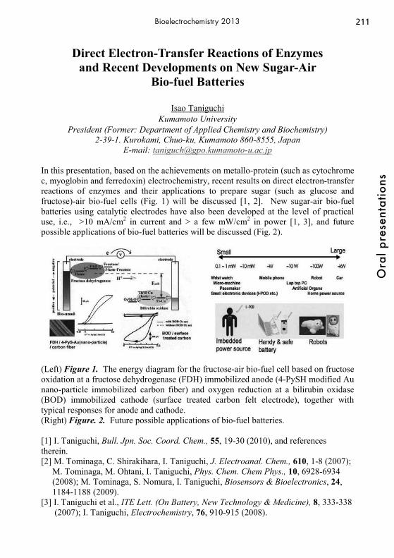
211
Ora
l pre
sent
atio
ns
Bioelectrochemistry 2013
Direct Electron-Transfer Reactions of Enzymes and Recent Developments on New Sugar-Air
Bio-fuel Batteries
Isao Taniguchi Kumamoto University
President (Former: Department of Applied Chemistry and Biochemistry) 2-39-1. Kurokami, Chuo-ku, Kumamoto 860-8555, Japan
E-mail: [email protected]
In this presentation, based on the achievements on metallo-protein (such as cytochrome c, myoglobin and ferredoxin) electrochemistry, recent results on direct electron-transfer reactions of enzymes and their applications to prepare sugar (such as glucose and fructose)-air bio-fuel cells (Fig. 1) will be discussed [1, 2]. New sugar-air bio-fuel batteries using catalytic electrodes have also been developed at the level of practical use, i.e., >10 mA/cm2 in current and > a few mW/cm2 in power [1, 3], and future possible applications of bio-fuel batteries will be discussed (Fig. 2).
(Left) Figure 1. The energy diagram for the fructose-air bio-fuel cell based on fructose oxidation at a fructose dehydrogenase (FDH) immobilized anode (4-PySH modified Au nano-particle immobilized carbon fiber) and oxygen reduction at a bilirubin oxidase (BOD) immobilized cathode (surface treated carbon felt electrode), together with typical responses for anode and cathode. (Right) Figure. 2. Future possible applications of bio-fuel batteries. [1] I. Taniguchi, Bull. Jpn. Soc. Coord. Chem., 55, 19-30 (2010), and references therein. [2] M. Tominaga, C. Shirakihara, I. Taniguchi, J. Electroanal. Chem., 610, 1-8 (2007); M. Tominaga, M. Ohtani, I. Taniguchi, Phys. Chem. Chem Phys., 10, 6928-6934 (2008); M. Tominaga, S. Nomura, I. Taniguchi, Biosensors & Bioelectronics, 24, 1184-1188 (2009). [3] I. Taniguchi et al., ITE Lett. (On Battery, New Technology & Medicine), 8, 333-338 (2007); I. Taniguchi, Electrochemistry, 76, 910-915 (2008).
212O
ral p
rese
ntat
ions
Bioelectrochemistry 2013
High-Intensity, Ultra-Short Pulsed Electric Field Exposure Initiates PIP2 Hydrolysis and Actin Cytoskeletal
Cortex Remodeling Gleb P. Tolstykh1, Hope T. Beier1, Marjorie A. Kuipers3, Gary L. Thompson1, Caleb C
Roth2, and Bennett L. Ibey3 1) National Research Council, Fort Sam Houston, TX, USA
2) General Dynamics Information Technology, Fort Sam Houston, TX, USA 3) Radio Frequency Bioeffects Branch, Human Effectiveness Directorate, Air
Force Research Laboratory, Fort Sam Houston, TX, USA Acute cell swelling and blebbing following exposure to nanosecond pulsed electric fields (nsPEF) has been reported [1,2]. We hypothesize that nsPEF-induced phosphatidylinositol-4,5-bisphosphate (PIP2) hydrolysis may initiate localized and transient remodeling of the actin filaments, which may be responsible for these morphological changes. PIP2 maintains normal mammalian cell homeostasis and anchors the proteins responsible for coupling of actin to the plasma membrane [3]. PIP2 is also heavily involved in phagocytosis, exocytosis, endocytosis, regulation of membrane ion channels and trafficking, cell osmoregulation and activation of intracellular enzymes [4-6]. By using pleckstrin homology (PH) domain of phospholipase C delta (PLCδ) fused with green fluorescent protein (EGFP) for visualization in the cell [7], we demonstrated that nsPEF exposure initiates a complex, intracellular phosphoinositide signaling pathway, which causes the activation of the protein kinase C (PKC) and reduction of PIP2 levels. We then transfected PLCδ-PH-EGFP into Chinese hamster ovary cells that stably expressed m-Apple-actin to demonstrate a correlation between PIP2 hydrolysis/depletion and actin cytoskeletal cortex dissociation after nsPEF exposure. We found that PIP2 hydrolysis and actin cortex dissociation was dependent on the nsPEF exposure dose. Thus, we believe that PIP2 hydrolysis is the origin of the well-documented cellular changes observed after nsPEF exposure. Acknowledgments: This work was supported by a grant from Air Force Office of Scientific Research (AFOSR-LRIR #09RH09COR) and was performed while authors (GPT, HTB, GLT) held a National Research Council Research Associate Award at the US Air Force Research Laboratory. References 1. Rassokhin, M.A. and A.G. Pakhomov, Electric field exposure triggers and guides
formation of pseudopod-like blebs in U937 monocytes. J Membr Biol, 2012. 245(9): p. 521-9.
2. Pakhomov, A.G., et al., Lipid nanopores can form a stable, ion channel-like conduction pathway in cell membrane. Biochem Biophys Res Commun, 2009. 385(2): p. 181-6.
3. McLaughlin, S., et al., PIP(2) and proteins: interactions, organization, and information flow. Annu Rev Biophys Biomol Struct, 2002. 31: p. 151-75.
4. Gamper, N. and M.S. Shapiro, Regulation of ion transport proteins by membrane phosphoinositides. Nat Rev Neurosci, 2007. 8(12): p. 921-34.
5. Gamper, N., Osmosensitivity through the PIP2 availability: just add water. J Physiol, 2010. 588(Pt 19): p. 3631-2.
6. Botelho, R.J., et al., Localized biphasic changes in phosphatidylinositol-4,5-bisphosphate at sites of phagocytosis. J Cell Biol, 2000. 151(7): p. 1353-68.
7. Horowitz, L.F., et al., Phospholipase C in living cells: activation, inhibition, Ca2+ requirement, and regulation of M current. J Gen Physiol, 2005. 126(3): p. 243-62.

213
Ora
l pre
sent
atio
ns
Bioelectrochemistry 2013
Electrochemical Lab-on-Chip Microsystems for Biomarker Analysis
Gerald A. Urban Laboratory for Sensors, Department of Microsystems Engineering (IMTEK),
University Freiburg, Germany Georges Koehler Allee 103
79110 Freiburg [email protected]
Micro- and Nanotechnology are actually entering life science and medical applications. Especially Lab-on-Chip and bioanalytical systems have attracted a lot of emphasis during the last years. Additionally nanoparticular and nanostructured materials in general demonstrate a high potential not only for drug delivery but also for the integration into fast, specific and highly sensitive analytical systems. This is proved by the recent advances in next generation sequencing devices as well as minimal invasive metabolic sensors. Microminiaturized flexible biochips were produced for in vivo applications as well as for cell based analysis especially of ROS, NO and HNO detection in tumor cell cultures. Nanomodification of electrodes exhibit sensitive and selective measurements of such species. Additionally microfluidic and microtechnological systems on chip exhibit the potential of decentralized pathogen detection. By using sophisticated microfluidics with integration of sensing and actuating systems completely detection systems based on RNA detection with implemented pathogen enrichment, bacteria lysis, purification and isothermal amplification modules can be realized. The innovative use of phase guides in the microfluidic chip enables the bubble free filling of analyt as well as gels for electrophoretic separation porpuses. Additionally they allow passive mixing which is important for mobile diagnostic devices. Their use and suitability to be integrated into highly-sensitive bioanalytical tests was demonstrated so far with gram negative and gram positive bacteria.
Fig.1 Lab-on-Chip device for bacteria detection

214O
ral p
rese
ntat
ions
Bioelectrochemistry 2013
Intrinsic Electroactivity of Membrane Proteins: Initial Findings and Future Prospects
Jan Vacek
Department of Medical Chemistry and Biochemistry, Faculty of Medicine and Dentistry, Palacky University, Hnevotinska 3, 775 15 Olomouc, Czech Republic
Membrane proteins take part in cell events such as transport of molecules via membranes, signal transduction and cell-cell interactions. The proteins associated with membranes comprise around 30 % of the human proteome. For this reason, new methods for the analysis of trace amounts of membrane proteins and study of their structure and interactions are being sought (1). Recently, it has been shown that the intrinsic electroactivity of membrane proteins can be studied after solubilization using non-ionic detergents such as C12E8 (2). For electrochemical analysis, chronopotentiometric and voltammetric techniques with mercury and carbon electrodes have been used under conditions similar to those usually used for water soluble (plasmatic) proteins. This approach (2) has enabled us to observe the intrinsic electroactivity of membrane proteins for the first time. For initial electrochemical studies, the model (trans)membrane protein (Na+/K+-ATPase) was used. The model selection was based on X-ray crystallography and NMR data and on a broad spectrum of physico-chemical parameters which are available for Na+/K+-ATPase (2). In addition to redox and electrocatalytic properties, Na+/K+-ATPase interactions with drugs (e.g. platinum cytostatics) and structural changes after ATP binding were studied (3). The methodology presented here is opening new areas in membrane protein research and the development of new electrochemical tools (sensors) for the study of molecular interactions and structural changes of membrane proteins. All these aspects are described in this contribution, including future prospects for research of intrinsic electroactivity of membrane proteins and its applications in biomedicine. References: (1) M.H.Y. Lam, I. Stagljar, Proteomics, 2012, 12, 1519. (2) M. Zatloukalova, E. Orolinova, M. Kubala, J. Hrbac, J. Vacek, Electroanalysis,
2012, 24, 1758. (3) M. Huliciak, J. Vacek, M. Sebela, E. Orolinova, J. Znaleziona, M. Havlikova, M.
Kubala, Biochem. Pharmacol., 2012, 83, 1507. Acknowledgement: This work was supported by the Palacky University, project No.
LF_2012_010.
215
Ora
l pre
sent
atio
ns
Bioelectrochemistry 2013
Microbial Electrolysis Cells for Production of Methane from CO2
Mieke C. A. A. van Eerten-Jansen, Annemiek ter Heijne, Cees J. N. Buisman, Hubertus
V. M. Hamelers Sub-department of Environmental Technology – Wageningen University
Bornse Weilanden 9, P.O. Box 17, 6700 AA, Wageningen, The Netherlands [email protected]
A methane-producing microbial electrolysis cell (MEC) is a technology to convert CO2 into methane, using electricity as an energy source and microorganisms as the catalyst. A methane-producing MEC provides the possibility to increase the fuel yield per hectare of land area, when the CO2 produced in biofuel production processes is converted to additional fuel methane. Besides increasing fuel yield per hectare of land area, this also results in more efficient use of land area, water, and nutrients. In this research, the performance of a methane-producing MEC was studied for 188 days in a flat-plate MEC design. Methane production rate and energy efficiency of the methane-producing MEC were investigated with time to elucidate the main bottlenecks limiting system performance. When using water as the electron donor at the anode during continuous operation, methane production rate was 6 L CH4/m3 per day at a cathode potential of -0.55V vs. normal hydrogen electrode with a coulombic efficiency of 23.1%. External electrical energy input was 73.5 kWh/m3 methane, resulting in a voltage efficiency of 13.4%. Consequently, overall energy efficiency was 3.1%. The maximum achieved energy efficiency was obtained in a yield test and was 51.3%. Analysis of internal resistance showed that in the short term, cathode and anode losses were dominant, but with time, also pH gradient and transport losses became more important. The results obtained in this study are used to discuss the possible contribution of methane-producing MECs to increase the fuel yield per hectare of land area.

216O
ral p
rese
ntat
ions
Bioelectrochemistry 2013
Fluorescence Spectroscopy for the Visualization of the Enzyme Distribution on Enzymatic Fuel Cell Electrodes
Miroslava Varničić*, Kai Sundmacher*,**, Tanja Vidaković-Koch** *Max-Planck-Institute for Dynamics of Complex Technical Systems
Sandtorstrasse 1, 39106, Magdeburg, Germany ** Otto-von-Guericke University Magdeburg
Universitätsplatz 2, 39106, Magdeburg, Germany E-mail address: [email protected]
Broader applications of redox enzymes as catalysts in bioelectrochemical systems like enzymatic fuel cells require significant increase of the catalytic current per geometrical surface area of the electrode. This goal can be possibly achieved by improvement of the electrode structure, for example by introduction of high surface area materials, resulting in 3-D electrodes [1]. This 3D structuring introduces various materials into electrode design, like enzymes (as catalytic elements), additives (like hydrogels for enhancing enzyme stability) and carbon nanomaterials (to increase active surface area per unit electrode volume and to enable electron conductivity). These components are commonly self-organized in the catalyst layer and their dispersion is unknown. For the optimal design of enzymatic electrodes, the visualization of the electrode structure and especially enzyme distribution in the catalyst layer is very important. Enzymes are protein structures and some of them, e.g. flavin enzymes (FAD), are fluorescent [2]. If enzymes lack its natural fluorescence they can be conveniently modified with a suitable fluorescence marker. This fluorescence can be further utilized to monitor the enzyme distribution in the catalyst layer. In this contribution we demonstrate for the first time the application of fluorescence spectroscopy for visualization of the enzyme dispersion in the catalyst layer. Horseradish peroxidase, isolated from Amoracia rusticana (EC 1.11.1.7) was chosen as model enzyme.
0.0 0.2 0.4 0.6
-24
-20
-16
-12
-8
-4
0
j / μ
A c
m-2
E / V vs. SCE
5% GA 3% GA 1% GA 0% GA
A)
B)
C)
Fig.1 Influence of the degree of enzyme cross-linking on electrode performance (A). Enzyme distribution visualized by fluorescence spectroscopy without (B) and with (C) cross linking. References: [1] I. Ivanov, T. Vidakovic-Koch and K. Sundmacher, J. Power Sources 196 (2011) 9260-9269. [2] H. Peter Lu, Luying Xun, X. Sunney Xie, Science 282 (1998) 1877-1882.

217
Ora
l pre
sent
atio
ns
Bioelectrochemistry 2013
Enriched diesel fed microbial fuel cell systems for enhanced remediation of petroleum hydrocarbon
contaminants Krishnaveni Venkidusamy, Mallavarapu Megharaj, Robin Lockington and Ravi Naidu Affiliations: Centre for Environmental Risk Assessment and Remediation (CERAR) and
CRC-Contamination Assessment and Remediation of the Environment (CRC CARE), Australia. University of South Australia, Mawson lakes, SA-5095.
Petroleum contamination of soil and ground water is a widespread and well recognized global environmental issue. Concerns about petroleum hydrocarbon contamination (PHC) and its potential risks to environment and human health have led to much research towards the development of numerous remediation technologies. An emerging green bioelectrochemical remediation (BER) technology, using microbial fuel cells (MFCs) which exploits the bio catalytic potential of electrochemically active microorganisms for the treatment of recalcitrant compounds while generating electricity in the process promises to be an effective approach to remediating PHC. Recent research shows that enriched biofilms result in excellent performance in terms of substrate degradation and increased electricity generation but their effects on hydrocarbon contaminant degradation is unknown. For this reason, we investigate the relationship between enriched biofilm anodes (EAMFC) and freshly inoculated new anodes (NAMFC) in diesel fed single chamber mediatorless microbial fuel cells (DMFC) using various techniques for the enhancement of PHC remediation with concomitant energy recovery. An anodophilic microbial consortium of an enriched MFC with a diesel concentration of about 800 mg/l exhibited complete disappearance of the diesel within 30 days whereas a freshly inoculated new anode MFC (NAMFC) showed only 83.4%diesel removal. The simultaneous voltage generation was 710±4mV and 450±5mV respectively. The behavior of enriched anodes at a higher concentration of diesel (8000 mg/l) was also studied. Scanning electron micrograph (SEM) revealed the formation of a thick covered biofilm on the enriched anodic electrode. High resolution imaging showed the presence of thin 55 nm diameter nano filaments emanating from the cells attached on the anodic surface. Anodic microbial community profiling confirmed that enrichment of diesel degrading exoelectrogenic bacteria had occurred.The most significant shift in the enriched anodic biofilm occurred within the obligate anaerobic bacteria such as Clostridia sp, Desulfosporosinus sp, whereas the NAMFC was dominated by an aerobic and facultative denitrifying population of bacteria related to Pseudomonas. Thus, quite a spectacular difference was seen between the bacterial communities of the two electrodes, suggesting that the thick bacterial film that developed on the enriched electrode favored the dominance of obligate anaerobes. Given its remarkable potential for biodegradation and electrochemical activity, a long term enrichment of the anodic electrode would seem to have considerable potential for bioagumentation operations using MFC in diesel contaminated sites. Further, identification of a biodegradative gene (alk B) involved in alkane utilization provided strong evidence of the catabolic pathway being used for diesel degradation in the DMFCs.

218O
ral p
rese
ntat
ions
Bioelectrochemistry 2013
Electric Field Enhancement of the Water-Driven, Permeabilizing Reorganization of Phospholipid Bilayers
P. Thomas Vernier1 and Mayya Tokman2
1Frank Reidy Research Center for Bioelectrics, Old Dominion University Norfolk, VA USA
2School of Natural Sciences, University of California, Merced Merced, CA USA
[email protected], [email protected] Despite widespread use of electric fields in biomedicine and biotechnology, and ongoing concerns among health practitioners, epidemiologists, regulatory bodies, and the general public regarding the possible harmful effects of exposure to low-level electric and magnetic fields, only a few plausible mechanisms for producing biologically significant changes in cells with low doses of electromagnetic energy have been proposed, and these remain insufficiently studied and poorly characterized. However extensive, our present largely phenomenological level of understanding can provide only a rough guide and is predictive only to a limited extent. One unambiguous biological target of electric field exposure is the lipid bilayer that forms the fabric of cellular membranes. Although the biological membrane is a thoroughly studied biomolecular assembly, the interactions of lipid bilayers and external electric fields have only recently been subjected to detailed analysis at the molecular level. Molecular dynamics (MD) simulations in particular have begun to shed light on the mechanism of electric-field-induced membrane permeabilization (electroporation), and the conclusions of these studies are consistent with larger-scale analytical models and experiments. Electropermeabilization is not normally considered a low-dose or low-field process, but molecular and continuum models both suggest that the electric field simply decreases the energy barrier for pore formation, and that nanoscale permeabilization events occur with lower fields, albeit at reduced frequencies and densities. Careful physical characterization of this process, including the identification of factors that enhance or inhibit pore formation, may for this reason contribute to our understanding of what is possible and what is likely to occur at doses below those normally used in electroporation protocols. We describe here the role of water in electropore creation, stabilization, and annihilation, as revealed in MD simulations. We argue from the standpoint of a detailed energetics analysis that the electric field-driven poration of a lipid bilayer is fundamentally an energy-minimizing reorganization of water at a low-permittivity interface. The rearrangement of phospholipids into what is known as a hydrophilic pore is a secondary process that follows the formation of a membrane-spanning water column and further lowers the energy of the system.
219
Ora
l pre
sent
atio
ns
Bioelectrochemistry 2013
Fundamental Studies of Charge Transport between Biomolecules and Electrodes
David H. Waldeck
Chemistry Department, University of Pittsburgh 219 Parkman Avenue, Pittsburgh PA 15260
[email protected] We report on the nature and mechanism of charge transfer between biomolecules and electrodes. A particular focus of the talk will be our most recent work on Peptide Nucleic Acid (PNA), a synthetic analog of DNA. We will compare the conductivity of single PNA oligonucleotides trapped within molecular junctions, formed using Scanning Probe Spectroscopy techniques, and the charge transfer properties of self-assembled monolayers of PNA duplexes. We will discuss how these different measurements investigate different aspects of the electron transfer. Although simple models predict a linear correlation between the molecular conductance and the charge-transfer rates, the experimental data show a power-law relationship within a specific class of structures, and a lack of correlation when a more diverse group of molecules are compared. We describe a recent theoretical model that can account for these differences by including variations in the energy barrier heights for charge transport and the bath-induced electronic decoherence experienced by the molecules in the two different measurements, STM-BJ and electrochemical rates.
220O
ral p
rese
ntat
ions
Bioelectrochemistry 2013
Electrochemical Control of the Adsorption of Surface Layer Proteins on Gold:
in-situ FTIR and SPM Investigations
Thomas Werzer1, Christian Zafiu2, Guenter Trettenhahn1, Uwe B. Sleytr2, Wolfgang Kautek*1
1University of Vienna, Department of Physical Chemistry Waehringer Strasse 42, A-1090, Vienna, Austria
2University of Natural Resources and Applied Life Sciences (BOKU) Centre for NanoBiotechnology, Muthgasse 11, A-1190, Vienna, Austria
Surface layer proteins form regular two-dimensional crystalline arrays in the nanometer regime (S-layers) on solid substrates by self-assembling singular protein units that are cross-linked with calcium ions [1]. Such layers have been successfully used in biotechnology [1], biomineralization [2,3] and biosensorics [4,5]. An outstanding feature of the S-layers is that their assembly and morphology can be electrochemically controlled and manipulated [6-9]. In situ experiments showed that adsorbed chaotropic anions in the electrochemical Helmholtz layer can be displaced by the proteins of the crystalline monolayer in a particular potential region [8,9]. On a negatively charged surface, a protein bilayer with different morphology was caused by strongly solvated chaotropic cations. In this context the influence of chaotropic and cosmotropic ions on the dynamics and the mechanism of the adsorption process of S-layer proteins of Lysinibacillus sphaericus CCM2177 were investigated by electrochemical in-situ techniques. [1] U.B. Sleytr, P. Messner, D. Pum, M. Sara, Angew. Chem. 38 (1999) 1035. [2] S. Dieluweit, D. Pum, U. B. Sleytr, Supramol. Sci. 5 (1998) 15. [3] S. Dieluweit, D. Pum, U.B. Sleytr, W. Kautek,
Materials Science and Engineering C 25 (2005) 727. [4] A. Neubauer, S. Pentzien, S. Reetz, W. Kautek, D. Pum, U. B. Sleytr,
Sens. Actuators B 40 (1997) 231. [5] D. Pum, A. Neubauer, U. B. Sleytr, S. Pentzien, S. Reetz, W. Kautek,
Ber. Bunsenges. Phys. Chem. 101 (1997) 1686. [6] M. Handrea, M. Sahre, A. Neubauer, U. B. Sleytr, W. Kautek,
Bioelectrochemistry 61 (2003) 1. [7] U. B. Sleytr, M. Sara, W. Kautek, EP 1 402 055 B1 (2007). [8] C. Zafiu, G. Trettenhahn, D. Pum, U.B Sleytr, and W. Kautek,
Phys. Chem. Chem. Phys. 13 (2011) 3478. [9] C. Zafiu, G. Trettenhahn, D. Pum, U.B Sleytr, and W. Kautek,
Phys. Chem. Chem. Phys. 13 (2011) 13232.

221
Ora
l pre
sent
atio
ns
Bioelectrochemistry 2013
How Gentle Can a Soft Electrode Array Be?
Gunther Wittstock,1 Andreas Lesch,1,2 Britta Vaske,1 Frank Meiners,1 Dmitry Momotenko,2 Fernando Cortés-Salazar,2 Hubert H. Girault2
1 Carl von Ossietzky University of Oldenburg, Department of Pure and Applied Chemistry D-26111 Oldenburg, Germany
2 Ecole Polytechnique Fédérale de Lausanne, Laboratoire d’Electrochimie Physique et Analytique, CH-1015 Lausanne, Switzerland
Recently, we introduced SECM electrodes that are manufactured in soft, sheet-like plastic foils that can brush surfaces [1]. Those probes have been expanded to individually addressable microelectrode arrays that allow the investigation of large surfaces [2, 3]. Another very appealing possibility in a biochemical context is the integration of microfluidic channels into the probe body allowing the injection of working solution [4, 5] or aspiration of minute sample volumes for simultaneous electrochemical and mass-spectrometric analysis [6]. The possibility to work on larger sample areas within reasonable imaging times holds great potential for biochemical analysis. However, the mechanical contact between the soft probe and the sample are a matter of concern. This contribution will present an estimation of the pressure exerted by soft probes [7] on a sample surface and practical tests for imaging self-assembled monolayers (SAM) on gold. It could be demonstrated that such monolayers can be imaged and surfaces features about 10 μm in size are visible on extended areas [8]. The absence of mechanically induced artifacts can be proven, whereas clear and reproducible chemical modification can be introduced into OEG-terminated SAM [8]. Pristine OEG-SAMs are resistant to protein adsorption and cell adhesion. This property is lifted after local chemical removal of the OEG units. The electrode arrays allow a very flexible and template-free electrochemical patterning of substrates for localized cell adhesion. [1] F. Cortes-Salazar, M. Träuble, F. Li, J.-M. Busnel, A.-L. Gassner, M. Hojeij, G.
Wittstock, H.H. Girault, Anal. Chem. 81 (2009) 6889. [2] F. Cortes-Salazar, D. Momotenko, H.H. Girault, A. Lesch, G. Wittstock, Anal.
Chem. 83 (2011) 1493. [3] F. Cortes-Salazar, D. Momotenko, A. Lesch, G. Wittstock, H.H. Girault, Anal.
Chem. 82 (2010) 10037. [4] D. Momotenko, F. Cortes-Salazar, A. Lesch, G. Wittstock, H.H. Girault, Anal.
Chem. 83 (2011) 5275. [5] F. Cortes-Salazar, A. Lesch, D. Momotenko, J.-M. Busnel, G. Wittstock, H.H.
Girault, Anal. Meth. 2 (2010) 817. [6] D. Momotenko, L. Qiao, F. Cortés-Salazar, A. Lesch, G. Wittstock, H.H. Girault,
Anal. Chem. 84 (2012) 6630. [7] A. Lesch, D. Momotenko, F. Cortes-Salazar, I. Wirth, U.M. Tefashe, F. Meiners,
B. Vaske, H.H. Girault, G. Wittstock, J. Electroanal. Chem. 666 (2012) 52. [8] A. Lesch, B. Vaske, F. Meiners, D. Momotenko, F. Cortes-Salazar, H.H. Girault,
G. Wittstock, Angew. Chem. Int. Ed. 51 (2012) 10413.
222O
ral p
rese
ntat
ions
Bioelectrochemistry 2013
Efficient bioelectrocatalysis of sulfite oxidase
U. Wollenberger, S. Frasca, T. Zeng, O. Rojas, J. Koetz , S. Leimkühler
University Potsdam Karl-Liebknecht Strasse 24-25, 14476 Golm, Germany
The presentation will cover attempts to achieve electrical communication between human sulfite oxidase (hSO) and electrodes, including surface immobilization, heterogeneous electron transfer of the domains, investigations of the catalytic behaviour of SO at electrode interfaces and biosensor applications. Sulfite oxidases (SO) from eukaryotic organisms are interesting enzymes for several reasons. They catalyze the oxidation of a small molecule, sulfite, to sulfate. The catalytic reaction takes place at the molybdenum containing catalytic domain. In the SO from vertebrates this catalytic domain is covalently linked by a flexible protein tether to the electron transfer domain Cyt b5. The latter transfers in the case of the human SO the electrons to Cyt c. Furthermore, the reduction potentials of the cofactors are low and the heme shows only a very slow reoxidation under ambient conditions. These properties are of particular importance from the point of practical application. Cyt c may be also used as electron shuttle to an electrode [1] or may be replaced by a chemically modified electrode[2], where self-assembled monolayers provide a charged surface for oriented adsorbtion of the enzyme. Recently we were able to further enhance the direct heterogeneous electron transfer after introduction of PEI capped gold nanoparticles [3]. For hSO at those surfaces the electron transfer to the catalytic Moco proceeds through the Cyt b5-domain and requires a domain motion for intramolecular electron transfer. Therefore the catalysis is strongly dependent on pH, ionic strength, and viscosity. With the nanoparticles a surface is provided for productive orientation of the enzyme, fast electron transfer to the Cyt b5 while retaining domain mobility. We will show the influence of the coupling procedure, type and size of nanoparticle on the efficiency of the bioelectrocatalysis and will also discuss alternative surfaces and binding strategies. [1] R. Spricigo, R. Dronov, K. V. Rajagopalan, F. Lisdat, S. Leimkühler, F. W. Scheller, U. Wollenberger, Soft Matter, 2008, 4, 972. [2] E. E. Ferapontova, T. Ruzgas, L. Gorton, Anal. Chem., 2003, 75, 4841; M. Sezer, R. Spricigo, T. Utesch, D. Millo, S. Leimkühler, M. A. Mroginski, U. Wollenberger, P. Hildebrandt, I. M. Weidinger Phys Chem Chem Phys., 2010, 12, 7894-7903. [3] S. Frasca, O. Rojas, J. Salewski, B. Neumann, K. Stiba, I.M. Weidinger, et al., Human sulfite oxidase electrochemistry on gold nanoparticles modified electrode, Bioelectrochemistry, 2012, 87, 33-41 Financial support by the Deutsche Forschungsgemeinschaft (Unicat Excellenzcluster), the BMBF (Taschentuchlabor) is gratefully acknowledged.

223
Ora
l pre
sent
atio
ns
Bioelectrochemistry 2013
Further insights into the catalytical properties of deglycosylated pyranose dehydrogenase from Agaricus meleagris recombinantly expressed in Pichia pastoris
Maria Yakovleva1, Anikó Killyéni1,2, Ionel Catalin Popescu2, Clemens K. Peterbauer3,
Lo Gorton1 Department of Biochemistry and Structural Biology, Lund University
PO Box 124, 221 00 Lund, Sweden Department of Physical Chemistry, Babes-Bolyai University, Cluj-Napoca, Romania
Division of Food Biotechnology, Department of Food Sciences and Technology, BOKU-University of Natural Resources and Applied Life Sciences, Muthgasse 18, A-
1190 Wien, Austria [email protected]
Pyranose dehydrogenase (PDH) belongs to the class of oxidoreductases, which can mono- and dioxidise a variety of sugars. PDH has no anomeric specificity and does not show any catalytic activity towards molecular oxygen, which makes it attractive for application in analytical chemistry, industrial production of rare sugars, and construction of biofuel cells [1]. In the present work the catalytical properties of glycosylated (gPDH) and deglycosylated PDH (dgPDH) from Agaricus meleagris recombinantly expressed in Pichia pastoris were studied. It was shown that further decomposition of dgPDH takes place depending on the storage conditions. Complete loss of an approximately 20 kDa fragment from the C-terminal was observed after 2.5 months storage at 4 °C resulting in formation of a stable decomposed deglycosylated PDH (ddgPDH). The decomposition procedure was followed by gel electrophoresis (SDS-PAGE) and enzyme activity measurements (ferrocenium). The corresponding molecular masses were determined to be ~93 kDa, ~65 kDa and ~45 kDa for gPDH, dgPDH and ddgPDH, respectively. An increase in activity from 370 U/mL to 2248 U/mL for ddgPDH compared to dgPDH (stored at -20 °C) was observed in 2.5 months. The effect of decomposition on the catalytic properties of ddgPDH formed was investigated using electrochemical measurements. All three forms of the enzyme were electrically “wired” to an osmium based redox polymer on the surface of graphite electrodes mounted into a flow-injection system (FIA). The current produced from oxidation of glucose by immobilised gPDH, dgPDH and ddgPDH was compared using FIA and cyclic voltammetry. A dramatic increase in the current density was observed when using ddgPDH (230 μA cm-2) compared with that for dgPDH (33 μA cm-2) and gPDH (19 μA cm-2) most likely due to the better intrinsic electron communication between the FAD containing active site of the smaller enzyme and the electrode surface. Additionally, the glycosylation sites of the enzyme were assigned using MALDI-MS in combination with tryptic digestion. References [1] C.K. Peterbauer, J. Volc, Appl. Microbiol. Biotechnol., 85 (2010) 837.

224O
ral p
rese
ntat
ions
Bioelectrochemistry 2013
Enzyme/MIP Architecture in a Novel Bio(mimetic)sensor
Aysu Yarman and Frieder W. Scheller Institute of Biochemistry and Biology, University of Potsdam, Karl-Liebknecht-Str. 24-
25, 14476 Golm, Germany [email protected]
Molecularly imprinted polymers (MIP) have been expected to substitute antibodies and enzymes in bioanalysis and chromatography. However, affinity and catalytic activity of MIPs are in general well below of the biological counterparts. Hybrids which are composed of a biocatalyst and MIP may combine the advantages of both components. But up to now only a few successful examples have been realised: Enzymes have been used as labels in MIP-based binding assays and the integration of the catalytic conversion of the analyte can directly generate an electrode-active product in MIP-based sensors. We applied for the first time a minienzyme-Microperoxidase-11 (MP-11) but also horseradish peroxidase (HRP) as a catalyst in combination with a MIP in order to eliminate the effect of interferences on drug indicating electrode. In the first step, the glassy carbon electrode was covered with a MIP layer which was formed by electropolymerisation of an o-phenylenediamine and resorcinol mixture in the presence of the target. As a model, the metabolite (aminoantipyrine) of the antipyretic drug aminopyrine was used. The rebinding of the analyte was clearly indicated by the signal suppression for the redox probe ferricyanide. Furthermore, the oxidation currents for aminoantipyrine on the imprinted electrode were 4.96-fold higher than the signal for ascorbic acid and 2.39-fold higher than that for uric acid. In the next step, we casted an MP-11/HRP containing chitosan capped gold nanoparticle mixture on top of the MIP layer. The peroxide dependent conversion of aminopyrine by MP-11 or HRP took place on top of a product-imprinted electropolymer on the indicator electrode. This arrangement resulted in the elimination of interfering signals by ascorbic acid and uric acid. The cathodic current of the MP-11/MIP electrode was linearly dependent up to 13μM on stepwise addition of aminopyrine. Application of HRP allowed the addition of 5-fold higher peroxide concentration as compared with MP-11. This resulted in an extended linear range up to 110 μM. Furthermore, the sensitivity of the HRP-based sensor was higher due to its higher catalytic activity. The analytical parameters for our hierarchical enzyme/MIP sensor compete very well with that for catalytic MIPs in the literature. Recently, we started to apply the same architecture to other drugs such as tamoxifen and paracetamol. In future we will include cytochrome P450 enzymes which metabolises almost 90% of the drugs which are applied today. Acknowledgements The authors gratefully acknowledge the financial support of BMBF and the Cluster of Excellence UniCAT.
225
Ora
l pre
sent
atio
ns
Bioelectrochemistry 2013
Electrochemical Assay on Osteoblastic Cells on a Sensor Chip
C. Yildirim1, M. Adamovski1, H. Leonhardt2, M. Gerhardt2, C. Beta2,
D. Benayahu3, U. Wollenberger1
1 Inst. of Biochemistry and Biology, University of Potsdam, Germany und 2 Inst. of Physics and Astronomy, University of Potsdam, Germany
3 Cell and Developmental Biology, Tel Aviv University, Israel [email protected]
Whole cells have the ability to present the intact sub-cellular machinery for the identification of a physiologically relevant event, and provide functional and analytical information [1]. Moreover biosensors incorporating mammalian cells have a distinct advantage of responding in a manner that can help to determine the physiological effect of a particular substance for better biological understanding and biomedical applications [2]. The behavior of the cells on the electrode surface is important and challenging point for such sensors. In our study we used mouse bone marrow stromal cell line (MBA-15), which exhibits osteoblastic phenotype in vitro [3]. We examined osteoblastic cells on the electrode surface by using electrochemical impedance spectroscopy and microscopy techniques. Furthermore, voltammetry was applied for an electrochemical enzyme assay to monitor differentiation of cells. Alkaline phosphatase (3.1.3.1, ALK-P) is one of the early differentiation markers in osteoblastic cell differentiation. In this study we demonstrate measurement of ALK-P activity on the sensor chip based on p-aminophenol oxidation and compare it with measurement of p-nitrophenol formation at 410 nm in spectrophotometer. In this case there is no need to disrupt the cells. This makes the online measurements with cells on the chips possible. We also examined the effect of changes in redox conditions on the ALK-P activity by using oxidizing and reducing agents.
[1] Ding et al. (2008), Curr Med Chem.; 15(30):3160-70. [2] Pancrazio et al. (1999), Annals of Biomedical Engineering; 27:697–711. [3] Benayahu et al. (1989) Journal of Cellular Physiology; 140(1):1–7

226O
ral p
rese
ntat
ions
Bioelectrochemistry 2013
Optimizing the Reactivity of Surface Confined Cobalt N4-Macrocyclics for the Electrocatalytic Oxidation of L-
cysteine by modulating the Co(II)/(I) formal potential of the catalyst
Miguel A. Gulppi, Gonzalo Ochoa, Maritza A.Páez, Jorge Pavez, José H. Zagal* Facultad de Química y Biología, Departamento de Química de los Materiales,
Universidad de Santiago de Chile, Casilla 40, Correo 33, Santiago 9170022, Chile. [email protected]
The redox potential of macrocyclic MN4 complexes confined on electrode surfaces is a very predictive reactivity index for their electrocatalytic activity for many reactions [1]. On the other hand, L-cysteine (R-SH) is a semi-essential amino acid, biosynthesized in humans.The thiol side chain in cysteine –SH participates in many enzymatic reactions, acting as a nucleophile. The thiol group (-SH) is susceptible to oxidization to give the disulfide derivative L-cystine R-SSR , which plays a very important structural role in many proteins. It is also used as a food additive. So its electrochemical detection in biological fluids and food samples provides a fast, reliable way of chemical analysis. Electrochemical sensors of this type can also be used coupled to chromatography [2a]. An screen printed electrodes using Co phthalocyanine and carbon have been developed for the detection of L-cysteine [2b,c]. In this work we have investigated the effect of the Co(II)/(I) formal potential on the catalytic activity of a series of a series of Co porphyrins, Co phthalocyanines and vitamin B12 (aquocobalamine) for the electrooxidation of L-cysteine. We have modulated the Co(II)/(I) by using electron-withdrawing or electron-donating groups located on the porphyrin or phthalocyanine ligand. A correlation of log I (at constant electrode potential) versus the Co(II)/(I) formal potential of the catalysts gives an asymmetrical parabolic correlation. This clearly shows that the search for better catalysts for this reaction point to those N4-macrocyclic complexes with Co(II)/(I) formal potentials around -1.0 V vs. SCE which correspond to an optimum situation for the interaction of the L-cysteine molecule with the Co center in the macrocyclic complex present on the OPG surface. Acknowledgements Work was supported by Fondecyt Project 1100773 and by Nucleo Milenio de Ingenieria Molecular P07- 006-F. References 1. (a)J.H. Zagal, S.Griveau S, J.F. Silva JF, T.Nyokong, F. Bedioui, Coord Chem Rev. 254 (2011) 2755; (b) J. Masa, K. Ozoemena, W. Schuhmann, J. H. Zagal J. Porphyrins Phthalocyanines (c) J. Masa, K. Ozoemena, W. Schuhmann,J. H. Zagal in “Electrocatalysis in Fuel Cells: A Non and Low Platinum Approach” M. Shao (ed) Springer, New York (in press) 2. X. Qi X, R.P. Baldwin, Electroanalysis 6 (1994) 353 (b) S.A.Wring, J.P.Hart, B.Birch B (1992) Electroanalysis 4 (1992) 229 (c) S.A. Wring, J.P. Hart, Analyst 117 (1992) 1281
227
Ora
l pre
sent
atio
ns
Bioelectrochemistry 2013
Electrochemically-assisted deposition of chitosan and sol-gel enzyme bio-composites for microfluidic biofuel cell
applications
Monika Żygowska1, Veronika Urbanova2,3, Mathieu Etienne2,3, Vladimir Ogurtsov1 and Gregoire Herzog2,3
1Tyndall National Institute, University College Cork, ‘Lee Maltings’, Cork, Ireland.
2Université de Lorraine, LCPME, UMR 7564, Villers-lès-Nancy, F-54600, France 3CNRS, LCPME, UMR 7564, Villers-lès-Nancy, F-54600, France
The ever-growing demand for miniaturized energy sources has triggered much research on harvesting energy from the surrounding environment. Enzymatic biofuel cells are promising candidates for small scale green energy sources [1]. Electrochemically-assisted deposition of biocatalysts enables their precise immobilization in confined space on the electrode surfaces. Encapsulation of biomolecules via electrodeposition in a sol-gel or chitosan matrix has been reported [2]. In this work, we describe the use of both encapsulating materials for dehydrogenase enzymes in the bioconversion of glucose or D-sorbitol on the bioanode. Bilirubin oxidase, electrodeposited on the biocathode, is used to catalyse the oxygen reduction [3]. Mediated electron transfer has been investigated for each enzyme catalyst using metal- and carbon-based electrodes. The catalytic behaviour of these biomolecules has been studied, both individually and in an assembled biofuel cell under static conditions, using buffer solutions containing appropriate redox substrates. Improved electrocatalytic activities of enzyme and mediator modified half cells have been verified in comparison to bare electrodes. Immobilization of selective enzymes in the channels of microfluidic biofuel cells is desirable as it takes advantage of the laminar flow enabling efficient energy conversion without the use of a membrane, simplifying device fabrication [4]. Additional work will focus on the characterization of enzymatic microfluidic biofuel cells in a flowing environment under the conditions of mediated electron transfer, in the presence of glucose/D-sorbitol and oxygen saturated buffer solutions. The authors are grateful to the Environment Protection Agency for funding this research through its STRIVE programme (2009-ET-MS-10-S2). [1] Ivanov, I.; Vidaković-Koch, T.; Sundmacher, K. Energies 2010, 3, 803-846 [2] Wang, Z.; Etienne, M.; Kuhn, A ; Walcarius, A. Electrochim. Acta 2011, 56, 9032-9040 [3] Salaj-Kosla, U.; Schuhmann, W; Magner, E.Electrochem.Commun. 2012, 16, 92-95 [4] Gallaway, J.; Calabrese Barton, S. J. Am. Chem. Soc. 2008, 130, 8527- 8536

In Vivo Cutaneous Electroporation of Human Host Defense Peptide LL-37 Accelerates Wound Healing
Martin C. Lam1,2, Gaëlle Vandermeulen2, Paolo E. Porporato3, Pierre Sonveaux3, Marcus Lehnhardt1, Frank Jacobsen1, Véronique Préat2* and Lars Steinstraesser1*
1 BG University Hospital Bergmannsheil, Department of Plastic Surgery, Burn Center, Ruhr University Bochum, Bürkle-de-la Camp Platz 1, D-44789, Bochum, Germany
2Université catholique de Louvain, Louvain Drug Research Institute, Pharmaceutics and Drug Delivery, Avenue E. Mounier, 73 B1 73 12, B-1200, Brussels, Belgium
3Université catholique de Louvain, Pole of Pharmacology, Medical School, Avenue Emmanuel Mounier 53 box B1.53.09, B-1200, Brussels, Belgium
[email protected] & [email protected] Objective: Viral gene therapy approaches delivering host defense peptide hCAP-18/LL-37 have shown promise in promoting wound healing and inhibiting bacterial growth. Our aim was to investigate if nonviral gene delivery of DNA encoding LL-37 using electroporation can promote wound healing. Methods: Two full-thickness skin wounds were created on the back of non-diabetic C57B6 and diabetic db/db mice using 4mm biopsy-punches. Fifty microgram DNA was intradermally injected in four quadrants of each wound followed by electroporation (EP). In another experiment, hind limb ischemia was achieved in C57B6 and 100 microgram DNA was intradermally injected above the ligation area followed by skin-targeted EP. Blood perfusion was evaluated over 12 days by Laser-Doppler-Imaging and the gastrocnemius muscle weight was compared. Wounds were randomly treated with plasmid pQE-hCAP-18/LL-37 or pQE-eGFP as negative control, both with CMV-promotor. EP was performed by one high- (700V/cm, 100 µs) and one low-voltage (200V/cm, 400 ms) electric pulses, delivered by a Cliniporator system (IGEA, Carpi, Italy) using plate-electrodes. Results: Non-invasive electrogene delivery of LL-37 accelerated reepithelialization of nondiabetic wounds significantly by 14% (p<0.01), and diabetic wounds by 19% (p<0.001) after 12 days, respectively. Cutaneous EP of LL-37 increased blood perfusion in hind limb ischemia by a factor two (85%±7 vs. 42%±11) (p<0.001) and reduced muscular atrophy, showing in average 8,4% higher muscle weight (p<0.05) at day 12. Other antimicrobial peptides tested were less efficient than LL-37 to promote wound healing. Conclusion: We introduced a novel non-viral gene therapeutic technique to success-fully treat acute, diabetic and ischemic wounds with LL-37.
228O
ral p
rese
ntat
ions
Bioelectrochemistry 2013

Abstracts of Poster Presentations
INTER
NATIO
NAL SO
CIETY OF ELECTR
OCHEM
ISTRY•The Bioelectrochemical Society
229
Post
ers
Bioelectrochemistry 2013
S1-001
Electrochemical communication of heterotrophically grown Rhodobacter capsulatus with graphite electrodes via
various polymeric mediators
Kamrul Hasan1, Vera Eßmann2, Kamil Górecki1, Sunil A. Patil1, Wolfgang Schuhmann2, Dónal Leech3, Cecilia Hägerhäll1, Lo Gorton1
1Department of Analytical Chemistry/Biochemistry and Structural Biology, Lund University, P.O. Box 124, SE-22100 Lund, Sweden
2Center for Electrochemical Sciences-CES, Ruhr-Universität Bochum, Universitätsstr. 150, 44780 Bochum, Germany
3School of Chemistry, National University of Ireland Galway, University Road, Galway, Ireland
*Corresponding author: [email protected]
Rhodobacter capsulatus is one of the most metabolically versatile non-sulfur purple bacteria and has the ability to grow under either anaerobic photosynthetic or aerobic dark conditions. Recently, we demonstrated its efficient electrical “wiring” with modified graphite electrodes to investigate the possible applicability in bioelectrochemical systems (BES). The bacterial cells embedded in an osmium redox polymer were able to generate a noticeable bioelectrocatalytic current (4.25 μA/cm²) using 2 mM succinate as substrate [1]. In this work bacterial cells grown under the same conditions as in [1] and a variety of polymeric mediators, such as osmium and dye-modified methacrylate-based redox polymers, differing in their potentials as well as chemical structures are investigated to improve microbial bioelectrocatalysis. In addition, a variety of substrates (e.g. succinate, malate, glucose, citrate, acetate, and glutamate) are considered, where succinate shows the highest current response. [1] Kamrul Hasan, Sunil A. Patil, Kamil Górecki, Donal Leech, Cecilia Hägerhäll, Lo Gorton, Electrochemical communication between heterotrophically grown Rhodobacter capsulatus with electrodes mediated by an osmium redox polymer, Bioelectrochemistry, (2012) in press.
230Po
ster
sBioelectrochemistry 2013

S1-002
Redox responsive regulation of microbial metabolism activity in an iron reducing bacterium
Shuji Nakanishi, Shoichi Matsuda, Kazuhito Hashimoto
RCAST, The University of Tokyo 7-3-1 Hongo, Bunkyo-ku, Tokyo,113-8656, Japan
Members of the genus Shewanella are ubiquitous Gram-negative bacteria and have the unique property of being able to utilize solid-state metal oxides for respiration when soluble electron acceptors are sparse or not accessible in the environment. Extracellular electron transfer by these bacteria, which is thought to be mediated through the expression of abundant outer membrane c-type cytochromes (OMCs), particularly OmcA and MtrC, has been extensively investigated. As the numerous metal oxides used by Shewanella for respiration, including ferrihydrite, hematite ( α -Fe2O3), goethite (α-FeOOH), lepidocrocite (γ-FeOOH), and pyrolusite (β-MnO2), have different energy level of the conduction band, it is worth examining the relationship between metabolism activity and energy level of the electron acceptor for deeper understanding of microbial activities in anaerobic subsurface environments. The metabolism ability can be investigated electrochemically by providing an electrode of an apropriate potential as the sole electron acceptor, which allows the capture of metabolic electrons and reversible tuning of the electron energy level of the solid-state electron acceptor. In the present work, we investigated the dependence of metabolism activity on the acceptor electron energy level using this electrochemical method, which revealed that the Shewanella changes it metabolism activity depending on the redox state of OMCs. In our experiments, a single chamber, three-electrode system was used to monitor the electrochemical behavior of the bacterium Shewanella oneidensis MR-1. A tin-doped In2O3 (ITO) glass substrate was used as the working electrode, which was mounted on the bottom of the reactor, and lactate was used as the sole carbon and electron source. The time courses of microbial electricity generation were observed at various anode potentials. At 0.0 V (vs. SHE), the microbial current continued to increase and reached 6 μA in 24 h. However, at 0.4 V, the microbial current exhibited constant value, approximately around 1 μA. To understand the details of the electrode potential dependence of microbial current generation, we set various kinds of electrode potential and current generation ability was plotted against the electrode potential. The microbial current first appeared at approximately -0.2 V, reached a maximum value at approximately -0.05 V, and then decreased in the potential region more positive than the redox potential of OMC. These results clearly indicated that metabolism activity was high at the negative potential regions where OMCs existed as reduced state, in contrast, metabolism activity was low at the positive potential regions where OMCs existed as oxidized state.
231
Post
ers
Bioelectrochemistry 2013
S1-003
Spectroelectrochemical Investigation on Glucose Oxidase: pH-dependent Determination of the Redox Potential
Stephan Vogt, Gilbert Nöll*
University of Siegen, Nöll Junior Research Group Adolf-Reichwein-Straße 2, 57068 Siegen
Many amperometric biosensors and biofuel cell anodes base on the flavoprotein glucose oxidase, which catalyzes the oxidation of β-D-glucose to δ-gluconolactone. Although more than 20,000 articles have been published about glucose oxidase, the redox potentials of this enzyme at different pH - especially at the physiological pH 7.4 - is still under debate. In the literature values in between -48 mV vs. NHE and -150 mV vs. NHE are mentioned for the pH around 7[1]. We have set up a computer-controlled and miniaturized UV-Vis-spectroelectrochemical cell to determine the redox potentials of different oxidoreductases which may be of interest for applications as catalystsin amperometric biosensors and biofuel cells. Glucose oxidase is investigated as a first model system. We will present UV-Vis-spectroelectrochemical measurements of glucose oxidase at different pH showing UV-Vis-spectra of the stepwise reduction and reoxidation. We could observe the fully oxidized flavoquinone, fully reduced hydroquinone, and singly reduced semiquinone species depending on the pH. The pH-dependent redox potentials of glucose oxidase obtained from titration curves (between pH 5 and 9) will be presented. The measurements were performed at least three times at each pH. [1] E1/2=-360 mV vs. Ag/AgCl (pH 7.2), Heller, A.; Phys. Chem. Chem. Phys., 2004 E1/2=-400 mV vs. SCE (pH 8.0), Willner et al., Angewandte Chemie, 2004 E1/2=-48 mV vs. NHE (pH 7.2), Kulys et al., J. Inorganic Biochemistry, 2006
232Po
ster
sBioelectrochemistry 2013
S2-001
Electrostatically-driven second sphere ligand switch between high and low reorganization energy forms of
native cytochrome c
Álvarez-Paggi D1, Castro MA1, Tortora V2, Radi R2, Murgida DH1
1INQUIMAE (CONICET-UBA) and 2Departamento de Bioquímica, Universidad de la República
Facultad de Ciencias Exactas y Naturales, Universidad de Buenos Aires, Ciudad Universitaria, Pab.2, piso1, C1428EHA-Buenos Aires, Argentina
Natural selection has evolved specialized redox proteins able to perform fast intra- and inter-molecular long range electron transfer (ET) reactions, in the milliseconds time scale and below, in spite of involving nearly null thermodynamic driving forces. Most of these nonadiabatic reactions are well described by the high temperature limit expression of Marcus semiclassical theory. Accordingly, the ET rate is proportional to the square of the electronic coupling matrix element, and to a Frank-Condon exponential term that accounts for the thermal accessibility of such degeneracy. The latter is dominated by the reorganization energy (λ) that accounts for both the energy required to distort the reactives towards the equilibrium configuration of the products and the reorganization of the solvent around the redox center. This parameter is of difficult experimental accessibility, and even in the case of model proteins such as cytochrome c (Cyt), a wide range of values have been reported, from 0.23 to 1.1 eV and beyond. We have been able to rationalize these results employing a combination of protein film voltammetry, time-resolved vibrational spectroelectrochemistry and molecular dynamics simulations to evaluate the reorganization energy of Cyt in biomimetic electrostatic complexes. The results reveal the existence of two native-like conformations of Cyt that present significantly different λ values. Conversion from the high to the low λ forms is triggered by electrostatic interactions, and involves the rupture of a weak H-bond between first (Met80) and second (Tyr67) sphere ligands of the heme iron as a distinctive feature of the conformational switch. Moreover, we identified the two flexible Ω loops as the transducers of the electrostatic signal. This fine-tuning effect is abolished in the Y67F Cyt mutant, which presents a λ value similar to the WT protein in electrostatic complexes. We propose that interactions of Cyt with the natural redox partner proteins activate a similar mechanism to minimize the reorganization energy of inter-protein electron transfer.
233
Post
ers
Bioelectrochemistry 2013
S2-002
Electrochemical Techniques as Versatile Tools for the Investigation of the Mechanism of Peptide-Induced
Membrane Permeabilization
Lucia Becucci*, Rolando Guidelli† *Department of Chemistry, University of Padova, Via Marzolo 1, 35131Padova, Italy
†Department of Chemistry, University of Florence, Via della Lastruccia 3, 50019 Sesto Fiorentino (Firenze) Italy
Ion flux across biological membranes is a fundamental aspect of cell function and plays a major role in many biological processes. The modulation of the ion flux by the transmembrane potential can be profitably investigated by experimental models of biological membranes (biomimetic membranes) reconstituted with biomolecules capable of inducing membrane permeabilization. To this end, several electrochemical approaches have been developed. Mercury supported mono- and bilayers have been successfully employed as suitable biomimetic membranes to investigate the mechanism of interaction of small proteins and polypeptides with biomembranes. The use of cyclic voltammetry turns out to be particularly appropriate to determine the voltage dependence of ion channels formed by polypeptide molecules with antibiotic activity, as well as to clarify the extent of their permeabilization efficiency (1,2). Phase-selective AC voltammetry is a powerful tool for evidencing qualitatively the nature of the interaction of macromolecules with mercury-supported phospholipid monolayers. Hydrophobic interactions involving the hydrocarbon tail region of the monolayer show a completely different effect on the AC voltammetric profile with respect to the hydrophilic interactions with the polar head region. The use of the computerized potential–step chronocoulometric technique is particularly suitable to elucidate the mechanism of pore formation. Nucleation-and-growth mechanism is clearly evidenced by the shape of the chronocoulometric curves obtained under proper experimental conditions (1-3). Finally, the use of electrochemical impedance spectroscopy allows useful information to be obtained about potential dependent effects of pore forming peptides or proteins on the different dielectric layers mimicking the biomimetic membrane structure (4). By the use of this technique the sequential steps of the penetration of a channel-forming polypeptide into an experimental model membrane may be evidenced through a proper elaboration of impedance data as a function of the applied potential. (1) L. Becucci, F. Maran, R. Guidelli, Biochim. Biophys. Acta 1818 (2012) 1656-1662; (2) L. Becucci, M. Papini, D. Muller, A. Scaloni, G. Veglia, R. Guidelli, Biochim, Biophys. Acta 1808 (2011) 2745-2752; (3) L. Becucci, M. Papini, R. Verardi, G. Veglia, R. Guidelli, Soft Matter 8 (2012) 3881-3888; (4) L. Becucci, M. R. Moncelli, R. Naumann, R. Guidelli, J. Am. Chem. Soc. 127 (2005) 13316-13323.
234Po
ster
sBioelectrochemistry 2013
S2-003
The Influence of Surface Charge on the Interactive Adsorption Behavior of Fibrinogen on a Gold Surface
Mahdi Dargahi and Sasha Omanovic
Department of Chemical Engineering, McGill University, Montreal, QC, Canada H3A 2B2
Protein adsorption is a complex and dynamic process involving protein/surface interactions, including hydrophobic/hydrophilic and electrostatic interactions, hydrogen and chemical bonding, van der Waals forces, all dependant on physico-chemical properties of the protein and substrate surface. Therefore, extensive research efforts have been made to understand the role of the above mentioned parameters on the protein/surface interactions. One of the important parameters is the substrate surface charge. In order to investigate the effect of surface charge on protein/surface interactions, many research groups have functionalized substrate surfaces with self-assembled monolayers offering different chemical and physico-chemical properties. However, this approach influences not only the substrate surface charge and its distribution, but also the surface chemistry and its other physico-chemical properties thus making it impossible to only study the influence of surface charge on the protein/surface interactions. We have used a model metal surface (gold) to investigate the adsorptive interactions of protein fibrinogen (FG) with the surface. The surface charge was electrochemically modulated by polarizing the surface in the electrochemical double-layer potential region, thus keeping other surface properties constant. Differential capacitance, polarization modulated infrared reflection absorption spectroscopy, and ellipsometry were used to independently investigate the interaction of FG with a gold substrate as a function of surface charge, in term of its adsorption kinetics and equilibrium. The results obtained by the three techniques showed a very good agreement. Equilibrium adsorption data was modeled using the Langmuir adsorption isotherm, which enabled calculation of the apparent Gibbs energy of adsorption. The obtained Gibbs energy of adsorption values were highly negative, indicating a spontaneous and strong adsorption of FG onto the gold surface at all surface potentials (charges) studied. With a positive increase in surface charge, the Gibbs energy of adsorption increased in the negative direction, indicating more favorable FG/surface adsorptive interactions. Kinetics measurements also showed that the FG adsorption kinetics is a strong function of surface charge. With the increase in surface charge to positive values, the FG adsorption kinetics also increases. The kinetic data were modeled using a two-step model. It was found that FG molecules reach a thermodynamically stable surface conformation faster at a positively-charged surface.
235
Post
ers
Bioelectrochemistry 2013
S2-004
Electrochemically-assisted Immobilization of Fibronectin on Metal Surfaces: Enhancement of Cell/Surface
Interactions
Mahdi Dargahia, Valentin Neleab, Aisha Mousab, Mari T. Kaartinenb,c, Sasha Omanovica
aDepartment of Chemical Engineering, McGill University, Montreal, QC, Canada H3A 2B2
bFaculty of Dentistry, McGill University, Montreal, QC, Canada H3A 2B2 cFaculty of Medicine, Department of Medicine, Division of Experimental Medicine,
McGill University, Montreal, QC, Canada H3A 1A3 [email protected]
Coating an implant surface with extracellular matrix protein fibronectin (FN) by simple physical adsorption is a common technique employed to improve the biocompatibility of the implant. The function of FN is to improve the attachment and proliferation of desired cells on the implant surface. FN is present in fluids in globular, closed conformation, but its open filamentous and fibrillar form is considered to be more stable and to have more bioactivity. However, the FN physisorption method does not offer control over the surface conformation of FN. In addition, such adsorbed FN easily desorbs from the implant surface due to the blood shear stress. Here, we present an electrochemically-based method for irreversible immobilization of FN on a gold surface. The method promotes change in FN surface conformation from closed to open and filamentous, which affects FN interaction with endothelial cells. When FN was adsorbed on a negatively-charged surface, it adopted an open (filamentous) conformation, while FN adsorbed on the positively-charged surface adopted a closed (globular) conformation. The surface modified by FN in the open conformation (negatively-charged surface) enhanced endothelial cell attachment significantly. A 60% increase in the amount of cells bound to this surface was observed in comparison to the naked surface, and a 40% increase in comparison to the surface modified by FN in the closed conformation (positively-charged surface). Experiments in a flow cell demonstrated that the electrochemically immobilized FN was highly stable on the metal surface, under fluid shear stress. The electrochemically-assisted FN immobilization method also allowed the formation of a stable FN monolayer on a 316L stainless steel surface.
236Po
ster
sBioelectrochemistry 2013

S2-005
Electrochemical Study of the Bcr-abl Tyrosine Kinase Inhibitor Danusertib
Oana Popa1,2, Victor C. Diculescu1
1Departamento de Quimica, Faculdade de Ciencia e Tecnologia, Universidade de Coimbra, 3004-535 Coimbra, Portugal
2 Faculty of Physics, University of Bucharest, 077125 Magurele-Bucharest, Romania [email protected]
Protein kinases represent a class of enzymes that modify other proteins through the chemical addition of a phosphate group from an ATP molecule to an aminoacid residue on a substrate protein, in a process named phosphorylation [1]. Phosphorylation processes are responsible for the regulation of cell proliferation, differentiation and transformation. Uncontrolled signalling is a frequent cause of inflammatory responses that leads to diseases such as cancer. These proteins have become an attractive target for the development of new anticancer therapies [2]. One of the most advanced clinical compounds currently available is danusertib, a pyrrolo-pyrazole derivative which exhibits inhibitory activity against Bcr-Abl tyrosine kinase, the biomarker of chronic myeloid leukaemia.
NN
O
HN
NHN
N
O
O
Scheme 1 – Chemical structure of danusertib
The electrochemical behaviour of danusertib was investigated at a glassy carbon electrode by cyclic, differential pulse and square wave voltammetry. Danusertib undergoes oxidation in a quasi-reversible and pH-dependent process. In acid electrolytes two consecutive, one electron and one proton anodic reactions were characterised. For alkaline media, a change in the oxidation mechanism was observed by the occurrence of new oxidation peaks. The electroactive centres of danusertib have been determined and an oxidation mechanism proposed. A method for the electroanalytical determination of danusertib with a LOD in the nM range was developed. References [1] J.A. Ubersax, J.E. Ferrell Jr, Nature Reviews: Molecular Cell Biology 8 (2007) 530. [2] P. Blume-Jensen, T. Hunter, Nature 411 (2011) 355.
237
Post
ers
Bioelectrochemistry 2013

S2-006
Is superoxide involved in human sulfite oxidase (hSOx) catalysis?
Thomas Dietz, Konstanze Stiba, Silke Leimkühler, Ulla Wollenberger
University of Potsdam, Institute for Biochemistry and Biology
Karl-Liebknecht-Straße 24-25 14476 Potsdam (Golm), Germany
[email protected] The human sulfite oxidase (hSOx) is a molybdenum- and heme-containing enzyme. The enzyme is a homodimer in solution and each monomer consists of a molybdenum-cofactor-containing domain (MD) and a cytochrome b5-containing heme-domain (HD). The two domains are connected via a flexible linker peptide. Sulfite is oxidized at the Moco-domain to sulfate by a two-electron transfer step. The electrons are then sequentially transferred via intramolecular electron transfer (IET) to the b5-domain. The physiological electron acceptor is cytochrome c [1]. The cytochrome c reduction is also observed when only the MD and sulfite are present. However, the reduction rate of cytochrome c is only 5% of the hSOx catalysed reaction [2]. Various sensor approaches were developed where cytochrome c is substituted by artificial mediators on an electrode. The basis for such a sensor is the confinement of all components with retained mobility on the electrode [3]. A slow reoxidation of the HD can be observed when O2 is present as electron acceptor. O2 consumption and H2O2 formation are also observed after immobilization of hSOx or MD on an electrode. In this work we present data for the sulfite oxidase reaction with O2 by a two-electron transfer step, which results in the formation of H2O2, or by a single electron transfer step, which leads to superoxide formation. [1] Kappler, U. and Bailey, S.; Molecular basis of intramolecular electron
transfer in sulfite-oxidizing enzymes is revealed by high resolution structure of a heterodimeric complex of the catalytic molybdopterin subunit and a c-type cytochrome subunit; Journal of Biological Chemistry, 280(26): 24999-25007, 2005.
[2] Wollenberger, U., Spricigo, R., Leimkühler, S. et al., Protein electrodes with direct electrochemical communication, in Biosensing for the 21st Century, Editors: R. Renneberg and F. Lisdat. Springer-Verlag Berlin. p. 19-64, 2008.
[3] Spricigo, R., Dronov, R., Rajagopalan, K.V. et al.; Electrocatalytically functional multilayer assembly of sulfite oxidase and cytochrome c; Soft Matter, 4(5): 972-978, 2008.
238Po
ster
sBioelectrochemistry 2013
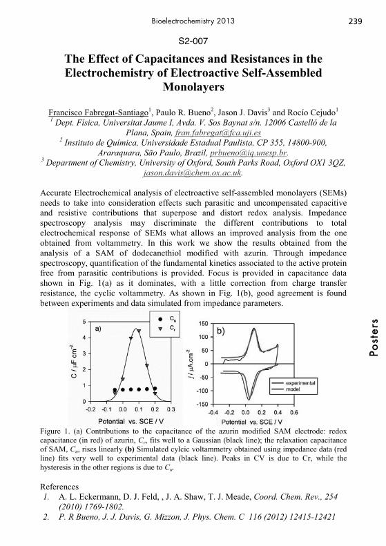
S2-007
The Effect of Capacitances and Resistances in the Electrochemistry of Electroactive Self-Assembled
Monolayers
Francisco Fabregat-Santiago1, Paulo R. Bueno2, Jason J. Davis3 and Rocío Cejudo1 1 Dept. Física, Universitat Jaume I, Avda. V. Sos Baynat s/n. 12006 Castelló de la
Plana, Spain, [email protected] 2 Instituto de Química, Universidade Estadual Paulista, CP 355, 14800-900,
Araraquara, São Paulo, Brazil, [email protected]. 3 Department of Chemistry, University of Oxford, South Parks Road, Oxford OX1 3QZ,
Accurate Electrochemical analysis of electroactive self-assembled monolayers (SEMs) needs to take into consideration effects such parasitic and uncompensated capacitive and resistive contributions that superpose and distort redox analysis. Impedance spectroscopy analysis may discriminate the different contributions to total electrochemical response of SEMs what allows an improved analysis from the one obtained from voltammetry. In this work we show the results obtained from the analysis of a SAM of dodecanethiol modified with azurin. Through impedance spectroscopy, quantification of the fundamental kinetics associated to the active protein free from parasitic contributions is provided. Focus is provided in capacitance data shown in Fig. 1(a) as it dominates, with a little correction from charge transfer resistance, the cyclic voltammetry. As shown in Fig. 1(b), good agreement is found between experiments and data simulated from impedance parameters.
Figure 1. (a) Contributions to the capacitance of the azurin modified SAM electrode: redox capacitance (in red) of azurin, Cr, fits well to a Gaussian (black line); the relaxation capacitance of SAM, Cu, rises linearly (b) Simulated cylcic voltammetry obtained using impedance data (red line) fits very well to experimental data (black line). Peaks in CV is due to Cr, while the hysteresis in the other regions is due to Cu. References 1. A. L. Eckermann, D. J. Feld, , J. A. Shaw, T. J. Meade, Coord. Chem. Rev., 254
(2010) 1769-1802. 2. P. R Bueno, J. J. Davis, G. Mizzon, J. Phys. Chem. C 116 (2012) 12415-12421
239
Post
ers
Bioelectrochemistry 2013

S2-008
Nanoscaled Protein Architectures with CDH on Electrodes for Selective Analyte Detection
S. C. Feifel, Artur Fandrich, R. Ludwig, L. Gorton, F. Lisdat
Biosystems Technology, Technical University of Applied Sciences Wildau Bahnhofstrasse 1, 15745 Wildau, Germany [email protected], [email protected]
The use of biological redox processes for the construction of sensors is a rather promising approach.[1] A spotlight here is the confinement of the catalytic reaction onto the electrode surface. A major advance was made by the use of the layer-by-layer self-assembly technique, leading to a significant increase in protein surface concentration. A number of protein multilayer designs have been reported which are stabilized by a polyelectrolyte or metallic nanoparticles. Such multilayer assemblies have recently been shown to allow incorporation of enzymes and establish communication to the electrode, thus allowing the construction of analytical signal chains.[2] By the use of different second building blocks in combination with redox proteins and enzymes the specific properties of the biomolecules (e.g. the catalytic ability) can be enhanced.[3,4] In this study we have been focused on the formation of bi-protein assemblies using cyt c as a redox protein, cellobiose dehydrogenase (CDH) as enzyme and non-conducting silica nanoparticles (SiNPs) and DNA as second building block for the construction of new protein multilayer architectures. It can be shown, that CDH can be incorporated into the protein multilayer arrangement in an active form, thus new electron transfer chain from lactose via CDH and multiple cyt c molecules toward the electrode can be constructed. In order to do so we have been applying the layer-by-layer technique with SiNPs and DNA for the formation of catalytically electro-active bi-protein multilayer electrodes. Furthermore we study the effect of the different second building blocks on the formation and electron transfer properties of such architectures. It has been found, that the second building block significantly influences the layered assembly and the electron transfer chain from substrate to the electrode. Also it needs to be mentioned, that the glycosylation of the enzyme has a strong impact on the catalytic activity of the constructed architectures. It can be shown that several catalytically active protein layers can be formed on the electrode surface, thus allowing the defined tuning of the sensitivity to lactose by the number of deposited layers. The approach is expected to have a considerable impact on the development of biosensors, and also represents a significant advance in modeling of biological electron transfer processes. References [1] I. Willner et al., Angew. Chem. Int. Ed. 2000, 39, 1180. [2] F. Lisdat et al., Chem. Commun. 2009, 283. [3] S. C. Feifel et al., Journal of Nanobiotechnology 2011, 9:59. [4] S. C. Feifel et al., Langmuir 2012, 28, 9189.
240Po
ster
sBioelectrochemistry 2013
S2-009
Effect of Deglycosylation on the Selectivity of Agaricus meleagris Pyranose Dehydrogenase Modified Electrodes
Anikó Killyéni1, Maria Yakovleva2, Lo Gorton2, Ionel Catalin Popescu1
1Department of Physical Chemistry, Babes-Bolyai University, Cluj-Napoca, Arany Janos Str. no. 11, RO-400028, Cluj Napoca, Romania
[email protected] 2Analytical Chemistry/Biochemistry and Structural Biology, Lund University
PO Box 124, SE-221 00 Lund, Sweden
Pyranose dehydrogenase (PDH) is an oxidoreductase with a covalently bound FAD cofactor, which displays broad substrate specificity and also a variable regioselectivity, being able to mono- and dioxidize a variety of sugars in their pyranose form. In the present work the bioelectrocatalytic activity of glycosylated (gPDH) and deglycosylated (dgPDH) PDH from Agaricus meleagris recombinantly expressed in Pichia pastoris was compared, by immobilizing them on graphite electrodes. Enzyme immobilization was done by adsorption from a mixture containing a mediator (Os redox polymer, Os-RP) and a cross-linking agent [poly(ethylene glycol) diglycidyl ether, 400]. The amperometric response of the obtained bioelectrodes was investigated under flow injection conditions. Previously, it was shown that deglycosylation of AmPDH results in: (i) an increase of the catalytic current, (ii) a decrease in the apparent Michaelis-Menten constant (KM
app) [1]. Indeed, the maximum catalytic current (Imax) measured for glucose at dgPDH/Os-RP electrodes (Imax = 3.35 ± 0.08 μA) was ~1.3 times higher than the similar value estimated for gPDH/Os-RP electrodes (Imax = 2.60 ± 0.06 μA). Additionally, deglycosylation of PDH resulted in a lower KM
app value (0.67 ± 0.06 mM) compared with that estimated for gPDH (4.65 ± 0.30 mM). Furthermore, deglycosylation of AmPDH changes the selectivity order from [glucose > fucose > galactose > cellobiose > xylose > sucrose] to [fucose > glucose > xylose > sucrose > galactose > cellobiose]. Moreover, the estimated value for Imax, measured for fructose at dgPDH/Os bioelectrodes was found to be ~176 nA, while for gPDH/Os-RP bioelectrodes no measurable signal was observed. Reference
[1]. Yakovleva, M. E.; Killyéni, A.; Ortiz, R.; Schulz, C.; MacAodha, D.; Conghaile, P. Ó.; Leech, D.; Popescu, I. C.; Gonaus, C.; Peterbauer, C. K.; Gorton, L., Electrochem. Commun. 2012, 24,120-122.
241
Post
ers
Bioelectrochemistry 2013
S2-010
Electrochemical regeneration of oxidized nicotinamide cofactors in a scalable reactor
Svenja Kochius, Jens Schrader, Dirk Holtmann
DECHEMA Research Institute Theodor-Heuss-Allee 25, 60486 Frankfurt, Germany
The selective oxidation of alcohols to the corresponding carbonyl group is one of the most fundamental transformations in organic synthesis. Using isolated alcohol dehydrogenases bears the promise of clean and efficient reaction procedures but is still hampered by some practical drawbacks impairing their application at preparative scale. Amongst them, efficient regeneration of the oxidized nicotinamide cofactors (NAD(P)+) remains an issue of intensive research. By now, several methods have been developed for cofactor recycling such as enzyme-coupled systems, substrate-coupled-systems, transition-metal catalyses, photochemical and electrochemical methods. Although high total turnover numbers (TTNs) are described for these approaches, the demand for an additional enzyme and/or substrate remains an important drawback of this approach. As the regeneration of NAD(P)+ from NAD(P)H is a redox process by nature, electrochemistry appears to be an obvious choice. Electrochemical oxidation of NAD(P)H represents a simple and cost effective process. No by-products are formed in this system, which facilitates the recovery of the desired product as it does not require a co-substrate. Nevertheless, some challenges have to be addressed: specially designed bioreactors are required for the synthesis reactions and the enzymes have not been evolved by nature for electrochemical conditions. Therefore, only a few examples exist where electrochemistry and enzyme catalysis are used in preparative synthesis. In such systems, a redox active mediator catalyses the electron transfer between NAD(P)H and the anode. An important challenge of the electrochemical NAD(P)+ regeneration lies in the heterogeneous nature of the reaction. As a result, diffusion limitation may impair the overall rate of the envisioned electroenzymatic reaction. However, three-dimensional electrodes are a promising technical basis to develop efficient electroenzymatic processes. In this study we present an approach for an efficient electrochemical NAD+ regeneration by using a scalable reactor based on a fixed bed system. First of all several mediators were screened according to their applicability to oxidize NADH in an electrochemical regeneration system. Among these ABTS showed the highest oxidation rate towards NADH with a turnover frequency of 1200 h-1. ABTS was used for further investigations in a 3-dimensional cell. Herein, oxidation parameters for ABTS were initially optimized. In a next step this regeneration system was coupled to the enzymatic conversion of glucose with glucose dehydrogenase. Substrate as well as enzyme showed good stability within the electrochemical system. To characterize the TTN and the space time yield (STY), concentrations of substrate, enzyme, cofactor and mediator were varied yielding TTNs of 930 for the mediator and of 93 for the cofactor as well as a maximum STY of 1.4 g l- 1h-1. It was also shown, that the electrode area had a significant influence on the reaction rate. The current efficiency (C.E.) was about 87%.
242Po
ster
sBioelectrochemistry 2013

S2-011
Electrochemical and Atomic Force Microscopy Studies of PduA Shell Protein Immobilisation on Gold Electrode
Surface
Nur Azura Mohd Said1 ,2, Mingzhi Liang3, Michael B. Prentice3 Grégoire Herzog4,5, & Vladimir I. Ogourtsov1
1Tyndall National Institute, Lee Maltings, University College Cork, Cork, Ireland 2Biotechnology Research Centre, Malaysian Agricultural Research & Development
Institute, Serdang, Selangor, Malaysia 3Department of Microbiology, University College Cork, Cork, Ireland
4Université de Lorraine, LCPME, UMR 7564, Villers-lès-Nancy, F-54600, France 5CNRS, LCPME, UMR 7564, Villers-lès-Nancy, F-54600, France
Hexameric carboxysome shell CcmK proteins assemble as a thin 2D crystal (one molecule thick) on an air-water interface. Thin pore-containing protein monolayers of this kind have potentially interesting electrochemical properties. PduA is a Ccmk-related hexameric microcompartment shell protein. We report successful immobilisation of histidine-tagged PduA produced from Lactobacillus reuteri on a 2 mm gold disc electrode. The gold surface was first activated with self-assembly monolayers of long chain 11-mercaptoundecanoic acid (11-MUA) followed by ester activation via 1-ethyl-3-(3-dimethylaminopropyl)-carbodiimide (EDC) and N-hydroxysuccinimide (NHS) reaction. The his-tag shell proteins were subsequently bound to the surface using a complex nitrilotriacetic acid (NTA) chelator coordinated with copper as the ligand metal. The immobilised shell proteins were characterized electrochemically in 1mM ferrocenecarboxylic acid in phosphate buffer saline (PBS), pH7.0, using cyclic voltammetry (CV) and electrochemical impedance spectroscopy (EIS) techniques. The obtained impedance spectra represented typical Randles circuit responses where a semicircle region followed by a straight line. Diameter of the semicircle increased with the SAM formation which confirms formation of the monolayer on the gold surface. The steric hindrance packing of the NHS ester has resulted in increment of the impedance spectra for the EDC and NHS steps. Following the attachment of the PduA shell proteins, the charge-transfer resistance of the impedance spectra (Rct), which relates to the semicircle diameter, continue to increase to the range of 125 kΩ indicating the successful immobilisation of the his-tag protein on the surface. The protein size attained from the particle analysis of the AFM images is in good agreement with the protein size as described from the literature. The results for both EIS and AFM techniques conducted have shown the successful immobilisation of the PduA protein on the gold electrode surface. The future work will include the study of the electrophysiological effect on adding glycerol to the surface and replacing copper with nickel as the chelating metal-NTA ligand. The authors would like to acknowledge the support of the Tyndall National Institute through the SFI-funded National Access Programme (Project NAP312)
243
Post
ers
Bioelectrochemistry 2013

S2-012
Electrochemical detection of DNA-Anticancer Antibody Rituximab Interaction
S. Carlos B. Oliveira, I.B. Santarino and A.M. Oliveira–Brett
Departamento de Química, Faculdade de Ciências e Tecnologia, Universidade de Coimbra, 3004-535 Coimbra, Portugal
[email protected] Rituximab (RTX) is a human/murine chimeric monoclonal (mAb) that specifically targets the transmembrane protein CD20 of B cells. This receptor is present in almost all malignant B-cells (more than 80%) and in normal differentiated B-lymphocytes (pre-B and mature B-lymphocytes). Rituximab efficiently kills both malignant and non-malignant CD20-positive cells, but B-cell recovering began at approximately 6 months following completion of treatment. RTX is FDA approved for the treatment of both indolent and aggressive B-cell non-Hodgkin's lymphomas (B-NHLs). Currently it is being increasingly used in the treatment of B-cell chronic lymphocytic leukemia (B-CLL), some autoimmune diseases and in type 1 diabetes, administered as monotherapy or in combination with chemotherapy and immunotherapy. However, the exact mechanism of RTX is still unknown and depends on the pathology type. The investigation of the interaction dsDNA-RTX has great importance to predict a new action mechanism as a genotoxic anticancer drug. The multilayer dsDNA-electrochemical biosensor, employing differential pulse voltammetry, was used for the in situ evaluation of the dsDNA-RTX interaction. The DNA damage was electrochemically detected following the changes in the oxidation peaks of guanosine and adenosine residues and monitoring the appearance of free DNA purine bases and their oxidation products, 8-oxoguanine (8-oxoGua), and 2,8-dyhydroxyadenine (2,8-oxoAde), biomarkers of DNA oxidative damage. The results showed guanosine and adenosine oxidation peak currents varying with incubation time, due to RTX binding to dsDNA, and leading to different modifications in the dsDNA structure. The RTX bound to dsDNA can still undergo oxidation. After long incubation time occurs the free guanine and adenine, and their oxidation peaks and the DNA oxidative damage was detected. The free guanine and adenine occur after the oxidation of the C1’ carbon of deoxyribose by RTX, causing their liberation from the double helix structure, and being an indicative of the DNA oxidative damage. Nondenaturing agarose gel-electrophoresis of dsDNA-RTX samples confirmed the occurrence of modifications in the dsDNA structure and the oxidative damage observed in the electrochemical results. The sensitivity of the multilayer dsDNA electrochemical biosensor offered the possibility to follow the interaction of RTX with DNA under different conditions and the results enabled a better understanding of the molecular mechanism of dsDNA-RTX interaction.
244Po
ster
sBioelectrochemistry 2013

S3-001
Investigation of the corrosion mechanism of WE43 Mg-alloy for biodegradable implant applications
M. Ascencio and S. Omanovic
Department of Chemical Engineering, McGill University 3610 University Street, Montreal, Canada
Magnesium is a promising material for the development of biodegradable implants mainly due to its biocompatibility and good mechanical properties. However, Mg corrodes too fast in the body leading to premature loss of mechanical properties and implant failure. Several Mg-alloys with improved corrosion properties have been investigated for biodegradable applications, however up to date no Mg-alloy with an adequate biocompatibility and corrosion resistance has been produced. Development of biodegradable Mg-alloys is limited due to a lack of understanding of the corrosion mechanisms occurring in the physiological environment and due to difficulties at emulating such environment in vitro. In this work, the short-term corrosion behaviour of a commercially available WE43 Mg-alloy in modified simulated body fluid (mSBF) is presented and a corrosion mechanism is proposed. Results obtained by x-ray diffraction, scanning electron microscopy, energy dispersive spectroscopy and x-ray photoelectron spectroscopy showed the formation of an amorphous phosphorous/calcium phosphate/carbonate-containing magnesium hydroxide corrosion layer. This was in agreement with the results obtained by attenuated total reflectance Fourier transform infrared spectroscopy and inductively coupled plasma, which evidenced the presence of phosphate and carbonate species in the corrosion layer and a decrease in the concentration of calcium and phosphorous ions in solution, respectively. Volcano-like deposits observed on the sample surface were related to the presence of cathodic zones where the reduction of water and formation of hydrogen gas occur. Hydrogen evolution and electrochemical impedance spectroscopy results showed a fast decrease in the corrosion rate during the first 24 hours of immersion, followed by a gradual decrease in the corrosion rate to a time of 48 hours. The onset of localized corrosion after 48 hours was then observed, as evidenced by a gradual increase in the corrosion rate and the presence of pits, observed by scanning electron microscopy and optical analyses. Finally, the following corrosion mechanism stages are proposed: a) Formation of a partially protective carbonate/phosphate-containing Mg-hydroxide corrosion layer involving the adsorption of Mg intermediates, b) Decrease in the corrosion rate due to a decrease in the porosity of the corrosion layer. This process is accompanied by a decrease in the adsorption of Mg intermediates and the presence of mass transport limitations and, c) Onset of pitting corrosion of a formed passive film, followed by a gradual increase in the corrosion rate and hydrogen evolution.
245
Post
ers
Bioelectrochemistry 2013
S3-002
New methodological approach for tracking systemic and local uptake of macromolecule enhanced by
electroporation in vivo
Tanja Blagus1, Bostjan Markelc1, Matej Rebersek2, Maja Cemazar1, Gregor Sersa1 1Institute of Oncology Ljubljana, Zaloska 2, 1000 Ljubljana, Slovenia
2University of Ljubljana, Faculty of Electrical engineering, Trzaska cesta 25, 1000 Ljubljana, Slovenia [email protected]
Transdermal delivery offers an attractive alternative to the conventional drug delivery methods of oral administration and intravenous injection. However, the stratum corneum acts as a barrier that limits the penetration of substances through the skin. Application of high-voltage pulses (electroporation, EP) to the skin increases its permeability and enables increased delivery of various substances into and through the skin. Several methods for in vitro and ex vivo determination of delivery through skin are available; however, in vivo methods are limited. Therefore, the aim of our study was to establish an easy and convenient method for non-invasive in vivo monitoring of transdermal drug delivery mediated by EP. Fluorescein-isothiocyanate dextran 4 kDa (FITC) was used as a model macromolecule to study the extent of transport into (local, topical) and through mouse skin into the bloodstream (transdermal, systemic) in vivo. Experiments were performed on female BALB/c mice. A patch with FITC (37,5 mg/ml) was applied on mouse skin 5 minutes before and immediately after EP. Multi- array electrodes were used for EP of skin. Parameters of EP were as follows: 1 electric pulse between the electrode pairs of 100 μs duration of different voltage to distance ratio (200, 400, 700, 1000, 1300 and 1600 V/cm). Altogether 24 electric pulses were delivered. As a positive control, we used a tape stripping method for the removal of the stratum corneum layer. Local (topical) delivery of FITC was determined by measurement of its fluorescence in the electroporated skin up to 7 days after EP. Systemic (transdermal) delivery of FITC was determined by capturing images of mouse tail every 5 minutes for one hour with digital camera attached to a fluorescence stereo-microscope. EP significantly enhanced transdermal transport of FITC into the bloodstream compared to the negative control. Systemic delivery of FITC through the skin by EP was increased up to ~5-fold with parameters of EP between 400 and 1300 V/cm. FITC fluorescence remaining in the electroporated skin (topical delivery) was the most pronounced in the skin area exposed to the electric pulses of the highest voltage (1600 V/cm) throughout the observation period (7 days). No skin damage was observed after EP. The obtained data of increased FITC fluorescence in mouse tail and in electroporated skin region without skin damage demonstrated that systemic (transdermal) and local (topical) delivery of macromolecules can be safely achieved with the application of high-voltage electric pulses with multi-array electrodes. We developed a new FITC based assay, which is suitable for determination of systemic as well as local delivery of the FITC.
246Po
ster
sBioelectrochemistry 2013

S3-003
Effects of Different Pulse Electric Field Parameters on Electropermeabilization of Fresh Rose Petals
Mustafa Fincan, Fatma Gundogdu, Betul Oskaybas, Seyma Avc
Department of Food Engineering, Erciyes University, Kayseri, TURKEY [email protected]
At hydrodistillation process of rose oil and hydrosol, one of the process parameter affecting distillation properties is tissue disintegration and permeabilization of intact cell membranes. The permeabilization is necessary for the reducing the resistance imposed by membranes and play an important role at diffusion of intracellular oil into water. Moreover, the realization of permeabilization before the distillation stage has considerable impacts on properties such as the distillation time, yield and the product composition [1]. Pulsed Electric Field (PEF) has recently been considered as a novel tissue disintegration method for non thermal permeabilizion of cellular membranes [2]. The objective of the present work was to study the effect of different PEF-related parameters on permeabilization of fresh rose petals using impedance changes. The rose petals submerged into water in a parallel plate type of treatment chamber equipped with impedance sensing electrodes with a 1 cm aperture were exposed to PEF using a Pulse generator. Field strength, pulse length, pulse number and pulse interval were selected to vary in the range 1.5-6 kV/cm, 10 μs – 999 μs, 1-99 and 100 msec-10 sec, respectively. The release of ionic species after PEF treatment was evaluated by measuring impedance at 100 kHz with a data sampling speed of at least 4 per second using an LCR meter connected to computer via an interface and a software. The disintegration levels after PEF were estimated based on the impedance of frozen-thawed tissue, and the approximate energy corresponding to different PEF treatments were calculated using the impedance value sampled after 0.25-1 s. At a conducted test whereby field strength was in the range of 3-4 kV/cm, the treatment resulted in a significant decrease in impedance and the kinetics of ionic species was most pronounced during about 10 min following the PEF treatment. Furthermore, comparing the impedance values of the samples at 1 MHz with those of PEF-treated samples indicated that the field strength level required for a complete permeabilization of rose petals was found considerably higher than those of soft plant tissues [2]. References 1-Handa S.S., Khanuja S.P.S., Longo G., Rakesh D.D.R., Extraction technologies for medicinal and aromatic plants, UNIDO and the International Centre for Science and High Technology (2008) 115-127. 2-Schilling S., Alber T., Toepfl S., Neidhart S., Knorr D., Schieber A., Carle R., Effects of pulsed electric field treatment of apple mash on juice yield and quality attributes of apple juices, Innovative Food Science and Emerging Technologies 8 (2007) 127–134.
247
Post
ers
Bioelectrochemistry 2013
S3-004
Optimization of electroporation protocol for extracting plasmid DNA from E. coli
Saša Haberl1, Marko Jarc2, Aleš Štrancar2, Damijan Miklavčič1
1University of Ljubljana, Faculty of Electrical Engineering Tržaška 25, 1000 Ljubljana, Slovenia
[email protected] 2 BIA Separations, d.o.o.
Mirce 21, 5270 Ajdovščina, Slovenia
In 70’s temporary increase in membrane permeability was achieved by means of electric pulses – electroporation [1]. With respect to prokaryotic cells, electroporation was used for the uptake of plasmid DNA (pDNA). Later also the possibility of pDNA extraction out of the cell was described – electroextraction [2]. Today there are several methods known for pDNA isolation (such as alkaline lysis), however considerable effort is needed for extracting pDNA [3]. The main advantages of electroextraction in comparison to alkaline lysis would be shorter time and reduced lysate volume, since no extra buffers are needed in electroextraction method. The aim of our study was to investigate how different pulse parameters and post-pulse incubation at different temperatures affect the efficiency of pDNA electroextraction from E. coli. The electroextraction was performed on 17h old culture of E.coli re-suspended and diluted (10-2) in distilled water. Samples were placed between the stainless steel plate electrodes with 1 mm gap. Different electric pulse parameters were used for extracting pDNA. Bacterial cells were incubated for 1h at 4°C or 37°C after pulse application. The extracted pDNA was quantified by high-performance liquid chromatography. Our results show that pulses with longer duration results in higher amount of extracted pDNA. This is consistent with the assumption that the size and number of the aqueous pores induced in membrane after electric pulses are increased with pulse duration [1]. Incubation of E. coli at 37°C post-pulse application gave a slightly higher amount of extracted pDNA compared to incubation at 4°C. This is somewhat surprising, since it is stipulated that at higher temperatures phospholipid molecules contain more saturated fatty acids, which causes less fragile membrane [4]. Thus incubation at 37°C post-pulse application should result in lower pDNA electroextraction. [1] E. Neumann, K. Rosenheck, Permeability changes induced by electric impulses in
vesicular membranes, J. Membr. Biol. 10 (1972) 290. [2] N.M. Calvin, P.C. Hanawalt, High-efficiency transformation of bacterial cells by
electroporation., J. Bacteriol. 170 (1988) 2796. [3] T.E.V. Aune, F.L. Aachmann, Methodologies to increase the transformation
efficiencies and the range of bacteria that can be transformed, Appl. Microbiol. Biotechnol. 85 (2010) 1313.
[4] T. Ohshima, K. Okuyama, M. Sato, Effect of culture temperature on high-voltage pulse sterilization of Escherichia coli, J. Electrostat. 55 (2002) 235.
248Po
ster
sBioelectrochemistry 2013

S3-005
Influence of calcium ions on the buildup and permeability of multilayer polymer films
Katarzyna Kilan, Lilianna Szyk-Warszyńska, Krzysztof Szczepanowicz, Piotr
Warszyński Jerzy Haber Institute of Catalysis and Surface Chemistry, Polish Academy of Sciences
Niezapominajek 8, 30-239 Cracow, Poland [email protected]
Multilayer polymer films are excellent candidates for functional membranes and coatings in molecular medicine applications. Polymers for such a purpose should be non-toxic, with minimal interaction with immune system (stealth effect) allowing easy modification with targeting ligands. Next critical factor after biocompatibility is defined permeability of such systems, especially in the field of drug delivery. The aim of our work was to correlate permeability of polymer multilayers with their mass or thickness controlled by presence of specified salts (NaCl, CaCl2) in buildup steps of those layers. Multilayer films consisting of biocompatible and natural polyelectrolytes (e.g. poly-L-arginine, modified pullulan, alginate) were constructed by layer-by-layer adsorption (LbL) technique. During formation of multilayers they were contacted with salts solutions having various ionic strength. The adsorbed mass and thickness of multilayers was determined by QCM-D (quartz crystal microbalance with dissipation) and correlated with one measured by ellipsometry in dry state. The electrochemical techniques (e.g. cyclic voltamperometry) were applied to determine permeability of those systems. For the poly-L-arginine/alginate multilayers we observed strong dependence of mass and thickness of the films on the concentration of calcium ions in rinsing solution, which was explained by crosslinking of alginate. Three electroactive agents with different molecular size were chosen to examine permeability. We observed decrease in anodic and cathodic currents with increasing number of polymer layers deposited on gold electrode (in comparison to bare electrode). Despite a significant mass increase of deposited films with increasing concentration of calcium ions in rinsing solutions shown in QCM-D experiments, considerable differences in permeability of multilayers were not evident.
249
Post
ers
Bioelectrochemistry 2013
S3-006
Electrochemical and spectroscopic characterization of PEDOT matrix containing iso-butyl-propanoic phenolate
Katarzyna Krukiewicz, Jerzy Zak
Faculty of Chemistry, Silesian University of Technology ul. M. Strzody 9, 44-100 Gliwice, Poland
[email protected] One of the greatest challenges in neuroprosthetics is the specific design of the surface of neural implant [1]. Apart from exhibiting desired electrical properties and electrochemical stability, it should also be biocompatible. Numerous studies have shown that some conjugated polymers, mainly polypyrrole and poly(3,4-ethylenedioxythiophene), do not cause adverse effects, such as inflammation or immunological reaction, when introduced into tissues [2]. Because of the fact that conjugated polymers exhibit ion-exchangeable properties, it is possible to immobilize biologically active compounds of ionic nature in a conducting polymer matrix [3]. The ability to release biomolecules in a controlled manner realizes the concept of smart drug delivery systems [4]. The aim of this work was to design biologically active and conducting coating that may be utilized in neuroprosthetics. Poly(3,4-ethylenedioxythiophene), PEDOT, was chosen as a matrix, while sodium iso-butyl-propanoic-phenolate, IBU, was chosen as a model biologically active compound. The immobilization of IBU was performed during a process of electrochemical polymerization by means of cyclic voltammetry. Obtained PEDOT:IBU matrix was characterized with electrochemical and spectroscopic methods including cyclic voltammetry, UVVis spectroelectrochemistry, Raman spectroscopy, IR spectroscopy and X-ray photoelectron spectroscopy. In order to verify whether PEDOT:IBU may be used as a drug delivery system, the conditions of electrochemically controlled drug release were studied. The financial support of the work was given by the European Union within the project SWIFT POKL.08.02.01-24-005/10. References [1] R.A. Green, N.H. Lovell., G.G. Wallace, L.A. Poole-Warren, Conducting polymers for neural interfaces: Challenges in developing an effective long-term implant, Biomaterials 29 (2008) 3393 [2] M.H. Vela, D.S de Jesus., C.M.C.M. Couto, A.N. Arau-jo, M.C.B.S.M. Montenegro, Electroimmobilization of MAO into a Polypyrrole Film and Its Utilization for Amperometric Flow Detection of Antidepressant Drugs, Electroanalysis 15 (2003) 133 [3] R. Wadhwa, C.F. Lagenaur, X.T. Cui, Electrochemically controlled release of dexamethasone from conducting polymer polypyrrole coated electrode, Journal of Controlled Release, 110 (2006) 531 [4] D. Svirskis, J. Travas-Sejdic, A. Rodgers, S. Garg, Electrochemically controlled drug delivery based on intrinsically conducting polymers, Journal of Controlled Release 146 (2010) 6
250Po
ster
sBioelectrochemistry 2013
S3-007
Improvement of both cell survival and efficacy for large plasmids electrotransfer in MSC
Léa L. Lesueur1, 2, 3, Lluis M. Mir1, 2, 3, Franck M. André1, 2, 3
1Université Paris-Sud, Laboratoire de Vectorologie et Thérapeutiques Anticancéreuses, UMR 8203, Orsay F-91405, France
2CNRS, Laboratoire de Vectorologie et Thérapeutiques Anticancéreuses, UMR 8203, Orsay F-91405, France
3Institut Gustave Roussy, Laboratoire de Vectorologie et Thérapeutiques Anticancéreuses, UMR 8203, Villejuif F-94805, France
Gene therapy is finally achieving its long awaited promise; indeed since 2006 several clinical successes have been reported. However all these trials were using viral approaches which present a variety of potential problems to the patient such as toxicity, immune responses, mutagenesis and oncogenesis. Among the alternative approaches used, nucleic acids electrotransfer presents certain advantages. The electrotransfer consists in the application of electric pulses allowing the permeabilisation of the cellular membranes and thus the entry of the DNA. Contrary to viral methods the electrotranfer is safer, enables nonpermanent expression of transgenes and allows transfection of large constructs. These advantages are particularly interesting for certain applications. We had previously developed an optimal electrogene transfer protocol for human Mesenchymal Stem Cells (hMSC) using a small reporter plasmid. However, for electrotransfer of a large plasmid (containing several genes) we observed drastically reduced expressions and survivals. Here we report diverse strategies that we have assessed to increase both cell survival and electrotransfer efficacy for large plasmids transfection into human MSC. These strategies include the addition of electrophoretic pulses, enzymatic treatments, the addition of surfactants, the modification of pulses parameters, as well as the overall procedures applied.
251
Post
ers
Bioelectrochemistry 2013

S3-008
Mechanisms associated with vascular-disrupting action of electrochemotherapy: intravital microscopy on a single
tumor blood vessel level
Bostjan Markelc1, Gregor Sersa1, Maja Cemazar1, 2 Affiliation: 1Department of Experimental Oncology, Institute of Oncology Ljubljana,
Zaloska 2, SI-1000 Ljubljana, Slovenia 2University of Primorska, Faculty of Health Sciences, Polje 42, SI-6310 Izola, Slovenia
Address: Zaloska 2, SI-1000 Ljubljana, Slovenia e-mail address: [email protected]
Electropermeabilization/electroporation (EP) is a physical method that, by application of electric pulses to cells, enables the introduction of molecules into the cells by increasing cell membrane permeability. The clinical use of EP in combination with cytotoxic drugs such as bleomycin or cisplatin is termed electrochemotherapy (ECT). EP is also increasingly used for the delivery of nucleic acids (plasmid DNA, siRNA etc) into cells in vitro and also into different tissues in vivo. The application of electric pulses to the tissue in vivo also has blood flow modifying effects. The aim of our study was to determine the effects of EP and ECT with bleomycin on the HT29 human colon carcinoma tumor blood vessels in the first hours after the therapy. The response of tumor blood vessels to EP and ECT was observed directly at the single blood vessel level by in vivo optical imaging in a dorsal window chamber in SCID mice with 70 kDa fluorescently labeled dextran (FD) in real time. The leakage of 70 kDa FD from tumor blood vessels was measured to determine the effects of EP and ECT on their permeability. In addition, functional vascular density (FVD), which describes the density of functional blood vessels in a given region of interest, and average diameter (DV) of perfused tumor blood vessels were determined in order to quantify the extent of the effects of EP and ECT on tumor blood vessels. EP and ECT induced a long lasting vascular lock, the onset of which was immediately after the application of electric pulses and affected the entire tumor vasculature. This was accompanied by the reduction of FVD in the tumor, as well as with the increase of DV of perfused tumor blood vessels. In case of ECT, the response of tumor blood vessels was more pronounced and resulted in the destruction of the tumor blood vessels within 24 hours. Additionally, EP and ECT induced a constriction of the blood vessels surrounding the tumor and increased their permeability for 70 kDa FD. This is the first study that shows the effects of EP and ECT on tumor blood vessels directly on a single blood vessel’s level in real time. The study confirms the proposed two phase model of blood flow modifying effects of EP and ECT on tumor blood vessels and provides additional proof that ECT has a vascular disrupting effect by showing destruction of small tumor blood vessels in real time. It further extends the model of blood flow modifying effects of EP and ECT on tumor blood vessels, providing evidence that their response to ECT starts to differ from EP already within the first hour after the therapy. Furthermore, it provides evidence that the blood vessels surrounding the tumor are also affected and respond to the application of electric pulses similarly as normal blood vessels.
252Po
ster
sBioelectrochemistry 2013
S3-009
EGT protocols: the role of electroporation based techniques, hyaluronidase and pH effects in the
permeabilization of tissue fibers.
N. Olaiz1, A. Soba1, P. Turjanski1, P. Chiarella2,3, S. De Santis3,4, VM. Fazio3,5, E. Signori2,3 and G. Marshall1
1 Laboratorio de Sistemas Complejos, Departamento de Computación, Facultad de Ciencias Exactas y Naturales, Universidad de Buenos Aires, 1428 Buenos Aires,
Argentina. 2Laboratory of Molecular Pathology and Experimental Oncology, CNR-IFT, Via Fosso
del Cavaliere 100, 00133 Rome, Italy. 3Laboratory of Molecular Medicine and Biotechnology, University Campus Bio-
Medico of Rome, Via A del Portillo 21, Rome, Italy 4Department of General and Environmental Physiology, University of Bari, Via
Amendola 165/A, 70125 Bari, Italy. 5Laboratory of Oncology, IRCCS ‘Casa Sollievo della Sofferenza’, Viale Padre Pio,
71013 San Giovanni Rotondo (FG), Italy. [email protected]
It is known that plasmid gene transfer efficiency in tissue fibers such as muscle can be significantly improved by the application of an electrotransfer based technique to the muscle following injection of bovine hyaluronidase and plasmid DNA. Despite a less damage in muscle fibers due to the pre-treatment by hyaluronidase, traces of necrosis still remain visible in the tissue with a concomitant loss of transfected tissue fibers and reduction in expression. Here we analyze with experimental measurements and theoretical modeling different factors producing tissue necrosis such as electric pulse parameters, electrode geometry as well as possible pH effects. Ways to optimize the EGT protocol are suggested.
253
Post
ers
Bioelectrochemistry 2013
S3-010
COST TD1104 Action – a Network for Development of Electroporation-based Technologies and Treatments
Damijan Miklavčič
University of Ljubljana, Faculty of Electrical Engineering Tržaška 25, 1000 Ljubljana, Slovenia
Exposure of biological cells to a sufficiently strong external electric field results in increased permeability of cell membranes, referred to as electroporation. Since all types of cells (animal, plant and microorganisms) can be effectively electroporated, without addition of viral or chemical compounds, electroporation is considered to be a universal method and a platform technology. Electroporation has become a widely used technology applicable to e.g. cancer treatment, gene transfection, food and biomass processing, and microbial inactivation. However, despite significant progress of electroporation-based applications, there is a lack of coordination and interdisciplinary exchange of knowledge between researchers from different scientific domains. Thus, critical mass for new major breakthroughs is missing. We therefore established cooperation between research groups working in different fields of electroporation. Cooperation in Science and Technology (COST) programme through which European Union funds networking and capacity-building activities between researchers working in different research fields seem ideal at this stage of electroporation and its applications development. This specific COST Action aims at: (i) providing necessary steps towards EU cooperation of science and technology to foster basic understanding of electroporation, (ii) improving communication between research groups, resulting in streamlining European R&D activities, and (iii) enabling further development of new and existing electroporation-based applications by integrating multidisciplinary research teams, as well as comprehensive training for Early-Stage Researchers (ESRs). Results of this COST Action will provide multiple societal, scientific, and technological benefits from improving existing electroporation-based applications and adding new ones in the field of medicine, biotechnology and environmental preservation. References: Miklavcic, D. Network for Development of Electroporation-Based Technologies and Treatments: COST D1104. J Membrane Biol 245: 591-598, 2012. www.electroporation.net
254Po
ster
sBioelectrochemistry 2013
S3-011
Laser-Pulse-Induced in-situ Diagnostics of Processes at Solid-Fluid Interfaces
Tristan Nagy, Oskar Armbruster, Ulrich Pacher, Günter Trettenhahn, Wolfgang Kautek
University of Vienna, Department of Physical Chemistry Währinger Strasse 42, A-1090, Vienna, Austria
Environmental impacts on functional nano- and microscale coating systems in microelectronics, medical, aeronautical, offshore and automotive technologies determine operational safety and sustainability. Recent developments in sensitive in-situ nanosecond pulse laser depassivation and spallation of films [1-3] and coatings connected with in-situ laser-induced breakdown spectroscopy (in-situ LIBS) for three-dimensional space-resolved chemical analysis and the determination of film thicknesses have been applied to the molecular and atomistic clarification of the composition, formation, nature, and behaviour of functional film and coating systems. Several effects related to plasma emission and material behaviour, sample geometry, and focusing conditions, as well as their impact on the reproducibility of data, were investigated and compared to numerical simulation results. Thus, fundamental aspects of electrochemical repassivation processes relevant for the environmental stability of technical surfaces can be monitored on-line. According to the widely-spread use of passive metals and alloys, e.g. titanium anchors, in implantology this may be from tremendous importance for future biomedical applications. [1] R. Oltra, E. Arenholz, P. Leiderer, W. Kautek, C. Fotakis, M. Autric, C.N. Afonso,
P. Wazen, Proc. SPIE 3885 (1999) 499. [2] A. Cortona, W. Kautek, Phys. Chem. Chem. Phys. 3 (2001) 5283. [3] G. Daminelli, P. Meja, A. Cortona, J. Krüger, M. Autric, W. Kautek, Proc. SPIE
4760 (2002) 239. [4] W. Kautek, G. Daminelli, Electrochim. Acta 48 (2003) 3249.
255
Post
ers
Bioelectrochemistry 2013

S3-012
Tailored Lipidic Cubic Phases as Novel Drug Delivery System
Ewa Nazaruk, MonikaSzlęzak, Ewa Górecka, Renata Bilewicz
Faculty of Chemistry, University of Warsaw, Pasteura 1, 02-093, Warsaw, Poland
At present, the cubic mesophases prepared by unsaturated monoglycerides or phytantriol (PT) are the most frequently investigated liquid crystal structures for drug delivery. Monoglycerides are non-toxic, biocompatible and biodegradable and show good chemical and physical stability of incorporated drugs and especially macromolecular drugs. The structure-forming lipids can absorb a certain amount of water and then spontaneously form gel-like phases with unique internal structures, into which drugs can be incorporated. Cubic phases can be attractive for controlled drug delivery because of the presence of small pore size. The compartmentalization in cubic mesophases can be used to introduce guest drugs of hydrophilic, lipophilic or amphiphilic nature. Hydrophilic drugs will be located close to the lipids polar head or in the water channels, whereas lipophilic drugs can be localized within the lipid bilayer and amphiphilic drugs in the interface. The diffusion of these molecules depends on the size of the molecules and the rate of transport can be modulated e.g by applying lipids with different acyl chains to change the charge of the water channels. Monoolein and phytantriol based cubic phase was used to immobilize doxorubicin, an anthracycline antibiotic used in cancer treatment. Monoolein and phytantriol phases were doped with pH dependent additive to modulate drug release. Drug diffusion and structural properties of monoolein- and phytatntriol – based cubic phases moped with doxorubicin were compared. Doxorubicin was implemented into the phytantriol/water systems to prepare cubic phases which would be resistant to lipases’ activity. X-Ray measurements were done to identify the properties of liquid crystalline phases. In wide angle range in all mesophases the diffused signal positioned at the angle corresponding to 4.5 Å distance was recorded, that is characteristic for the phases with liquid like order of alkyl chains. In low angle range the Bragg reflections characteristic to the long range positional order were detected. SAXS data collected for non-doped phytantriol based cubic phase consisting of 27% H2O shows formation of Pn3m structure at ambient temperature. Doping Phyt/H2O system with 1 wt% Dox at ambient temperature results in Ia3d cubic phase formation.
256Po
ster
sBioelectrochemistry 2013
S3-013
A Nanosecond, High-Voltage Pulse Generator for Electric Pulse Application to Low Conductivity Liquid Media
S. Romeo1, G. Pataro2, A. Sannino1, O. Zeni1, G. Ferrari2,3, M. R. Scarfì1 1CNR, Institute for Electromagnetic Sensing of the Environment (IREA)
Via Diocleziano328, 80124, Naples, Italy [email protected];
2 Department of Industrial Engineering, University of Salerno via Ponte don Melillo, 84084, Fisciano (SA), Italy
3ProdAl scarl, University of Salerno (Italy), via Ponte don Melillo, 84084, Fisciano (SA), Italy
The usage of pulsed power technologies has found large acceptance, over the past decades, for several new applications in the fields of medicine, biotechnology, environment and food processing. As a matter of fact, intense pulsed electric fields (PEF), when applied to cells or tissues, have been demonstrated to induce permeabilization of the cell membranes, an effect that is known as electroporation [1]. The method is efficient for molecule delivery across cell membranes, but also for cancer treatment (electrochemotherapy) [2]. Moreover, the treatment is effective with respect to liquid food pasteurization, water decontamination, and, in general, for food processing [3]. All the applications cited above are based on the usage of electric pulses with durations in the μs to ms time-scale. More recently, pulses with duration in the sub-μs and ns time scale have been demonstrated to interact with both the plasma and the intracellular membranes, showing promising applications both for medicine and for industry [4]. In all cases, pulse generators providing flexible exposure conditions (pulse amplitude, duration, repetition rate) are strongly desirable in the framework of research studies aimed at investigating the biological effects of PEF and at developing their applications. In this work, a ns, high-voltage pulse generator based on the Blumlein pulse forming network concept will be presented, that has been realized for in vitro application of PEF on biological loads. In particular, the pulse generator has been suited for pulse application to high impedance loads (hundreds of Ohms), by employing microstrip transmission lines that can be easily homemade with the desired physical and electromagnetic characteristics. The system will be used to apply PEF to low-conductivity liquid media placed in batch PEF treatment chamber, in order to evaluate the release of metals from the electrodes into the liquids. Preliminary results of these measurements will be also presented. References
1. T. Y. Tsong, Biophys. J., 60:297-306, 1991 2. M. Breton and L. M. Mir, Bioelectromagnetics, 33(2):106-123, 2012. 3. H. Akiyama, et al., IEEE Trans. Diel. Elect. Insul., 14(5):1051-1064, 2007. 4. R. P. Joshi and K. H. Schoenbach, Critical Reviews in Biomedical
Engineering, 38:255-304, 2010.
257
Post
ers
Bioelectrochemistry 2013

S3-014
Liver Segmentation for Electrochemotherapy Treatment Planning
Denis Pavliha1, Maja M. Mušič2, Gregor Serša2, Damijan Miklavčič1
1 University of Ljubljana, Faculty of Electrical Engineering, Ljubljana, Slovenia 2 Institute of Oncology, Ljubljana, Slovenia
Electrochemotherapy (ECT) enhances chemotherapy outcome due to permeabilization of targeted cell membranes, which allows the chemotherapeutic drug to easily traverse tumor cell membranes (Serša et al., 2008). ECT has been in clinical use for treating skin melanoma (Marty et al., 2006) using fixed geometry electrodes; in such case, it is required to follow standard operating procedures for a successful ECT treatment (Mir et al., 2006). Recently, ECT has been tested in clinical trials for treating deep-seated metastases, e.g. in the liver (Edhemović et al., 2011). In case of deep-seated tumors, patient-specific treatment planning of ECT is required (Pavliha et al., 2012); treatment planning is based on the patient’s medical images and begins with image segmentation, i.e. extraction of the relevant tissue segments from the two-dimensional medical images’ collection in order to later generate a representative three-dimensional model of the patient’s inner structure. In order to evaluate different liver segmentation approaches for liver ECT treatment planning, we implemented three different segmentation algorithms: region growing, adaptive threshold, and active contours (Kass et al., 1987). An additional postprocessing step has been applied after every algorithm in order to further eliminate possible anomalies that may be produced during segmentation. The implemented algorithms have different parameters that allow adjusting the functioning of each algorithm, hence production of different segmentation results. The algorithms were optimized using a training set of seven real case models previously manually segmented by a radiologist with the aim to validate the segmentation algorithms and provide a robust framework for as automatic as possible ECT treatment planning. Namely, validation is an important step of developing procedures for operative planning since it ensures the produced data will be accurately generated. Therefore, validation allowed us to modify the segmentation algorithms in order to provide correct results that are most similar to the training-set cases manually segmented by the radiologist and will allow us further developments towards establishing an automatic yet robust ECT treatment planning. Edhemović, I. et al., 2011. Technol. Cancer Res. Treat 10, 475–485. Kass, M. et al., 1987. Int. J. of Comp. Vis 1, 321–331. Marty, M. et al., 2006. EJC Supplements 4, 3–13. Mir, L.M. et al., 2006. EJC Supplements 4, 14–25. Pavliha, D. et al., 2012. Bioelectrochemistry 87, 265–273. Serša, G. et al., 2008. EJSO 34, 232–240.
258Po
ster
sBioelectrochemistry 2013

S3-015
Molecular dynamics simulation of Archaea Aeropyrum pernix membrane
Andraž Polak1, Peter Kramar1, Damijan Miklavčič1, Mounir Tarek2
1University of Ljubljana, Faculty of electrical engineering, Tržaška cesta 25, 1000 Ljubljana, Slovenia
2CNRS-Université de Loraine, UMR 7565 Campus Science Grignard BP 70239, 54506 Vandoeuvre-lès-Nancy, Cedex, France
Aeropyrum pernix (Ap) is aerobic hyperthermophilic Archaea, which grows in a coastal solfataric vent at Kodakara, Juma Island, Japan. This organism can live in harsh environments with temperatures up to 100 °C. These membranes are composed of two lipids: 2,3-di-O-sesterterpanyl-sn-glicerol-1-phospho-myo-inositol (AI) and 2,3-di-O-sesterterpanyl-sn-glicerol-1-phospho-1'(2'-O-α-D-glucosyl)-myo-inositol (AGI) [1]. Due to their high stability, these membranes are interesting to study in the prospect of using them in drug delivery systems. Here we present preliminary results on the investigation of the structure and electrical properties of Archea lipid bilayers using Molecular Dynamics simulations. The membrane model was composed of AI and AGI molecules, at molecular rations consistent with Aeropyrum pernix -Archea membranes composition, embedded in high KCl salt concentration solution. We used the CHARMM 36 all-atom carbohydrate and lipid force field, and the parameters of ether linkages adopted from DPhPC-ether model [2]. The MD simulations were carried out at constant pressure 1 Atmosphere and temperature 323 K using NAMD. Several properties of these Archea lipid bilayers, extracted from the MD simulations were compared to those of pure DPPC Phosphatidylcholine lipid bilayers modeled under similar conditions. Among these, the area per lipid, the membrane dipole potential... We then present preliminary results of the electroporation of these lipid bilayers when subject to high electric fields, and discuss the similarities and differences of the molecular processes taking place, in comparison to electrporation of simple PC based lipid bilayers. [1] H. Morii, H. Yagi, H. Akutsu, N. Nomura, Y. Sako, and Y. Koga, “A novel phosphoglycolipid archaetidyl(glucosyl)inositol with two sesterterpanyl chains from the aerobic hyperthermophilic archaeon Aeropyrum pernix K1,” Biochimica et Biophysica Acta (BBA) - Molecular and Cell Biology of Lipids, vol. 1436, no. 3, pp. 426–436, Jan. 1999. [2] K. Shinoda, W. Shinoda, T. Baba, and M. Mikami, “Comparative molecular dynamics study of ether- and ester-linked phospholipid bilayers.,” The Journal of chemical physics, vol. 121, no. 19, pp. 9648–54, Nov. 2004.
259
Post
ers
Bioelectrochemistry 2013

S3-016
Electrotransfer of pStaby: A new safe and efficient DNA vaccine vector devoid of antibiotic resistance marker
G. Vandermeulen1, K. Vanvarenberg1, O. Schakman2, C. Szpirer3 and V. Préat1
1Université catholique de Louvain, Louvain Drug Research Institute, Pharmaceutics and Drug Delivery, 1200 Brussels, Belgium; 2 Université catholique de Louvain, Institut de Recherche Expérimentale et Clinique, Pole of Endocrinology, Diabetes, and Nutrition, 1200 Brussels, Belgium; 3Delphi Genetics, 6041 Gosselies, Belgium
The development of DNA vaccines requires both an adequate plasmid vector and a potent delivery method. During plasmid production, the selection of bacteria which contain the plasmid of interest is necessary. If the use of an antibiotic resistance gene as selection marker is efficient, it raises several concerns that are often pointed out by the regulatory authorities. Among the emerging technologies for plasmid selection, the use of a toxin-antitoxin system is particularly attractive. This study aimed to demonstrate the lack of toxicity of a new toxin-antitoxin based plasmid vector delivered by electrotransfer and its efficiency for inducing immune response to gp160. Here, the antidote gene (ccdA) is introduced in the pStaby plasmid under the control of a constitutive promoter. Expression of the poison gene (ccdB) by the bacteria is strongly repressed in the presence of the plasmid but, when the plasmid is lost, the antidote is degraded and the production of the toxin is induced, causing cell death. First, toxicity assays were performed in vitro on B16F10 and 293T cells. Death to live cell ratio was obtained from results of LDH and MTT assays and no in vitro toxicity was observed for the pStaby plasmid. we also evaluated in vivo the toxicity of pStaby administered by electrotransfer. We injected 100 μg of plasmid in 30 μl of PBS into each tibial cranial muscle. Then, we placed the leg between plate electrodes and we delivered 8 square-wave electric pulses (200 V/cm 20 ms 2 Hz). The open-field test was used to assess a nonforced ambulation and we noticed an accommodation effect when measures were performed daily but no difference was observed between treated or untreated mice. The grip strength test was used to measure the strength of combined fore limb-hindlimb muscles. No effect of the electrotransfer treatment was observed. The creatine kinase level in mice sera measured 15 days after electrotransfer were not modified. Put together, data from this in vivo experiment showed that muscle electrotransfer of pStaby did not influence the behavior of mice. Finally, the gp160 sequence was subcloned in pStaby. gp160 envelope glycoprotein is known to be an important target for HIV-1 vaccines. Mice received one priming and two boosts by muscle electrotransfer, with two weeks between each administration. Cytokine assays on splenocytes from immunized animals showed that γ-IFN and IL-2 were produced after stimulation with the antigen but no IL-10 and IL-4 were detected. This suggests that mice were efficiently immunized by electrotransfer of pStabygp160 plasmids with Th1-orientation of the immune response. In conclusion, electrotransfer of pStaby appears as a safe and efficient method for DNA vaccination. The authors acknowledge the financial support from the Biowin Project (DNAVAC) of the Walloon Region, Belgium.
260Po
ster
sBioelectrochemistry 2013
S3-017
Doxorubicin Delivery Enhanced by Electroporation to Colon Adenocarcinoma Cells with P-gp Overexpression
Julita Kulbacka1, Jolanta Saczko1, Nina Rembialkowska1, Anna Choromanska1,
Katarzyna Biezunska-Kusiak1, Joanna Rossowska2, Malgorzata Daczewska3, Malgorzata Kotulska4
1) Department of Medical Biochemistry, Medical University, Chalubinskiego 10, 50-368 Wroclaw, Poland; 2) Institute of Immunology and Experimental Therapy, Polish
Academy of Sciences, Rudolfa Weigla 12 St., 53-114 Wroclaw, Poland; 3) Department of General Zoology, Zoological Institute, University of Wroclaw, Sienkiewicza 21 St.,
50-335 Wroclaw; 4) Institute of Biomedical Engineering and Instrumentation, Wroclaw University of Technology, Wybrzeze Wyspianskiego 27 St., 50-370 Wroclaw
During electroporation this process electropores induced by the influence of electromagnetic field are generated and additional path of transport of macromolecules is available. The aim of this study was evaluation of electroporation efficiency for doxorubicin transport to human colon cancer cells with overexpression of P-glycoprotein. For experiments LoVo and LoVoDX cells were used. Expression of P-glycprotein was detected by immunofluorescence. MTT assay was applied for cellular viability. In the experiments were applied EP parameters: 250, 1250 and 2000 V/cm, 50 μs, 5 pulses in each case. The doxorubicin concentration was 10 μg/ml. The efficiency of macromolecules transport was examined cytofluorymetrically by assessing the degree of penetration of propidium iodide (PI). The sensitivity of both cell lines was dependent on the parameters used during EP. Electroporation enhanced doxorubicin effect in resistant cell line.
261
Post
ers
Bioelectrochemistry 2013

S3-018
System For Nanoporation of Biological Cells Based on Optically-Triggered High-Voltage Spark-Gap Switch
S. Balevicius1, V. Stankevic1, N. Zurauskiene1, E. Shatkovskis2, A. Stirke1,
A. Bitinaite1,3, R. Saule1,3, R. Maciuleviciene3, and G. Saulis1,3* 1Semiconductor Physics Institute, Center for Physical Sciences and Technology,
Vilnius, 01108, Lithuania 2Vilnius Gediminas Technical University, Vilnius, 10223, Lithuania
3Laboratory of Biophysics for Bionanotechnology and Medicine, Faculty of Natural Sciences, Vytautas Magnus University, Kaunas, 44248, Lithuania
*E-mail: [email protected]
For investigations of electroporation of biological cells with high-voltage (up to 100 kV) pulses of nanosecond-duration, methods and equipment for the generation and registration of short, yet powerful electrical pulses, are of great importance. Here, we present the system for electroporation of cells suspended in liquid media. The system consists of the electric pulse generator based on spark-gap switch and a coaxial cuvette with a 0.03-ml active volume and 1-mm distance between 28.26 mm2 square disk-shape electrodes. The spark-gap is optically triggered by a 0.45-ns duration and 1-mJ energy laser pulse (wavelength 1062 nm). A 75-Ohm impedance transmission line with a 1:100 attenuator and the 6-GHz wideband real-time oscilloscope were used to monitor the pulse. It is able to generate near-perfect square-wave pulses (rise and fall times <0.5 ns) with the duration of 10, 40, 60 or 90 ns. The maximal pulse amplitude is 12.5 kV. The main advantage of the system is the ability to generate single pulses with the amplitude and duration precisely set in advance. The system was tested on human erythrocytes. The fraction of electroporated cells was determined from the extent of hemolysis after long (20–24 h) incubation in 0.63% NaCl solution at 4 oC. The dependence of the fraction of electroporated cells on the amplitude of the electric field pulse was determined for pulses with the duration from 10 to 95 ns. For the 95-ns duration pulse, the amplitude required to electroporate 50% of cells was 11 kV/cm. When the pulse duration was decreased to 40 ns, the amplitude of the pulse electroporating 50% of cells increased to 60 kV/cm.
262Po
ster
sBioelectrochemistry 2013
S3-019
Alternative Therapy Protocols For Tumours Treatment: Dna Vaccination Mediated By Electrotransfer
Pieranna Chiarella1,2, Stefania De Santis1,3, Emanuela. Massi1, Mariangela. De Robertis1,
Vito Michele Fazio1,4, Emanuela Signori2 1Laboratory of Molecular Medicine and Biotechnology, CIR, University Campus Bio-
Medico of Rome, Via Alvaro del Portillo 21, 00128 Rome, Italy 2Laboratory of Molecular Pathology and Experimental Oncology, CNR-IFT, Via Fosso
del Cavaliere 100, 00133 Rome, Italy. 3Department of General and Environmental Physiology, University of Bari, Via
Amendola 165/A, 70125 Bari, Italy. 4Laboratory of Oncology, IRCCS ‘Casa Sollievo della Sofferenza’, Viale Padre Pio,
71013 San Giovanni Rotondo (FG), Italy. [email protected]; Phone: +390649934232; Fax:+390649934257
DNA vaccines are emerging as a promising approach for introducing foreign antigens into the host, inducing protective immunity against malignant tumours as they stimulate both cellular and humoural immune responses against the encoded antigen. Our group has been studying several strategies to enhance the DNA vaccination efficacy such as antigen optimisation, co-expression of cytokines and co-stimulatory molecules in the same vector, use of adjuvants and delivery systems. A safe tool to deliver a large amount of naked DNA to tissues has been rapidly developed over the last decade. This technology is commonly known as electrotransfer (ET). When this technology is coupled with a pre-treatment by hyaluronidase, enhanced transfection of muscular fibres and increased expression of the encoded antigen are obtained. Recently we investigated the role of ET in recruiting and triggering cells involved in antigen presentation and immune response. We also investigated the role of hyaluronidase and its contribution to the inflammatory reaction induced by ET, hypothesizing a possible role in supporting the adjuvancy of ET. Mouse skeletal muscle treated by ET and hyaluronidase were collected and analyzed at different time points. We demonstrated that EP induces transient morphological changes in the muscle with early production of endogenous cytokines responsible for signalling danger at the local level. It also causes the recruitment of inflammatory cells independently of the DNA injection and the activation of a danger pro-inflammatory pathway, resulting in T-lymphocyte migration. Our results demonstrated also that hyaluronidase amplified the effect of ET in terms of inflammatory cells recruitment into the skeletal muscle and enhanced the early release of inflammatory cytokines Il-1β and IL-6. Although hyaluronidase is not comparable to ET as pro-inflammatory signal, it contributes to enhance the pro-inflammatory effect of ET occurring in the skeletal muscle. These observations are important for optimizing DNA vaccination protocols based on ET as this delivery strategy can be considered as good adjuvant in genetic vaccination protocols and an important and very promising tool in the future of DNA vaccination therapy to be employed both in preclinical and clinical therapeutic protocols against cancer in the field of veterinary and human medicine.
263
Post
ers
Bioelectrochemistry 2013

S3-020
Plasma Electrolytic Oxidation of Ti-13Nb-13Zr Alloy - Corrosion and Bioactivity Investigations
Anna Klimczyka), Magdalena Widziołekb), Artur Macieja), Tadeusz Gorewodac),
Agnieszka Krząkałaa), Marzena Kik-Jaworskad), Joanna Michalskae), Grzegorz Tylkob), Beata Cwalinaf), Anna M. Osyczkab), Wojciech Simkaa)
a) Faculty of Chemistry, Silesian University of Technology B. Krzywoustego 6, 44-100 Gliwice, Poland
[email protected] b) Faculty of Biology and Earth Sciences, Jagiellonian University, Krakow, Poland
c) Institute of Non Ferrous Metals, Gliwice, Poland d) Department of Biopharmacy, Silesian Academy of Medicine, Sosnowiec, Poland e) Faculty of Materials Science and Metallurgy, Silesian University of Technology,
Katowice, Poland f) Faculty Of Energy And Environmental Engineering, Silesian University of
Technology, Gliwice, Poland
The growing requirements for long-term implants and doubt regarding the bioinertness of titanium alloys containing vanadium have contributed to the growing number of investigations of vanadium-free titanium alloys. Among the studied alloys, the most promising metallic biomaterials are the following types: titanium-aluminum-niobium (Ti-6Al-7Nb), titanium-niobium-zirconium (Ti-13Nb-13Zr), titanium-niobium-tantalum-zirconium (Ti-29Nb-13Ta-4.5Zr) and titanium-niobium (Ti-50Nb). These alloys are characterised by much greater biocompatibility and corrosion resistance than the alloys containing vanadium. They also have a lower Young’s modulus (below 100 GPa). Modification of the surface of a titanium implant by plasma electrolytic oxidation (PEO) is relatively easy and inexpensive. It permits good adhesion to the substrate and allows for the use of homogeneous oxide coatings, which can be enriched in biocompatible elements (i.e., silicon) during the process. This technique results in the formation of numerous micropores on the surface of the oxidising metal. The ions contained in a bath solution can penetrate into the oxide layer in the course of the glow discharge effect (breakdown of the oxide layer) on the sample substrate, which occurs during the PEO.
The Ti-13Nb-13Zr alloy used in this investigations has a cylindrical shape with a diameter of 9.5 mm and a height of 7 mm. Samples preparation methodology has been presented in our previous reports. Electrochemical corrosion as well as biocorrosion with use of sulphate reducing bacteria (SRB) were determined. Bioactivity of surface modified Ti-13Nb-13Zr alloy was determined with use of simulate body fluid (SBF) and adult human bone marrow stromal cells.
It was found that PEO parameter plays important role in formation of an oxide layer on the Ti-13Nb-13Zr alloy. The corrosion resistance (electrochemical and biocorrosion) as well as bioactivity increases with formation of thick Si-rich coating on the titanium alloy.
This work was supported by the research projects numbers: IP 2010 0377 70 (WS), 2011/01/B/NZ4/00664 (AMO), NN518291940 (BC).
264Po
ster
sBioelectrochemistry 2013

S3-021
Anodic Oxidation of Tantalum in Silicate Solutions
Wojciech Simkaa), Maciej Sowaa), Robert P. Sochab), Joanna Michalskac), Agnieszka Krząkałaa)
a) Faculty of Chemistry, Silesian University of Technology B. Krzywoustego 6, 44-100 Gliwice, Poland
[email protected] b) Jerzy Haber Institute of Catalysis and Surface Chemistry PAS, Krakow, Poland c) Faculty of Materials Science and Metallurgy, Silesian University of Technology,
Katowice, Poland
In the past, refractory metals such as Ti, V, Zr, Nb, Hf, Ta and Re have been investigated for their application as biomaterials for implants because of their good corrosion resistance and satisfying mechanical properties. Titanium and its alloys seemed to dominate this area, especially alloys containing V and Al. However, two latter metals after prolonged exposure to physiological fluids induce health issues, owing to vanadium cytotoxicity and aluminum bone growth inhibition properties, thus efforts has been made to replace them. Ta is becoming increasingly popular as dopant in place of V and Al alloys. Plasma electrolytic oxidation (PEO) of tantalum may lead to advantageous results, owing to improved corrosion resistance of coatings produced during this process, as well as their extended surface area, with relatively low cost of treatment. Commercially pure tantalum metal sheets were cut into 10 x 10 mm plate samples. Each sample was welded with titanium wire for fixing purposes, polished, etched in H2SO4/HF bath, washed with distilled water, and ultrasonically cleaned in distilled water. Experiment composed of series of anodization processes of metal samples in K2SiO3 solution with varying both concentration and voltage. Anodization cell consisted from a vessel equipped with an outer shell, through which cooling water was flowing to remove the heat liberated during treatment, ferromagnetic stirring bar with external magnetic stirrer, titanium cathode and Ta sample anode. Treated samples were subjected to analysis using scanning electron microscopy (SEM), energy-dispersive X-ray spectrometry (EDX), X-Ray photoelectron spectroscopy (XPS) and corrosion measurements. Tantalum based coatings treated in potassium silicate solution develop very extensive surfaces with large amount of silicon oxide incorporated into the oxide layer. Rough and highly porous structures were obtained for higher concentrations of K2SiO3. Increasing concentration of electrolyte during treatment increases the amount of bath components incorporation into the oxide layer (silicon oxide, in particular). However, microcracks being formed due to thermal stresses across the surface of the coatings may limit their application.
This work was supported by the Polish Ministry of Science and Education under the research project No. IP 2011 0494 71 during 2012-2014.
265
Post
ers
Bioelectrochemistry 2013
S4-001
New tools for electrochemical biomarkers detection implying Solid State biological
recognition kits on Nano-Modified electrodes
1Harold E Braustein, 1Clementiy Levkov, 2Isabella E Braustein, 1Judith Rishpon Tel Aviv University, 2 Israeli Health Ministry Heaquarters
Ramat Aviv, Jerusalem [email protected]
We present here a novel detection technology based on polypropylene micro tips filled with beads modified with functional groups in a flow system and electrochemical detector. Immunoglobulin G (IgG) and C reactive proteins (CRP) were tested in serum samples as a model system. Antibodies against the target proteins were bound to the beads. The electrochemical detection was done on screen printed electrodes (SPE) modified with carbon nanotubes (CNT) and amperometric flow cell connected to a mini potetentiostat were employed. CRP and IgG detection has been established. A linear detection range of 0-40 ng CRP per ml was found, using anti-CRP antibodies and for IgG a linear detection range between 0-10 ng/ml using antiIgG samples from its occurrence. were bound to the beads. For the electrochemical detection screen printed electrodes (SPE) modified with carbon nanotubes (CNT) and amperometric flow cell connected to a mini potetentiostat were employed. CRP and IgG detection has been established. A linear detection range of 0-40 ng CRP per ml was found, using anti-CRP antibodies and for IgG a linear detection range between 0-10 ng/ml using antiIgG samples from its occurrence.
266Po
ster
sBioelectrochemistry 2013
S4-002
Anna Koper (Chemistry, Maria Curie-Sklodowska University, Lublin, Poland), Malgorzata Grabarczyk
The Different Electrochemical Aspects of Studies of Humic Substances as a Natural Biopolymers Present in
Environmental Samples Anna Koper, Malgorzata Grabarczyk, Cecylia Wardak, Agnieszka Nosal-Wiercińska
Faculty of Chemistry, Maria Curie-Sklodowska University M.C. Sklodowska sq.3, 20-031 Lublin, Poland
Humic substances (HS) are naturally occurring complex polymeric oxidation products of decaying plant and animal wastes. The study of these materials is important from an environmental perspective because they are abundantly found around the world in soil and waters. Considering the analysis of humic substances in environmental samples by electrochemical methods we can distinguish two different aspects. First is the quantitative determination of HS using the voltammetric methods. The another aspect is connected with interfering properties of HS in electrochemical determination of metals. HS are operationally subdivided according to their solubility into humic acids (HA) and fulvic acids (FA). A few electrochemical methods exist to determine HA in natural waters. One is based on the interaction of molybdenum with HA, forming Mo(VI)–humic acid complexes that adsorb on the electrode and are detected by their reduction using cathodic stripping voltammetry (CSV) [1]. Humic acids are known to form complexes with iron (as Fe(III)) in natural waters. It is demonstrated here that seawater adsorb on the mercury drop electrode and cause a reduction peak for complexed iron in adsorptive cathodic stripping voltammetry which is much enhanced by a catalytic effect in the presence of bromated [2]. This reaction is used for a sensitive method for the determination of HA in natural waters including seawater. Humic acids cause interference nearly in all voltammetric methods of metals determination. Such substances can easily adsorbed at the surface of the working electrode, which cause fouling or passivation of the electrode and reduce or disturb the voltammetric signal of the determined ion. In some voltammetric procedures the adsorptive properties of Amberlite XAD-7 or XAD-16 resin were successfully applied for elimination of interferences from organic matter [3]. This goal of this communication is to describe two different aspects for the electrochemical studies of humic substances: the quantitative determination of HS in natural water samples and the examination and elimination of negative influence of HS on voltammetric signal of determined metal ions. 1. F. Quentel, C. Elleouet, C.L. Madec, Electroanalysis 1992, 4, 707 2. L.M. Laglera, G. Battaglia, C.M.G. Berg, Anal. Chim. Acta 2007, 599, 58–66 3. M. Grabarczyk, A. Koper, Talanta 2011, 84, 393-399
267
Post
ers
Bioelectrochemistry 2013

S4-003
Phenyl Layers – Matrix for Specific Immobilization of Biologically Important Compounds
Agata Kowalczyk, Michał Fau, Anna M. Nowicka, Marcin Strawski, Zbigniew Stojek
Faculty of Chemistry, University of Warsaw, ul. Pasteura 1, PL-02-093 Warsaw, Poland
e-mail: [email protected]
The derivatization of surfaces is often required to improve the performance of many materials and to change their surface properties. It is often critical for some particular applications. Many molecules and biomolecules, after the direct contact with conducting surfaces, especially with noble metals, can be deactivated or even denaturated. Therefore, a modification of the conducting surfaces with an organic monolayer with specific functional end groups for the covalent attachment of various molecules has received great attention. The deposited organic layer fulfills its function well when it is sufficiently stabile and conductive and evenly covers the modified surface without any defects. These conditions are well fulfilled by the phenyl films. The binding of the aryl groups to the electrode surface is likely a two-step process involving the electrochemical generation of aryl radicals which subsequently react with the surface. The diazonium cations can be generated either in situ through a classic diazotation of a primary amine in aqueous media or ex situ by reduction of an appropriate diazonium salt in an aprotic medium. In the case of the aqueous solutions the diazonium salt was synthesized in situ. There was a risk in the in situ method: the amine derivative could become a component in the modifying layer or the diazonium salt was transformed into diazohydoxide and diazoate. In this work we present the potentiostatic accumulation of a thin regular layer of thickness close to monolayer of the phenyl groups at the Au surface under in situ conditions. The application of the appropriate electroreduction potential and the appropriate molar ratio of the substrates significantly limited the influence of the side reactions, for example the hydrolysis of the diazonium salt, on the stability and functionality of the formed layer. The accumulation at different potentials and application of various molar ratios of the substrates were investigated using an electrochemical quartz crystal microbalance (EQCM). The resulting modified gold electrodes were characterized using atomic force microscopy (AFM), contact angle measurements and electrochemical impedance spectroscopy (EIS) in the presence of electroactive species in the solution (Fe(CN)6
3-/4-). The nature of the chemical bonding between the gold surface and the phenyl groups was determined by applying the spectroscopic methods. We have examined the usefulness of the attached layers for immobilization of DNA and enzymes.
268Po
ster
sBioelectrochemistry 2013

S4-004
Structural Diversity and Nanomechanical Stability of Solid-Supported Phospholipid Bilayers Formed by Vesicle
Fusion
Jan Pawlowski, Slawomir Sek Univeristy of Warsaw, Department of Chemistry
Pasteura 1, 02093 Warsaw, Poland [email protected]
Solid supported phospholipid bilayers formed by fusion of small unilamellar vesicles have been extensively used as models of biological membranes and have provided invaluable information on lipid-lipid and lipid-protein interactions in these membranes [1]. Multicomponent biological membranes would be organized in functional microdomains called rafts, this lateral segregation can be driven by lipid-lipid interactions [2]. The existence of lipid microdomains areas that differ in lipid composition from other areas in the membrane was the object of intensive research. Basic questions like the size, the kinetics of formation, or the transbilayer organization of lipid rafts are still a matter of discussion [3]. Evidence for the presence of a category of microdomains enriched in sphingolipids and cholesterol, designed as lipid rafts, has been accumulating over the past several years. Atomic force microscopy (AFM) has been used to characterize the formation of a biomimetic bilayer composed of 1,2-dioleoyl-sn-glycero-3-phosphocholine (DOPC) and N-(hexadecanoyl)-sphing-4-enine-1-phosphocholine (HSM) on mica and gold surfaces. AFM allowed to study the behavior of two-phase lipid mixtures at the mesoscopic scale. As a first step to investigate lipid rafts properties, HSM was combined with the unsaturated DOPC. Bilayer films have been made of various phospholipid binary mixtures. AFM examination has shown differences in the topography and elastic properties of DOPC/HSM layers in the dependence of compounds concentration. HSM-enriched microdomains adopt a variety of size, shape and mesoscopic structure. Gold solid support allowed the application of an electric potential across the film and the study of its influence on the structure and integrity of the bilayers. Coupling electrochemical measurements with AFM was employed to characterize the properties of the film of bilayers as a function of the potential applied to the gold electrode. Present work is an introduction to the studies of model cell membranes however it shows how the nature of observed microdomains is readily elucidated due to the information retrieved from AFM and electrochemistry. These instructions are an example of what a properly prepared meeting abstract should look like. Proper column and margin measurements are indicated. [1] M.C. Giocondi, D. Yamamoto, E. Lesniewska, P.E. Milheit, T. Ando, C. Le Grimellec, Biochim. et Biophys. Acta, 1798, (2010), 703-718 [2] O. Szekely, Y. Schilt, A. Steiner, U. Raviv, Langmuir, 27, (2011), 14767-14775 [3] R.F.M. de Almeida, L.M.S. Loura, M. Prieto, Chem. Phys. Lipids, 157, (2009), 61-77
269
Post
ers
Bioelectrochemistry 2013
S4-005
Control of Small Molecules Diffusion in Temperature-Responsive Polymers Films at Heatable Electrode
Piyanut Pinyou a, Natalia Guerrero Alburquerque b, Erik Wischerhoff b, Jan Szeponik c,
André Laschewsky d, Nicolas Plumeré a, Wolfgang Schuhmann a a Analytische Chemie - Elektroanalytik & Sensorik; Center of Electrochemical
Sciences-CES, Ruhr-Universität Bochum; Universitätstr. 150; D-44780 Bochum Germany
bFraunhofer Institute for Applied Polymer Research; Geiselbergstr. 69; D-14476 Postdam – Golm, Germany
c BST Bio Sensor Technology GmbH; Buchholzer Str. 55-61;D-13156 Berlin – Niederschönhausen
d Universität Potsdam, Inst. Chemie, Karl-Liebknecht-Str.24-25 ;D-14476 Potsdam – Golm, Germany
[email protected] Temperature responsive polymers have received great attention due to their tunable phase transition. By changing the temperature of the surrounding environment, the property of the hydrogels can be modulated and controlled. This feature renders temperature responsive polymers very useful for the application in many research fields. In this study, polymers bearing 2-(4-benzoylphenoxy) ethyl methacrylate (BPEM), which serves as a photosensitive crosslinking unit, were synthesized. Gold electrodes were modified with cystamine to create self-assembled monolayers, and subsequently modified with the polymer under UV-irradiation. After excitation by UV-light, the benzophenone groups attached to the copolymer produce reactive radicals, leading to crosslinking and immobilization of the hydrogel film on the electrode surface. The diffusion of the ferricyanide/ferrocyanide couple as redox-active probe within the hydrogel film was investigated electrochemically as a function of temperature. Changes in the reversibility and intensity of the peak current for the reduction and oxidation of the ferricyanide/ferrocyanide redox couple were found to correlate with the phase transition of the hydrogel film. Thus, diffusion of the redox probe within the polymer can be modulated by tuning the temperature applied at the heatable electrode. The same strategy was used to control the diffusion properties of enzyme substrates, and thus to control the catalytic activity of enzymes embedded in the hydrogel film. [1] J. Bünsow, A. Enzenberg, K. Pohl, W. Schuhmann, D. Johannsmann,
Electroanalysis 2009, 22, 978-984. [2] J. Buller, A. Laschewsky, E. Wischerhoff, Soft Matter (in press, doi:
10.1039/c2sm26879e).
270Po
ster
sBioelectrochemistry 2013
S4-006
Ultra Fast Cyclic Voltammetry for Bioelectrochemical Assays
Snizhko Dmytro, Rozhitskii Mykola
Kharkiv National University of Radio Electronics, Laboratory of Analytical Optochemotronics
61166, Lenin ave. 14, Kharkiv, Ukraine, e-mail: [email protected], [email protected]
The development of techniques and for electrochemical measurements continues as result of electrochemical processes understanding and utilization of modern nanoelectronics and novel instrumentation construction concepts. The area where utilization of advanced technologies is deciding is nanoelectrochemistry which overcomes resolving main limitations of traditional electrochemistry and determines new investigation possibilities and applications. For successful nanoelectrochemistry utilization new technology and techniques ought to be used among which the problem of ultra fast electrochemical measurements is among leading. For its realization the ultra fast potentiostate (UFP) development and realization need to be accomplished. In the work problem of the ultra fast potentiostate (UFP) construction was investigated. A number of UFP parameters essential for bioelectrochemical investigations was defined and tested. Among them are rate of potential sweep, electrochemical kinetics, cell and ultramicroelectrodes parameters and some other. The developed UFP unique feathers are described in a content of it utilization for molecular processes between biomolecules as well as biomolecules and surfaces in single cells on microscopic and nanoscopic level. Application of UFP opens the possibilities of at extremely fast scan rates. At these conditions very thin diffusion layers (comparable to nanometers) are generated. The nanoscale objects electrochemical behavior is possible to study in a very small sample volume near microelectrode involved in electrochemical processes. Ultra fast measurements are applicable for the determination of electron transfer rate of electroactive species on electrode. The UFP has attractive properties for bioanalytics and bioobjects investigation such as cells and different biological substances as enzymes catalytic reaction. This work was supported by Science and Technology Center in Ukraine Project #5067 (Project Manager – M. Rozhitskii).
271
Post
ers
Bioelectrochemistry 2013
S4-007
An innovative route for the functionalization of PANI: Comparative study of attached vs. adsorbed ferrocene
C. Sanchisa, H. J. Salavagioneb, J. M. Sansanoc, E. Morallóna
(a) Dpto. Química-Física & Instituto de Materiales - University of Alicante, Ap.99 San Vicente del Raspeig (SPAIN), [email protected]
(b) Dpto. Física e Ingeniería & Instituto de Ciencia y Tecnología de Polímeros - CSIC, C/ Juan de la Cierva 3, Madrid (SPAIN)
(c) Dpto. Química Orgánica & Insituto de Síntesis Orgánica- University of Alicante, San Vicente del Raspeig (SPAIN)
The use of a redox mediator as an electron shuttle between modified electrodes and redox proteins is one of the classical strategies to achieve the electrical regeneration of enzymes and a milestone in bioelectrochemical studies. One of the most appealing approaches is the use of conducting polymers functionalized with covalently attached redox mediators, which constitute the base for the fabrication of 2nd generation biosensors. Polypyrrole (PPy) has been extensively used due to the ease of preparation of covalently modified derivatives of pyrrole, which are successfully used to coat electrodes by means of either homo- or copolymerization [1]. On the other hand, Polyaniline (PANI) stands as one of the less popular conducting coatings for such a purpose. This is mainly due to its poor electroactivity at neutral pH, but also to the fact that modified monomers polymerise to render a coating which no longer resembles the original PANI [2]. In this work we present an innovative strategy for the preparation of PANI covalently modified with redox mediators, which is based on a post-synthetic modification route of the polymer that highly preserves its original properties. The insertion of primary amino groups in the rings of PANI is first performed in order to obtain a derivative which we call aminated polyaniline (APAN). Once this is achieved the mediator provided with a carboxylic acid moiety is attached through the formation of a peptide bond, using the carbodiimide reaction. In the present case, ferrocenecarboxylic acid is used as the redox mediator. The voltammetric characterization of PANI covalently modified with ferrocene shows a rapid degradation probably due to the lability of ferrocenium, a result of the hydrolytic cleavage of the complex. This is not an unexpected result according to previous studies already reporting such a problem [3]. Surprisingly, a control experiment performed with self-doped PANI and ferrocene where the mediator is merely adsorbed on the polymer coating, presents optimum stability and efficient electron transfer to Cytochrome C at neutral pH. [1] S. Cosnier, Biosens. Bioelectron. 14 (1999) 443. [2] A. Mulchandani, C-L. Wang, H.H. Weetall, Anal. Chem. 67 (1995) 94. [3] P.N. Bartlett, V.Q. Bradford, R.G. Whitaker, Talanta 38 (1991) 57.
272Po
ster
sBioelectrochemistry 2013
S4-008
Ordered biomaterials in electrochemical sensors
Sylwia Strzalkowskaa, Tomasz Sokalskic, Magdalena Maj-Zurawskaa, Wladyslaw Wieczorekb, Andrzej Lewenstamc
a University of Warsaw, Faculty of Chemistry, Pasteura 1, 02-093 Warsaw, Poland b Warsaw University of Technology, Faculty of Chemistry, Politechniki 1, 00-660
Warsaw, Poland c Process Chemistry Centre, c/o Laboratory of Analytical Chemistry, Åbo Akademi
University, Biskopsgatan 8, FIN-20500 Åbo-Turku, Finland [email protected]
Due to many applications ranging from the detection of disease-causing and food-contaminating organisms to forensic and environmental research [1-4], in the last few decades, we have observed increased interest in electrochemical biosensors, especially the ones containing various forms of DNA. The sensor composite layers, consisting of nanomaterials and combination of a polymer are a relatively novel idea of organized structures showing many attractive properties, including improved elecrochemical behavior [5,6]. In this work, the glassy carbon (GC) electrode layer construction of the polymer [poly(3-octylthiophene-2,5-diyl)] combined with multiwall carbon nanotubes (MWCNTs) were used in the ordered architecture of the matrices holding DNAs. Various methods: differential pulse voltamperometry (DPV), cyclic voltamperometry (CV) and the atomic force microscopy (AFM) as well as scanning electron microscopy (SEM) were used to characterize the sensor. Except of GC, another type of the electrode, the reticulated vitreous carbon (RVC) foam electrode, was used to study the DNA layers without the composite. The oxidation signals of nucleic acid bases were significantly enhanced because of the larger active area compared to the GC electrode. In the present of a typical redox indicator the interactions were observed resulted in the shift of the peak potentials and in the changed signals height. References [1] A.M. Oliveira-Brett, J.A.P. Piedade, L.A Silva, V.C. Diculescu, Anal. Biochemistry 332 (2004) 321. [2] S. Rauf, J.J. Gooding, K. Akhtar, M.A. Ghauri, M. Rahman, M.A. Anwar, A.M. Khalid, Journal of Pharmaceutical and Biomedical Analysis 37 (2005) 205. [3] D. Maciejewska, I. Szpakowska, I.Wolska, M. Niemyjska, M. Mascini, M. Maj- Zurawska, Bioelectrochemistry 69 (2006) 1. [4] A. Palińska, A. Grodzka, H. Elzanowska, B. Kepska, E. Zwierkowska, S. Achmatowicz, M. Maj-Zurawska, Electroanalysis, 22 (2010) 1306. [5] N.K. Guimard, N. Gomez, C.E. Schmidt, Prog. Polym. Sci. 32 (2007) 876. [6] J.Li, Q. Liu, Y. Liu, S. Liu, S. Yao, Anal. Biochemistry 346 (2005) 107.
273
Post
ers
Bioelectrochemistry 2013
S4-009
Electrochemical and Scanning Probe Microscopy Studies of Laccase on Au(111) Surfaces
Christoph Traunsteiner, Julia Kunze
Technische Universität München James-Franck-Str. 1, 85748 Garching
In fuel cell systems and metal-air batteries, the oxygen reduction reaction (ORR) at the cathode side is still one of the performance-limiting factors. Extensive efforts are made to improve existing catalysts and to find new materials with lower overpotentials for oxygen reduction in order to improve its sluggish kinetics. Enzymes like laccase are ideal candidates for the investigation of the oxygen reduction reaction due to their high redox potentials and their high catalytic activity towards the ORR. In this work, laccase molecules are immobilized on Au(111) surfaces in order to be studied by electrochemical methods and scanning probe techniques. One challenging task is tethering the enzymes onto the Au(111) surface without altering their tertiary structure in order to preserve the activity of the molecules. Linker molecules like thiols consisting of a sulfhydryl group, an organic spacer and a functional group are attached to the gold surface via self-assembly to establish an interlayer between the highly charged Au(111) surface and the biomolecules. For enzyme immobilization, the thiol’s functional group facing the electrolyte has to be adjusted to the enzyme structure to guarantee a proper enzyme orientation and to enable direct electron transport from the electrode to the enzyme redox center [1]. In this work, functional groups similar to the enzyme’s natural substrates are used. Their oxidation state can be controlled by changing the potential applied to the electrode. In the presence of enzymes, ORR activity was detectable with cyclic voltammetry at potentials sufficiently negative to keep the functional group in a reduced state. This activity vanished when the functional groups were oxidised and thus were no longer accessible to the enzymes. In order to improve the enzymes’ accessibility to the linker molecules, mixed monolayers consisting of linkers and shorter thiols, called spacers, are used. Electrochemical Impedance Spectroscopy (EIS) studies are performed to determine and optimize the ratio of linkers and spacers on the surface. In-situ scanning probe techniques like electrochemical scanning tunnelling microscopy (EC-STM) and scanning electrochemical potential microscopy (SECPM) are used to determine enzyme coverage and orientation. Particularly SECPM provides the unique ability of contactless probing of non-conductive surfaces or surfaces with low conductivity, thus allowing imaging of sensitive molecules such as enzymes [2]. [1] S. Shleev et al., Electroanalysis 18 (2006), 1901 [2] C. Baier and U. Stimming, Angew. Chem. Int. Ed. 48 (2009), 5542.
274Po
ster
sBioelectrochemistry 2013

S4-010
Coating of gold substrates with polyaniline through electrografting of diazonium salts
A. Vacca, M. Mascia, S. Rizzardini, S. Palmas
Dipartimento di Ingegneria Meccanica Chimica e dei Materiali Università degli Studi di Cagliari Piazza D’armi, 09123 Cagliari
email: [email protected] Coupling between cells and bioelectronic devices may require the functionalization of the surface exposed to the medium where cells are growing. Functionalization of conducting and semiconducting surfaces is then a very important issue in the fields of bioelectronics, clinical diagnostics and biological sensing. To this aim, several methods have been devised and employed, such as self assembling monolayers (SAMs) and electropolymerization. Electrode surface modification by the electrochemical reduction of aryl diazonium salts is a promising alternative to conventional electrode modification schemes. Electroreduction of the diazonium produces an aryl radical that can then graft to the surface forming a stable covalent bond. A significant advantage of diazonium electrodeposition is the possibility to control surface coverage and density of the resulting film, moreover diazonium salts deposition with suitable moieties may allow to link conducting polymers to give a very good cellular adhesion. Polyaniline (PANI) is one of the well-studied conducting polymers, possesses intriguing electrical, electrochemical, and optical properties and has been extensively utilized for preparation of electrochemical biosensors, cellular adhesion and tissue engineering. In this work a layer by layer approach was followed to obtain stable coating of PANI on gold substrates (see figure).
Gold substrates were characterized by cyclic voltammetries. 4-nitro- benzenediazonium were then electrodeposited onto gold from organic solution by cyclic voltammetries. The effect of different solvent on the surface concentration and the thickness of the layer has been also analysed; ionic
liquids have been used as solvent for the electroreduction of diazonium salts. The functionalized gold was characterized by electrochemical impedence spectroscopy and cyclic voltammetry using aqueous ferro/ferrycyanide as redox probe. In order to obtain a reactive terminal moiety which can be used to bond a biomolecules, the nitro group was electrochemically reduced to amino group. The amino moiety was then treated with gluteraldehyde as a linker. The further step was the chemical bonding with polyaniline previously synthesized by interfacial chemistry. Results were compared with those obtained by direct electropolymerization of anyline.
275
Post
ers
Bioelectrochemistry 2013
S4-011
Novel Design of Impedimetric Affinity Biosensor based on Metal-Protein Hybrids and a New Polymeric Adhesion
Layer
Tal Yoetz-Kopelman1,2, Hila Moscovitch-Dagan1,2, Gil Mor1, Yosi Shacahm-Diamand2, Amihay Freeman1
1Department of Molecular Microbiology and Biotechnology; 2Department of Physical Electronics, Tel Aviv University, Tel Aviv 69978, Israel
"Lab-on-a-chip" affinity based biosensing is expected to revolutionize the field of clinical diagnostics by providing bedside and real time analysis by means of straightforward electrical measurements. A growing need for miniature, portable, fast, stable, sensitive and cost-effective analyzers, is calling for novel designs of affinity based sensors. The objective of this work is to design a novel impedimetric biosensor based on metal-protein hybrids (MPH) immobilized on electrode's surface by a new polymeric layer for direct detection of biorecognition events. Immobilization of the protein probe on electrode's surface remains one of the main hurdles in the field of biosensors. A novel adhesive basal layer made of Polyglutaraldehyde (PGA), synthesized in our lab, was constructed on the surface of gold electrode for the immobilization of the protein probe. The strongly self-adsorbed PGA layer provides a novel metal surface activator for subsequent protein immobilization. Using the PGA as adhesive, chemically reactive yet neutral layer, enabled monitoring of the stepwise modification process of electrode's surface by electrochemical impedance spectroscopy (EIS), as well as the detection of a ligand molecule (avidin-biotin model). Our results indicated that PGA is a high-quality novel metal surface activator for protein immobilization for impedimetric biosensor systems. The metallic coating of the MPH probe is achieved by electroless deposition (ELD) of silver on the surface of the protein while maintaining its activity and solubility [1-2]. The MPH probe is inherently expected to increase sensitivity of the impedimetric biosensor. When an analyte binds to a sensing molecule (probe) immobilized on electrode's surface, the analyte-probe-electrode may be presented as capacitors in a series. In this case, the smallest capacitor introduced into the interface has the largest impact on the overall measured capacitance. As the primary capacity increases in the case of the conductive biological structure, a bigger change is expected to occur by binding of the analyte. Preliminary comparison of EIS measurements affected by untreated protein and silver-protein hybrid, adsorbed to electrode's surface were conducted. Significant differences in impedance spectra were recorded: reduced overall impedance and charge transfer resistance and increased double layer capacitance were observed for the MPH compared with the untreated protein. This observation is in accord with previous amperometric studies carried out in our lab demonstrating nanowiring of the active site of a MPH (glucose oxidase) to electrode's surface [1]. [1] H. Dagan-Moscovich et al., J. Physical Chemistry C 111(15) (2007) 5766. [2] G. Mor et al., J. Physical Chemistry C 115(46) (2011) 22695.
276Po
ster
sBioelectrochemistry 2013

S4-012
An amperometric urea biosensor based on in-situ secretory antibody modified electrode from wild winter
Jasmine petal cells and inductive effect of urea
Yongchun ZHU*, Chunyan PANG, Hongyan GAO, Yue Dong, JIe Lu Key University Laboratory on separation and analysis of complex systems of Liaoning
Universities, College of Chemistry and Life Science, Shenyang Normal University Addre Shenyang, Liaoning 110034 P. R. of China
Fax:086-024-86592548; Tel: 86-024-86593377ss; e-mail [email protected]
Biosensors based on the bio-recognition units such as enzymes, DNA, apatomers, proteins, whole cells, to obtained high selectivity and high sensitivity for the detections of targets. Among the many bio-recognition units, the plant cells, especially in flower petals, are good cell resources for biosensor development, due to the secretion responses of plant cells to physical and chemical stimuli from its environment, and releasing some secretory antibodies to recognize the targets and to alarm other nearby cells of the plant, event other plants surronded to release the same antibodies to the physical or chemical stimuli. This behavior represents the real biological recognition process with high selectivity and sensitivity. If these antibodies would be used in the development of biosensors, the biosensor can overcome many problems such as the recognition resources, stability, sensetivity, selectivity, life-time. In the resent studies, the secretory antibodies from fresh petals of wild winter Jasmine (figure I) by physical mashing were modified in-situ on the solid carbon paste electrode surface, and served as the working electrode of the electrochemical biosensor. The modified biomolecules show a oxidation reaction peak at about 0.4V in the differential pulse votammetric curves (figure II). The oxidation peak current inreases with the scan rate indicating the adsorption of the modifed biomolecules on the electrode surface, and decreases with concentration logarithm of urea (figure III and IV) indicating the interaction of the biomolecules with urea to form an electrochemical inactive species on the electrode. In the fluorecent spectrophotometric study, it was fonud that urea in the range of 10-6—10-8 M shows an inductive effect on the release of the recectory antibodies, which increases the concentration of antibodies and improves the recognition activity to urea in solution.
0.0 0.1 0.2 0.3 0.4 0.5 0.6
-15-10
-505
1015202530
a
21
i/μA
E/V(vs.SCE)
9.0
9.5
10.0
10.5
11.0
11.5
12.0I
2
1
b
i / μ
Α
-18 -16 -14 -12 -10 -8 -6 -4
6
8
10
12
14
16II
i p / μ
A
log(c)
Figure I. The CV curves (a) and DPV curves (b) of the biosensor with urea(1);without
urea(2) in 0.1 M KCl electrolyte. PH=7.5; Quiet time: 2 s; scan rate: 0.05 v/s; urea concentration of 1.0×10⁻⁵ M.
Figure II. The relationship between anodic peak current and logarithm of concentration of urea.
277
Post
ers
Bioelectrochemistry 2013
S5-001
Fluorescence Microscopy of Isolated Mitochondria Coupled with Electrochemical Detection of Reactive
Oxygen Species at The Single Organelle Level
Emmanuel Suraniti1, Suresh Vajrala1, Salem Ben-Amor1, Michel Rigoulet2, Anne Devin2, Neso Sojic1, Stéphane Arbault1
1 University of Bordeaux, Institute of Molecular Sciences, CNRS UMR 5255, Analytical NanoSystems Group, ENSCBP, 33607 Pessac, FRANCE
2 University Bordeaux 2, Institute of Cell Biochemistry and Genetics, CNRS UMR 5095, 33077 Bordeaux, FRANCE
Mitochondria are not anymore considered solely as the ATP factory in cells. They are involved in many metabolic cellular pathways, and in particular in the production of Reactive Oxygen Species (ROS). ROS are a side-product of oxidative phosphorylation, which couples at the respiratory chain the reduction of dioxygen in water with the phosphorylation of ADP in ATP. Dysfunctions of mitochondria lead to an increase in the production of ROS that affect lipids, proteins, nuclear DNA. Accurate determination of the quantity of ROS produced by mitochondria, until now controversial, is then of importance for understanding their role in cell metabolism. Analysis at the single mitochondrion level presents great interest to this respect. Electrochemistry had already shown to be an efficient technique for detection and quantification of ROS at single cells, by use of UltraMicroElectrodes (UME). Recently, electrochemical detection of ROS was performed on very large populations of mitochondria using UME. Moreover electrochemical quantification of O2 is a usual tool for measuring respiration of large populations of mitochondria, and could be adapted to the level of single organelle. On the other hand, different fluorescent markers have been used to follow the evolution of the physical and biochemical state of the mitochondrial network in living cells. But until now, fluorescence microscopy was to our knowledge poorly used for studying single isolated mitochondria. Performing simultaneously fluorescence and electrochemical measurements at the single mitochondrion level might permit to correlate its respiration rate and production of ROS with its biochemical and physical state. We will present the study of isolated mitochondria immobilized on hydrophilic PDMS surfaces. By use of effectors, we were able to modulate the biochemical state of isolated mitochondria while following the evolution of their individual NADH fluorescence. Simultaneously, an UME (1-3 μm diameter) was positioned at a micrometric distance from a single mitochondrion in order to measure local variations of oxygen or hydrogen peroxide. Both information could be coupled and revealed individual dynamics of complex metabolic responses.
278Po
ster
sBioelectrochemistry 2013

S5-002
Simple label-based electrochemical assay for detection of microRNAs as potential cancer biomarkers
Martin Bartošík,1,2 Mojmír Trefulka,2 Roman Hrstka,1 Bořivoj Vojtěšek,1
Emil Paleček1,2
1 Masaryk Memorial Cancer Institute, Žlutý kopec 7, 656 53 Brno, Czech Republic 2 Institute of Biophysics AS CR, v.v.i., Královopolská 135, 612 65 Brno, Czech Republic
e-mail address: [email protected]
It was shown that misregulation of gene expression may often lead to carcinogenesis by promoting overexpression of oncogenes or by inhibiting synthesis of tumor suppressors. For instance, altered expression levels of specific microRNAs (miRNAs) - short RNA molecules which regulate gene expression by binding to mRNA - were observed in certain cancer cells. In contrast to currently available methods, electrochemistry has an advantage of cheaper instrumentation and faster assay times. Here we show that miRNAs are modified with an electroactive complex composed of osmium(VI) and nitrogenous ligand, such as bipyridine, by binding to the 3’-end of the RNA, but not to deoxyribose in DNA (Scheme 1). Based on our previous paper [1], we observed redox peaks from such modified miRNAs at carbon electrodes and more sensitive electrocatalytic peak at mercury-based electrodes. By combining the miRNA labeling with a magnetic beads-based hybridization procedure, we successfully detected specific microRNAs previously developed to silence recently discovered oncogene AGR2 [2].
Scheme 1. Specific binding of Os(VI)-based complex to the ribose residue at the 3’-end of RNA (left), but not to bases or deoxyribose residues in DNA (right).
References [1] Trefulka M, Bartošík M, Paleček E, Electrochem. Commun. 12 (2010) 1760. [2] Hrstka R, Nenutil R, Fourtouna A, Maslon MM, Naughton C, Langdon S, Murray E, Larionov A, Petrakova K, Muller P, Dixon MJ, Hupp TR, Vojtesek B., Oncogene 29 (2010) 4838-47. Acknowledgments We would like to thank RECAMO CZ.1.05/2.1.00/03.0101 and GACR P301/11/2055.
279
Post
ers
Bioelectrochemistry 2013

S5-003
First Electrochemically active Protein MIP
Maria Bosserdt, Nenad Gajovic-Eichelmann, F.W. Scheller Fraunhofer Institute for Biomedical Engineering (IBMT)
Am Mühlenberg 13, 14476 Potsdam-Golm The combination of Protein Imprinted Polymers (PIPs) with efficient electron transfer is a new concept for characterizing electroactive proteins such as cytochrome c (cyt c). Electropolymerisation allows in one step the preparation of PIP and its integration with the transducer surface. Nevertheless, the choice of monomers for PIP synthesis poses a challenge. The natural monomer scopoletin (7-Hydroxy-6-methoxycoumarin), which is isolated from plant roots, is water-soluble and can be electropolymerized from dilute aqueous solutions in the presence of high protein concentrations. Therefore it is a good candidate for PIP preparation. The polymerization of scopoletin in presence of cyt c is carried out at a low potential. This is an advantage, because the protein remains native. The specific interaction between an imprinted polymer and its template can be assessed by the investigation of monomer-template interactions. It is shown for the first time that the complex formation between scopoletin and cyt c in a solution can be tracked by fluorescence titration. The mathematical model of the binding leads to the Stern-Volmer plot. The affinity constant was determined to be Ka = (29300 ± 180) M-1. For the first time we describe the preparation of a PIP, where the redox protein cyt c exhibits direct electron transfer (DET) with the underlying electrode. For this reason poly-scopoletin is electrodeposited in presence of cyt c on the surface of a gold electrode which is covered with a self-assembled monolayer ( SAM) of mercaptoundecanoic acid (MUA). The binding of the cyt c was studied with cyclic voltammetry. In the absence of the MUA layer no DET for cyt c is found and the pseudo peroxidatic activity of the scopoletin-entrapped protein (for the oxidation of Amplex® Red) is three times lower than in the presence of MUA. The electroactive surface concentration of bound cyt c to the MUA/MIP is almost two times higher than compared with that of the non-imprinted (NIP) control polymer. The surface concentration for the cyt c/MUA-MIP of (0.53 ± 0.15) pmoles/cm2 reflects almost 25 percent of the protein monolayer at the MUA covered electrode without the electropolymer. The peak current depends linearly on the scan rate and the rate constante ks was determined to be ks = 19 s-1 The specificity was studied with the competitive binding of the target protein cyt c in the presence of myoglobin, lysozyme and bovine serum albumin (BSA). The competitive binding experiments show that the molecular shape and the charge of proteins influence the DET of the electroactive protein. The authors gratefully acknowledge the financial support of BMBF (0311993) of Germany. This work is a part of UniCat, the Cluster of Excellence in the field of catalysis coordinated by TU Berlin and financially supported by Deutsche Forschungsgemeinschaft (DFG) within the framework of the German Excellence Initiative (EXC 314).
280Po
ster
sBioelectrochemistry 2013

S5-004
0
20
40
60
80
100
CuCd
NiCo 0
1020
3040
50
[Inhib
itor]
/ μM
Inhibitor Effect of Heavy Metal Cations at Redox-Mediated Enzyme Biosensors
M. Emilia Ghica1, Ricardo C. Carvalho1, Aisha Attar2,
Aziz Amine2, Christopher M.A. Brett1 1Departamento de Química, Faculdade de Ciências e Tecnologia,
Universidade de Coimbra, 3004-535 Coimbra, Portugal 2Faculty of Science and Techniques, University Hassan II Mohammedia,
BP 146 Mohammedia 20650, Morocco [email protected]
Heavy metal ions can be stimulatory, inhibitory or even toxic in biochemical reactions depending on their concentrations. A trace level of many metals is required for activation of the function of many enzymes but an excessive amount can lead to inhibition or toxicity. This is mostly due to the chemical binding of heavy metal ions to the enzymes, resulting in the disruption of enzyme structure and activity [1]. Biosensors have become an important tool for detection of chemical and biological components for clinical, food and environmental monitoring owing to their high specificity and sensitivity, rapid response, low cost, relatively compact size and user-friendly operation [2], which can be exploited for the measurement of inhibition effects by toxic species. The present work focuses on the comparison of inhibitor effect of heavy metal cations at glucose oxidase biosensors. Different redox mediators have been used, including metal hexacyanoferrates (cobalt and copper) and a polyphenazine (poly(neutral red)) [3]. The determination of the cations cadmium, cobalt, copper and nickel was carried out by fixed potential amperometry in the presence of glucose enzyme substrate. Values of I10 and I50 corresponding to 10 % and 50 % of inhibition were calculated for each metal ion and the degree of inhibition was compared between the different redox mediator-based biosensors. In each case the type of inhibition was evaluated. Electrochemical impedance spectro-scopy was also used for characteri-sation, confirming the results obtained by amperometry.
3D plot of the inhibition for CoHCF/GOx biosensor at
-0.35 V, 0.1 M NaPBS, pH 7.0. [1] C. Li, H.H.P. Fang, Chemosphere 67 (2007) 668-673. [2] A. Amine, H. Mohammadi, I. Bourais, G. Palleschi, Biosens. Bioelectron. 21
(2006) 1405-1423. [3] M.E. Ghica, C.M.A. Brett, Microchim. Acta 163 (2008) 185-193.
281
Post
ers
Bioelectrochemistry 2013
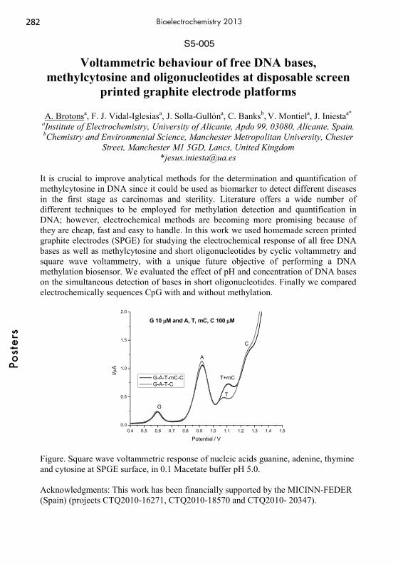
S5-005
Voltammetric behaviour of free DNA bases, methylcytosine and oligonucleotides at disposable screen
printed graphite electrode platforms
A. Brotonsa, F. J. Vidal-Iglesiasa, J. Solla-Gullóna, C. Banksb, V. Montiela, J. Iniestaa* aInstitute of Electrochemistry, University of Alicante, Apdo 99, 03080, Alicante, Spain. bChemistry and Environmental Science, Manchester Metropolitan University, Chester
Street, Manchester M1 5GD, Lancs, United Kingdom *[email protected]
It is crucial to improve analytical methods for the determination and quantification of methylcytosine in DNA since it could be used as biomarker to detect different diseases in the first stage as carcinomas and sterility. Literature offers a wide number of different techniques to be employed for methylation detection and quantification in DNA; however, electrochemical methods are becoming more promising because of they are cheap, fast and easy to handle. In this work we used homemade screen printed graphite electrodes (SPGE) for studying the electrochemical response of all free DNA bases as well as methylcytosine and short oligonucleotides by cyclic voltammetry and square wave voltammetry, with a unique future objective of performing a DNA methylation biosensor. We evaluated the effect of pH and concentration of DNA bases on the simultaneous detection of bases in short oligonucleotides. Finally we compared electrochemically sequences CpG with and without methylation.
0.4 0.5 0.6 0.7 0.8 0.9 1.0 1.1 1.2 1.3 1.4 1.50.0
0.5
1.0
1.5
2.0
I/μA
Potential / V
G-A-T-mC-C G-A-T-C
G
A
T+mC
T
C
G 10 μM and A, T, mC, C 100 μM
Figure. Square wave voltammetric response of nucleic acids guanine, adenine, thymine and cytosine at SPGE surface, in 0.1 Macetate buffer pH 5.0. Acknowledgments: This work has been financially supported by the MICINN-FEDER (Spain) (projects CTQ2010-16271, CTQ2010-18570 and CTQ2010- 20347).
282Po
ster
sBioelectrochemistry 2013
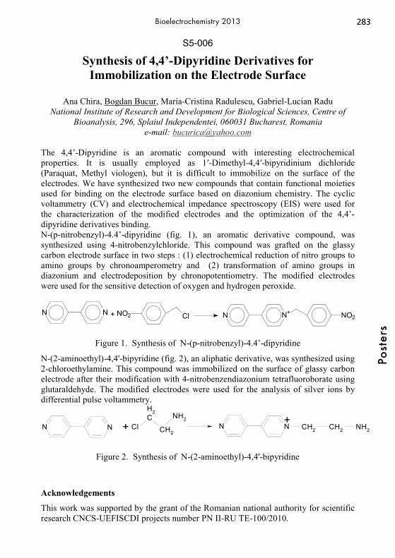
S5-006
Synthesis of 4,4’-Dipyridine Derivatives for Immobilization on the Electrode Surface
Ana Chira, Bogdan Bucur, Maria-Cristina Radulescu, Gabriel-Lucian Radu
National Institute of Research and Development for Biological Sciences, Centre of Bioanalysis, 296, Splaiul Independentei, 060031 Bucharest, Romania
e-mail: [email protected] The 4,4’-Dipyridine is an aromatic compound with interesting electrochemical properties. It is usually employed as 1′-Dimethyl-4,4′-bipyridinium dichloride (Paraquat, Methyl viologen), but it is difficult to immobilize on the surface of the electrodes. We have synthesized two new compounds that contain functional moieties used for binding on the electrode surface based on diazonium chemistry. The cyclic voltammetry (CV) and electrochemical impedance spectroscopy (EIS) were used for the characterization of the modified electrodes and the optimization of the 4,4’-dipyridine derivatives binding. N-(p-nitrobenzyl)-4.4’-dipyridine (fig. 1), an aromatic derivative compound, was synthesized using 4-nitrobenzylchloride. This compound was grafted on the glassy carbon electrode surface in two steps : (1) electrochemical reduction of nitro groups to amino groups by chronoamperometry and (2) transformation of amino groups in diazonium and electrodeposition by chronopotentiometry. The modified electrodes were used for the sensitive detection of oxygen and hydrogen peroxide.
Figure 1. Synthesis of N-(p-nitrobenzyl)-4.4’-dipyridine
N-(2-aminoethyl)-4,4'-bipyridine (fig. 2), an aliphatic derivative, was synthesized using 2-chloroethylamine. This compound was immobilized on the surface of glassy carbon electrode after their modification with 4-nitrobenzendiazonium tetrafluoroborate using glutaraldehyde. The modified electrodes were used for the analysis of silver ions by differential pulse voltammetry.
N N + ClCH2
CH2
NH2N N CH2 CH2 NH2
+
Figure 2. Synthesis of N-(2-aminoethyl)-4,4'-bipyridine
Acknowledgements
This work was supported by the grant of the Romanian national authority for scientific research CNCS-UEFISCDI projects number PN II-RU TE-100/2010.
N N + NO2 Cl N N+ NO2
283
Post
ers
Bioelectrochemistry 2013
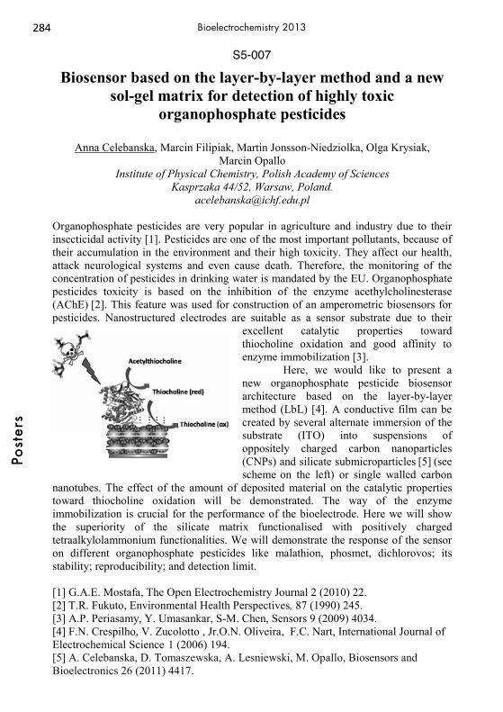
S5-007
Biosensor based on the layer-by-layer method and a new sol-gel matrix for detection of highly toxic
organophosphate pesticides
Anna Celebanska, Marcin Filipiak, Martin Jonsson-Niedziolka, Olga Krysiak, Marcin Opallo
Institute of Physical Chemistry, Polish Academy of Sciences Kasprzaka 44/52, Warsaw, Poland.
Organophosphate pesticides are very popular in agriculture and industry due to their insecticidal activity [1]. Pesticides are one of the most important pollutants, because of their accumulation in the environment and their high toxicity. They affect our health, attack neurological systems and even cause death. Therefore, the monitoring of the concentration of pesticides in drinking water is mandated by the EU. Organophosphate pesticides toxicity is based on the inhibition of the enzyme acethylcholinesterase (AChE) [2]. This feature was used for construction of an amperometric biosensors for pesticides. Nanostructured electrodes are suitable as a sensor substrate due to their
excellent catalytic properties toward thiocholine oxidation and good affinity to enzyme immobilization [3].
Here, we would like to present a new organophosphate pesticide biosensor architecture based on the layer-by-layer method (LbL) [4]. A conductive film can be created by several alternate immersion of the substrate (ITO) into suspensions of oppositely charged carbon nanoparticles (CNPs) and silicate submicroparticles [5] (see scheme on the left) or single walled carbon
nanotubes. The effect of the amount of deposited material on the catalytic properties toward thiocholine oxidation will be demonstrated. The way of the enzyme immobilization is crucial for the performance of the bioelectrode. Here we will show the superiority of the silicate matrix functionalised with positively charged tetraalkylolammonium functionalities. We will demonstrate the response of the sensor on different organophosphate pesticides like malathion, phosmet, dichlorovos; its stability; reproducibility; and detection limit.
[1] G.A.E. Mostafa, The Open Electrochemistry Journal 2 (2010) 22. [2] T.R. Fukuto, Environmental Health Perspectives, 87 (1990) 245. [3] A.P. Periasamy, Y. Umasankar, S-M. Chen, Sensors 9 (2009) 4034. [4] F.N. Crespilho, V. Zucolotto , Jr.O.N. Oliveira, F.C. Nart, International Journal of Electrochemical Science 1 (2006) 194. [5] A. Celebanska, D. Tomaszewska, A. Lesniewski, M. Opallo, Biosensors and Bioelectronics 26 (2011) 4417.
284Po
ster
sBioelectrochemistry 2013

S5-008
New Osmium Loaded Hybrid Microgels as Biosensors with Controlled Redox Centers Nano-Gaps
Andrea Contin1, Sascha Pöller1, Dmitrii A. Guschin1, Véronique Lapeyre2, Valerie Ravaine2, Wolfgang Schuhmann1
1Analytische Chemie -Elektroanalytik & Sensorik; Center of Electrochemical Sciences - CES, Ruhr-Universität Bochum; D-44780 Bochum Germany
2Institut des Sciences Moléculaires - UMR 5255 - Université de Bordeaux 1; ENSCBP - Institut Polytechnique de Bordeaux; 33607 Pessac - France
In the past years composite microgels have been intensively studied due to the possibility to enhance the general features of pure microgels and to additionally also add peculiar features imparted from the nature of the second component within the microgel. A rational selection of starting materials and polymerization conditions may lead to a useful and controlled combination of the final properties of the composite microgels. The most common additives were clays, metal oxides and different kind of biomaterials such as proteins[1] or coniugated polymers aiming on changes of the characteristics of pure microgels such as colloidal stability, catalytic activity etc. Following the work carried by Ravaine et al.[2], a poly( isopropylacrylamide/methy-lenebisacrylamide) core-shell microgel has been modified through the addition of phenylboronic acid derivatives, seeking for enhanced affinity towards a “bio-target”, namely glucose. In fact, phenylboronic acid forms a stable complex with glucose at pH values close to its pKa (8.8). Furthermore, the formation of the mentioned complex in-creases the solubility of boronic acid, leading to the swelling of the “hosting” microgel nanoparticle. The until now unexplored application of these new hybrid polymers in electrochemical applications is of high interest. Adding specifically selected monomers to the microgel structure allowed us to load redox centers, namely Os-complexes, onto the latter. The presence of active redox mediators allows to analyze the electron transfer rate of electron hopping among redox centers, which is above all distance dependent. The action of an enzyme which modifies the key parameter “microgel swelling degree/ re-dox centers gaps”, seems to work as a perfect “trigger” to reduce the distance between Osmium complexes. Using glucose oxidase, which catalyzes the oxidation of glucose to gluconic acid, the enzymatically catalyzed conversion is leading to a substantial change of the swelling properties which in turn is leading to a modulation of the electron-transfer rate within the polymer. First experiments showing the polymer de-sign and synthesis and the feasibility of the suggested approach will be presented. [1] S. Su, M. M. Ali, C. D.M. Filipe, L. Yingfu, R. Pelton, Macromol. 9 (2008) 935-8 [2] V. Lapeyre, C. Ancla, B. Catargi, V. Ravaine, J. Coll. Interf. Sci. 327 (2008) 316–323
285
Post
ers
Bioelectrochemistry 2013

S5-009
Electrochemical immunoassay for the detection of MUC1 cancer biomarker
Cecilia Cristea1, Anca Florea1, 2, Giovanna Marrazza2, Robert Săndulescu1
1 Analytical Chemistry Department, Faculty of Pharmacy, Iuliu Haţieganu University of Medicine and Pharmacy, Pasteur 6, 400349, Cluj-Napoca, Romania
[email protected] 2 Ugo Schiff ChemistryDepartment, Universita degli studi di Firenze
Via della Lastruccia 3, 50019 Sesto Fiorentino, Italy Considerable efforts have been made during the last years for the development of precise, rapid, sensitive and selective immunosensors for cancer biomarker detection. Increased levels of MUC1 protein are associated with cancer [1]. MUC1 is a transmembrane protein, heavily O-glycosylated, which is physiologically expressed on the apical plasma membrane of most secretory epithelia. In case of malignant processes MUC1 is overexpressed and underglycosylated and is involved in tumor progression, serving as tumor marker [2]. Aptamers have been widely used as sensing elements for biosensor development for their unique advantages such as small size, low cost, easy to synthesize in vitro and good storage properties [3]. The goal of the present work is to develop new screening devices for the detection of Mucin 1 (MUC1) protein, using two different approaches: an ELISA-like antibodies sandwich assay and an aptamer-based assay on graphite screen printed electrodes. Magnetic beads (MBs) were used as immobilization platforms in order to achieve this goal. The primary antibody and the biotinylated aptamer were immobilized on Protein G and streptavidin modified MBs respectively, in order to capture the MUC1 antigen. The sandwich assay was then performed by adding a secondary anti-MUC1 antibody and a third alkaline phosphatase (AP)-labeled antibody. The modified MBs are then captured by a magnet on the surface of a graphite working electrode and after the addition of the AP substrate (1-naphthyl-phosphate) the 1-naphthol produced during the enzymatic reaction is detected using differential pulse voltammetry (DPV). The experimental conditions regarding the incubation time and concentration of antibodies and aptamer solutions were optimized and LODs of -0.62 ppb for the antibodies assay and 1.63 ppb for the aptamer-based assay in a linear range of 0-10 ppb were obtained. This study provides a new appealing tool for the detection of cancer biomarker MUC1 with the possibility for future medical applications. References [1] SJ. Gendler. Mammary Gland Biol Neoplasia 2001, 6, 339; [2] C. Van Elssen, P. Frings, F. Bot, K. Van de Vijver, M. Huls, Histopathology, 2010, 57(4), 597-606; [3] S. Centi, S. Tombelli, M. Minunni, M. Mascini, Anal. Chem. 2007, 79, 1466-1473.
286Po
ster
sBioelectrochemistry 2013
S5-010
Novel DNA Redox Labels at Silver Solid Amalgam Electrode
Ales Danhel1, Ludek Havran1, Hana Pivonkova1, Veronika Raindlova2, Michal Hocek2,
and Miroslav Fojta1 1 Institute of Biophysics, Academy of Sciences of the Czech Republic, v.v.i.,
Kralovopolska 135, CZ-61265 Brno, Czech Republic 2 Institute of Organic Chemistry and Biochemistry, Academy of Sciences of the Czech
Republic, v.v.i., Flemingovo nam. 2, CZ-16610 Prague 6, Czech Republic [email protected]
Electrochemical behaviour of recently designed DNA redox labels at mercury meniscus modified silver solid amalgam electrode (m-AgSAE) using cyclic voltammetry, adsorptive stripping voltammetry (AdSV) and transfer stripping voltammetry (TSV) will be presented in this work. Mechanically stable and robust m-AgSAE represents, in combination with voltammetric methods, convenient tool for development of sensors applicable in detection of bio-molecules, such as oligonucleotides (ONs) and/or nucleic acids [1,2]. Attachment of an appropriate DNA redox label to the selected nucleotide and its successful incorporation to the oligonucleotide or DNA [3-5] may increase sensitivity, selectivity and signal response diversity of studied bio-molecules using the m-AgSAE. This approach to sensing of the DNA utilizing m-AgSAE may be further used for development of novel, and sufficiently sensitive and robust detectors applicable for bioassays in toxicological, biological, and medicinal applications concerning to the completion of genome sequencing, identification of the relationship between the individual genome characteristics, cellular functions or diseases, and in development of widely accessible and decentralized diagnostic methods [6]. [1] M. Fojta, L. Havran, H. Pivonkova, P. Horakova, M. Hocek, Curr. Org. Chem., 15 (2011) 2936-2942. [2] M. Hocek, M. Fojta, Chem. Soc. Rev., 40 (2011) 5802-5814. [3] V. Raindlova, R. Pohl, M. Sanda, M. Hocek, Angew. Chem., Int. Ed., 49 (2010) 1064-1066. [4] V. Raindlova, R. Pohl, B. Klepetarova, L. Havran, E. Simkova, P. Horakova, H. Pivonkova, M. Fojta, M. Hocek, ChemPlusChem, 77 (2012) 652-662. [5] P. Horakova, H. Macickova-Cahova, H. Pivonkova, J. Spacek, L. Havran, M. Hocek, M. Fojta, Org. Biomol. Chem., 9 (2011) 1366-1371. [6] E. Palecek, M. Bartosik, Chem. Rev., 112 (2012) 3427–3481.
287
Post
ers
Bioelectrochemistry 2013

S5-011
Surface Modification of a Novel Benzimidazole Containing Polymer for Biomolecule Immobilization and
Biosensing Applications
Sema Demirci Uzun 1, Naime Ünlü Akbaşoğlu2, Fulya Ekiz Kanik 3, Duygu Kozanoğlu4, Emren Nalbant Esentürk2,4, Suna Timur5, Levent Toppare1,2,3,6
1 Department of Polymer Science and Technology, Middle East Technical University 2Department of Chemistry, Middle East Technical University
3Department of Biotechnology, Middle East Technical University 4Department of Micro and Nanotechnology, Middle East Technical University
5Department of Biochemistry, Ege University 6The Center for Solar Energy Research and Application (GÜNAM), Middle East
Technical University [email protected]
Conducting polymers (CPs) with their unique electronic, electrochemical and optical properties are useful in improving many technological applications. A biosensor, one of which, can be easily fabricated using conducting polymers as appropriate immobilization matrices for biomolecules. Furthermore, the use of these fascinating polymers for biomolecule immobilizations enhance stability, sensitivity and efficient electron transfer ability for the biosensors.1,2 In this study, graphite electrode surfaces were modified with single walled carbon nanotubes (SWCNT), gold nanoparticles (AuNPs) and both of these nanostructures. Subsequently, a novel monomer; 4-(4,7-di(thiophen-2-yl)-3a,7a-dihydro-1H-benzo[d] imidazol-2-yl)benzaldehyde was electropolymerized on these modified electrodes. Glucose oxidase (GOx) molecules were covalently attached onto the polymer coated graphite electrodes. During this process, imine bond formation between aldehyde groups of the polymer and amine groups of the enzyme molecules was successfully achieved. Constructed biosensors were tested at -0.7 V vs. Ag/AgCl AgCl in amperometric techniques and responses are correlated with the substrate concentration to determine the biosensor performances. X-Ray photoelectron spectroscopy (XPS) and transmission electron microscope (TEM) were used to characterize surface properties and morphologies for each step during the preparation of biosensors. The modified poly-4-(4,7-di(thiophen-2-yl)-1H-benzo[d]imidazol-2-yl)benzaldehyde surfaces showed excellent immobilization matrix properties in terms of their good kinetic parameters (Km
app and Imax) and low LOD values with high operational and storage stabilities for glucose sensing. Moreover, the combination of SWCNT and AuNPs coating with the polymer provides large surface areas for the immobilization of proteins. References [1] Chen, W., Cai, S., Ren, Q.-Q., Wen, W. and Zhao, Y.-D., Analyst, 137, 49-58, 2012. [2] Chauhan, N., Narang, J. and Pundir, C. S., Analyst, 136, 1938-1945, 2011.
288Po
ster
sBioelectrochemistry 2013
S5-012
Development of a Conducting Polymer Based Amperometric Acetylcholine Biosensor for Pesticide
Detection
Fulya Ekiz Kanika, Marit Kolbb, Suna Timurc, Müfit Bahadirb, Levent Topparea,d,e,f PhD candidate, PhD, Prof. Dr., Prof. Dr., Prof. Dr.
a Department of Biotechnology, Middle East Technical University, Ankara 06800, Turkey
b Institute of Environmental and Sustainable Chemistry, Technische Universität Braunschweig, Hagenring 30, Braunschweig 38106, Germany
c Department of Biochemistry, Faculty of Science, Ege University, Izmir 35100, Turkey d Department of Chemistry, Middle East Technical University, Ankara 06800, Turkey e Department of Polymer Science and Technology, Middle East Technical University,
Ankara 06800, Turkey f The Center for Solar Energy Research and Application (GUNAM), Middle East
Technical University, Ankara 06800, Turkey [email protected], [email protected], [email protected], [email protected],
Conjugated polymers are highlighted with their distinct properties serving as multipurpose materials in many different application fields such as solar cells [1] and imaging [2]. According to the tunable properties, they can be applied for enzyme immobilization for mechanically robust and stable biosensors [3]. CPs are considered as appropriate host for enzymes since the enzyme molecules can easily and sufficiently anchored on the electrode surface assisted by the CPs. An amperometric acetylcholine biosensor was prepared by the generation of CP; poly(4-(2,5-di(thiophen-2-yl)-1H-pyrrol-1-yl)benzenamine) (poly(SNS-NH2)) on the graphite electrodes. Fabrication of amperometric acetylcholine biosensor for pesticide detection by co-immobilization of acetylcholinesterase (AChE) and choline oxidase (ChO) onto poly(SNS-NH2) films using covalent binding is described. Characterization of resulting acetylcholine biosensor was discovered in terms of optimum pH, enzyme loading, linear response range and shelf-life. Linear range and shelf-life were found as 0.12-10 mM and 4 weeks, respectively. The designed biosensor was employed in determination of paraoxon-ethyl in spiked tap water samples according to the inhibition principle of the pesticides. Satisfactory results were obtained for paraoxon-ethyl determination in water samples compared to a reference procedure. [1] M. Karakus, D. H. Apaydn, D. E. Yldz, L. Toppare, A. Cirpan, Polymer 53 (2012) 1198-1202. [3] M. Ak, A. Durmus, L. Toppare, Solid State Sci. 9 (2007) 843-849. [3] F. Ekiz, F. Oguzkaya, M. Akin, S. Timur, C. Tanyeli, L. Toppare, J. Mater. Chem., 21 (2011) 12337–12343.
289
Post
ers
Bioelectrochemistry 2013

S5-013
Opposing effects of riboflavin and methylene blue on DNA oxidation
Hanna Elzanowska, Agnieszka Gniazdowska, Adriana Palinska-Saadi, Magdalena
Maj-Zurawska Faculty of Chemistry, University of Warsaw, Pasteura 1, 02-093 Warsaw, Poland
Tel: +48 22 8220211, e-mail: [email protected] Although the oxidation of nucleic acid bases on carbon electrodes is often diminished, e.g. by anticancer drugs, cis-platin or carboplatin [1], the opposite effect is also observed in some other cases [2]. In our earlier studies [3-5], strong enhancement of plasmid DNA oxidation by methylene blue (MB), a typical DNA intercalator, or by deposited metal (Pd) nanoparticles [6] has been documented. Now, we compare these results with the effect of riboflavin (RF) interactions on the oxidation of the pGEX-4T-2 plasmid in both supercoiled and linear forms. Slow accumulation of the plasmid on a glassy carbon (GC) electrode from highly diluted solutions (pg/mL) allows for the formation of a stable plasmid DNA layer as judged by the oxidation signals >1 V detected by SW, DP and AC voltammetry. The incorporation of either RF, or MB is shown as oxidation of these substances in their typical potential ranges from -0.5 V to ca. 0 V, and also at high potentials close to 1 V, leading in case of MB to a significant enhancement of the DNA signal. Surprisingly, such enhancement is not observed for RF, even though the interactions of RF with the plasmid are visible as a shift of the redox potential of RF oxidation. This result is confirmed by the competitive incorporation of MB in the presence of RF. The differences in DNA-RF interactions are also detected when two different forms of the pGEX-4T-2 plasmid, supercoiled and linear, are used for the formation of a DNA layer on the GC electrode. References [1] G. Bagni, D. Osella, E. Sturchio. M. Mascini, Anal. Chim. Acta 573-574 (2006) 81. [2] J. Labuda, A.M. Oliveira Brett, G. Evtugyn, M. Fojta, M. Mascini, M. Ozsoz, I.
Palchetti, E. Palecek, and J. Wang, Pure Appl. Chem. 82 (2010) 1161. [3] A. Palinska, A. Grodzka, H. Elzanowska, B. Kepska, E. Zwierkowska, S.
Achmatowicz, M. Maj-Zurawska; Electroanalysis 22 (2010) 1306. [4] A. Gniazdowska, A. Palinska, E. Krawczyk, H. Elzanowska, M. Maj-Zurawska, Supercoiled and linear plasmid DNAs interactions with methylene blue, BIOELECHEM-D-12-00046R1. [5] A. Palinska-Saadi, E. Krawczyk, K. Michalska, H. Elzanowska, M. Maj-Zurawska, Electrochemical detection of supercoiled and linear plasmid DNA, BIOELECHEM-D-11-00376R1. [6] H. Elzanowska, A. Ogorek, P. Kulesza, 61st Annual Meeting of the International
Society of Electrochemistry, September 26th, October 1, 2010, Niece, France.
290Po
ster
sBioelectrochemistry 2013

S5-014
Carbazole Unit and TiO2 Nanoparticles Containing Biosensor and Its Applications
Fatma Bilge Emre1, Funda Sayılkan1, Yasemin Aslan Udum2, Levent Toppare3, 4,5, 6
1 Faculty of Education, İnönü University 2 Department of Advenced Technologies, Institute of Science andTechnology, Gazi
University 3 Department of Chemistry, Middle East Technical University
4 Department of Biotechnology, Middle East Technical University 5 Department of Polymer Science and Technology, Middle East Technical University
6 The Center for Solar Energy Research and Application (GÜNAM ), Middle East Technical University
[email protected], [email protected], [email protected], [email protected]
Conducting polymers are utilized as a platform for immobilization of biomolecules. These novel materials can be prefered in some areas like organic light emitting diodes, biosensor applications [1]. They are biocompatible with enzymes. Enzyme modified electrodes have several advanteges such as high stability, fast response and low cost. Metal oxide nano materials are suitable for designing new and improved sensing devices, especially electrochemical sensors and biosensors studies. These are used in many areas such as sensor, catalyst, antibacterial applications [2], personal care products. Especialy TiO2 is an attractive material for use of biosensors, Metal oxide nanomaterials have good properties such as high specific surface area, high reactivity. Nanopartices provide immobilization of biomolecules. These materials was catalysed electrochemical reactions via electron transfer between electrode surfaces and proteins, labeling of biomolecules and even acting as reactant [3]. In this study, firstly we synhesized nano-sized TiO2 powders at low-temperature via hydrothermal methods. Nano-TiO2 powders has been turned into the sol in water to have easy interaction with GOx. Nano-TiO2 powders was characterized with XRD and particle size analyzer. After that 4-methly carbozol-3-carboxylic acid was electrochemically deposited on graphite electrodes and used as immobilization matrix for biosensing studies. After electrochemical deposition of the polymeric matrix TiO2 and glucose oxidase (GOx) was immobilized on the modified electrodes as the model enzyme. The decrease in oxygen level as a result of enzymatic reaction was monitored at −0.7 V vs Ag/AgCl (3.0 M KCl) and correlated with substrate concentration. The biosensor was used for several parameters such as a operational storage and stabilities. Surface characterizations were done with SEM. [1] Marek Trojanowicz, Microchim. Acta 143, 75–91,(2003. [2] Zhaobo Wang, Guıcun Lı , Hongruı Peng, Zhıkun Zhang, Journal Of Materıals Science 40, 6433–6438, 2005. [3] Xiliang Luo, Aoife Morrin, Anthony J. Killard, Malcolm R. Smyth, Electroanalysis 4, 319 – 326, 2006.
291
Post
ers
Bioelectrochemistry 2013

S5-015
Benzoimidazole Based Conducting Polymer and PMMA/Clay Nanocomposite Containing Biosensor and Its
Applications
Fatma Bilge Emre1, Melis Kesik2, Fulya Ekiz Kanik3, Evren Aslan Gurel4, Levent Toppare2, 3, 5, 6
1 Faculty of Education, İnönü University 2 Department of Chemistry, Middle East Technical University
3 Department of Biotechnology, Middle East Technical University 4EMPA Swiss Federal Laboratories for Materials Science and Technology, Laboratory
for Protection and Physiology, Switzerland 5 Department of Polymer Science and Technology, Middle East Technical University
6 The Center for Solar Energy Research and Application (GÜNAM ), Middle East Technical University
[email protected], [email protected], [email protected], [email protected], [email protected]
Conducting polymers are recent generation of polymeric materials which opened the way of progress in achieving new type of polymers for several applications. These novel materials can be used in several areas such as electrochromic devices, photovoltaics, organic light emitting diodes and biosensor applications [1]. Conducting polymers have recently been considered as suitable matrices for biomolecules and have been used to improve the properties of various biomedical devices [2]. Using electropolymerization, several properties such as morphology and thickness can be easily controlled. Clays have been used in extensive areas such as pharmacy, cosmetics and ion exchanges. Laponite is a synthetic smectite that resembles the natural clay mineral hectorite in both structure and composition. Via modifications with different polymers, laponite nanomaterials can be prepared such as PMMA/clay nanocomposites. In this work firstly 4,7-bis(2,3-dihydrothieno[3,4-b][1,4]dioxin-5-yl)-2-phenyl-2,7a-dihydro-1H-benzo[d]imidazole) was electrochemicaly polymerized on a graphite electrode in order to use as an immobilization matrix for biosensing studies. After electrochemical deposition of the polymeric matrix, PMMA/clay nanocomposites and glucose oxidase (GOx) were consecutively immobilized onto the polymer coated electrode surface. Due to the lack of functional groups in the structure of the polymer, matrix was functionalized with clay nanoparticles to enhance the stability of the enzymes on the electrode surface and improve sensing ability of the biosensor. Amperometric measurement technique was used in biosensor applications. In this study, the decrease in oxygen level as a result of enzymatic reaction was monitored at -0.7 V vs Ag/AgCl and related with the substrate concentration. [1] Heeger, A. J. Chemical Society Reviews, 39, 2354-2371, 2010. [2] Ravichondran, R., Sundarrajan, S., Venugopal, J.,Mukherjee, S. andRamakrishna, S. Journal of the Royal Society Interface, 7, 559-579, 2010. [3] Mousty C., Applied Clay Science, 27, 159-177, 2004.
292Po
ster
sBioelectrochemistry 2013

S5-016
Electrochemical Detection of Superoxide Using SOR Immobilized on Carbon Nanotubes
Fabiane Caxico de Abreu2, Jonathan Caranto1 , Donald Kurtz1, and Carlos D. Garcia1
1 Department of Chemistry, The University of Texas at San Antonio, One UTSA Circle, San Antonio, TX 78249
2Institute of Chemistry and Biotechnology, Federal University of Alagoas, Brazil [email protected]
Superoxide, O2
-, is one of the reactive oxygen species that are ubiquitously produced in biological respiration and metabolism. It is closely related to a number of biological processes, such as aging, ischemia–perfusion and inflammation. Because of this, the detection of O2
- has become interest to scientists from many fields. Among other strategies for the analysis of O2
-, electrochemical reduction is one of the most interesting ones because it can enable its analysis at low concentrations and in biological matrices. In order to improve the selectivity of the analysis, superoxide reductase (SOR) can be used. SOR is a non-heme iron protein that catalyzes the reduction of superoxide to hydrogen peroxide1. In this project, a third generation biosensor for superoxide anion (O2
-) was developed by immobilizing SOR on the surface of carbon nanotubes (CNT). The SOR was obtained by expression of a plasmid containing Thermotoga maritima SOR (Tm SOR) with an N-terminal 6X-HisTag, obtained from the Joint Center for Structural Genomics. Molecular modeling calculations (Autodoc) showed that SOR has more affinity for Multiwall CNT (-40.8 Kcal/mol) than Singlewall CNT (-22 Kcal/mol). The high efficiency of the electron transfer of SOR and the selectivity of SOR toward O2
- enabled a sensitive electrochemical measurement of O2- at –200 mV (vs.
Ag/AgCl/ClSAT). Under the optimized solution conditions (0.1 M phosphate buffer, pH 8.2), a detection limit of 0.10μM was obtained. Remarkably, the biosensor enabled the detection without significant interferences from physiological levels of ascorbic or uric acids. (a) (b)
50 100 150 200 250 300 350 400 450-4.2
-4.0
-3.8
-3.6
-3.4
-3.2
Cur
rent
/ A
E / V vs Ag|AgCl|Cl- sat
Figures:(a) SOR on the MWCNT according to the AutoDock Vina molecular docking program; (b) Chronoamperometric curve of i-t of the O2
- in the PBS 8.2, Edet= -0.2V Reference: 1. Emerson J. P , Coulter, E. D. , Cabelli, D. E., Phillips, R. S. and Kurtz, D. M. Biochemistry, 41 (13), 2002, 4348. Acknowledgements The financial support provided by CNPq, INCT, UFAL and UTSA.
293
Post
ers
Bioelectrochemistry 2013
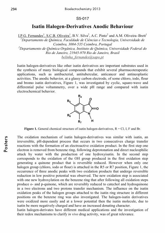
S5-017
Isatin Halogen-Derivatives Anodic Behaviour
I.P.G. Fernandes1, S.C.B. Oliveira1, B.V. Silva2, A.C. Pinto2 and A.M. Oliveira–Brett1 1Departamento de Química, Faculdade de Ciências e Tecnologia, Universidade de
Coimbra, 3004-535 Coimbra, Portugal 2Departamento de Química Orgânica, Instituto de Química, Universidade Federal do
Rio de Janeiro, 21945-970 Rio de Janeiro, Brasil [email protected]
Isatin halogen-derivatives like other isatin derivatives are important substrates used in the synthesis of many biological compounds that exhibit several pharmacoterapeutic applications, such as antibacterial, antitubercular, anticancer and antineoplastic activities. The anodic behavior, at a glassy carbon electrode, of some chloro, iodo, flour and bromo isatin derivatives, Figure 1, was investigated by cyclic, square-wave and differential pulse voltammetry, over a wide pH range and compared with isatin electrochemical behavior.
N
O
O
H
R5
R6
R4
R7
Figure 1. General chemical structure of isatin halogen-derivatives, R = Cl, I, F and Br.
The oxidation mechanism of isatin halogen-derivatives was similar with isatin, an irreversible, pH-dependent process that occurs in two consecutives charge transfer reactions with the formation of an electroactive oxidation product. In the first step one electron is removed from benzene ring, following deprotonation and direct nucleophilic attack by water with the production of one hydroxyisatin. In the second step corresponds to the oxidation of the OH group produced in the first oxidation step generating a quinone product that is reversible reduced. However when only one halogen group (chloro, iodo or flour) is attached in the R5 or R7 position, Figure 1, the occurrence of three anodic peaks with two oxidation products that undergo reversible reduction in low positive potential was observed. The new oxidation step is associated with one new hydroxylation on the benzene ring that after following all oxidation steps produce o- and p-quinone, which are reversibly reduced to catechol and hydroquinone in a two electrons and two protons transfer mechanism. The influence on the isatin oxidation peaks of the halogen groups attached to the isatin ring structure in different positions on the benzene ring was also investigated. The halogen-isatin derivatives were oxidized more easily and at a lower potential then the isatin molecule, due to isatin be more negatively charged and have an increased donating character. Isatin halogen-derivates have different medical applications and the investigation of their redox mechanisms to clarify in vivo drug activity, was of great relevance.
294Po
ster
sBioelectrochemistry 2013
S5-018
Detection of biomolecules at nanostructured surfaces by combining of electrochemistry and spectroscopy
Stefanie Grützke, Sascha Pöller, Magdalena Gebala, Wolfgang Schuhmann
Analytische Chemie – Elektroanalytik & Sensorik Ruhr-Universität Bochum, Universitätsstr. 150, 44780 Bochum, Germany
[email protected] The intercalation process of small compounds into double stranded DNA (dsDNA) has been in the focus of research for the past four decades. Electrochemical impedance spectroscopy (EIS) is a very sensitive method to characterize DNA-modified electrode surfaces as well as investigate the intercalation process.[1] Furthermore, many of the compounds which may intercalate into dsDNA are SERS (surface enhanced Raman scattering) active and can therefore not only be analyzed by EIS but also be visualized by means of Raman spectroscopy.[2] Not only the intercalation process can be detected but also different redox states may be observed in this way. For example a system of a lactate and glucose dehydrogenase entrapped within an electrocatalyst film can be used to investigate electrocatalytic NADH oxidation.[3] The change of the redox potential will also lead to a change of the Raman signal which can be detected. To achieve a surface enhancement of the Raman signal specific conditions are necessary. Especially the presence of “hot spots” areas is needed where the electromagnetic field is strongly localized caused by nanoscale junctions between suitable metallic nanoobjects.[4] The development of nanostructured surfaces which can be used to enhance the Raman signal of the biomolecules are in the focus of many studies.[5] The new aspect of our research is the electrochemical modification of a (bio)func-tionalized surface and the simultaneous detection of these changes with Raman spectroscopy especially SERS. As a result we achieve two related information of spectroscopy and electrochemistry. In this study we present the results of the local electrochemical modification of (bio)molecules on a nanostructured electrode which were afterwards detected by surface enhanced Raman scattering. [1] M. Gebala, L. Stoica, S. Neugebauer, W. Schuhmann, Electroanalysis 21(2009) 325 [2] S. Grützke, S. Abdali, W. Schuhmann, M. Gebala, Electrochem. Commun. 19
(2012) 59 [3] E. Al-Jawadi, S. Pöller, R. Haddad, W. Schuhmann, Microchim Acta 177 (2012)
405 [4] M. Abdelsalam, P.N. Bartlett, A.E. Russel, J.J. Baumberg, E.J. Calvo, N.G.
Tognalli, A. Fainstein, Langmuir 24 (2008)7018 [5] R. Szamocki, P. Massé, S. Ravaine, V. Ravaine, R. Hempelmann, A. Kuhn, J.
Mater. Chem. 19 (2008) 409
295
Post
ers
Bioelectrochemistry 2013

S5-019
Electrochemical determination of antihypertensive drug irbesartan in pharmaceuticals
V. K. Guptaa,b, R. Jainc, S. Agarwalc, R. Mishrac, A. Dwivedic
a Department of Chemistry, Indian Institute of Technology Roorkee, Roorkee 247 667, India
b Department of Chemistry, King Fahd University of Petroleum and Minerals, Dhahran 31261, Saudi Arabia
c School of Studies in Chemistry, Jiwaji University, Gwalior 474011, India E-mail: [email protected]
A sensitive voltammetric method has been developed for the determination of irbesartan in a Britton–Robinson buffer medium. Irbesartan exhibited a well-defined cathodic peak over the entire pH range from 2.0 to 12.0. The mechanism of reduction was postulated on the basis of controlled potential electrolysis, coulometry, and spectral analysis. Under optimal conditions, a linear response of irbesartan was obtained in the range from 3.0 × 10⁻⁵ to 5.7 × 10⁻³ mol L−1 and with a limit of detection of 5.33 × 10⁻⁷ mol L−1. The effect of cationic surfactant on the voltammetric reduction peak of irbesartan in Britton–Robinson buffer is also described.
Chemical structure of irbesartan.
296Po
ster
sBioelectrochemistry 2013
S5-020
Electrochemical analysis of DNA structure transitions
Luděk Havran, Pavlína Vidláková, Hana Pivoňková, Iva Kejnovská, Michaela Vorlíčková, Miroslav Fojta
Institute of Biophysics ASCR,v.v.i. Královopolská 135, 612 65 Brno, Czech Republic
Nucleic acids are electroactive species producing set of intrinsic voltammetric signals at different types of working electrodes [1]. At mercury electrodes, adenine and cytosine produce a cathodic peak CA (due to its reduction). Guanine gives anodic signal G, which is obtained in cyclic voltammetry (CV) or in anodic stripping modes (due to oxidation of reduction product of guanine that is reduced at very negative potentials). At carbon electrodes guanine and adenine undergo oxidation at positive potentials and produce oxidations signals Aox and Gox [2]. The nucleic acids, depending on their nucleotide sequences, can exhibit a variety of conformational transitions and adopt different secondary structures such as hairpins, triplexes, left-handed Z-DNA or quadruplexes. Some of them may occur in natural DNAs as local structures stabilized by negative DNA supercoiling and it is generally accepted that (at least) some of them have biological (regulatory) functions [3]. In vitro the “non-B” structures can be created using model synthetic oligonucleotides (ON) of suitable base sequence [4]. In this contribution we studied influence of forming of G-quadruplexes (in G-rich ON) and hairpins on electrochemical behavior synthetic ON at mercury and carbon electrodes. Literature [1] E. Paleček, M. Bartošík, Electrochemistry of Nucleic Acids. Chemical Reviews 112
(2012) 3427-3481 [2] E. Paleček, F. Jelen, In Electrochemistry of nucleic acids and proteins Towards
electrochemical sensors for genomics and proteomics (Eds: E. Paleček, F. Scheller, J. Wang), Elsevier, Amsterdam, 2005, Vol. 1, pp 74-174.
[3] Paleček, E. Critical Reviews in Biochemistry and Molecular Biology 1991, 26, 151-226.
[4] Blackburn, G. M.; Gait, M. J. Nucleic acids in chemistry and biology; Oxford university press: Oxford, 1996.
Acknowledgments This work was supported by the GACR (P206/12/2378, P206/12/G151).
297
Post
ers
Bioelectrochemistry 2013

S5-021
Impedimetric Microbial Sensor for Real-Time Monitoring of Phage Infection of Escherichia coli
Jia Shin Ho1, Ming Soon Cheng1, Vincent T. K. Chow2, Chee-Seng Toh1
1Division of Chemistry and Biological Chemistry, School of Physical and Mathematical Sciences, Nanyang Technological University
21 Nanyang Link, Singapore 637371 2Department of Microbiology, Yong Loo Ling School of Medicine, National University
of Singapore, Singapore [email protected]
Microbial sensors have attracted great interest in healthcare applications and environmental monitoring because of the suitability for the detection of important contaminants including cells [1] and viruses [2]. In the present work, a label free impedimetric microbial sensor using self-assembled monolayers (SAMs) modified Au electrode is developed for the monitoring of morphological change induced by non-lytic M13 filamentous bacteriophage infection of Escherichia coli (E. coli) cells [3]. The antibody immobilization and cell binding to the SAMs block the electrode surface and this leads to the increase in charge transfer resistance. Upon bacteriophage infection, damage to the lipopolysaccharide (LPS) layers causes morphological change and loss of structural rigidity of E. coli cells. This results in the aggregation of Fe(CN)6
3-/4- at the electrode surface and increases the electron transfer rate. Faradaic impedance spectroscopy is used to monitor the consequent decrease in electron transfer resistance due to the bacterium-phage interaction. This cell-based biosensor serves as a potential alternative for the in-situ monitoring of virus infection process due to its rapid and inexpensive operation procedure. The label-free procedure and ability to detect morphological change of E. coli cells within 3 h is well ahead of traditional microscopic methods which require significantly longer times. This work is presently extended toward the investigation of real-time monitoring of live infection of human and mosquito cells by dengue viruses. References: [1] M. S. Cheng, S. H. Lau, V. T. Chow, C. S. Toh, Environ. Sci. Technol. 45 (2011) 6453-6459. [2] M. S. Cheng, J. S. Ho, C. H. Tan, P. S. J. Wong, L. C. Ng, C. S. Toh, Anal. Chim. Acta 725 (2012) 74-80. [3] M. S. Cheng, J. S. Ho, S. H. Lau, V. T. Chow, C. S. Toh, Submitted. Notice that all papers will be published in the Meeting´s Book of Abstracts and are copy-right of the International Society of Electrochemistry.
298Po
ster
sBioelectrochemistry 2013

S5-022
Development of the 3rd Generation Hydrogen Biosensor
Evgeniia Konishcheva, Oleg Voronin, Arkady Karyakin M.V. Lomonosov Moscow State University
1-3 Leninskie Gory, Moscow, Russian Federation [email protected]
Hydrogen is a key metabolite of many anaerobic heterotrophic bacteria. Development of methods of hydrogen production is under a great interest of many researches [1]. Therefore monitoring of its accumulation in bioreactor is significantly important. Chromatography allows hydrogen determination in a gas phase, but it gives no possibility to measure hydrogen dissolved in solution. Moreover, the hydrogen determination is important from the beginning of a lag-phase till a stationary phase. In order to measure dissolved hydrogen in bacterial culture during their growth the 3rd generation biosensor based on hydrogenase was developed. It was previously shown in [2, 3] that hydrogenase immobilized on carbon filament materials modified with electroactive polymers provides direct electron transfer. For development of the hydrogen biosensor we proposed to immobilize hydrogenase from T. roseopersicina on a screen-printed electrode. For the biosensor development electrodes with addition of 10% of carbon black into graphite paste was used. The enzyme was immobilized onto the surface by adsorption from a water solution. Nafion membrane was used for improving operational stability of the biosensor up to 10 hours. The biosensor was operated under both voltammetry (dynamic range was 1.7-56μM of hydrogen in H2
/Ar mixture) and amperometry (dynamic range was 1.7-56μM hydrogen in H2
/Ar mixture, sensitivity was 0.63A·M-1·cm-2). Thus, the 3rd generation hydrogen biosensor based on modified screen-printed electrodes was developed. The operational stability of the sensor is longer than a lag-phase of hydrogen-producing bacteria that use glucose as a substrate. It was shown that the biosensor could operate in both potentiometry and amperometry modes. References [1] S. Venkata Mohan and M. Prathima Devi, Bioresource Technology 123 (2012) 627-
635. [2] A.A. Karyakin, et al., Angewandte Chemie International Edition 46 (2007) 7244-
7246. [3] O.G. Voronin, et al., International Journal of Hydrogen Energy 37 (2012) 10585-
10589.
299
Post
ers
Bioelectrochemistry 2013
S5-023
Charge Transfer Processes in Ferrocene-containing Polymer Brushes
M. Lillethorupa,b, K. Torbensena, M. Cecattoa, S. U. Pedersena,c, K. Daasbjerga,b,c
a Department of Chemistry, University of Aarhus, Langelandsgade 140, DK-8000 Aarhus C, Denmark
b Center for Oxygen Microscopy and imaging (COMI), Department of Chemistry, University of Aarhus, Langelandsgade 140, DK-8000 Aarhus C, Denmark
c Interdisciplinary Nanoscience Center (iNANO), Institute of Physics and Astronomy, Ny Munkegade 120, DK-8000 Aarhus C, Denmark
e-mail: [email protected]
Electrodes functionalized with redox polymers have attracted considerable attention because of their numerous important applications [1,2]. This includes the construction of electron transfer interfaces for amperometric biosensors, biofuel cells, and nanoelectronic devices. Recent advances in the living polymerization techniques, such as atom transfer radical polymerization (ATRP), offer the possibility to synthesize well ordered redox polymer brush structures. Such ordered polymer films represent an interesting platform in the study of charge transport phenomena as well as electron mediation.
We have developed electrodes modified with poly(ferrocenylmethyl methacrylate) brushes and investigated the charge transport through these surface films. Interestingly, the rate of electron transfer through the redox polymer brush system significantly depends on the nature of the anchoring layer between the substrate and the polymer chains as evidenced from electrochemical measurements as well as by ellipsometry, X-ray photoelectron spectroscopy (XPS), and polarization modulation-infrared reflection absorption spectroscopy (PM-IRRAS). The anchoring film originates from a diazonium-based ATRP-initiator containing a tertiary alkyl bromide functionality[3] and simply by changing the grafting conditions the characteristics of the initiator film may easily be varied. It was found that the apparent rate constant of electron transfer decreases exponentially with the thickness of this layer. A subject of on-going investigations is the post functionalization of these ferrocene-containing polymer brushes with a redox enzyme. Here the electron mediating function of the redox centers within the polymer chains will be studied along with investigation of the redox enzyme.
[1] G. Wang, L. Zhang, J. Zhang, Chem. Soc. Rev. 41 (2012) 797–828.
[2] A. Harper, M.R. Anderson, Sensors 10 (2010) 8248–8274.
[3] J. Iruthayaraj, S. Chernyy, M. Lillethorup, M. Ceccato, T. Røn, M. Hinge, P. Kingshott, F. Besenbacher, S.U. Pedersen, K. Daasbjerg, Langmuir 27 (2011) 1070–1078.
300Po
ster
sBioelectrochemistry 2013
S5-024
Development of a voltammetric and an amperometric immunoassay for E. coli detection
G. Göbel, C. Nietzold, R. Brunner and F. Lisdat
Biosystems Technology, University of Applied Sciences Wildau Bahnhofstr. 1, 15745 Wildau, Germany
In the present study two electrochemical immunoassays for the detection of Escherichia coli are reported. For the detection via cyclic voltammetry in a solution of the redox marker potassium ferro-/ferricyanide first anti-E.coli-antibodies (α-E.coli) are coupled covalently onto the surface of an ITO-(indium tin oxide)-electrode. The voltammetric measurements of the α-E.coli-ITO-electrodes after capturing the bacteria show decreased peak currents and increased peak separations indicating the hindered electron transfer by the bound bacteria. For the amplification of the sensor signal gold nanoparticles with bound α-E.coli are prepared. After incubation of the α-E.coli-ITO-electrodes in this nanoparticles solution the cyclic voltammetry reveals significantly higher peak currents and a slightly decreased peak separation for different E.coli-concentrations. The use of gold nanoparticles reduces the detection limit of the assay down to 102-103 CFU/ml. Furthermore also the distinction of the bacteria concentration in samples is improved significantly in the range up to 108 CFU/ml because of the more pronounced differences of the peak currents for different E.coli-concentrations. UV/Vis measurements of the transparent ITO-electrodes with bound bacteria and gold nanoparticles allow the E. coli-detection in the same concentration range. Additionally investigations to the influence of the nanoparticles size (14 nm and 40 nm) reveal a higher sensitivity for the larger gold nanoparticles in the voltammetric measurements while the UV/Vis-detection is not influenced in such a pronounced way. Finally the specificity of the assay is proved by incubation of the electrodes with Micrococcus luteus (up to 108 CFU/ml) where no current response can be detected. In an alternative approach α-E.coli antibodies are covalently coupled onto a gold electrode. Then this electrode is immersed in solutions of different E.coli-concentrations and subsequently incubated in HRP(horse radish peroxidase)-conjugated α-E.coli-antibodies. Amperometric measurements of the reduction current in the presence of hydroquinone and hydrogen peroxide at -260 mV (vs. Ag/AgCl, 1 M KCl) reveal a current signal in a detection range from 104 to 108 CFU/ml. In order to improve the detection limit and the sensitivity of the system different incubation methods (shaking, stirring and ultrasound) are investigated. Incubation in a stirred bacteria suspension leads to a lower detection limit of 101 CFU / ml. This study demonstrates two possibilities of rather simple but sensitive electrochemical bacteria detection.
301
Post
ers
Bioelectrochemistry 2013

S5-025
Screen-printed electrodes (SPEs) in investigation of DNA interactions with Ethidium Bromide and Methylene Blue
Magdalena Maj-Zurawska1, Adriana Palinska-Saadi1, Aleksandra Szajerska1, Piotr
Kozłowski2, Hanna Elzanowska1
1Faculty of Chemistry, University of Warsaw, Pasteura 1, 02-093 Warsaw, Poland 2Faculty of Biology, University of Warsaw, Miecznikowa 1, 02-096 Warsaw, Poland
Fax: +48 22 8225996, e-mail: [email protected] Both ethidium bromide (EtBr) and methylene blue (MB) are widely used in DNA preparation and purification and also in developing DNA sensors [1]. These dyes can therefore be used as probes of interactions between various forms of DNA. According to our earlier investigations [2-4], the electrochemical study of DNA-MB interactions can also be used to distinguish between various forms of plasmid DNA. Now, we have focused on the effect of EtBr in comparison to MB in intercalation into a supercoiled plasmid pUC18/ScERH. Two procedures were used to observe initial and long term DNA-dye interactions. The screen-printed (SP) electrode was either immersed in the plasmid pH 4.7 solution with the intercalator successively added or the SP was placed in a number of solutions containing plasmid and an increasing amount of the intercalator. As before [2-4], the enhancement of the DNA oxidation signals > 1V vs. Ag/AgCl was observed. The ratio of DNA and various dye signals were used to determine the similarities and differences between EtBr and MB effect on the plasmid oxidation. In case of EtBr, an additional signal at potentials less positive than observed for either EtBr or plasmid can be attributed to specific changes in the electrochemistry of the DNA-EtBr complex. In addition to the electrochemical studies also fluorescence of the dyes has been measured, thus proving various modes of plasmid interactions with these intercalators.
References: [1] S. Rauf, J.J. Gooding, K. Akhtar, M.A. Ghauri, M. Rahman, M.A. Anwar, A.M. Khalid; Journal of Pharmaceutical and Biomedical Analysis 2005, 37, 205 [2] A. Palinska, A. Grodzka, H. Elzanowska, B. Kepska, E. Zwierkowska, S. Achmatowicz, M. Maj-Zurawska; Electroanalysis 2010, 22, 1306 [3] A. Gniazdowska, A. Palinska, E. Krawczyk, H. Elzanowska, M. Maj-Zurawska, Supercoiled and linear plasmid DNAs interactions with methylene blue, BIOELECHEM-D-12-00046R1. [4] A. Palinska-Saadi, E. Krawczyk, K. Michalska, H. Elzanowska, M. Maj-Zurawska, Electrochemical detection of supercoiled and linear plasmid DNA, BIOELECHEM-D-11-00376R1.
302Po
ster
sBioelectrochemistry 2013
S5-026
Porphyrin-based Anion Selective Electrodes and Their Application as Detectors in FIA Systems
M. Mroczkiewicz, J. Zajda, M. Pietrzak, Ł. Górski, E. Malinowska
Department of Microbioanalytics, Faculty of Chemistry, Warsaw University of Technology, Noakowskiego 3, 00-664 Warsaw, Poland
Electrochemical sensors became recently very widespread analytical tools, mainly due to simplicity and low cost of the analysis. Among them, ion-selective electrodes (ISEs) with polymeric membranes are the most frequently used, mainly thanks to their obvious advantages, such as selectivity, fast and reversible response and wide range of potential analytes, that can be determined using ISEs [1,2]. Metalloporphyrins have emerged recently as particularly interesting ISE ionophore species [3], yielding anion-selective sensors with selectivity patterns significantly different from the sequence based on ions’ free energy of hydration (so-called Hofmeister selectivity pattern). Therefore, metalloporphyrin-based ISEs allow the determination of hydrophilic anions, such as fluoride or acetate [4,5,6]. In the framework of this presentation, some important applications of Zr(IV)-tetraphenyl porphyrin – based acetate-selective electrodes as well as Al(III)-tetraazaporphine - based fluoride-selective electrodes as detectors in Flow-Injection Systems will be shown. Systems dedicated to acetylcholine and acetylcholinesterase’s inhibitors determination were constructed and studied. Enzymatic hydrolysis was applied to generate acetate ions from acetylcholine, employing Acetylcholinesterase, EC 3.1.1.7, immobilized on a solid support in flow – through reactor. The optimization of enzyme immobilization and configuration of flow injection analysis system was performed [6]. Moreover, novel FIA system for the determination of total concentration of water-soluble aliphatic organic acids (so-called volatile fatty acids, VFAs) was developed. Detection part of that system consisted of two polymeric membrane ISEs with membranes differing in selectivity. Final response of the system is a result of combination of EMF signals from both electrodes. System was successfully adapted for analysis of VFAs in samples from methane fermentation reactor [7]. Moreover, the possibility of application of fluoride-selective electrodes, as detector in FIA systems utilizing enzymatic reactions in various configurations (biosensor and enzymatic flow-through reactor) was evaluated. Different methods of enzyme (Acid Phosphatase, EC 3.1.3.2) immobilization have been tested, including covalent bonding via -NH2 and –COOH groups (both on specially designed resins and on sensor membranes), and cross-linking with glutaraldehyde (CLEAs, cross-linked enzyme aggregates). References: [1] P. Buhlmann, E. Pretsch, E. Bakker, Chem. Rev. 98 (1998) 1593. [2] E. Bakker, P. Buhlmann, E. Pretsch, Chem. Rev. 97 (1997) 3083. [3] Ł. Górski,E. Malinowska,P.Parzuchowski, W. Zhang, M.E. Meyerhoff, Electroanal 15 (2003) 1229. [4] Ł. Górski, M. Mroczkiewicz, M. Pietrzak,E. Malinowska, Anal.Chim.Acta 633 (2009) 181. [5] M. Pietrzak, M. Mroczkiewicz, E. Malinowska, Electroanal 24 (2012) 173. [6] Ł. Górski, M. Mroczkiewicz, M. Pietrzak, E. Malinowska, Anal. Chim. Acta 644 (2009) 30. [7] M. Mroczkiewicz, Ł. Górski, A. Zamojska-Jaroszewicz, E. Malinowska, Talanta 85 (2011) 2047.
303
Post
ers
Bioelectrochemistry 2013

S5-027
Electrochemical studies of the hemin modified graphite nanostructures as a new biosensor for H2O2
N. Hosseininasaba, S. Shahrokhiana,b, H. Naghibi
aDepartment of Chemistry, Sharif University of Technology, Tehran 11155-9516, Iran bInstitute for Nanoscience and Technology, Sharif University of Technology, Tehran,
Iran H2O2 is an important substance due to its various applications in the food production, pulp and paper bleaching, oxidation of organic substances, sterilization, liquid-based fuel cells [1-3]. Moreover, H2O2 is a product of many enzymatic reactions in which H2O2 concentration could be related to concentration of other substrate present in the reactions. These properties of H2O2 provide considerable attention for fabricating new method for its determination. Different analytical methods, such as spectrophotometric, fluorometric, titration are used but electrochemical methods due to their low cost, high sensitivity and easy for using have been widely considered as suitable methods [4]. With development of nanotechnology, nanostructures are used in fabrication of biosensor as a good matrix for immobilization of biomolecules and increase the electron transfer between the biomolecules and electrode surface [5-6]. In this work, graphite nanoparticles were functionalized by simple method to provide amine groups on their surfaces. The amine groups not only provide better dispersion but also lead covalent bond between hemin molecules and graphite nanoparticles. Hence, these graphite nanoparticles were employed for immobilization hemin on the surface of glassy carbon electrode. Afterward, this biosensor has been used for determination of H2O2. We used Cyclic voltammetry and electrochemical impedance spectroscopy for investigation the mechanism of reaction and H2O2 concentration. Keyword: Graphite, nanostructure, SEM, cyclic voltametry, biosensor. References [1] A. Salimi, R. Hallaj, S. Soltanian, H. Mamkhezri, Anal. Chim. Acta. 594 ( 2007) 24. [2] Y. Usui, K. Sato, M. Tanaka, Angew. Chem. Int. Ed., 42 ( 2003) 5623. [3] M. Yamamoto, M. Nishioka, M. Sadakata, J. Electrostat. 55 (2002) 173. [4] W. Jia, M. Guo, Z. Zheng, T. Yu, Y. Wang, E. G. Rodriguez, Y. Leia, Electroanal.
19 ( 2008) 2153. [5] X. Zhang, G.Wang, X. Liu, J. Wu, M. Li, J. Gu, H. Liu, B. Fang , J. Phys. Chem. C.
112 ( 2008) 16845. [6] A. Kaushik, R. Khana, P. R. Solankia, P. Pandeya, J. Alamb,S. Ahmadb, Biosens. Bioelectron. 24 (2008) 676.
304Po
ster
sBioelectrochemistry 2013

S5-028
Structure and stability of adsorbed laccase
Piotr Olejnik,Barbara Palys Department of Chemistry, University of Warsaw, ul. Pasteura 1, 02-093 Warsaw,
Poland. e-mail:[email protected]
Preservation of the immobilized biomolecules catalytic activity is a primary challenge for producing biosensors based on nanostructured films. The laccase layers adsorbed on bare and modified gold electrodes were studied by polarization modulation infrared spectroscopy (PM-IRRAS). Influence of the enzyme oxidation state on the infrared spectra is discussed. The stability of the enzyme tertiary structure was studied by the kinetics of the H-D exchange for adsorbed and native enzyme. The laccase activity was investigated by linear sweep voltammetry. According to the PM-IRRAS data, laccase was not denaturated upon adsorption. The activity of the enzyme was correlated with the orientation of the enzyme molecule on the electrode surface.
305
Post
ers
Bioelectrochemistry 2013

S5-029
In situ Electrochemical and Gel-Electrophoresis Evaluation of Anticancer Drug Temozolomide and its
Metabolites-DNA Interaction
Ilanna Campelo Lopes, Severino Carlos B. Oliveira, Ana Maria Oliveira-Brett Departamento de Química, Faculdade de Ciências e Tecnologia, Universidade de
Coimbra, 3004-535, Coimbra, Portugal [email protected]
Temozolomide (TMZ), Scheme 1, is an antineoplastic alkylating agent with activity against serious and aggressive types of brain tumours. It has been postulated that TMZ exerts its antitumor activity via its spontaneous degradation at physiological pH, Scheme 1. The in vitro evaluation of the interaction with double-stranded DNA (dsDNA) of TMZ and its final metabolites, 5-aminoimidazole-4-carboxamide (AIC) and methyldiazonium ion, was studied using differential pulse voltammetry at a glassy carbon electrode. DNA oxidative damage was electrochemically detected following the occurrence of the oxidation peaks of 8-oxoguanine/2,8-dyhydroxyadenine.
N
NN
N
O
CH3
ONH2
N
NHHN
NN
CH3
ONH2
N
NH
ONH2
NH2
CH3
N+
N
TMZ AICMTIC
Methyldiazonium ion
Scheme 1. Hydrolysis cascade at physiological pH of TMZ to 5-(3-methyltriazen-1-
yl)imidazole-4-carboxamide (MTIC) and its final metabolites, AIC and methyldiazonium ion. Differential pulse voltammetry showed the decrease of the dsDNA oxidation peaks with incubation time with TMZ and AIC/methyldiazonium ion due to dsDNA condensation. Using the multilayer dsDNA-electrochemical biosensor to investigate in situ dsDNA interaction with TMZ and AIC/methyldiazonium ion confirmed the dsDNA condensation and showed a specific interaction between guanosine residues and TMZ metabolites, since the free guanine oxidation peak was detected. The occurrence of DNA bases oxidative damage caused by TMZ metabolites was also detected electrochemically by the appearance of the 8-oxoguanine/ 2,8-dyhydroxyadenine oxidation peaks. Non-denaturing agarose gel-electrophoresis of dsDNA-AIC/methyldiazonium ion samples confirmed the occurrence of dsDNA condensation and oxidative damage observed by electrochemistry. The importance of the dsDNA-electrochemical biosensor in the in situ evaluation of TMZ-dsDNA interactions was clearly demonstrated.
306Po
ster
sBioelectrochemistry 2013
S5-030
Virgin olive oil ortho-phenols electrochemical behaviour
A.M. Oliveira-Brett 1, T.A. Enache 1, A. Amine 2, C.M.A. Brett 1 1 Departamento de Química, Faculdade de Ciências e Tecnologia, Universidade de
Coimbra, 3004-535 Coimbra, Portugal 2 Laboratoire Analyses Chimiques et Biocapteurs, Faculté des Sciences et Techniques,
Université HassanII-Mohammedia, B.P. 146, 20800 Mohammedia, Morocco [email protected]
Virgin olive oil (VOO) phenolic compounds have positive effects on certain physiological parameters, such as plasma lipoproteins, oxidative damage, inflammatory markers, platelet and cellular function, and bone health. These compounds show an important, wide range, of biological properties such as anti-inflammatory, antibacterial, antitumor, anticonvulsant and antioxidant. The most important antioxidant activity is related to the free radical-scavenging ability, by breaking the chain of reactions triggered by free radicals, and has been shown that the degree of antioxidant activity is correlated with the number of hydroxyl groups. In particular, ortho-phenolic substitution, as in hydroxytyrosol, gives high antioxidant ability, while a single hydroxyl substitution, as in tyrosol, does not confer any activity, since tyrosol does not protect LDL from chemically induced oxidation. As a consequence of the antioxidant activity and health benefits there is increasing interest in the determination of the concentration of VOO ortho-phenols. Therefore, an electroanalytical methodology was developed for the determination of the total ortho-phenol content of VOO with high sensitivity and reproducibility. The selectivity of the voltammetric methods for the electrochemical detection of ortho-phenols with respect to mono-phenols, at screen-printed electrodes (SPEs), was studied in detail by cyclic voltammetry, using phenol, catechol, tyrosol (T), hydroxytyrosol (HT), ferulic acid (FA) and caffeic acid (CA), as model compounds for ortho- and mono-phenols. The oxidation of ortho-phenols and mono-phenols occurs following different mechanisms, and at different potentials. Using screen-printed electrodes and square wave voltammetry, an HT detection limit of 0.65 μM, corresponding to 2.0 mg/kg was obtained. The electroanalytical methodology developed was applied to the determination of ortho-phenol content in fresh and one-year-old VOO, using HT as external standard. The HT equivalent determined for one year old VOO samples was 6 mg/kg, for fresh VOO samples 30 mg/kg, and recoveries in the range of 78-93% of HT standard were obtained. The effect of VOO matrix components on the HT standard response was investigated.
307
Post
ers
Bioelectrochemistry 2013
S5-031
Biosensing of Endocrine Disruptors: Two Case Studies
Sanaz Pilehvar, Jahangir Ahmad Rather, Karolien De Wael Environmental Analysis Research Group, Department of Chemistry, University of
Antwerp, Groenenborgerlaan 171, B-2020 Antwerp, Belgium [email protected]
Endocrine disrupting chemicals (EDCs) are exogenous substances that alter the function(s) of endocrine system and cause adverse health effects. Different products such as pesticides, plasticizers and persistent pollutants are highly suspected to induce endocrine-disrupting effects. Various techniques are available for EDCs detection such as high performance liquid chromatography (HPLC), liquid chromatography–mass spectrometry (LC–MS), gas chromatography–mass spectrometry (GC-MS), fluorimetry, enzyme-linked immunosorbent assay (ELISA), molecular imprinting technique and supercritical fluid extraction (SFE). Although these techniques have high accuracy with low detection limits, they are expensive, time-consuming and, due to the complexity of the environmental matrices, pre-concentration of the samples is required for the analysis. In response to these limitations, a large number of efforts have been directed towards the development of simple and effective methods for the determination of the EDCs and the fabrication of biosensors has generated tremendous interest in this area. Electrochemical biosensors incorporating enzymes with nanomaterials combine the recognition and catalytic properties of enzymes with the electronic properties of various nanomaterials with synergistic properties originating from the components of the hybrid composites. Herein, we fabricate biosensors for the detection of two EDCs: Butylparaben (BP) and Matairesinol (MR). Among them parabens are considered as Category 1 EDCs by the European Commission in 2012, because they interfere with normal hormone function and cause breast cancer . MR is a plant lignin found mainly in ax seeds, rye and has estrogen-like structure. In gastrointestinal tract, MR structure is changed into metabolite enterodil which is known to have estrogenic properties. Therefore, trustable data on the phytoestrogen matairesinol is needed to assess the health implications on humans and animals. In the present work, the determination of BP and MR was done by the fabrication of biosensors consisting of tyrosinase immobilized in a matrix of polymers and carbon nanotubes. The BP concentration was linear over the range from 2.1 to 35.4 μM with the detection limit (LOD) of 0.1 μM. The fabricated biosensor was successfully applied to the detection of BP in real-life cosmetic samples with good recovery ranging from 98.5% to 102.8%. The MR concentration was linear over range from 180 nM to 4.33 μM with LOD of 37 nM.
Fig. 1: Fabrication of biosensor for detection of (A)MR and (B)BP
A)
B)
308Po
ster
sBioelectrochemistry 2013
S5-032
Novel 3D Integration Technology for Whole Cell Bio-Electrochemical Sensor
Heftsi Ragonesa David Schreibera, Alexandra Inberga, Olga Berkha,
Amihay Freemanb and Yosi Shacham-Diamanda a Department of Physical Electronics, Tel-Aviv University, Tel-Aviv, 69978, Israel b Department of Molecular Microbiology and Biotechnology, Tel-Aviv University
Tel-Aviv, 69978, Israel [email protected]
3D integration allows vertical stacking of electronic, electrochemical, and other micro-system technologies. In this work we present novel 3-dimenational electrochemical sensor on a polymeric substrate using through-substrate via contacts. The via conductor is polymer based, providing a flexible “all polymer” biochip. The flexible chip consists of a PDMS substrate comprises of an electrochemical cell with two gold electrodes (working and counter) and an Ag/AgCl quasi-reference. The metal electrodes are fabricated by conventional electroplating and patterning methods. While in conventional 2D design all electrodes and contacts are located on the upper face of the biochip, in the new 3D integration the electrodes are located on one side of the substrate while the contacts are located at the back side, allowing a 3D electrical signals flow to the potentiostat and signal processing units. The electrical communication between the bio-chip front and backside was enabled by conducting vias fabricated by cast molding. The via-contacts were filled with conductive PDMS containing 70 wt% Ag powder exhibiting resistance through 2 mm diameter and 1.5 mm long vias lower than 1Ω. Electrochemical characterization of the chip was carried by measuring the redox behavior of p-aminophenyl phosphate in a cyclic voltammetry analysis. The 3-D sensor exhibited stable voltammetric signatures in repeated tests. These results show that the described system is suitable for future in-vitro & in-vivo upward measurements.
309
Post
ers
Bioelectrochemistry 2013

S5-033
Triazole–Acridine Conjugates: Redox Mechanisms and in situ Electrochemical Evaluation of Interaction with DNA
A.D.R. Pontinha1, Silvia Sparapani2, Stephen Neidle2 and Ana Maria Oliveira-Brett1 1Departamento de Química, Faculdade de Ciências e Tecnologia, Universidade de
Coimbra, 3004-535, Coimbra, Portugal 2CRUK Biomolecular Struture Group, UCL School of Pharmacy, University College
London, London WC1N 1AX, UK [email protected]
Acridines are heterocyclic structures widely studied as antitumour, antiparasitic and antibacterial agents, a number of which have been shown to stabilise quadruplex DNA structures. A great diversity of acridine derivatives has been synthesized with the purpose of significantly increasing binding affinity and selectivity for human telomeric quadruplex. The core polyaromatic ring of acridines has a high affinity for duplex DNA leading to intercalation and stacking and a recently-designed series of disubstituted triazole-linked acridine compounds has shown selectivity to human telomeric sequences. Two members of a series of triazole-acridine conjugate compounds, designated as GL15 and GL7, Scheme 1, bind with high selectivity to telomeric quadruplex DNA. Redox mechanisms and in situ electrochemical interaction of GL15 and GL7 with dsDNA were investigated using a DNA-electrochemical biosensors.
NN
NN
NH
O
N
N
NN
NH
O
N
NN
NN
NH
O
N
N
NN N
H
O
N
Scheme 1 - Chemical structures: (A) GL15 and (B) GL7. The redox properties of GL15 and GL7 involve a complex, pH-dependent, adsorption-controlled irreversible process and were investigated using cyclic, differential pulse, and square wave voltammetry at a glassy carbon electrode. The interaction between dsDNA and GL15 or GL7 was investigated in incubated solutions using dsDNA-, poly[G]-, and poly[A]-electrochemical biosensors. It was demonstrated that the interaction is time-dependent, both GL15 and GL7 interacting with dsDNA, causing condensation of dsDNA morphological structure but not oxidative damage.
B
A
310Po
ster
sBioelectrochemistry 2013

S5-034
Sensor Based on Semiconductor Nanostructures for Polycyclic Aromatic Hydrocarbons Detection in Water
Objects
Olga A. Sushko, Mykola M. Rozhitskii Laboratory of Analytical Optochemotronics, Kharkiv National University of Radio
Electronics 14, Lenin ave., Kharkiv, 61166, Ukraine
e-mail: [email protected]
Every year in the world's water basins fall thousands of chemicals with unpredictable effects, many of them are hazardous for human being among which are polycyclic aromatic hydrocarbons (PAHs). That is why their detection and content monitoring are quite important Modern methods and means for the determination of PAH have several disadvantages such as high detection limit, low selectivity, long duration and complexity of the analysis procedure. The work sensor based on semiconductor nanostructures avoiding of these disadvantages is described. The proposed nanophotonic sensor device contains an optically transparent working electrode modified by a thin layer of semiconductor nanomaterials like quantum dots (QDs) that play a role of detector elements for PAHs determination. QDs are luminophores but in comparison with organic luminophores they have a much narrower luminescence spectra, high luminescence intensity and quantum yield, stability for photobleaching. QDs converted to ionic form in electrochemical processes on working electrode react with oppositely charged analyte particles (PAHs) formed on sensor’s auxiliary electrode, resulting in emission of an analytical signal from excited QD light quanta hν the number of which is a measure of PAHs content. The peculiarities of the developed sensor and analytical system as a whole if compared with known one are high selectivity, reproducibility, simplicity and cheapness of construction. The developed sensor contains the follow elements: sample inlet, working ІTO electrode, substrate, auxiliary glassy carbon electrode, laying, connector for connection the working electrode, hole for connection the auxiliary electrode, working chamber, layer of luminescent QDs detector elements, sample outlet. The developed sensor operation was tested on a model solutions contained such widespread and dangerous PAH as benzo[a]pyrene. The obtained data show good metrological characteristics (a low detection limit, high reproducibility and some others).
311
Post
ers
Bioelectrochemistry 2013
S5-035
A Sepiolite Modified Conducting Polymer Based Biosensor
Saniye Söylemez1, Fulya Ekiz Kanik2, Simge Tarkuc3, Yasemin Arslan Udum4, Levent
Toppare1,2,5,6
PhD candidate, PhD candidate, PhD, Assoc. Prof. Dr., Prof. Dr. 1Department of Chemistry, Middle East Technical University, Turkey
2Department of Biotechnology, Middle East Technical University, Turkey 3Chemical Engineering, Delft University of Technology, The Netherlands
4Institute of Science and Technology, Department of Advanced Technologies, Gazi University, Turkey
5Department of Polymer Science and Technology, Middle East Technical University, Turkey
6The Center for Solar Energy Research and Application (GÜNAM), Middle East Technical University, Turkey
[email protected], [email protected], [email protected], [email protected], [email protected]
Conducting polymers (CPs) are suitable matrices for enzyme immobilization since conducting polymer based enzyme electrodes exhibit high operational stability and fast response [1]. Clays have been widely used for the designs of biosensors. Sepiolite is a clay mineral and exhibits many advantageous features as enzyme immobilization matrix such as chemical and thermal stabilities, high specific surface area, porous structure and high adsorption capacities [2]. In this study, (10,13-bis(2,3-dihydrothieno[3,4-b][1,4]dioxin-5-yl)dibenzo[a,c]phenazine was synthesized according to a previous study for the biosensor construction [3]. Electrochemical polymerization of this monomer was achieved on a graphite electrode. Cholesterol oxidase (ChOx) and sepiolite mixture was immobilized onto the polymer coated graphite electrode. Adsorption technique was used as the immobilization technique. This technique is enhanced via strong π-π stacking of aromatic groups in the structures of polymer backbone and enzyme molecules to stabilize tertiary structure of proteins. Amperometric measurement technique was used in biosensor applications. In amperometric studies, the decrease in oxygen level as a result of enzymatic reaction was monitored at -0.7 V vs. Ag/AgCl and correlated with the substrate concentration. Determination of cholesterol is very important for some health problems [4]. Hence, it is important to develop new cholesterol biosensors. In conclusion, a novel cholesterol biosensor was developed in. The constructed enzyme electrodes have good KM
app, Imax and very low limit of detection value with the substantial shelf life. [1] Singh, S., Solanki, P.R., Pandey, M.K., Malhotra, B.D., 2006. Sensors Actuat. B-Chem. 115, 534-541. [2] Cengiz, S., Cavaş, L., Yurdakoç, K., 2012. Appl. Clay Sci. 65-66, 114-120. [3] Tarkuc, S., Udum A.Y., Toppare, L., 2010. J. Electroanal.Chem. 643, 89-93. [4] Singh, S., Chaubey, A., Malhotra, B.D., 2004. Anal. Chim. Acta. 502, 229-234.
312Po
ster
sBioelectrochemistry 2013

S5-036
Utilization of Controlled Length Homopolymer Tails Synthesised by Terminal Deoxyribonucleic Transferase for
Electrochemical Detection and DNA Manipulation
Jan Špačeka, Adam Silberb, Luděk Havrana and Miroslav Fojtaa
a Institute of Biophysics, v.v.i., Academy of Sciences of the Czech Republic, Kralovopolska 135, CZ-612 65, Brno, CzechRepublic.
E-mails:[email protected] b Department of Applied Mathematics, Faculty of Electrical Engineering and Computer
Science, VSB-Technical University of Ostrava,Tr. 17. listopadu 15, Ostrava-Poruba, Czech Republic. E-mail: [email protected]
Labeling of DNA with homopolymer chains by terminal deoxyribonucleic transferase (TdT) was used previously in molecular biology applications (homopolymer tailing [1]
and electrochemical labeling [2]). Main problem of these applications was the inability to control the length of product of this reaction properly, making this method not fit for a wider use. We present a new method of homopolymer synthesis utilizing dideoxy nucleotides (ddN) as terminators of polymerization in addition to deoxynucleotides (dN) in reaction mediated by the TdT. We created mathematical models of this reaction predicting lengths of the products based on the ratio between dN and ddN which turned to correspond with experimental data due to the fact that TdT is not able to discriminate between ddN and dN. Now we are able to create homopolymer tails of desired lengths on any type of DNA (short single stranded oligonucleotides, double stranded 3’ or 5’ overhang or fragments resulting from restriction cleavage – the only condition required is free 3’OH group) without need for knowing the DNA concentration and from all four natural nucleotides as well as with some modified (labeled) nucleotides. This approach proved a useful tool for further applications: We used polyA for manipulation with DNA (for example purification before measurements) and polyA in combination with polyT for coupling of DNA strands. We used electrochemically active nitrophenyl-labelled 7-deazaguanosine triphosphate for detection of hybridization of oligonucleotides and currently working on detection of hybridization with other types of DNA including PCR products and genomic DNA. We were also able to detect relative average length of DNA fragments obtained from restriction cleavage of calf thymus DNA. The latter experiment was designed as a model of a detection of DNA damage, since most of DNA damage can be enzymatically transformed into 3’ OH end [3]. [1] Cahova-Kucharikova, K., et al. (2005). Analytical Chemistry 77(9): 2920-2927. [2] Deng, G. and R. Wu (1981). Nucleic Acids Research 9(16): 4173-4188. [3] Horakova, P., et al. (2011). Organic & Biomolecular Chemistry 9(5): 1366-1371.
313
Post
ers
Bioelectrochemistry 2013

S5-037
Amperometric Tyrosinase Biosensor Based on Multi Wall Carbon Nanotubes Imobilized on the Surface of Carbon
Paste Electrode for the Determination of Trolox Antioxidant Capacity
Milan Sýs1, Dai Long Vu1 Bruna Pekec2, Kurt Kalcher2 and Karel Vytřas1
1Department of Analytical Chemistry, Faculty of Chemical Technology, University of Pardubice, Studentská 573, Pardubice 532 10, Czech Republic
2Institute of Chemistry – Analytical Chemistry, Karl-Franzens University Graz, A-8010 Graz, Austria
[email protected] This paper describes the behavior of Trolox (6-hydroxy-2,5,7,8-tetramethylchroman-2-carboxylic acid) on the carbon paste electrode modified with multi walled carbon nanotubes (MWCNTs) and subsequently immobilized with tyrosinase enzyme by Nafion. MWCNTs offer possibility to increase the current signal due to their catalytic effect. Redox characteristic of Trolox was described by cyclic voltammetry. Trolox is used as standard for comparison of antioxidant capacity as Trolox equivalent antioxidant capacity (TEAC) for different kinds of real samples. The parameters of cyclic voltammetry were following: potential range from -0.5 V to +1.3 V vs. Ag/AgCl scan rate 0.1 V·s−1, potential step 5 mV and number of scans (n=5-10). All experiments were performed in 0.1 M phosphate buffer, pH 7.0 at 25 ± 1°C. For determination of TEAC was used hydrodynamic amperometry under optimized conditions. Keywords: Amperometry; Biosensor; Carbon paste electrode; Multi walled carbon nanotubes; Nafion; Trolox; Tyrosinase; Vitamin E.
314Po
ster
sBioelectrochemistry 2013

S5-038
Frequency Domain Identification for non-Invasive Glucose Measurements
Sven Verguts1, Oscar Olarte2, Wendy Van Moer2,3, Kurt Barbé2, Yves Van Ingelgem1,
Annick Hubin1 1Dept. SURF, Vrije Universiteit Brussel, Pleinlaan 2, B-1050 Brussels, Belgium 2Dept. ELEC, Vrije Universiteit Brussel, Pleinlaan 2, B-1050 Brussels, Belgium
3University of Gävle, Kungsbäcksvägen 47, 801 76 Gävle, Sweden e-mail: [email protected], [email protected]
Recently, a promising non-invasive glucose measurement technique, based on dielectric spectroscopy, has been developed. This technique allows measuring the properties of the system as a function of the frequency and allows distinguishing between different involved processes. When developing a non-invasive glucose measurement system, one needs to deal with different noise sources that must be identified and quantified in order to provide confidence bounds for the estimated glucose level. Hence, advanced signal processing techniques are needed to quantify, detect and discriminate the presence of noise sources as well as the non-linear distortions inherent to the system. Using odd random phase multisine (ORPM), as an excitation signal, allows us to identify the best linear approximation (BLA), the levels of noise and non-linear contributions in the system [1]. With the obtained information, a model in the frequency domain is developed. Monitoring the behavior of the poles and zeros of the synthesized system enables us to discriminate between physiological and pathological glucose concentrations. The previous procedure can be applied over different matrices (mixtures of various substances) employing incremental experimentation (adding elements to make the analyzed matrix more complex). That procedure will identify and discriminate changes in the model or model behavior due to the glucose concentration or related factors [2][3]. Preliminary results employing a simple matrix of NaCl at different glucose concentrations reveal the capability of the described methodology to sense different glucose concentration. This result supports the hypothesis that a one-to-one relationship exists between the impedance changes and the glucose concentration. References [1] R. Pintelon, J. Schoukens, “System identification, a Frequency Domain Approach,” John Wiley and Sons, 2001. [2] O. Olarte, W. Van Moer, K. Barbé, Y. Van Ingelgem, A. Hubin, “Using Random Phase Multisines to Perform Non-Invasive Glucose Measurements,” IEEE MEMEA 2011, Bari, Italy, 29-30 May 2011, pp.300 – 304. [3] O. Olarte, W. Van Moer, K. Barbé, S. Verguts, Y. Van Ingelgem, A. Hubin, “Using the Best Linear Approximation as a First Step to a New Non-Invasive Glucose Measurement,” IEEE I2MTC, Graz, Austria, May 13-16, 2012, - 2747.
315
Post
ers
Bioelectrochemistry 2013

S5-039
Electrochemical analysis of DNA multiply labeled with selected organic electroactive tags
P. Vidlaková1, J. Balintová2, R. Pohl2, L. Havran1, M. Fojta1 and M. Hocek2
1 Institute of Biophysics, v.v.i. Academy of Sciences of the Czech Republic Královopolská 135, 61265 Brno (Czech Republic)
2 Institute of Organic Chemistry and Biochemistry Academy of Sciences of the Czech Republic Flemingovo nám. 2, 16610 Prague 6 (Czech Republic)
[email protected] Nucleic acids are electroactive species that can be reduced and/or oxidized at different types of working electrodes [1]. For application of electroanalytical techniques in analysis of DNA sequences or DNA interactions it is convenient to use DNA probes containing chemically modified nucleosides [2]. When the DNA or oligonucleotide are chemically modified, their electrochemical responses can be changed depending on electrochemical activity of the introduced moiety. Attachment of a new electroactive group can give rise to a new signal. Electroactive labels can be used for development of DNA biosensor or bioassays. Electrochemically labeled DNA can be prepared using primer extension with KOD polymerase. Not only natural dNTPs, but also chemically modified dNTPs bearing different electroactive groups (e.g. anthraquinone and nitrophenyl) can be used as substrates of e.g., KOD polymerase. For primer extension and DNA labeling only one type of labelled dNTPs or a suitable combination of several types of labeled dNTPs simultaneously introduced in a DNA (oligonucleotide) molecule can be used. Distinction of multiple labels can be attained not only on the basis of their different redox signals, bust also through their conversion to products differing in their electrochemical properties from the originally introduces moieties. This work was supported by,GACR (P206/12/2378, P206/12/G151) and GA ASCR (IAA400040901). References [1] E. Palecek, F. Jelen: In Electrochemistry of nucleic acids and proteins Towards electrochemical sensors for genomics and proteomics Palecek, E., Scheller, F., Wang, J., Elsevier, 2005, Amsterodam [2] M. Fojta, L. Havran, H. Pivoňková, P. Horáková, M. Hocek: Current Organic Chemistry 2011, 15, 2936-2949
316Po
ster
sBioelectrochemistry 2013

S5-040
Electrooxidation Chemistry of Quercetin-3-Gallate
Martina Zatloukalová1, 2, Teodor Adrian Enache1, Vladimír Křen3, Jitka Ulrichová2, Jan Vacek2, Ana Maria Oliveira-Brett1
1Department of Chemistry, Faculty of Science and Technology, University of Coimbra, 3004-535 Coimbra, Portugal
2 Department of Medical Chemistry and Biochemistry, Faculty of Medicine and Dentistry, Palacký University, Hněvotínská 3, 775 15 Olomouc, Czech Republic
3 Institute of Microbiology, Academy of Sciences of the Czech Republic, Vídeňská 1083,142 20 Prague, Czech Republic
Quercetin is a polyphenolic compound widely distributed in nature, belonging to the class of flavonoids (flavonols), and together with its derivatives are recognized to possess high biological and pharmacological activity due to their electron/proton donor ability. Among the flavonol derivatives, quercetin-3-gallate (QG), Fig. 1, is of particular interest because of the high antioxidant activity derived from quercetin and gallic acid moieties.
OHO
OH O
O
OH
OH
OOH
OH
OH
A C
B
D
Fig. 1 Chemical structure of quercetin-3-gallate (QG).
The redox properties of QG has been studied by cyclic, differential and square wave voltammetric techniques using a glassy carbon electrode, and its electrooxidation mechanism was compared with the electrochemical behaviour of quercetin, gallic acid and its methyl ester. The anodic behaviour of QG follows a pH-dependent mechanism, associated with each electroactive group (catechol, pyrogallol and resorcinol), which occurs in three successive steps. The first two electron transfer reactions occur at a lower potential, corresponding to the reversible oxidation of the catechol and pyrogallol moieties, and were followed by irreversible oxidation reaction on the A-ring, at more positive potential. The electrochemical characterization of QG brought useful data about antioxidant activity and chemical reactivity of flavonols. Acknowledgements: This work was supported by the Czech Science Foundation (P301/11/0767). The ERASMUS Programme (EU) and Grant LF_2012_010 are gratefully acknowledged as well.
317
Post
ers
Bioelectrochemistry 2013

S5-041
Electrochemical DNA biosensor for Hg2+ detection
Robert Ziółkowski, Łukasz Górski, Elżbieta Malinowska Institute of Biotechnology, Department of Microbioanalytics, Faculty of Chemistry,
Warsaw University of Technology ; 00-664 Warsaw, Poland; Noakowskiego 3; Warsaw, 00-664, Poland
[email protected] Because of its toxicity and accumulative character rapid and accurate methods allowing for detection of trace level of mercury ion are very desirable. Till now mercury ions detection methods can be divided into classical ones (e.g. AAS, ICP-OES or ICP-MS) and methods based on sensor systems (fluorophores, anodic stripping voltammetry, polymeric materials, proteins). Classical methods require sophisticated instrumentation, skilled personnel and time-consuming sample pre-treatment. Also most sensor systems have limitations such as low sensitivity, poor selectivity or incompatibility with aqueous environments. In recent years DNA biosensors dedicated to mercury ion detection were constructed. The detection is based on coordinate, highly selective, interaction between Hg2+ and bis-thymine. Nevertheless, despite relatively simple procedure these systems involve sophisticated chemicals (e.g. nanoparticles, fluorophores, quenchers, labelled oligonucleotides). Based on our previous experience with electrochemical detection of uranyl ions, we present simple, relatively fast and most of all cheap method for mercury ions detection. Recognition layer of DNA biosensor is composed of 15 thymine oligonucleotides and/or 6-mercapto-1-hexanol. Biosensor was subjected to electrochemical marker and then to mercury ions. Difference in analytical signal intensity allows for detection of nanomolar mercury ion concentration. This, together with high selectivity, simplicity and low cost is in our opinion interesting alternative to presently existing methods. Acknowledgments This work was financed by the National Science Centre research project 2011/01/N/ST4/03330
318Po
ster
sBioelectrochemistry 2013
S6-001
Application of polyoxometallate-modified gold nanoparticles to oxidation of glucose at physiological pH
Magdalena Blicharska, Anna Dobrzeniecka, Sylwia Zoladek and Pawel J. Kulesza
Departament of Chemistry, University of Warsaw, Pasteura 1, PL-02-093,Warsaw, Poland
In recent years there has been growing interest in methods of preparation and studies of gold nanoparticles (AuNP). Their unique properties create capabilities wide spectrum of application, particularly in electrocatalysis, fuel cells[1] and sensors[2]. The goal of our studies is the examination of electrocatalytic activity of polyoxometallate (phosphododecamolybdates-PMo12)- modified gold nanoparticles (AuNP-PMo12), prepared according to procedure described already[3], towards glucose oxidation in 0.1 M phosphate buffer. Polyoxometallates act herein as a stabilizer for AuNP. The other type of stabilizers and their influence on AuNP characteristics will be also discussed. The further studies will be focused on the influence of functional groups at graphitic matrix of multiwalled carbon nanotubes (MWCNT’s) on AuNP-PMo12 , with great attention paid to glucose electrooxidation process. The AuNP-PMo12 will be anchored at two types of functionalized MWCNT’s(Scheme 1), namely acid oxidised (O-MWCNT’s) and MWCNT’s with incorporated nitrogen into graphitic network (N-MWCNT’s), prepared according to previous reports[4,5]. The systems will be subjected to physical characterization, like Scanning Electron Microscopy (SEM) and Transmission Electron Microscopy (TEM) to verify their nanostructure morphology.
Scheme 1. The synthesis procedure of multiwalled carbon nanotubes modified with phosphomolybdates stabilized gold nanoparticles (AuNP-PMo12) . [1] X.Wang, M.Falk, R.Ortiz, H.Matsumurda, J.Bobacka, R.Ludwig, M.Bergelin, L.Gorton, S.Shleev, Biosensors and Bioelectronics, 31 (2012) 219 [2] N.German, A.Ramanavicius, J.Voronovic, A.Ramanoviciene, Colloids and Surfaces A: Physicochem. Eng. Aspects, 413 (2012) 224 [3] S. Zoladek, I. A. Rutkowska, K. Skorupska, B. Palys, P. J. Kulesza, Electrochimica Acta, 56 (2011) 10744 [4] M. Skunik, P. Kulesza, Anal. Chim. Acta 631 (2009) 153 [5] J. Y. Choi, R. S. Hsu, Z. Chen, J. Phys. Chem, 114 (2010) 8048
319
Post
ers
Bioelectrochemistry 2013

S6-002
Application of polyoxometallate-modified gold nanoparticles to oxidation of glucose at physiological pH
Magdalena Blicharska, Anna Dobrzeniecka, Sylwia Zoladek and Pawel J. Kulesza
Departament of Chemistry, University of Warsaw, Pasteura 1, PL-02-093,Warsaw, Poland
In recent years there has been growing interest in methods of preparation and studies of gold nanoparticles (AuNP). Their unique properties create capabilities wide spectrum of application, particularly in electrocatalysis, fuel cells[1] and sensors[2]. The goal of our studies is the examination of electrocatalytic activity of polyoxometallate (phosphododecamolybdates-PMo12)- modified gold nanoparticles (AuNP-PMo12), prepared according to procedure described already[3], towards glucose oxidation in 0.1 M phosphate buffer. Polyoxometallates act herein as a stabilizer for AuNP. The other type of stabilizers and their influence on AuNP characteristics will be also discussed. The further studies will be focused on the influence of functional groups at graphitic matrix of multiwalled carbon nanotubes (MWCNT’s) on AuNP-PMo12 , with great attention paid to glucose electrooxidation process. The AuNP-PMo12 will be anchored at two types of functionalized MWCNT’s(Scheme 1), namely acid oxidised (O-MWCNT’s) and MWCNT’s with incorporated nitrogen into graphitic network (N-MWCNT’s), prepared according to previous reports[4,5]. The systems will be subjected to physical characterization, like Scanning Electron Microscopy (SEM) and Transmission Electron Microscopy (TEM) to verify their nanostructure morphology.
Scheme 1. The synthesis procedure of multiwalled carbon nanotubes modified with phosphomolybdates stabilized gold nanoparticles (AuNP-PMo12) . [1] X.Wang, M.Falk, R.Ortiz, H.Matsumurda, J.Bobacka, R.Ludwig, M.Bergelin, L.Gorton, S.Shleev, Biosensors and Bioelectronics, 31 (2012) 219 [2] N.German, A.Ramanavicius, J.Voronovic, A.Ramanoviciene, Colloids and Surfaces A: Physicochem. Eng. Aspects, 413 (2012) 224 [3] S. Zoladek, I. A. Rutkowska, K. Skorupska, B. Palys, P. J. Kulesza, Electrochimica Acta, 56 (2011) 10744 [4] M. Skunik, P. Kulesza, Anal. Chim. Acta 631 (2009) 153 [5] J. Y. Choi, R. S. Hsu, Z. Chen, J. Phys. Chem, 114 (2010) 8048
320Po
ster
sBioelectrochemistry 2013

S6-003
Effect of Heavy Metals from Wastewater on Electricity Generation in Microbial Fuel Cells
Tunc Catala, Carole Abourachedb, Shoutao Xub, Hakan Bermekc, Hong Liub
aUskudar University, Department of Molecular Biology and Genetics, Altunizade 34662, Istanbul, Turkey
bOregon State University, Biological and Ecological Engineering Department, Corvallis, 97331, Oregon, USA
cIstanbul Technical University, Department of Molecular Biology and Genetics, Maslak 34469, Istanbul, Turkey
e-mail: [email protected] The effect of heavy metals (Cu, Ni and Pb) commonly found in industrial wastewaters on electricity generation was investigated using single-chamber air-cathode microbial fuel cell (MFCs). Electro-active microorganisms were enriched using sodium acetate at 1000 Ohm external resistance. Concentrations of heavy metals gradually increased in each operation from 0 mg L-1 to 80 mg L-1 examining two different external resistances (330 and 1000 Ohm). While 50 mg/L of Cu or Ni significantly inhibited electricity generation, Pb did not cause notable decreases in electricity generation at concentrations of up to 30 mg L-1 .With the addition of Cu or Pb, a precipitation was observed in MFCs. Cu precipitation was either due to extracellular polysaccharide complex formation or other mechanisms. The inhibitory effects of heavy metals on electricity generation are found in the following order: Cu >Ni>Pb. Denaturing gradient gel electrophoresis (DGGE) of PCR-amplified 16S rRNA gene segments extracted from anode biofilm bacteria showed the influence of heavy metals on the anode microbial populations. In conclusion, microbial fuel cells have a potential to be used in the treatment of heavy metal contaminated wastewaters producing direct electricity.
321
Post
ers
Bioelectrochemistry 2013

S6-004
Bioelectrocatalytic and electrocatalytic oxygen and hydrogen peroxide reduction at multicomponent films in a
physiological pH electrolyte.
Anna Dobrzenieckaa*, Aleksandar Zeradjaninb,c, Wolfgang Schuhmannb,c, Pawel. J. Kuleszaa
a ) Department of Chemistry, University of Warsaw, Pasteura 1, PL-02-093 Warsaw, Poland
b) Analytische Chemie – Elekroanalytik & Sensorik, Ruhr-Universität Bochum, Universitätsstr. 150, D-44780 Bochum, Germany
c) Center for Electrochemical Sciences – CES; Ruhr-Universität Bochum, Universitätsstr. 150, D-44780 Bochum, Germany
The oxygen reduction reaction (ORR) plays a vital role in fuel cell research. Understanding the mechanism of the ORR or its pathway is a fundamental prerequisite for further development of efficient and stable catalysts. Oxygen may be reduced in a direct 4e- pathway to form water or through a 2e- pathway with hydrogen peroxide generation. The hydrogen peroxide can be further reduced in a second 2e- transfer reaction or be chemically decomposed to form water and oxygen. Thus, the observed number (n) of electrons exchanged during the ORR varies depending on the nature of the reaction pathway. The measurements were performed in a phosphate buffer electrolyte of neutral pH. Cobalt protoporphyrin(IX) (CoP) adsorbed on multiwalled carbon nanotubes (CNTs) to form a CNTs/CoP porous composite material was chosen as a model system. We already proved that both CNTs and CoP individually catalyze oxygen reduction, while the composite CNT/CoP film exhibited a higher onset potential than any of the individual components[1]. In contrast with the CNTs, the composite film was able to further chemically decompose the hydrogen peroxide. The number of electrons exchanged, as determined by means of RC-SECM, was 2.6. Hydrogen peroxide is therefore the main product of the ORR on the CNT/CoP composite catalyst. In order to realize the four electron reduction pathway of O2 molecule to water, the concept of bifunctional electrocatalysis [2], whereby a substance capable of chemically decomposing or electocatalically reducing H2O2, for example horseradish peroxidase (HRP) is added to the catalyst system. An interesting alternative to horseradish peroxidase was seen in an artificial peroxidase analogue namely nanoparticulated Prussian Blue (PB). Thus, stable multicomponent systems, CNT/HR /CoP or CNT/PB/CoP, operated at a physiological pH with a propensity for four electron reduction of oxygen to water were obtained. [1] A. Dobrzeniecka, A. Zeradjanin, J. Masa, A. Puschhof, J. Stroka, W. Schuhmann, P.J. Kulesza, Catalysis Today (2012), 10.1016/j.cattod.2012.03.060 [2] B. Kowalewska, M. Skunik, K. Karnicka, K. Miecznikowski, M. Chojak, G. Ginalska, A. Belcarz, P.J. Kulesza, Electrochim. Acta, 53 (2008) 2408.
322Po
ster
sBioelectrochemistry 2013
S6-005
Development of integrated mediating systems utilizing ultra-thin films of conducting polymers and
functionalized carbon nanotubes for bioelectrocatalytic oxidation of glucose
Marta Gierwatowska, Barbara Kowalewska, Pawel J. Kulesza
Department of Chemistry, University of Warsaw, Pasteura 1, PL-02-093 Warsaw, Poland,
[email protected] The main goal of our research was to design, characterize and evaluate utility of a novel multi-component integrated mediating system capable of effective transfer of charge to a bioelectrocatalyst (glucose oxidase) during oxidation of glucose in neutral media. An important issue was to develop and to utilize multifunctional interfaces characterized by appropriate rigidity, fast charge propagation dyanamics and porosity permitting unimpeded access of the reactant to catalytically active sites. Our integrating mediating system utilized and ultra-thin film (inner layer) of the conducting polymer, e.g. poly(3,4-ethylenodioxythiophene) or PEDOT) properly admixed with properly functionalized carbon nanotubes. The use of conducting polymer was dictated by the need to produce a robust dense but conducting nterface at the glassy carbon electrode surface. Multiwalled carbon nanotubes stabilized with 4-(pyrrole-1-yl) benzoic acid (PyBA) and subsequently modified with a redox mediator, tetrathiafulvalene (TTF) [1,2] were utilized to allow controlled charge transfers (at the appropriate potential) to the enzyme active sites. The presence of 4-(pyrrole-1-yl) benzoic acid in such integrated systems improved the stability (through interacting with positively charged sites of the conducting polymer) and introduced new functional (carboxyl) groups, which facilitated the enzyme immobilization. Application TTF (trapped within PyBA layers) made the effective flow of electrons from the enzyme (glucose oxidase) redox centres to the electrode substrate (glassy carbon) feasible. Carbon nanotubes, that were evenly distributed both in the conducting polymer and the enzyme layers, formed a three-dimensional "nanowire” network improving the overall electronic conductivity of the biocatalytic film. The well defined peak for the oxidation of glucose was recorded in 0.1M phosphate buffer (pH = 7.0). This research is of importance to development of the biofuel cell and sensor technologies. References [1] B. Kowalewska, P.J. Kulesza, Electroanalysis, 2009, 21, 351. [2] B. Kowalewska, M. Skunik, K. Karnicka, K. Miecznikowski, M. Chojak, G. Ginalska, A. Belcarz, P.J. Kulesza, Electrochim. Acta, 2008, 53, 2404.
323
Post
ers
Bioelectrochemistry 2013

S6-006
Modified Pyrolized Photoresist Bioelectrodes for Membraneless Glucose/O2 Enzyme Microfluidic Fuel Cells
Maria José González-Guerrero1, Juan Pablo Esquivel1, Neus Sabaté1, F. Javier del
Campo1, Shelley D. Minteer2
1Instituto de Microelectrónica de Barcelona, IMB-CNM (CSIC) Campus Universidad Autónoma de Barcelona, 08193 –Bellaterra, Barcelona, Spain.
[email protected] 2Department of Chemistry, University of Utah, Salt Lake City, Utah 84112, USA.
This works presents a new scheme to increase the power density of glucose microbiofuel cells (GBFs). We successfully created for the first time a microfluidic biofuel cell using pyrolized photoresist film (PPF) electrodes at both anode and cathode. Electron transfer at the bio-anode is mediated by a ferrocenium based polyethyleneimine polymer linked to glucose oxidase [1]. The bio-cathode, on the other hand, uses laccase and hydroxyated carbon nanotubes (MWCNTs) modified with anthracene groups, allowing direct electron transfer from the carbon material to the enzyme [2]. Electroactive material immobilized at PPF electrodes showed better power output and stability than gold electrodes. Performance of the microfluidic device was recorded at different flow rates, showing an open circuit voltage of 0.74V. At room temperature with a flow rate of 55μl/min, this fuel cell displayed a maximum power density of 269.5μW/cm2, and a maximum current density of 1.14mA/cm2.
.
Figure 1. Enzymatic PPF-based Microfluidic BioFuel Cell consisting of an An-MWCNT/laccase/TBAB-Nafion biocathode and a GOx/Fc-C6-LPEI bioanode (A). Assembly used during caracterization based on all-polymer technology (B). Representative polarization and power curve of a glucose/O2 microfluidic fuel cell. The anolyte consists of 100 mM sodium phosphate, 100 mM glucose at pH 7.4 with an air-satured 150 mM citrate buffer at pH 4.5 catholyte.
[1] Meredith, M. T.; Kao, D.-Y.; Hickey, D.; Schmidtke, D. W.; Glatzhofer, D. T. J. Electrochem. Soc. 2011, 158, B166. [2] Meredith, M. T.; Minson, M.; Hickey, D.; Artyshkova, K.; Glatzhofer, D. T.; Minteer, S. D. ACS Catal. 2011, 1, 1683.
A B Power Curve
Polarization
324Po
ster
sBioelectrochemistry 2013

S6-007
Coupled enzymatic and inorganic oxygen electroreduction reactions to increase performances of a microfluidic
biofuel cell.
F. M. Cuevas-Muñiza,b, B. López-Gonzáleza,c, M. Guerra-Balcázara, C. Innocentb, L. Renaudd, J. Ledesma-Garcíaa, L. G. Arriagac, S. Tingryb
a División de Investigación y Posgrado, Facultad de Ingeniería, Universidad Autónoma de Querétaro, Cerro de las campanas s/n, 76010, Santiago de Querétaro, México.
b Institut Européen des Membranes, CNRS UMR 5635, Place Eugène Bataillon, CC 047, 34095 Montpellier, Cedex 5, France.
c Centro de Investigación y Desarrollo Tecnológico en Electroquímica, Parque Tecnológico Querétaro Sanfandila, Pedro Escobedo, 76703, Querétaro, México.
d Institut des Nanotechnologies de Lyon, CNRS UMR 5270, 43 bd du 11 novembre 1918, Université Lyon 1, Lyon, F-69622, France e-mail address: [email protected]
In this work the effect of the electrode material (Pt/C and Vulcan XC-72R) was compared towards the oxygen reduction reaction (ORR) in microfluidic glucose/O2 biofuel cell. This device operates with parallel flow of the fuel and oxidant streams within a microchannel in laminar regime without mixing. Glucose was oxidized at the anode by the enzyme glucose oxidase (GOx) and oxygen was reduced by the enzyme laccase at the cathode. However, in electrochemical laminar flow system where convection transport dominates, the use of dissolved O2 is one of the main limitations of these systems due to its low concentration and low diffusion of reactants from the bulk to the electrode surface. We demonstrated in this work the benefit effect of coupling enzyme and platinum mediated ORR at the cathode on Pt/C electrode material, due to the proximity of reduction potentials of both reactions. This device delivered 3 times more power density than the enzymatic biofuel cell based on carbon vulcan electrode. The efficiency of the microfluidic devices was compared based on the open circuit voltage, and the delivered current density and power density. Keywords: Biofuel cell, ORR, carbon vulcan, Pt/C, Laccase, Glucose oxidase.
0 50 100 150 200 2500.0
0.1
0.2
0.3
0.4
Lacase Pt/C Lacase-Pt/C
Current density / μA cm-2
Pot
entia
l / V
0
5
10
15
20
Pow
er density / μW cm
-2
Polarization and power density curves of glucose microfluidic biofuel cell using three
different cathodes (fill: voltage, hollow: power density).
325
Post
ers
Bioelectrochemistry 2013

S6-008
Membrane-based UV-powered low wattage biofuel cell sensor system
Jia Shin Ho, Chee-Seng Toh
Division of Chemistry and Biological Chemistry, School of Physical and Mathematical Sciences, Nanyang Technological University
21 Nanyang Link, Singapore 637371 [email protected]
Biofuel cell sensor systems provide an interesting and practical low-power approach to the monitoring of important classes of analytes including chemicals [1,2], biochemicals [1,2] and biological reagents [3] such as viruses [4], which utilize the analytes as substrates for powering the biofuel cells. Herein, we describe a low wattage ultraviolet light powered anode in a biofuel cell developed using a nanoporous membrane structure coated on one side with titania particles and the other side is coated with a redox polymer-modified with the glucose oxidase enzyme. The titania particles-embedded side oxidizes substrates including methanol, phenols and living E.coli cells which are potential environmental pollutants. The oxidation process at the titania particles-embedded polymer is completed with the reduction of co-product produced by the enzyme-glucose substrate reaction at the polymer-enzyme cathode. The measurement of current generated from this ultraviolet light-biofuel cell is potentially useful for the monitoring of redox pollutants including organics and bacteria coliforms, while at same time, helps deactivate the polluting substrates. References: [1] E. Katz, A. F. Buckmann, I. Willner, J. Am. Chem. Soc. 123 (2001) 10752-10753. [2] A. Zebda, C. Gondran, A. Le Goff, M. Holzinger, P. Cinquin, S. Cosnier, Nature Commun. (2011) 2:370 doi: 10.1038/ncomms1365. [3] L. P. Wong, Y.Y. Wei, C.S. Toh, J. Electroanal. Chem. 671 (2012) 80-84. [4] Y.Y. Wei, L. P. Wong, C.S. Toh, submitted. Notice that all papers will be published in the Meeting´s Book of Abstracts and are copy-right of the International Society of Electrochemistry.
326Po
ster
sBioelectrochemistry 2013
S6-009
Kinetics of bioelectrocatalytic glucose oxidation by Escherichia Coli in the presence of exogenic mediators
I. A. Kazarinov, A. A. Ignatova, M. N. Naumova, and O. V. Ignatov
Saratov State University named after N.G. Chernyshevsky 83 Astrakhanskaya Str., Saratov 410012, Russian Federation
e-mail: [email protected]
Despite numerous research papers in the field of mediator electrodes based on bacterial cells, only few of them were aimed at quantitative determination of the catalytic activity of such systems, at search of general principles of the quantitative description of electronic transport efficiency in the substrate–microbic cell–mediator–electrode system. The objective of this research was design of kinetic models and description of the mechanisms of the enzymatic reactions proceeding on the microbic mediator bioanode on the basis of Escherichia coli. A mathematical model based on Michaelis—Menten’s equation and considering the distribution constants of the substrate and mediators between the internal cellular environment and the analyzed solution was applied to analyze experimental data:
[ ]
[ ]SS
К клетpS =,
, [ ][ ]ок
клетокрМ М
МК ,
, = ,
where KS,p and KM,p are the distribution constants of the substrate and mediator between the internal cellular environment and the external solution, respectively
КS,клет and КМ,клет are Michaelis' constants for the substratum and the oxidized form of the electronic mediators, respectively;
Sклет and S the substratum inside and outside of the bacterial cell, respectively; Mок, клет and Mок the oxidized form of the mediator inside and outside of the
bacterial cell, respectively; Imax the maximum reaction rate. The catalytic activity of cells in the presence of methylene blue and gallocyanine was characterized by three parameters, namely: the maximum reaction rate (Imax) and the Michaelis to distribution constant ratio for the substrate (KS,cell/KS,p) and for the electronic acceptors (KM,cell/KM,p). The parameters Imax, KS,cell/KS,p, and KM,cellt/KM were determined from experimental data graphically by linearization of Michaelis’ equation:
окрМ
клM
MIКK
II111
max,
,
max
⋅⋅
+= и [ ]SIК
KII pS
клетS 111
max,
,
max
⋅⋅
+= .
The Imax/(KM,cell/KM,p) ratio provides an efficiency indicator for electron transport mediators. The Imax/(KM,cell/KM,p) ratio for methylene blue and gallocyanine is 4.3 and 2.8, respectively, therefore, the diffusion rate of methylene blue between the catalyst and the contacting solution is higher than for gallocyanine.
,][/][/1 ,,,,
max
окрМклетМpSклетS MКKSКKII++
=
327
Post
ers
Bioelectrochemistry 2013

S6-019
An Innovative Procedure for the Construction of Microbial Bioanodes for the Treatment of Paper Mill
Effluents in Microbial Fuel Cells
Stéphanie F. Ketepa,b; Alain Bergelb; Marie Bertrandc; Wafa Achouakc; Eric Fouresta aCentre Technique du Papier, 341 rue de la papeterie, 38400 Saint Martin d’Hères,
France bLaboratoire de Génie Chimique, CNRS, Université de Toulouse, 4 allée Emile Monso
BP 84234, 31432 Toulouse, France cCEA, DSV, IBEB, SBVME, Laboratoire d’Ecologie Microbienne de la Rhizosphère et
des Environnements Extrêmes (LEMiRE), Saint-Paul-lez-Durance, France Presenting author : Tel.: +33 534323627; postal address: bLaboratoire de Génie Chimique CNRS, Université de Toulouse, 4 allée Emile Monso BP84234, 31432
Toulouse, France. E-mail address: [email protected] Microbial fuel cells (MFCs) convert directly to electrical energy the chemical energy contained in a large variety of organic compounds. The oxidation of the organic compounds is catalyzed by a microbial biofilm that grow spontaneously on the anode surface. It is thus a very attractive challenge to develop MFCs to treat the large amounts of organic matter contained in effluents from the pulp and paper industries. In this framework, developing appropriate microbial bioanodes is an essential target. The purpose of the present work was to design microbial bioanodes able to oxidize paper mill effluent with the highest possible current density at the lowest potential. The bioanodes were formed on graphite electrodes under potentiostatic control using a three-electrode set-up. Primary biofilms were formed under polarisation at -0.2 V/SCE using the raw effluent as inoculum source. The biofilms obtained were scratched from the electrode surface and used as inoculum into a sterilized fresh medium to form secondary biofilms. The same operation was then repeated to form tertiary biofilms, while decreasing the potential to -0.4 V/SCE. This innovative procedure that combined scratching/inoculation and decreasing the applied potential allowed bioanodes produce current densities of 6 A/m2 at potential as low as -0.4 V/SCE. In contrast, applying -0.4 V/SCE initially to form the primary biofilms did not lead to the production of current. DGGE analysis showed a strong selection of the microbial communities that made up the successive anodic biofilms, with the final predominance of the species Desulfuromonas acetexigens that has not been described as electroactive yet. The results will also be discussed in terms of pH variation, which played an essential role on electro active biofilms. This work is part of the Agri-Elec project (ANR-08-BIOE-001) supported by French Agence Nationale de la Recherche (ANR).
328Po
ster
sBioelectrochemistry 2013
S6-010
Biofilm formation on graphite anode surfaces for application to microbial electrochemical cells
D. Leech1 P. Jana1, R. Sapireddy1, P. Kavanagh1, K. Katuri2, A. Kumar3, P. Lens3
1School of Chemistry National University of Ireland Galway, Ireland
2Water Desalination and Reuse Centre King Abdullah University of Science and Technology, Saudi Arabia
3UNESCO-IHE Institute for Water Education, Holland [email protected]
There is an increasing interest in the role of the anode potential, cell configuration and the anode surface structure and chemistry in selecting for, and inducing electroactive bacterial (EAB) growth on solid surfaces, because of potential application in microbial fuel and electrochemical cell technology. We report here on the voltammetric behaviour of EAB films, induced to grow on carbon-based electrodes under different growth conditions. The approach initially examines the role of an applied controlled-potential to affect biofilm electrochemical catalytic activity compared with the behavior of control biofilms using single-culture studies (Geobacter sulfurreducens) and mixed-culture studies on model fuel/wastes. Increased biofilm current production is observed to occur as a function of increasing the applied anode potential (up to +0.2 V vs Ag/AgCl), in single-chamber electrochemical cell configuration. In contrast highest currents are observed at lower applied anode potential (−0.2 V vs Ag/AgCl), in dual-chamber, membrane-spearated, electrochemical cell configuration. Tailoring of the carbon surface chemistry, using aryldiazonium salt reduction, can provide a method to affect initial EAB biofilm adhesion, growth and electrical power production in a microbial fuel cell. Funding agency acknowledgement: Funding from Science Foundation Ireland, Charles Parsons Award (CP06-E006)
329
Post
ers
Bioelectrochemistry 2013
S6-011
Biocathode based in Laccase on Vulcan XC-72 prepared by adsorption method.
B. López-Gonzáleza, F. M. Cuevas-Muñizb, M. Guerra-Balcázarb, V. Vallejo-Becerrab,
L. G. Arriagac, J. Ledesma-Garcíab*
a División de Estudios de Posgrado, Facultad de Química, Universidad Autónoma de Querétaro
Cerro de las campanas s/n, 76010, Santiago de Querétaro, México. b División de Investigación y Posgrado, Facultad de Ingeniería, Universidad Autónoma
de Querétaro Cerro de las campanas s/n, 76010, Santiago de Querétaro, México.
c Centro de Investigación y Desarrollo Tecnológico en Electroquímica Parque Tecnológico Querétaro Sanfandila, Pedro Escobedo, 76703, Querétaro, México.
*e-mail address:[email protected]
In this work the adsorption of Trametes versicolor Laccase over carbon vulcan was studied. Free Laccase parameters, such as pH and temperature, have been determinated and were compared with immobilized Laccase. Laccase was immobilized on carbon mediatorless and immobilized with ABTS as mediator on carbon, these materials were characterized by FTIR, BET and point of zero charge (PZC). The characterization evidenced the modification of the supports with the enzyme and the mediator ABTS. The materials were evaluated for the oxygen reduction reaction (ORR) in phosphate buffer solution (pH 5 and 7). Keywords: Oxygen reduction reaction, ABTS, enzyme immobilization.
330Po
ster
sBioelectrochemistry 2013

S6-012
Biobattery Based on Carbon Nanotube Structured Biocathode and Plated Zinc Anode
Dominika Łyp, Krzysztof Stolarczyk, Paweł Krysiński, Renata Bilewicz
Faculty of Chemistry, University of Warsaw Pasteura 1, Warszawa 02-093, Poland e-mail address: [email protected]
Recently we have shown the construction of an efficient zinc – air biobattery with laccase as the cathode catalyst. In the present study we present a biobattery with plated zinc anode and a cathode based on arylated carbon nanotubes (SWCNTs) and laccase used under stationary conditions and in the flow system [1,2]. The parameters of the cell: open circuit potential and power were evaluated and potentials of each of the electrodes were monitored under biobattery working conditions and different external resistances. At low currents (below 1mA/cm2) flowing through the cell the cathode is working as the biocathode but above certain value of current it starts to work as a simple carbon nanotubes modified electrode without the contribution of enzyme in the catalytic process of oxygen reduction. The conditions of this change of electrode characteristics was carefully investigated. The biobattery was used to power small sensing devices.
Scheme of flow cell
References 1. K. Stolarczyk, M. Sepelowska, D. Lyp, K. Zelechowska, J.F. Biernat, J. Rogalski, K.D. Farmer, K.N. Roberts, R. Bilewicz, Bioelectrochem. 87 (2012) 154. 2. K. Stolarczyk, D. Łyp, K. Żelechowska, J. F. Biernat, J. Rogalski, R. Bilewicz, Electrochim. Acta 79 (2012) 74.
331
Post
ers
Bioelectrochemistry 2013
S6-013
Screening Different Sludges from Sewage Treatment Led to Different Microbial Electrodes
Rimboud Mickaël1, Desmond Elie2, Erable Benjamin1, Bouchez Théodore2
and Bergel Alain1 1 Laboratoire de Génie Chimique, Université de Toulouse – CNRS, 4 allée Emile
Monso, BP84234 F-31432 Toulouse, France 2 IRSTEA – Unité de Recherche Hydrosystèmes et Bioprocédés, 1 rue Pierre-Gilles de
Gennes, CS 10030, 92761 Antony, France [email protected]
BioElectrochemical System (BES), e.g. Microbial Fuel Cells and Microbial Electrolysis Cells (MFCs, MECs) relies on microbial electrodes. These electrodes are constituted by a biofilm of microorganisms that forms spontaneously on the electrode surface; they achieve the catalysis of the electrochemical oxidation of a large variety of organic compounds and can also catalyze some reductions like oxygen reduction. Numerous examples of MFCs and MECs including microbial anodes formed from sludges and wastewaters have been reported in literature1. The advantages of these media are obvious: they provide both the microorganisms that will form the biofilm and the dissolved organic matter that will be oxidized by the bioanode. The BES constitutes in the same time a mean to treat wastewaters and to harvest electrons, either for electricity production (MFC) or for electrosynthesis of chemicals (MEC). In the work presented here, a screening of three different effluents, coming from different steps of a wastewater treatment plant, primary sludge, biological sludge and aerated sludge, was realized. The samples were tested using identical electrochemical conditions (3-electrode cell, raw effluents without any addition or dilution). A current related to the oxidation of the volatile fatty acids (VFAs) contained in the effluent was recorded under chronoamperometry with the biological sludges, up to 10 A m-2. Cyclic voltammogramms displayed a response characteristic of catalytic oxidation. In contrast, no current was observed with the primary sludges. This will be explained by the different nature of these two effluents. When using the aerated sludges as inoculum in a nitrifying medium, a signal corresponding to the reduction of the dissolved oxygen was observed. Current densities down to -4 A m-2 were obtained under air bubbling. These results will be put in perspective with the different bacteria that were identified by pyrosequencing in the different biofilms. Aknowledgment: This work was part of the BioRare project funded by the ANR (ANR-10-BTBR-02-03). The authors thank Dr. Laure Renvoise from Suez Environnement for supplying the sludges. 1 B.E. Logan, K. Rabaey, Science 337 (2012) 686-690
332Po
ster
sBioelectrochemistry 2013

S6-014
Comparison of commercial and custom made microbial fuel cells, using bacterial substrates from wastewater
treatment plants of Latvia as a substrate.
Z. Rutkovska1, I. Dimanta1, A. Gruduls1, J. Kleperis2, V. Nikolajeva1 Department of Microbiology and Biotechnology, University of Latvia1, Institute of
Solid State Physics, University of Latvia2 Kronvalda Blvd. 4, Riga, Latvia, LV-15861, Kengaraga street 8, Riga, Latvia, LV-10632
[email protected] Microbial fuel cells (MFCs) are devices that use bacteria as the catalyst to oxidize organic and inorganic matter and generate current [1]. MFC research has been rapidly evolved in the past decade, whereas studies in Latvia have emerged only in recent years. An operational MFC prototype was designed and constructed in our laboratory. For comparison in several parallel experiments with commercial (Adams & Chittenden Scientific Glass) and constructed MFCs wastewaters (containing natural bacterial substrates from wastewater treatment plant “Daugavgriva” in Riga, Latvia) were used. Voltage and amperage readings were fixed and analysed using electrochemical voltametry system for both MFC potential assessment. For identification of the wastewater microbial consortia and determination of dominating species two identification methods (BBL™ Crystal™ ID) were used. For taxonomy analysis molecular methods (e.g. 16S rDNA sequencing) were applied. Microbial biofilm prevalence on MFC anodes were examined using scanning electron microscope (SEM). Results in the report will be discussed in detail.
Fig. 1. Construction of custom made MFC reactor, proton exchange membrane
containing bridge and electrodes. Acknowledgements We acknowledge the support of Latvian National Research Programme in Energetics, Microbial Strain Collection of Latvia, Student Council of University of Latvia and The Fund of University of Latvia for the scholarship to Zane Rutkovska. References [1] Aelterman P., Rabaey K., Clauwaert P., Verstraete W., 2006. Microbial fuel cells for wastewater treatment. Water Science & Technology 54, 8, 9–15.
333
Post
ers
Bioelectrochemistry 2013
S6-015
Electrochemical Activation of Carbon Dioxide to Formate by Moorella thermoacetica and Clostridium
formicoaceticum
Jieun Song and Woonsup Shin Department of Chemistry and Interdisciplinary Program of Integrated
Biotechnology, Sogang University, Seoul 121-742, Korea
Email: [email protected]
The carbon dioxide conversion is important not only in removing the greenhouse gas but also in utilizing this most abundant carbon compound toward useful organic compounds. CO2 activation is fundamentally an energy-required process and the development of efficient catalysts is a key issue. In nature, microbes including acetogens and methanogens exhibit C1 chemistry capable of activating CO2 to single or multi-carbon compounds. We found that acetogenic bacteria, Moorella thermoacetica(Mt) and Clostridium formicoaceticum(Cf) are efficient catalysts for the electrochemical conversion of CO2 to formate. The current efficiency is reached to 80% for Mt and 100% for Cf when those are electrolyzed in 1 atm CO2 saturated 0.1 M phosphate buffer solution (pH 7.0) at -0.58 V vs. NHE which is near the equilibrium potential of CO2/formate.1 The comparison of the two microorganism in carbon dioxide activation will be discussed. [1] Song, J.; Kim, Y.; Lim, M.; Lee, H.; Lee, J. I.; Shin, W., ChemSusChem, 4(5), 587 (2011).
334Po
ster
sBioelectrochemistry 2013

S6-016
Ultra-sensitive Conductometric Detection of Pesticides Based on Inhibition of Esterase Activity from Arthrospira
platensis
Nadèje Tekaya 1,2, Olga Saiapina 3, Hatem Ben Ouada 4, Florence Lagarde 1, Hafedh Ben Ouada 2, Nicole Jaffrezic-Renault 1
1Univ. Lyon, ISA, CNRS/ENS/UCBL UMR 5280, Villeurbanne, France 2Univ. Monastir-LIMA- Faculté des Sciences de Monastir - Monastir 5000, Tunisie
3Laboratory of Biomolecular Electronics, Institute of Molecular Biology and Genetics of National Academy of Sciences of Ukraine, 150 Zabolotnogo St., 03680, Kyiv
4Univ. Monastir- INSTM, Route de Khniss –Monastir 5000, Tunisie [email protected], Tel. +33611901065.
In this study, enzymatic conductometric biosensor, using immobilized Arthtrospira platensis cells, for the detection of pesticides in water, was elaborated. Cyanobacterium cells were immobilized, using PAH-coated gold nanoparticles, on gold interdigitated electrodes. Cholinesterase (ChE) activity was inhibited in the presence of pesticides and a variation of the local conductivity was measured after addition of the substrate Acetylthiocholine Chloride (AChCl). The Michealis-Menten constant (Km) was evaluated to be 1.8mM through a calibration curve of the substrate (Fig.1). Inhibition of ChE was observed with Paraxon-Methyl, Parathion-Methyl, Triazine and Diuron with a detection limit of 10-18M, 10-20M, 10-20M and 10-12M, respectively and the half maximal inhibitory concentration (IC50) was determined at 10-16M, 10-20M, 10-18M and 10-06M, respectively. An important decrease of response time τ90% was recorded for ChE activity response towards the substrate AChCl after 30 minutes cell exposure to pesticides. τ90% varied from 80 minutes to 2 seconds. On the sensor surface, scanning electron microscopy (SEM) images revealed an evolution of the cyanobacterium’s external surface in presence of pesticides (Fig.2). Lifetime of the Arthrospira platensis-based biosensor was estimated to be more than 25 days.
Fig.1. Calibration curve of the (ChE)
activity with AChCl concentration. The mean values and error bars have been calculated from 3 experimentations with different biosensors, under the same experimental conditions (Na2
HPO4, 5mmol/L at pH 5.2).
Fig.2. SEM images of Arthrospira
platensis cells coated with PAH-coated gold nanoparticules+BSA, immobilized
on the working electrode.
335
Post
ers
Bioelectrochemistry 2013

S6-017
Bioelectrochemical Production of Caproate and Caprylate from Acetate by Mixed Cultures
Mieke C. A. A. van Eerten-Jansena, Annemiek ter Heijnea, Tim I. M. Grootscholtena, Kirsten J. J. Steinbuscha,b, Tom H. J. A. Sleutelsc, Hubertus V. M. Hamelersa,c, Cees J.
N. Buismana,c
a Sub-department of Environmental Technology, Wageningen University, Bornse Weilanden 9, 6708 WG Wageningen, The Netherlands.
bWaste2Chemical B.V., Verbindingsweg 4, 6703 HC Wageningen, The Netherlands c Wetsus, Centre of Excellence for Sustainable Water Technology, Agora 1,
P.O. Box 1113, 8900 CC Leeuwarden, The Netherlands. [email protected]
The use of mixed cultures to convert waste biomass into medium chain fatty acids, precursors for renewable fuels or chemicals, is a promising route. To convert waste biomass into medium chain fatty acids, an external electron donor in the form of hydrogen or ethanol needs to be added. This study investigated whether the cathode of a bioelectrochemical system can be used as the electron donor for the conversion of acetate into medium chain fatty acids. We show that medium chain fatty acids were produced in a bioelectrochemical system at -0.9 V vs. NHE cathode potential, without addition of an external mediator. Caproate, butyrate and smaller fractions of caprylate were the main products formed from acetate. In-situ produced hydrogen was likely involved as an electron donor for the reduction of acetate. Electron and carbon balances revealed that 45% of the electrons in electric current and acetate, and 31% of the carbon from acetate were recovered in the formed products. This study showed for the first time production of medium chain fatty acids caproate and caprylate from acetate at the cathode of bioelectrochemical systems, and offers new opportunities for application of bioelectrochemical systems.
336Po
ster
sBioelectrochemistry 2013

S6-018
A comparative study of carbon-based electrodes for direct electron transfer with multicopper oxidases
Jeevanthi Vivekananthan, Wolfgang Schuhmann
Analytische Chemie -Elektroanalytik & Sensorik; Center of Electrochemical Sciences - CES, Ruhr-Universität Bochum; Universitätsstr. 150; D-44780 Bochum
Germany [email protected]
Over the past years carbon-based materials were intensively investigated for different kind of applications in biotechnology. In biofuel cell research carbon nanotubes, carbon microfibers and carbon nanoballs can be used to increase the electrochemical accessible surface area leading finally to a higher current density. Multicopper oxidases (MCOs) have attracted increasing attention as biocatalysts for biofuel cell cathodes, due to their ability to reduce O2 to water at comparatively high potentials. In the present work, preliminary results for the direct electron transfer (DET) of bilirubin oxidase, from Myrothecium verrucaria, on various carbon-based electrodes, namely GE (graphite rod), CMF/GE (carbon microfibers/ graphite), CNT/GE (carbon nanotubes/graphite), NB/GE (carbon nanoballs/graphite), CNT/CMF/GE (carbon nanotubes/carbon microfibers/graphite) (Fig.1b) and NB/CMF/GE (carbon nanoballs/carbon microfiber/graphite), will be presented (Fig.1a). The enzyme was immobilized at the electrode surface using glutaraldehyde as a crosslinker. The electrode material that proves best was used to immobilize and test other MCOs. Future aim of this work is to build an enzymatic biofuel cell, consisting of a high performance biocathode and bioanode based on these carbon materials. a) b)
-100 0 100 200 300 400 500 600 700
-400
-300
-200
-100
0
100
i/μA
/cm
²
E/mV
GE CMFGE CNTGE NBGE CNTCMFGE NBCMF
Fig.1: a) Biocatalytic currents of M.v. BOD on different carbon-based electrodes b) SEM-image of CNT on CMF.
337
Post
ers
Bioelectrochemistry 2013
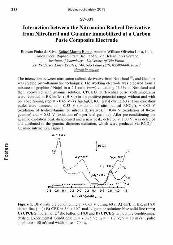
S7-001
Interaction between the Nitroanion Radical Derivative from Nitrofural and Guanine immobilized at a Carbon
Paste Composite Electrode
Robson Pinho da Silva, Rafael Martos Buoro, Antonio William Oliveira Lima, Luis Carlos Cides, Raphael Prata Bacil and Silvia Helena Pires Serrano
Institute of Chemistry – University of São Paulo Av. Professor Lineu Prestes, 748, São Paulo (SP), 05508-000, Brazil
[email protected] The interaction between nitro anion radical, derivative from Nitrofural (1), and Guanine was studied by voltammetric techniques. The working electrode was prepared from a mixture of graphite / Nujol in a 2:1 ratio (w/w) containing 13.3% of Nitrofural and then, recovered with guanine solution, CPCEG. Differential pulse voltammograms were recorded in BR buffer (pH 8.0) in the positive potential range, without and with pre conditioning step at - 0.65 V (vs Ag/AgCl, KCl (sat)) during 60 s. Four oxidation peaks were detected at: - 0.53 V (oxidation of nitro radical RNO2
•-), + 0.08 V (oxidation of hydroxylamine or nitrous derivative), + 0.44 V (oxidation of 8-oxo guanine) and + 0.81 V (oxidation of superficial guanine). After pre-conditioning the guanine oxidation peak disappeared and a new peak, detected at 1.00 V, was detected and attributed to the guanine dimmers oxidation, which were produced via RNO2
•- - Guanine interaction, Figure 1.
. Figure 1. DPV with pré conditioning at - 0.65 V during 60 s: A) CPE in BR, pH 8.0 dotted line (........); B) CPE in 5,0 x 10⁻⁶ mol L-1guanine solution: blue solid line (____); C) CPCEG in 0.2 mol L-1 BR buffer, pH 8.0 and D) CPCEG without pre conditioning, dashed. Experimental Conditions: Ei = - 0.75 V; Ef = + 1.2 V, ν = 10 mVs-1, pulse amplitude = 50 mV and width pulse = 70 ms. (1) Julião M. S. S., Ferreira E. I., Ferreira, N.G., Serrano S.H.P.; Electrochim. Acta 51 (2006) 5080–5086
338Po
ster
sBioelectrochemistry 2013

S7-002
Simultaneous Detection of Purine Derivatives at a Dopamine Pyrolytic Graphite Modified Electrode
Rafael Martos Buoro, Robson Pinho da Silva, Antonio William Oliveira Lima and
Silvia Helena Pires Serrano Institute of Chemistry – University of São Paulo
Av. Professor Lineu Prestes, 748, São Paulo (SP), 05508-000, SP, Brazil [email protected]
The direct determination of Xanthine by voltammetric methods with conventional electrodes is complicated by the presence of ascorbic acid (AA), dopamine (DA), hypoxanthine (HX) and uric acid (UA) usually present in urine samples. Using DPV, uric acid, xanthine (XA) and hypoxanthine (HXA) were simultaneously detected at Dopamine Pyrolytic Graphite Modified Electrode (DPGME), previously reported (1). The detection limit, in Phosphate Buffer Solution (PBS), for XA in presence of 5.0 x10-
5 mol L-1 HXA was 2.3 x10⁻⁶ mol L-1 (with sensibility of 2.8 A mol-1 L-1 cm2), while the detection limit for HXA in presence of 5.0 x10⁻⁵ mol L-1 XA was 5.6 x10⁻⁶ mol L-1 (with sensibility of 1.4 A mol-1 L cm-2). XA and HXA were determined in urine samples and the values founded were 0.47 mM e 5.9 mM, respectively. The oxidation peak potentials, obtained by cyclic voltammetry were 0.051, 0.393, 0.765 and 1.08 V vs Ag/AgCl, KCl(sat) for AA, UA, XA and HXA, respectively, Figure 1.
Figure 1. Cyclic Voltammograms obtained with: (dotted line) PGE and (Continuous line) DPGME in PBS, pH 6.5 containing 1.0 x 10⁻³ mol L-1AA; 1.0 x 10⁻⁴ mol L-1 UA; 5.0 x 10⁻⁵ mol L-1 XA and 5.0 x 10⁻⁵ mol L-1 HXA. Scan rate = 100 mV s-1, Ei = - 0.1 V; Eλ = + 1.3 V and Ef = - 0.1V. (1) da Silva R. P., Lima A. W. O., Serrano S.H.P., Anal. Chim. Acta, 612 (2008) 89-98
339
Post
ers
Bioelectrochemistry 2013
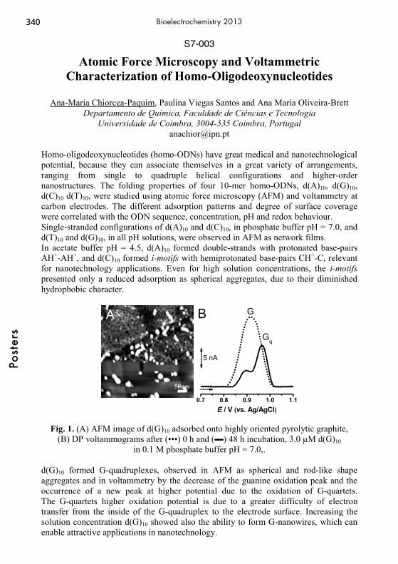
S7-003
Atomic Force Microscopy and Voltammetric Characterization of Homo-Oligodeoxynucleotides
Ana-Maria Chiorcea-Paquim, Paulina Viegas Santos and Ana Maria Oliveira-Brett
Departamento de Química, Faculdade de Ciências e Tecnologia Universidade de Coimbra, 3004-535 Coimbra, Portugal
Homo-oligodeoxynucleotides (homo-ODNs) have great medical and nanotechnological potential, because they can associate themselves in a great variety of arrangements, ranging from single to quadruple helical configurations and higher-order nanostructures. The folding properties of four 10-mer homo-ODNs, d(A)10, d(G)10, d(C)10 d(T)10, were studied using atomic force microscopy (AFM) and voltammetry at carbon electrodes. The different adsorption patterns and degree of surface coverage were correlated with the ODN sequence, concentration, pH and redox behaviour. Single-stranded configurations of d(A)10 and d(C)10, in phosphate buffer pH = 7.0, and d(T)10 and d(G)10, in all pH solutions, were observed in AFM as network films. In acetate buffer pH = 4.5, d(A)10 formed double-strands with protonated base-pairs AH+-AH+, and d(C)10 formed i-motifs with hemiprotonated base-pairs CH+-C, relevant for nanotechnology applications. Even for high solution concentrations, the i-motifs presented only a reduced adsorption as spherical aggregates, due to their diminished hydrophobic character.
A
0.7 0.8 0.9 1.0 1.1
Gq
G
5 nA
E / V (vs. Ag/AgCl)
B
Fig. 1. (A) AFM image of d(G)10 adsorbed onto highly oriented pyrolytic graphite, (B) DP voltammograms after (•••) 0 h and () 48 h incubation, 3.0 μM d(G)10
in 0.1 M phosphate buffer pH = 7.0,. d(G)10 formed G-quadruplexes, observed in AFM as spherical and rod-like shape aggregates and in voltammetry by the decrease of the guanine oxidation peak and the occurrence of a new peak at higher potential due to the oxidation of G-quartets. The G-quartets higher oxidation potential is due to a greater difficulty of electron transfer from the inside of the G-quadruplex to the electrode surface. Increasing the solution concentration d(G)10 showed also the ability to form G-nanowires, which can enable attractive applications in nanotechnology.
340Po
ster
sBioelectrochemistry 2013

S7-004
Laccase Enzyme Immobilization in Conducting Polymer Matrix through Magnetite Nanoparticles
Balázs Endrodi, Attila Kormányos, Csaba Janáky, Csaba Visy
Department of Physical Chemistry and Materials Science, University of Szeged Rerrich square 1, Szeged H-6720, Hungary
Email: [email protected]
Conducting polymer based (nano)composite electrodes are still in the focus of research and development, because these materials can be used for several different applications. Although iron-oxalate doped polypyrrole and PEDOT [1] layers are proved to be active in the electro-catalytic oxygen reduction reaction, it was demonstrated earlier that incorporation of different ferrites can enhance this activity, assumingly due to the redox switching between Fe3+/Fe2+ [2]. The electrocatalytic activity of such hybrids can be further increased by immobilization of different biologically active molecules into the polymer matrix. In this study, we present a new and efficacious method for specific binding of laccase enzyme onto magnetite nanoparticles by forming oxalate groups on the magnetite’s surface by potassium tetraoxalate treatment. The enzyme covered magnetite nanoparticles were successfully incorporated into polypyrrole matrix by galvanostatic polymerization. The presence of the laccase was proved by EQCN, TEM and FT-IR spectroscopy. Electrochemical behaviour of the formed hybrids was characterized by cyclic voltammetry. In saturated oxygen atmosphere, the reduction surplus – related to the electrochemical reduction of oxygen – was remarkable in the case of the laccase containing layer. Kinetic aspects of the oxygen reduction reaction of laccase containing layers were investigated by linear voltammetry, performed on a rotating disk electrode and a complete, pure 4-electron route was found which is promising for fuel cell applications, as it leads to water formation without involvement of considerable hydrogen-peroxide.
-1,0 -0,8 -0,6 -0,4 -0,2 0,0
-80
-60
-40
-20
0
20
40
I / μ
A
E / V
Polypyrrole Polypyrrole/Magnetite Polypyrrole/Magnetite/Laccase
[1] G. Bencsik; Z. Lukacs; C.Visy, Analyst 2010, 135 (2), 375-380. [2] C. Janáky, et al., J. Phys. Chem. C, 2010, 114, 19338–19344. Supported by TÁMOP 4.2.4.A/2-11-1-2012-0001 National Excellence Programme
341
Post
ers
Bioelectrochemistry 2013

S7-005
Electrochemical Surface Treatment of 316L Stainless Steel for Biomedical Applications
Sajjad Habibzadeh1, Grishma Hirode1, Sasha Omanovic1, Dominique Shum-Tim2
1 Department of Chemical Engineering, McGill University, 3610 university Street, Montreal, Quebec, Canada H3A 2B2
2 Division of Cardiac Surgery & Surgical Research, McGill University Health Center, Montreal, Quebec, Canada
Metallic coronary stents, which are mainly made of 316L stainless steel (SS), are medical devices (implants) that can provide endovascular scaffolding in order to relive the vascular obstruction and minimize a risk of myocardial infraction (hart attack). However, the surface of a SS stent is thrombogenic and provokes tissue reaction. One component of the process of thrombosis is the activation and aggregation of platelets. Since nature of a metal surface is crucial to the blood compatibility, employing a suitable surface treatment can improve the biocompatibility of the stent. In this work, electrochemical polishing (EP) was applied as a SS stent surface treatment method. The SS surface was anodically polarized at cell voltages of 2.5, 4 and 10 V in an appropriate electrolyte, at 60oC. A range of experimental techniques were used in the investigation of surface roughness, topographical, morphological, electrochemical/corrosion, and chemical properties of EP surfaces such as, atomic force microscopy (AFM), scanning electron microscopy (SEM), impedance spectroscopy (EIS), anodic polarization, X-ray photoelectron spectroscopy (XPS). In addition, the attachment and morphology of platelet-rich plasma (PRP) with the EP was assessed. The average surface roughness (Ra) measured by AFM depended on the voltage difference applied. The lowest value was obtained at 4 V (36 nm), whereas the values on the unmodified (control) surface and EP surfaces electropolished at 2.5V and 10 V were 123 nm, 78 nm and 67 nm, respectively. EIS results confirmed that the corrosion stability of all EP surfaces was higher than that of the control surface. XPS results revealed the chromium enrichment in the passive oxide films on the EP samples, and the oxygen content close to the outer film surface was relatively the largest on the sample prepared at 4V. Hence, one of the origins of the increased corrosion resistance of EP samples was the formation of a thicker and more Cr-rich passive oxide film, in comparison to the film formed on the control substrate surface.PRP results showed a relatively abundant accumulation of platelets on the control surface, while significantly fewer platelets were attached to the EP surfaces. Thus, a ca. 93% reduction of the number of attached platelets was obtained on the EP sample prepared at 4 V, in comparison to the control surface. In conclusion, EP of a SS surface produces the surface that is more corrosion resistant and ‘unfriendly’ for platelet attachment, rendering the surface more biocompatible.
342Po
ster
sBioelectrochemistry 2013

S7-006
Electrochemical reaction system to overcome enzyme instabilities
Dirk Holtmann, Laura Getrey, Jens Schrader
DECHEMA-Research Institute Theodor-Heuss-Allee 25
60486 Frankfurt am Main, Germany [email protected]
Electroenzymatic processes are interesting approaches due to the combination of the advantages of enzymes and electrochemical steps. Chloroperoxidase (CPO, EC 1.11.1.10) from the filamentous fungus Caldariomyces fumago is a versatile heme-dependent peroxidase requiring hydrogen peroxide and chloride, bromide or iodide for the halogenation of organic substrates. In addition to halogenations CPO catalyses hydrogen peroxide-supported oxidation, the dismutation of hydrogen peroxide, and some cytochrome P450 monooxygenase-like reactions. Nevertheless, its use in preparative or industrial scale reactions has been hindered by instability towards the co-substrate hydrogen peroxide. In order to avoid the irreversible inactivation hydrogen peroxide can be generated electrochemically on a low, but sufficient level for catalytic activity. Here we describe a gas diffusion electrode (GDE) as a new type of electrode material for electroenzymatic processes. In this study we have investigated the chlorination of monochlorodimedone (MCD) and thymol, the sulfoxidation of thioanisole and the oxidation of indole. The reaction system was described by the total turnover number (ttn) as quotient of the moles of formed product and added enzyme and the space-time-yield (STY) as the mass of a product formed per volume of the reactor and time unit.
Substrate/ Reaction c(CPO)
[nM]
Electrode surface
area [cm²]
STY
[g L-1 d-1]
TTN
[-]
MCD, chlorination 2.5 5.5 8.4 1,173,000
MCD, chlorination 12.4 5.5 52 217,00
Thymol, chlorination 10 5.5 20.5 154,000
Thioanisole, sulfoxidation 100 16.5 19.8 83,600
Indole, oxidation 100 16.5 8.3 39,000
Our data illustrate that an electro-enzymatic process with a reaction system based on a GDE is possible. We found space time yields and TTN. Our results demonstrate that the ttn of the system can be improved by using the reaction system. The GDE-peroxidase-system presented is a promising tool for applications in white biotechnology and for the production of chiral pharmaceuticals.
343
Post
ers
Bioelectrochemistry 2013

S7-007
Electrochemical behavior of duplex stainless steel in the presence of sulphate-reducing bacteria biofilms
Joanna Michalska1, Marzena Jaworska-Kik2, Weronika Dec3, Wojciech Simka4
1Department of Materials Science, Silesian University of Technology, Poland Krasinskiego Street 8, 40-019 Katowice
[email protected] 2Department of Biopharmacy, Medical University of Silesia, Poland
Narcyzow Street 1, 41-200 Sosnowiec 3Department of Environmental Biotechnology, Silesian University of Technology,
Poland B. Krzywoustego Street 6, 44-100 Gliwice
4Faculty of Chemistry, Silesian University of Technology, Poland B. Krzywoustego Street 6, 44-100 Gliwice
The formation of biofilms on metal surfaces is the initial stage of biofouling and microbiologically influenced corrosion (MIC). The kinetics of microorganism’s adsorption on metal surface is very important to understand biofilm formation and this can be a starting point for controlling biofouling and to develop effective methods to prevent biocorrosion. In the present work, the effect of pure cultures of sulphate-reducing bacteria (Desulfovibrio desulfuricans) on the biofilm formation on duplex stainless steel was studied. The open circuit potential measurements were used to monitor the attachement activity of bacteria on steel surface. It was proved that sulphate-reducing bacteria biofilms caused the “enoblement” of the steel surface, which result in an increase of corrosion potential. Pre-exposure in sulphate-reducing bacteria led also to increased corrosion current densities. Localized corrosion mechanism in the presence of sulphate-reducing bacteria biofilms was studied electrochemically. Cyclic potentiodynamic polarization curves were measured on steel after its exposure to bacteria. Steel coupons in solution-annealed condition were exposed to different standard and wild strains of Desulfovibrio desulfuricans, for different time intervals. Polarization studies performed after exposition to sulphate-reducing bacteria mediums revealed enhanced corrosion of duplex stainless steel. Etching of the duplex structure, pitting as well as crevice attack were noticed on the steel surface. The nature and mechanism of SRB attack on DSS were discussed. Electrochemical studies were completed with surface characterization including the structure and configuration of biofilms, which was carried out using scanning electron microscopy. Special attention was paid to the role of steel microstructure in the biofilm formation. ACKNOWLEDGMENT Scientific work was supported by the Polish Ministry of Science and Higher Education under research project No. N507 230040 (2011-2013).
344Po
ster
sBioelectrochemistry 2013
S7-008
Double intrachain histidine-tag for isotropic self-assembly of redox enzyme on electrode surfaces
Frank Müller, Joerg Henig, Tarik Abdulazim, Thore Schmidt, Martin Winkler, Thomas
Happe, Nicolas Plumeré. Center for Electrochemical Sciences - CES, Ruhr-Universität, Bochum, D-44780
(Germany) [email protected]
In biosensing and biofuel cell applications, control of enzyme orientation on surfaces is crucial to ensure accessibility to the active site and efficient electrochemical communication between the electrode’s surface and the enzyme’s redox site. Affinity binding between a terminal histidine-tag and an electrode surface functionalized with metal complexes allows for oriented immobilization of fully active enzyme monolayers.[1] However the applications of this approaches are limited since the possible location of the terminal His-tag on the protein limits the choices in orientation. This issue may be solved by using intrachain histidine residues [2] from the surface of the enzyme. However, in comparison to the terminal his-tag, the stability of the binding is significantly lower. We circumvent this issue by introducing a double intrachain histidine-tag as binding site at the protein. Based on SPR investigations we demonstrate the increased binding affinity of protein bearing two sets of His-X3-His (two histidine separated by two amino acids) in alpha helixes on NTA-Ni(II) modified surfaces. We demonstrate that the single his-tagged protein dissociated from the NTA-Ni(II) surface via both metal-histidine dissocation and metal ion transfer processes. The presence of two binding sites in double his-tagged protein on the other hand prevents both dissociation pathways and thus yields stable protein monolayers. The binding and electrocatalytic activity of a ferredoxin NADP+ reductase with the double intrachain his-tag on glassy carbon electrodes modified with NTA-Zn(II) and NTA-Cu(II) will be given as example. Acknowledgement Financial support by the EU and the state NRW in the frame work of the HighTech.NRW programme is gratefully acknowledged. [1] a) R. Blankespoor, B. Limoges, B. Schöllhorn, J.-L. Syssa-Magalé, D. Yazidi,
Langmuir 2005, 21, 3362–3375. b) V. Balland, S. Lecomte, B. Limoges, Langmuir 2009, 25, 6532–6542. c) W. H. Campbell, J. Henig, N. Plumeré, Bioelectrochemistry.
[2] a) F. H. Arnold, B. L. Haymore, Science 1991, 252, 1796–1797. b) J. Madoz-Gúrpide, J. M. Abad, J. Fernández-Recio, M. Vélez, L. Vázquez, C. Gómez-Moreno, V. M. Fernández, J. Am. Chem. Soc. 2000, 122, 9808–9817.
345
Post
ers
Bioelectrochemistry 2013

S7-009
A comparative voltammetric study of the redox behavior of 6-benzylaminopurine and its derivatives on mercury
and pencil graphite electrodes
Iveta Pilarova1,2, Rudolf Navratil1, and Libuse Trnkova*1,3 1Department of Chemistry, Faculty of Science, Masaryk University,
Kotlarska 2, CZ-611 37 Brno, Czech Republic 2Central European Institute of Technology-CEITEC MU,
Zerotinovo namesti 617/9, CZ-601 77 Brno, Czech Republic, European Union 3CEITEC, University of Technology,
Technicka 3058/10, CZ-616 00 Brno, Czech Republic, European Union *[email protected]
Redox processes of 6-benzylaminopurine (6-BAP), as one of the most important naturally occurring cytokinins (phytohormones), were studied on mercury and graphite electrodes using cyclic and differential pulse voltammetry [1,2]. Nowadays, great attention is paid to the newly prepared derivatives of 6-BAP exhibiting cytostatic effects. From the point of view of important signaling and cytotoxic functions of BAP derivatives, the knowledge of the ability of these compounds to diffuse across cell membranes into the intracellular space is also important. The aim of our contribution is a comparative voltammetric study of the redox behavior of 6-BAP and its chlorinated and methoxylated derivatives on a hanging mercury drop electrode (HMDE) and a pencil graphite electrode (PeGE). The voltammetric experiment was performed in phosphate-acetate buffer solutions containing 10% v/v CH3OH and different concentrations of NaCl (from 0.08 M to 1 M). Linear sweep voltammetry (LSV) in connection with elimination voltammetry with linear scan (EVLS) [3-5] allowed to determine not only the mechanism of reduction and oxidation processes on both electrodes but also important parameters required for the determination of diffusion coefficients. The diffusion characteristic, studied also using a rotating disk electrode (RDE), was evaluated in terms of the influence of ionic strength, the substituent (-Cl and -OCH3), and its position on the benzene ring. . Acknowledgement This research has been supported by Project 106/09/H035 of the GA CR, the CEITEC – Central European Institute of Technology Project CZ.1.05/1.1.00/02.0068, and by project MUNI/A/0992/2009 of the Ministry of Education of the Czech Republic. References [1] G. Dryhurst, Electrochemistry of Biological Molecules, London, 1977. [2] D. Tarkowska, et al., Collect. Czech. Chem. Commun., 68 (2003) 1076-1093. [3] L. Trnkova in: V. Adam, R. Kizek (Eds.), Utilizing of Bio-electrochemical Methods in Biological Research, Research Signpost, Kerala, India, 2007, Ch. 4, p. 51-74. [4] L. Trnkova, F. Jelen, M. Ozsoz, in: M. Ozsoz (Ed.), Electrochemical DNA Biosensors, Pan Stanford Publishing, Singapore, 2012, Ch. 11, p. 355-378. [5] I. Pilarova, P. Lubal, L. Trnkova, Electroanalysis, 24 (2012) 349-356.
346Po
ster
sBioelectrochemistry 2013

S7-010
On the Role of Early Stages of Molecular Oxygen Activation in Bioelectrochemistry and Nanotechnology
A. A. Revina
Frumkin Institute of Physical Chemistry and Electrochemistry, Russian Academy of Sciences,
Leninskii pr. 31, Moscow, 119071 Russia e-mail: [email protected]
Modern approaches to revealing the formation mechanism and determining the physicochemical properties and functional activity of nanosized structures, are directly associated with the study of the molecular oxygen effect. In the end of XIX century, A.N. Bakh, C. Engle, and W.Wild in their works (1897) concerning the so-called oxygen activation formulated the fundamentals of the peroxide theory of oxidation processes for the first time. A.N. Frumkin in his lecture (1948) entitled “Adsorption and Oxidation Processes”dedicated to the 50th Anniversary of the Bakh’s peroxide theory of oxidation traced its further advancement in biology, physical chemistry, and electrochemistry. On the base of studying oxygen adsorption at activated coals it was found that the adsorption yielded “active centers CxO2”, and the carbon surface acquired positive charge; more strongly bound oxidized forms appear only at high oxygen concentration and long exposure. Importantly, when studying the electrochemistry of platinum sols, N.A. Bakh and A.A. Rakov (1937) detected reversible oxygen-containing centers that transformed to stable oxide forms at some experimental conditions. These results were emphasized by A.N. Frumkin as directly related to the problem of the mechanism of the oxidation elementary act, to which Academician A.N. Bakh was paying attention till his last days. The method of pulse radiolysis with detecting of intermediate particles of the liquid-phase organics oxidation allowed identifying alkyl (Rj) and peroxide radicals(RO2), superoxide radical-anions, the formation of reversible oxocomplexes, charge-transfer complexes M+δ…O2
-δ for phtalocyanines, enzymes, carotenoids, and flavonoids (Revina1995). Studying the mechanism of metal nanoparticles formation in reverse-micelle systems allowed demonstrating the extreme importance of the fine effects and the role of labile reversible oxocomplexes on both the particles' stability and formation, - in the area of nanotechnology and related applications.
347
Post
ers
Bioelectrochemistry 2013
S7-011
Hemiprotonated C – C+ base pairing investigated by electrochemical and spectral methods
Libuse Trnkova1,3*, Libor Gurecky1, Sylvie Dohnalikova1, Iveta Pilarova1,2 , and Paula
Toimil Loureiro1,4 1Department of Chemistry, Faculty of Science, Masaryk University,
Kotlarska 2, CZ-611 37 Brno, Czech Republic 2Central European Institute of Technology – CEITEC, University of Technology,
Technicka 10, CZ-616 00 Brno, Czech Republic 3CEITEC Masaryk University, Zerotinovo namesti 9, CZ-601 77 Brno, Czech Republic
4Biophysics & Interfaces Group, Department of Applied Physics, Faculty of Physics, University of Santiago de Compostela, E-15782 Santiago de Compostela, Spain
*[email protected] It is generally known that under slightly acidic pH conditions oligodeoxynucleotides (ODNs) rich in cytosine (C) can form i-motifs (intercalated hemiprotonated C – C+ base pairing). These structures can be responsible for i-tetraplexes in both centromeric and telomeric regions of human chromosomes. There are speculations that the presence of i-motif structures correlates with the expansion of the triplet repeat sequences associated with neurological disorders [1-3]. The present work deals with the electrochemical and spectral investigation of short ODNs containing different numbers of C. Using linear sweep voltammetry (LSV) and elimination voltammetry with linear scan (EVLS) we studied not only the effect of the number of C in the ODNs chain on the reduction signals in buffered and non-buffered solutions but also the effect of pH and ionic strength. The reduction C peaks, recorded at the mercury electrode in the potential range from –1.1 V to –1.7 V vs. Ag/AgCl/3MKCl, are influenced by ODN conformation changes which were confirmed by circular dichroism and UV-Vis spectra. The intermolecular or intramolecular folded i-motif structures were found for all ODNs except dC3. The electrochemical and spectral experiments were completed with the results of gel electrophoresis of ODNs on PAGE. Acknowledgement This research has been supported by the following projects: (a) Project CEITEC – Central European Institute of Technology CZ.1.05/1.1.00/02.0068, (b) Project MUNI/A/0992/2009, and (c) Project CZ.1.07/2.3.00/30.0009 of the Ministry of Education of the Czech Republic. References [1] T. Simonsson, M. Pribylova, M. Vorlickova, Biochem. Biophys. Res. Commun., 278 (2000) 158-166. [2] J. L. Leroy, K. Gehring, et al., Biochemistry, 32 (1993) 6019-6031. [3] J. L. Leroy, M. Gueron, et al., Nucl. Acids Res., 22 (1994) 1600-1606. [4] L. Trnkova, F. Jelen, M. Ozsoz, in: M. Ozsoz (Ed.), Electrochemical DNA Biosensors, Pan Stanford Publishing, Singapore, 2012, Ch. 11, p. 355-378.
348Po
ster
sBioelectrochemistry 2013

S7-012
Development of Neural Stimulating Electrodes Based on Iridium-Ruthenium Mixed Oxides
Nehar Ullah and Sasha Omanovic Department of Chemical Engineering, McGill University
3610 University St., Montreal, Qc, H3A 2B2, Canada [email protected]
Several emerging clinical applications require the use of neural prosthetics devices. These applications range from brain stimulation to decreasing of symptoms of various diseases such as Parkinson’s, epilepsy, uncontrolled night urination, interfacing the brain with artificial limbs, and vision restoration. In all such applications a neural electrode works as a bridge between the external electronic control device and the human biological system (neurons), to enable the transfer of information between completely different systems, electronic and biological. Much attention has been given to the design/functionality of neural electrodes, which includes its proper size, sensitivity and reaction of the human tissue to a foreign body element (neural electrode), its stability, service time, fouling, etc. Selectivity is one of the major issues in the clinical applications of neural electrodes. This occurs mostly during the activation of neurons, and is the result of the large size of the implanted neural electrode relative to the neighboring neurons. Hence, neural electrodes should be of small size, but should have the capability of delivering sufficiently high charge density without causing any negative side effects to the functionality of neurons and the surrounding tissues. The aim of the work was to develop new oxide-based coatings for stimulating electrodes that would offer both higher intrinsic and extrinsic charge injection, than stimulating electrodes currently used in practice. This would enable miniturization of the electrodes, and also narrow down the cyclization potential limits, thus decreasing the negative effect of the stimulating electrodes to the surrounding tissues. The initial development of the coatings was based on producing Ir/Ru oxide coatings of various compositions, prepared by thermal decomposition on a Ti substrate. Experiments showed that specific Ir/Ru-Ox coatings offered a significantly increased, electrochemically-active surface area than the currently used state-of-the-art, Ir-oxide (control). These binary coatings were also capable of delivering significantly higher charge than the control, both from the intrinsic and extrinsic point of view. The produced coatings were demonstrated to be stable, with improved charge-delivery performance with cycling use, due to the gradual increase in surface roughness.
349
Post
ers
Bioelectrochemistry 2013
S7-013
Laccase – Substrate Interaction – Surface Plasmon Resonance Study
Agnieszka Więckowska
Faculty of Chemistry, University of Warsaw ul. Pasteura 1, 02093 Warsaw, Poland
Surface plasmon resonance is a great tool for the observation of the interactions between molecules in real time. While one type of molecules is immobilized to the surface of SPR sensor, the other are in solution and passed over the surface. The changes of the resonance angle depend on many factors including the binding of biomolecules events [1]. Laccase, EC1.10.3.2, is widely distributed in natural world and can be produced by plants and fungi. Laccase catalyses the oxidation of orto- and para-diphenols, polyphenols, polyamines, lignins, and aryl diamines and some inorganic compounds coupled to the reduction of oxygen to water. In the active centre of fungal laccase four copper atoms are placed and they are involved in the electron transfer from a reducing substrate towards molecular oxygen with its eventual reduction to water. Laccases are being investigated for a variety of practical applications, i.e. electrocatalytic reduction of dioxygen [2] and oxidation of phenolic substrates [3]. In this project we employ surface plasmon resonance (SPR) as well as Quartz Crystal Microbalance (QCM) for the observation of interaction between enzyme - laccase and gold surface, surface covered with gold nanoparticles and surface modified with natural substrates as syringic acid ot anthraquinone derivatives to bind laccase in the right orientation, and to retain the contact between enzyme and the gold surface which can act as the electrode in biofuel cell. References [1] F. Markey Bia Journal 1999, 1, 14. [2] P. Rowiński, R. Bilewicz, M.J. Stebe, E. Rogalska, Anal. Chem. 2004, 76, 283. [3] R.S. Freire, N. Duran, L.T. Kubota, Talanta 2001, 54, 681.
350Po
ster
sBioelectrochemistry 2013

S7-014
Electrochemical Oxidation of the Isoquinoline Alkaloid Berberine in Aqueous Medium
Jana Skopalová1, Barbora Papoušková1, Martina Zatloukalová2, Jan Vacek2
1Department of Analytical Chemistry, Faculty of Science, Palacký University, 17. listopadu 12, 771 46 Olomouc, Czech Republic
2 Department of Medical Chemistry and Biochemistry, Faculty of Medicine and Dentistry, Palacký University, Hněvotínská 3, 775 15 Olomouc, Czech Republic
[email protected], [email protected]
Electrochemical oxidation of berberine in aqueous medium was studied by cyclic and differential pulse voltammetry at a glassy carbon electrode (GCE). Two anodic peaks of the quaternary form of berberine (Fig. 1, left) were observed at +1.2 V and +1.4 V (vs. SCE) in acidic and neutral solutions. When the anodic polarization exceeded the value of +1.1 V, the redox active film is formed on the GCE surface. The formation of adsorbed film was well-documented by quasireversible redox couple at +0.25 V which was studied in redox cycling experiments.
Fig. 1 Chemical structure of berberine quaternary salt (left) and berberine pseudobase
(right).
In alkaline medium, a new anodic peak at +0.5 V appeared due to oxidation of berberine pseudobase (Fig. 1, right) to 8-oxoberberine. Solutions of berberine at different pH were subjected to controlled potential electrolysis on platinum gauze electrode and analyzed using liquid chromatography equipped with electrospray ionization/quadrupole time-of-flight mass spectrometry. The main water soluble monomeric product of berberine oxidation under physiological-near experimental conditions, OP1, was identified as demethyleneberberine cation. Acknowledgements: This work was supported by the Czech Science Foundation (P503/11/P312) and by the Ministry of Education, Youth and Sports of the Czech Republic (grant projects CZ.1.05/2.1.00/03.0058 of Operational Program Research.
351
Post
ers
Bioelectrochemistry 2013

S8-001
Highly Sensitive Platinum-Black Coated Platinum Electrodes for Electrochemical Detection of Oxidative
Stress in Microchannels
Y. Li, C. Sella, F. Lemaître, M. Guille-Collignon, L. Thouin, C. Amatore UMR CNRS 8640 PASTEUR, Ecole Normale Supérieure, Département de Chimie
24 rue Lhomond, 75231 Paris Cedex 05, France [email protected]
Microuidic devices have attracted signicant attentions for bioanalytical applications due to numerous and well-known advantages. They become new and effective tools for single-cell analysis, in particular for real-time monitoring of stimulated release [1]. In this context, oxidative stress is one of the metabolic situations of interest. Quantitative determination of reactive oxygen species (ROS) and reactive nitrogen species (RNS) are still required to provide data that accurately reflect their concentration in cells. Electrochemical techniques are a logical choice for monitoring oxidative bursts [2-3]. However selectivity and sensitivity, as well as long-term stability of electrode responses for ROS and RNS detections need to be sufficiently addressed. Hence, the high catalytic activity of Pt-black provides a viable approach for monitoring single-cell release in microfluidic devices. We thus investigated the electrochemical detection of H2O2 and NO2
- at highly sensitive Pt-black microband electrodes [4]. Characterization and optimization of Pt-black deposits were performed according to the active surface and roughness factor. Electrode activation was achieved by using a specific procedure which led to a higher stability of electrode responses versus time. The electrode performances were then studied over a wide range of applied potential and compared to data obtained from bare-Pt electrodes. Steady-state currents were also compared to theoretical predictions based on convective mass transport at microchannel electrodes [5,6]. Results show that Pt-black deposits improve drastically the performances of H2O2 and NO2
- detections. In each case, the oxidation mechanism at Pt-black electrodes is discussed in terms of active sites at the nanometric level and higher porosity of Pt-black deposits facilitating the electron transfer. Highly sensitive Pt-black electrodes were characterized with 5 decades of linear concentration range and low detection limits down to 10 nM for both species. In addition, a microfluidic device was specifically designed for cell culture and online monitoring of oxidative bursts from stimulated single cells. A discussion about application of Pt-black in microfluidic devices is presented. [1] Y. Wang, Z.-Z. Chen, Q.-L. Li, Microchim. Acta, 2010, 168, 177. [2] C. Amatore, S. Arbault, M. Guille, F. Lemaitre, Chem. Rev., 2008, 108, 2585. [3] S. Borgmann, Ana. Bioanal. Chem., 2009, 394, 95. [4] Y. Li, C. Sella, F. Lemaître, M. Guille-Collignon, L. Thouin, C. Amatore,
Electroanalysis, in press. [5] C. Amatore, N. Da Mota, C. Sella, L. Thouin, Anal. Chem., 2007, 79, 8502. [6] C. Amatore, N. Da Mota, C. Sella, L. Thouin, Anal. Chem., 2008, 80, 4976.
352Po
ster
sBioelectrochemistry 2013
S8-002
Interaction of Anthracycline Drug with Model Membrane System
Dorota Nieciecka, Agata Królikowska, Aleksandra Joniec, Paweł Krysiński
University of Warsaw, Department of Chemistry, Pasteura 1, 02-093 Warsaw, Poland
[email protected] Anthracycline drugs belong to the group of antibiotics used in the treatment of a wide range of cancer. The mechanism of their action consist in interact with DNA by intercalation and then inhibit the replication process. Despite of high efficiency, these drugs cause cardiac toxicity and this leads to the limitations of their usage in therapy. We have investigated the interaction of anthracyclines with biomimetic membranes because the mechanism of drug crossing through the lipid bilayer in cells is still unknown. Biomimetic layers for investigations of anthracycline partition were formed by Langmuir – Blodgett (L-B) technique onto gold electrodes. The properties of such films were characterized by an inhibition of K4Fe(CN)6 reduction processes, IR spectroscopy and by ellipsometry. Molecules of studied drugs have two redox-active centers, therefore the interaction of drug with membranes can be monitored by electrochemical technique such as cyclic voltammetry. Researches show that the drug is easily and quickly adsorbed on different types of the obtained Langmuir-Blodgett membranes. The electrochemical results were supported by quartz crystal microbalance (QCM), Surface Enhanced Raman Spectroscopy (SERS), IR Spectroscopy ( PMIRRAS) and Surface Plasmon Resonance (SPR).
353
Post
ers
Bioelectrochemistry 2013

S8-003
Study of cancer cells altered processes by mapping both glucose uptake and lactate release
Alice Soldàa,b, Stefania Rapinoa,b, Pier Giuseppe Peliccia, Francesco Paoluccib, Marco
Giorgioa a European Institute of Oncology (Campus IEO), Via Adamello 16, 20139 Milan, Italy b University of Bologna, Chemistry Department “G. Ciamician”, Via Selmi 2, 40126
Bologna, Italy e-mail: [email protected]
Most cells use glucose as a fuel source. Glucose is metabolized by glycolysis in a multi-step set of reactions resulting in the creation of pyruvate. In typical normal cells, much of this pyruvate enters the mitochondria where it is oxidized by the Krebs Cycle to generate ATP to meet the cell’s energy demands. However, in cancer cells, much of the pyruvate from glycolysis is directed away from the mitochondria to create lactate. Lactate production is typically restricted to anaerobic conditions nevertheless cancer cells preferentially channel glucose towards lactate production even when oxygen is plentiful, a process termed “aerobic gycolysis” or the Warburg Effect. [1] The past decade has seen a growth in the application of biosensors to micro and nano-meter level investigations in a wide variety of disciplines. Rapid development, both in miniaturization techniques and in understanding of biological processes, has accelerated the expansion of biosensors in clinical applications. Enzyme-based sensors are very interesting because they offer high selectivity toward a single analyte, based on their structural complementarities, and the opportunity to improve sensitivity, time scale and information content. Furthermore, they are irreplaceable tools for non- invasive study of glycolytic switching (or metabolism alterations) at cellular level. [2,3] Enzyme-based ultramicroelectrode (UME) in conjunction with scanning electrochemical microscopy (SECM) can be developed as a useful technique for studying cell metabolic fluxes, since it can map electrochemical activity across the entire surface of a single cell and can record dynamic changes, examining the metabolic differences between nonmetastatic and metastatic human cells. [2] Indeed it was demonstrated that under aerobic conditions, cancer cells metabolize approximately tenfold, more glucose to lactate in a given time than normal cells. [1] Information about the way a cell performs glycolysis could be acquired by using modified UME biosensor with glucose oxidase (GOx) and lactate oxidase (LOx) in combination with SECM. This apparatus increases the spatial and time resolution of metabolic study, that will be used as powerful tools for studying the altered processes that affect cancerous cells by mapping both glucose uptake and lactate release. References [1] W.H. Koppenol, P.L. Bounds, C.V. Dang, Nature Reviews Cancer. 11 (2011) 325. [2] J.P. Wilburn, D.E. Cliffel, Anal. Chem. 80 (2008) 2717. [3] C. Kurzawa, A. Hengstenberg, W. Schuhmann, Anal. Chem. 74 (2002) 355.
354Po
ster
sBioelectrochemistry 2013
S8-004
Study of cancer cells altered processes by mapping both glucose uptake and lactate release
Alice Soldàa,b, Stefania Rapinoa,b, Pier Giuseppe Peliccia, Francesco Paoluccib, Marco
Giorgioa a European Institute of Oncology (Campus IEO), Via Adamello 16, 20139 Milan, Italy b University of Bologna, Chemistry Department “G. Ciamician”, Via Selmi 2, 40126
Bologna, Italy e-mail: [email protected]
Most cells use glucose as a fuel source. Glucose is metabolized by glycolysis in a multi-step set of reactions resulting in the creation of pyruvate. In typical normal cells, much of this pyruvate enters the mitochondria where it is oxidized by the Krebs Cycle to generate ATP to meet the cell’s energy demands. However, in cancer cells, much of the pyruvate from glycolysis is directed away from the mitochondria to create lactate. Lactate production is typically restricted to anaerobic conditions nevertheless cancer cells preferentially channel glucose towards lactate production even when oxygen is plentiful, a process termed “aerobic gycolysis” or the Warburg Effect. [1] The past decade has seen a growth in the application of biosensors to micro and nano-meter level investigations in a wide variety of disciplines. Rapid development, both in miniaturization techniques and in understanding of biological processes, has accelerated the expansion of biosensors in clinical applications. Enzyme-based sensors are very interesting because they offer high selectivity toward a single analyte, based on their structural complementarities, and the opportunity to improve sensitivity, time scale and information content. Furthermore, they are irreplaceable tools for non- invasive study of glycolytic switching (or metabolism alterations) at cellular level. [2,3] Enzyme-based ultramicroelectrode (UME) in conjunction with scanning electrochemical microscopy (SECM) can be developed as a useful technique for studying cell metabolic fluxes, since it can map electrochemical activity across the entire surface of a single cell and can record dynamic changes, examining the metabolic differences between nonmetastatic and metastatic human cells. [2] Indeed it was demonstrated that under aerobic conditions, cancer cells metabolize approximately tenfold, more glucose to lactate in a given time than normal cells. [1] Information about the way a cell performs glycolysis could be acquired by using modified UME biosensor with glucose oxidase (GOx) and lactate oxidase (LOx) in combination with SECM. This apparatus increases the spatial and time resolution of metabolic study, that will be used as powerful tools for studying the altered processes that affect cancerous cells by mapping both glucose uptake and lactate release. References [1] W.H. Koppenol, P.L. Bounds, C.V. Dang, Nature Reviews Cancer. 11 (2011) 325. [2] J.P. Wilburn, D.E. Cliffel, Anal. Chem. 80 (2008) 2717. [3] C. Kurzawa, A. Hengstenberg, W. Schuhmann, Anal. Chem. 74 (2002) 355.
A motorized stretching device in combination with
scanning electrochemical microscopy: Towards detection of ATP at bone cells
Charlotte Steinbach1, Elena Hecht1, Stefanie Weber1, Astrid Liedert2, Anita Ignatius2,
Boris Mizaikoff1 and Christine Kranz1
[1] Institute of Analytical and Bioanalytical Chemistry, University of Ulm, Albert-Einstein-Allee 11, 89081 Ulm
[2] Institute of Orthopaedic Research and Biomechanics, University of Ulm Helmholtzstraße 14, 89081, Ulm
Mechanical forces play a significant role in cell biology. External forces can induce a multitude of cellular processes, which ranges from vital cell functions such as proliferation and differentiation to inducing pathological processes. In order to study in vitro cell responses to mechanical strain, an uniaxial stretching device was developed. The device enables applying reproducible strain levels to an PDMS-membrane with cultivated cells at physiological to hyperphysiological levels (1). The stretching device is conceptualized that it can be implemented in a scanning electrochemical microscope (SECM). This combination of analytical tools enables detecting the immediate response of cultivated bone cells towards strain. Adenosine-5`-triphosphate (ATP), as important autocrine and paracrine mediator molecule, is involved in cell processes like proliferation. In bone-resorbing cells such as the receptor-protein-tyrosine-phosphatase-zeta (PTPRζ)-osteoblastic cells, it is released due to mechanical stress and controls bone formation and regeneration (2,3) by the activation of P2 receptors. PTPRζ-osteoblastic cells and their deficient knock-out mutants show a different behavior concerning proliferation and differentiation in response to stretch. Thus, the ATP levels above these cells are expected to be altered. ATP microbiosensors in combination with the SECM implemented stretching device are used to investigate changes in ATP levels in close vicinity to the cell surface. Positioning and the detection of released ATP due to stretch is obtained by a dual-microelectrode assembly. The first bare electrode is used for recording current-distance curves and the second electrode is modified with an enzymatic layer consisting of glucose oxidase and hexokinase [4,5].. First results of localized ATP detection above PTPRζ-osteoblastic cells and their knock-out mutants will be presented. Both cell types will be stretched up to 7 % and the differences in the measured ATP levels will be discussed. [1] E. Hecht, P. Knittel, E. Felder, P. Dietl, B. Mizaikoff, C. Kranz, Analyst 137
(2012) 5208 [2] K.A. Buckley, S.L. Golding, J.M. Rice, J.P. Dillon, J.A. Gallagher, The FASEB
Journal 17 (2003) 1406 [3] J.P. Reyes, S.M. Sims, S.J. Dixon, Frontiers in Bioscience S3 (2011) 1101 [4] A. Kueng, C. Kranz, B. Mizaikoff, Biosensors and Bioelectronics 21 (2005) 346 [5] F. Scheller, D. Pfeiffer, Anal Chimica Acta 117 (1980) 383
355
Post
ers
Bioelectrochemistry 2013
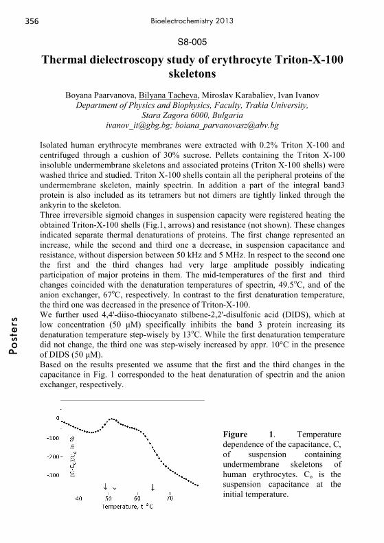
S8-005
Thermal dielectroscopy study of erythrocyte Triton-X-100 skeletons
Boyana Paarvanova, Bilyana Tacheva, Miroslav Karabaliev, Ivan Ivanov
Department of Physics and Biophysics, Faculty, Trakia University, Stara Zagora 6000, Bulgaria
[email protected]; [email protected]
Isolated human erythrocyte membranes were extracted with 0.2% Triton X-100 and centrifuged through a cushion of 30% sucrose. Pellets containing the Triton X-100 insoluble undermembrane skeletons and associated proteins (Triton X-100 shells) were washed thrice and studied. Triton X-100 shells contain all the peripheral proteins of the undermembrane skeleton, mainly spectrin. In addition a part of the integral band3 protein is also included as its tetramers but not dimers are tightly linked through the ankyrin to the skeleton. Three irreversible sigmoid changes in suspension capacity were registered heating the obtained Triton-X-100 shells (Fig.1, arrows) and resistance (not shown). These changes indicated separate thermal denaturations of proteins. The first change represented an increase, while the second and third one a decrease, in suspension capacitance and resistance, without dispersion between 50 kHz and 5 MHz. In respect to the second one the first and the third changes had very large amplitude possibly indicating participation of major proteins in them. The mid-temperatures of the first and third changes coincided with the denaturation temperatures of spectrin, 49.5oC, and of the anion exchanger, 67oC, respectively. In contrast to the first denaturation temperature, the third one was decreased in the presence of Triton-X-100. We further used 4,4'-diiso-thiocyanato stilbene-2,2'-disulfonic acid (DIDS), which at low concentration (50 μM) specifically inhibits the band 3 protein increasing its denaturation temperature step-wisely by 13oC. While the first denaturation temperature did not change, the third one was step-wisely increased by appr. 10°C in the presence of DIDS (50 μM). Based on the results presented we assume that the first and the third changes in the capacitance in Fig. 1 corresponded to the heat denaturation of spectrin and the anion exchanger, respectively.
Figure 1. Temperature dependence of the capacitance, C, of suspension containing undermembrane skeletons of human erythrocytes. Co is the suspension capacitance at the initial temperature.
356Po
ster
sBioelectrochemistry 2013
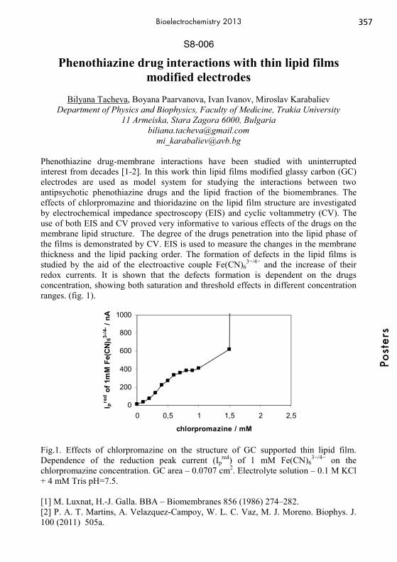
S8-006
Phenothiazine drug interactions with thin lipid films modified electrodes
Bilyana Tacheva, Boyana Paarvanova, Ivan Ivanov, Miroslav Karabaliev
Department of Physics and Biophysics, Faculty of Medicine, Trakia University 11 Armeiska, Stara Zagora 6000, Bulgaria
[email protected] [email protected]
Phenothiazine drug-membrane interactions have been studied with uninterrupted interest from decades [1-2]. In this work thin lipid films modified glassy carbon (GC) electrodes are used as model system for studying the interactions between two antipsychotic phenothiazine drugs and the lipid fraction of the biomembranes. The effects of chlorpromazine and thioridazine on the lipid film structure are investigated by electrochemical impedance spectroscopy (EIS) and cyclic voltammetry (CV). The use of both EIS and CV proved very informative to various effects of the drugs on the membrane lipid structure. The degree of the drugs penetration into the lipid phase of the films is demonstrated by CV. EIS is used to measure the changes in the membrane thickness and the lipid packing order. The formation of defects in the lipid films is studied by the aid of the electroactive couple Fe(CN)6
3−/4− and the increase of their redox currents. It is shown that the defects formation is dependent on the drugs concentration, showing both saturation and threshold effects in different concentration ranges. (fig. 1).
0
200
400
600
800
1000
0 0,5 1 1,5 2 2,5
chlorpromazine / mM
I pre
d of 1
mM
Fe(
CN)
63-/4
- / nA
Fig.1. Effects of chlorpromazine on the structure of GC supported thin lipid film. Dependence of the reduction peak current (Ip
red) of 1 mM Fe(CN)63−/4− on the
chlorpromazine concentration. GC area – 0.0707 cm2. Electrolyte solution – 0.1 M KCl + 4 mM Tris pH=7.5. [1] M. Luxnat, H.-J. Galla. BBA – Biomembranes 856 (1986) 274–282. [2] P. A. T. Martins, A. Velazquez-Campoy, W. L. C. Vaz, M. J. Moreno. Biophys. J. 100 (2011) 505a.
357
Post
ers
Bioelectrochemistry 2013

359Bioelectrochemistry 2013
Index
Abdulazim, Tarik, S7-008Abou Hamdan, Abbas, (Thu S2)14:00Abriata, Luciano, (Thu S2)17:20Achouak, Wafa, (Tue S6)12:00Adamovski, M., (Thu S8)14:30Adamovski, Miriam, (Thu S8)14:50Agarwal, Shilpi, S5-019Ahmad Rather, Jahangir, S5-031Akbasoglu Ünlü, Naime, S5-011Akhter, Zareen, (Thu S2)17:40Al-Mayouf, Abdullah, (Wed S5)17:40Al Hoshan, Mansour, (Wed S5)17:40Almeida, Thiago, S6-001Alvarez de Eulate, Eva, (Tue S5)14:00Alvarez Gallego, Yolanda, (Wed S6)15:10Alvarez, Julia, (Wed S7)15:50Alvarez-Paggi, Damián, (Thu S2)17:20,
(Thu S2)17:20, S2-001Amal, Rose, (Wed S5)11:40Amato, L., (Thu S8)14:30Amatore, Christian, (Mon S1)10:00,
(Wed S7)16:40, (Thu S8)11:00, S8-001Amine, Aziz, S5-004André, Franck, (Mon S3)15:10, S3-007Aquino Neto, Sidney, S6-001Arbault, Stéphane, (Tue S5)12:20, (Wed
S7)16:40, (Thu S5)11:40, S5-001Armbruster, Oskar, S3-011Armstrong, Fraser, (Tue)09:00Arriaga, Gerardo, S6-007Arrigan, Damien, (Tue S5)14:00Ascencio-Pinedo, Mario Alberto, S3-001Aslan Gurel, Evren, S5-015Aslan Udum, Yasemin, S5-014Atanassov, Plamen, (Mon S1)14:50,
(Wed S6)10:00, (Wed S6)17:20Attar, Aisha, S5-004Augustin, Ewa, (Mon S3)12:20Avcý, Seyma, S3-003
BBabanova, Sofia, (Wed S6)10:00Bachmann, Till. T., (Thu S5)12:20Bacil, Raphael Prata, S7-002Badalyan, Artavazd, (Thu S5)17:20
Baffert, Carole, (Thu S2)14:00Bahadir, Müfit, S5-012Bajd, Franci, (Wed S3)12:20Bajracharya, Suman, (Wed S6)15:10Balevicius, Saulius, S3-018Balintová, Jana, (Thu S5)15:30Bandarenka, Aliaksandr, (Thu S8)15:10Banks, Craig E., S5-005Barbé, Kurt, S5-038Baronian, Keith, (Thu S2)17:00Barsan, Madalina, (Wed S5)15:50Bartlett, Philip, (Mon S4)17:20,
(Tue S5)10:00, (Wed S5)17:40, S4-005Bartošík, Martin, S5-002Becucci, Lucia, S2-002Bedioui, Fethi, (Thu S5)16:40Beese, Pascal, (Thu S7)17:20Beier, Hope, (Tue S3)11:00Ben Ouada, Hafedh, S6-016Ben-Amor, Salem, (Tue S5)12:20, S5-001Benayahu, Dafna, (Thu S8)14:50Bergel, Alain, (Tue S6)12:00Beta, Carsten, (Thu S8)14:50Bettazzi, Francesca, (Thu S5)14:50Bezerra Oliveira, Carlos Severino, S5-017Biernat, Jan F., (Wed S6)14:30,
(Wed S6)17:00Biezunska-Kusiak, Katarzyna, S3-017Bilewicz, Renata, (Wed S6)14:30, (Wed
S6)17:00, S3-012, S6-012Birss, Viola, (Wed S5)16:40Bitinaite, Aiste, S3-018Blagus, Tanja, S3-002Blicharska, Magdalena, S6-002Boisen, A., (Thu S8)14:30Bonhommeau, Sébastien, (Thu S5)14:30Bosserdt, Maria, S5-003Bottari, Serge, (Tue S5)12:20Bouffier, Laurent, (Thu S5)14:30Bragos, Ramon, (Thu S8)15:10Brand, Izabella, (Thu S8)12:00Braustein, Harold, (Tue S5)11:40, S4-001Breton, Marie, (Mon S3)11:30,
(Tue S3)12:00Brett, Christopher, (Wed S5)15:50, S5-004

360 Bioelectrochemistry 2013
Brotons, Ariadna, (Mon S4)12:20, S5-005Brunner, Robert, S5-024Brust, Mathias, (Thu S2)17:40Brzostek, Katarzyna, (Tue S6)11:20Bucur, Bogdan, S5-006Bueno, Paulo R., S2-007Buess-Herman, Claudine, (Mon S4)11:20Buisman, Cees JN, (Wed S6)11:20, S6-017Buller, Jens, (Mon S4)12:00Buoro, Rafael Martos, S7-001, S7-002Buzzeo, Marisa, (Thu S2)15:50Bystrzejewski, Michal, (Thu S7)16:40CCadossi, Ruggero, (Mon S3)11:00Campbell, Holly, (Wed S5)16:40Campos, Rui, (Thu S5)15:10, (Thu S8)15:30Caranto, Jonathan, S5-016Cardoso, Franciane P., S6-001Carminati, M., (Thu S8)14:30Carvalho, Ricardo, S5-004Castro, María A., S2-001Catal, Tunc, S6-003Cecatto, Marcel, S5-023Cejudo, Rocío, S2-007Celebanska, Anna, (Wed S6)14:50, S5-007,Cemazar, Maja, (Mon S3)15:30, S3-002,
S3-008Chamas, Alexandre, (Thu S2)17:00Cheng, Ming Soon, S5-021Chiarella, Pieranna, S3-009, S3-019Chiorcea-Paquim, Ana-Maria, S7-003Chira, Ana, S5-006Cho, Brian, (Mon S1)11:00Chockalingham, Muthukumar, (Thu)09:00Chopinet, Louise, (Wed S3)12:00Choromanska, Anna, S3-017Choudhury, Moinul, (Thu)09:00Chow, Vincent T. K., S5-021Ciaccafava, Alexandre, (Mon S4)11:40,
(Mon S1)15:30, (Tue S6)14:00Ciampi, Simone, (Thu)09:00Clark, Claire, (Wed S5)17:20Clausmeyer, Jan, (Thu S7)17:40Coman, V., (Thu S8)14:30Condon, Pierre-Alexis, (Thu S5)14:30Contin, Andrea, S5-008Coronado, Javier, (Wed S6)17:40Corović, Selma, (Wed S3)12:20
Cortes-Salazar, Fernando, (Mon S1)17:30Costentin, Cyrille, (Thu S2)14:50Crain, Jason, (Thu S5)12:20Crepaldi, Laís B., S6-001Cristea, Cecilia, S5-009Cuevas-Muñiz, Francisco, S6-007, S6-011Cwalina, Beata, S3-021
Dda Silva, Luis Carlos Cides, S7-002da Silva, Robson Pinho, S7-001, S7-002Danhel, Aleš, (Thu S5)15:30Daasbjerg, Kim, S5-023Daczewska, Malgorzata, S3-017Dague, Etienne, (Wed S3)12:00Dalak, Ekin, (Wed S6)15:10Dalmay, Claire, (Tue S3)14:00Danhel, Ales, S5-010Darchen, François, (Wed S7)16:40Dargahi, Mahdi, S2-003, S2-004Davis, Jason J., S2-007de Almeida, Rodrigo F. M., (Mon S4)15:30De Andrade, Adalgisa, S6-001De Poulpiquet, Anne, (Mon S4)11:40,
(Mon S1)15:30, (Tue S6)14:00De Robertis, Mariangela, S3-019De Santis, Stefania, S3-009, S3-019De Wael, Karolien, (Mon S4)11:00, S5-031Dec, Weronika, S7-007del Campo, F. Javier, S6-006Delia, Marie-Line, (Tue S6)12:00Dementin, Sébastien, (Thu S2)14:00Demirci, Sema, S5-035Demirci Uzun, Sema, S5-011Deng, Huimin, (Mon S1)17:00Derylo, Maria, (Mon S3)12:00Devin, Anne, (Tue S5)12:20, S5-001Dewan, Faizunnahar, (Thu S2)15:50Diculescu, Victor, S2-005Dierich, Marlen, (Thu S5)17:20Dietz, Thomas, S2-006Dimaki, M., (Thu S8)14:30Dimanta, Ilze, S6-014Dimova, Rumiana, (Wed S3)11:00Dobrzeniecka, Anna, S6-002, S6-004Dohnalikova, Sylvie, S7-011Dominguez-Benetton, Xochitl,
(Wed S6)15:10Donate, Amy, (Mon S3)14:30

361Bioelectrochemistry 2013
Doneux, Thomas, (Mon S4)11:20Dong, Yue, S4-012Donten, Mateusz, (Thu S7)16:40Donten, Mikolaj, (Mon S3)12:20,
(Thu S7)16:40Dubinin, Alexander, (Thu S8)11:40Duca, Matteo, (Thu S2)14:50Dufva, M., (Thu S8)14:30Dwivedi, A., S5-019EEing, Christian, (Mon S3)17:10Ekiz Kanik, Fulya, S5-011, S5-012, S5-015,
S5-035Elzanowska, Hanna, (Wed S5)16:40, S5-013,
S5-025Emnéus, Jenny, (Thu S8)14:30Emre, Fatma Bilge, S5-014, S5-015Enache, Teodor Adrian, S5-040Endrodi, Balazs, S7-004Enning, Dennis, (Thu S7)17:20Esquivel, Juan Pablo, S6-006Eßmann, Vera, S1-001Etienne, Mathieu, (Tue S5)11:20,
(Tue S5)11:20, (Wed S6)15:30FFabregat-Santiago, Francisco, S2-007Fandrich, Artur, (Mon S4)12:00, S2-008Fau, Michal, S4-003Fazio, Vito Michele, S3-009, S3-019Feifel, Sven Christian, S2-008Feldman, Ben, (Mon S1)11:00Feliu, Juan Miguel, (Mon S4)12:20Fenga, Paula G., S6-001Ferapontova, Elena E., (Thu S5)14:00,
(Thu S5)15:10, (Thu S2)15:10Ferrari, Giovanna, (Thu S8)14:30, S3-013Ferri, Stefano, (Mon S4)15:10Filipiak, Marcin, S5-007Fincan, Mustafa, S3-003Fletcher, Sharon, (Tue S5)14:00Florea, Anca, S5-009Fojta, Miroslav, (Thu S5)15:30, S5-010,
S5-020Fourmond, Vincent, (Thu S2)14:00Français, Olivier, (Tue S3)14:00Frasca, Stefano, (Thu S2)11:00Freeman, Neville, (Thu S5)11:20
Frey, Wolfgang, (Mon S3)17:10, (Mon S3)17:30
Fulcrand, Rémy, (Wed S7)16:40, (Thu S8)11:00
GGadiou, Roger, (Mon S4)11:40Gajovic-Eichelmann, Nenad, S5-003Galdino, Fabiane, S5-016Gale, Bruce, (Tue S6)11:40Gamero Quijano, Alonso, (Thu S2)16:40Gao, Hongyan, S4-012Gao, Zhiqiang, (Mon S1)17:00Garcia, Carlos, S5-016Garcia, Kristen, (Wed S6)10:00Garcia, Sergio Omar, (Wed S6)17:20Garrido Fernandes, Isabel Patricia, S5-017Garrido, Jose A., (Mon S4)14:30Garrigue, Patrick, (Thu S5)14:30Gaus, Katharina, (Thu)09:00Gebala, Magdalena, (Mon S1)15:50,
(Thu S5)12:00, S5-018, S5-036Gerhardt, Matthias, (Thu S8)14:50Getrey, Laura, S7-006Ghach, Wissam, (Tue S5)11:20Ghanem, Mohamed, (Wed S5)17:40Ghica, M. Emilia, (Wed S5)15:50, S5-004Ghio, A., (Thu S8)14:30Giersberg, Martin, (Thu S2)17:00Gierwatowska, Marta, S6-005Giorgio, Marco, S8-003Girault, Hubert, (Mon S1)17:30Giudici-Orticoni, Marie-Thérèse,
(Mon S4)11:40, (Tue S6)14:00Glargaard Hansen, Allan, (Tue S6)10:00Glithero, Nick, (Wed S5)17:20Gniazdowska, Agnieszka, S5-013Godon, Jean-Jacques, (Tue S6)12:00Göbel, Gero, S5-024Goettel, Martina, (Mon S3)17:10Gongazde, Ekaterina, (Mon S4)15:50González-Guerrero, Maria José, S6-006Gooding, Justin, (Wed S5)11:40, (Thu 09:00Górecka, Ewa, S3-012Górecki, Kamil, S1-001Gorewoda, Tadeusz, S3-021Górski, Łukasz, S5-026, S5-041Gorton, Lo, S1-001, (Tue S5)12:00,
(Wed S6)11:00, (Wed S6)12:20, (Wed
362 Bioelectrochemistry 2013
S5)17:20, (Thu S2)12:00, (Thu S8)14:00, S2-008, S2-009
Gowrishankar, Thiruvallur, (Mon S3)10:00Grabarczyk, Malgorzata, S4-002Grabiec, Arnold, (Mon S3)12:00Grootscholten, Tim, S6-017Groppi, Jessica, S4-005(Mon S4)17:20Gruduls, Arturs, S6-014Grützke, Stefanie, S5-018Guerra-Balcázar, Minerva, S6-007, S6-011Guerrero Alburquerque, Natalia, S4-005Guidelli, Rolando, (Thu S2)12:20, S2-002Guille Collignon, Manon, (Wed S7)16:40,
(Thu S8)11:00, S8-001Gulppi, Miguel A, (Wed S7)17:00Gundogdu, Fatma, S3-003Guo, Siqi, (Mon S3)14:30Gupta, Vinod Kumar, S5-019Gurecky, Libor, S7-011Gusbeth, Christian, (Mon S3)17:10,
(Mon S3)17:30Guschin, Dmitrii A., S5-008HHaberl, Sasa, S3-004Habibzadeh, Sajjad, S7-005Hägerhäll, Cecilia, S1-001, (Thu S8)14:00Hamelers, Hubertus VM, (Wed S6)11:20,
S6-017Hamidi, Hassan, (Wed S6)12:20Happe, Thomas, S7-008Hartwich, Gerhard, (Mon S1)15:50Hasan, Kamrul, S1-001, (Wed S6)12:20,
(Thu S8)14:00Hashimoto, Kazuhito, (Mon S1)14:00Hason, Stanislav, (Wed S5)17:00Havran, Ludek, (Thu S5)15:30, S5-010,
S5-020Hecht, Elena, (Wed S7)14:30, S8-004Heiskanen, A., (Thu S8)14:30Heller, Adam, (Mon)09:00Heller, Richard, (Mon S3)14:30Henig, Jörg, (Thu S7)17:40, S7-008Herzog, Gregoire, (Wed S6)15:30, S2-011Hibbert, D. Brynn, (Wed S5)11:40Hirode, Grishma, S7-005Ho, Jia Shin, S5-021, S6-008Hocek, Michal, (Thu S5)15:30, S5-010Holtmann, Dirk, S2-010, S7-006
Horaguchi, Yohei, (Mon S4)15:10Horsman, Michael R., (Thu S5)15:10Hrstka, Roman, S5-002Hubin, Annick, S5-038Huerta, Francisco, (Thu S2)16:40
IIbey, Bennett, (Tue S3)11:00Iglic, Ales, (Mon S4)15:50, (Mon S4)15:50Ignatius, Anita, S8-004Ignatov, Oleg, S6-009Ignatova, Anna, S6-009Iniesta, Jesus, (Mon S4)12:20, S5-005Innocent, Christophe, (Tue S6)14:00, S6-007Ivanov, Ivan T., S8-005, S8-006JJacobsen, Frank, (Mon S3)14:50Jaffrezic-Renault, Nicole, S6-016Jain, R., S5-019Jan, Vacek, S5-040Jana, Partha, S6-010Janaky, Csaba, S7-004Jarc, Marko, S3-004Jarzebinska, Anita, (Mon S3)12:20Jaworska-Kik, Marzena, S7-007Jeuken, Lars, (Mon S4)14:00Joniec, Aleksandra, S8-002Jönsson-Niedziolka, Martin, (Mon S1)15:30,
(Wed S6)14:50, S5-007Jouannot, Ouardane, (Wed S7)16:40Juhaniewicz, Joanna, (Wed S7)15:10KKřen, Vladimír, S5-040Kaartinen, Mari T., S2-004Kakhki, Somayeh, (Wed S5)15:50Kalcher, Kurt, S5-037Kannegaard Karlsen, Kasper, (Tue S6)10:00Karabaliev, Miroslav, S8-005, S8-006Karaskiewicz, Maciej, (Wed S6)14:30Karyakin, Arkady, (Thu S5)17:40, S5-022Katakis, Ioanis, (Mon S1)12:00Kataky, Ritu, (Thu S8)15:30Katuri, Krishna, S6-010Kausar Janjua, Naveed, (Thu S2)17:40Kautek, Wolfgang, (Thu S2)11:40, S3-011Kavanagh, Paul, S6-010Kazarinov, Ivan, (Tue S6)12:20, S6-009Kejnovská, Iva, S5-020
363Bioelectrochemistry 2013
Keller, S., (Thu S8)14:30Kelm, Sorge, (Thu S8)12:00Keresztes, Zs., (Thu S8)14:30Kerzenmacher, Sven, (Wed S6)11:40Kesik, Melis, S5-015Kik-Jaworska, Marzena, S3-021Kilan, Katarzyna, S3-005Kilburn, Jeremy, S4-005(Mon S4)17:20Killyéni, Aniko, (Tue S5)12:00, S2-009Kizling, Michał, (Wed S6)17:00Kleber, Christoph, (Wed S5)15:10Kleperis, Janis, S6-014Klimczyk, Anna, S3-021Knittel, Peter, (Wed S7)14:30Kocak, Izzet, (Wed S5)17:40Koch, Karl-Wilhelm, (Thu S8)12:00Kochius, Svenja, S2-010Koetz, Joachim, (Thu S2)11:00Kojima, Katsuhiro, (Mon S4)15:10Kokaia, M., (Thu S8)14:30Kolb, Marit, S5-012Konishcheva, Evgeniia, S5-022Koper, Anna, S4-002Kormanyos, Attila, S7-004Koster, Dominique, (Wed S6)16:40Kotulska, Malgorzata, (Mon S3)12:00,
S3-017Kowalczyk, Agata, (Mon S3)12:20, (Thu
S7)16:40, S4-003Kowalewska, Barbara, (Wed S5)14:30,
S5-023, S6-005Kozanoglu, Duygu, S5-011Kozlowski, Piotr, S5-025Kraatz, Heinz-Bernhard, (Wed S5)10:00Kräß, Corinna, (Wed S6)11:40Kramar, Peter, (Mon S4)15:50, S3-015Kranjc, Matej, (Wed S3)12:20Kranz, Christine, (Wed S7)14:30, S8-004Krause, Steffi, (Wed S5)11:20Krol, Anne, (Thu S8)15:30Królikowska, Agata, S8-002Krukiewicz, Katarzyna, S3-006Krysiak, Olga, S5-007Krysinski, Pawel, (Mon S3)12:20, S6-012,
S8-002Krząkała, Agnieszka, S3-020, S3-021Kuhn, Alexander, (Wed S7)17:20Kuipers, Marjorie, (Tue S3)11:00Kulbacka, Julita, (Mon S3)12:00, S3-017
Kulesza, Pawel, (Tue S6)11:20, (Wed S5)14:30, S5-023, S6-002, S6-004, S6-005
Kumar, Amit, S6-010Kunze, Gotthard, (Thu S2)17:00Kunze, Julia, S4-009Kurtz, Donald, S5-016Kutner, Wlodzimierz, (Wed S5)14:00
LLagarde, Florence, S6-016Lam, Martin, (Mon S3)14:50Landini, E., (Thu S8)14:30Lapeyre, Véronique, S5-008Laschewsky, André, (Mon S4)12:00, S4-005Lazerges, Mathieu, (Thu S5)16:40Le Pioufle, Bruno, (Tue S3)14:00Lecomte, Sophie, (Thu S5)14:30Ledesma-García, Janet, S6-007, S6-011Leech, Donal, (Mon S1)15:10,
(Thu S8)14:00, S6-010Léger, Christophe, (Thu S2)14:00Lehnhardt, Marcus, (Mon S3)14:50Leimkühler, Silke, (Thu S2)11:00,
(Thu S5)17:20, S2-006Lemaître, Frédéric, (Wed S7)16:40,
(Thu S8)11:00, S8-001Lens, Piet, S6-010Leonhardt, Helmar, (Thu S8)14:50Leray, Isabelle, (Mon S3)11:30Lesueur, Léa, (Mon S3)15:10, S3-007Lewenstam, Andrzej, S4-008Li, Jianbo, (Wed S3)11:40Li, Yun, (Thu S8)11:00, S8-001Liang, Mingzhi, S2-011Liedert, Astrid, S8-004Liew, Aaron, (Mon S3)15:10Lillethorup, Mie, S5-023Limoges, Benoit, (Mon S4)10:00Lin, Hao, (Wed S3)11:40Lindholm-Sethson, Britta, (Thu S8)15:50,
S8-002Lipkowski, Jacek, (Wed S7)15:10Lisdat, Fred, (Mon S4)12:00, (Mon
S1)16:40, S2-008, S5-024Liu, Baohong, (Mon S1)17:30Liu, Zenghe, (Mon S1)11:00Lockington, Robin, (Wed S6)12:00Loget, Gabriel, (Wed S7)17:20

364 Bioelectrochemistry 2013
Lojou, Elisabeth, (Mon S4)11:40, (Mon S1)15:30, (Tue S6)14:00
Lopes, Ilanna, S5-029López González, Berenice, S6-007, S6-011Lotowska, Weronika, (Tue S6)11:20Lu, Jie, S4-012Lu, Xun, (Thu)09:00Ludwig, Roland, (Wed S6)11:00,
(Thu S2)12:00, S2-008Lundberg, Cathryn, (Mon S3)14:30Lyp, Dominika, (Wed S6)17:00, S6-012
MMušič, Maja, S3-014MacDonald, Daniel, (Thu S5)12:20Macek Lebar, Alenka, (Mon S4)15:50Maciej, Artur, S3-021Maciuleviciene, Ruta, S3-018Magner, Edmond, (Mon S4)14:50Maj-Zurawska, Magdalena, S4-008, S5-013,
S5-025Malinowska, Elżbieta, S5-026, S5-041Mano, Nicolas, (Mon S1)14:30Manojlovic, Dragan, (Wed S7)17:20,
(Thu S5)11:40Marinesco, Stephane, (Thu S8)12:20Markelc, Bostjan, S3-002, S3-008Marken, Frank, (Mon S1)15:30Marques, Helena, (Mon S4)11:40,
(Mon S1)15:30Marquês, Joaquim T., (Mon S4)15:30Marrazza, Giovanna, S5-009Marshall, Guillermo, S3-009, S3-009Martinez Serrano, A., (Thu S8)14:30Massi, Emanuela, S3-019Matsue, Tomokazu, (Wed S7)14:00Mayrhofer, Karl J.J., (Thu S7)17:20Mazerska, Zofia, (Mon S3)12:20Megharaj, Mallavarapu, (Wed S6)12:00Meiners, Frank, (Wed S5)12:20Ménová, Petra, (Thu S5)15:30Meredith, Matthew T., S6-001Meunier, Anne, (Wed S7)16:40, (Thu
S8)11:00, (Thu S5)11:00Meynaq, Mohammad Yaser Khani,
S8-002(Thu S8)15:50Michalska, Joanna, S3-020, S3-021, S7-007Miklavčič, Damijan, (Wed S3)10:00, (Wed
S3)12:20, S3-004, S3-010, S3-014, S3-015
Millo, Diego, (Wed S7)14:50Milutinovic, Milena, (Thu S5)11:40Minteer, Shelley, (Tue S6)11:40, (Wed
S6)14:00, S6-001, S6-006Mir, Lluis M., (Mon S3)11:30,
(Mon S3)15:10, (Tue S3)12:00, (Wed S3)10:00, S3-007
Mishra, R., S5-019Mizaikoff, Boris, S8-004Mohd Said, Nur Azura, S2-011Montiel, Vicente, (Mon S4)12:20, S5-005Montilla, Francisco, (Thu S2)16:40Morallón, Emilia, (Thu S2)16:40, S4-007Morotomi-Yano, Keiko, (Tue S3)10:00Mount, Andrew, (Thu S5)11:20,
(Thu S5)12:20Mousa, Aisha, S2-004Mroczkiewicz, Monika, S5-026Müller, Frank, S7-008Murago, Elizabeth, (Wed S5)11:40Murgida, Daniel, (Thu S2)17:20, S2-001
NNaghibi, Hamid, S5-027Nagy, Tristan, S3-011Naidu, Ravi, (Wed S6)12:00Nakanishi, Shuji, S1-002Nalbant Esentürk, Emren, S5-011Narváez Villarrubia, Claudia,
(Wed S6)17:20Naumova, Mariya, S6-009Navratil, Rudolf, S7-009Nazaruk, Ewa, S3-012Neidle, Stephen, S5-033Nelea, Valentin, S2-004Neumann, Eberhard, (Wed)09:00Newsholme, Philip, (Tue S5)14:00Nieciecka, Dorota, S8-002Niedziolka-Jonsson, Joanna, (Mon S1)15:30Nietzold, Carolin, S5-024Nikolajeva, Vizma, S6-014Nöll, Gilbert, S1-003, (Thu S5)15:50Nosal-Wiercińska, Agnieszka, S4-002Nowicka, Anna M., (Mon S3)12:20,
(Thu S7)16:40, S4-003Nullmeier, Martina, (Thu S8)12:00OO’Brien, Timothy, (Mon S3)15:10O’Sullivan, Shane, (Tue S5)14:00
365Bioelectrochemistry 2013
Ochoa, Gonzalo, (Wed S7)17:00Ogourtsov, Vladimir, (Wed S6)15:30, S2-011Olaiz, Nahuel, S3-009Olarte, Oscar, S5-038Olejnik, Piotr, (Mon S4)17:00, S5-028Oliveira Lima, Antonio William, S7-001,
S7-002Oliveira, Severino Carlos, S2-012, S5-029Oliveira-Brett, Ana Maria, (Thu S8)11:20,
S2-012, S5-017, S5-029, S5-030, S5-033, S5-040, S7-003
Omanovic, Sasha, S2-003, S2-004, S3-001, S7-005, S7-012
Opallo, Marcin, (Mon S1)15:30, (Wed S6)14:50, S5-007
Orio, Julie, (Mon S3)12:00Ortiz, Roberto, (Wed S6)11:00Oskaybas, Betul, S3-003Osyczka, Anna, S3-021PPałys, Barbara, (Wed S5)12:00,
(Wed S5)15:30, S5-028, S5-036Paarvanova, Boyana, S8-005, S8-006Pacher, Ulrich, S3-011Paez, Maritza A., (Wed S7)17:00Pakhomov, Andrei, (Tue S3)11:20Palchetti, Ilaria, (Thu S5)14:50Palecek, Emil, (Thu S2)11:20, S5-002Palinska-Saadi, Adriana, S5-013, S5-025Palys, Barbara, (Mon S4)17:00Pang, Chunyan, S4-012Pant, Deepak, (Wed S6)15:10Paolucci, Francesco, S8-003Papouškova, Barbora, S7-014Pasco, Neil, (Wed S5)17:20Pataro, Gianpiero, (Mon S3)17:50, S3-013Patil, Sunil A., (Thu S2)14:30,
(Thu S8)14:00, S1-001Pavez, Jorge, (Wed S7)17:00Pavliha, Denis, S3-014Pavlin, Darja, (Mon S3)15:30Pawlowski, Jan, S4-004Pecoraro, Vincent, (Thu S2)14:50Pekec, Bruna, S5-037Pelicci, Pier Giuseppe, S8-003Perrier, Michel, (Wed S6)17:40Perutkova, Sarka, (Mon S4)15:50Peterbauer, Clemens K., (Tue S5)12:00
Pietrzak, Mariusz, S5-026Pilarova, Iveta, S7-009, S7-011Pilehvar, Sanaz, S5-031Pinczewska, Aleksandra, S4-005
(Mon S4)17:20Pinto, Angelo, S5-017Pinyou, Piyanut, S4-005Pires Serrano, Silvia Helena, S7-001, S7-002Pishko, Michael, (Mon S1)11:30Pivonkova, Hana, S5-010, S5-020Pliquett, Uwe, (Tue S3)11:40Plumeré, Nicolas, (Thu S7)17:40, S4-005,
S7-008Pöller, Sascha, (Wed S6)16:40,
(Thu S5)17:20, S5-008, S5-018Polak, Andraz, S3-015Popa, Oana, S2-005Popescu, Ionel Catalin, (Tue S5)12:00,
S2-009Porporato, Paolo E., (Mon S3)14:50Pourhaghighi, Reza, (Mon S1)17:30Preat, Veronique, (Mon S3)14:50,
(Mon S3)14:50, S3-016, S3-016Prentice, Michael, S2-011Prieto, Francisco, (Wed S7)15:50
QQiao, Liang, (Mon S1)17:30RRabaey, Korneel, (Thu S2)14:30Raczkowska, Adrianna, (Tue S6)11:20Radi, Rafael, S2-001Radu, Gabriel-Lucian, S5-006Radulescu, Maria-Cristina, S5-006Ragones, Heftsi, S5-032Raindlova, Veronika, (Thu S5)15:30, S5-010Raiteri, R., (Thu S8)14:30Ramos Moreno, T., (Thu S8)14:30Rant, Ulrich, (Thu S5)10:00Rapino, Stefania, S8-003Rasmussen, Michelle, (Wed S6)14:00Raso, Javier, (Mon S3)16:40Ravaine, Valerie, S5-008Rebersek, Matej, S3-002Reid, Russell, (Tue S6)11:40Rembialkowska, Nina, S3-017Ren, Yuqian, (Mon S1)17:00Renaud, Louis, S6-007

366 Bioelectrochemistry 2013
Revina, Alexandra, S7-010Richard, Nuccitelli, (Tue S3)11:40Rieder, Annika, (Mon S3)17:30Rigoulet, Michel, (Tue S5)12:20, S5-001Rimboud, Mickaël, S6-013Robert, Marc, (Thu S2)14:50Rodaite-Riseviciene, Raminta,
(Wed S3)11:20Rodes, Antonio, (Wed S7)15:50Rodrigues Pontinha, Ana Dora, S5-033Rogalski, Jerzy, (Wed S6)14:30,
(Wed S6)17:00Rojas, Oscar, (Thu S2)11:00Rols, Marie-Pierre, (Mon S3)12:00,
(Mon S3)15:50, (Wed S3)12:00Romeo, Stefania, S3-013Rosazza, Christelle, (Mon S3)15:50Rossowska, Joanna, S3-017Roth, Caleb, (Tue S3)11:00Rousseau, Raphaël, (Tue S6)12:00Roy, Jared, (Wed S6)10:00Rozhitskii, Mykola, S4-006, S5-034Rubenwolf, Stefanie, (Wed S6)11:40Rueda, Manuela, (Wed S7)15:50Rutkovska, Zane, S6-014Rutkowska, Iwona A, (Tue S6)11:20Słoniewska, Anna, S5-036(Wed S5)15:30
SSabahat, Sana, (Thu S2)17:40Sabaté, Neus, S6-006Sabourin, D., (Thu S8)14:30Saczko, Jolanta, (Mon S3)12:00, S3-017Sadik, Mohamed, (Wed S3)11:40Saiapina, Olga, S6-016Saito, Shoko, (Mon S4)15:10Salavagione, Horacio J., S4-007Salvatore, Princia, (Tue S6)10:00Sampietro, M., (Thu S8)14:30Sanchez, Benjamin, (Thu S8)15:10Sanchis, Carlos, S4-007Sandulescu, Robert, S5-009Sané, Sabine, (Wed S6)11:40Sannino, Anna, S3-013Sansano, Jose Miguel, S4-007Santaella, Catherine, (Tue S6)12:00Santarino, Inês, S2-012Sapireddy, Raghavulu, S6-010Saule, Rita, (Wed S3)11:20, S3-018
Saulis, Gintautas, (Wed S3)11:20, S3-018Savéant, Jean-Michel, (Thu S2)14:50Sayilkan, Funda, S5-014Scarfì, Maria Rosaria, S3-013Schakman, Olivier, S3-016Scheller, Frieder Wolfram, (Wed S5)14:50,
S5-003Schmidt, Thore, S7-008Schmuser, Ilka, (Thu S5)11:20Schrader, Jens, S2-010, S7-006Schröder, Uwe, (Thu S2)10:00Schuhmann, Wolfgang, S1-001, (Mon
S1)15:50, (Wed S6)16:40, (Wed S5)17:20, (Thu S5)12:00, (Thu S5)17:20, (Thu S7)17:40, S4-005, S5-008, S5-018, S5-036, S6-004, S6-018
Schulte, Albert, (Wed S7)17:40Schulz, Christopher, (Thu S2)12:00Schulze, Holger, (Thu S5)12:20Schwartz, Thomas, (Mon S3)17:30Sek, Slawomir, (Wed S7)15:10,
(Wed S7)15:30, S4-004, S7-011Sekretaryova, Alina, (Thu S5)17:40Sella, Catherine, (Thu S8)11:00, S8-001Semenov, Iurii, (Tue S3)11:20Sentic, Milica, (Wed S7)17:20,
(Thu S5)11:40Sersa, Gregor, (Mon S3)14:00, (Mon
S3)15:30, S3-002, S3-008Serša, Gregor, S3-014Serša, Igor, (Wed S3)12:20Seta, Ewelina, (Tue S6)11:20Shams, Esmaeil, (Wed S5)15:50Shan, Jerry, (Wed S3)11:40Sharma, Mohita, (Wed S6)15:10Shatkovskis, E., S3-018Shen, Wei, (Mon S1)17:00Shin, Woonsup, (Mon S1)12:20, S6-015Shirley, Shawna, (Mon S3)14:30Shleev, Sergey, (Mon S1)14:50,
(Wed S6)17:20Shreiber, David, (Wed S3)11:40Shum-Tim, Dominique, S7-005Shvets-Teneta-Gurii, T.B, (Thu S8)11:40Signori, Emanuela, S3-009, S3-019Silva, Barbara, S5-017Silve, Aude, (Mon S3)11:30Simka, Wojciech, S3-020, S3-021Skopalova, Jana, S7-014

367Bioelectrochemistry 2013
Sleutels, Tom HJA, S6-017Sleytr, Uwe, (Thu S2)11:40Sloniewska, Anna, (Mon S4)17:00Smith, Kyle, (Mon S3)10:00Snitka, Valentinas, (Wed S3)11:20Snizhko, Dmytro, S4-006Soba, Alejandro, S3-009Socha, Robert, S3-020Sode, Koji, (Mon S4)15:10Sojic, Neso, (Tue S5)12:20, (Wed S7)17:20,
(Thu S5)11:40, (Thu S5)14:30, S5-001Sokalski, Tomasz, S4-008Solda, Alice, S8-003Solla-Gullón, Jose, (Mon S4)12:20, S5-005Son, Reuben, (Mon S3)10:00Sonveaux, Pierre, (Mon S3)14:50Sosna, Maciej, (Thu S2)15:10Sowa, Maciej, S3-020Soylemez, Saniye, S5-035Spaček, Jan, (Thu S5)15:30, S5-036Sparapani, Silvia, S5-033Spoto, Giuseppe, (Wed S5)11:00Stankevic, Vitalij, S3-018Steinbach, Charlotte, (Wed S7)14:30, S8-004Steinbusch, Kirsten JJ, S6-017Steinstraesser, Lars, (Mon S3)14:50Stiba, Konstanze, S2-006Stirke, Arunas, S3-018Stoica, Anca-Iulia, (Wed S5)15:10Stojek, Zbigniew, (Mon S3)12:20,
(Thu S7)16:40, S4-003Stolarczyk, Krzysztof, (Wed S6)17:00,
S6-012Straessner, Ralf, (Mon S3)17:10Strancar, Ales, S3-004Stratmann, Lutz, S5-036(Thu S5)12:00Stratmann, Martin, (Thu S7)17:20Strawski, Marcin, S4-003Stroka, Jadwiga, S6-002Strzalkowska, Sylwia, S4-008Su, Qiang, (Thu S5)17:00Sukhorukov, Gleb, (Wed S5)11:20Sultana, Reshma, (Thu S5)11:20Suraniti, Emmanuel, S5-001Sushko, Olga, S5-034Svendsen, W.E., (Thu S8)14:30Swiech, Olga, S4-010(Mon S4)17:40Swietlikowska, Agnieszka, (Mon S4)17:00,
(Wed S5)12:00
Syed, Shahida, (Thu S5)12:20Sýs, Milan, S5-037Szajerska, Aleksandra, S5-025Szaniawska, Ewelina, (Tue S6)11:20Szczepanowicz, Krzysztof, S3-005Szeponik, Jan, S4-005Szlêzak, Monika, S3-012Szot, Katarzyna, (Mon S1)15:30,
(Wed S6)14:50Szpirer, Cédric, S3-016Szyk-Warszyñska, Lilianna, S3-005
TTacheva, Bilyana, S8-005, S8-006Tal, Vanna.-T., (Thu S5)16:40Talaga, David, (Thu S5)14:30Taniguchi, Isao, (Tue S6)11:00Tard, Cédric, (Thu S2)14:50Tarek, Mounir, (Wed S3)10:00, S3-015Tartakovsky, Boris, (Wed S6)17:40Tavahodi, Mojtaba, (Thu S2)12:00Teissie, Justin, (Mon S3)12:00Teixidor, Francesc, (Wed S5)15:10Tekaya, Nadèje, S6-016Ter Heijne, Annemiek, (Wed S6)11:20,
S6-017Terry, Jonathan, (Thu S5)11:20Theanponkrang, Somjai, (Wed S7)17:40Thompson, Gary, (Tue S3)11:00Thouin, Laurent, (Thu S8)11:00, S8-001Timur, Suna, S5-011, S5-012Tingry, Sophie, (Tue S6)14:00, S6-007Tobolkina, Elena, (Mon S1)17:30Toh, Chee-Seng, S5-021, S6-008Toimil Loureiro, Paula, S7-011Tokman, Mayya, (Tue S3)12:20Tolstykh, Gleb, (Tue S3)11:00Tomaszewska, Dorota, (Wed S6)14:50Toppare, Levent, S5-011, S5-012, S5-014,
S5-015, S5-035Torbensen, Kristian, S5-023Torres, A. Carolina, (Wed S5)15:50Tórtora, Verónica, S2-001Tozon, Natasa, (Mon S3)15:30Traunsteiner, Christoph, S4-009Trefulka, Mojmír, S5-002Trettenhahn, Günter, (Thu S2)11:40, S3-011Triffaux, Eléonore, (Mon S4)11:20Trnkova, Libuse, S7-009, S7-011

368 Bioelectrochemistry 2013
Troshin, G.I., (Thu S8)11:40Turjanski, Pablo, S3-009Tylko, Grzegorz, S3-021UUllah, Nehar, S7-012Ulrichova, Jitka, S5-040Ulstrup, Jens, (Tue S6)10:00Urban, Gerald, (Tue S5)11:00Urbanova, Veronika, (Wed S6)15:30Uttrup Pedersen, Steen, S5-023VVacca, Annalisa, S4-010Vacek, Jan, (Thu S2)15:30, S7-014Vajrala, Suresh, S5-001Vallejo Becerra, Vanessa, S6-011van Eerten-Jansen, Mieke CAA,
(Wed S6)11:20, S6-017Van Ingelgem, Yves, S5-038Van Moer, Wendy, S5-038Vanbroekhoven, Karolien, (Wed S6)15:10Vandermeulen, Gaelle, (Mon S3)14:50,
S3-016Vanvarenberg, Kevin, S3-016Varnicic, Miroslava, (Wed S6)15:50Vasylieva, Natalia, (Thu S8)12:20Velikonja, Aljaž, (Mon S4)15:50Venkidusamy, Krishnaveni, (Wed S6)12:00Venzlaff, Hendrik, (Thu S7)17:20Vergani, M., (Thu S8)14:30Verguts, Sven, S5-038Vernier, P Thomas, (Tue S3)12:20Viana, Ana S., (Mon S4)15:30Vidal-Iglesias, Francisco José,
(Mon S4)12:20, S5-005Vidláková, Pavlína, (Thu S5)15:30, S5-020,
S5-039Viegas Santos, Paulina, S7-003Vijayan, Vimal, (Thu S2)17:00Vila, Alejandro, (Thu S2)17:20Vinas, Clara, (Wed S5)15:10Visy, Csaba, S7-004Vivekananthan, Jeevanthi, S6-018Voccia, Diego, (Thu S5)14:50Vogt, Stephan, S1-003Vojtìšek, Boøivoj, S5-002Vorlícková, Michaela, S5-020Voronin, Oleg, S5-022
Vu, Dai Long, S5-037Vychodilová, Zdenka, (Thu S5)15:30Vytøas, Karel, S5-037
WWalcarius, Alain, (Tue S5)11:20Waldeck, David, (Mon S4)16:40Walker, Lindsey, (Thu S2)15:50Walton, Anthony, (Thu S5)11:20Wang, Jian, (Wed S5)11:20Wardak, Cecylia, S4-002Warszyñski, Piotr, S3-005Watkinson, Michael, (Wed S5)11:20Weaver, James, (Mon S3)10:00Weber, Stefanie, S8-004Wegener, Joachim, (Thu S8)10:00Weingart, Helge, (Wed S7)17:40Werzer, Thomas, (Thu S2)11:40Wezgowiec, Joanna, (Mon S3)12:00Widdel, Friedrich, (Thu S7)17:20Widziołek, Magdalena, S3-021Wieckowska, Agnieszka, S7-013Wieczorek, Wladyslaw, S4-008Winkler, Martin, S7-008Winterhalter, Mathias, (Wed S7)17:40Wischerhoff, Erik, (Mon S4)12:00, S4-005Wittstock, Gunther, (Thu S7)17:00Wollenberger, Ulla, (Thu S2)11:00,
(Thu S8)14:30, (Thu S8)14:50, (Thu S5)17:20, S2-006
YYakovleva, Maria, (Tue S5)12:00, S2-009Yano, Ken-ichi, (Tue S3)10:00Yarman, Aysu, (Wed S5)14:50Yildirim, Cigdem, (Thu S8)14:50Yoetz-Kopelman, Tal, S4-011Yu, Miao, (Wed S3)11:40ZZafiu, Christian, (Thu S2)11:40Zagal, José H, (Wed S7)17:00Zajda, Joanna, S5-026Zak, Jerzy, S3-006Zatloukalova, Martina, S5-040, S7-014Zelechowska, Kamila, (Wed S6)14:30,
(Wed S6)17:00Zeng, Ting, (Thu S2)11:00Zeni, Olga, S3-013Zeradjanin, Aleksandar, S6-004

369Bioelectrochemistry 2013
Zhang, Jingdong, (Tue S6)10:00Zhu, Yongchun, S4-012Zimdars, Andreas, (Mon S1)15:50Ziółkowski, Robert, S5-041Zitare, Ulises, (Thu S2)17:20Zloczewska, Adrianna, (Wed S6)14:50
Zoladek, Sylwia, (Tue S6)11:20, S6-002Zor, K., (Thu S8)14:30Zumbusch, Andreas, (Mon S3)15:50Zurauskiene, Nerija, S3-018Zygowska, Monika, (Wed S6)15:30

372 Bioelectrochemistry 2013
Electroanalytical Instruments
• Potentiostats/Galvanostats • Polarography • Electrodes • Dedicated Systems for:
- Corrosion - Energy Storage - Sensors - Teaching and practical courses
Deutsche METROHM GmbH & Co. KG In den Birken 3 70794 Filderstadt Tel. +49 711 7 70 88 - 0 Fax +49 711 7 70 88 – 55 [email protected] www.metrohm.de