Biodegradation and Mineralization of Polystyrene by ...
7
Biodegradation and Mineralization of Polystyrene by Plastic-Eating Mealworms: Part 2. Role of Gut Microorganisms Yu Yang, † Jun Yang,* ,† Wei-Min Wu, ‡ Jiao Zhao, § Yiling Song, ∥ Longcheng Gao, † Ruifu Yang, § and Lei Jiang* ,† † Key Laboratory of Bio-Inspired Smart Interfacial Science and Technology of Ministry of Education, School of Chemistry and Environment, and ∥ School of Biological Science and Medical Engineering, Beihang University, Beijing 100191, People’s Republic of China ‡ Department of Civil and Environmental Engineering, William & Cloy Codiga Resource Recovery Research Center, Center for Sustainable Development & Global Competitiveness, Stanford University, Stanford, California 94305-4020, United States § Shenzhen Key Laboratory of Bioenergy, BGI-Shenzhen, Shenzhen, Guangdong 518083, People’s Republic of China * S Supporting Information ABSTRACT: The role of gut bacteria of mealworms (the larvae of Tenebrio molitor Linnaeus) in polystyrene (PS) degradation was investigated. Gentamicin was the most effective inhibitor of gut bacteria among six antibiotics tested. Gut bacterial activities were essentially suppressed by feeding gentamicin food (30 mg/g) for 10 days. Gentamicin-feeding mealworms lost the ability to depolymerize PS and mineralize PS into CO 2 , as determined by characterizing worm fecula and feeding with 13 C-labeled PS. A PS-degrading bacterial strain was isolated from the guts of the mealworms, Exiguobacterium sp. strain YT2, which could form biofilm on PS film over a 28 day incubation period and made obvious pits and cavities (0.2−0.3 mm in width) on PS film surfaces associated with decreases in hydrophobicity and the formation of C−O polar groups. A suspension culture of strain YT2 (10 8 cells/mL) was able to degrade 7.4 ± 0.4% of the PS pieces (2500 mg/L) over a 60 day incubation period. The molecular weight of the residual PS pieces was lower, and the release of water-soluble daughter products was detected. The results indicated the essential role of gut bacteria in PS biodegradation and mineralization, confirmed the presence of PS-degrading gut bacteria, and demonstrated the biodegradation of PS by mealworms. ■ INTRODUCTION Global production of petroleum-based plastic has grown 200- fold from 1.5 million tons in 1950 to 299 million tons in 2013. 1,2 Tremendous consumption of plastic has produced large amounts of plastic waste, which has aroused global environmental concern. 2−7 Polystyrene (PS), which is a common petroleum-based plastic with Styrofoam (expanded PS foam) as a major product, showed an annual global production of approximately 21 million tons in 2013. 1 Although styrene monomers 8,9 and oligomers 10 are susceptible to biodegradation, PS is generally considered non-biodegradable as a result of its high molecular weight and highly stable structure. 11 Using a 14 C-labled PS tracer, previous investigations have shown that the rate of PS biodegradation in different microbial consortia, such as soil, sewage sludge, decaying garbage, or manure, ranged from 0.01 to less than 3% within 4 months. 12−14 A few bacteria isolated from soil were found to be capable of colonizing PS film surfaces, 15,16 and researchers have claimed success in the isolation of PS-degrading micro- organisms, such as Rhodococcus ruber . 15 However, no convincing evidence has been provided that these isolates were deposited in any culture collection center or changed the physical and chemical properties of PS. Some mandibulate insects and stored-product insect pests found in storehouses and kitchens are able to chew and eat plastic packages of grain. 17−21 In our companion paper (10.1021/acs.est.5b02661), we reported that the larvae of Tenebrio molitor Linnaeus (Coleoptera: Tenebrionidae), which are commonly known as mealworms, are able to chew and eat Styrofoam. Analyses of fecula egested showed that the cleavage of long-chain PS molecules and the formation of low- molecular-weight metabolites occurred in the gut. On the basis of the carbon balance of the ingested Styrofoam and 13 C- tracer tests, the ingested PS was mainly mineralized to CO 2 Received: May 31, 2015 Revised: September 14, 2015 Accepted: September 21, 2015 Published: September 21, 2015 Article pubs.acs.org/est © 2015 American Chemical Society 12087 DOI: 10.1021/acs.est.5b02663 Environ. Sci. Technol. 2015, 49, 12087−12093
Transcript of Biodegradation and Mineralization of Polystyrene by ...

Biodegradation and Mineralization of Polystyrene by Plastic-EatingMealworms: Part 2. Role of Gut MicroorganismsYu Yang,† Jun Yang,*,† Wei-Min Wu,‡ Jiao Zhao,§ Yiling Song,∥ Longcheng Gao,† Ruifu Yang,§
and Lei Jiang*,†
†Key Laboratory of Bio-Inspired Smart Interfacial Science and Technology of Ministry of Education, School of Chemistry andEnvironment, and ∥School of Biological Science and Medical Engineering, Beihang University, Beijing 100191, People’s Republic ofChina‡Department of Civil and Environmental Engineering, William & Cloy Codiga Resource Recovery Research Center, Center forSustainable Development & Global Competitiveness, Stanford University, Stanford, California 94305-4020, United States§Shenzhen Key Laboratory of Bioenergy, BGI-Shenzhen, Shenzhen, Guangdong 518083, People’s Republic of China
*S Supporting Information
ABSTRACT: The role of gut bacteria of mealworms (thelarvae of Tenebrio molitor Linnaeus) in polystyrene (PS)degradation was investigated. Gentamicin was the mosteffective inhibitor of gut bacteria among six antibiotics tested.Gut bacterial activities were essentially suppressed by feedinggentamicin food (30 mg/g) for 10 days. Gentamicin-feedingmealworms lost the ability to depolymerize PS and mineralizePS into CO2, as determined by characterizing worm fecula andfeeding with 13C-labeled PS. A PS-degrading bacterial strainwas isolated from the guts of the mealworms, Exiguobacteriumsp. strain YT2, which could form biofilm on PS film over a 28day incubation period and made obvious pits and cavities(0.2−0.3 mm in width) on PS film surfaces associated withdecreases in hydrophobicity and the formation of C−O polargroups. A suspension culture of strain YT2 (108 cells/mL) was able to degrade 7.4 ± 0.4% of the PS pieces (2500 mg/L) over a60 day incubation period. The molecular weight of the residual PS pieces was lower, and the release of water-soluble daughterproducts was detected. The results indicated the essential role of gut bacteria in PS biodegradation and mineralization, confirmedthe presence of PS-degrading gut bacteria, and demonstrated the biodegradation of PS by mealworms.
■ INTRODUCTION
Global production of petroleum-based plastic has grown 200-fold from 1.5 million tons in 1950 to 299 million tons in2013.1,2 Tremendous consumption of plastic has producedlarge amounts of plastic waste, which has aroused globalenvironmental concern.2−7 Polystyrene (PS), which is acommon petroleum-based plastic with Styrofoam (expandedPS foam) as a major product, showed an annual globalproduction of approximately 21 million tons in 2013.1 Althoughstyrene monomers8,9 and oligomers10 are susceptible tobiodegradation, PS is generally considered non-biodegradableas a result of its high molecular weight and highly stablestructure.11 Using a 14C-labled PS tracer, previous investigationshave shown that the rate of PS biodegradation in differentmicrobial consortia, such as soil, sewage sludge, decayinggarbage, or manure, ranged from 0.01 to less than 3% within 4months.12−14 A few bacteria isolated from soil were found to becapable of colonizing PS film surfaces,15,16 and researchers haveclaimed success in the isolation of PS-degrading micro-organisms, such as Rhodococcus ruber.15 However, no
convincing evidence has been provided that these isolateswere deposited in any culture collection center or changed thephysical and chemical properties of PS.Some mandibulate insects and stored-product insect pests
found in storehouses and kitchens are able to chew and eatplastic packages of grain.17−21 In our companion paper(10.1021/acs.est.5b02661), we reported that the larvae ofTenebrio molitor Linnaeus (Coleoptera: Tenebrionidae), whichare commonly known as mealworms, are able to chew and eatStyrofoam. Analyses of fecula egested showed that the cleavageof long-chain PS molecules and the formation of low-molecular-weight metabolites occurred in the gut. On thebasis of the carbon balance of the ingested Styrofoam and 13C-tracer tests, the ingested PS was mainly mineralized to CO2
Received: May 31, 2015Revised: September 14, 2015Accepted: September 21, 2015Published: September 21, 2015
Article
pubs.acs.org/est
© 2015 American Chemical Society 12087 DOI: 10.1021/acs.est.5b02663Environ. Sci. Technol. 2015, 49, 12087−12093
(47.7%), with a small fraction incorporated into lipid biomass(10.1021/acs.est.5b02661).The midgut lumen of the mealworm harbors a variety of
microorganisms that play an important role in the digestion ofrefractory food.22 The number of cultivable microorganisms inthe gut of each mealworm is approximately 5−6 × 10⁵ colony-forming units (CFU). Gut microbial populations are influencedby food. Antibiotics, such as nystatin and ampicillin, cansuppress gut microbiota.22 Thus, if gut microbiota are essentialfor PS biodegradation, the suppression of gut microbiota byantibiotics will negatively impact PS degradation. The isolationof PS-degrading bacteria from the gut of the mealworm isanother approach to establish the essential role of microbiota inPS degradation. As described elsewhere, we recently isolatedtwo polyethylene (PE)-degrading bacterial strains, Bacillus sp.YP1 and Enterobacter asburiae YT1, from the PE plastic-eatingwaxworm gut and identified the presence of PE-degradingmicroorganisms in this environment.20,21
In this study, we attempted to determine whether antibioticsuppression of gut microorganisms would impair the capabilityof the mealworm for PS biodegradation and mineralization andto isolate PS-degrading bacterial strains from the mealworm gutto understand the role of mealworm gut microbiota in PSbiodegradation.
■ MATERIALS AND METHODSTest Materials and Medium. Detailed information
regarding test materials is described in our companion paper(10.1021/acs.est.5b02661). Styrofoam feedstock was obtainedfrom SINOPEC Beijing Yanshan Company, Beijing, China.Both α 13C-labeled and β 13C-labeled PS were purchased fromSigma-Aldrich, St. Louis, MO. To prepare PS film for microbialdegradation, a Styrofoam sample was dissolved in xylenesolvent (0.03 g/mL). The solution was then spread on a glassplate. After 5 h, the formed films were taken off the glass plateand fixed in a hood at room temperature for 3 days. The filmswere then rinsed with methanol solvent, followed by deionizedwater, and dried again prior to use. The thickness of the filmsprepared was approximately 0.02 mm. According to a gaschromatographic (GC) analysis, no residual xylene wasdetected in the PS film. The PS film was cut into 50 × 50mm square sheets for the growth of bacterial biofilm on an agarplate and small 3 × 3 mm pieces for the growth of bacterialsuspension in a liquid medium.Mealworms (the larvae of T. molitor Linnaeus) were
purchased from Daxing Insect Breeding Plant, Beijing, China.Liquid carbon-free basal medium (LCFBM) was prepared
with deionized water and contained (per 1000 mL) 0.7 g ofKH2PO4, 0.7 g of K2HPO4, 0.7 g of MgSO4·7H2O, 1.0 g ofNH4NO3, 0.005 g of NaCl, 0.002 g of FeSO4·7H2O, 0.002 g ofZnSO4·7H2O, and 0.001 g of MnSO4·H2O according to theASTM standard for determining the resistance of plastics tobacteria (ASTM G22-76).23 LCFBM was used for theenrichment of PS-degrading microorganisms and character-ization of the degradation of PS films. Carbon-free basal agarmedium (CFBAM) was prepared by adding 15 g of agar to1000 mL of LCFBM. The Luria-Bertaini (LB) medium wasprepared with deionized water containing (per 1000 mL) 10 gof bacteriological tryptone, 5 g of yeast extract, 10 g of NaCl,and 15 g of agar. The liquid nutrient broth (NB) medium wasprepared by dissolving 3 g of beef extract, 10 g ofbacteriological tryptone, and 5 g of NaCl in 1 L of deionizedwater. The pH of the NB medium was adjusted to
approximately 7.0. Saline water (SW) was prepared bydissolving 8.5 g of NaCl (0.85%, w/v) with 1 L of deionizedwater, and the pH was adjusted to 7.2. All media were sterilizedby autoclaving at 121 °C for 30 min.
Antibiotic Suppression Treatment Assay. The morphol-ogy of the microorganisms in the midgut contents ofmealworms was observed by an environment scanning electronmicroscope (ESEM, Quanta FEG250, FEI Company, Hillsboro,OR).A group of 15 mealworms fed with Styrofoam as a sole diet
for 2 weeks was collected. The surfaces of the worms weresterilized by immersion in 75% ethanol for 1 min and thenrinsed 2 times with sterile SW. Next, their guts were drawn outand pooled in a 10 mL centrifuge tube containing 5 mL of SW.After being shaken on a vortex mixer for 5 min, the gut tissueswere carefully removed with a pipet. The gut suspension wasprepared for a subsequent antibiotic screening test.Six different antibiotics, including ampicillin, chlorampheni-
col, erythromycin, gentamicin, tetracycline, and vancomycin,were screened for their ability to inhibit mealworm gut bacteriabased on the inhibition halos test (Figure S1 of the SupportingInformation) described in the literature.24 The gut suspension(100 μL) was inoculated and spread across a LB medium plate.Subsequently, the discs of six antibiotics (30 μg of antibioticsper disc) were placed on the surface of the inoculated plates.Discs containing no antibiotic served as controls. The plateswere incubated in triplicate for 24 h at 30 °C, and then theinhibition halos were measured. The antibiotic generating thelargest inhibition halo was selected to prepare the antibiotic dietfor suppressing gut bacteria.Three groups of mealworms (400 in each group) were fed
with the selected antibiotic diet (30 mg/g of bran food) for 10days, whereas the other three control groups were fed withnormal bran without antibiotic. At 0, 3, 7, and 10 days, 15mealworms were randomly collected from each group toprepare a gut suspension, as described above. The suppressionof gut bacteria was assessed on the basis of the results of thebacterial numbers estimated by the series dilution method ofplate counting.24 After bacterial number counting on day 10,the remaining antibiotic-treated mealworms were subsequentlyfed with Styrofoam and 13C-labeled PS according to theprevious procedure and their fecula was collected for physicaland chemical characterization. CO2 released was collected for astable isotopic test (10.1021/acs.est.5b02661).
Isolation of PS-Degrading Microorganisms. A group of50 mealworms fed with Styrofoam as a sole diet for 2 weekswas collected to prepare a gut cell suspension as an inoculum ofPS-degrading bacterial enrichment, as described above. Thissuspension was transferred to a 250 mL Erlenmeyer flask thatcontained 1 g of small PS pieces and 80 mL of LCFBM. Thisflask was incubated on a rotary shaker (120 rpm) at ambienttemperature. After 60 days, the residual PS pieces wereremoved, and the enrichment was spread across plates with LBagar. After incubation for 24 h at ambient temperature (22−24°C), the colonies were picked and spread to other plates withfresh LB agar medium, where they were kept until purecolonies of isolates were obtained on the basis of observationsof the morphologies of colonies formed on the same plate andmicroscopic examinations of cell morphology.
Preliminary Screening of Isolates for PS-DegradingAbility. The bacterial isolates were grown in the NB mediumfor 12 h. The cells were then collected via centrifugation(10 000 rpm) and rinsed with SW to remove residual medium.
Environmental Science & Technology Article
DOI: 10.1021/acs.est.5b02663Environ. Sci. Technol. 2015, 49, 12087−12093
12088

This procedure was repeated 3 times. Next, the collected cellswere resuspended in SW to obtain a cell suspension with aconcentration of approximately 108 cells per mL. The meltedsterile CFBAM (15 mL) was poured into a glass Petri dish (90mm diameter) and cooled to ambient temperature. The cellsuspension (0.5 mL) was spread homogeneously across thesurface of the CFBAM plate, which was subsequently coveredwith a PS film (50 × 50 mm). In the uninoculated controls, thePS films were added without the inoculation of the cellsuspension. CFBAM plates inoculated with the cell suspensionwithout the added PS film were used as controls to determinewhether the isolates could grow on agar alone. All plates wereincubated at ambient temperature (22−24 °C) and 85%relative humidity for 28 days. Three plates were prepared foreach isolated strain. Growth on PS was preliminarilydetermined by observing the formation of a biofilm andcounting the number of the isolate grown on the PS filmsurface after 28 days. The numbers of bacteria that had grownon the PS film surface were counted using the previouslydescribed procedure.20 The isolate, which had a high cellnumber on the PS film surface, was targeted for furthercharacterization.Fluorescence Microscope and Scanning Electron
Microscope (SEM) Observation of Biofilm. The biofilmof the isolated strain on the PS film sheet was examined using aSEM (Quanta FEG250, FEI Company, Hillsboro, OR).20 Theviability of the bacterial cells was characterized in situ using afluorescence microscope (BX51, Olympus, Tokyo, Japan) afterstaining with the LIVE/DEAD BacLight Bacterial ViabilityKit.20,25,26
Analysis of Surface Properties of PS Film. To examinethe changes in the physicochemical properties of PS film, thebiofilm on the PS film sheet on the CFBAM plate wascompletely removed by mixing the biofilm with 2% (w/v)aqueous sodium dodecyl sulfate (SDS) for 4 h and then rinsingit with deionized water according to the washing procedurereported by Sivan et al.26 Following treatment, no cells wereobserved on the surface of the PS film sheet. The PS film sheetsin the uninoculated control were also treated using the sameprocedure. Afterward, the change in the hydrophobicity of thePS film sheet surface was determined by measuring the watercontact angle (WCA) using a contact angle measuring device(OCA40, DataPhysics, Filderstadt, Germany). Surface chemicalcomponents were investigated using X-ray photoelectronspectroscopy (XPS, Thermo Scientific, Waltham, MA). Duringthe XPS spectra analysis, scanning was carried out over abroadband energy range (0−1200 eV) at an electron takeoffangle of 90° from the sample areas less than 1 mm in diameter.The overlapping peaks in the C 1s region were resolved intotheir individual components using a peak-fitting program(XPSPEAK, version 3.0).Biodegradation Assays. PS biodegradation by the isolated
bacterial strain was characterized by weight loss and molecularweight shifts after 60 days of incubation in the LCFBM. Thecell suspension of strain YT2 was prepared by resuspending thecollected cells grown on the NB medium with SW, as describedabove. PS pieces (100 mg) and cell suspension (10 mL) wereadded to a 150 mL Erlenmeyer flask with 40 mL of LCFBM.The inoculated cell concentration was approximately 108 cellsper mL. A flask without inoculation served as the uninoculatedcontrol. Both flasks were incubated on a rotary shaker (120rpm) at ambient temperature. At the end of a 60 day incubationperiod, the residual PS pieces were collected, washed according
to the previously described procedure of Sivan et al., and driedat ambient temperature for the measurement of residualweight.20,26
The washed residual PS pieces were randomly sampled for amolecular weight distribution analysis using gel permeationchromatography (GPC, V2000, Waters, Milford, MA). Todetermine the daughter products of PS degradation, the liquidculture of strain YT2 harvested was centrifuged at 10 000 rpmfor 15 min and the supernatant was filtered via a 0.22 μmmembrane filter. The filtrate was freeze-dried and thenredissolved in 0.1 mL of dichloromethane. The water-solubledaughter products were identified by a gas chromatograph/mass spectrometer (GC/MS, Agilent 5975, Palo Alto, CA)equipped with a DB-5 capillary column. The oven temperaturewas first held at 50 °C for 1 min, then increased to 250 °C at 10°C/min, and held at 250 °C for 5 min.
Sequencing and Phylogenetic Analysis. The genomicDNA needed for 16S rDNA amplification of the isolated strainswas extracted from cells grown in the late log phase using aconventional proteinase K treatment and phenol−chloroformextraction. Amplification of the 5′ end of the gene wasperformed with the universal primers 8-F (5-AGAGTTTGA-TYMTGGCTCAG-3′) and 1942-R (5′-GGTTACCTTGT-TACGACTT-3′). The obtained sequences were aligned withorganisms present in the GenBank database using the BasicLocal Alignment Search Tool (BLAST) created by the NationalCenter for Biotechnology Information, Bethesda, MD. Thesequence of our isolated strain was deposited in GenBank.
■ RESULTS AND DISCUSSIONEffect of Antibiotics on the Activity of Gut Bacteria. A
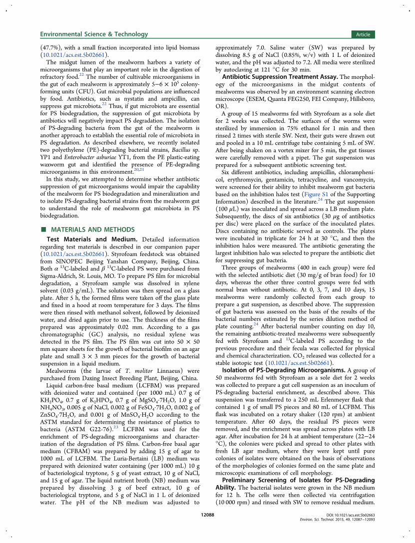
great number of bacterial cells with various morphotypes (cocciand short and long rods) inhabited the midgut content of theStyrofoam-eating mealworm, as observed by ESEM (Figure 1a).This observation indicated that the midgut of the mealwormdid harbor a diversity of microorganisms.22
The results of the antibiotic screening test indicated that 3 ofthe 6 tested antibiotics were able to inhibit the growth of gutbacteria in the LB medium with the formation of clearinhibition halos (Figure S1 of the Supporting Information).The effective antibiotics were ampicillin, gentamicin, andtetracycline. Among these antibiotics, gentamicin showed thebest ability to inhibit the growth of gut bacteria, with clearerand broader halos (Figure S1 of the Supporting Information).Gentamicin is well-known as a bactericidal antibiotic that worksby irreversibly binding the 30S subunit of the bacterialribosome, interrupting protein synthesis. Consequently,gentamicin was selected for the suppression of mealworm gutbacteria.When mealworms were fed with gentamicin-containing bran
(30 mg/g of bran), gut bacteria were significantly suppressedon the basis of the estimation of gut bacterial numbers by theseries dilution method of plate counting (Figure 1b). After 10days, there were no viable bacterial colonies in the LB mediumplate inoculated with gut suspension, indicating that the level ofgut bacteria was too low to grow in the LB medium.Subsequently, these gentamicin-treated mealworms were
tested for their PS-degrading capability. The gentamicin-treatedand control (untreated) mealworms were fed with Styrofoam,and the molecular weights of their fecula were analyzed. Incomparison to the molecular weight of the feedstock Styrofoam(Mn = 40 430, and Mw = 124 200), the Mn (32 260) and Mw(98 330) of the fecula of the control mealworms without
Environmental Science & Technology Article
DOI: 10.1021/acs.est.5b02663Environ. Sci. Technol. 2015, 49, 12087−12093
12089
gentamicin treatment (Figure 1c) dropped significantly by 8170and 25 870, respectively. However, the fecula of the gentamicin-treated mealworms had a Mn of 39 620 and a Mw of 122 650(Figure 1c), which were decreased insignificantly by only 810and 1550, respectively. This result indicated that thesuppression of gut microbiota by gentamicin impaired theability of the mealworms to depolymerize PS.On day 10, the gentamicin-treated mealworms were also fed
with jelly food containing α13C- or β13C-labled PS for 16 days.CO2 in the off air was collected for the analysis of 13C. Theresults showed that the gentamicin-treated worms did notproduce 13C-enriched CO2, whereas the untreated control did(t test; p < 0.001; Figure 1d). This result indicated that thesuppression of gut bacteria impaired the ability of themealworms to mineralize PS.Preliminary Screening of PS-Degrading Isolates. A
total of 13 bacterial cultures were isolated by picking upcolonies formed from the enrichment of PS-eating mealwormguts (Table S1 of the Supporting Information). Afterward, wecharacterized biofilm formation on the PS film sheets to screenthe culture for potential PS biodegradation because theformation of a biofilm enables microorganisms to use efficientlynon-soluble substrates, as described previously for thecharacterization of PE-degrading bacterial strains.20,25,26
Accordingly, screenings for potential PS-degrading bacteriaamong these isolated cultures were performed in terms of thenumber of the cells in the biofilm colonized on the PS film inthe CFBAM plates (Figure S2 of the Supporting Information).Among the 13 isolates, one non-spore forming Gram-positive
bacterium was the most abundant (9.3 ± 0.3 × 10⁸ CFU/cm2)
when grown as a biofilm on the PS film sheet (Figure S2 of theSupporting Information). This strain was taxonomicallyidentified as Exiguobacterium sp. strain YT2 based on its 16SrRNA sequence (Figure S3 of the Supporting Information)and, consequently, was selected as a candidate of potential PSdegraders for further study. The sequence of strain YT2 hasbeen deposited in GenBank under reference KP731587. TheExiguobacterium sp. strain YT2 has been deposited at the ChinaGeneral Microbiological Culture Collection Center(CGMCCC 10521).
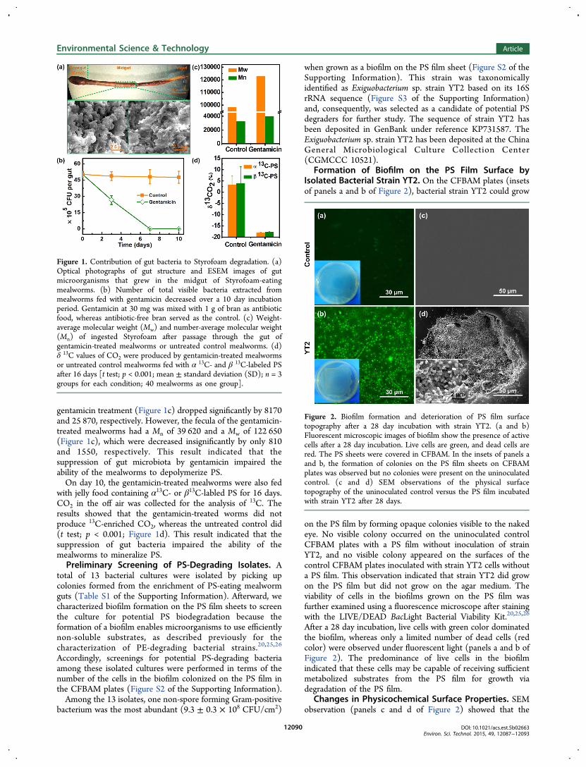
Formation of Biofilm on the PS Film Surface byIsolated Bacterial Strain YT2. On the CFBAM plates (insetsof panels a and b of Figure 2), bacterial strain YT2 could grow
on the PS film by forming opaque colonies visible to the nakedeye. No visible colony occurred on the uninoculated controlCFBAM plates with a PS film without inoculation of strainYT2, and no visible colony appeared on the surfaces of thecontrol CFBAM plates inoculated with strain YT2 cells withouta PS film. This observation indicated that strain YT2 did growon the PS film but did not grow on the agar medium. Theviability of cells in the biofilms grown on the PS film wasfurther examined using a fluorescence microscope after stainingwith the LIVE/DEAD BacLight Bacterial Viability Kit.20,25,26
After a 28 day incubation, live cells with green color dominatedthe biofilm, whereas only a limited number of dead cells (redcolor) were observed under fluorescent light (panels a and b ofFigure 2). The predominance of live cells in the biofilmindicated that these cells may be capable of receiving sufficientmetabolized substrates from the PS film for growth viadegradation of the PS film.
Changes in Physicochemical Surface Properties. SEMobservation (panels c and d of Figure 2) showed that the
Figure 1. Contribution of gut bacteria to Styrofoam degradation. (a)Optical photographs of gut structure and ESEM images of gutmicroorganisms that grew in the midgut of Styrofoam-eatingmealworms. (b) Number of total visible bacteria extracted frommealworms fed with gentamicin decreased over a 10 day incubationperiod. Gentamicin at 30 mg was mixed with 1 g of bran as antibioticfood, whereas antibiotic-free bran served as the control. (c) Weight-average molecular weight (Mw) and number-average molecular weight(Mn) of ingested Styrofoam after passage through the gut ofgentamicin-treated mealworms or untreated control mealworms. (d)δ 13C values of CO2 were produced by gentamicin-treated mealwormsor untreated control mealworms fed with α 13C- and β 13C-labeled PSafter 16 days [t test; p < 0.001; mean ± standard deviation (SD); n = 3groups for each condition; 40 mealworms as one group].
Figure 2. Biofilm formation and deterioration of PS film surfacetopography after a 28 day incubation with strain YT2. (a and b)Fluorescent microscopic images of biofilm show the presence of activecells after a 28 day incubation. Live cells are green, and dead cells arered. The PS sheets were covered in CFBAM. In the insets of panels aand b, the formation of colonies on the PS film sheets on CFBAMplates was observed but no colonies were present on the uninoculatedcontrol. (c and d) SEM observations of the physical surfacetopography of the uninoculated control versus the PS film incubatedwith strain YT2 after 28 days.
Environmental Science & Technology Article
DOI: 10.1021/acs.est.5b02663Environ. Sci. Technol. 2015, 49, 12087−12093
12090

biofilm of strain YT2 generated obvious surface deterioration,with the formation of pits and cavities on the surface of the PSfilm. The surface of the uninoculated control was smooth anddid not have any defects. The typical cavities on the surface ofthe PS film had a maximum width of approximately 200 × 200μm (Figure 2d). This result indicated that strain YT2 is capableof degrading the PS film and damaging the PS physicalintegrity.The water contact angle (WCA) was used to analyze changes
in surface hydrophobicity. After the formed biofilm wascompletely removed from the PS film samples, the WCA ofthe surface of the PS film inoculated with strain YT2 was 80.8± 3.0° (n = 5), which was much lower than the WCA of theuninoculated control (95.8° ± 1.6°; n = 5; Figure 3a). This
result indicated that the formation of the biofilm by strain YT2also decreased the hydrophobicity of the tested PS samples.This decline in hydrophobicity would reduce resistance tosubsequent degradation by bacterial cells. A similar observationwas reported previously in characterizing two PE-degradingbacterial strains.20
X-ray photoelectron spectroscopy (XPS) was used to analyzechanges in surface chemical components and functional groups.Figure 3b shows the XPS scanning spectra (0−900 eV) for thePS film incubated with strain YT2 versus the uninoculatedcontrol. In the uninoculated control, only surface carbon (284.8eV) and a limited amount of oxygen (532.3 eV) were observed.The spectrum of the PS sample with strain YT2 showed thatthe amount of oxygen increased significantly, whereas theamount of carbon appeared constant. A comparison of the XPSspectra of C 1s on the PS film surface inoculated with strainYT2 versus the uninoculated control (Figure 3c) demonstratedthat the peak-fitting result of C 1s for the uninoculated controlshowed only one peak at 284.8 eV, which was assigned to a−C−C− group. For the strain YT2-incubated samples, in
addition to the peak at 284.8 eV, another peak appeared at286.5 eV and was assigned to the −C−O− group, implyingoxidation to alcohol- and carboxylic-acid-like compounds.27
The O/C ratios of the strain YT2-incubated samples wereremarkably higher than the O/C ratios of the uninoculatedcontrols (0.10 versus 0.02). The uninoculated controls showed100% relative abundances of −C−C− group peaks and did nothave any −C−O− group peaks. However, the relativeabundances of the −C−C− and −C−O− group peaks of thestrain YT2-incubated samples were 91 and 9%, respectively.These results indicated that strain YT2 was capable of attackingor oxidizing the PS structure to produce more polar derivatives.
Weight Loss and the Molecular Weight Decrease ofthe PS Samples by Strain YT2. Degradation efficiency canbe directly measured by the weight loss of a sample. The weightloss of the PS samples inoculated with strain YT2 is presentedin Figure 4a. After a 60 day incubation with strain YT2 in
LCFBM, the weight loss of the PS pieces was 7.4 ± 0.4%, whichwas much higher than the 0.8% weight loss elicited by R. ruberC208 over a 56 day period as reported by Mor et al.15
The molecular weight and molecular weight distribution(MWD) of the PS samples after a 60 day incubation weredetermined using GPC. The molecular weights (Mn/Mw) of thestrain YT2-incubated PS sample versus the uninoculatedcontrol were 37 480/110 070 and 40 430/124 200, respectively,which represented a ∼7−11% reduction from the control PS(Figure 4b). The MWDs of the PS samples incubated with thestrain YT2 showed a clear negative trend compared to the
Figure 3. Surface chemical analysis of the PS samples of theuninoculated control and the samples inoculated with strain YT2 aftera 28 day incubation. (a) WCAs of the PS films inoculated with thestrain YT2 decreased in comparison to the WCAs of the uninoculatedcontrol, indicating a decrease in surface hydrophobicity. (b) XPSscanning and (c) C 1s spectra of the uninoculated control and theresidual PS films inoculated with strain YT2.
Figure 4. Characterization of PS biodegradation by strain YT2 inLCFBM compared to the control (uninoculated medium) after a 60day incubation (mean value ± SD; n = 3). (a) Change in the dryweight of the PS pieces. (b) Molecular weight (Mw/Mn) and (c)MWD shift of the residues inoculated with strain YT2 versus theuninoculated control.
Environmental Science & Technology Article
DOI: 10.1021/acs.est.5b02663Environ. Sci. Technol. 2015, 49, 12087−12093
12091

uninoculated control PS (Figure 4c). The decrease in MWDsuggested that cleavage/depolymerization of the PS long-chainstructure occurred and that lower molecular weight fragmentswere formed in the presence of strain YT2. In addition, theanalysis of samples extracted from LCFBM using GC/MSindicated that more than 40 peaks were found in the mediumwith strain YT2 but that no peaks were found in the control(Figure S4 and Table S2 of the Supporting Information).In summary, the weight loss and molecular weight decrease
of the PS samples supported the conclusion that strain YT2,which was isolated from the mealworm gut, was capable ofdegrading PS.Implications. Our antibiotic test results confirmed that
antibiotic suppression of gut bacteria impaired the ability of themealworm to depolymerize long-chain PS molecules andfurther mineralize the metabolites to CO2. This study is thefirst to report the presence of PS-degrading bacteria in the gutsof mealworms. We enriched a mixed culture from Styrofoam-eating mealworm gut content using PS pieces as the sole carbonsource and isolated 13 pure bacterial cultures. Among them,one isolated bacterial Exiguobacterium sp. strain YT2 wasselected, and PS degradation by strain YT2 was confirmed bynot only bacterial growth on the PS film, which causes changesin surface topography, decreases in hydrophobicity, and theformation of carbonyl groups, but also the measurement ofweight loss and the identification of the loss of molecularweight and the release of water-soluble daughter products. Bycombination of the results of antibiotic suppression andmicrobial culture-dependent isolation approaches, this studyhas provided evidence that mealworm gut microbiota play anessential role in PS biodegradation in the gut. In this study, aLB agar medium was used for the isolation of PE- and PS-degrading bacteria. As a result of the high NaCl content (10 g/L), this medium was a selective medium. Further research isneeded to test or develop other media for optimal isolation ofPE- and PS-degrading bacteria.On the basis of the short retention time of gut contents (<24
h) and up to 47.7% carbon conversion into CO2 by themealworms (10.1021/acs.est.5b02661), the PS degradationefficiency (e.g., 7.4% over 60 days) demonstrated that theisolated bacterial strain YT2 outside the living host appears toshow much poorer PS degradation efficiency than thatdemonstrated in the gut system. The PS degradation by strainYT2 outside the worm gut could be limited by unknown factorsbecause less energy was generated for cell growth. Similar lowapparent yield coefficients were observed when PE-degrading E.asburiae YT1 and Baccilus sp. YP1 grew on PE films with 0.82and 0.66 g of cells/g of PE, respectively.20
We could expect that PS degradation in mealworm guts isanalogous to microbial degradation of lignocelluloses inruminating mammals and wood in termites for the mutualbenefits of the metabolism of microbial consortia and host. Thegut of the mealworm can be considered an efficient bioreactor.Physicochemical “treatments” (by chewing, ingesting, mixingwith gut contents, etc.) together with the activity of enzymessecreted by the worm are also possibly critical for the success ofrapid PS degradation in this bioreactor. More research will beconducted to fully understand the synergic actions betweenworm digestion and microbial metabolism and to betterunderstand the enzymatic systems involved in the biodegrada-tion of PS as well as other plastics, such as PE, which could behelpful in the development of promising remediationapproaches for plastic wastes.
■ ASSOCIATED CONTENT
*S Supporting InformationThe Supporting Information is available free of charge on theACS Publications website at DOI: 10.1021/acs.est.5b02663.
Results of different antibiotics on gut bacterial growth(Figure S1), changes in bacterial cell numbers on the PSfilm incubated with 13 different isolated bacterial cultures(Figure S2), 16S RNA gene-based neighbor-joiningphylogenetic tree of strain YT2 (Figure S3), GC of theextract from the culture inoculated with strain YT2 andthe uninoculated control after 60 days (Figure S4), atotal of 13 bacterial strains isolated from the PSenrichment of gut microbes (Table S1), and GC/MSresults of water-soluble products from PS pieces in thepresence and absence of strain YT2 (Table S2) (PDF)
■ AUTHOR INFORMATION
Corresponding Authors*Telephone/Fax: +86-10-8233-8552. E-mail: [email protected].*Telephone/Fax: +86-10-8262-1396. E-mail: [email protected].
NotesThe authors declare no competing financial interest.
■ ACKNOWLEDGMENTS
This research was supported by the National Natural ScienceFoundation of China (NSFC, Grants 51373006 and20477002), the State Basic Research Program of China(Grant 2014CB931800), and the Shenzhen Key Laboratoryof Bioenergy (Grant CXB201005240001A). Wei-Min Wu is afunded international collaborator under NSFC Grant51373006.
■ REFERENCES(1) PlasticsEurope. PlasticsThe Facts 2014/2015An Analysis ofEuropean Latest Plastics Production, Demand and Waste Data;PlasticsEurope: Brussels, Belgium, 2014; www.plasticseurope.org/documents/document/20150227150049-final_plastics_the_facts_2014_2015_260215.pdf.(2) Panda, A. K.; Singh, R. K.; Mishra, D. K. Thermolysis of wasteplastics to liquid fuel: A suitable method for plastic waste managementand manufacture of value added products-A world prospective.Renewable Sustainable Energy Rev. 2010, 14 (1), 233−248.(3) Barnes, D. K.; Galgani, F.; Thompson, R. C.; Barlaz, M.Accumulation and fragmentation of plastic debris in global environ-ments. Philos. Trans. R. Soc., B 2009, 364 (1526), 1985−1998.(4) Law, K. L.; Moret-Ferguson, S.; Maximenko, N. A.; Proskurowski,G.; Peacock, E. E.; Hafner, J.; Reddy, C. M. Plastic accumulation in theNorth Atlantic subtropical gyre. Science 2010, 329 (5996), 1185−1188.(5) Rochman, C. M.; Browne, M. A.; Halpern, B. S.; Hentschel, B. T.;Hoh, E.; Karapanagioti, H. K.; Rios-Mendoza, L. M.; Takada, H.; Teh,S.; Thompson, R. C. Policy: Classify plastic waste as hazardous. Nature2013, 494 (7436), 169−171.(6) Cozar, A.; Echevarría, F.; Gonzalez-Gordillo, J. I.; Irigoien, X.;Ubeda, B.; Hernandez-Leon, S.; Palma, A. T.; Navarro, S.; García-de-Lomas, J.; Ruiz, A.; Fernandez-de-Puelles, M. L.; Duarte, C. M. Plasticdebris in the open ocean. Proc. Natl. Acad. Sci. U. S. A. 2014, 111 (28),10239−10244.(7) Jambeck, J. R.; Geyer, R.; Wilcox, C.; Siegler, T. R.; Perryman,M.; Andrady, A.; Narayan, R.; Law, K. L. Plastic waste inputs from landinto the ocean. Science 2015, 347 (6223), 768−771.
Environmental Science & Technology Article
DOI: 10.1021/acs.est.5b02663Environ. Sci. Technol. 2015, 49, 12087−12093
12092

(8) Velasco, A.; Alonso, S.; García, J. L.; Perera, J.; Díaz, E. Geneticand functional analysis of the styrene catabolic cluster of Pseudomonassp. strain Y2. J. Bacteriol. 1998, 180 (5), 1063−1071.(9) O’Leary, N. D.; O’Connor, K. E.; Dobson, A. D. W.Biochemistry, genetics and physiology of microbial styrene degrada-tion. FEMS Microbiol. Rev. 2002, 26 (4), 403−417.(10) Tsuchii, A.; Suzuki, T.; Takahara, Y. Microbial degradation ofstyrene oligomer. Agric. Biol. Chem. 1977, 41 (12), 2417−2421.(11) Gautam, R.; Bassi, A. S.; Yanful, E. K. A review ofbiodegradation of synthetic plastic and foams. Appl. Biochem.Biotechnol. 2007, 141, 85−108.(12) Guillet, J. E.; Regulski, T. W.; McAneney, T. B. Biodegradabilityof photodegraded polymers II: tracer studies of biooxidation of EcolytePS polystyrene. Environ. Sci. Technol. 1974, 8, 923−925.(13) Sielicki, M.; Focht, D. D.; Martin, J. P. Microbial degradation of[14C] polystyrene and 1, 3-diphenylbutane. Can. J. Microbiol. 1978, 24,798−803.(14) Kaplan, D. L.; Hartenstein, R.; Sutter, J. Biodegradation ofpolystyrene, poly(methyl methacrylate), and phenol formaldehyde.Appl. Environ. Microbiol. 1979, 38, 551−553.(15) Mor, R.; Sivan, A. Biofilm formation and partial biodegradationof polystyrene by the actinomycete Rhodococcus ruber. Biodegradation2008, 19, 851−858.(16) Atiq, N.; Ahmed, S.; Ali, M. I.; Ahmad, B.; Robson, G. Isolationand identification of polystyrene biodegrading bacteria from soil. Afr. J.Microbiol. Res. 2010, 4, 1537−1541.(17) Gerhardt, P. D.; Lindgren, D. L. Penetration of packaging films:film materials used for food packaging tested for resistance to somecommon stored-product insects. Calif. Agric. 1954, 8, 3−4.(18) Browditch, T. G. Penetration of polyvinyl chloride andpolypropylene packaging films by Ephestia cautella (Lepidoptera:Pyralidae) and Plodia interpunctella (Lepidoptera: Pyralidae) larvae,and Tribolium confusum (Coleoptera: Tenebrionidae) adults. J. Econ.Entomol. 1997, 90, 1028−1031.(19) Riudavets, J.; Salas, I.; Pons, M. J. Damage characteristicsproduced by insect pests in packaging film. J. Stored Prod. Res. 2007,43, 564−570.(20) Yang, J.; Yang, Y.; Wu, W.-M.; Zhao, J.; Jiang, L. Evidence ofpolyethylene biodegradation by bacterial strains from the gut ofplastic-eating waxworms. Environ. Sci. Technol. 2014, 48 (23), 13776−13784.(21) Yang, Y.; Chen, J.; Wu, W.-M.; Zhao, J.; Yang, J. Completegenome sequence of Bacillus sp. YP1, a polyethylene-degradingbacterium from waxworm’s gut. J. Biotechnol. 2015, 200, 77−78.(22) Genta, F. A.; Dillon, R. J.; Terra, W. R.; Ferreira, C. Potentialrole for gut microbiota in cell wall digestion and glucosidedetoxification in Tenebrio molitor larvae. J. Insect Physiol. 2006, 52(6), 593−601.(23) ASTM International. ASTM G22-76. Standard Practice forDetermining Resistance of Plastics to Bacteria; ASTM International: WestConshohocken, PA, 1996.(24) Visotto, L. E.; Oliveira, M. G. A.; Guedes, R. N. C.; Ribon, A. O.B.; Good-God, P. I. V. Contribution of gut bacteria to digestion anddevelopment of the velvetbean caterpillar, Anticarsia gemmatalis. J.Insect Physiol. 2009, 55 (3), 185−191.(25) Gilan, I.; Hadar, Y.; Sivan, A. Colonization, biofilm formationand biodegradation of polyethylene by a strain of Rhodococcus ruber.Appl. Microbiol. Biotechnol. 2004, 65 (1), 97−104.(26) Sivan, A.; Szanto, M.; Pavlov, V. Biofilm development of thepolyethylene-degrading bacterium Rhodococcus ruber. Appl. Microbiol.Biotechnol. 2006, 72 (2), 346−352.(27) Shang, J.; Chai, M.; Zhu, Y. Solid-phase photocatalyticdegradation of polystyrene plastic with TiO2 as photocatalyst. J.Solid State Chem. 2003, 174 (1), 104−110.
Environmental Science & Technology Article
DOI: 10.1021/acs.est.5b02663Environ. Sci. Technol. 2015, 49, 12087−12093
12093