Biocompatibility and mechanical properties of diamond-like...
13
Biocompatibility and mechanical properties of diamond-like coatings on cobalt-chromium-molybdenum steel and titanium-aluminum- vanadium biomedical alloys C. Hinu ¨ ber, 1,2 C. Kleemann, 1,2 R. J. Friederichs, 3 L. Haubold, 1 H. J. Scheibe, 1 T. Schuelke, 1 C. Boehlert, 3 M. J. Baumann 3 1 Fraunhofer Center for Coatings and Laser Applications, East Lansing, Michigan 48823 2 Coating Technology Division, Technical University Dresden, Mechanical Engineering, Dresden 01069, Germany 3 Department of Chemical Engineering and Materials Science, College of Engineering, Michigan State University, East Lansing, Michigan 48824 Received 13 June 2007; revised 12 November 2009; accepted 11 March 2010 Published online 20 July 2010 in Wiley Online Library (wileyonlinelibrary.com). DOI: 10.1002/jbm.a.32851 Abstract: Diamond-like carbon (DLC) films are favored for wear components because of diamond-like hardness, low friction, low wear, and high corrosion resistance (Schultz et al., Mat-wiss u Werkstofftech 2004;35:924–928; Lappalainen et al., J Biomed Mater Res B Appl Biomater 2003;66B:410– 413; Tiainen, Diam Relat Mater 2001;10:153–160). Several studies have demonstrated their inertness, nontoxicity, and the biocompatibility, which has led to interest among manu- facturers of surgical implants (Allen et al., J Biomed Mater Res B Appl Biomater 2001;58:319–328; Uzumaki et al., Diam Relat Mater 2006;15:982–988; Hauert, Diam Relat Mater 2003;12:583–589; Grill, Diam Relat Mater 2003;12:166–170). In this study, hydrogen-free amorphous, tetrahedrally bonded DLC films (ta-C) were deposited at low temperatures by phys- ical vapor deposition on medical grade Co28Cr6Mo steel and the titanium alloy Ti6Al4V (Scheibe et al., Surf Coat Tech 1996;85:209–214). The mechanical performance of the ta-C was characterized by measuring its surface roughness, con- tact angle, adhesion, and wear behavior, whereas the bio- compatibility was assessed by osteoblast (OB) attachment and cell viability via Live/Dead assay. There was no statistical difference found in the wettability as measured by contact angle measurements for the ta-C coated and the uncoated samples of either Co28Cr6Mo or Ti6Al4V. Rockwell C indenta- tion and dynamic scratch testing on 2–10 lm thick ta-C films on Co28Cr6Mo substrates showed excellent adhesion with HF1 grade and up to 48 N for the critical load L C2 during scratch testing. The ta-C coating reduced the wear from 3.5 10 5 mm 3 /Nm for an uncoated control sample (uncoated Co28Cr6Mo against uncoated stainless steel) to 1.1 10 7 mm 3 /Nm (coated Co28Cr6Mo against uncoated stainless steel) in reciprocating pin-on-disk testing. The lowest wear factor of 3.9 10 10 mm 3 /Nm was measured using a ta-C coated steel ball running against a ta-C coated and polished Co28Cr6Mo disk. Student’s t-test found that the ta-C coating had no statistically significant (p < 0.05) effect on OB attach- ment, when compared with the uncoated control samples. There was no significant difference (p < 0.05) in the Live/ Dead assay results in cell death between the ta-C coated Co28Cr6Mo and Ti6Al4V samples and the uncoated controls. Therefore, these ta-C coatings show improved wear and cor- rosion (Dorner-Reisel et al., Diam Relat Mater 2003;11:823– 827; Affato et al., J Biomed Mater Res B Appl Biomater 2000;53:221–226; Dorner-Reisel et al., Surf Coat Tech 2004;177–178:830–837; Kim et al., Diam Relat Mater 2004;14:35–41) performance and excellent in vitro cyto-com- patibility, when compared with currently used uncoated Co28Cr6Mo and Ti6Al4V implant materials. V C 2010 Wiley Peri- odicals, Inc. J Biomed Mater Res Part A: 95A: 388–400, 2010. Key Words: diamond-like coatings, ta-C films, physical vapor deposition, Ti6Al4V, Co28Cr6Mo INTRODUCTION According to the National Center for Health Statistics (165,000 in 2001) and the American Academy of Orthope- dic Surgery (198,000 in 2002), approximately 200,000 total hip joint replacements are performed annually in the United States. 1,2 A leading cause for hip joint replacement is osteo- arthritis, which affects more than 20 million Americans. 3,4 Hip replacement surgery increased by 38% between 1995 and 2004 and is expected to more than double within the next 25 years. 4,5 Of this total, there were approximately 40,000 revision surgeries. 5 Because the average age of patients requiring hip replacements is decreasing and the average life expectancy is increasing, the number of hip re- vision surgeries is also expected to rise. Thus, it becomes critical to increase the lifetime of a hip replacement to mini- mize the number of revision surgeries. One of the primary issues impeding orthopedic implant longevity is the high wear rate of commonly used material wear couples such as Co28Cr6Mo/ultra high molecular weight polyethylene (UHMWPE) and Ti6Al4V/UHMWPE found in total hip and total knee replacements. 3,6 There is evidence that wear de- bris, especially UHMWPE particulates, lead to wear particle Correspondence to: M. J. Baumann; e-mail: [email protected] 388 V C 2010 WILEY PERIODICALS, INC.
Transcript of Biocompatibility and mechanical properties of diamond-like...

Biocompatibility and mechanical properties of diamond-like coatingson cobalt-chromium-molybdenum steel and titanium-aluminum-vanadium biomedical alloys
C. Hinuber,1,2 C. Kleemann,1,2 R. J. Friederichs,3 L. Haubold,1 H. J. Scheibe,1 T. Schuelke,1
C. Boehlert,3 M. J. Baumann3
1Fraunhofer Center for Coatings and Laser Applications, East Lansing, Michigan 488232Coating Technology Division, Technical University Dresden, Mechanical Engineering, Dresden 01069, Germany3Department of Chemical Engineering and Materials Science, College of Engineering, Michigan State University,
East Lansing, Michigan 48824
Received 13 June 2007; revised 12 November 2009; accepted 11 March 2010
Published online 20 July 2010 in Wiley Online Library (wileyonlinelibrary.com). DOI: 10.1002/jbm.a.32851
Abstract: Diamond-like carbon (DLC) films are favored for
wear components because of diamond-like hardness, low
friction, low wear, and high corrosion resistance (Schultz
et al., Mat-wiss u Werkstofftech 2004;35:924–928; Lappalainen
et al., J Biomed Mater Res B Appl Biomater 2003;66B:410–
413; Tiainen, Diam Relat Mater 2001;10:153–160). Several
studies have demonstrated their inertness, nontoxicity, and
the biocompatibility, which has led to interest among manu-
facturers of surgical implants (Allen et al., J Biomed Mater
Res B Appl Biomater 2001;58:319–328; Uzumaki et al., Diam
Relat Mater 2006;15:982–988; Hauert, Diam Relat Mater
2003;12:583–589; Grill, Diam Relat Mater 2003;12:166–170). In
this study, hydrogen-free amorphous, tetrahedrally bonded
DLC films (ta-C) were deposited at low temperatures by phys-
ical vapor deposition on medical grade Co28Cr6Mo steel and
the titanium alloy Ti6Al4V (Scheibe et al., Surf Coat Tech
1996;85:209–214). The mechanical performance of the ta-C
was characterized by measuring its surface roughness, con-
tact angle, adhesion, and wear behavior, whereas the bio-
compatibility was assessed by osteoblast (OB) attachment
and cell viability via Live/Dead assay. There was no statistical
difference found in the wettability as measured by contact
angle measurements for the ta-C coated and the uncoated
samples of either Co28Cr6Mo or Ti6Al4V. Rockwell C indenta-
tion and dynamic scratch testing on 2–10 lm thick ta-C films
on Co28Cr6Mo substrates showed excellent adhesion with
HF1 grade and up to 48 N for the critical load LC2 during
scratch testing. The ta-C coating reduced the wear from 3.5 �10�5 mm3/Nm for an uncoated control sample (uncoated
Co28Cr6Mo against uncoated stainless steel) to 1.1 � 10�7
mm3/Nm (coated Co28Cr6Mo against uncoated stainless
steel) in reciprocating pin-on-disk testing. The lowest wear
factor of 3.9 � 10�10 mm3/Nm was measured using a ta-C
coated steel ball running against a ta-C coated and polished
Co28Cr6Mo disk. Student’s t-test found that the ta-C coating
had no statistically significant (p < 0.05) effect on OB attach-
ment, when compared with the uncoated control samples.
There was no significant difference (p < 0.05) in the Live/
Dead assay results in cell death between the ta-C coated
Co28Cr6Mo and Ti6Al4V samples and the uncoated controls.
Therefore, these ta-C coatings show improved wear and cor-
rosion (Dorner-Reisel et al., Diam Relat Mater 2003;11:823–
827; Affato et al., J Biomed Mater Res B Appl Biomater
2000;53:221–226; Dorner-Reisel et al., Surf Coat Tech
2004;177–178:830–837; Kim et al., Diam Relat Mater
2004;14:35–41) performance and excellent in vitro cyto-com-
patibility, when compared with currently used uncoated
Co28Cr6Mo and Ti6Al4V implant materials. VC 2010 Wiley Peri-
odicals, Inc. J Biomed Mater Res Part A: 95A: 388–400, 2010.
Key Words: diamond-like coatings, ta-C films, physical vapor
deposition, Ti6Al4V, Co28Cr6Mo
INTRODUCTION
According to the National Center for Health Statistics(165,000 in 2001) and the American Academy of Orthope-dic Surgery (198,000 in 2002), approximately 200,000 totalhip joint replacements are performed annually in the UnitedStates.1,2 A leading cause for hip joint replacement is osteo-arthritis, which affects more than 20 million Americans.3,4
Hip replacement surgery increased by 38% between 1995and 2004 and is expected to more than double within thenext 25 years.4,5 Of this total, there were approximately40,000 revision surgeries.5 Because the average age of
patients requiring hip replacements is decreasing and theaverage life expectancy is increasing, the number of hip re-vision surgeries is also expected to rise. Thus, it becomescritical to increase the lifetime of a hip replacement to mini-mize the number of revision surgeries. One of the primaryissues impeding orthopedic implant longevity is the highwear rate of commonly used material wear couples such asCo28Cr6Mo/ultra high molecular weight polyethylene(UHMWPE) and Ti6Al4V/UHMWPE found in total hip andtotal knee replacements.3,6 There is evidence that wear de-bris, especially UHMWPE particulates, lead to wear particle
Correspondence to: M. J. Baumann; e-mail: [email protected]
388 VC 2010 WILEY PERIODICALS, INC.

induced aseptic loosening.6–10 Wear products from metallicimplants have also been shown to be harmful to the sur-rounding tissue.5,9,11–13 Co-and Cr-ions are considered to betoxic or even carcinogenic and have been shown to promoteinflammation and reduced cell activity.7,8,11,12,14–16
Coating metallic prostheses to obtain a wear and corro-sion protective surface is not a new idea. Diamond-like car-bon (DLC) is a class of amorphous carbon thin film materi-als that are composed of a mixture of graphite-type (sp2
hybridized, trigonal planar covalent bonding) and diamond-type (sp3 hybridized, tetragonal tetrahedral covalent bond-ing) atomic bonds. This unique amorphous atomic structureleads to material properties such as low friction coefficientsand wear rates, which depend on the actual fraction of dia-mond-type bonds and on the amount of addition chemicalelements such as hydrogen in the films.17 Recently, DLCmaterials have been the subjects of extensive investigationbecause of their potential in biomedical applications. Numer-ous authors note the potential of DLC coatings to act as aprotective coating in orthopedic applications and have inves-tigated their mechanical properties.7,8,18,19 Each of thesestudies showed that DLC coatings provide highly desirablemechanical properties including diamond-like hardness andextremely low wear rates and therefore are preferred candi-dates for articulating orthopedic devices. DLC coatings canpotentially eliminate wear debris and are widely consideredto be inert, corrosion resistant, and biocompatible.6,20–24
However, a 2003 in vivo study examined a catastrophic fail-ure of a particular DLC coating caused by microscopic coatingdefect (pinholes) induced delamination and subsequent abra-sion but did not include detailed information about the type(sp3 content, chemical composition) of DLC coating used.25 Inrecent years, the focus has been on improving the adhesionand reducing defects of the DLC coatings and on investigatingtheir wear and friction properties.22,26 DLC coatings also pro-vide a desirable surface for other medical applications. Addi-tions of Ag, Si, N, CaO, and F to the DLC coatings were foundto further add functionality, such as antibacterial and antith-rombogenic behavior, increased chemical inertness, andimproved biocompatibility for blood contacting devices orcardiovascular applications.12,27–30
This study reports on the wear behavior and in vitro bio-compatibility of a hydrogen-free and tetrahedrally bondedamorphous carbon coating (ta-C) using a murine osteoblast(OB) cell line. Surface roughness and contact angle measure-ments were preformed to characterize the surfaces of the dif-ferent wear couples. Results from scratch tests and pin-on-disktesting on 2–10 lm thick ta-C coatings on Co28Cr6Mo sampleswere performed to assess the mechanical integrity of the coat-ing on typical biomedical implant grade material. OB attach-ment data and LIVE-DEAD assay results were collected to eval-uate the biocompatibility of these ta-C coated Co28Cr6Mo(ASTM F1537) and Ti6Al4V (ASTM F 136) materials.
METHODS
Preparation of hydrogen-free DLC ta-C coatingsBefore the coating process, all samples were cleaned in anultrasonic bath in ethanol for 10 min followed by a deter-
gent (MUCAPUR-AF) based washing process (5 min inMIELE Professional IR8000 washer). After washing, thesamples were dried and then kept at 80�C in a dry environ-ment until they were placed into the physical vapor deposi-tion (PVD) system. The PVD system consists of a chamberwith a rotating cylindrical sample holder, three direct cur-rent cathodic arc evaporators to deposit adhesion layersand a Laser-ArcoV
R
module (Fraunhofer Institute for Materi-als and Beam Technology, Dresden, Germany) that isattached to the main chamber. The ta-C coatings were pre-pared using the Laser-ArcoV
R
module, which is a laser-con-trolled pulsed high current cathodic vacuum arc evaporationprocess.31,32 The module houses a vertically rotating cylin-drical graphite target. A Nd-YAG laser, a beam scanner opti-cal system, and a high current pulsed power supply are alsopart of the LaserArcoV
R
module. After loading the samples inthe coating system and evacuating to high vacuum (<0.1Pa), the samples were plasma cleaned with an Ar high volt-age direct current discharge before deposition. Next a thinadhesion layer (<200 nm) was deposited using the directcurrent arc evaporators with a material transition gradientfrom Cr to C. Subsequently the ta-C film was depositedusing the Laser-ArcoV
R
module. The laser controls the igni-tion location along the graphite target and the timing of thepulsed arc discharges. A repetition frequency of 500 Hz andpeak arc currents of 1500 A were used. This energetic car-bon evaporation process generates fully ionized carbonplasma pulses, which rapidly expand from the solid graphitecathode toward the substrates. The carbon ion energy dis-tribution in these plasmas peaks at comparatively high ener-gies in the order of several tens of electron volts (dependingon the maximal arc current), which is sufficient to bury car-bon ions several atomic layers deep under the surface dur-ing film growth (‘‘subplantation’’). This subplantation effectis enabling for the formation of tetragonal diamond-like sp3
carbon bonds33 that lead to hard and dense coatings with ahigh Young’s modulus. These materials are in the literaturereferred to as ‘‘ta-C’’ coatings.17 Because the described pro-cess synthesizes the ta-C coatings from a solid graphitesource, there is no hydrogen incorporated into the films.Depending on the specific deposition conditions (i.e., sub-strate temperature, residual gas pressure, incident angle,etc.) the resulting DLC coatings can be tailored to addressthe requirements of typical wear applications. An exampleis the adjustment of the coating’s Young’s modulus, whichcan be varied between 350 and 700 GPa.26,32 The substratesurface temperature during the coating process is controlledby parameters such as the repetition frequency and thepeak current of the arc pulses. It is typically kept below150�C to avoid the relaxation of diamond-like sp3 bondsto form graphitic sp2, which would render the coating less‘‘diamond-like.’’
Surface roughnessCharacterization of surface roughness is necessary duringevaluation of different wear couples. The surface roughnessaverage (Ra) and the 10 point maximum peak average (Rz)were measured with a profilometer (Veeco DEKTAK 6M)
ORIGINAL ARTICLE
JOURNAL OF BIOMEDICAL MATERIALS RESEARCH A | NOV 2010 VOL 95A, ISSUE 2 389

using a stylus radius of 2.5 lm, a scanning length of 20mm, and a lateral resolution of 0.5 lm with a minimum ofthree scans per sample.
Contact angle measurementsTo determine the hydrophobicity of the prepared surfaces,contact angle measurements were made using a VCA optima(AST Products, Billerica, MA) tester. Measurements were
carried out with a droplet of 2 lL doubly distilled water.Both before and after ta-C deposition, the contact angle wasmeasured on three samples three separate times.
Adhesion testsThe ta-C coating adhesion to the metal substrates was eval-uated using both Rockwell C indentation and scratch testing,standard techniques commonly used to quantify the
FIGURE 1. Optical micrographs showing typical Rockwell C indentation test results. (A) stainless steel coupon with unpolished film; thickness
4 lm. (B) Co28Cr6Mo coupon with polished film; thickness 10 lm; (C) schematic reference chart to qualify Rockwell C indication tests.
390 HINUBER ET AL. BIOCOMPATIBILITY AND MECHANICAL PROPERTIES OF DIAMOND-LIKE COATINGS

interfacial strength of coating-substrate systems. During theRockwell C adhesion test, a cone-shaped diamond tip with aradius of 200 lm and a tip angle of 120� was perpendicu-larly indented into the coating with a force of 1472 N.Depending on the strength of the coating-substrate inter-face, the force introduced by the indenter generates a net-work of microscopic cracks around the indentation site ormay even cause the coating to delaminate around the inden-tation. The result of the test is judged qualitatively by com-paring optical microscope images of the crack network andthe degree of delamination to an adhesion quality chart andclassifying the image within six levels from HF1 throughHF6 (Fig. 1). The coating adhesion was also evaluated usinga commercial scratch tester having a spherical diamond in-denter with a radius of 200 lm. Indentations were madeperpendicularly to the coating surface, while scratchingalong the surface with the load increasing from zero to acritical load, which was identified as that load where thecoating-substrate interface began to fail. The scratches wereanalyzed in an optical microscope. The first critical load,LC1, was defined when initial cracking occurred next to the
scratch and the second critical load, LC2, indicates a brokenand delaminated film (Fig. 2).
Wear testsA pin-on-disk tribometer with reciprocating motion wasused to determine the coefficient of friction (COF) and wearrates of the wear couples. The tests were performed byapplying a normal load to a pin or ball, pressing it against aflat disk and moving it over a distance of 10 mm in a recip-rocating motion. Sets of 50,000 test cycles (one cycle is 10mm forward and 10 mm back) were performed for a totalsliding contact distance of 1000 m with each set taking� 8.7 h. Note that while the actual contact pressures inhuman hip joints are on the order of 10–20 MPa based onjoint dimensions and average human weight, because of themuch smaller contact area in the wear tester (pin or ballpressing against a flat sample), most of the experimentswere performed at significantly higher contact pressures inexcess of 1200 MPa, which is more typical for industrialbearing testing. The contact pressure was reduced to 30–40MPa when UHMWPE was used as the disk material. The
FIGURE 2. Optical micrographs of typical scratch test results. (A) 1 lm ta-C film, initial crack formation during scratch test, critical load LC1. (B)
5 lm ta-C film, large area cracks and film delamination during scratch test, critical load LC2.
ORIGINAL ARTICLE
JOURNAL OF BIOMEDICAL MATERIALS RESEARCH A | NOV 2010 VOL 95A, ISSUE 2 391

tests were performed in dry (30–40% humidity, air temper-ature 20–25�C) and lubricated conditions. Phosphate buf-fered saline (PBS) and Dulbecco’s Media II (D-Media 2) sol-utions were used as lubricants and the temperaturemaintained at 37.1�C. The tip of the pin with the appliedload is interchangeable with spherical (balls) or flat (pins)wear counterparts. Commonly available ball bearing materi-als such as tungsten carbide (hard metal), aluminum oxide(sapphire) and stainless steel (Inox 440C) were used as abaseline and custom-made pins fabricated from Co28Cr6Mowith respect to the medical application. For particular casestudies, the ball and pin materials were coated with ta-C.The disk materials were ta-C coated and uncoatedCo28Cr6Mo. Further reference measurements were per-formed on UHMWPE disk samples and sapphire, Inox aswell as Co28Cr6Mo wear couples.
Measurements were performed with surfaces as depos-ited and postpolished. For the polishing of the ta-C coatingsas well as improving the surface finish of the UHMWPEdisks, commercial diamond polishing equipment was used.Fig. 3 shows scanning electron microscopy (SEM) photomi-
crographs of cross sections through an unpolished and apolished ta-C coated Co28Cr6Mo sample.
During the test, the contacting ball/pin and disk surfaceslose material as a result of wear. Optical and sometimes sec-ondary electron microscopy was used to measure thedimensions of the worn area on the test specimen. The vol-ume of the material removed from the initially sphericalball was calculated based on the measured radius of theworn off area and the ball radius. The volume of the weartrack on the flat disk was calculated based on the lengthand the cross-sectional area of the wear track. At least fiveprofilometer scans were performed per wear track to deter-mine the cross sectional area. The wear factor, hereexpressed in mm3/Nm, is defined as the ratio of the wearvolume to the applied normal force normalized by the slid-ing distance. We have reported the wear factors for the pin/ball, the disk, and the total wear rate as the sum of the indi-vidual wear factors.
Cell culture systemAttachment and cell death were used as preliminary toolsto assess early stage biocompatibility of the ta-C coatings.Murine MC3T3-E1 OB cells were cultured using completemedia, a-minimum essential media (Invitrogen, Carlsbad,CA) supplemented with 10% fetal bovine serum (AtlantaBiologicals, Lawrenceville, GA). OBs were incubated at 37�Cand 5% CO2/95% air under humid conditions. To preventconfluency, OBs were enzymatically removed from the cul-ture surface with 0.5% Trypsin-ethylenediaminetetraaceticacid 10� (Invitrogen, Carlsbad, CA) every 2–3 days.34 Fol-lowing established procedures, the OBs were counted usinga hemacytometer.35,36 OBs were cultured to passagesbetween 29 and 34. Complete media and Trypsin were dec-anted, and OBs were re-suspended in fresh complete media.Ti6Al4V/ta-C coated samples (Ti6Al4V substrate with a ta-Ccoated surface) and the uncoated control Ti6Al4V sampleswere placed in six-well polystyrene cell culture plates andseeded with OBs at a density of 11,320 cells/cm2 to ensureconfluency, using surface tension to form a bead-like dropof suspended OBs in complete media on each sample sur-face. This same procedure was repeated using Co28Cr6Mo/ta-C coated samples (Co28Cr6Mo substrate with a ta-Ccoated surface) and the uncoated control Co28Cr6Mo sam-ples. A pipette tip was used to spread the suspended OBs toensure complete coverage of the sample surface.
Cell attachment studyFollowing standard protocols developed in our lab, cellswere plated onto sterile coated and uncoated samples.Seeded samples were incubated at 37�C and 5% CO2/95%air under humid conditions for 4 h, the media aspirated andthe OBs washed twice with 1� PBS. Cells were fixed with3.7% formaldehyde for 10 min at room temperature,washed twice with 1� PBS, permeabilized for 3–5 min in0.1% Triton ��100 in 1� PBS and washed twice with 1�PBS. Hoechst nucleus immuno-fluorescence stain (Hoechst33342 stain Invitrogen) was applied to each sample underdark conditions and incubated for 30 min. After incubation,
FIGURE 3. Scanning electron micrographs of (A) unpolished ta-C and
(B) polished ta-C films on Co28Cr6Mo substrates.
392 HINUBER ET AL. BIOCOMPATIBILITY AND MECHANICAL PROPERTIES OF DIAMOND-LIKE COATINGS
the stain was aspirated and the samples washed twice with1� PBS and mounted using a 1:1 solution of 1� PBS toglycerol. The OBs were imaged using a fluorescent micro-scope with an attached digital camera. Hoechst stain fluo-resces at a wavelength of kemission ¼ 465 nm when exposedto light with a wavelength of kexcitation ¼ 355 nm. Twentyrepresentative images were taken per sample to calculateOB attachment. Counting nuclei over each image and calcu-lating the average quantified the number of attached cellsper field. The total number of attached cells on the wholesurface was then calculated using the average amount ofcells per field. After imaging, the samples were washed with1� PBS and reverse osmosis water and then wiped with70% ethanol 30% reverse osmosis water. Finally, the sam-ples were placed in a sonic bath for 10 min in 95% by vol-ume ethanol, air-dried and placed in a desiccator at lowpressure for storage.
LIVE/DEAD viability assaySeeded samples were incubated at 37�C and 5% CO2/95%air under humid conditions for 24 h. After the incubationperiod, media and nonadherent cells were washed off thesample with 1� PBS and were centrifuged for 5 min at1000 rpm and 4�C. The liquid was decanted, and nonadher-ent cells were re-suspended in 0.1 mL 1� PBS. Using sur-face tension, the nonadherent cells were plated on the re-spective sample and incubated at 37�C and 5% CO2/95%air under humid conditions for 30 min so that cells wouldsettle to the surface. 0.5 mL of freshly combined LIVE/DEADVR assay (Molecular Probes, Eugene, OR), dilutedaccording to the manufacturer’s procedure (4 lM Ethidiumhomodimer—1 and 2 lM calcein AM in 1� PBS), wasplaced on each sample surface using surface tension andincubated at 37�C and 5% CO2/95% air under humid condi-tions for 30 min. After incubation, 30 lL of fresh LIVE/DEAD assay was added to each sample. Samples were theninverted in a six-well culture plate and 10 digital imageswere taken per sample under the fluorescent microscopenot more than 1 h after incubation. After imaging, sampleswere washed with 1� PBS and reverse osmosis water andwiped with a solution of 70% ethanol/30% reverse osmosiswater. Finally, the samples were placed in a sonic bath for10 min in 95% by volume ethanol, air-dried and placed in adesiccator at low pressure for storage.
Statistical analysisTrials for attachment and LIVE/DEAD studies were run intriplicate with three samples per substrate material eitheruncoated (control) or coated with ta-C amorphous diamond-
like carbon. Statistical significance was assessed via Stu-dent’s t-test, with p < 0.05 indicating statistical significance.
MicroscopyTa-C coated Ti6Al4V and Co28Cr6Mo samples with andwithout OBs attached were imaged using a scanning elec-tron microscope. Specimens with OBs were fixed for 10 minwith 3.7% formaldehyde and dehydrated by placing samplesin 25%, 50%, 75%, and 95% ethanol in water for 10 min ateach concentration. The final step was a further 10 min in100% ethanol. The samples were then critical point driedand gold coated using an EMSCOPE SC500 sputter coateroperated at 20 mA for 3 min to form a 21-nm thick goldcoating.
RESULTS AND DISCUSSION
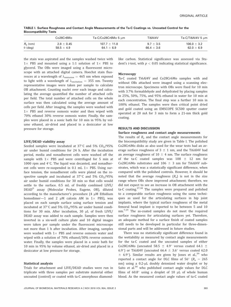
Surface roughness and contact angle measurementsThe results of Ra and the contact angle measurements forthe biocompatibility study are given in Table I. The polishedCo28Cr6Mo disks as also used for the wear tests had an av-erage surface roughness of 3 6 1 nm, and the Ti6Al4V hadan average roughness of 10 6 4 nm. The surface roughnessof the ta-C coated samples was 108 6 12 nm forCo28Cr6Mo substrates and 106 6 3 nm for Ti6Al4V sub-strates, which was a statistically significant difference, whencompared with the polished controls. However, it should benoted that the average roughness (Ra) is not in the sizerange where OBs show improved attachment; therefore, wedid not expect to see an increase in OB attachment with theta-C coating.37,38 The samples were prepared and polishedto a comparable surface roughness using the same techni-ques as used for the articulating surfaces in hip jointimplants, where the typical surface roughness of the metalfemoral head implant is reported to be between 5 and 10nm.7,19 The as-coated samples do not meet the requiredsurface roughness for articulating surfaces yet. Therefore,an adequate method for a surface finish of coated samplesstill needs to be developed in particular for three-dimen-sional parts and will be addressed in future studies.
There was no statistically significant difference found inthe wettability as measured by contact angle measurementsfor the ta-C coated and the uncoated samples of eitherCo28Cr6Mo (uncoated 58.5 6 4.9� versus coated 64.1 66.9�) or Ti6Al4V (uncoated 66.4 6 3.6� versus coated 62.06 6.9�). Similar results are given by Jones et al.,39 whoreported a contact angle for DLC films of 56� (Ra ¼ 265nm) using a 0.2-lL doubly deionized water droplet or bySaito et al.30 who published contact angle values for DLCfilms of 60.8� using a droplet of 10 lL of whole humanblood. As the measured contact angle values of ta-C coated
TABLE I. Surface Roughness and Contact Angle Measurements of the Ta-C Coatings vs. Uncoated Control for the
Biocompatibility Tests
Co28Cr6Mo Ta-C/Co28Cr6Mo 5 lm Ti6Al4V Ta-C/Ti6Al4V 5 lm
Ra (nm) 2.8 6 0.45 107.7 6 11.8 9.7 6 3.5 106.0 6 3.2y (deg) 58.5 6 4.9 64.1 6 6.9 66.4 6 3.6 62.0 6 6.9
ORIGINAL ARTICLE
JOURNAL OF BIOMEDICAL MATERIALS RESEARCH A | NOV 2010 VOL 95A, ISSUE 2 393

and uncoated control samples show no significant differen-ces, and cell attachment has been shown to be related tothe surface wettability,30 our results showing no significantchange in cell attachment are in agreement with these wett-ability values.
Ta-C coating adhesionAdhesion testing was performed on stainless steel andCo28Cr6Mo substrates. The deposited ta-C coatings rangedfrom 2 lm to 10 lm in thickness. Polished and unpolishedcoatings were tested. All ta-C coated samples showed thehighest level of coating adhesion (HF1) as it can be deter-mined by Rockwell C indentation testing (Table II and Fig.1). Independent of the substrate material, the coating thick-ness and whether the surfaces were polished, the ta-C coat-ings did not delaminate or significantly crack around theedge of the indentation.
The results of the scratch test experiments are given inTable II and were found to be more sensitive to the coatingthickness and surface roughness than the Rockwell C testresults. Thicker coatings are harder to delaminate becausethe influence of the substrate is reduced and the coatingbecomes more self-supporting. The highest critical load val-ues of 48 N for LC2 (broken and delaminated film) and 20 Nfor LC1 (first cracks appear along the scratch) were meas-ured on the Co28Cr6Mo with 10 lm thick polished ta-Ccoatings. In comparison, reported critical loads for hard sub-strates ranged from 5 to 10 N for LC2 and were less than 5N for LC1.
40 We attribute the improved adhesion perform-ance of our ta-C coatings to the carefully engineered gradi-ent interface layer between the substrate and the ta-C coat-ing. Examples for scratches causing LC1/C2 type failures areshown in Figure 3.
In summary, the ta-C coatings investigated adhered verywell to the Co28Cr6Mo substrates with an increased scratchresistance in case of the thickest (� 10 lm) and smoothest(polished) coatings. However, it is also important to pointout that, for thin film coatings in general, a (sometimes sub-stantially) delayed coating delamination has been observedin the literature, which may be caused by defects such aspinholes in the coating. Microscopic defects in a coating in acorrosive environment may lead to an erosion of the sub-strate material with a subsequent coating delamination.41
Even though we did not observe this type of delaminationduring wear testing in simulated body fluids, we still planon future studies with coated substrates in more corrosive
environments with simulated body fluids (both isotonic sa-line and cell culture media supplemented with bovine se-rum) and temperatures for an extended time,42 using moreaggressive acidic solutions as proposed elsewhere43 andunder cyclic fatigue loading conditions.
Wear test resultsAll of the wear test results are compiled in Table III. Foreach wear couple, the table lists the materials, their surfaceroughness and coating thickness (if not coated, the thick-ness is listed as 0 lm), the lubrication conditions and themeasured coefficient of friction and the wear factors forpin/ball, disk, and combined. PBS and Dulbecco’s Media II(D-Media 2) were used as lubricants to simulate an in vivosynovial joint at normal body temperature, 37.1�C. Bovinesynovial fluid should be used as a lubricant in future stud-ies because it is a closer model to the in vivo human envi-ronment. A zero wear factor for either pin or disk indicatesthat there was no measurable wear. This was the case formetal and sapphire balls (Pin Wear) running againstUHMWPE as well as for ta-C coated and polished disks(disk wear) running against ta-C coated pins.
Selected data from the table are shown in Figure 4,where the total wear factor is plotted for material combina-tions running in PBS lubrication. The Co28Cr6Mo/Inox440C pairing was coated with ta-C and compared with anuncoated pairing of the same materials. The ta-C coatingreduced wear from 3.50 � 10�5 mm3/Nm from an uncoatedcontrol (Co28Cr6Mo/Inox 440C) to 1.11 � 10�7 mm3/Nm(coated Co28Cr6Mo/coated Inox 440C). The highest wearfactor measured was on the order of 3.7 � 10�4 mm3/Nmand obtained for uncoated and unpolished UHMWPE run-ning against uncoated sapphire pins. As a result of polish-ing, the UHMWPE disk before the test (thus reducing theaverage roughness Ra from more than 100 nm to less than10 nm) the wear factor was improved by a factor of at least100 versus Co28Cr6Mo (2.5 � 10�6 mm3/Nm) or sapphire(3.3 � 10�6 mm3/Nm). An as-deposited and unpolished ta-C coated disk running against a ta-C coated pin improvesthe wear factor to about 4.6 � 10�7 mm3/Nm. Tungstencarbide and sapphire balls against polished ta-C disksyielded wear factors on the order of 3.4 � 10�8 mm3/Nm.Another 100-fold wear factor reduction can be achieved byrunning a polished ta-C coated disk against ta-C coated pins.These tests did not show any wear on the disk, yielding thelowest measured combined wear factors in the order of 3.9
TABLE II. Results of the Ta-C Coating Adhesion Tests on Metallic Substrates
Substrate MaterialTa-C
Thickness (lm)
Roughness (nm)Ta-C Polished/Unpolished
Rockwell Scratch (N)
Rz Ra Grade LC1 LC2
Stainless steel 2 923 97 Unpolished HF1 4.4 19.8Stainless steel 4 964 107 Unpolished HF1 8 25.6Stainless steel 5 906 111 Unpolished HF1 8.3 22.3Stainless steel 10 1136 131 Unpolished HF1 17.2 27
115 8 Polished HF1 16 31Co28Cr6Mo 10 1326 142 Unpolished HF1 22 32.4
42 5 Polished HF1 20 48.4
394 HINUBER ET AL. BIOCOMPATIBILITY AND MECHANICAL PROPERTIES OF DIAMOND-LIKE COATINGS

TABLEIII.WearTestResultsFrom
Pin-on-D
iskTribometer
WearCouple
Disk/Pin
Disk
Ball/Pin
Lubrica
nt
COF
Pin
Wear
(mm
3/N
m)
Disk
Wear
(mm
3/N
m)
TotalWear
(mm
3/N
m)
Total
Data
Points
Standard
Deviation
(%)
Surface
Material
Ta-C
Thickn
ess
(lm)
Roughness
Material
Ta-C
Thickn
ess
(lm)
Ra/nm
Rz/nm
CoCrM
o/SS
Polish
ed
Co28Cr6Mo
04
58
Inox440C
0Dry
0.51
1.23E�0
63.37E�0
53.50E�0
55
9PE/Al2O3
Unpolish
ed
UHMWPE
0107
931
Al2O3
0PBS
0.40
0.00Eþ0
03.67E�0
43.67E�0
45
37
PE/Al2O3
Polish
ed
UHMWPE
07
88
Al2O3
0PBS
0.08
0.00Eþ0
03.34E�0
63.34E�0
610
22
PE/CoCrM
oPolish
ed
UHMWPE
07
88
PIN
Co28Cr6Mo
0PBS
0.11
0.00Eþ0
02.50E�0
62.50E�0
620
23
PE/SS
Unpolish
ed
UHMWPE
0107
931
Inox440C
0PBS
0.42
0.00Eþ0
01.94E�0
41.94E�0
410
27
Ta-C/Al2O3
Unpolish
ed
Co28Cr6Mo
10
132
1114
Al2O3
0PBS
0.13
1.78E�0
51.97E�0
61.98E�0
515
10
Ta-C/Al2O3
Polish
ed
Co28Cr6Mo
10
557
Al2O3
0D-M
edia
20.14
4.46E�0
91.06E�0
71.10E�0
75
29
Ta-C/Al2O3
Polish
ed
Co28Cr6Mo
10
557
Al2O3
0PBS
0.07
1.37E�0
94.08E�0
84.22E�0
815
15
Ta-C/SS
Unpolish
ed
Co28Cr6Mo
5124
982
Inox440C
0Dry
0.10
1.08E�0
74.20E�0
75.28E�0
75
8Ta-C/SS
Polish
ed
Co28Cr6Mo
53
23
Inox440C
0Dry
0.11
1.98E�0
92.59E�0
82.79E�0
85
14
Ta-C/ta-C
Unpolish
ed
Co28Cr6Mo
5124
982
Inox440C
2Dry
0.45
6.55E�1
15.14E�0
75.14E�0
75
19
Ta-C/ta-C
Unpolish
ed
Co28Cr6Mo
10
132
1114
PIN
Co28Cr6Mo
2PBS
0.09
1.87E�0
72.77E�0
74.64E�0
75
12
Ta-C/ta-C
Polish
ed
Co28Cr6Mo
53
23
Inox440C
2Dry
0.10
4.28E�0
86.85E�0
81.11E�0
75
16
Ta-C/ta-C
Polish
ed
Co28Cr6Mo
10
542
PIN
Co28Cr6Mo
2PBS
0.05
4.87E�1
00.00Eþ0
04.87E�1
02
15
Ta-C/ta-C
Polish
ed
Co28Cr6Mo
53
23
Inox440C
2PBS
0.15
3.94E�1
00.00Eþ0
03.94E�1
02
15
Ta-C/W
CUnpolish
ed
Co28Cr6Mo
10
132
1114
WC
0PBS
0.08
1.89E�0
64.84E�0
72.37E�0
620
19
Ta-C/W
CPolish
ed
Co28Cr6Mo
10
557
WC
0D-M
edia
20.10
9.58E�0
91.61E�0
71.70E�0
75
31
Ta-C/W
CPolish
ed
Co28Cr6Mo
10
557
WC
0PBS
0.06
1.72E�0
92.47E�0
82.64E�0
810
29
ORIGINAL ARTICLE
JOURNAL OF BIOMEDICAL MATERIALS RESEARCH A | NOV 2010 VOL 95A, ISSUE 2 395

� 10�10 mm3/Nm. Overall, the measured wear factors spansix orders of magnitude and are in excellent agreement withpublished data.44
Based on these results, the most important factor inreducing the wear rate is the bearing surface material. Hipsimulator studies are extremely beneficial and necessary incharacterization of a wear surface for use in the body. Lap-palainen et al. 7 have shown the superior wear behavior(six orders of magnitude less wear) of amorphous diamondcoatings compared with industry standard materials,CoCrMo, and UHMWPE, in vitro with hip simulator testing.Results such as these have encouraged our research groupto pursue hip simulator testing that will be the topic offuture publications. A similar study is envisioned that willuse similar industry standard implant material pairings andhip simulator testing conditions in accordance with theliterature.
Surface roughness and the particular lubrication condi-tions are also important and can improve the wear factorby several orders of magnitude. Beginning in the 1990s,there have been several studies that found similar resultsfor DLC coatings. Thomson et al. 45 found that diamond-likefilms neither increase the physical damage, the toxicity northe inflammatory reaction of mouse fibroblasts. In a follow-up study, they later demonstrated that human fibroblastsand human OBs grown on DLC showed normal cellulargrowth and no in vitro cytotoxicity.46 McColl et al. 47 foundno negative effects of DLC on mouse fibroblasts (3T3-L1). Alater study by this group on DLC coated polystyrene sub-strates confirmed this finding by reporting normal cellulargrowth and no cytotoxicity.48 In research that is very similarto this study, Kornu et al. 49 found that diamond-like coatedTi6Al4V alloys as well as DLC coated CoCrMo alloys hadsimilar attachment behavior using mouse OBs (MC3T3-E2),when compared with uncoated CoCrMo and Ti6Al4V disks.Dowling et al. 50 demonstrated enhanced biocompatibility ofDLC coatings on titanium alloys, which acted as a diffusion
barrier and therefore dramatically decreased the rate ofmetal ion release. In an in vivo (sheep) study, Allen et al.20
showed good biocompatibility of DLC coated CoCrMo alloysamples as well as in an in vitro model using DLC coatedpolystyrene wells and human OBs. Li and Gu51 investigatedthe attachment of different cell types (macrophages, granu-locytes, and fibroblasts) on DLC-coated polymethylmetha-crylate samples and showed that DLC coatings provide a de-sirable surface for normal cell growth and morphology offibroblasts (3T3). A recent study by Van et al. evaluated thein vivo behavior of tetrahedral amorphous DLC coated sili-con in SV129 mice over a period of 6 months. Tissueresponse was mild and localized to the implantation site.52
Cell attachment studyTo determine early indicators of biocompatibility, the attach-ment behavior of a standard mouse OB cell line (MC3T3-E1) was evaluated by seeding the OBs onto uncoatedCo28Cr6Mo and uncoated Ti6Al4V control samples as wellas onto ta-C coated Co28Cr6Mo and ta-C coated Ti6Al4Vsamples and culturing for 4 h at 37�C in a humid atmos-phere of 95% air/5% CO2. Cells were subsequently stainedand visualized after incubation and the percent of attachedcells was quantified and plotted as shown in Figure 5. Therewas no statistically significant difference found between thepercent OB attachment on the ta-C coated Co28Cr6Mo sam-ples with 7.54 6 0.43% of the cells attaching, when com-pared with 8.21 6 0.55% of the cells attaching to theuncoated Co28Cr6Mo controls. Similarly, the ta-C coatedTi6Al4V samples showed 8.10 6 0.26% of cells attaching,which was not statistically different from the 10.32 61.49% of cells attaching to the uncoated Ti6Al4V controls.
FIGURE 4. Total wear factors (pin wear þ disk wear) in phosphate
buffered saline solution.
FIGURE 5. MC3T3-E1 osteoblast-like cell attachment after 4 h incuba-
tion at 37�C in a humidified atmosphere of 95% air and 5% CO2. OB
cell number was determined using the Hoechst nucleus staining
method and fluorescence microscopy. OBs were plated at equivalent
cell number per surface area. Values are expressed as a percentage of
OBs attached 6 SE with each condition done in triplicate. Neither the
OB attachment for the ta-C Co28Cr6Mo nor the ta-C Ti6Al4V was stat-
istically different (p < 0.05) from the uncoated Co28Cr6Mo and
Ti6Al4V, respectively.
396 HINUBER ET AL. BIOCOMPATIBILITY AND MECHANICAL PROPERTIES OF DIAMOND-LIKE COATINGS

Although exposure to Co28Cr6Mo and Ti6Al4V particulateshas been shown to alter osteoclast behavior and may have adeleterious effect on bone fixation to these materials and/orcontribute to aseptic loosening by inhibiting bone forma-tion,10,53,54 Ti6Al4V alloys have been shown to be morefavorable to OB attachment, when compared with CoCrMoalloys.55 No statistical difference was found between the ta-C coated Ti6Al4V or Co28Cr6Mo and their uncoated con-trols therefore indicating comparable early in vitro cyto-compatibility of these ta-C coatings.
Live-Dead StudyTo determine whether the ta-C could induce OB death, theOBs were incubated with Calcein AM (which fluorescesgreen for live cells) and ethidium homodimer (which fluo-resces red in dying cells). These OBs were then seeded onboth the ta-C coated Ti6Al4V and uncoated controls and theta-C coated Co28Cr6Mo and uncoated controls in the samemanner as for the cell attachment study but with an incuba-tion period of 24 h. Representative Live/Dead fluorescencemicroscopy images of the ta-C coated ta-C coatedCo28Cr6Mo and the uncoated Co28Cr6Mo controls are
FIGURE 6. Fluorescence microscope imaging using Live/DeadVR assay for OBs after 24 h incubation in a humid atmosphere of (A) ta-C coated
Co28Cr6Mo, (B) Co28Cr6Mo control, (C) ta-C coated Ti6Al4V, and (D) Ti6Al4V control samples. [Color figure can be viewed in the online issue,
which is available at wileyonlinelibrary.com.]
FIGURE 7. Live/Dead results showing the percent OB death after 24 h
incubation at 37�C in a humid atmosphere of 95% air/5% CO2. OB cell
number was determined using Live/DeadVR assay and fluorescence mi-
croscopy. Cells were counted using the software Image-Pro Plus, ver-
sion 5.1 Media Cybernetics. OBs were plated at equivalent cell
number per surface area and values are expressed as a percentage of
OBs attached 6 SE with each condition done in triplicate. No statisti-
cally significant (p < 0.05) differences in cell death were detected in
either the ta-C coated Ti6Al4V and uncoated Ti6Al4V pairing or the ta-
C coated Co28Cr6Mo and uncoated Co28Cr6Mo pairing.
ORIGINAL ARTICLE
JOURNAL OF BIOMEDICAL MATERIALS RESEARCH A | NOV 2010 VOL 95A, ISSUE 2 397

given in Figure 6(a,b), whereas the ta-C coated Ti6Al4V andthe uncoated Ti6Al4V controls are shown in Figure 6(c,d).Each image depicts a homogenous distribution of living OBs
(green) on the sample surfaces with relatively few deadcells (red). Quantifying the data from the Live/Dead viabilityassay photomicrographs and examining the percent cell
FIGURE 8. Scanning electron microscopy images showing (A) ta-C coated Co28Cr6Mo surface, (B) ta-C coated Co28Cr6Mo with OBs, (C)
Co28Cr6Mo control with OBs, (D) ta-C coated Ti6Al4V surface, (E) ta-C coated Ti6Al4V with OBs, (F) Ti6Al4V control with OBs, (G) ta-C coated
Co28Cr6Mo (5000�), and (H) uncoated Co28Cr6Mo (5000�).
398 HINUBER ET AL. BIOCOMPATIBILITY AND MECHANICAL PROPERTIES OF DIAMOND-LIKE COATINGS

death as a function of the total number of cells for the ta-Ccoated Ti6Al4V and uncoated controls and the ta-C coatedCo28Cr6Mo and uncoated controls, we find that the occur-rence of cell death is not statistically different for the ta-C-coated alloys in comparison with the uncoated alloys asshown in Figure 7. The ta-C coated Co28Cr6Mo sampleshad an average of 5.45 6 1.52% dead cells in comparisonwith 2.56 6 0.65% dead cells on the uncoated Co28Cr6Mocontrol samples. For the ta-C coated Ti6Al4V samples, theaverage cell death was 7.05 6 1.40% in comparison with3.2 6 1.24% for the uncoated control Ti6Al4V samples. Stu-dent’s t-test found no statistically significant differencebetween the fraction of dead cells on ta-C coated alloys,when compared with the uncoated samples, thus indicatingthat the ta-C coating on these two substrates is not cyto-toxic in terms of early cell behavior.
SEM imagingSEM images were taken of the ta-C coated Co28Cr6Mo andTi6Al4V samples with and without OBs as well as theuncoated control samples seeded with OBs and are shownin Figure 8. The SEM photomicrographs of the ta-C coatedCo28Cr6Mo and Ti6Al4V alloys [Fig. 8(a,d,g)] show a rela-tively uniform, smooth surface with a similar topographyfor both the ta-C coated Co28Cr6Mo and ta-C coatedTi6Al4V alloys. The polished surface of a Co28Cr6Mo sam-ple is shown at 5000� [Fig. 8(h)] for comparison to the ta-C coated Co28Cr6Mo 5000� image [Fig. 8(g)]. The polisheduncoated Co28Cr6Mo sample has a smooth surface [Fig.8(h)], whereas the ta-C coated Co28Cr6Mo sample has auniform globular surface structure [Fig. 8(g)]. The ta-Ccoated Co28Cr6Mo and Ti6Al4V and uncoated controlCo28Cr6Mo and Ti6Al4V samples, seeded with OBs [Fig.8(b,e,c,f), respectively] illustrate the OB morphology. In eachcase, the OBs appear well adhered and spiculated on theuncoated Co28Cr6Mo and Ti6Al4V samples [Fig. 8(c,f)]. TheOBs on the ta-C coated surfaces [Fig. 8(b,e)] are similarlyspread out in this commonly noted typical polygonalshape.35 There is no visible difference in cell morphology onthe ta-C coated Co28Cr6Mo and Ti6Al4V surfaces in com-parison with their uncoated Co28Cr6Mo or the Ti6Al4V con-trols. Independent of the surface treatment, the OBs wereable to attach and begin to spread.
CONCLUSIONS
The reduced wear exhibited by the ta-C coated substratesalong with relative biocompatibility indicate that this ta-Ccoating shows promise for increasing lifetimes for compo-nents with articulating surfaces such as total hip and kneereplacement implants. The cell attachment and LIVE/DEADresults along with the SEM analysis of the cell morphologyindicate that the ta-C coating on either Co28Cr6Mo orTi6Al4V has a comparable biocompatibility to the controluncoated Co28Cr6Mo and Ti6Al4V, commonly used orthope-dic implant materials. The continuous development in thearea of ta-C synthesis and coating stack design has lead tosignificant improvements in coating adhesion and load bear-ing capacity.
ACKNOWLEDGMENTS
The authors thank Symmetry Medical Inc. of Lansing, Michi-gan, for sponsoring this research program and for donating allthe biomedical gradematerials used in this study.
References1. Bren L. Joint replacement: An inside look. FDA Consumer Maga-
zine 2004;FDA 04-1335C.
2. AAOS. Number of arthroplasties to increase dramatically. 2002.
3. Los Angelos County Department of Health Services PH. Arthritis—
The leading cause of disability. LA Health 2006:1–6.
4. Mann D. Sharp increase in artificial knees and hips by 2030.
WebMD Medical News; 2006.
5. Kurtz S, Lau E, Zhao K, Mowat F, Ong K, Halpern M. The future
burden of hip and knee revisions: US predictions from 2005 to
2030, 73rd Annual Meeting of the American Academy of Ortho-
paedic Surgeons, Chicago, IL, March 22–26, 2006; p SE53.
6. Grill A. Diamond-like carbon coatings as biocompatible materi-
als—An overview. Diam Relat Mater 2003;12:166–170.
7. Lappalainen R, Selenius M, Anttila A, Yrjo T, Konttinen Y, Santa-
virta S. Reduction of wear in total hip replacement prostheses by
amorphous diamond coatings. J Biomed Mater Res B Appl Bio-
mater 2003;66B:410–413.
8. Dorner-Reisel A, Schuerer C, Mueller E. The wear resistance of
DLC coated and uncoated Co28Cr6Mo knee prostheses. Diam
Relat Mater 2003;11:823–827.
9. Lewis G. Polyethylene wear in total hip and knee arthroplasties.
J Biomed Mater Res B Appl Biomater 1997;38:55–75.
10. Archibeck M, Jacobs J, Roebuck K, Glant T. The basic science of
periprosthetic osteolysis. J Bone Joint Surg Am 2000;83:
1478–2000.
11. Affato S, Frigo M, Toni A. An in vitro investigation of dlc carbon
as a femoral head coating. J Biomed Mater Res B Appl Biomater
2000;53:221–226.
12. Galante J, Lemons J, Spector M, PD Wilson J, Wright T. The bio-
logic effects of implant materials. J Orthop Res 1991;9:760–775.
13. Rodgers S, Howie D, Graves S, Pearcy M, Haynes D. In vitro
human monocyte response to wear particles of titanium alloy
containing vanadium or niobium. J Bone Joint Surg 1997;79B:
311–315.
14. Savarino L, Granchi D, Ciapetti G, Stea S, Donati M, Zinghi G,
Fontanesi G, Rotini R, Montanaro L. Effects of metal ions on white
blood cells of patients with failed total joint arthroplasties.
J Biomed Mater Res 1999;47:543–550.
15. Pioletti D, Takei H, Kwon S, Wood D, Sung K-LP. The cytotoxic
effect of titanium particles phagocytosed by osteoblasts.
J Biomed Mater Res 1999;46:399–407.
16. Allen M, Myer B, Millett P, Rushton N. The effects of particulate
cobalt, chromium and cobalt-chromium alloy on human osteo-
blast-like cells in vitro. J Bone Joint Surg 1997;79B:475–482.
17. VDI. VDI2840, Carbon Films—Basic Knowledge, Film Types and
Properties. Volume VDI Handbook Materials Technology: Verein
Deutscher Ingenieure; 2005.
18. Schultz H, Weihnacht V, Scheibe H, Schultrich B. Wear investiga-
tions on superhard amorphous carbon films. Mat-wiss u Werk-
stofftech 2004;35:924–928.
19. Tiainen V-M. Amorphous carbon as a bio-mechanical coating—
Mechanical properties and biological applications. Diam Relat
Mater 2001;10:153–160.
20. Allen M, Myer B, Rushton N. In vitro and in vivo investigations
into the biocompatibilty of DLC coatings for orthopedic applica-
tions. J Biomed Mater Res B Appl Biomater 2001;58:319–328.
21. Uzumaki E, Lambert C, Belangero W, Freire C, Zavaglia C. Evalua-
tion of diamond-like carbon coating produced by plasma immer-
sion for orthopedic applications. Diam Relat Mater 2006;15:
982–988.
22. Hauert R. A review of modified DLC coatings for biological appli-
cations. Diam Relat Mater 2003;12:583–589.
23. Dorner-Reisel A, Schurer C, Irmer G, Muller E. Electrochemical
corrosion behaviour of uncoated and DLC coated medical grade
Co28Cr6Mo. Surf Coat Tech 2004;177–178:830–837.
ORIGINAL ARTICLE
JOURNAL OF BIOMEDICAL MATERIALS RESEARCH A | NOV 2010 VOL 95A, ISSUE 2 399

24. Kim H, Ahn S, Kim J-G, Park S, Lee K-R. Corrosion performance
of diamond-like carbon coated Ti alloy in the simulated body fluid
environment. Diam Relat Mater 2004;14:35–41.
25. Taeger G, Podleska L, Schmidt B, Ziegler M, Nast-Kolb D. Com-
parison of DLC and Al2O3 articulating with PE in total hip arthro-
plasty. Mat-wiss u Werkstofftech 2003;34:1094–1100.
26. Hauert R, Muller U. An overview on tailored tribological and bio-
logical behavior of diamond-like carbon. Diam Relat Mater 2003;
12:171–177.
27. Morrison M, Buchanan R, Liaw P, Berry C, Brigmon R, Riester L,
Abernathy H, Jin C, Narayan R. Electrochemical and antimicrobial
properties of diamondlike carbon metal composite film. Diam
Relat Mater 2006;15:138–146.
28. Hasebe T, Shimada A, Suzuki T, Matsuoka Y, Saito T, Yohena S,
Kamijo A, Shiraga N, Higuchi M, Kimura K, Yoshimura H, Kuri-
bayashi S. Fluorinated diamond like carbon as antithrombogenic
coating for blood contacting devices. J Biomed Mater Res 2006;
76A:86–94.
29. Dorner-Reisel A. Strukturanpassung und Beeinflussung funk-
tionaler Eigenschaften amorpher und nanokristalliner diaman-
tahnlicher Kohlenstoffschichten durch den Einbau von Elementen,
Verbindungen und Dotierungen. Freiberg, Germany; 2003.
30. Saito T, Hasebe T, Yohena S, Matsuoka Y, Kamijo A, Takahashi K,
Suzuki T. Antithrombogenicity of fluorinated diamond-like carbon
films. Diam Relat Mater 2005;14:1116–1119.
31. Scheibe H-J, Drescher D, Schultrich B, Falz M, Leonhardt G, Wild-
berg R. The laser-arc: A new industrial technology for effective
deposition of hard amorphous carbon films. Surf Coat Tech 1996;
85:209–214.
32. Scheibe H-J, Schultrich B, Ziegele H, Siemroth P. Deposition of
superhard amorphous carbon films by pulsed arc sources. IEEE T
Plasma Sci 1997;25:685–688.
33. Lifshitz Y, Kasi SR, Rabalais JW. Subplantation model for film
growth from hyperspezial species: Application to diamond. Phys
Rev Lett 1989;62:1290–1293.
34. Shu R, McMullen R, Baumann MJ, McCabe LR. Hydroxyapatite
accelerates differentiation and suppresses growth of MC3T3-E1
osteoblasts. J Biomed Mater Res 2003;67A:1196–1204.
35. Smith I, Baumann M, Case E. Induced microcracking affects
osteoblast attachment on hydroxyapatite scaffolds for tissue engi-
neering. Am J Biochem Biotechnol 2006;2:105–110.
36. Smith I, Baumann M, McCabe L. MC3T3-E1 osteoblast attachment
and proliferation on porous hydroxyapatite scaffolds fabricated
with nanophase powder. Int J Nanomed 2006;1:189–194.
37. Keller J, Schneider G, Stanford C, Kellog B. Effects of implant
microtopography on osteoblast cell attachment. Implant Dent
2003;12:175–181.
38. Degasne I, Basle M, Demais V, Hure G, Lesourd M, Grolleau B,
Mercier L, Chappard D. Effects of roughness, fibronectin, and vi-
tronectin on attachment, spreading, and proliferation of human
osteoblast-like cells (Saos-2) on titanium surfaces. Calcif Tissue
Int 1999;64:499–507.
39. Jones M, McColl I, Grant D, Parker K, Parker T. Protein adsorption
and platelet attachment and activation on TiN. TiC, DLC coatings
on Ti for cardiovascular applications. J Biomed Mater Res 2000;
53:413–421.
40. Ronkainen H, Koskinen J, Varjus S, Holmberg K. Load-carrying
capacity evaluation of coating/substrate systems for hydrogen-
free and hydrogenated diamond-like carbon films. Tribol Lett.
1999;6:63–73.
41. Lappalainen R, Santavirta SS. Potential coatings in total hip
replacement. Clin Orthop Relat Res 2005;430:72–79.
42. Tiainen V-M. Amorphous carbon as a bio-mechanical coating—
Mechanical properties and biological applications. Diam Relat
Mater 2001;10:153–160.
43. Lappalainen R, Heinonen H, Anttila A, Santavirta S. Some rele-
vant issues related to the use of amorphous diamond coatings
for medical applications. Diam Relat Mater 1998;7:482–485.
44. Santavirta SS, Lappalainen R, Pekko P, Anttila A, Konttinen
YT. The counterface, surface smoothness, tolerances, and
coatings in total joint prostheses. Clin Orthop Relat Res 1999;
369:92–102.
45. Thomson A, Law F, Rushton N, Franks J. Biomcompatibility of di-
amond-like carbon coating. Biomaterials 1991;12:37–40.
46. Allen M, Law F, Rushton N. The effects of diamond-like carbon
coatings on macrophages, fibroblasts and osteoblast-like cells in
vitro. Clin Mater 1994;17:1–10.
47. McColl I, Grant D, Green S, Wood J, Parker T, Parker K, Goruppa
A, Braithwaite N. Low temperature plasma-assisted chemical
vapour deposition of amorphous carbon films for biomedical-
polymeric substrates. Diam Relat Mater 1993;3:83–87.
48. Parker T, Parker K, McColl I, Grant D, Wood J. The biocompatibil-
ity of low temperature diamond-like carbon films: A transmission
electron microscopy, scanning electron microscopy and cytotoxic-
ity study. Diam Relat Mater 1994;3:1120–1123.
49. Kornu R, Maloney W, Kelly M, Smith R. Osteoblast adhesion to
orthopaedic implant alloys: Effects of cell adhesion molecules
and diamond-like coating. Orthop Res 1996;14:871–877.
50. Dowling D, Kola P, Donnelly K, Kelly T, Brumitt K, Lloyd L, Eloy R,
Therin M, Weill N. Evaluation of diamond-like carbon coated
orthopaedic implants. Diam Relat Mater 1997;6:390–393.
51. Li D, Gu H. Cell attachment on diamond-like carbon coating. J
Mater Sci 2002;25:7–13.
52. Van DL, Padera R, Friedmann T, Sullivan J, Langer R, Kohane D.
In vivo evaluation of tetrahedral amorphous carbon. Biomaterials
2005;26:465–473.
53. Neale SD, Haynes DR, Howie DW, Murray DW, Athanasou NA.
The effect of particle phagocytosis and metallic wear particles on
osteoclast formation and bone resorption in vitro. J Arthroplasty
2000;15:654–662.
54. Lohmann CH, Schwartz Z, Koster G, Jahn U, Buchhorn GH, Mac-
Dougall MJ, Casasola D, Liu Y, Sylvia VL, Dean DD, Boyan BD.
Phagocytosis of wear debris by osteoblasts affects differentiation
and local factor production in a manner dependent on particle
composition. Biomaterials 2000;21:551–561.
55. Price N, Bendall SP, Frondoza C, Jinnah RH, Hungerford DS.
Human osteoblast-like cells (MG63) proliferate on a bioactive
glass surface. J Biomed Mater Res 1997;37:394–400.
400 HINUBER ET AL. BIOCOMPATIBILITY AND MECHANICAL PROPERTIES OF DIAMOND-LIKE COATINGS