Biochemistry of neurotransmitters - Med Study...
46
Biochemistry of neurotransmitters Dr. Mamoun Ahram Neuroscience 2015
Transcript of Biochemistry of neurotransmitters - Med Study...

Biochemistry of neurotransmitters
Dr. Mamoun AhramNeuroscience2015

References
This lecture
Mark’s Basic Medical Biochemistry, 4th ed, pp. 908-918
http://what-when-how.com/neuroscience/neurotransmitters-the-neuron-part-1/

What is a neurotransmitter?
A chemical substance that:Is synthesized and stored in a presynaptic neuron (the enzymes needed for its synthesis must be present in the neuron), Is released at a synapse following depolarization of the nerve terminal (usually dependent on influx of calcium ions), binds to receptors on the postsynaptic cell and/or presynaptic terminal, elicits rapid-onset and rapidly reversible responses in the target cell,Is removed or inactivated from the synaptic cleft.

Types of neurotransmitters
Small-moleculeAmines (acetylcholine, epinepherine, dopamine, histmaine, etc.)
Amino acids (glutamate, aspartate)
Neuropeptides
• Gases (nitric oxide)

Note the differences
Onset and duration of action
Concentration for action and receptor binding
Concentration of [Ca+] for release
Site of synthesis, modification
Fate

NEUROPEPTIDES

Introduction
More than 50 neuropeptides have been described Behavior
Pain perception
Memory
Appetite
Thirst
Temperature
Homeostasis
Sleep

Neuropeptides: neurohormones or neurotransmitters?
Neurohormones: when neurons secrete their peptides into the vascular system to be transported to a relatively distant target
Neurotransmitter: Many axon terminals of neurosecretory cells secrete their products at the synapse to directly affect a post synaptic cell
Neuropeptides can do both – depends on nerve terminal

Classification of neuropeptides
Neuropeptides can be grouped into families based on similarities in their amino acid sequences.
Neuropeptide Families
Tachykinins: substance P, bombesin, substance Insulins: insulin, insulin-like growth factors Somatostatins: somatostatin, pancreatic polypeptide Gastrins: gastrin, cholecystokininOpioids: opiocortins, enkephalins, dynorphin
Opiate Family
Name Amino Acid Sequence
Leu-enkephalin
Tyr-Gly-Gly-Phe-Leu-OH
Met-enkephalin
Tyr-Gly-Gly-Phe-Met-OH
Beta-endorphin
Tyr-Gly-Gly-Phe-Met-Thr-Ser-Glu-Lys-Ser-Gln-Thr-Pro-Leu-Val-Thr-Leu-Phe-Lys-Asn-Ala-Ile-Val-Lys-Asn-Ala-
His-Lys-Gly-Gln-His-OH
DynorphinTyr-Gly-Gly-Phe-Leu-Arg-Arg-Ile-Arg-Pro-Lys-Leu-Lys-Trp-Asp-Asn-Gln-OH

Stages of actionSynthesis (ER and Golgi apparatus)
Packaging into large-dense core vesicles (with modifying enzymes)
Transport (fast-axonal transport)During the transport, proteases cleave the precursor neuropeptide into the final mature form.
ReleaseThey are released gradually over time in response to general increases in the level of intracellular calcium.
Action (prolonged)
Termination by diffusion and degradation

Diversity: alternative splicing
Alternative splicing of mRNA leads to translation of distinct precursors, and subsequent processing leads to unique mature peptides.
Example is the substance P mRNA

Diversity: proteolytic, differential, sequential processingNeuropeptides are produced from a longer precursor protein by
Proteolytic processing.
Vesicular packaging of different proteases that recognize different cleavage sequences
Hiding a proteolytic site by post-translational modifications (example: addition of a carbohydrate side chain.)
Tissue-specific
Processing of the pro-opiomelanocortin (POMC)precursor proceeds in an ordered, stepwise fashion.
Some of the reactions are tissue specific. ACTH,adrenocorticotropic hormone; CLIP, corticotropin-
like intermediate lobe peptide; JP, joining peptide;
LPH, lipotropin; MSH, melanocyte-stimulating
hormone; PC, prohormone convertase.

The levels of regulation of neuropeptide expression

Role of calcium
Vesicles are located further away from the presynaptic
membrane and away from place of Ca influx

Neuropeptides
The endogenous opiates
Neuropeptide Y
Galanin
Pituitary adenylate cyclase–activating peptide (PACAP)
Melanocyte-stimulating hormone (MSH)
Neurokinin A (NKA)
Substance P (SP)
Neurotensin
Calcitonin-gene–related protein (CGRP)
Vasoactive intestinal polypeptide (VIP)

SMALL-MOLECULE NEUROTRANSMITTERS

Types of small-molecule neurotransmitter
Nitrogen-containing molecules amino acids and their derivatives
intermediates of glycolysis and the Krebs cycle (TCA cycle)
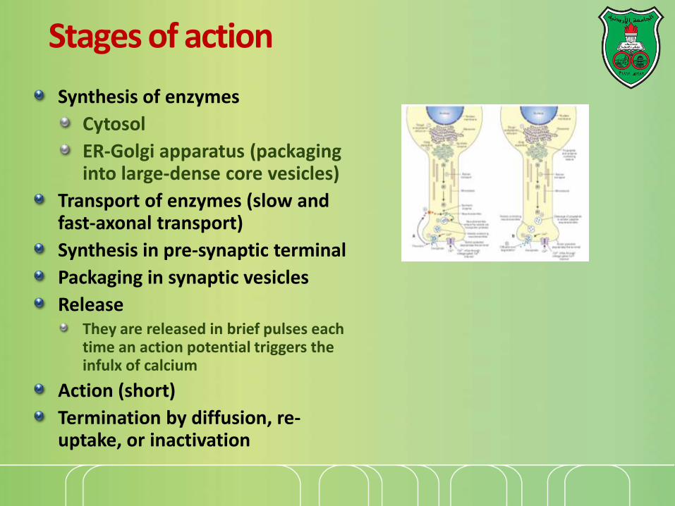
Stages of action
Synthesis of enzymes
Cytosol
ER-Golgi apparatus (packaging into large-dense core vesicles)
Transport of enzymes (slow and fast-axonal transport)
Synthesis in pre-synaptic terminal
Packaging in synaptic vesicles
ReleaseThey are released in brief pulses each time an action potential triggers the infulx of calcium
Action (short)
Termination by diffusion, re-uptake, or inactivation

mM2 [Ca+] =
uM1 .0[Ca+] =
uM100 -50[Ca+] =


Notes
Role of cofactorsS-adenosylmethionine (methyl transfer)
Pyrodoxal phosphate (vitamin B6): transamination, decarboxylation
Tetrahydrobiopterin (BH4)

TYROSINE-DERIVED NEUROTRANSMITTERS
Dopamine, norepinephrine, and epinephrine

Rate-limitingstep
Pyridoxal phosphate vesicular
Vitamin B12 or folate
Diet/
liver
phenylalanine hydroxylase
cytoplasm

50%
10%


Leaking

COMT and MAO
Parkinson’s
disease
Inactivation is
dependent on SAM
and vitamin B12 and
folate

Regulation
• Tyrosine hydroxylase– Short term
• Inhibition by free cytosolic catecholamines
• Catecholamines compete with BH4 binding to enzyme
• Activation by depolarization
– Tight binding to BH4 following phosphorylation by PKA, CAM kinases, PKC
– Long-term (plus dopamine -hyroxylase)

TRYPTOPHAN-DERIVED NEUROTRANSMITTERS
Serotonin and melatonin

BH4
Serotonin
5-hydroxyindoleacetic
acid
urine
Antidepressants , called selective serotonin re-uptake inhibitors (SSRIs), like Prozac® inhibit the reuptake process resulting in prolonged serotonin presence in the synaptic cleft.

Melatonin
Serotonin synthesized in the pineal gland serves as a precursor for the synthesis of melatonin, which is a neurohormone involved in regulating
sleep patterns
Seasonal and circadian (daily) rythyms
Dark-light cycle

GLUTAMATE AND ASPARTATE

Glutamate and aspartate
Nonessential amino acids
Do not cross BBB must be synthesized in neurons
Main synthetic compartmentsneurons
glial cells
Both are excitatory neurotransmitters.

Synthesis of glutamate
Sources:
Glycolysis Krebs cycle Transamination or dehydrogenation
Glutamine (deamination)
Another source: aspartate
Removalexcitatory amino acid carrier-1 (EAAC1)
glutamate transporter-1 (GLT-1) and glutamate—aspartate transporter (GLAST)
GABA
glutaminaseGlutamine
synthetase
transaminase1
2
3
-KG
Glu
Dehydro

Sources of glutamate (supplementary)

Aspartate
A vesicular uptake mechanism for aspartate has not yet been demonstrated, somewhat weakening the case for considering aspartate to be a neurotransmitter
Precursor: oxaloacetate (transamination)

Glycine
The major inhibitory neurotransmitter in the spical cord
Synthesized from serine by serine hydroxymethyltransferase through 3-phosphoglycerate
Removal: high-affinity transporter
Folic
acid

OTHERS

GABA
GABA is present in high concentrations (millimolar) in many brain regions.
These concentrations are about 1,000 times higher than concentrations of the classical monoamine neurotransmitters in the same regions.
The GABA shunt is a closed-loop process with the dual purpose of producing and conserving the supply of GABA.

GABA shunt

Diet
Membrane
PL
Synthesis of acetylcholine
Choline + acetylcoenzyme-A by choline acetyltransferase in cytoplasm
Transported into and stored in vesicles.
Removal: hydrolysis by acetylcholinesterase

Histamine
it does not penetrate the blood—brain barrier and, hence, must be synthesized.
Pyridoxal phosphate
Astrocytes
(MAO)Neuron
X

Inactivation of histamine

Nitric oxide (NO)
Glutamate is released (1) and acts on NMDA receptors located on the post-synaptic neuron (2)
Ca2+ enters the postsynaptic neuron and binds with calmodulin activating NOS (3) resulting in formation of NO and citrulline from L-arginine (4).
NO stimulates guanylate cyclase forming cGMP (5), which results in a physiological response (6)
No can diffuse out: a) to the presynaptic terminal (retrograde messenger) (7) prolonging effect and b) into adjacent neurons (8) and glial cells (9) stimulating guanylate cyclase.
Half-life: 2-4 seconds
NO is inhibited by hemoglobin and other
heme proteins which bind it tightly

Is NO a neurotransmitter?
Yes, but:
It is not stored in vesicles
It is not released by calcium-dependent exocytosis (it diffuses)
Its inactivation is passive (there is no active process that terminates its action)
It decays spontaneously
It does not interact with receptors on target cells
Its sphere of action depends on the extent to which it diffuses, and its action is not confined to the conventional presynaptic-postsynaptic direction.
NO acts as a retrograde messenger and regulates the function of axon terminals presynaptic to the neuron in which it is synthesized.

NO synthase
Isoform I (nNOS or cNOS)
Neurons and epithelial cells
activated by the influx of extracellular calcium
isoform II (iNOS)
Macrophages and smooth muscle cells
induced by cytokines
and isoform III (eNOS)
Endothelial cells lining blood vessels
activated by the influx of extracellular calcium
All three isoforms require BH2 as a cofactor and nicotinamide adenine dinucleotide phosphate (NADPH) as a coenzyme