BIOCHEMISTRY Amino Acids and Proteins · Amino Acid Function and Protein Classification • Amino...
52
BIOCHEMISTRY Amino Acids and Proteins BIOB111 CHEMISTRY & BIOCHEMISTRY Session 14
Transcript of BIOCHEMISTRY Amino Acids and Proteins · Amino Acid Function and Protein Classification • Amino...

BIOCHEMISTRY
Amino Acids and Proteins
BIOB111
CHEMISTRY & BIOCHEMISTRY
Session 14

Session Plan
• Characteristics of Proteins
• Amino Acids: Building Blocks for Proteins
• Essential Amino Acids
• Properties of Amino Acids
• Amino Acids as Buffers
• Cysteine – a Unique Amino Acid
• Biochemically Important Small Peptides
• Primary, Secondary, Tertiary & Quaternary Structure of Proteins
• Protein Hydrolysis
• Protein Denaturation
• Structural and Functional Classification of Proteins
Stoker 2014, Figure 20-22 p729

Functional groups in Proteins
Stoker 2014, p650
Protein
Amino Acid
Amine
Carboxylic acid

Introduction to Proteins
• Proteins are essential macronutrients which are important for
life
– Proteins are obtained mainly from animal products and other sources,
such as nuts and legumes
• Proteins are also called Polypeptides and are unbranched
polymers of Amino Acids (AAs)
– The amino acid building blocks or units, are organic compounds made
of carbon, hydrogen, nitrogen, oxygen or sulfur
• Next to water, proteins are the most abundant molecules in all
cell
– 15% of overall cellular mass

Amino Acids
• Amino acid (AA) = organic compound with both
–NH2 (Amino group) & –COOH (Carboxyl group) functional groups
– All AAs found in proteins are α-Amino acids
Stoker 2014, p695

Amino Acids
• 20 Standard AAs are divided into 4 groups based on the properties of the R groups:
– Non-polar
– Polar Neutral
– Polar Acidic
– Polar Basic
• More than 700 different AAs are known
– 20 standard AAs = AAs normally found in proteins
• R group (Side chain) = different for each amino acid
– R groups vary in:
• Size
• Shape
• Charge
• Acidity
• Functional groups
• Chemical reactivity

Classification of Amino Acids
• Non-polar AA
– Hydrophobic
• Polar Neutral AA
– Hydrophilic with neutral side chains
• Polar Acidic AA
– Hydrophilic with acidic side chains
• Polar Basic AA
– Hydrophilic with basic side chains
• Sulphur-Containing AA
– Methionine, Cysteine
Non-Polar
Polar
S-containing
AcidicBasic

9 out of the 20 standard AAs.
*Are essential amino acids
*Must be obtained from the diet,
as the body can not make them.
The 20 Amino Acids Required for Protein Synthesis

Acid-Base Properties of Amino Acids
• Amino acids contain both:
– An acidic group (–COOH)
– An alkaline/basic group (–NH2)
Stoker 2014, p700

Acid-Base Properties of Amino Acids
• In a neutral aqueous solution:– Carboxyl groups donate H+, producing a negatively charged ion
–COOH → –COO- + H+
– Amino groups accept H+ & produce a positively charged ion
–NH2 + H+ → –NH3+
• An internal acid-base reaction occurs on the same amino acid– Produces the zwitterion
Zwitterion
Stoker 2014, p701

Isoelectric Point (pI) or (IEP)
Isoelectric point:
• The pH, at which a specific amino acid exists as a zwitterion & its net charge is zero
– At IEP the amino acid are not attracted towards applied electric field because they carry zero net charge
– Different AAs have different IEPs
• Dependent on the structure of the amino acid– Acidic and basic amino acids have very different pIs
– Acids must gain a hydrogen to be neutral (low pH)
– Bases must loss a hydrogen to be neutral (high pH)
Stoker 2014, Table 20-3 p702

Zwitterion & pH Change
• Zwitterion structure changes when the pH of solution is altered
• In a solution more alkaline than the isoelectric point:– Zwitterion donates a H+ (from both amino and carboxyl groups) & forms a negatively charged ion
• The amino acid behaves as an ACID.
Stoker 2014, p702

Amino Acid Forms in Solutions • In a solution 3 different amino acid forms exist
• The equilibrium shifts with pH change
– Zwitterions
– Positive ions
– Negative ions
Alanine Zwitterion
At pI (pH=6)
Charge = 0
Alanine Positive Ion
At pH<pI
Charge = 1+
Alanine Negative Ion
At pH>pI
Charge = 1-

Cysteine – A Unique Amino Acid• The only standard amino acid containing
a Sulfhydryl group (–SH) (thiol group)– Cysteine readily oxidizes & forms a disulfide linkage with another cysteine
– 2 Cysteine residues linked via a disulfide bond help maintain
protein structure, stability & function• Disulfide bonds contributes to tertiary, quaternary protein structure
Stoker 2014, p703
+

Attempt Socrative questions: 1 to 3
Google Socrative and go to the student login
Room name:
City name followed by 1 or 2 (e.g. PERTH1)
1 for 1st session of the week and 2 for 2nd session of the week

Amino Acid Function and Protein Classification• Amino acids are the building block of proteins, support metabolism
and are important energy source
• Proteins are classified based on chemical composition:
– Simple proteins
• Consist only of amino acids.
• More than 1 protein subunit may be present
– Complex / Conjugated proteins
• Consists of 1 or more protein chains & 1 or more prosthetic groups
– Prosthetic group: organic or inorganic non-amino acid component
Lipoproteins – lipid prosthetic group
Glycoproteins – carbohydrate prosthetic group
Metalloproteins – specific metal (Fe, Zn, Cu) prosthetic group

Amino Acids in Proteins: Peptide Bond
• Amino acids are linked together in proteins by peptide bonds
– Covalent Amide bond found between amino acids in a peptide chain
• Peptide bond formation:
Carboxyl group (–COOH ) of one amino acid interacts with the
Amine group (–NH2) of another amino acid
– Produces an Amide + a molecule of H2O
Stoker 2014, p704

Amino acid chain: Peptides
Stoker 2014, p704-5
PEPTIDE
An unbranched chain of amino acids held
together by Peptide Bonds
DIPEPTIDE
2 amino acids linked via peptide bond
TRIPEPTIDE
3 amino acids linked via peptide bond
OLIGOPEPTIDE
10-20 amino acids linked via peptide bond
POLYPEPTIDE
Long unbranched chain of 20+ amino acids
linked via peptide bond
A peptide chain has 2 different ends:
– The end carrying a free –NH3+ is called
the N-terminal end
– The end carrying a free –COO- is called
the C-terminal end
• By convention, the sequence of AAs in
a peptide is always written with
N-terminal on the left & the C-terminal
on the right

Important Small Peptides
• ANTIOXIDANTSGlutathione
– Tripeptide (Glu–Cys–Gly) present in high levels in most cells
• Protects cells from highly reactive oxygen species (peroxides & superoxides)
– Produced during normal metabolism
– Immune response
• Detailed action of Glutathione is discussed in Nutritional Biochemistry.
Stoker 2014, p708

Important Small Peptides
• NEUROTRANSMITTERS
– Enkephalins = pentapeptide produced by the brain
• Bind at receptor sites within the brain to reduce pain
– Responsible for athlete’s “high” despite an injury
• Morphine & Codeine bind to Enkephalin receptors
– Long-lasting painkillers
• 2 best-known Enkephalins:
– Met-enkephalin: Tyr–Gly–Gly–Phe–Met
– Leu-enkephalin: Tyr–Gly–Gly–Phe–Leu

Important Small Peptides
• SMALL PEPTIDE HORMONES– Oxytocin
– Vassopressin = Antidiuretic hormone (ADH)• Both are Nonapeptides (9 AAs)
– Differ in the amino acids in positions 3 & 8
• Produced by the Hypothalamus – Stored in the Pituitary gland
• 6 AA held in a loop by disulfide bonds between 2 Cysteine's
Stoker 2014, p708

General Structure of Proteins• The 3-D structure of all proteins (monomeric or multimeric),
is more complex than that of carbohydrates or lipids
• 4 levels of protein structure:
– Primary Structure (1ᵒ) : amino acid sequence
– Secondary Structure (2ᵒ): α-helices, β-strands,
random coil (absence of 2ᵒ structure)
– Tertiary Structure (3ᵒ): Overall arrangement of protein structure within one peptide chain
– Quaternary Structure (4ᵒ): Overall arrangement of protein structure involving multiple peptide chains
• Only oligomeric (multimeric) proteins have this type of structure

Primary Structure (1ᵒ) of Proteins• Primary structure is the sequence of
amino acids in a protein chain– Amino acids are bonded together via peptide bonds
– Involves the number, type & order of attachment of the amino acids
– Every protein has a different amino acid sequence
Stoker 2014, Figure 20-11 p718
• Insulin was the 1st
protein to have its primary structure determined
– 51 amino acids in 2 chains, linked via disulfide bonds.
• Chain A (21 AAs) & Chain B (30 AAs)
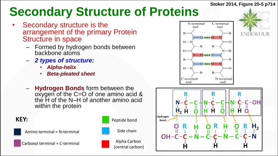
Secondary Structure of Proteins• Secondary structure is the
arrangement of the primary Protein Structure in space
– Formed by hydrogen bonds between backbone atoms
– 2 types of structure:• Alpha-helix
• Beta-pleated sheet
– Hydrogen Bonds form between the oxygen of the C=O of one amino acid & the H of the N–H of another amino acid within the protein
Stoker 2014, Figure 20-5 p714
Amino terminal = N-terminal
Alpha Carbon(central carbon)
Side chain
Peptide bond
Carboxyl terminal = C-terminal
KEY:

α-Helix
• Shape of a coiled spring (Helix)
• Hydrogen bonds are formed between C=O (i) and NH of the 4th
amino acid (i+3) down the same protein chain
– 4th if C=O residue considered 1st
• The R-groups stay outside of the helix
Stoker 2014, Figure 20-6 p715

β-Pleated Sheet
• β-Pleated Sheet is formed between 2 protein chain segments side by side– Linked by Hydrogen bonds
– The R-groups are above & below the sheet
Stoker 2014, Figure 20-7 p716
In proteins, where β-Pleated Sheet involves a single protein chain, several U-turns are formed• This “U-turn structure” is the most
frequent type of β-Pleated Sheet

Unstructured Segments• Most proteins have only part
of the structure forming
-helix or -sheet – Possible to have both -helix & -pleated sheet within the same protein
• Unstructured segments – Potions of a protein that have
neither -helix nor -pleated sheet
• Impart flexibility to proteins.
Stoker 2014, Figure 20-8 p716

Tertiary Structure of Proteins
• Tertiary protein structure:– Formed by interactions between the side chains (R-groups) of the
amino acids within a protein• Result in a more complex 3-D arrangement of the protein in space
• 4 types of interactions: – a) Disulfide bonds: The strongest interactions (covalent bond)
• Between two cysteine’s
– b) Electrostatic interactions (Salt Bridges)• Interactions between an acidic & a basic R-group (+ve/-ve charge int.)
– c) Hydrogen bonds: between polar R-groups
– d) Hydrophobic attractions: between non-polar R-groups

Stoker 2014, Figure 20-12 p719
Interactions in the Tertiary Structure

Stoker 2014, Figure 20-13 p719,720
Tertiary Structure of a Protein
Human insulin, a small 2-chain
protein, has both intra-chain &
inter-chain disulfide linkages as
part of its tertiary structure
Tertiary Structure of the
Single-chain Protein
Myoglobin

Quaternary Structure of Proteins
Quaternary Structure of Proteins
• Highest level of protein organization
• Found only in multimeric proteins that have 2 or more polypeptide chains (subunits) in their structure.
– Usually an even number of subunits (2 = dimer, 4 = tetramer,…)
• Subunits are held together by the same interactions as tertiary structure– Hydrogen bonds, disulfide bonds, hydrophobic & electrostatic interactions
• 4ᵒ structure easily disrupted by very small changes in cellular conditions – Protein chains fall apart, resulting in a temporary loss of protein activity
– When normal conditions are restored the multimer automatically reforms & regains its function

Stoker 2014, Figure 20-15 p721
Tertiary & Quaternary Structures of the Oxygen-carrying
Protein Hemoglobin.
It is a Tetramer with 2 identical α-chains & 2 identical β-chains.
Each chain contains a haeme group, where
oxygen binds.

Stoker 2014, p722
Within a single protein chain, what types of interactions between
amino acids give the protein its shape?
Is it be possible for the 1st and 100th amino acids
within a protein chain to form an interaction that contributes
to the shape of the protein? Why/Why not?
What types of interactions allow a multi-subunit protein
to be held together? Are these interactions the same or
different from interactions that hold together a single-unit protein?
G
Key concept: 3° and 4° protein structure

Attempt Socrative questions: 4 to 6
Google Socrative and go to the student login
Room name:
City name followed by 1 or 2 (e.g. PERTH1)
1 for 1st session of the week and 2 for 2nd session of the week

Protein Hydrolysis
• Protein Hydrolysis is the reverse of peptide bond formation
– Peptide bond is broken• The amine & carboxyl groups
are regenerated
• The protein splits into smaller peptides & AAs
• In the lab protein hydrolysis requires water, acid or base & heat
• In the body enzymes catalyze protein hydrolysis
Stoker 2014, p723
• Protein hydrolysis is necessary for digestion of dietary proteins
– Digestive enzymes catalyze protein hydrolysis & break the peptide bond
• Produces free amino acids
• Free amino acids are absorbed from the gut into bloodstream & transported to body cells for synthesis of new proteins
• Protein hydrolysis also occurs in cells– Old proteins are broken down to liberate
amino acids • Used to produce new proteins
– Hydrolysis of cellular proteins & their re-synthesis is a continuous process.
Protein hydrolysis

Protein Denaturation• Protein denaturation involves disruption of the protein’s characteristic 3-D quaternary, tertiary &
secondary structures
– Primary structure is not affected
• Protein denaturation leads to partial or complete loss of function
– Protein function depends on protein structure
– Small denaturation changes can be reversed & the protein does eventually “re-fold” =
Renaturation
– Major denaturation changes are irreversible
• Denatured proteins lose their water solubility & precipitate in a solution – Coagulation.
• Egg white (a concentrates solution of protein Albumin) forms a white solid when heated
– Cooking food denatures the protein but doesn’t change protein nutritional value
• Easy for digestive enzymes to hydrolyse the protein
& kills microorganisms by denaturing their proteins

Protein Denaturation by Heat and Chemicals• Cauterization: heat used in surgery to seal
blood vessels or small wounds
• Sterilization of surgical instruments, at high
temperature & high pressure, in autoclave
denatures bacterial proteins & enzymes
• Canning foods
• Fever
– Body temperature may rise to 400C without serious
consequences
– Temperature greater that 400C, can inactivate
enzymes, esp. in CNS, leading to dysfunction & death
• Change in pH
– In the stomach Hydrochloric acid denatures
proteins
– In yoghurt Lactic acid produced by fermenting
bacteria denatures milk proteins
• Alcohol denatures bacterial proteins
– Used as disinfectant >>> hence swabbing skin
prior to injection
• Agitation stretches peptide chains until
weak bonds break
– Whipping egg whites
• Heavy metals (Hg2+, Pb2+) & reducing
agents disrupt disulfide bonds, changing
the tertiary structure of proteins

Stoker 2014, Table 20-5 p726
Protein denaturation
involves loss of the
protein’s 3-D structure.
Complete loss of such
structure produces a
random-coil protein
strand.
Protein Denaturation

What can cause a protein to denature?
After protein denaturation, which of the four levels of
protein structure remain in tract and which are broken down?
Will the protein be functional or non-functional
after denaturation? Why/why not?
G
Key concept: protein denaturation

Attempt Socrative questions: 7 to 10
Google Socrative and go to the student login
Room name:
City name followed by 1 or 2 (e.g. PERTH1)
1 for 1st session of the week and 2 for 2nd session of the week

Classification of Proteins based on shape
• Protein classification is
based on molecular shape
– Determined by tertiary &
quaternary structures
3 types:
• Fibrous Proteins– Protein molecules with elongated shape
– Structural proteins
• Provide support & protection
– Most abundant in the body
• Total mass is greater than globular proteins
– Tend to aggregate together to form macromolecular
structures, e.g., hair, nails
Globular Proteins
– Molecules with peptide chains folded into
spherical or globular shapes
• More numerous than Fibrous proteins
• Functional proteins
– Involved in metabolism, enzymes,
transport & regulatory molecules,
intracellular signaling molecules
Membrane proteins
– Associated with cell membranes
• Non-polar R-groups are inside & polar R-
groups outside of the molecule
• Generally water-insoluble
• Help in transport of molecules across the
membrane

Stoker 2014, Table 20-6 p727

Stoker 2014, Figure 20-20 p728
α-Keratin structure in hair Collagen
• Collagen is the most abundant protein in the body (30% of total proteins)
– Main structural protein in connective tissues, bones, tendons, skin, cartilage, ligaments, blood vessels etc.
• Triple helix – formed by 3 peptide chains wrapping around each other
– Rich in Proline (up to 20%), Glycine, Hydroxyproline & Hydroxylysine
• Many triple helices combine into collagen fibrils– Cross-linking of fibrils gives collagen its strength
• The greater the cross-linking, the more rigid the fibril– Stiffening of collagen associated with ageing.

Collagen
Stoker 2014, Figure 20-22 p729 Stoker 2014, Table 20-7 p728
Electron micrograph of collagen fibers.

Haemoglobin• Haemoglobin is a globular protein that transports oxygen
in blood from lungs to tissues
• Haemoglobin is a tetrameric protein (4 protein chains) with 4 Haeme groups, each containing an Fe atom
– Fe ion interacts with oxygen
– Each Haemoglobin molecule can transport up to 4 oxygen molecules at a time
Stoker 2014, p729

Stoker 2014, Table 20-4 p710

Functional Classification of Proteins
• Proteins play crucial roles in most biochemical processes
• The diversity of functions exhibited by proteins far exceeds the role of other biochemical molecules
• The functional versatility of proteins stems from:
– Ability to bind small molecules specifically & strongly to themselves
– Ability to bind other proteins & form fiber-like structures
– Ability to bind & be integrated into cell membranes

Functional Classification of Proteins• Catalytic proteins = Enzymes
– Every biochemical reaction in the body requires an enzyme
• Defense proteins = Immunoglobulins / Antibodies– Vital for immune system function
• Transport proteins– Transport small molecules elsewhere in the body & release them on demand
• E.g. Haemoglobin, Lipoproteins
• Messenger proteins– Transmit signals to coordinate biochemical processes between different cells, tissues & organs
• E.g. hormones: Insulin, Glucagon, human growth hormone
• Contractile proteins– Required for movement
• E.g. Actin & Myosin proteins in muscles

Functional Classification of Proteins• Structural proteins
– Provide structural support
– E.g. Collagen (cartilage, tendons) & Keratin (skin, hair, nails)
• Trans-membrane proteins– Help control the movement of small molecules & ions across the cell membrane
• E.g. Ion channels
• Storage proteins– Bind & store small molecules or atoms
• E.g. Ferritin (Fe-storing protein), Myoglobin (oxygen-storing protein)
• Regulatory proteins– Form receptors to which messenger molecules bind, usually on the exterior of cell membrane
• Nutrient proteins– Particularly important in the early stages of life from embryo to infant
• E.g. Casein (milk), Ovalbumin (egg white)

Readings & Resources• Stoker, HS 2014, General, Organic and Biological Chemistry, 7th edn,
Brooks/Cole, Cengage Learning, Belmont, CA.
• Stoker, HS 2004, General, Organic and Biological Chemistry, 3rd edn, Houghton Mifflin, Boston, MA.
• Timberlake, KC 2014, General, organic, and biological chemistry: structures of life, 4th edn, Pearson, Boston, MA.
• Alberts, B, Johnson, A, Lewis, J, Raff, M, Roberts, K & Walter P 2008, Molecular biology of the cell, 5th edn, Garland Science, New York.
• Berg, JM, Tymoczko, JL & Stryer, L 2012, Biochemistry, 7th edn, W.H. Freeman, New York.
• Dominiczak, MH 2007, Flesh and bones of metabolism, Elsevier Mosby, Edinburgh.
• Tortora, GJ & Derrickson, B 2014, Principles of Anatomy and Physiology, 14th edn, John Wiley & Sons, Hoboken, NJ.
• Tortora, GJ & Grabowski, SR 2003, Principles of Anatomy and Physiology, 10th edn, John Wiley & Sons, New York, NY.