BIOCHEMICAL PHARMACOLOGY OF … able of Contents (continued) Section Page VI APPLICATION OF...
41
AMRL-TR-68- 132 BIOCHEMICAL PHARMACOLOGY OF ' HYDRAZINES TOXICITY ARTHUR FURST WALDEMAR R. GUSTAVSON ROBERT S. deROP? Institute of Chemical Biology University of San Francisco D DJ- MAY 1969 ISP_4 1969 This doctiment has been approved for public release and sale; its 4istribution is unlimited. AEROSPACE MEDICAL RESEARCH LABORATORY AEROSPACE MEDICAL DIVISION AIR FORCE SYSTEMS COMMAND WBIGHT-PATTERSON AIR FORCE BASE, OHIO
Transcript of BIOCHEMICAL PHARMACOLOGY OF … able of Contents (continued) Section Page VI APPLICATION OF...

AMRL-TR-68- 132
BIOCHEMICAL PHARMACOLOGY OF 'HYDRAZINES TOXICITY
ARTHUR FURST
WALDEMAR R. GUSTAVSON
ROBERT S. deROP?
Institute of Chemical Biology
University of San Francisco
D DJ-
MAY 1969 ISP_4 1969
This doctiment has been approved for publicrelease and sale; its 4istribution is unlimited.
AEROSPACE MEDICAL RESEARCH LABORATORYAEROSPACE MEDICAL DIVISIONAIR FORCE SYSTEMS COMMAND
WBIGHT-PATTERSON AIR FORCE BASE, OHIO

AMRL-TR-68-132
BIOCHEMICAL PHARMACOLOGY OFHYDRAZINES TOXICITY
ARTHUR FURST
WALDEMAR R. GUSTAVSON
ROBERT S. deROPP
This document has been approved for publicrelease and sale; its distribution is unlimited.

Foreword
This study was initiated by the Toxic tlazards Division of the AerospaceMedical Reserch Laboratory. The research was performed by the Institute ofChemical Biology, University of Sani Francisco, San Francisco, California 94117,under Contract No. AF 33(615).3829. Kenneth C. Back, Phi), Chief, ToxicologyBr.a., ' - .#1-ir wt monitor for the Aerospace Medical Research Laboratory.The work was performed in support of Project 6302, "Toxic Hazards of Propel-lants and Materials," Task 630202, "lPharmacology-Biochemistry," and WorkUnit 019, "Research on the Biochemical Pharmacology of Hydrazines Toxicity."This study was started in March 1966 and was completed in February 1968.
The authors wish to express their appreciation to Mr. George Ledin, Jr., forhis help in experimental design and statistical evaluation of data. The authorsalso thank Mr. Joseph Castagna, Jr., Miss Sandra Stratman, Miss Marion Doyle,and Mrs. Helen Graham for their laboratory assistance.
This technical report has been reviewed and is approved.
C. H. KIIATOCIIVlL, Colonel, USAF, MCCommanderAerospace I edical Research Laboratory

Abstract
The toxic action of 1,1-dimethylhydrazine (UDMH) and monomethylhy-drazine (NMIt1) may be mediated by the inactivation of pyridoxal in the brain.One poss...aity considered was the formation of a hydrazone between the pyri-doxals and the substituted hydrazine. Pyridoxal dependent enzymes were in-vestigated. UI)MII and MMII inhibited both glutamic acid decarboxylase andI)OPA decarboxylase. Transaminases (amino transferases) which required a-ketog-lutaric acid as a substrate were not affected by the hydrazines tested. Furtherwork was conducted to refine an ultrasensitive bioassay method for the detectionof each congener of the vitamin BK1 group. The microorganisms investigated forthe assay were a neurospora and a yeast. Some indirect evidence was obtainedwhich implies that UI)MII injected intraperitoneally can be detected in thecentral nervous system. A mathematical model for hydrazines-induced convul-sions was developed. It is now possible to predict the time lapse after adminis-traition of the convulsigen and the onset of seizure if only three data points aregiven.
.ii!1

Table of ContentsSection Page
I INTRODUCTION --.-- 1
11 EFFECT OF UDMH, MMH AND THEIR B,HYDRAZONES ON THE ACTIVITY OFBRAIN DECARBOXYLASES 3
Introduction 3
Materials and Methods -- ---- 3
Results------ -- --- ----------------------------- ------ 5
Discussion and Conclusions ----- -------- ------ - --- ------ I- -------- 9
III THE EFFECT OF SOME HYDRAZINES ANDHYDRAZONES ON BRAIN TRANSAMINASES - ----- --- ---- ------- 10
Introduction ----------- --- ------ ----- ------- -- ----- ------- ---- - -- 10
M e th o d s --- -- -- -- -- -- --- ---.. ..... ... --- -- -- ---- ----- ----- -.- - 11
Results---------------------------- -------- - --------------- --12
Discussion and Conclusions-- ----- ----- ----- ------- ------------------- 12
IV DEVELOPMENT OF AN ULTRASENSITIVEQUANTITATIVE BIOASSAY METHOD FORVITAMIN B, USING NEUROSPORA ... 13
Introduction -----------------------------------...--- 13
Methods .13
Results 16
Discussion and Conclusions 19
V USE OF YEAST FOR BIOASSAYOF VITAMIN Bo 20
Introduction 20
Materials and Methods 20
Results 21
Discussion and Ct..clusions 2
iv

f able of Contents (continued)
Section Page
VI APPLICATION OF ULTRASENSITIVEQUANTITATIVE BIOASSAY METHODSFOR TIlE I)ETECTION OF VITAMIN B,,IN RODENT BRAINS 23
Introduction -_ 23
Methods 23
Results ... 24
Discussion and Conclusions .. .. 25
VII EVII)ENCE THAT INJECTED UDMH ISFOUNI) IN THE CNS - .. 26
Introduction -........... 26
Materials and Methods ..... .. 26
Results ....... 26
Discussion and Conclusions ----- . 27
VIII DEVELOPMENT OF A MATHEMATICALMODEL FOR TIlE CONVULSION PROCESSINDUCEI) BY HYDFIAZINFES 28
APPENDIX - PREPARATION OF N. SITOPILA FOR BIOASSAY 30
REFERENCES 31
v

List of TablesTable Page
1 GLUTAMIC I)ECARBOXYLASE ACTIVITY IN BRAINS OFMICE CONVULSED WITH HYDRAZINES COMPAREDWITH THAT OF SALINE INJECTED CONTROLS 6
II DOPA DECARBOXYLASE ACTIVITY IN BRAINSOF CONVULSED MICE COMPARED WITH THATOF SALINE INJECTED CONTROLS -.... ... .. . 7
III GLUTAMIC DECARBOXYLASE ACTIVITY (Ag/g per hour)OF BRAIN HOMOGENATES INCUBATED INTHE PRESENCE OF HYDRAZINES AND HYDRAZONES---------------- 8
IV DOPA DECARBOXYLASE ACTIVITY (jsg/g per hour) OFBRAIN HOMOGENATES INCUBATED IN THE PRESENCEOF HYDRAZINES AND HYDRAZONES .. ....... .............................--------- 9
V PERCENTAGE INHIBITION OF CLUTAMIC PYRUVICTRANSAMINASE IN THE MOUSEBRAIN BY MbMI AND UDMH--------- -........ ..... --... .......... 12
VI NEUROSPORA SITOPHILA GROWTHCOMPARISON OF THE TWO METHODS OFGLASSWARE CONTAMINATION -...........- ---- -------- ---------- 16
VII EFFECT OF TEMPERATURE ON THE GROWTHOF N. SITOPHILA ----- -- ------.. --.......------- --......-.... ...... 17
VIII DRY WEIGHT OF N. SITOPHILA FROM 5 jgPAL ELUTED FROM PAPER .... ... I ------- 18
IX DETECTION AND ASSAY OF PICOGRAMAMOUNTS OF PAL --------- ----------------------.---.--- 18
X DETECTION AND ASSAY OF PAL IN THENANOGRAM RANGE OF CONCENTRATIONWITH SACCHAROMYCES CARLSBERGENSIS .....--------. ------- 21
XI PICOGRAM RANGE ASSAY OF PYRIDOXALIICI USING S. CARLSBERGENSIS 21
XII DETECTION AND ASSAY OF PAL ISOLATED FROMRODENT BRAINS BY PAPER CHROMATOGRAPHYAND ASSAY BY N. SITOPHILA 25
XIII PALP-UDMH INHIBITION STUDY 25
XIV RELATIVE POTENCY 29
vi
Sedion IINTRODUCTION
The rocket fuels 1,_1-dimethylhydrazine (UHMIH) and monomethylhydrazine (MMH) areconvulsant agents. All species investigated responded similarly to these convulsigens (ref 1, 2, 3).Characteristic of the convulsion process after an intraperitoneal injection of these agents is atime lapse between their administration and the o, et of the seizure. For a related substitutedhydrazine, thiosemicarbazide (TSC), Jenney and Lee (ref 4) suggested a quantitative represent-ation. A straight line was approximated when the log,., dose was plotted against the time lapseprior to the onset of the convulsion. This relationship had a limited applicability for it was linearonly in a relatively small dose range. The dose-time relationships for UDMH and MMH werestudied by O'Brien et al. (ref 5) for rats, and by Furst and Custavson (ref 6) for mice. The lattergroup included data for the pyridoxal (and 5-phosphate) hydrazones.
The fact that the pyridoxal hydrazones are more toxic than the alkylhydrazines themselves(ref 6) suggests that the mechanism of action of the hydrazines involves brain pyridoxal. Indirectevidence for the involvement and perhaps inhibition of pyridoxal was given by Dubnick et al.(ref 7) who found that the pyridoxal antagonist 4-deoxypyridoxal exacerbates the toxicity of thehydrazine compound phenelzine. McCormick and Snell (ref 8) proposed a mechanism for thio-semicarbazide (TSC) toxicity; they postulated that the TSC combined with pyridoxal formed ahydrazone, which was a kinase inhibitor. In the presence of the inhibitor, pyridoxal could not bephosphorylated to pyridoxal-5-phosphate. No direct evidence was presented that TSC or the hy-drazone crossed the blood-brain-barrier.
Pyridoxal-5-phosphate plays an important role in amino acid metabolism in the brain; it is acoenzyme for all nonoxidative transformations of the amino acids (ref 9, 10) such as decarboxyla-tion, transamination, racemization, f-elimination and y-elimination. Inactivation of this coenzymeby inhibition or by lowering of its absolute content in the rodent brain would be reflected in the de-creased activity of the decarboxylases and transaminases. The enzymes are essential for the forma-tion and metabolism of a number of brain biogenic amines like serotonin, norepinepherine, dopa-mine and GABA.
This project was undertaken to determine the biochemical mechanism of convulsant action ofthe alkylhydrazines, UDMH and MMI. The areas investigated were:
1. The change in pyridoxal content of rodent brain, before and after induced convulsion. Thisrequired developing an ultrasensitive detection method for the individual vitamin Bo congenersin rodent brains.
2. The potential inhibition by the alkylhydrazines of decarbxoxylase, in rodent brains.
3. The potential inhibition by the alkylhydrazines of some brain transaminases.
4. The detection of UDMH in rodent brain after intraperitoneal administration.
5. The development of a more exact mathematical expression of the relationship between thedose of the convulsigen and the time lapse before the onset of the seizure. The equation wouldhold over the entire dosage range and provide useful information for prospective studies.
Throughout this report, the following ab~breviations are used for the hydrazine compounds:
1, .-dimethylhydrazine ( UDMII) and monomethylhydrazine (MMtl). The congeners of theVitamin B group will be designated as: pyridoxine or pyridoxal (POL), pyridoxamine (PAMINE),pyridoxal (PAL). Their respective 5.phosphates will be indicated as in pyridoxal-5-phosphate(PALP). The hydrazones of PAL, or PALIP will be shown with a dash followed by the respectivehydrazine as in: PAL-MMII, PALP-UI)MII. Other abbreviations of chemical cimpounds are ex-plained at the approximate place of their introduction in the text.
2

Section 11EFFECT OF UDMH, MMH AND THEIR B6
HYDRAZONES ON THE ACTIVITY OFBRAIN DECARBOXYLASES
INTRODUCTIONThe toxic mechanism of action of convulsant hydrazines U'I)MlI and MMII may involve the
inactivation or depletion of PALI) in the central nervous system. As part of this program, a studywas made of the inhibitory effect of these alky'1hydrazines and their p ridoxal h) drazones on two(lecarboxylation enzymes, glutamnic (lecarbOxylasse and l)OPA (lccarIbox lase. Both of the enzymesare pyridoxal-dependent.
The reactions are:
CO011 COOT-i
Cli 2 glutamnicGH
U1712 decarboxylase C 2 + co-,
glutamic acid CABA
H HC C
II\ )OPA 1HGC C-GIL2-GH-GOOII o HG C-C11 2-CH 2-NH-2 + CO2
1 11 N112 decarhoxylase I H110-C CI-I HO-C CI
C
HO
DOPA DOPamine
IEnzyme activity can he measured by the amnount of amine formed ( ref 11), or by the C0 2liberated. This study uitilized ITC carboxyl-labeked amino acids, the liberated I 'CO,. being trappedand1 counted in a liquid-scintillation couinter. The reaction '%essels N~ere designed in the Institute ofChemical Biology, and the mnethod used w~as that of deC 1opp and Furst ( ref 12).
MATERIALS AND METHODSMale Swiss-albino mice (Berkeley Pacific Laboratories) \%eighing 18-20 g. were randomized,
then injected intraperitoneally with a sufficient amount of an unbuffered saline solution of alkyl-hydrazine ( 11I)MI I Eastman Organic Chemicals, MN!II: Aldrich Chemical) or the PAL or PALPhyclrazones (synthesized in our laboratory) to indluce convulsions (ref 6). The followving com-
3

pounds were used: MMII, U\)M I, PAL-MM11, PAL-UDMI, PALP-UDM11, PALP-MMH. Miceinjected with saline only were used as controls. Two mice were included in each group, and the ex-periments were replic,,ted three to five times. Promptly after convulsion the mice were decapitatedand, upon immediate removal their brains were weighed and homogenized with a teflon glassiz.l1ogenizer in thee volumes of 0.02 Mt ice-cold phophate buffer at p11 7. The homogenate wasolaced in a dialysis bag (%4"-tubing) and dialyzed overnight against one liter of phosphate bufferat 4' C. The contents of the dialysis bag were rinsed into a graduated cylinder with buffer and thevolume adjusted to give the equivalent of either 25 or 12.5 mg of tissue per ml.
Radioactive substrates used in these experiments were DL-3, 4-dihydroxyphen-ilalanine-l-C(New England Nuclear. NEN-245), with a specific activity of 2.36 ic/mM; and DL-glutamic-' 4 Cacid ( New England Nucl, CM.\1O22) specific activity 2.5 nic/mM. The labeled substrates weredissolved in sutfficifnt phos ,ate buffer to make a solution containing 1 ing of the L-isomer per mland stored froen. For use, tihe\ \%ere diluted with the corresponding cold amino acid always at aconcentration of 1 ing of ,he L-isomer per mi. In practice, a solution giving about 600 CPM perjig of L-isomer pro\ed satisfactory. The ten-to-fifteen-fold dilution of the hot substrate with coldwas prepared 10 mi at a time and stored frozen.
PALI) (Nutritional Biochemical Corp.) was dissolved in buffer to give a 1 mg/ml solution,diluted for use to give a final concentration of 1, 10 and 100 ,g/ml. The cofactor was stored frozenbut lost its activity in solution when kept longer than six weeks.
The hydrazines and hydrazones were dissolved in buffer and diluted for use to give final con-centrations of 1 x 10- 3 M, 10-1M, and 10-"M. They were subsequently found to be unstable in so-lution, a factor which introduced a variable in some of the earlier experiments. In later work,fresh solutions were made each day.
The enzyme reaction was carried out in the vessel shown in Figure 1. It consisted of twoparts, the reaction chamber bearing the male joint and the absorption chamber hearing the f#'male joint. Before use, the two halves were connected.
E
Figure 1. Enxyme Reaction Vessel
4
One ml of brain suspension was diluted to give the desired tissue concentration, then addedto the reaction vessel containing PALP and the hydrazines or the hydrazones, when these were tobe tested in vitro, to give a total volume of 1.9 ml. The two openings were sealed with serum caps(Aloe Scientific #V72400E); the air was replaced by nitrogen uinder reduced pressure by the in-sertion of a hypodermic needle through the cap which was attached by a three-way stopcock toa tank of nitrogen and a pump. The substrate (100 /g of the L-isomer in one ml of buffer) wasadded to the enzyme after 10 minutes preincubation on a Dubnoff shaking incubator at 37"C.Blanks were prepared by immersing the reaction %essels in a beaker of boiling water for 2 minutesto inactivate the enzyme before adding the substrate. Incubation time -%as 30 minutes. The reac-tion was stopped by injection of 0.1 ml 3N sulfuric acid through the serum cap into the reactionvessel. The absorbing vessel was injected with 0.4 ml of phenylethylamine in methanol (1:3).Then 0.1 ml of a saturated solution of cold sodium carbonate was added to the reaciion vessel toflush out dissolved "CO.. The absorption vessel was disconnected and the phenylethylamine wasrinsed quantitatively by means of a disposable pipette into a scintillation vial with 10 ml of scintil-lant solution (6 g PPO, 200 mg POPOP in 1 liter toluene). The vials were counted in a PackardTri-Carb liquid scintillation spectrometer. The amount of decarboxylation %%as expressed as lg ofL-isomer decarboxylated per grain of brain tissue per hour.
A series of in vitro experiments was performed. Brains from mice given no injection of con-vulsive agent were obtained, homogenized and prepared as previously described. Tile final tissuelevel in these experiments was 25 mg/mil. To test the possible inhibitory effect of UDMH, MMH,PAL-NMMH and PALP-UDMII, these compounds were added directly to the reaction mixture atconcentrations of 1 x 10-s, 10-1, 10- 5M and at 0. Also added was PALP at levels of 0, 1, 10, 100/tg/ml.
RESULTSGlutamic Decarboxylase Activity of Brains from Convulsed Mice
In Table 1, the glutaie decarboxylase activity of brain tissue from mice convulsed from var-
ious hydravines and hydrazones is compared with that of control mice injected only with saline.The dialyzed brain homogenates still possessed decarboxylase activity even %%hen no PALP wasadded. This activity was increased about four-fold in the controls when I ).g/mul of PALP wasadded. A further small increase occurred with 10 /ig/mil PALIP. but when the level was raised to100 lig/mil, the cofactor exerted a slight inl,.bitory effect.
All of the six compounds tested decreased the glutamic decarbox% lase activity of the brainsfrom convulsed mice. This lowering was greatest when no PALP was added to the reaction mix-ture. Tihe most nctive compounds %%ere MM!I and UlNIII, both producing an inhibition of about70% in the absence of PALP. Ths inhibition could not he attributed to hydrazine passively car-ried over in the brain itself since all such material would hame been removed by dialysis. Theenzyme was inactivated and the inactivation \%as partially reversed b\ the addition of PALP.
The PAL and PAIP hydrazones though inhibitory, were less so than the free hydrazinesthemselves.
5

TABLE I
GLUTAMIC DECARBOXYLASE ACTIVITY IN BRAINS OFMICE CONVULSED WITH IIYDRAZINES COMPARED
WITH THAT OF SALINE INJECTED CONTROLS
Values: j.g L-glutainic acid decarboxylated per gram of tissue per hour
Concen-No. of PALP tration (jig/ml)
COMS.POUNI) Exp. 0 1 10 100
M \' 5 30 249 319 201
Saline Control 101 409 450 354
% Inhibition 70.3% 39.1% 29.1% 43.2%
UDMII 3 28 280 324 276
Saline Control 101 412 455 429
% Inhibition 72.3% 32.0% 28.8% 35.7%
PAL-MMH 3 64 312 324 240
Saline Control 108 392 394 372
% Inhibition 10.7% 20.4% 17.8% 35.5%
PAL-UDlMH 3 69 280 284 224
Saline Control 98 382 395 296
% Inhibition 29.6% 26.7% 28.1% 24.3%
PALP-MMH 1 65 148 372 264
Saline Control 11(0 413 462 365
% Inhibition 40.917, 64.2% 19.5% 27.7%
PALP-UI)MII 3 48 270 294 2.8
Saline Control 95 420 ,440 360
% Inhibition 49.5%/ 35.7% 33.2% 31.1%
DOPA Decarboxylaso Activity of Brains from Convulsed Mic,In Table 1I, the I)OPA decarboxylase activity of brain tissue fron mice convulsed with var-
ions hydraztmes and hvdrazones is compared %% ith that of saline-inje.ted controls. MMII significant-ly reduced enzyme activity in these preparations but onlh in the absence of PAIP. None of theother comiimunds affected significantly the activity of this en/, me under these experimental con-(litins.

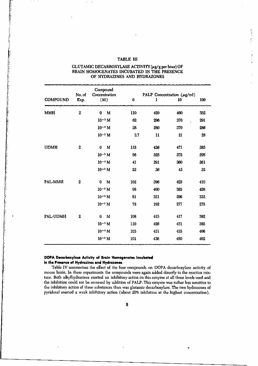
Glutamic Decarboxylase Activity of Brain HomogenatesIncubated in the Presence of Hydrazines and Hydrazones
Table III summarizes the effect of two hydrazines and their PAL hydrazones on glutamic de-carboxylase of mouse brain when they were added directly to the reaction mixture.
Both MMH and UDMH inhibited the action of this enzyme at a concentration of I x 10- 3M,and the inhibition was not totally reversed by any of the levels of PALP added. At two lowerlevels of these substances (1 x 10- 4 and 10--SM), there was still some inhibition of the enzymewhich was not reversed by PALP. The two hydrazones, however, were much less inhibitory andin some experiments, not inhibitory at all. It is possible that such inhibitory action as these com-pounds appeared to exert was due to breakdown products formed in solution.
TABLE II
DOPA DECARBOXYLASE ACTIVITY IN BRAINS OFCONVULSED MICE COMPARED WITH THAT
OF SALINE INJECTED CONTROLS
Values; pg DOPA decarboxylated per gram of tissue per hour
No. of Concen-COMPOUND Exp. 0 PALP tration (g/ml)
1 10 100
MMH 2 60 200 255 165Saline Control 121 402 465 600% Inhibition 50.4% 50.2% 45.2% 72.5%
UDMI 2 137 350 497 575Saline Control 115 410 487 685% Inhibition -19.1% 14.6% -2.1% 16.1%
PAL-MMIl 2 118 545 500 500Saline Control 98 465 525 545% Inhibition -20.4% -17.2% 4.8% 8.3%
PAL-UI)MH 2 143 610 655 780Saline Control 182 565 685 700% Inhibition 21.4% -8.0% 4.4% -11.4%
PALP-MMII 2 120 575 582 545Saline Control 121 562 570 650% Inhibition 0.8% -2.7% -1.4% 16.2%
PALP-UDM I 2 136 550 585 457Saline Cor. 120 690 660 580% Inhibition -13.3% 20.3% 11.4% 21.2%
7
F
TABLE III
GLUTAMIC DECARBOXYLASE ACTIVITY (ug/g per hour) OFBRAIN HOMOGENATES INCUBATED IN THE PRESENCE
OF HYDRAZINES AND HYDRAZONES
CompoundNo. of Concentration PALP Concentration (Itg/ml)
COMPOUND Exp. (M) 0 1 10 100
MMH 20 M 110 420 460 352
10- 5 M 62 296 370 291
10- 4M 38 280 370 28610- 3 M 3.7 11 21 28
UDMH 2 0 M 115 436 471 385
10- 5 M 56 325 375 295
10- 4 M 41 291 360 261
10- 3 M 22 36 43 32
PAL-MMH 2 0 M 102 396 425 410
10-1 M 98 400 385 426
10- 4 M 81 321 296 33210-3 M 78 192 277 275
PAL-UDMH 2 0 M 108 415 417 392
10-5 M 110 426 431 385
10-4 M 103 431 433 406
10-1 M 101 436 450 402
DOPA Decarboxylase Activity of Brain Homogenates incubatedin the Presence of Hydrazinos and Hydraxones
Table IV summarizes the effect of the four compounds on DOPA decarboxylase activity ofmouse brain. In these experiments the compounds were again added directly to the reaction mix-ture. Both alkylhydrazines exerted an inhibitory action on this enzyme at all three levels used andthe inhibition could not be reversed by addition of PALP. This enzyme was rather less sensitive tothe inhibitory action of these substances than was glutamic decarboxylase. The two hydrazones ofpyridoxal exerted a weak inhibitory action (about 20% inhibition at the highest concentration).
8

TABLE IV
DOPA DECARBOXYLASE ACTIVITY (,ug/g per hour) OFBRAIN HOMOGENATES INCUBATED IN THE
PRESENCE OF HYDRAZINES AND HYDRAZONES
CompoundNo. of Concentration PALP Concentration (pAg/inl)
COMPOUND Exp. (M) 0 1 10 100
MMH 2 0 M 122 408 495 68110-5 M 131 310 445 41510- 4 M 90 336 295 36010-3 M 42 102 133 103
UDMH 2 0 M 116 392 483 65310-5 M 75 278 385 39710- 4 M 51 164 370 33310- 3 M 50 54 122 228
PAL-MMH 2 0 M 120 420 492 660
10-' M 117 390 490 62510- 4 M 121 340 425 54510 :' M 120 370 460 560
PAL-UDMII 2 0 M 125 410 470 67010- M 111 397 380 67210-4 NI 131 39.1 425 59510- : Ni 1(y 345 3(X) 575
DISCUSSION AND CONCLUSIONSNone of the hydrazines or hydrazones tested could he classified as potent enzyme inhibitors
when compared, for example, with a-hydrazino-methyl-DOPA, which gives alx)ut 50% inhibi-tion at 1 x 10-"N. The compounds however, significantly lower the activity of lth glutamic andDOPA decarhoxylase in brains of mice which had been convulsed. The fact that this inhibitioncould not be completely reversed simply by addition of PALP suggests that the removal ofPALP by its combination with hydrazine is not the only mechanism by which it exerts its effect.,lso, since convulsions can be induced by pyridoxal hydrazones confirms this view. In vitro, how-ever, the combination of hydrazine with the cofactor probably is largely responsible for the in-hibition. Under these conditions, the pyridoxal hydrazones are only weakly inhibitory or not in-hibitory at all.
It must be borne in mind that the activity of these compounds in the body may be due to oneof the breakdown products or a product of their interaction with body tissues. This possibility de-serves further investigation.
9
\p
Section IIITHE EFFECT OF SOME HYDRAZINES AND
HYDRAZONES ON BRAIN TRANSAMINASESINTRODUCTION
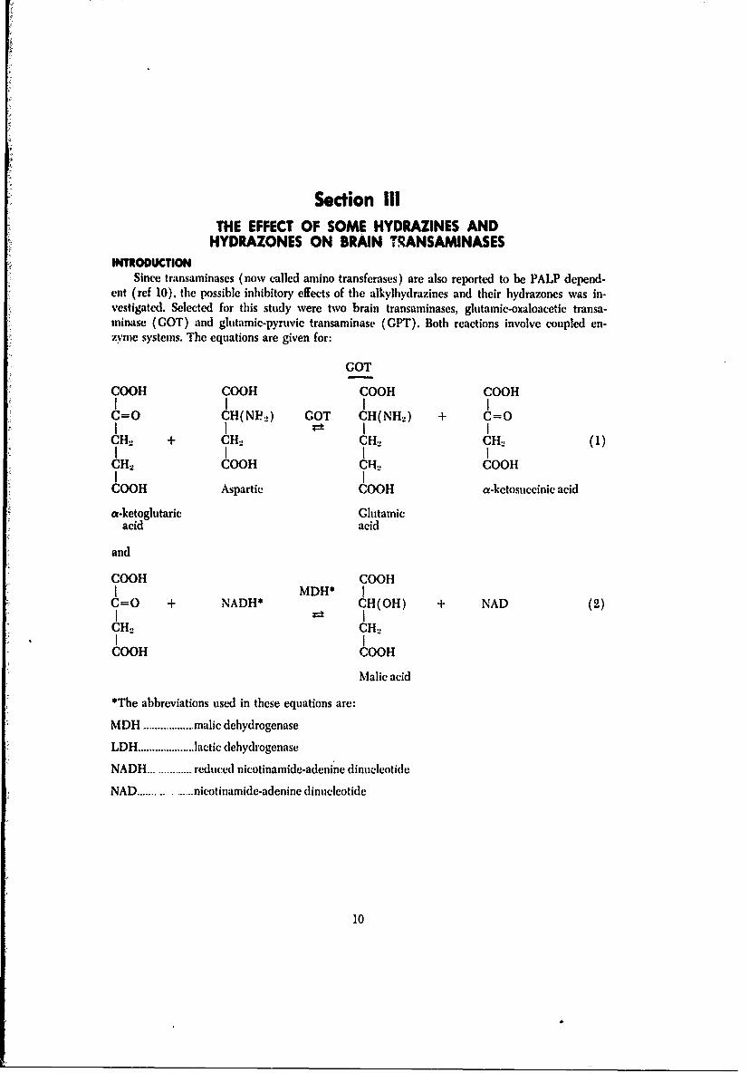
Since transaminases (nowv called amino transferases) are also reported to be PALP depend-ent (ref 10). the possible inhibitory effects of the alkylbydrazines and their hydrazones was in-vestigated. Selected for this study were two brain transaininases, glutamic-oxaloacetic transa-minase (GOT) and glutamnic-pyruvic transaminase (ClPT). Both reactions involve coupled en-zyme systems. The equations are given for:
GOT
COOH COOH COOH COOH
C=O H(NVF.,) COT CH(NH..) + C=O
CHJ + C;H:. CH2 CH2 1
CH 2 COO)H 010011OG
COO)H Aspartic COOH a-ketosuccinic acid
a-ketoglutaric Glutamicacid acid
and
COOH COOHIMDH* I
L;=0 + NADH* CH(OH) + NAD (2)
CH 2 CH2
COOH 1COOH
Malic acid
*The abbreviations used in these equations are:
MDH ........... malic dehydrogenase
LDH ........... lactic dehydrogenase
NADH...... ... reduced nicotinamide-adenine dinucleotide
NAD ...........nicotinamide-adenine dinucleotide
10

CPT
COOH COOH COOH COOHI i IC=O CH(NHt) CH(NH2) + C=O (3)i i I ICU + CH. UPT CH2 CH.
CU2 CUI I
COOH Alanine COOH Pyruvic acid
and
COOH COOtlLDH [
C=O + NADH r-t CH(OH) + NAD (4)
CUH3 CH3Lactic acid
The change: NADH-- NAD in reactions (2) and (4) can readily be measured by de-termining the decrease in the optical density at 340 mjt for a given time period. The rate ofchange for this reading is related to activity of the respective enzyme.
This work was designed to show whether the six compounds under investigation (UDMH,MMH, PAL-UDMH, PAL-MMH, PALP-UDMIt, PALP-MMH) would influence the rate of actionof brain transaminases when added directly to the reaction mixtures at levels ranging fromI X 10-3 to Ix 10-6 M.
METHODSPreparation of Enzyme
Adult male Swiss-albino mice were decapitated and the brains removed and weighed. Aver-age brain weight was 420mg. Brains were homogenized in a teflon-glass homogenizer in fourtimes their weight of borate buffer p!l 8.2. This was further diluted to give one part tissue in fiftyof homogenate. The pr"paration was dialyzed against berate buffer overnight. The COT activitydid not change when the dialyzed homogenate was stored frozen, but the activity of the GPTdecreased rapidly For this reason brain homogenates used for study of the latter enzymes wereprepared from fresh brain. Before use, the homogenate was placed in an ultracentrift'ge and spunat 100,000 x g for 30 minutes. The clear supernatant provided the source of the transaminase.
Assay ProcedureKits (Worthington) prepared for the determination of GOT and CPT activity in serum were
used in these experiments. Reagents in the kit were dissolved in 2.2 ml of water. The brain enzymepreparation was added to a cuvette in a volume of 0.5 ml and the hydrazines in 0.3 ml giving areaction mixture final volume of 3.0 ml. Materials to be tested were added to the reaction mixtureat concentrations of I x 10-6, I x 10- 5, 1 x 10- 4 and 1 x 10⁻³M. A mixture to which buffer onlywas added provided the control; PALP was added to give final concentrations of 0, 1, 10 and 100pg/ml.
Immediately ujxmn mixing the solutions, the reaction was initiated, a stopwatch was started,and the optical density (01)) was read at one minute intervals. Results were expressed as aver-age change in 01) per minute.
11

RESULTSPercentage inhibition of glutamic-pyruvic transaminase in mouse brain by MMIt and UDMH
is shown in table V (means of three estimates).
TABLE V
PERCENTAGE INHIBITION OF GLUTAMIC-PYRUVICTRANSAMINASE IN THE MOUSE BRAIN BY
MMH AND UDMH
CONCENTRATION
COMPOUND OM 1 x 10- 5M 1 x 10- 4M 1 x 10⁻³M
MMH 0% 4.9% 16.3% 70.1%
UDMH 0% 6.4% 15.7% 55.4%
The remaining compounds (PAL-MMH, PAL-UDMH, PAL-MMH, PALP-UDMH) weretested over a range from 1 x 10"M to 1 x 10- 4M but had no significant inhibitory effect on thiEenzyme.
Clutamic-oxaloacetic transaminase was not inhibited by MMH or UDMH over the range1 x 10-1 to 1 x 10-'M or by the four hydrazones over a range of 1 x 10- 6 to 1 x 10- 4 M. Additionof PALP to give concentrations of 1, 10 or 100 fpg/ml did not affect the rate of the reaction.
DISCUSSION AND CONCLUSIONSAlthough pyridoxal is reported to be the coenzyme in enzymatic transaminases, especially be-
tween a-ketoglutaric acid and a number of L-aliphatic amino acids, the vitamin does not seem tobe inactivated by the alkyihydrazines except at very high concentrations. Since in vitro formationof the hydrazones is accomplished simply by mixing PALP, or PAL with either UDMH or MMH,it can be assumed that the hydrazones thus formed are not inhibitory as evidenced by their failureto modify the enzyme reactions.
The conclusion can be drawn that the mechanism of convulsant action of the alkylhydrazinesdoes not involve these transaminases investigated. It is of interest that the rate of transaminasewas not enhanced by the addition of exogenous PALP.
12

Section IVDEVELOPMENT OF AN ULTRASENSITIVEQUANTITATIVE BIOASSAY METHOD FOR
VITAMIN B6 USING NEV'ROSPORAINTRODUCniON
An assay method for the determination of vitamin B,; content in mouse brains had to be de-veloped before achieving the objectives of this project; i.e., to detect minute changes in PALP inbrains just after convulsions induced by MMH or UDMH. It was anticipated that the initial Bitlevels per mouse brain would be in the nanogram range and that the change would have to bedetected at the picogram levels. No chemical analysis is sensitive at these low levels, although arecently introduced gas chromatographic method may have promise (ref 13); but these proce-dures have not been applied to tissues.
Microbiological assays are unusually sensitive, and a method for the determination of totalvitamin B, was developed by Atkin et al (ref 14) employing a yeast. This method was modifiedby Snell and Rannefeld (ref 15), and when a commercial medium was introduced more uniformresults were obtained (ref 16). After the x-ray mutants of the fungus, Neurospora sitophila, be-came available it was found that one of the strains was vitamin B6 dependent; this strain was thenused for a bioassay of this vitamin (ref 17, 18).
The lower limit for detection of vitamin B6 by these methods as reported in the literature wasin the microgram range, values not sensitive enough for this work. Furthermore these proce-dures were not developed for the.detection of individual congeners. A reproducible separation ofthe individual components of the vitamin Bt group has already been achieved in these laboratories(ref 19) by the use of paper chromatography; the objective of this phase of the work was to de-velop an ultrasensitive bioassay method that could quantitatively determine PALP and PAL in thenecessary nanogram or picogram range. Neurospora sitophila was the microorganism chosen forthis project.
Many variables were investigated and attempts were made to find the optimum conditions forreproducible results with the bioassay method. Different experiments were conducted to deter-mine: a) the best way to clean the glassware, b) if shaking the cultures had an effect on thegrowth rates, c) the temperature sensitivity of the neurospora, d) a method of preserving theinoculum through lyophilization to minimize loss of sensitivity of the fungus to PAL due to appar-ent back mutation, e) if the fungus was inhibited, or was growth supported by the PAL or PALPhydrazones, f) the optimum conditions for the assay.
METHODSA. Preparation of the Glassware
The nature of the experimental work required a very meticulous prx.eture for laboratoryglassware cleaning. The procedure consisted of the following steps:
(1) All glassware was washed with Alconox solution, then rinsed with distilled wat.r.(2) The glassware was soaked in a bath of diluted sodium dichromate solution (120 g
Na.,Cr2O7 + 1600 ml concentrated sulfuric acid 2(X) ml wditer).
(3) The glassware was rinsed ten times with distilled water and ten times withl distilled-deionized water (d.d-w).
(4) Finally, the glassware was dried and covered to prevent contamination from the air.
13

This method was compared with a control method which consisted of washing the glasswarewith Alconox solution and rinsing with distilled water only (Step 1). The rates and total amountof growth cultures of Neurospora sitophila were compared by growing the organism in Basal Me-dium containing 100 nanograms PAL per flask; ten-125ml Erlenmeyer flasks were prepared thesame way. Also, the extent of growth was estimated visually on a 0-5 scale. The mycelium wasthen harvested, dried in a desiccator and weighed to the nearest 0.1 mg.
B. Shake - Flasks
Comparisons were made on growth rates between continuous shaking of flasks and allowingthe vessels to remain undisturbed at the same temperature.
C. Temperature Sensitivity of N. sitophila
The temperature sensitivity of N. sitophila was investigated as follows:
(1) A serial dilution with d-d-w was used to prepare a pyridoxal HC1 solution of 100picogram/ml from a standard solution. Sterilition was done by Scitz filtering.
(2) Erlenmeyer flasks (125 ml) with assay medium were prepared in triplicate. Controlscontained 10 ml Basal Medium + 10 ml d-d-w and assay flasks had 10 ml Base.l Me-dium + 9 ml d-d-w. All flasks with medium were autoclaved at 121"C for 20 minutes.
(3) To each assay flask was added 1 ml of pyridoxal solution. The final concentration was5 picograms of PALP/ml of medium.
(4) Spores from three N. sitophila slant cultures were placed in suspension in 10 ml steriled-d-w. The turbidity of this suspension was adjusted with sterile d-d-w to read 5%transmittance at 590 m/L in a Bausch and Lomb Spectronic-20 spectrophotometer.Each flask was inoculated with 0.2 ml of this mixture.
(5) The cult.res were incubated on a Dubnoff shaker for 20 hours. The experiments wereconducted at the following temperatures: 21, 23, 25, 26, 27 and 30°C.
(6) The mycelium was harvested by centrifuging the cultures at 33,000 rpm for 20 min-utes. The pellet was then removed, blotted on filter paper and dried to constant weightin a vacuum desiccator over CaCI.
(7) The weights of the mycelial iats thus obtained were recorded.
D. Preservation of the Neurospora by Lyophilization
Mycelia of a one-week-old subculture of the Neurospora were transferred into a preserva-tive medium (1% monosodium ghtamate and 3% dextrose in 10% PVP). The mixture was in-jected into liquid nitrogen using a syringe with a 16-gauge needle. The resultant freeze-driedbead-like Neurospora was then transferred into flamed ampules; the transferring was carried outwith the aid of a flamed forceps which was also dipped into liquid nitrogen to facilitate the pro-cedure. One half of the ampules were put into a nitrogen atmosphere (by holding the open am-pule under liquid nitrogen) and the other half remained in air. Al! ampules were then sealed withan acetylene torch.
The lyophilized supply was subeultured in slants imade of Bacto Neurospora subculture me-dia. A suspension made of the lyophilized Neurospora was made into an inlocumlmnm and testedwith the hioassay of B,;. The bioassay was carried out in Petri dishes containing Baeto-pyridoxine
14

media and Difco Agar; the hardened media provided a firm surface for the support of strips ofchromatographic paper (Whatman No. 4). Sterile paper strips were spotted with measuredamount (1 mg, 10-1 mg and 10- 2 rag) of PAL hydrochloride and then placed on these Petri disheswhich were then incubated at 300C for 5 days.
E. Will Hydrazones Support or Inhibit Growth of Neurospora?
Studies were made to see if PAL-UDMH or PALP-UDMH could be a source of vitamin B,,for, or be inhibitory to the growth of N. sitophila.
A stock solution of PALP hydrochloride was made in d-d-w at a concentration of 10 nano-grams/ml, and Seitz filtered. Stock solutions at a concentration of 1000 nanograms/ml were madeo'f PAL-UDMH and PALP-UDMH. Experiments were conducted according to the following:
(1) Basal medium, 10 ml, was added to a series of 125 ml Erlenmeyer flasks. To some wereadded just 10 ml d-d-w; to others were added 9 ml d-d-w and 1 ml of PALP stock solution; to athird set was added 10 ml d-d-w, and 0.5 ml of stock PAL-UDMH.
(2) To a series of 10 ml Basal Medium in flhsks were added 1 ml of stock PALP solution andincreasing volumes of the PALP-UDMH stock solution, so that the concentration of PALP-UDMHranged from 100 to 900 nanograms per flask. These experiments were repeated with PAL-UDMH.
(3) To a third group of flasks were added 10 ml Basal Medium, no PALP, but increasingamounts of PAL-UDMH, or PALP-UDMH as the only source of vitamin B6. Concentrations ofthese hydrazones ranged from 100-800 nanogram/flask. Controls for this series were d-d-w alone,and PALP at a level of 10 nanogram/flask.
All groups were then treated the same. Each flask was inoculated with 0.2 ml of N. sitophilaspore suspension originally made up to read 5% transmittance at 590p.. The flasks were pluggedand incubated in a Dubnoff shaker for 20 hours at 27'C. The medium was then centrifuged, andmycelium pellets were removed, blotted, dried and weighed.
F. The Final Assay Procedure Developed
N. Sitophila (ATCC 9276) was grown on Neurospora sub-culture Agar plates at 25"C. A re-serve supply of cultures was prepared by the lyophilization technique described to insure geneticintegrity of the strain. A homogenous inoculum was available for each successive assay and a newtube was used !or each new assay. (See Appendix for final lyophilization procedure.) Each in-oculum was prepared by suspending spores from a two-day old subculture of the fungus ind-d-w, and this was diluted to a standard turbidity of 5% transmittance at 590 mnj in a Bauschand Lomb Spectronic 20 spectrophotometer.
A standard solution of PAL hydrochloride was prepared and by serial dilution in d-d-w a stocksolution was obtained with a concentration of PAL (not hydrochloride) 2A) picograms/mil. Thiswas sterilized by passing through a Seitz filter. Ileat inactivated this vitamin.
Assay vessels were 125 ml Erlenmeyer flasks containing 10 nil of basal mnedmun, these weresterilized by autoclaving at 121'C for 20 minutes. Sterile PAL solutions were ad.lt in an aunountso that successive flasks contained 0 to 7 picograms of I pilograrn increments. Final dilution to 20ml was completed using d-d-w.
These flasks were inoculated with 0.2 ml of a fresh N. sitophila spore suspension. 1 he cultureswere then suspended in a l)ubnoff shaker and agitated in the water bath for 20 hours at 26"C.
15

Upon completion of the gowth perio!, cultures were tested for purity and the mycelia wereharvested by centrifuging the cultures in centrifuge tubes for 20 minutes at 33,000 rpm. Thesupernatant fluvt was discarded and the mycelial pellet was removed, dried between two pieces offilter paper to rem-ove excess moisture and then dried at constant weight over CaCI2 in a vacuumdesiccator for 12 hrs. The dry mats were weighed to nearest 0.1 mg.
The same preo,,dure was used to assay POL and PAMINE, except that these members of thevitamin B, complex were prepared as a stock solution at 40 picogrAms/m!.
In order to test the applicability of the N. sitophila assay procedure for the detection of paperchromatographed vitamin B, congeners, the following method was employed:
PAL hydrochloride was spotted on 1 x 1 t.ip Watmann Paper #4 at a concentration of 5ptg/spot. Following the .procedure of Custavson et al (ref 19), the paper was developed in a pyri-dine:butanol:water solvent system. After drying, the pper was examined under ultraviolet light,the fluorescent spot was marked and cut out. The PAL was eluted by agitating the paper for 15minutes with 10 ml t' ardified Basal Mediumn (pl 4.4). The eluate was Seitz filtered and addedto 10 ml sterile d-d-w in the assay flasks using sterile technique; strips spotted with saline actedas controls.
The flasks were then inoculated with the 0.2 ml of prepared spore suspension, incubated andthe mycelia harvested, dried and weighed.
This pr'cedure was repeated using PAL at the picogram level.
RESUITSA. Detection of the vitamin B,1 by the neurospora was markedly facilitated with chromic acid-washed glassware. A comparison of the two methods (in visual and dry-weight observations) isgiven in table VI. It can be scen that the qualitat:ve est.mation for extent of growth was unreli-able for this level of PAL.
TABLE VI
NEUROSPORA SITOPHILA GROWTH. COMPARISON OFTHE TWO METHODS OF GLASSWARE CONTAMINATION
(Visual Ranking (5 best growth) and Dry Weight Determinations (mg))
FLASK NUMBER 1 2 3 4 5 6 7 8 9 10
Acid VISUAL 5 5 4 5 5 5 3 4 3 5
Washed DRY-WT. 67.5 57.0 37.5 66.5 54.0 60.5 24,0 33.0 11.0 20.5
Alconox VISUAL 5 5 5 5 3 3 3 3 4 4
Washed DRY-WT. 34 0 57.5 5-4.0 60.5 11.0 13.0 66.5 19.3 22.7 22.5
Visual Av ge Ranking Dry Weight Average Determination
Acid xWashed 4.40 43.15 mg
Aionox Washed 4.00 36.40 mg
16

B. The method employing shaking gave much more uniform results than that without the mix-ing; therefore this step became an integral part of every procedure used.
C. The neurospora used for bioassay (Beadle 299) appears to be temperature sensitive. Theoptimum temperature range for growth in shake cultures appears to be 26-27°C. Above and be-low these temperatures growth is less and the culture's dependency on PAL is substantially de-creased. Results of a typical experiment are given in table VII.
TABLE VII
EFFECT OF TEMPERATURE ON THE GROWTH OFN. SITOPHILA
Dry Weight in mg
Temp. (C) 21 23 25 26 27 30
Controls 1. 3.5 3.0 3.4 3.0 .4 1.2
(no PAL) 2. 3.5 3.0 2.8 2.5 1.4 1.0
3. 3.4 3.5 3.0 3.1 1.4 1.0
av. 3.5 3.2 3.1 2.9 1.4 1.1
Assay 1. 3.5 3.4 3.1 3.4 2.4 1.3
5 picograms/ml 2. 3.7 3.0 3.4 3.4 2.6 1.3PAL 3. 3.6 3.5 3.1 3.5 2.5 1.0
av. 3.6 3.3 3.2 3.4 2.5 1.2
Difference
Test-Controls 0.1 0.1 0.1 0.5 1.1 0.1
D. The lyophilized neurospora bxth in the nitrogen and air atmospheres coid he subculturedeasily. After 48 hours growth was noted and after 5 (lays sporulation occurred. In the preliminaryassay using Petri dishes adequate growth was noted in three days. Visually it could easily be es-timated that the growth could he qualitatively related at the ing level to the amount of PALspotted on the paper.
E. The hydrazones were not a source of vitamin B1, for the fungus. Evidently under the condi.tions of the experiment, neither the PAL-UDMII or the PALIP-U)MtII hydrolyzed to liberate thefree vitamin. The weight of mycelium harvested in the first sol of expcriments was 2.4 mg k henno PAL or hydrazones was added, and the same \alue \hen only hydrazone was present. For thePALP controls, some growth was obtained.
When a constant amount of PALP was added to the basal medium ( 10 nanogramns/flask ' andincreasing amounts of PALI-UDMII (from 0-800 nanogramns/flask) \%ere added, no differences inmyceliumn weight were found from flask to flask.
No concentration of PAL-UI)MII increased the dry weight of harvested myceliim, ave( rage
17

weights ranged from 2.5 to 2.8 mg which were tie same as for the d.d-w control. At 900 and 1000nanograms/flask of PALP-UDMH, the harvested mycelium weighed from 3.3-3.8 mg.
F. The harvested mycelia obtained from the growth of N. sitophila on 5/Lg PAL eluted from thechromatographed paper strips are summarized in table VIII. Slight growth was always noted fromthe controls.
TABLE VIII
DRY WEIGHT OF N. SITOPHILA FROM 5pg PAL ELUTEDFROM PAPER
Flask No. Test Culture Flask No. Controls
1 11).4 5 4.8
2 6.6 6 5.03 9.4 7 3.0
4 7.6 8 3.0Av. 8.5 3.7
The values of mycelia obtained when PAL was used at the picograr levels are given intable IX.
TABLE IXDETECTION AND ASSAY OF PICOGRAM AMOUNTS
OF PAL
Dry weight mycelia in mg
Trial 0 p/ml 1 p/ml 5 p/ml 7 p/ml
I 2.0 2.0 4.5 4.5
2.6 4.0 4.0 5.0
2.5 2.5 4.0 5.0Ave. 2.3 2.8 4.1 4.8
11 4.5 3.2 7.5 6.6
2.5 - 2.5 6.2
2.8 2.5 3.3 4.5
Ave. 3.2 2.8 4.4 5.8
III 1.3 1.5 3.0 3.5
1.4 1.6 3.0 3.8
1.4 1.6 3.0 4.0
Ave. 1.4 1.6 3.0 3,8
POI, and PAMINE values are not as reliable as PAL ir the low picogram range.
18

DISCUSSION AND CONCLUSIONSA bioassay method for the detection and estimation of individual members of the vitamin Bo
complex has been developed. Using the fungus, N. sitophila, PAL can be detected as low as 5-7picograms/ml. The method depends upon growing the mycelium as shake cultures, harvesting thegrowth, and weighing the final mats.
All congeners of this vitamin can be separated by means of paper chromatography, elutedfrom the paper with Basal Medium and assayed. The method is more sensitive for PAL than POLor PAMINE.
Loss of sensitivity of the fungus to vitan'.'i Bn was noted on successive culturing on artificialmedia. A method was developed to preserve i. -w cultures by lyophilization. It is assumed thatthe !o.,s of dependence of the micro-organism on vitamin B6 was due to back mutations.
The temperature dependence of N. sitophila was studied, and the range 26-27°C was found
to be optimum for growth.
This fungus is not inhibited by PAL-UDMH or PAL-MMH, but at very high levels of PALP-UDMHI, 1,000 nanograms/ml, some growth of the Neurospora is noted.
19
Section VUSE OF YEAST FOR BIOASSAY OF VITAMIN B6
INTRODUCTIONTests were run to establish whether a yeast system, S. carlsbergensis (culture 4228, Standard
Brands,.N. Y.), was suitable for detection of vitamin B,1 congeners in the nanogram and picogramranges (ref 15, 16). This assay system was intended as a cross-check for the Neurospora methoddescribed in the previous section. Also, this yeast exhibited a greater sensitivity to PALP thanNeurospora.
MATERIALS AND METHODSVitamin-free yeast biase was obtained from Difco Laboratories, Baltimore, Md., and lyophil-
ized cultures of S. carlsbergensis (ATCC 4228) were purchased from the American Type Cul-ture Collection, Rockville, Md.
Experiments were conducted to find the optimum conditions for the assay. Growth was esti-mated by counting particles on a Coulter Counter, and by turbidometrie measurements using twocolorimeters; a Klett colorimeter, and a Bausch and Lomb Spectronic 20 spectrometer at 590 m.The latter instrument proved most satisfactory and was used for this section.
Standard solutions of PAL hydrochloride were prepared in concentrations of 50 nano-grams/ml and 50 picograms/ml. The basal medium, Difco Vitamin-Free Yeast Base, was madeby dissolving 16.79 g in 100 ml d-d-w. All solutions were Seitz-filtered and stored in sterile cotton-stoppered flask. (Filtration was used in place of heat sterilization because of the heat lability ofPAL.)
The assay medium was prepared in sterile, cotton plugged 16 mm, culture tubes according tothe following scheme:
Cone. of pyridoxal Nanograms/mlper 1 ml of finalmedium 0 1 3 5 7
ml of sterile 9 8.8 8.4 8.0 7.611,O in tube
ml of sterile 1.0 1.0 1.0 1.0 1.0basal medium
ml of sterile PALHCI standard 0 0.2 0.6 1.0 1.1solution (50 nano/nil)added
Tubes of assay media thus prepared were inoculated with 0.1 ml of a suspension of S. carls-bergensis obtained from suspending one loopful of growth at 260C from a potato-dextrose agarslant culture in 10 ml of sterile d.d-w. To inFure a consistent amount of inoculum in successiveassays, the transmittance (%T) at 590 mF of the yeast suspension was adjusted to 20 using steriled-d-w.
20
assay.,;,~~~~~
~ ~ ~ mi
th Ir
n mta c 1%T
aa~ 590a in
ol thel yeas
susp nsio
wa ad
e to1
sn
rle

Inoculated cultures in triplicate were incubated on a rapid shaker at 260C for 4.5 days. Theextent of growth was measured optically by a change in %T as compared to pure d-d-w which wasused as a reference. Control flasks were used to test cultures for purity.
RESULTSTable X gives an example of data from a typical experiment.
TABLE X
DETECTION AND ASSAY OF PAL IN THE NANOGRAMRANGE OF CONCENTRATION WITH SACCHAROMYCES
CARLSBERGENSIS
% Transmittance at 590 mu diluted 1:1 with d-d-w
Nanograms/ml
Tube 0 1 3 5 7
1 31.0 32.0 25.0 19.0 12.02 30.0 35.0 25.0 21.5 7.53 32.0 36.0 26.0 18.0 15.0
Average 31.0 34.3 25.3 19.5 11.5
Corrected averages for undiluted cultures:15.5 17.15 12.6 9.75 5.75
To assay the vitamin B0 group at the picogram level, certain modifications were made in thesetechniques (ref 20). These are:
1. The slant cultures were grown at 310C rather than 26"C.2. The assay was run at 300C rather than 26"C.
3. The incubation period was shortened from 4.5 days to 3.
4. The inoculum (1 loopful of growth from a potato-dextrose agar slant culture in 10 mld-d-w) was incubated on a shaker 1 hour at 280C before use.
5. Tubes of the assay medium were inoculated with 0.2 ml of the yeast suspension rather than0.1 ml.
The data of an experiment are given in table XI.
TABLE XI
PICOGRAM RANGE ASSAY OF PYRIDOXAL 11C USINGS. CARLSBERGENSIS
% Transmittance at 590 npPicogram/ml
Tube 0 1 3 5 7
1 19.7 13.1 21.3 17.9 12.02 38.5 41.5 17.0 12.2 15.03 broke 35.5 23.9 26.1 17.4
Average 29.1 30.0 20.7 18.7 11.1
21

DISCUSSION AND CONCLUSIONSThe lyophilized culture of Saccharomyces carlsbergensis obtained from ATCC was successful-
ly subcultured. Since direct transfer of the lyophilized culture to potato-dextrose agar was as suc-cessful as the transfer to nutrient broth and then subsequent transfer to potato-dextrose agar, theintermediate step was abandoned.
Most of the problems of erratic growth such as contamination and sensitivity of detectionwere overcome and detection was greatly facilitated. The feasibility of several detecion methodswas explored. The observations obtained with one method were checked against similar data fromother methods and were found in very good agreement.
The % transmittance data for the yeast bioassays were obtained on both a Klett Colorimeterand a Bausch and Lomb spectronic 20 spectrophotometer. When the readings with the Klett foryeast cultures were completed, a check against Coulter Counter counts was taken on the same cul-tures. Klett readings did not correlate well with the total cell population data obtained with theCoulter Counter. Hence the Klett Colorimeter was judged unreliable and its use was discontinued.The Bausch and Lomb Spectrophotometer was found to be a more reliable instrument. The con-stant refining of the techniques of the yeast bioassay procedure produced the desired result of ob-taining pyridoxal sensitivity of the organism in the picogram range of concentration. Though morework on this method is indicated, the procedure has been used to assay PALP in individual mousebrains.
22

Section VIAPPLICATION OF ULTRASENSITIVE QUANTITATIVE
BIOASSAY METHODS FOR THE DETECTION OFVITAMIN B6 IN RODENT BRAINS
INTRODUCTIONThe ultra-sensitive bioassay nethods, although not perfected, were found to be useful and
were applied for the detection of Vitamin B6 congeners in the brain. However, for this section onlyPAL and PALP were investigated. The method used involved paper chromatography to separatethe components, elution of the individual components from the strips, and finally the bioassayprocedures described in the two previous sections. Concentrations standards were in the pico-gram range.
This section consists of two parts:
A. The analysis of PAL and PALP in mouse brains.
B. The effect of PALP-UDMH on growth of Neurospora.
METHODS
Part AFor both sections A and B, mouse brains were prepared for chromatography by the following
procedure:
(1) Two groups of mice were used. The first group was injected I.P. at a dose level of 500mg/Kg of UI)MH in saline. The control group was injected with a comparable volume of normalsaline.
(2) After death by convulsions in the experimental group or cervical fracture in the controlgroup, the entire brains were immediately removed, quickly frozen in liquid nitrogen, andweighed.
(3) Five mouse brains from each group were pooled and 1 nil of pyridine was added forevery gram of brain tissue. Each mixture was homogenized for one minute, using an ultrasonicprobe. ( Bio-Sonic Instrument). Samples were kept cold at all times in a bath of acetone and dryice.
(4) The homogenates were centrifuged for 5 minutes at 2500 rpm.
The supernatant from the centrifuged hoiuogenate was chromatographed on WhatmanPaper #4 by the procedure established by Gustavson ct al (ref 19). Brain supernates and stand-ards were diluted 10X before spotting on chromatography strips (0.1 ml of homogenate super-natant and 0.9 ml of a pyridine:butanol: water in ratio of 1.2:saturated).
The zones ("spots") of pyridoxal were cut from the chroniatographed strips and the PAL waseluted in 10 ml Basal Medium. These areas were determined by control Rf values.
23
Various methods of elution were tested and the best results were obtained when a combina-tion of the following procedures were used:
(1) To avoid the destruction of PAL by light, the strips were chromatographed and the"spots" were cut out in the dark.
(2) Basal medium was used for elution but the pit was adjusted to 5.0 so that PAL wouldbe recovered more efficiently.
(3) A slight increase of temperature increased the recovery of PAL by elution, therefore this
step was done in an incubator at 3700 without light.
(4) The beakers wdre intermittently agitated.
(5) The elution process was carried out for at least one hour to obtain maximum recoveryof PAL.
(6) Two millipore filtration systems (Swinney Adapters with syringe and needle attach-ments) were introduced in place of the Seitz. This allowed separation of the experimental and con-trol eluates. A Seitz filter was used to sterilize the known assay solution.
(7) To the sterile eluates in assay flasks were added 10 ml aliquots of sterile d-d-w.
(8) Each flask was inoculated with 0.2 mi N. sitophila spore suspension and the cultureswere incubated at 270C for 20 hours in a Dubnoff shaker.
(9) The myeelium was harvested and the dry weights recorded.
For PALP detection and quantitative assay, the same procedure was used, but the elutingsolvent was I ml vitamin-free yeast base plus 9 ml d-d-w. The flask was inoculated with S. earls-bergensis.
Part IThe assay procedure followed that given in Part A, with the following modifications: instead
of eluting PAL or PALP from the paper strips, solutions of PALP-UDMII of known concentrationwere added to 10 ml of assay medium. To a series of flasks was added I ml of a solution contain-ing 1 tg/ml of PALP. To each flask was also added PALP-UDMH in increasing concentrationfrom 100 to 10(X nanograms/flask.
RESULTS
Part AThe data from the determination of PAL in rodent brains is given in table XII. The weight
of dry mycelium was converted to picograms PAL/ml of brain homogenate. Three separate trialsare recorded.
Part !From the results given in table XIlII, it can be noted that PALP-UDMII was not an inhibitor
of the neurospora used. The slightly greater dryweights of mycelium noted at the higher con-centrations of the hydrazone were not statistically significant.
24
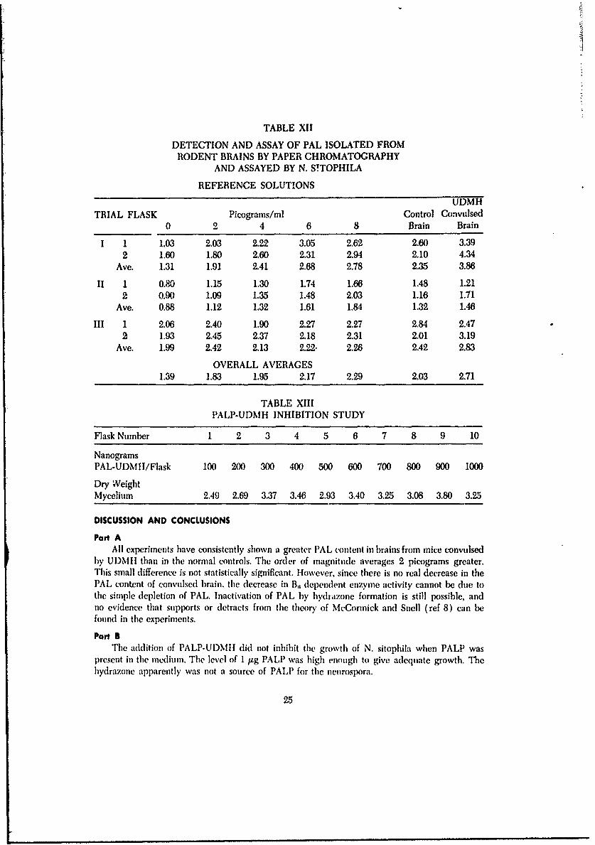
TABLE XII
DETECTION AND ASSAY OF PAL ISOLATED FROMRODENT BRAINS BY PAPER CHROMATOGRAPHY
AND ASSAYED BY N. S!TOPHILA
REFERENCE SOLUTIONS
UDMHTRIAL FLASK Picograms/ml Control Convulsed
0 2 4 6 8 Brain Brain
I 1 1.03 2.03 2.22 3.05 2.62 2.60 3.392 1.60 1.80 2.60 2.31 2.94 2.10 4.34
Ave. 1.31 1.91 2.41 2.68 2.78 2.35 3.86
II 1 0.80 1.15 1.30 1.74 1.66 1.48 1.212 0.90 1.09 1.35 1.48 2.03 1.16 1.71
Ave. 0.88 1.12 1.32 1.61 1.84 1.32 1.46
III 1 2.06 2.40 1.90 2.27 2.27 2.84 2.472 1.93 2.45 2.37 2.18 2.31 2.01 3.19
Ave. 1.99 2.42 2.13 2.22- 2.26 2A2 2.83
OVERALL AVERAGES1.39 1.83 1.95 2.17 2.29 2.03 2.71
TABLE XIII
PALP-UDMH INHIBITION STUDY
Flask Number 1 2 3 4 5 6 7 8 9 10
NanogramsPAL-UDMII/Flask 100 200 300 400 500 600 700 800 900 1000
Dry WeightMycelium 2.49 2.69 3.37 3.46 2.93 3.40 3.25 3.08 3.80 3.25
DISCUSSION AND CONCLUSIONS
Part AAll experiments have consistently shown a greater PAL content in brains from mice convulsed
by UDMII than in the normal controls. The order of magnitude averages 2 picograms greater.This small difference is not statistically significant. However, since there is no real decrease in thePAL content of convulsed brain, the decrease in B, dependent enzyme activity cannot be due tothe simple depletion of PAL. Inactivation of PAL by hydrazone formation is still 1x)ssible, andno evidence that supports or detracts from the theory of McCormick and Snell (ref 8) can befound in the experiments.
Part BThe addition of PALP-UI)MII did not inhibit the growth of N. sitophila when PALP was
present in the medium. The level of 1 j.g PALP was high enough to give adequate growth. Thehydrazone apparently was not a source of PALP for the neurospxora.
25

Section V11EVIDENCE THAT INJECTED UDMH IS FOUND
IN THE CNS
INTRODUCTIONIndirect evidence is presented that UDMH may cross the blood-brain barrier after intraperi-
toneal administration. Until now, most theories of convulsive action of U131\11 and \11\11 as-sunnedl that these a]lkylbyd razintes did penetrate the brain barrier and exerted their action in theCNS. No supporting data were presenteA by others.
lIn brief, the circumstantial evidence is: following UI)MII induiced convulsions inl mice, thleb~rains were removed, p~rep~ared as previously de~scribed butt PALIP was added to the supernatantprior to chromatography. Examination of the paper strip revealed a new Ilo(WCscent spot wvhichis not p~resenlt in the supernatant of control b~rains. ile chromatographic 11r value indicated thenew compound to be PALP-UDMH.
MATERIALS AND METHODSThese animals were convulsed with UI)MII, and the brains were r('n'ovt'd and prep~ared as
described in Section VI, Methods; Part A. The supernatants w~ere spotted ..n Whatmnan #4 paperand chromatographed in the dark using pyridine: butanol: water as describt(l in Gustavson, et al(ref 19). After the paper strips wvere dried, they were examine(' ider liltzaviolet light.
The experiments wvere repeated but this time a known qunantity of PALI) w~as added to thehomogenates prior to centrifugation. These supernates wvere again chromnatographed and examinedfor fluorescence.
In sulbsed'.ent experimnents, PAL, PAL-UDM II, PALP- U DM11 were chroniatographedl singlyand in all conibinations. These compounds were also added individually to brains from UDIconvulsed mice and to controls. These brains wtere prepared, supernatants were spottedl onl paperandl chromatographed.
The paper strips were exposedl to ammnoniumn hydroxide vapors, and thle 11r v'alues were mvas-tired for all fluorescent spots.
RESULTSNo fluorescent spots were noted on the paper strips chromatographecl from the convulsed or
control b)rain preparations. WVhen the experiments were repeated hut with PA I P added to #I-homiogenates, the ultraviolet light revealed a new -dark snot" fluorescence in convuIdsedl brainpreparations, hut not in controls. 'The fluorescent spot was . nhianced wthen exposed to Illlllollilunlhydroxide vapors. The 11, of the "dark spot" was idlentical to that ofPA -tI) .
When PAIP-UI)Mil was added to control or convulsedl brain, or was adlded to the solventsystein, and each preparation was chronatographed, the salme Hrf \'.tale was obtained for the "darkspot." All spots reacted sim.ilarly to ammnonium hydroxide vapors.
A mnixture of PAL.II)NIII and PALP-LIDM 11 was chroinatographed. anit( separation wvaseasily achieved, and the expected lit values were found. After exposuire to animonium hydroxidevapors, andl after exaunination uinder uiltravioltet light, only\ PALP-UI)\l II ga~ e the characteristic(lark spot."
26

L
DISCUSSION AND CONCLUSIONSAll of the evidence points to the fact that the "dark spot" is PALP-UDMH. Under ultraviolet
light, free UDMH does not fluoresce; the hydrazone has a marked fluorescence. It is assumedthat the hydrazone is easily formed in brain tissue. Tle evidence comes from the rapidity withwhich the compound is made by simply mixing the reactants in aiueous solution.
To date this appears to be the first evidence that UDMII does appear in the rodent brainafter an intraperitoneal administration of the hydrazine agent.
27
Section VIIIDEVELOPMENT OF A MATHEMATICAL MODEL
FOR THE CONVULSION PROCESS INDUCEDBY HYDRAZINES
ts short duration ihakes the convulsion process one of the ,hoSt difficult toxicological pro-cesses to study. The symptoms preceding the actual seizure are varied, ani induced convu!sionsdo not have exact pre-established patterns, although some signs occur with greater frequencythan others. Precision equipment for measuring these signs was not available, and all symptomaticcharacteristics were ohserved without their more fine-detailed descriptions.
Each time a mouse convulsed, a certain selection of the most frequent symptoms - mydriasis,iosis, exophthalos, enophthahnos, astasia, trismus, piloerection, catalepsy, lethargy, grand mal,
and the more or less definitively final conditions of a sequence of petit rnals followed by opistho-tonos - was observexl. The mathematical model had to reflect the severity of these symptoms asa function of tile administered dose, and also provide an accurate description of the time lapseprior to the onset of the seizure.
The first series of convulsion experiments produced the necessary data on tile basis of whichthe prop sed model (ref 21) was written. With the aid of several Fortran IV computer programsit was possible to obtain values of time lapse (min.) over a very wide range of dose (mg/Kg). Thesurprising agreement between the experimental data points and the computed values led us towork out a more refined mathematical model incorporating a most valuable feature - its prospec-tive (predictive) capability. All consequent convulsion experiments were therefore shortened toobtaining a few baseline points, or threshold values, and then for each convulsigen a computer-ized table of expected lag times was obtained. The success of this approach made it possible toecononize in many respects especially in the number of rolents to he expended and thus cutdown on lalmiratory tine.
In essenc &he model utilized in this work is described in its elementary form by Ledin et al(ref 21 ). Thc successful application of this novel approach warranted a paper by Furst et al (ref22).
If the data from a convulsion experiment (lag time vs dose of convulsant are plotted onregular c(ordinate paper with the dose on the abscissa, a hyperboloid curve is obtained; this curveis of the hyperbolic-exponential type, as best least-squares linearization is achieved only after in-verse-ruling and logarithmic transformations. A simple logarithmic transformation (log dose vstime) is insufficient as it provides linearization only over a small (lose range. Ilie convulsion equa-tion is then a mnemiber of the family
T Ke
where T is tile time lapse prior to convulsion or death, 1) is drug dose, p( I))/j(I)) is a polynomialratio of negative degree (the generalized polynomial hyperbo icity property), e is the base ofnatural logarithms (,, 2.71828 ... ) and K is a constant to be determined from experimental in-formation. ll equation used in our initial prediction trials with the computer was
+ T,,V-o-., + C ( I), + c ( - , ()-l)2_)1)-1D., ) ( 1)-I),} (l - ):i "'
28

where I)o is the "minimal dose" (that is, minimum dose for which convulsions take place and alsothe highest dose which does not induce convulsions) and the doses D, and D. are two suitablychosen experimental doses. The time To is the minimal time of response (that is, the lag time cor-responding to the maximum dose). The coefficients cO, cI, c2 ,. . were computed from these initialexperimental points. A sequence of predicted times were obtained by substituting a monotoni-cally increasing sequence of (loses D at increments of 50 mg/Kg, and these times were found inexcellent correspmndence with the experimentally observed values. This equation, with new con-stants, was tested for each substituted alkylhydrazine and hydrazone utilized in this study.
The inverse of the area under each convulsion or death curve (from I)o to a value I)) of aparticular cwnvulsigen turned out to provide a measure of the intensity of the specific symptomsresulting from the administration of the agent. These estimates, a new concept in the literatureare best coined as "convulsivility" (convlsant ability) and "lethality," and their usefulness inestablishing a new basis for ranking a group of chemical agents is exemplified in table XIV wherethe potency is given as a ratio with UDMII taken as the arbitrary unit. These estimates also givean indirect jIudgment of potency or a conditional comparison of effectiveness, for they can be re-lated to various therapeutic indexes and fit in very well with accepted criterions of mortality orpoisonous efficacy.
TABLE XIV
Relative Potency
(Trapezoidal Estimates)
HYDRAZINES tlYDRAZONLa
Hlydra-zinc
Anhy- PAL- PAL- PALP- PALP-UDMII MMII drous UI)MII MMI UI)MII MMII
"Convtl'ivility" 1.00 6.24 6.95 2.3.13 19.58 16.71 18.26
"L(thality" 1.(X) 7.73 5.92 18.82 17.40 21.39 17.44
29
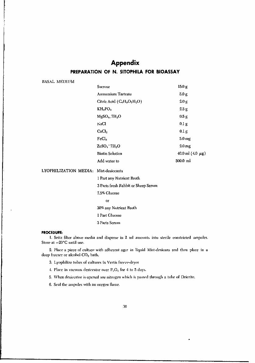
AppendixPREPARATION OF N. SITOPHILA FOR BIOASSAY
BASAL MEIUtMSucrose 15.0 g
Ammonium Tartrate 5.0 g
Citric Acid (G,;li,1l. 2 I_0) 2.0 g
KI' 2PO4 2.5 gM gSO,, 71120 0.5 g
NaGI 0.1 g
CaCl2 0.1 gFeCl.-I 5.0 mg
ZnSO, 71120 2.0 mg
Biotin Solution 40.0 ml (4.0 I&g)
Add water to 500.0 ml
LYOPHILIZATION MEDIA: Mist-desiccants
I Part any Nutrient Broth
3 Parts fresh flabbit or Sheep Serum
7.5% Glucose
or
30% any Nutrient Broth
I Part Glucose
3 Parts Serum
PROCEDURE:1. Seitz filter alx)ve mnedia and (disperse in 2 til amounts into sterile constrictedl ampules.
Store at -20*C until Ilse.
2. Place -. piece of culture with adherent agar in liquid Iviit-(lesicafs and then place it] adleep) freezer or alcohiol-GO.. bath.
3. Lyophilize tubes of cultures in \'crtis freeve-dryer
4. Place in vacuum dlesiccator over P..0O, for 4 to 5 days.
5. When desiccator is opened Ilse nitrogen which is passed through a tube of lDrierite.
6. Seal the aruptles with anl oxygen flame.
30

References1. Back, K. C., and A. A. Thomas, 1963. "Pharmacology and Toxicology of 1, 1-Dimethylhydra-
zinc (UDMH)," American Industrial Hygiene Association Journal, V. 24, 23-27.
2. Fairchild, M. D., and M. B. Sterinan, 1965. 1, 1-Dimethylhydrazine Effects on Central Excit-atory and Inhibitory Mechanisms in Cats, AMRL-TR-65-142, Aerospace Medical ResearchLaboratories, Wright-Patterson Air Force Base, Ohio 1-33.
3. Reeves, J. L., 1961. Influence of Large Doses of Pyridoxine llydrochloride on the Convul-sigenic Activity of UDMII in Monkeys, USAF Aerospace Medical Center Bulletin 62-31,School of Aerospace Medicine, Brooks Air Force Base, Texas 1-5.
4. Jenney, E. H., and L. 1). Lee, 1951. "The Convulsant Effect of Semicarbazide," Journal ofPharmacology and Experimental Therapeutics, V. 103, 349.
5. O'Brien, R. D., M. Kirkpatrick, and P. S. Miller, 1964. "Poisoning of the Rat by Htydrazine andAlkylhydrazines," Toxicology aui Applied Pharmacology, V. 6, 371-377.
6. Furst, A. and W. R. Custavson, 1967. "A Comparison of Alkylhydrazines and their B6 -Hydra-zones as Convulsant Agents," Proceedings of the Society for Experimental Biology and Medi-cine, V. 124, 172-175.
7. Dubnick, B., G. A. Leson, and C. C. Scott, 1960. "Effect of Forms of B. on the Acute Toxicityof Hydrazines," Toxicology and Applied u harmazology, V. 2, 403-409.
8. McCormick, D. B., and E. E. Snell, 1959. "Pyridoxine Kinase of Human Brain and its Inhibi-tion by Hydrazine Derivatives," Proceedings of the National Academy of Science, V. 45, 1371-1379.
9. Snell, E. E., P. M. Fasella, A. E. Braunstein, and A. R. Fanell, 1963. Chemical and BiologicalAspects of Pyridoxal Catalysts, Pergamon Press, New York, 579-594.
10. Wagner, A. F., and K. Folkers, 1964. Vi!amins and Coenzymes, Interscience Publishing Coin-pany, New York, p. 172.
11. Weissbach, It., W. Voenberg, and S. Udenfriend, 1960. "Enzymatic Decarboxylation of
a-Methylamino Acids," Biochemistry, Biophysics Research Communications, V. 3, 225-227.
12. deflopp, 1R. S. and A. Furst, 1966. "Effect of Analogs of Phenylalanine and Tryptophan onKinetics of I)OPA I)ecarboxylase in Rat Brain," Brain Research, V. 2, 323-332.
13. Korytynk, W., G. Fricke and B. Paul, 1966. "Pyridoxine Chemistry (XII) Gas Chromatographyof Compouinds in the Vitamin B6 Group," Analytical Biochemistry, V. 7, 66-75.
14. Atkin, L., A. Schultz, W. Williams, and C. Frey, 1943. "Yeast Microbiological Methods for I)e-termination of Vitamins," Indust, ial and Engineering Chemistry Analytical Edition, V. 15,1,41-144.
15. Snell, E E., and A. N. llannefeld, 1945. '"i'he Vitamin B, Group: The Vitamin Activity of Py-ridoxal and Pyriodoxamuine for Various Organisms." Journal of Biological Chemistry, V. 157,475-489.
16. ulrley, N. A., 1960. "Note on the Modification of the Atkin Method for Vitamin B, Assay,"Journal of the Associated Offickil Agricultural Chemists, V. 43, .13-45.
31

17. Stokes, J. A. Lerson, J. WxKlward, and J. W. Foster, 1943. "A Neurospora Assay for Pyridox-ine," Journal of Biological Chemistry, V. 150, 17-24.
18. Barton-Wright, E. C., 1952. Microbiological Assay of the Vitamin B Complex anl Amino Acids,Pitman Publishing Company, New York, p. 80.
19. Gustavson, W. R., G. l xdin, Jr., and A. Furst, 1966. "Variation of Rr of Vitamin B,; in Groupwith p1l," Journal of Chromnutography, V. 24, 288-290.
20. Tsuji, K., 1966. "Liquid Nitrogen Preservation of S. Carlsbergensis and its Use in a BiologicalAssay of Vitamin B,;," Applied Microbiology, V. 14, 456-461.
21. Ledin, G., Jr., W. 1R. Gustavson, and A. Furst, 1967. "Response-Time Considerations and Math-ematical Aspects of Convulsions Induced by Ilydrazines," Proceedings of the Vestern Phar-macology Society, V. 10, 61-63.
22. Furst, A. F., G. Ledin, Jr., and W. 1. Gustavson, 1967. "A Mathematical Model for Convul-sions Induc d by Alkylhydrazines," The Pharncologist, V. 9.
32

Security Classification
DOCUMENT CONTROL DATA . R & D(Security classllcation of title, body of abstract and indexing annotation must be entered when the overall report Is classlifed)
I ORIGINATING ACTIVITY .Corpor&te author) Za. REPORT SECURITY CLASSIFICATION
Institute of Chemical Biology UNCLASSIFIEDUniversity of San Francisco 2b. GROUP N/ASan Francisco, California 94117
3 REPORT TITLE
BIOCHEMICAL PHARMACOLOGY OF HYDRAZINES TOXICITY
4 DESCRIPTIVE NOTES (Type Of report and Inclusive dates)
Final Report, March 1966-February 19685 AU THORISI (Fir&f name, middle Initial, last ndme)
Arthur FurstWaldemar R. GustavsonRobert S. deRopp
6 RPORT DATE Is. TOTAL NO. OF PAGES REFS
May 1969 32J 2268 CONTRACT OR GRANT NO AF 33(615)-3829 9a. ORIGINA TOR'S REPORT NUMBERIS)
b. PROJECT NO 6302
Task Nu. 630202 9b. OTHER REPORT NOS4 (Any other numbers that may beasa,edthis report)
d. Work Unit No. 630202019 AMRL-TR-68-13210 DISTRIBUTION STATEMENT
This document has been approved for publicrelease and sale; its distribution is unlimited.
11 SUPPLEMENTARY NOTES 12. SPONSORING MILITARY ACTIVITY
Aerospace Medical Research Laboratory,
Aerospace Medical Div., Air Force SystemsCommand, Wright-Patterson AFB, OH 45433
13 ABSTRACT
The toxic action of 1, 1-dimethylhydrazine (UDMH) and monomethyihydrazine (MMH)may be mediated by the inactivation of phridoxal in the brain. One possibility con-sidered was the formation of a hydrazone between the pyridoxals and the substitutedhydrazine. Pyridoxal dependent enzymes were investigated. UDMH and MMH inhibitedboth glutamic acid decarboxylase and DOPA decarboxylase. Transaminases (aminotransferases) which required alpha-ketoglutaric acid as a substrate were not affectedby the hydrazines tested. Further work was conducted to refine an ultrasensitive bio-assay method for the detection of each congener of the vitamin B6 group. The micro-organisms investigated for the assay were a neurospora and a yeast. Some indirectov'idence was obtained which implies that UDMH injected intraperitoneally can beditected in the central nervous system. A mathematical model for hydrazines-inducedconvulsions was developed. It is now possible to predict the time lapse after adminis-tration of the convulsigen and the onset of seizure if only three data points are given.
FORMDDI NOr 61473 _________

Security Classification14 LINK A LINK S LINK C
KEY WORDS
ROLE WT ROLE WT ROLE WT
Hydrazines1, 1-Dimethylhydrazine (UDMH)Monomethylhydrazine (MMH)PyridoxineVitamin BPharmacologyBiochemistryToxicologyBioassay MethodsRodents
Securtty C s10 Ificaton