Positive Modulatory Interactions of NMDA Receptor GluN1/2B ...
Biochemical Modulatory and Protective Effects of the Hydroalcoholic Extract of Scrophularia striata on the Hepatotoxicity of Silver Nanoparticles
in the Rat Model
Masoud Shamohamadi1, Mehrdad Pooyanmehr2* , Ali Maleki3, Lida Haghnazari4
1.Faculty of Veterinary Medicine, Razi University, Kermanshah, Iran 2.Department of Basic Sciences, Faculty of Veterinary Medicine, Razi University, Kermanshah, Iran 3.Department of Medical Biology Research Center, Kermanshah University of Medical Sciences, Kermanshah, Iran 4.Department of Clinical Biochemistry, Kermanshah University of Medical Sciences, Kermanshah, Iran
Correspondence Mehrdad Pooyanmehr, Department of Basic Sciences, Faculty of Veterinary Medicine, Razi University, Kermanshah, Iran Tel: +98 (083) 38320041, Fax: +98 (083) 38320041, Email: [email protected] Received: 2020-12-06 Accepted: 2021-03-17
Iranian Journal of Veterinary Medicine Volume 15- Issue 03
Original Article
Online ISSN : 2252-0554
Abstract BACKGROUND: Silver nanoparticles (AgNPs) are widely used in various products. On the other hand, they can cause a variety of toxicity in living organisms, such as biochemical changes and oxidative stress in the liver. Scroph-ularia striata plant can affect the toxicity of AgNPs in diverse parts of the body due to the potent antioxidant compounds. OBJECTIVES: The present study aimed to investigate the modulatory impact of the hydroalcoholic extract of Scrophularia striata on the hepatotoxicity and oxidative stress caused by AgNPs in male Wistar rats. The measured hepatic enzymes and serum biochemical metabolites included alanine aminotransferase, aspartate aminotransferase, alkaline phosphatase, lactate dehydrogenase, gamma-glutamyl transferase, albumin, Globulin, total protein, blood urea nitrogen, creatinine, total bilirubin, and direct bilirubin. In addition, the assessed blood oxidative stress markers entailed malondialdehyde, total antioxidant capacity, catalase (CAT), superoxide dismutase (SOD), and glutathione peroxidase (GPx). METHODS: A total of 30 male rats with an average weight of 200±20 g were randomly assigned to five experi-mental groups of six. Animals in group 1 as the negative control received 2 ml distilled water and in group 2 as positive control received 200 ppm AgNPs (i.e., hepatotoxic dose). The rats in groups 3, 4, and 5 received 20, 60, and 180 mg/kg Scrophularia striata extract and 200 ppm AgNPs in 30 days, respectively. The animals were sacri-ficed under slight anesthesia 24 h after the last treatment. RESULTS: Hepatic enzymes, serum biochemical metabolites, and oxidative stress markers, mainly CAT, SOD, and GPx in groups 4 and 5 were significantly different from the positive and negative control groups (P<0.05). CONCLUSIONS: Scrophularia striata plant owing to the presence of some special ingredients, such as flavonoids can compensate for the side effects of AgNPs in the body. KEYWORDS: Hepatic enzymes, Hepatotoxicity, Oxidative stress, Scrophularia striata, Silver nanoparticle
Copyright © 2021. This is an open-access article distributed under the terms of the Creative Commons Attribution- 4.0 International License which permits Share, copy and redistribution of the material in any medium or format or adapt, remix, transform, and build upon the material for any purpose, even commercially.
How to Cite This Article Shamohamadi, M ., Pooyanmehr, M., Maleki, A., Haghnazari, L. (2021). Biochemical Modulatory and Protective Effects of the
Hydroalcoholic Extract of Scrophularia striata on the Hepatotoxicity of Silver Nanoparticles in the Rat Model. Iranian Journal of Veterinary Medicine, 15(3), 311-324.
10.22059/IJVM.2021.308066.1005119
Effect of Scrophularia on Hepatotoxicity of Silver Nanoparticles Masoud Shamohamadi et al.
312 Iran J Vet Med., Vol 15, No 3 (Summer 2021)
Introduction Metal nanoparticles (NPs) have attracted much at-
tention due to their extensive use in biomedicine and industry (Bindhu et al., 2015; Abdelmoneim et al., 2016; Agrawal et al., 2018). Several highly neces-sary properties have been noted for NPs that make them useful for specific applications. At the same time, they may also be associated with undesirable biological/toxicological reactivity (Oberdörster et al., 2010; Adeyemi et al., 2015). Studies have shown that NPs can pass through cell membranes and inter-act with biomolecules causing damage to DNA and proteins, as well as altering the cell (Ahamed et al., 2008; Iversen et al., 2009; Maneewattanapinyo et al., 2011). Moreover, these particles might result in neurotoxicity due to crossing the blood-brain barrier (Rahman et al., 2009; Gonzalez et al., 2017). There-fore, there are growing concerns about the safety of NPs for human health and the environment that ne-cessitate further research on the safety of metal NPs (Adeyemi et al., 2014). Studies on some metal-based NPs (e.g. Ag, Au, and Cu) demonstrated their toxic properties at some doses (Pan et al., 2007). Among the nanomaterials, silver NPs (AgNPs), as a product of nanotechnology, has gained interest because of their distinctive properties, such as good conductiv-ity, chemical stability, and catalytic effect along with antibacterial, antifungal, antiviral, and anti-inflam-matory activities (Ivask et al., 2014; Franci et al., 2015). The toxicity of AgNPs was reported to be de-pendent on various factors, including particle size, shape, and capping agent (León-Silva et al., 2016). Toxicity induced by AgNPs and the role of oxidative stress in this process were demonstrated in human cells (Kim and Choi, 2009; Adeyemi and Orekoya, 2014). On the other hand, medicinal plants and nat-ural products have been used for centuries as traditional treatments for numerous diseases (Cock et al., 2015). Most pharmacological activities of me-dicinal plants are primarily attributed to their phytochemical constituents (Gautam and Kumar, 2012; Chung et al., 2016). In the western provinces of Iran, the snapdragon plant with the scientific name Scrophularia striata Boiss has traditional medical usage. Different extracts of this plant are tradition-ally used in treating infectious diseases. The pharmaceutical activities and positive effects of
Scrophularia striata extract, including wound heal-ing (Ghashghaii et al., 2017; Haddadi et al., 2019), antibacterial effects (Zamanian et al., 2013), anti-in-flammatory activities (Mahboubi et al., 2013), analgesic impact (Nasri et al., 2013), anti-cancer properties (Rezaie et al., 2010), antioxidant capacity (Javan et al., 2015), reducing edema, T-cells prolif-eration, and nitric oxide production inhibition have been studied (Azadmehr et al., 2009). A review of the literature revealed that no study has been per-formed on the protective effects of Scrophularia striata against the toxicity of AgNPs. Consequently, the present study was conducted to determine the bi-ochemical modulatory and protective impacts of the hydroalcoholic extract of Scrophularia striata on the hepatotoxicity of AgNPs in the rat model.
Materials and Methods Plant Sample Collection
The medicinal plant Scrophularia striata was col-lected from mountains around Kermanshah province, Iran in the spring. A botanist confirmed the plant specification with the code NO: 42801, Kuh.
Preparation of Hydroalcoholic Extract First, the plant was cleaned and dried at a temper-
ature of 25°C without exposure to direct sunshine (in the shade). An amount of 250 g of the powdered leaf was extracted with 750 mL ethanol in a cold macer-ation process for 48 h. The crude aqueous extract was concentrated using a rotary evaporator. The evaporator was kept at a temperature of 40°C. The mixture was allowed to settle before the concentra-tion process followed by elutriating and filtering the supernatant with a Cartouche paper. The dry extract was obtained at a value of 0.22 mg (Nikbakht-Bru-jeni et al., 2013).
Preparation of Nanoparticles The AgNPs solution with a concentration of 4000
PPM was prepared from the Pishgaman Iranian Na-nomaterial Company (Iran). The AgNPs were 5-8 nm in diameter and were filtered using a 0.22 𝜇𝜇M filter by UV-Visible spectrophotometry (Biotek Epoch, USA), and inductively coupled plasma opti-cal emission spectrometry (ICP-OES, Cambridge, United Kingdom). Moreover, the physical and
Masoud Shamohamadi et al.
Iranian Journal of Veterinary Medicine
Iran J Vet Med., Vol 15, No 3 (Summer 2021) 313
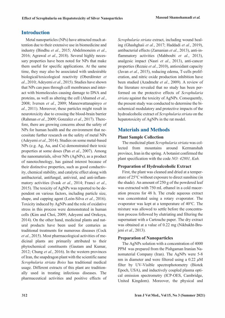
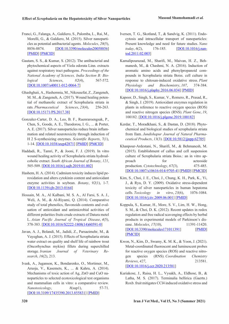
chemical properties were confirmed by scanning electron microscopy (SEM) and transmission elec-tron microscopy (TEM) (Figure 1). Standardization was performed by Pishgaman Iran Nanomaterials Company. Briefly, the preparation and characteriza-tion of AgNPs were carried out by adding 100 mM silver nitrate to 1% (w/v) tannic acid solution (pH adjusted at 8 by adding 150 mM potassium car-bonate) of polyvinyl pyrrolidone (PVP). The AgNP solution became pale yellow. To obtain the toxic concentration of 200 ppm, the original solution was diluted to 20 units, following the manufacturer's in-structions. Finally, animals were gavaged daily for 30 days. The hepatotoxicity of NPs at a dose of 200 ppm was determined based on similar studies (Shariatzadeh et al., 2016; Prakash et al., 2017).
Figure 1. a) SEM and b) TEM images of AgNPs
Experimental Animals Animals and Treatments
The study design was approved by the Research Deputy of Razi University, Kermanshah, Iran with the ethical code of NO: 396.2.038 on 30.1.2018. The handling of animals was and consistent with relevant guidelines as approved by the Institutional Ethics Committee. A total of 30 male Wistar rats weighing 200±20 g were acclimatized for two weeks. The an-imals were housed in standard plastic cages at a temperature of 25±3°C under standard environmen-tal and nutritional conditions of the light and dark cycles of 12 h with free access to standard food and clean water.
Study Groups Group 1 (NC: negative control): received 2 mL of distilled water
Group 2 (PC: positive control): received 200 ppm AgNPs
Group 3 (A): received 20 mg/kg of Scrophularia striata extract and 200 ppm AgNPs
Group 4 (B): received 60 mg/kg of Scrophularia striata extract and 200 ppm AgNPs
Group 5 (C): received 180 mg/kg of Scrophularia striata extract and 200 ppm AgNPs
The animals were given oral daily treatments for 30 days. The election of doses used is premised on previous reports (Naqvi et al., 2010; Agarwal et al., 2013).
Animals Euthanasia At the end of treatment (30 days), animals were
euthanized by the intraperitoneal injection of 1 mg/kg xylazine HCl (xylazine 2%, Alfasan, Nether-lands) and 0.5 mg/kg ketamine HCl (ketamine 5%, Trittau, Germany). Serum Biochemical Analysis
Blood samples were collected into standard labor-atory Eppendorf tubes. The collected blood speci-mens were centrifuged at 12000 g for 10 min (Het-tich, United Kingdom), and the sera were kept at 4°C until measurements. The parameters were assessed using an automated biochemical analyzer (SK3002-4040, Sinothinker, China).
Hepatic Enzymes and Serum Biochemical Metabolites Hepatic enzymes, including alanine aminotrans-
ferase (ALT), aspartate aminotransferase (AST), and alkaline phosphatase (ALP) were measured by com-mercial kits (Randox, United Kingdom). In addition, lactate dehydrogenase (LDH), gamma-glutamyl transferase (GGT), albumin (ALB), globulin, total protein (TP), blood urea nitrogen (BUN), creatinine (Cr), total bilirubin (TBIL), and direct bilirubin (DBIL) were assessed using commercial kits (Pars Azmoon Co., Iran) and UV/V spectrophotometer (Shimadzu, Kyoto, Japan) in rats serum samples (Azadmehr et al., 2009).
Oxidative Stress Assays Malondialdehyde (MDA) based on reaction by
optical absorption spectrophotometry (Shimadzu, Kyoto, Japan), total antioxidant capacity (TAC) ac-cording to the ABTS [2,2'-azino-bis(3-ethylbenzo-thiazoline-6-sulfonic acid)] radical reduction, and cation antioxidant molecules by using Randox kits (UK, Randox), catalase (CAT) by using Enzyme Ac-tivity kit (Nactaz™-Catalase), superoxide dismutase
Effect of Scrophularia on the Hepatotoxicity of Silver Nanoparticles Masoud Shamohamadi et al.
314 Iran J Vet Med., Vol 15, No 3 (Summer 2021)
(SOD) and glutathione peroxidase (GPx) based on Formazone optical absorption in comparison with a standard curve were determined (Shariatzadeh et al., 2016).
The present study was approved (Use of labora-tory animals) by the research council of Razi University with the code NO: 396.2.038, adopted on 30.1.2018.
Data Analysis The data were statistically analyzed by one-way
analysis of variance and Tukey post-hoc test using the SPSS software version 18 (IBM, Chicago, Ill., USA). All results are shown as mean±SEM and P-value<0.05 is considered significant.
Results The results of the current study showed that all
studied factors (hepatic enzymes and serum metabo-lites) increased in the positive control group, compared to the negative control group. Further-more, the administration of Scrophularia striata hydroalcoholic extract in treatment groups (20, 60, and 180 mg/kg) along with 200 ppm AgNPs (dose of
hepatotoxicity) caused alterations in serum metabo-lites in groups 4 (60 mg/kg) and 5 (180 mg/kg), compared to the positive control group (P<0.05). Our findings revealed that Scrophularia striata hy-droalcoholic extract could modulate the cytotoxic effects of AgNPs in rats.
Accordingly, the dosage of the extract at concen-trations 20, 60, and 180 mg/kg (groups 3, 4, and 5) caused the levels of ALP, ALT, and AST to get close to the negative control. The order of the intensity of the extract effect on ALT (76.41±4.53 IU/L), AST (154.17±55.75 IU/L), and ALP (270.71±8.73 IU/L) was concentrations 60, 180, and 60 mg/kg (Table 1). The results showed that the levels of LDH and GGT changed, in comparison with the positive and nega-tive controls. The highest dosage of Scrophularia striata extract at concentration 180 mg/kg (group 5) led to altered LDH (838.07±101.27 IU/L) and GGT (6.36±0.87 IU/L) levels, in comparison with the pos-itive and negative controls (P<0.05) (Table 1).
Moreover, the daily gavage of AgNP caused re-ductions in the levels of serum ALB, Glo, TP, and an elevation in the level of serum Cr and BUN, com-pared to the negative control. The high (180 mg/kg)
Table 1. The biochemical modulatory effects of Scrophularia Striata extract on Hepatic Enzymes & serum metabolites caused by the hepatotoxicity of AgNPs 200 PPM
Parameters NC * PC # A B C
ALT(IU/L) 67.7±1.78 107.75±3.81 80.41±2.75b 76.41±4.53a 81.44±14.72b AST (IU/L) 87.9±2.65 235.4±4.59 184.31±2.92c 160.71±11.85b 154.17±55.75a ALP (IU/L) 204.7±3.59 345.2±3.62 302.21±4.6c 270.71±8.73b 273.79±49.22b LDH (IU/L) 651.16±7.07 947.2±4.2 871.41±2.4c 859.07±20.77b 838.07±101.27a GGT (IU/L) 5.1±0.37 7.4±0.66 6.8±0.28b 6.4±0.43b 6.36±0.87b Alb (mg/dL) 3.7±0.29 2.5±0.28 3.1±0.26a 3.21±0.47a 3.48±0.6a Glo (mg/dL) 3.7±0.52 2.9±0.62 3.1±0.32a 2.7±0.46b 3.3±0.16a
TP (mg/dL) 7.4±0.51 5.4±0.47 5.9±0.34b 6.5±0.57a 6.38±0.8a BUN (mg/dL) 13±1.05 41.31±2.26 32.3±1.4c 35.2±2.52c 30.48±9.75b Cr (mg/dL) 0.40±0.03 0.80±0.07 0.76±0.05b 0.72±0.07b 0.68±0.14a
BIL.T (mg/dL) 0.55±0.04 0.76±0.03 0.75±0.08c 0.72±0.02c 0.68±0.02b BIL.D (mg/dL) 0.11±0.01 0.19±0.02 0.19±0.01b 0.17±0.03a 0.16±0.01a
Notice: The values of the small letters a, b & c indicate a significant difference between the studied groups in comparison with the PC # and got close to the NC* groups respectively (P<0.05). NC: Negative control, PC: Positive control, A: 20mg/kg, B: 60mg/kg, C: 180mg/kg
Masoud Shamohamadi et al.
Iranian Journal of Veterinary Medicine
Iran J Vet Med., Vol 15, No 3 (Summer 2021) 315
and moderate (60 mg/kg) doses of Scrophularia stri-ata extract administered to groups 5 and 4 resulted in higher levels of Alb (3.48±0.6 and 3.21±0.47 mg/dL), Glo (3.3±0.16 and 2.7±0.46mg/dL), and TP (6.38±0.8 and 6.5±0.57 mg/dL), compared to the positive control and got close to the negative control (P<0.05). The highest dose of Scrophularia striata extract (180 mg/kg) diminished BUN (30.48±9.75 mg/dL) and Cr (0.68±0.14 mg/dL) (P<0.05), (Table 1).
Furthermore, the levels of serum TBIL and DBIL increased with a 30-day daily gavage of 200 ppm AgNPs for the positive control, in comparison with the negative control (Table 1). The daily administra-tion of the extract (20, 60, 180 mg/kg) in three treatment groups along with 200 ppm AgNPs caused changes in serum TBIL and DBIL. Based on our re-
sults, significant differences were observed between the groups of concentrations 20, 60, and 180 mg/kg, especially at the concentrations of 180 mg/kg with
0.68±0.02 and 0.16±0.01, in comparison with the positive control with 0.76±0.03 and 0.19±0.02 and the negative control with 0.55±0.04 and 0.11±0.01 (P<0.05) (Table 1).
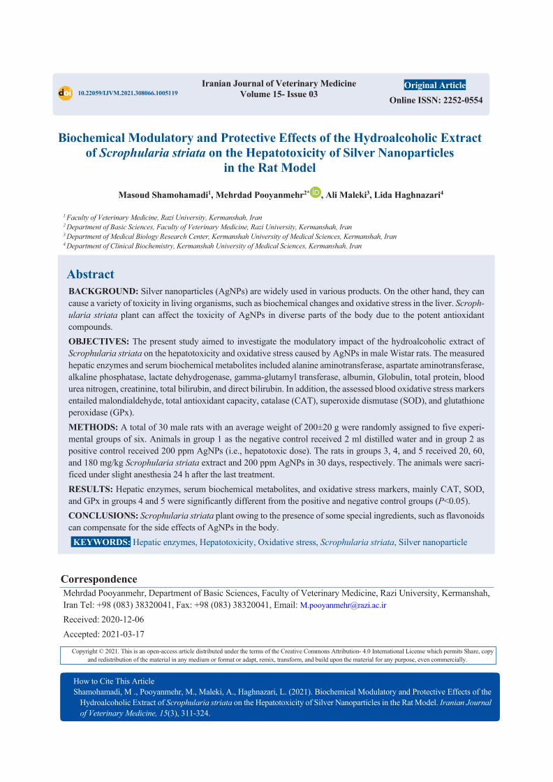
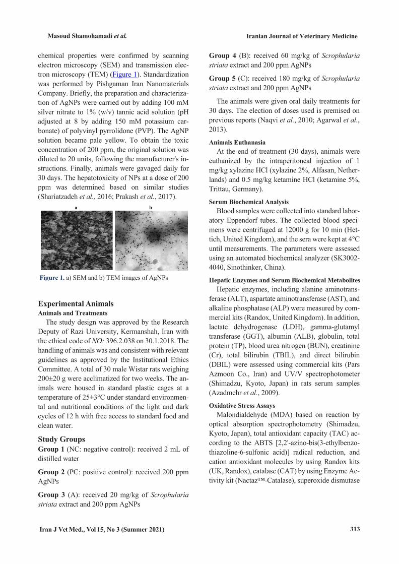
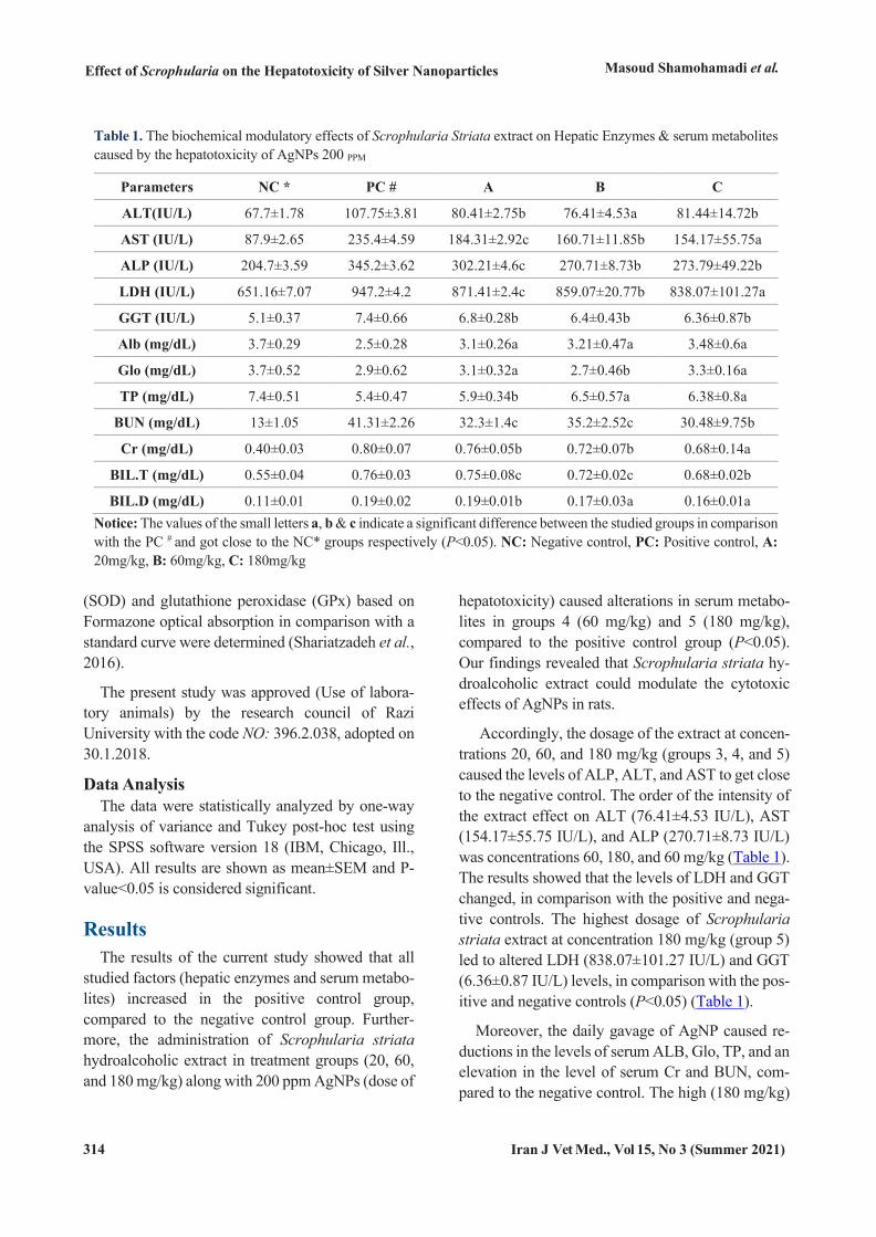
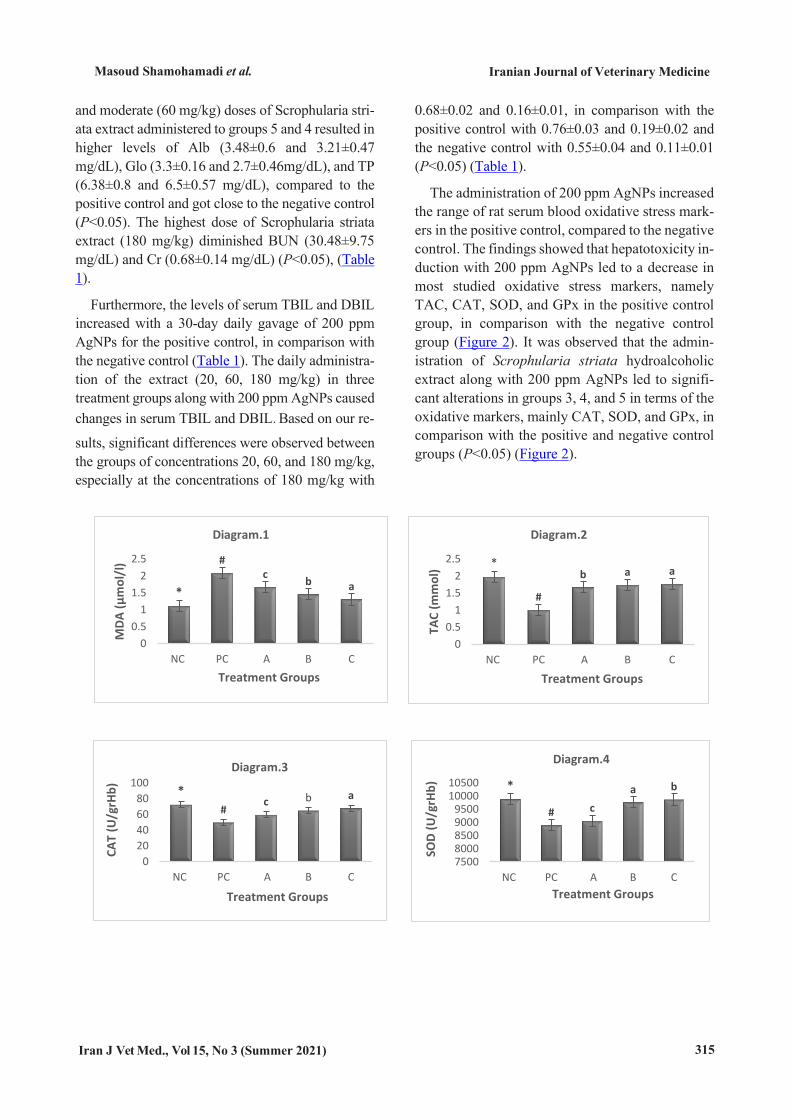
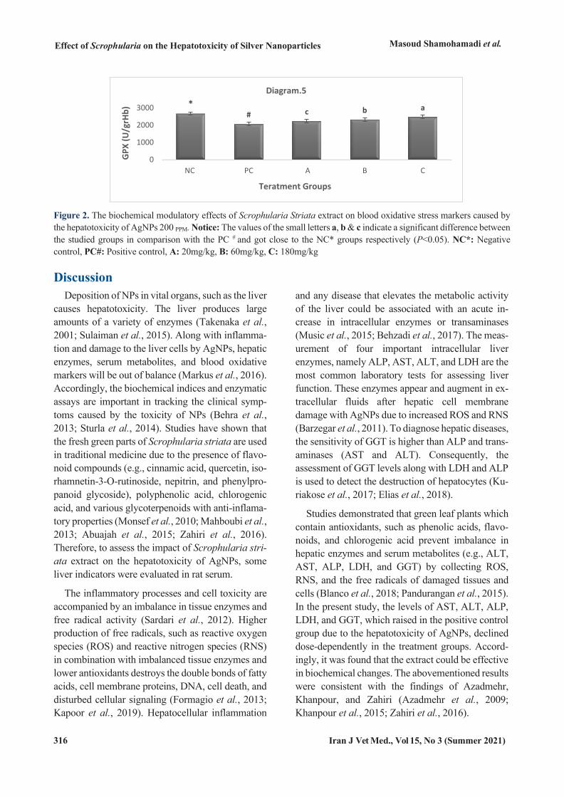
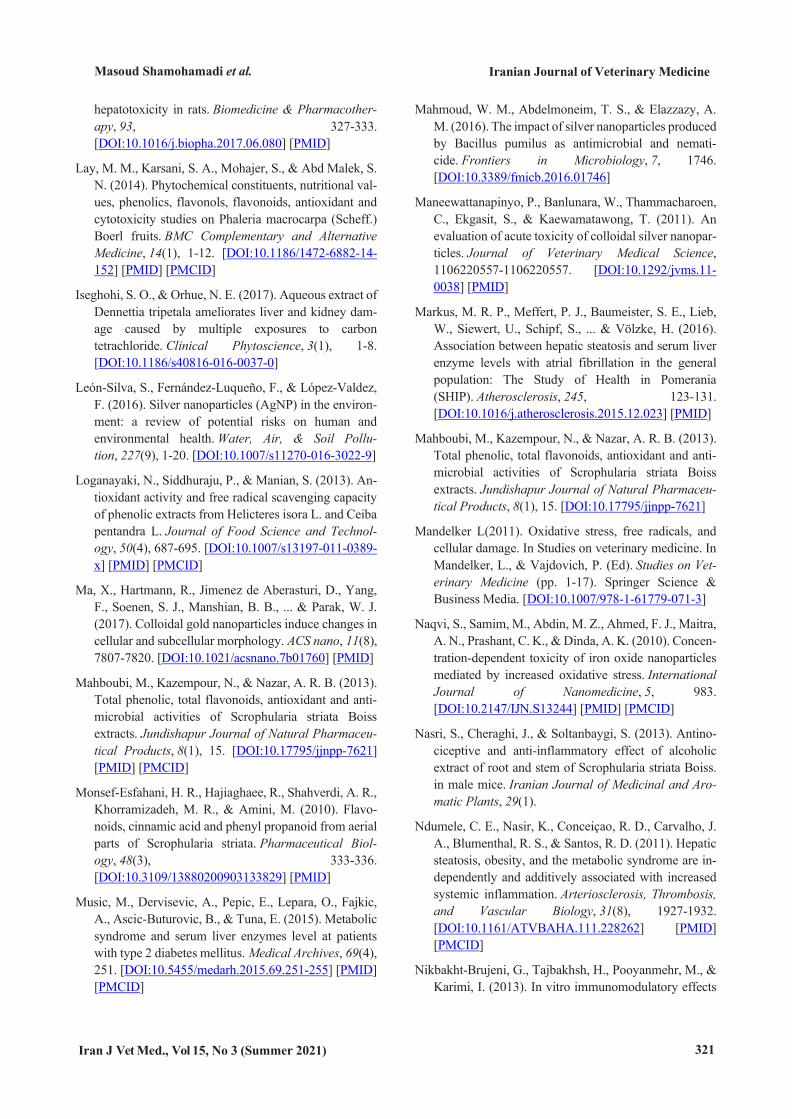
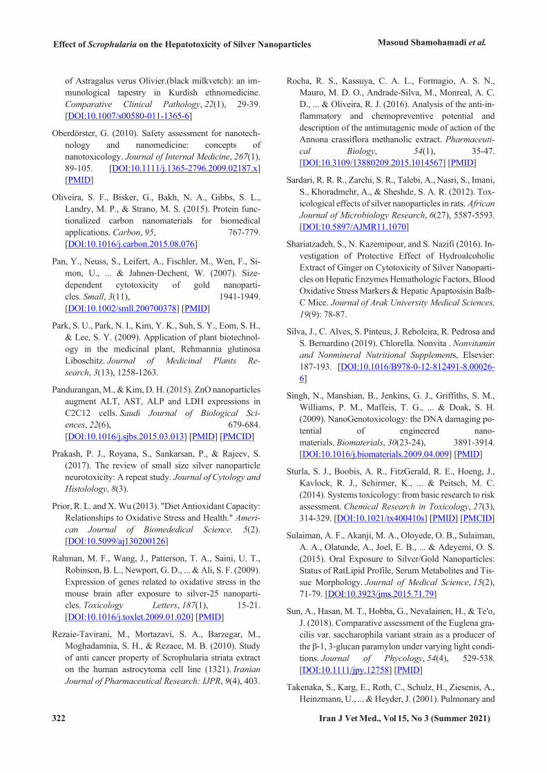
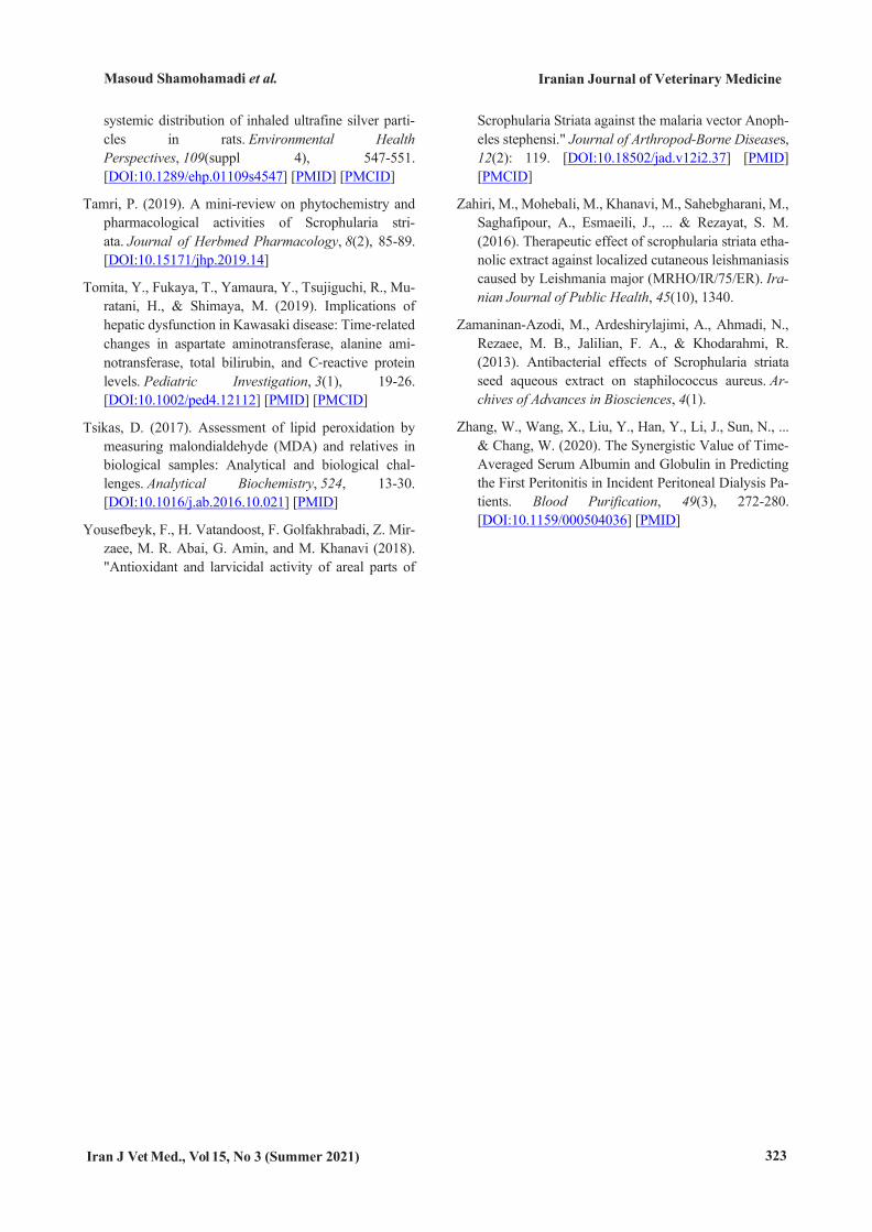
The administration of 200 ppm AgNPs increased the range of rat serum blood oxidative stress mark-ers in the positive control, compared to the negative control. The findings showed that hepatotoxicity in-duction with 200 ppm AgNPs led to a decrease in most studied oxidative stress markers, namely TAC, CAT, SOD, and GPx in the positive control group, in comparison with the negative control group (Figure 2). It was observed that the admin-istration of Scrophularia striata hydroalcoholic extract along with 200 ppm AgNPs led to signifi-cant alterations in groups 3, 4, and 5 in terms of the oxidative markers, mainly CAT, SOD, and GPx, in comparison with the positive and negative control groups (P<0.05) (Figure 2).
*
#c b a
00.5
11.5
22.5
NC PC A B C
MDA
(µm
ol/l
)
Treatment Groups
Diagram.1
*
#
b a a
00.5
11.5
22.5
NC PC A B C
TAC
(mm
ol)
Treatment Groups
Diagram.2
*#
c b a
020406080
100
NC PC A B C
CAT
(U/g
rHb)
Treatment Groups
Diagram.3*
# ca b
75008000850090009500
1000010500
NC PC A B C
SOD
(U/g
rHb)
Treatment Groups
Diagram.4
Effect of Scrophularia on the Hepatotoxicity of Silver Nanoparticles Masoud Shamohamadi et al.
316 Iran J Vet Med., Vol 15, No 3 (Summer 2021)
Figure 2. The biochemical modulatory effects of Scrophularia Striata extract on blood oxidative stress markers caused by the hepatotoxicity of AgNPs 200 PPM. Notice: The values of the small letters a, b & c indicate a significant difference between the studied groups in comparison with the PC # and got close to the NC* groups respectively (P<0.05). NC*: Negative control, PC#: Positive control, A: 20mg/kg, B: 60mg/kg, C: 180mg/kg
Discussion Deposition of NPs in vital organs, such as the liver
causes hepatotoxicity. The liver produces large amounts of a variety of enzymes (Takenaka et al., 2001; Sulaiman et al., 2015). Along with inflamma-tion and damage to the liver cells by AgNPs, hepatic enzymes, serum metabolites, and blood oxidative markers will be out of balance (Markus et al., 2016). Accordingly, the biochemical indices and enzymatic assays are important in tracking the clinical symp-toms caused by the toxicity of NPs (Behra et al., 2013; Sturla et al., 2014). Studies have shown that the fresh green parts of Scrophularia striata are used in traditional medicine due to the presence of flavo-noid compounds (e.g., cinnamic acid, quercetin, iso-rhamnetin-3-O-rutinoside, nepitrin, and phenylpro-panoid glycoside), polyphenolic acid, chlorogenic acid, and various glycoterpenoids with anti-inflama-tory properties (Monsef et al., 2010; Mahboubi et al., 2013; Abuajah et al., 2015; Zahiri et al., 2016). Therefore, to assess the impact of Scrophularia stri-ata extract on the hepatotoxicity of AgNPs, some liver indicators were evaluated in rat serum.
The inflammatory processes and cell toxicity are accompanied by an imbalance in tissue enzymes and free radical activity (Sardari et al., 2012). Higher production of free radicals, such as reactive oxygen species (ROS) and reactive nitrogen species (RNS) in combination with imbalanced tissue enzymes and lower antioxidants destroys the double bonds of fatty acids, cell membrane proteins, DNA, cell death, and disturbed cellular signaling (Formagio et al., 2013; Kapoor et al., 2019). Hepatocellular inflammation
and any disease that elevates the metabolic activity of the liver could be associated with an acute in-crease in intracellular enzymes or transaminases (Music et al., 2015; Behzadi et al., 2017). The meas-urement of four important intracellular liver enzymes, namely ALP, AST, ALT, and LDH are the most common laboratory tests for assessing liver function. These enzymes appear and augment in ex-tracellular fluids after hepatic cell membrane damage with AgNPs due to increased ROS and RNS (Barzegar et al., 2011). To diagnose hepatic diseases, the sensitivity of GGT is higher than ALP and trans-aminases (AST and ALT). Consequently, the assessment of GGT levels along with LDH and ALP is used to detect the destruction of hepatocytes (Ku-riakose et al., 2017; Elias et al., 2018).
Studies demonstrated that green leaf plants which contain antioxidants, such as phenolic acids, flavo-noids, and chlorogenic acid prevent imbalance in hepatic enzymes and serum metabolites (e.g., ALT, AST, ALP, LDH, and GGT) by collecting ROS, RNS, and the free radicals of damaged tissues and cells (Blanco et al., 2018; Pandurangan et al., 2015). In the present study, the levels of AST, ALT, ALP, LDH, and GGT, which raised in the positive control group due to the hepatotoxicity of AgNPs, declined dose-dependently in the treatment groups. Accord-ingly, it was found that the extract could be effective in biochemical changes. The abovementioned results were consistent with the findings of Azadmehr, Khanpour, and Zahiri (Azadmehr et al., 2009; Khanpour et al., 2015; Zahiri et al., 2016).
*# c b a
0
1000
2000
3000
NC PC A B C
GPX
(U/g
rHb)
Teratment Groups
Diagram.5
Masoud Shamohamadi et al.
Iranian Journal of Veterinary Medicine
Iran J Vet Med., Vol 15, No 3 (Summer 2021) 317
Moreover, ALB, Glo, and TP represent the rate of protein synthesized by the liver. The normal concen-trations of these biomarkers are disrupted in liver injury (Adeyemi et al., 2012; Zhang et al., 2020). The hepatotoxic dose diminished serum ALB, GLO, and Cr levels. The concentrations of these bi-omarkers (ALB, GLO, and TP) altered in the three treatments groups, compared to the positive control and negative control groups. The latter results were in line with the studies performed by Bahrami, Lay, and Lawal (Bahrami, 2011; Lay et al., 2014; Lawal et al., 2017).
Ammonia contains nitrogen and is produced by the liver. The urea is transported by the blood from the liver to the kidneys. The measurement of BUN and Cr was a part of the serum biochemical profile for diagnosing AgNPs-induced liver damage and evaluating the effectiveness of the extract. The BIL results from the disintegration of hemoglobin by liver cells. The serum levels of this biomarker in-crease with further destruction of the liver cells and RBCs (Singh et al., 2009; Adeyemi et al., 2012; To-mita et al., 2019). Therefore, serum DBIL and TBIL levels indicate liver activity in the disintegration and excretion of substances. The modulatory effects of Scrophularia striata extract were revealed in the lev-els of these parameters in the treatment groups, compared to the positive control and negative con-trol groups. As a result, BUN, Cr, DBIL, and TBIL concentrations decreased in the treatment groups in a dose-dependent manner. The mentioned results are consistent with other investigations on the impact of Scrophularia striata (Kamalipourazad et al., 2016; Falahi et al., 2018; Tamri, 2019).
Free radicals are very active chemically and play an important role in many diseases. The antioxidant defense naturally neutralizes the influences of these radicals (Prior and Wu, 2013; Rocha et al., 2016). As described above, raised production of free radicals, such as ROS and RNS disrupts the natural mecha-nism of cellular signaling pathways by misbalancing and weakening the antioxidant defense. The in-creased production of ROS and RNS along with severe oxidative cellular damage cause an elevation in the oxidative stress biomarkers, such as MDA (Tsikas, 2017; Kwon et al., 2021). Scrophularia stri-ata extract was found to be effective in the three
treatments groups, in comparison with the positive and negative control groups (Figure 2). Consistent with this result, Mandelkeret indicated the antioxi-dant activity of this agent that led to MDA reduction (Mandelkeret et al., 2011).
Antioxidant enzymes, including CAT, SOD, and GPx are powerful free radical scavengers. Collabo-ration between distinct antioxidants provides higher protection against the attack of ROS and RNS (Ndumele et al., 2011). The TAC reflects the total antioxidant activity of the body. Therefore, TAC measurement can provide more information than as-sessing each antioxidant component individually (Alonso et al., 2014). Scrophularia striata extract was observed to affect the level of this parameter in the treatment groups A, B, and C (Figure 2). The lat-ter finding was in line with the results of Shariatzadeh et al. (Shariatzadeh et al., 2016).
The accumulation of hydrogen peroxide (H2O2) in the cells causes the oxidation of DNA, proteins, and fats resulting in mutation and cell perdition (Ma et al., 2017). The CAT is an antioxidant enzyme in-volved in the detoxification of H2O2 by catalyzing the conversion of two H2O2 molecules to oxygen and two molecules of water. The highest levels of CAT are found in the liver, kidneys, and erythrocytes. The measurement of CAT enzyme activity is a standard and reliable way to assess biological samples, such as serum (Hashem, 2014). We observed that the ex-tract influenced the levels of this parameter in the treatment of groups A, B, and C (Figure 2). The re-sults of Hossain et al. concerning the antioxidant activity of medicinal plants and CAT changes are congruent with the results of the current study (Hoss-ain et al., 2014).
Superoxide is a byproduct of the secondary me-tabolism of oxygen and the lack of control can cause different damages to cells. The SOD is the first and most important antioxidant enzyme in all aerobic or-ganisms that are directly involved in the reduction of reactive oxygen metabolites. This enzyme catalyzes the superoxide and H2O2 produced in the cell to an oxygen molecule (O2). Consequently, the SOD is a remarkable antioxidant present in almost all the cells exposed to oxygen (Markus et al., 2016).
Moreover, GPx is an enzyme with peroxidase ac-tivity and plays an important biological role in
Effect of Scrophularia on the Hepatotoxicity of Silver Nanoparticles Masoud Shamohamadi et al.
318 Iran J Vet Med., Vol 15, No 3 (Summer 2021)
protecting organisms against oxidative damage. This enzyme prevents the formation of free radicals by re-ducing H2O2 and a wide range of organic peroxides (Adedara et al., 2018). Therefore, evaluating the ac-tivity of the GPx enzyme can be a diagnostic tool for inflammatory and cellular liver damage caused by the toxic effects of AgNPS. There are reports of ox-idative stress markers imbalances due to the hepatotoxic impacts of AgNPs similar to the present investigation (Ali et al., 2013; Nasser et al., 2009; Hassan et al., 2019; Majid et al., 2009). In this study, the protective effects of Scrophularia striata extract against 200 ppm AgNPs were observed in terms of the antioxidant parameters in the treatment groups A, B, and C, which is consistent with the results of Lo-ganayaki and Ahmed regarding antioxidant activity and free radical scavenging (Loganayaki et al., 2013; Ahmed et al., 2019). Therefore, data analysis re-vealed that altering liver enzymes, serum bioche-mical metabolites, and oxidative stress factors could probably be attributed to the antioxidants, such as phenolic acids, flavonols, and flavonoids in Scroph-ularia striata (Sun et al., 2018; Silva et al., 2019).
Conclusion The AgNPs are widely used in various products
and can cause toxicity for a variety of organs in liv-ing organisms, especially hepatoxicity along with
alteration in hepatic enzymes, serum biochemical metabolites, and blood oxidative markers. On the other hand, the medicinal plant Scrophularia striata with a range of antioxidant properties can be useful as a substitute for chemical compounds. The results of this study demonstrated that the protective effects of the hydroalcoholic extract of Scrophularia striata against the hepatotoxicity of AgNPs are most likely due to the presence of flavonoids. However, further investigation is required to elucidate the cellular and molecular signaling pathways involved in the im-pacts of Scrophularia striata extract.
Acknowledgments A part of the data in this research is related to the
graduate student thesis of veterinary medicine with No: 7087460, Date: 19.11. 2018, in the Faculty of Veterinary Medicine, Razi University, Iran. The re-searchers are extremely grateful to the laboratory experts involved in the experiments.
Conflict of Interest The authors declare that they have no conflict of
interest. The authors alone are responsible for the content of the paper.
References Abuajah, C. I., Ogbonna, A. C., & Osuji, C. M. (2015).
Functional components and medicinal properties of food: a review. Journal of Food Science and Technol-ogy, 52(5), 2522-2529. [DOI:10.1007/s13197-014-1396-5] [PMID] [PMCID]
Adedara, I. A., Anao, O. O., Forcados, G. E., Awogbindin, I. O., Agbowo, A., Ola-Davies, O. E., ... & Farombi, E. O. (2018). Low doses of multi-walled carbon nano-tubes elicit hepatotoxicity in rats with markers of oxidative stress and induction of pro-inflammatory cy-tokines. Biochemical and Biophysical Research Communications, 503(4), 3167-3173. [DOI:10.1016/j.bbrc.2018.08.112] [PMID]
Adeyemi, O. S., Akanji, M. A., & Ekanem, J. T. (2012). Ethanolic extract of Psidium guajava influences pro-tein and bilirubin levels in Trypanosoma brucei brucei
infected rats. Journal of Biolojical Sciences, 12(2), 111-116. [DOI:10.3923/jbs.2012.111.116]
Adeyemi, O. S., Adewumi, I., & Faniyan, T. O. (2015). Silver nanoparticles influenced rat serum metabolites and tissue morphology. Journal of Basic and Clinical Physiology and Pharmacology, 26(4), 355-361. [DOI:10.1515/jbcpp-2013-0092] [PMID]
Adeyemi, O. S., & Faniyan, T. O. (2014). Antioxidant sta-tus of rats administered silver nanoparticles orally. Journal of Taibah University Medical Sciences, 9(3), 182-186. [DOI:10.1016/j.jtumed.2014.03.002]
Adeyemi, O. S., & Orekoya, B. T. (2014). Lipid profile and oxidative stress markers in Wistar rats following oral and repeated exposure to fijk herbal mixture. Jour-nal of Toxicology, 2014. [DOI:10.1155/2014/876035] [PMID] [PMCID]
Masoud Shamohamadi et al.
Iranian Journal of Veterinary Medicine
Iran J Vet Med., Vol 15, No 3 (Summer 2021) 319
Agarwal, M., M. Murugan, A. Sharma, R. Rai, A. Kamboj, H. Sharma and S. K. Roy (2013). Nanoparticles and its toxic effects: A review. International Journal of Cur-rent Microbiology and Applied Sciences, 2(10): 76-82.
Agrawal, S., Bhatt, M., Rai, S. K., Bhatt, A., Dangwal, P., & Agrawal, P. K. (2018). Silver nanoparticles and its potential applications: A review. Journal of Pharma-cognosy and Phytochemistry, 7, 930-937.
Ahamed, M., Karns, M., Goodson, M., Rowe, J., Hussain, S. M., Schlager, J. J., & Hong, Y. (2008). DNA dam-age response to different surface chemistry of silver nanoparticles in mammalian cells. Toxicology and Ap-plied Pharmacology, 233(3), 404-410. [DOI:10.1016/j.taap.2008.09.015] [PMID]
Ahmed, O. M., Abdul-Hamid, M. M., El-Bakry, A. M., Mohamed, H. M., & Abdel Rahman, E. S. (2019). Ca-mellia sinensis and epicatechin abate doxorubicin-induced hepatotoxicity in male Wistar rats via their modulatory effects on oxidative stress, inflammation, and apoptosis. Journal of Applied Pharmaceutical Sci-ence, 9(04), 030-044. [DOI:10.7324/JAPS.2019.90405]
Alonso, A., Misialek, J. R., Amiin, M. A., Hoogeveen, R. C., Chen, L. Y., Agarwal, S. K., ... & Selvin, E. (2014). Circulating levels of liver enzymes and incidence of atrial fibrillation: the Atherosclerosis Risk in Commu-nities cohort. Heart, 100(19), 1511-1516. [DOI:10.1136/heartjnl-2014-305756] [PMID] [PMCID]
Azadmehr, A., Afshari, A., Baradaran, B., Hajiaghaee, R., Rezazadeh, S., & Monsef-Esfahani, H. (2009). Sup-pression of nitric oxide production in activated murine peritoneal macrophages in vitro and ex vivo by Scroph-ularia striata ethanolic extract. Journal of Ethnopharmacology, 124(1), 166-169. [DOI:10.1016/j.jep.2009.03.042] [PMID]
Bahrami, A. M. (2011). The effectiveness of Scrophularia Striata on Newcastle disease. Australian Journal of Basic and Applied Sciences, 5(12), 2883-2888.
Barzegar, A., & Moosavi-Movahedi, A. A. (2011). Intra-cellular ROS protection efficiency and free radical-scavenging activity of curcumin. PloS one, 6(10), e26012. [DOI:10.1371/journal.pone.0026012] [PMID] [PMCID]
Behra, R., Sigg, L., Clift, M. J., Herzog, F., Minghetti, M., Johnston, B., ... & Rothen-Rutishauser, B. (2013). Bi-oavailability of silver nanoparticles and ions: from a chemical and biochemical perspective. Journal of the
Royal Society Interface, 10(87), 20130396. [DOI:10.1098/rsif.2013.0396] [PMID] [PMCID]
Behzadi, S., Serpooshan, V., Tao, W., Hamaly, M. A., Al-kawareek, M. Y., Dreaden, E. C., ... & Mahmoudi, M. (2017). Cellular uptake of nanoparticles: journey in-side the cell. Chemical Society Reviews, 46(14), 4218-4244. [DOI:10.1039/C6CS00636A] [PMID] [PMCID]
Bindhu, M. R., & Umadevi, M. (2015). Antibacterial and catalytic activities of green synthesized silver nanopar-ticles. Spectrochimica Acta Part A: Molecular and Biomolecular Spectroscopy, 135, 373-378. [DOI:10.1016/j.saa.2014.07.045] [PMID]
Blanco, J., Tomás-Hernández, S., García, T., Mulero, M., Gómez, M., Domingo, J. L., & Sánchez, D. J. (2018). Oral exposure to silver nanoparticles increases oxida-tive stress markers in the liver of male rats and deregulates the insulin signalling pathway and p53 and cleaved caspase 3 protein expression. Food and Chem-ical Toxicology, 115, 398-404. [DOI:10.1016/j.fct.2018.03.039] [PMID]
Chung, I. M., Park, I., Seung-Hyun, K., Thiruvengadam, M., & Rajakumar, G. (2016). Plant-mediated synthesis of silver nanoparticles: their characteristic properties and therapeutic applications. Nanoscale Research Let-ters, 11(1), 1-14. [DOI:10.1186/s11671-016-1257-4] [PMID] [PMCID]
Cock, I. E. (2015). The medicinal properties and phyto-chemistry of plants of the genus Terminalia (Combretaceae). Inflammopharmacology, 23(5), 203-229. [DOI:10.1007/s10787-015-0246-z] [PMID]
Elias, S. E. S., Eltom, A., Osman, A. L., & Babker, A. M. (2018). Gamma glutamyl transferase and lactate dehy-drogenase as biochemical markers of severity of preeclampsia among Sudanese pregnant women. Int J Reprod Contracept Obstet Gynecol, 7(8), 3020-3. [DOI:10.18203/2320-1770.ijrcog20183294]
Falahi, H., Sharifi, M., Maivan, H. Z., & Chashmi, N. A. (2018). Phenylethanoid glycosides accumulation in roots of Scrophularia striata as a response to water stress. Environmental and Experimental Botany, 147, 13-21. [DOI:10.1016/j.envexpbot.2017.11.003]
Formagio, A. S., Kassuya, C. A., Neto, F. F., Volobuff, C. R., Iriguchi, E. K., do C Vieira, M., & Foglio, M. A. (2013). The flavonoid content and antiproliferative, hypoglycaemic, anti-inflammatory and free radical scavenging activities of Annona dioica St. Hill. BMC Complementary and Alternative Medicine, 13(1), 1-8. [DOI:10.1186/1472-6882-13-14] [PMID] [PMCID]
Effect of Scrophularia on the Hepatotoxicity of Silver Nanoparticles Masoud Shamohamadi et al.
320 Iran J Vet Med., Vol 15, No 3 (Summer 2021)
Franci, G., Falanga, A., Galdiero, S., Palomba, L., Rai, M., Morelli, G., & Galdiero, M. (2015). Silver nanoparti-cles as potential antibacterial agents. Molecules, 20(5), 8856-8874. [DOI:10.3390/molecules20058856] [PMID] [PMCID]
Gautam, S. S., & Kumar, S. (2012). The antibacterial and phytochemical aspects of Viola odorata Linn. extracts against respiratory tract pathogens. Proceedings of the National Academy of Sciences, India Section B: Bio-logical Sciences, 82(4), 567-572. [DOI:10.1007/s40011-012-0064-7]
Ghashghaii, A., Hashemnia, M., Nikousefat, Z., Zangeneh, M. M., & Zangeneh, A. (2017). Wound healing poten-tial of methanolic extract of Scrophularia striata in rats. Pharmaceutical Sciences, 23(4), 256-263. [DOI:10.15171/PS.2017.38]
Gonzalez-Carter, D. A., Leo, B. F., Ruenraroengsak, P., Chen, S., Goode, A. E., Theodorou, I. G., ... & Porter, A. E. (2017). Silver nanoparticles reduce brain inflam-mation and related neurotoxicity through induction of H 2 S-synthesizing enzymes. Scientific Reports, 7(1), 1-14. [DOI:10.1038/srep42871] [PMID] [PMCID]
Haddadi, R., Tamri, P., & Jooni, F. J. (2019). In vitro wound healing activity of Scrophularia striata hydroal-coholic extract. South African Journal of Botany, 121, 505-509. [DOI:10.1016/j.sajb.2019.01.002]
Hashem, H. A. (2014). Cadmium toxicity induces lipid pe-roxidation and alters cytokinin content and antioxidant enzyme activities in soybean. Botany, 92(1), 1-7. [DOI:10.1139/cjb-2013-0164]
Hossain, M. A., Al Kalbani, M. S. A., Al Farsi, S. A. J., Weli, A. M., & Al-Riyami, Q. (2014). Comparative study of total phenolics, flavonoids contents and eval-uation of antioxidant and antimicrobial activities of different polarities fruits crude extracts of Datura metel L. Asian Pacific Journal of Tropical Disease, 4(5), 378-383. [DOI:10.1016/S2222-1808(14)60591-0]
Javan, A. J., Bolandi, M., Jadidi, Z., Parsaeimehr, M., & Vayeghan, A. J. (2015). Effects of Scrophularia striata water extract on quality and shelf life of rainbow trout (Oncorhynchus mykiss) fillets during superchilled storage. Iranian Journal of Veterinary Re-search, 16(2), 213.
Ivask, A., Juganson, K., Bondarenko, O., Mortimer, M., Aruoja, V., Kasemets, K., ... & Kahru, A. (2014). Mechanisms of toxic action of Ag, ZnO and CuO na-noparticles to selected ecotoxicological test organisms and mammalian cells in vitro: a comparative review. Nanotoxicology, 8(sup1), 57-71. [DOI:10.3109/17435390.2013.855831] [PMID]
Iversen, T. G., Skotland, T., & Sandvig, K. (2011). Endo-cytosis and intracellular transport of nanoparticles: Present knowledge and need for future studies. Nano today, 6(2), 176-185. [DOI:10.1016/j.nan-tod.2011.02.003]
Kamalipourazad, M., Sharifi, M., Maivan, H. Z., Beh-manesh, M., & Chashmi, N. A. (2016). Induction of aromatic amino acids and phenylpropanoid com-pounds in Scrophularia striata Boiss. cell culture in response to chitosan-induced oxidative stress. Plant Physiology and Biochemistry, 107, 374-384. [DOI:10.1016/j.plaphy.2016.06.034] [PMID]
Kapoor, D., Singh, S., Kumar, V., Romero, R., Prasad, R., & Singh, J. (2019). Antioxidant enzymes regulation in plants in reference to reactive oxygen species (ROS) and reactive nitrogen species (RNS). Plant Gene, 19, 100182. [DOI:10.1016/j.plgene.2019.100182]
Kerdar, T., Moradkhani, S., & Dastan, D. (2018). Phyto-chemical and biological studies of scrophularia striata from Ilam. Jundishapur Journal of Natural Pharma-ceutical Products, 13(3). [DOI:10.5812/jjnpp.62705]
Khanpour-Ardestani, N., Sharifi, M., & Behmanesh, M. (2015). Establishment of callus and cell suspension culture of Scrophularia striata Boiss.: an in vitro ap-proach for acteoside production. Cytotechnology, 67(3), 475-485. [DOI:10.1007/s10616-014-9705-4] [PMID] [PMCID]
Kim, S., Choi, J. E., Choi, J., Chung, K. H., Park, K., Yi, J., & Ryu, D. Y. (2009). Oxidative stress-dependent toxicity of silver nanoparticles in human hepatoma cells. Toxicology in vitro, 23(6), 1076-1084. [DOI:10.1016/j.tiv.2009.06.001] [PMID]
Koppula, S., Kumar, H., More, S. V., Lim, H. W., Hong, S. M., & Choi, D. K. (2012). Recent updates in redox regulation and free radical scavenging effects by herbal products in experimental models of Parkinson’s dis-ease. Molecules, 17(10), 11391-11420. [DOI:10.3390/molecules171011391] [PMID] [PMCID]
Kwon, N., Kim, D., Swamy, K. M. K., & Yoon, J. (2021). Metal-coordinated fluorescent and luminescent probes for reactive oxygen species (ROS) and reactive nitro-gen species (RNS). Coordination Chemistry Reviews, 427, 213581. [DOI:10.1016/j.ccr.2020.213581]
Kuriakose, J., Raisa, H. L., Vysakh, A., Eldhose, B., & Latha, M. S. (2017). Terminalia bellirica (Gaertn.) Roxb. fruit mitigates CCl4 induced oxidative stress and
Masoud Shamohamadi et al.
Iranian Journal of Veterinary Medicine
Iran J Vet Med., Vol 15, No 3 (Summer 2021) 321
hepatotoxicity in rats. Biomedicine & Pharmacother-apy, 93, 327-333. [DOI:10.1016/j.biopha.2017.06.080] [PMID]
Lay, M. M., Karsani, S. A., Mohajer, S., & Abd Malek, S. N. (2014). Phytochemical constituents, nutritional val-ues, phenolics, flavonols, flavonoids, antioxidant and cytotoxicity studies on Phaleria macrocarpa (Scheff.) Boerl fruits. BMC Complementary and Alternative Medicine, 14(1), 1-12. [DOI:10.1186/1472-6882-14-152] [PMID] [PMCID]
Iseghohi, S. O., & Orhue, N. E. (2017). Aqueous extract of Dennettia tripetala ameliorates liver and kidney dam-age caused by multiple exposures to carbon tetrachloride. Clinical Phytoscience, 3(1), 1-8. [DOI:10.1186/s40816-016-0037-0]
León-Silva, S., Fernández-Luqueño, F., & López-Valdez, F. (2016). Silver nanoparticles (AgNP) in the environ-ment: a review of potential risks on human and environmental health. Water, Air, & Soil Pollu-tion, 227(9), 1-20. [DOI:10.1007/s11270-016-3022-9]
Loganayaki, N., Siddhuraju, P., & Manian, S. (2013). An-tioxidant activity and free radical scavenging capacity of phenolic extracts from Helicteres isora L. and Ceiba pentandra L. Journal of Food Science and Technol-ogy, 50(4), 687-695. [DOI:10.1007/s13197-011-0389-x] [PMID] [PMCID]
Ma, X., Hartmann, R., Jimenez de Aberasturi, D., Yang, F., Soenen, S. J., Manshian, B. B., ... & Parak, W. J. (2017). Colloidal gold nanoparticles induce changes in cellular and subcellular morphology. ACS nano, 11(8), 7807-7820. [DOI:10.1021/acsnano.7b01760] [PMID]
Mahboubi, M., Kazempour, N., & Nazar, A. R. B. (2013). Total phenolic, total flavonoids, antioxidant and anti-microbial activities of Scrophularia striata Boiss extracts. Jundishapur Journal of Natural Pharmaceu-tical Products, 8(1), 15. [DOI:10.17795/jjnpp-7621] [PMID] [PMCID]
Monsef-Esfahani, H. R., Hajiaghaee, R., Shahverdi, A. R., Khorramizadeh, M. R., & Amini, M. (2010). Flavo-noids, cinnamic acid and phenyl propanoid from aerial parts of Scrophularia striata. Pharmaceutical Biol-ogy, 48(3), 333-336. [DOI:10.3109/13880200903133829] [PMID]
Music, M., Dervisevic, A., Pepic, E., Lepara, O., Fajkic, A., Ascic-Buturovic, B., & Tuna, E. (2015). Metabolic syndrome and serum liver enzymes level at patients with type 2 diabetes mellitus. Medical Archives, 69(4), 251. [DOI:10.5455/medarh.2015.69.251-255] [PMID] [PMCID]
Mahmoud, W. M., Abdelmoneim, T. S., & Elazzazy, A. M. (2016). The impact of silver nanoparticles produced by Bacillus pumilus as antimicrobial and nemati-cide. Frontiers in Microbiology, 7, 1746. [DOI:10.3389/fmicb.2016.01746]
Maneewattanapinyo, P., Banlunara, W., Thammacharoen, C., Ekgasit, S., & Kaewamatawong, T. (2011). An evaluation of acute toxicity of colloidal silver nanopar-ticles. Journal of Veterinary Medical Science, 1106220557-1106220557. [DOI:10.1292/jvms.11-0038] [PMID]
Markus, M. R. P., Meffert, P. J., Baumeister, S. E., Lieb, W., Siewert, U., Schipf, S., ... & Völzke, H. (2016). Association between hepatic steatosis and serum liver enzyme levels with atrial fibrillation in the general population: The Study of Health in Pomerania (SHIP). Atherosclerosis, 245, 123-131. [DOI:10.1016/j.atherosclerosis.2015.12.023] [PMID]
Mahboubi, M., Kazempour, N., & Nazar, A. R. B. (2013). Total phenolic, total flavonoids, antioxidant and anti-microbial activities of Scrophularia striata Boiss extracts. Jundishapur Journal of Natural Pharmaceu-tical Products, 8(1), 15. [DOI:10.17795/jjnpp-7621]
Mandelker L(2011). Oxidative stress, free radicals, and cellular damage. In Studies on veterinary medicine. In Mandelker, L., & Vajdovich, P. (Ed). Studies on Vet-erinary Medicine (pp. 1-17). Springer Science & Business Media. [DOI:10.1007/978-1-61779-071-3]
Naqvi, S., Samim, M., Abdin, M. Z., Ahmed, F. J., Maitra, A. N., Prashant, C. K., & Dinda, A. K. (2010). Concen-tration-dependent toxicity of iron oxide nanoparticles mediated by increased oxidative stress. International Journal of Nanomedicine, 5, 983. [DOI:10.2147/IJN.S13244] [PMID] [PMCID]
Nasri, S., Cheraghi, J., & Soltanbaygi, S. (2013). Antino-ciceptive and anti-inflammatory effect of alcoholic extract of root and stem of Scrophularia striata Boiss. in male mice. Iranian Journal of Medicinal and Aro-matic Plants, 29(1).
Ndumele, C. E., Nasir, K., Conceiçao, R. D., Carvalho, J. A., Blumenthal, R. S., & Santos, R. D. (2011). Hepatic steatosis, obesity, and the metabolic syndrome are in-dependently and additively associated with increased systemic inflammation. Arteriosclerosis, Thrombosis, and Vascular Biology, 31(8), 1927-1932. [DOI:10.1161/ATVBAHA.111.228262] [PMID] [PMCID]
Nikbakht-Brujeni, G., Tajbakhsh, H., Pooyanmehr, M., & Karimi, I. (2013). In vitro immunomodulatory effects
Effect of Scrophularia on the Hepatotoxicity of Silver Nanoparticles Masoud Shamohamadi et al.
322 Iran J Vet Med., Vol 15, No 3 (Summer 2021)
of Astragalus verus Olivier.(black milkvetch): an im-munological tapestry in Kurdish ethnomedicine. Comparative Clinical Pathology, 22(1), 29-39. [DOI:10.1007/s00580-011-1365-6]
Oberdörster, G. (2010). Safety assessment for nanotech-nology and nanomedicine: concepts of nanotoxicology. Journal of Internal Medicine, 267(1), 89-105. [DOI:10.1111/j.1365-2796.2009.02187.x] [PMID]
Oliveira, S. F., Bisker, G., Bakh, N. A., Gibbs, S. L., Landry, M. P., & Strano, M. S. (2015). Protein func-tionalized carbon nanomaterials for biomedical applications. Carbon, 95, 767-779. [DOI:10.1016/j.carbon.2015.08.076]
Pan, Y., Neuss, S., Leifert, A., Fischler, M., Wen, F., Si-mon, U., ... & Jahnen‐Dechent, W. (2007). Size‐dependent cytotoxicity of gold nanoparti-cles. Small, 3(11), 1941-1949. [DOI:10.1002/smll.200700378] [PMID]
Park, S. U., Park, N. I., Kim, Y. K., Suh, S. Y., Eom, S. H., & Lee, S. Y. (2009). Application of plant biotechnol-ogy in the medicinal plant, Rehmannia glutinosa Liboschitz. Journal of Medicinal Plants Re-search, 3(13), 1258-1263.
Pandurangan, M., & Kim, D. H. (2015). ZnO nanoparticles augment ALT, AST, ALP and LDH expressions in C2C12 cells. Saudi Journal of Biological Sci-ences, 22(6), 679-684. [DOI:10.1016/j.sjbs.2015.03.013] [PMID] [PMCID]
Prakash, P. J., Royana, S., Sankarsan, P., & Rajeev, S. (2017). The review of small size silver nanoparticle neurotoxicity: A repeat study. Journal of Cytology and Histolology, 8(3).
Prior, R. L. and X. Wu (2013). "Diet Antioxidant Capacity: Relationships to Oxidative Stress and Health." Ameri-can Journal of Biomededical Science, 5(2). [DOI:10.5099/aj130200126]
Rahman, M. F., Wang, J., Patterson, T. A., Saini, U. T., Robinson, B. L., Newport, G. D., ... & Ali, S. F. (2009). Expression of genes related to oxidative stress in the mouse brain after exposure to silver-25 nanoparti-cles. Toxicology Letters, 187(1), 15-21. [DOI:10.1016/j.toxlet.2009.01.020] [PMID]
Rezaie-Tavirani, M., Mortazavi, S. A., Barzegar, M., Moghadamnia, S. H., & Rezaee, M. B. (2010). Study of anti cancer property of Scrophularia striata extract on the human astrocytoma cell line (1321). Iranian Journal of Pharmaceutical Research: IJPR, 9(4), 403.
Rocha, R. S., Kassuya, C. A. L., Formagio, A. S. N., Mauro, M. D. O., Andrade-Silva, M., Monreal, A. C. D., ... & Oliveira, R. J. (2016). Analysis of the anti-in-flammatory and chemopreventive potential and description of the antimutagenic mode of action of the Annona crassiflora methanolic extract. Pharmaceuti-cal Biology, 54(1), 35-47. [DOI:10.3109/13880209.2015.1014567] [PMID]
Sardari, R. R. R., Zarchi, S. R., Talebi, A., Nasri, S., Imani, S., Khoradmehr, A., & Sheshde, S. A. R. (2012). Tox-icological effects of silver nanoparticles in rats. African Journal of Microbiology Research, 6(27), 5587-5593. [DOI:10.5897/AJMR11.1070]
Shariatzadeh, S., N. Kazemipour, and S. Nazifi (2016). In-vestigation of Protective Effect of Hydroalcoholic Extract of Ginger on Cytotoxicity of Silver Nanoparti-cles on Hepatic Enzymes Hemathologic Factors, Blood Oxidative Stress Markers & Hepatic Apaptosisin Balb-C Mice. Journal of Arak University Medical Sciences, 19(9): 78-87.
Silva, J., C. Alves, S. Pinteus, J. Reboleira, R. Pedrosa and S. Bernardino (2019). Chlorella. Nonvita . Nonvitamin and Nonmineral Nutritional Supplements, Elsevier: 187-193. [DOI:10.1016/B978-0-12-812491-8.00026-6]
Singh, N., Manshian, B., Jenkins, G. J., Griffiths, S. M., Williams, P. M., Maffeis, T. G., ... & Doak, S. H. (2009). NanoGenotoxicology: the DNA damaging po-tential of engineered nano-materials. Biomaterials, 30(23-24), 3891-3914. [DOI:10.1016/j.biomaterials.2009.04.009] [PMID]
Sturla, S. J., Boobis, A. R., FitzGerald, R. E., Hoeng, J., Kavlock, R. J., Schirmer, K., ... & Peitsch, M. C. (2014). Systems toxicology: from basic research to risk assessment. Chemical Research in Toxicology, 27(3), 314-329. [DOI:10.1021/tx400410s] [PMID] [PMCID]
Sulaiman, A. F., Akanji, M. A., Oloyede, O. B., Sulaiman, A. A., Olatunde, A., Joel, E. B., ... & Adeyemi, O. S. (2015). Oral Exposure to Silver/Gold Nanoparticles: Status of RatLipid Profile, Serum Metabolites and Tis-sue Morphology. Journal of Medical Science, 15(2), 71-79. [DOI:10.3923/jms.2015.71.79]
Sun, A., Hasan, M. T., Hobba, G., Nevalainen, H., & Te'o, J. (2018). Comparative assessment of the Euglena gra-cilis var. saccharophila variant strain as a producer of the β‐1, 3‐glucan paramylon under varying light condi-tions. Journal of Phycology, 54(4), 529-538. [DOI:10.1111/jpy.12758] [PMID]
Takenaka, S., Karg, E., Roth, C., Schulz, H., Ziesenis, A., Heinzmann, U., ... & Heyder, J. (2001). Pulmonary and
Masoud Shamohamadi et al.
Iranian Journal of Veterinary Medicine
Iran J Vet Med., Vol 15, No 3 (Summer 2021) 323
systemic distribution of inhaled ultrafine silver parti-cles in rats. Environmental Health Perspectives, 109(suppl 4), 547-551. [DOI:10.1289/ehp.01109s4547] [PMID] [PMCID]
Tamri, P. (2019). A mini-review on phytochemistry and pharmacological activities of Scrophularia stri-ata. Journal of Herbmed Pharmacology, 8(2), 85-89. [DOI:10.15171/jhp.2019.14]
Tomita, Y., Fukaya, T., Yamaura, Y., Tsujiguchi, R., Mu-ratani, H., & Shimaya, M. (2019). Implications of hepatic dysfunction in Kawasaki disease: Time‐related changes in aspartate aminotransferase, alanine ami-notransferase, total bilirubin, and C‐reactive protein levels. Pediatric Investigation, 3(1), 19-26. [DOI:10.1002/ped4.12112] [PMID] [PMCID]
Tsikas, D. (2017). Assessment of lipid peroxidation by measuring malondialdehyde (MDA) and relatives in biological samples: Analytical and biological chal-lenges. Analytical Biochemistry, 524, 13-30. [DOI:10.1016/j.ab.2016.10.021] [PMID]
Yousefbeyk, F., H. Vatandoost, F. Golfakhrabadi, Z. Mir-zaee, M. R. Abai, G. Amin, and M. Khanavi (2018). "Antioxidant and larvicidal activity of areal parts of
Scrophularia Striata against the malaria vector Anoph-eles stephensi." Journal of Arthropod-Borne Diseases, 12(2): 119. [DOI:10.18502/jad.v12i2.37] [PMID] [PMCID]
Zahiri, M., Mohebali, M., Khanavi, M., Sahebgharani, M., Saghafipour, A., Esmaeili, J., ... & Rezayat, S. M. (2016). Therapeutic effect of scrophularia striata etha-nolic extract against localized cutaneous leishmaniasis caused by Leishmania major (MRHO/IR/75/ER). Ira-nian Journal of Public Health, 45(10), 1340.
Zamaninan-Azodi, M., Ardeshirylajimi, A., Ahmadi, N., Rezaee, M. B., Jalilian, F. A., & Khodarahmi, R. (2013). Antibacterial effects of Scrophularia striata seed aqueous extract on staphilococcus aureus. Ar-chives of Advances in Biosciences, 4(1).
Zhang, W., Wang, X., Liu, Y., Han, Y., Li, J., Sun, N., ... & Chang, W. (2020). The Synergistic Value of Time-Averaged Serum Albumin and Globulin in Predicting the First Peritonitis in Incident Peritoneal Dialysis Pa-tients. Blood Purification, 49(3), 272-280. [DOI:10.1159/000504036] [PMID]
324- 311 ، 3 شماره ، 15 دوره ، 1400 ، ایران دامی طب مجله
Scrophularia( بیوشیمیایی عصاره هیدروالکلی تشنه داري کنندگی و تعدیل بررسی اثر محافظتی striata ( به دلیل سمیت کبدي نانوذرات نقره در مدل موش صحرایی
4، لیدا حق نظري 3، علی ملکی * 2، مهرداد پویانمهر 1شامحمدي مسعود
دانشکده دامپزشکی ، دانشگاه رازي، کرمانشاه ، ایران . 1 گروه علوم پایه، دانشکده دامپزشکی، دانشگاه رازي، کرمانشاه، ایران .2 شناسی پزشکی، دانشگاه علوم پزشکی کرمانشاه ، کرمانشاه، ایران یست گروه مرکز تحقیقات ز .3 گروه بیوشیمی بالینی، دانشگاه علوم پزشکی کرمانشاه، کرمانشاه، ایران . 4
)1399 ماه اسفند 27: نهایی پذیرش ،1399 ماه آذر 16: مقاله دریافت (
Iranian Journal of Veterinary Medicine
Abstracts in Persian Language ISSN Online 2252-0554
ه د ی ک چ
هاي ایجاد سمیت در ارگانیسم قادر به شود. از طرف دیگر به طور گسترده اي در محصولات مختلف استفاده می (AgNPs)نانوذرات نقره : مطالعه زمینه اکسیدانی به دلیل داشتن ترکیبات آنتی Scrophularia striataگیاه زنده به ویژه تغییرات فاکتورهاي بیوشیمیایی و استرس اکسیداتیو در کبد شوند.
. میت نانوذرات نقره در نقاط مختلف بدن تأثیر بگذارد تواند بر اثر س قدرتمند می
هاي علیه اثرات سمیت کبدي نانوذرات نقره در آنزیم Scrophularia Striataکنندگی عصاره هیدروالکلی هدف از این مطالعه بررسی اثر تعدیل :هدف متابولیت از برخی ( کبدي، بیوشیمیایی سرم و ALT, AST, ALP, LDH, GGT, Alb, GLO, TP, BUN, Cr, BIL.T, BIL.Dهاي (
.هاي صحرایی نر نژاد ویستار بود در موش (MDA, TAC, CAT, SOD, GPX)نشانگرهاي استرس اکسیداتیو خون
حیوانات به تیمار شدند. روز 30تایی به مدت 6گروه 5طور تصادفی در گرم به 200 ± 20نژاد ویستار با وزن متوسط سر موش صحرایی نر 30: ر کا روش 180و mg/kg 60,20شاهد مثبت) و ( 2نانوذرات نقره (دوز سمیت کبدي) در گروه ppm200شاهد منفی) ، ( 1لیتر آب مقطر را در گروه میلی 2ترتیب ها تحت بیهوشی ساعت بعد از آخرین تیمار، موش 24 .دریافت کردند 5و 4،3هاي نانوذرات نقره در گروه ppm 200و Scrophularia Striataعصاره
.سبک و مرگ آسان قرار گرفتند
GPXو CAT ،SODطور عمده در هاي بیوشیمیایی سرم و نشانگرهاي استرس اکسیداتیو، به هاي کبدي، متابولیت روي آنزیم کنندگیاثرات تعدیل : نتایج نشان دادند) در مقایسه با گروه کنترل مثبت و کنترل منفی 60 و mg/kg 180( 5و 4ها مورد مطالعه به خصوص در گروه هايداري بین گروه اختلاف معنی
)5 /0P < (.
تواند برخی از تغییرات ناشی از مانند فلاونوئیدها می به دلیل داشتن برخی از از ترکیبات ویژه Scrophularia Striataاحتمالا گیاه : نهایی گیري نتیجه . عوارض جانبی نانوذرات نقره در بدن را تا حدودي جبران و اصلاح کند
لاریافو استرس اکسیداتیو، نانوذرات، استریاتا اسکرو ،هاي کبدي سمیت کبدي، آنزیم : کلیدي هاي واژه
[email protected] : ایمیل گروه علوم پایه، دانشکده دامپزشکی، دانشگاه رازي، کرمانشاه، ایران. ، مهرداد پویانمهر : مسئول نویسندة
10.22059/IJVM.2021.308066.1005119