Biochemical Characterization of Two Major Proteins of the ...
12
International Journal of Biochemistry & Physiology Biochemical Characterization of Two Major Proteins of the Hamster Sperm Acrosomal Matrix Int J Biochem Physiol Biochemical Characterization of Two Major Proteins of the Hamster Sperm Acrosomal Matrix Nagdas SK 1* , West K 1 , Carr K 1 and Raychoudhury S 2 1 Department of Chemistry and Physics, Fayetteville State University, USA 2 Department of Biology, Chemistry and Environmental Health Science, Benedict College, Columbia *Corresponding author: Subir K Nagdas, Department of Chemistry and Physics, Fayetteville State University, 1200 Murchison Road, Fayetteville, NC 28301, USA, Tel: 910-672-2073; Email: [email protected] Abstract An acrosomal fraction, termed the ALM (acrosomal lamina complex) was isolated from hamster cauda epididymal spermatozoa that contain specific domains of the acrosomal matrix and an adherent detergent-insoluble complex, termed the acrosomal lamina, which is derived from the outer acrosomal membrane. By SDS-PAGE, the ALM fraction exhibited two major polypeptides of Mr=29,000 (ALM29) and Mr=22,000 (ALM22). We have also shown that an ALM complex binds both acrosin and N-acetylglucosaminidase (NAGA) in a dose-dependent manner and acrosin binds to the ALM29 polypeptide only. The objective of the present study is to investigate the binding efficiency of acrosin to the V8 protease-generated peptides of ALM29 and ALM22 polypeptides, to examine the presence of phosphate groups on both ALM29 and ALM22 polypeptides and to identify the binding competency of dephosphorylated ALM29 polypeptide to acrosin. Diagonal gel electrophoresis was performed to investigate whether both ALM29 and ALM22 polypeptides are joined by disulfide bridge(s). Both polypeptides migrate on a diagonal line with a comparable Rf in both non-reduced and reduced conditions suggesting that both polypeptides are not joined by disulfide bridge(s). All V8 protease-digested peptides of ALM29 and ALM22 showed immunoreactive bands when stained with anti-ALM22 antibody suggesting that all peptides are antigenically related family. Both ALM29 and ALM22 polypeptides were stripped out of the ALM complex by high pH (pH-11) extraction and were treated with alkaline phosphatase followed by immunoblot analysis. Both ALM29 and ALM22 polypeptides showed a reduction in size (~3 kDa) by alkaline phosphatase treatment. A sedimentation assay was employed to determine whether alkanine phosphate treated ALM29 polypeptide possesses acrosin binding. Dephosphorylated ALM29 polypeptide revealed a significant reduction (~50%) in acrosin binding in comparison with acrosin to a native ALM29 polypeptide. Our studies conclude that both ALM29 and ALM22 polypeptides are phosphorylated and demonstrate the role of phosphate group of ALM29 polypeptide in acrosin binding. Keywords: Hamster Sperm; Acrosome; Acrosomal Protein; Acrosin Research Article Volume 1 Issue 1 Received Date: July 12, 2016 Published Date: July 21, 2016
Transcript of Biochemical Characterization of Two Major Proteins of the ...

International Journal of Biochemistry & Physiology
Biochemical Characterization of Two Major Proteins of the Hamster Sperm Acrosomal Matrix Int J Biochem Physiol
Biochemical Characterization of Two Major Proteins
of the Hamster Sperm Acrosomal Matrix
Nagdas SK1*, West K1, Carr K1 and Raychoudhury S2
1Department of Chemistry and Physics, Fayetteville State University, USA
2Department of Biology, Chemistry and Environmental Health Science, Benedict
College, Columbia
*Corresponding author: Subir K Nagdas, Department of Chemistry and Physics, Fayetteville State University, 1200
Murchison Road, Fayetteville, NC 28301, USA, Tel: 910-672-2073; Email: [email protected]
.Abstract
An acrosomal fraction, termed the ALM (acrosomal lamina complex) was isolated from hamster cauda epididymal
spermatozoa that contain specific domains of the acrosomal matrix and an adherent detergent-insoluble complex,
termed the acrosomal lamina, which is derived from the outer acrosomal membrane. By SDS-PAGE, the ALM fraction
exhibited two major polypeptides of Mr=29,000 (ALM29) and Mr=22,000 (ALM22). We have also shown that an ALM
complex binds both acrosin and N-acetylglucosaminidase (NAGA) in a dose-dependent manner and acrosin binds to
the ALM29 polypeptide only. The objective of the present study is to investigate the binding efficiency of acrosin to
the V8 protease-generated peptides of ALM29 and ALM22 polypeptides, to examine the presence of phosphate groups
on both ALM29 and ALM22 polypeptides and to identify the binding competency of dephosphorylated ALM29
polypeptide to acrosin. Diagonal gel electrophoresis was performed to investigate whether both ALM29 and ALM22
polypeptides are joined by disulfide bridge(s). Both polypeptides migrate on a diagonal line with a comparable Rf in
both non-reduced and reduced conditions suggesting that both polypeptides are not joined by disulfide bridge(s). All
V8 protease-digested peptides of ALM29 and ALM22 showed immunoreactive bands when stained with anti-ALM22
antibody suggesting that all peptides are antigenically related family. Both ALM29 and ALM22 polypeptides were
stripped out of the ALM complex by high pH (pH-11) extraction and were treated with alkaline phosphatase followed
by immunoblot analysis. Both ALM29 and ALM22 polypeptides showed a reduction in size (~3 kDa) by alkaline
phosphatase treatment. A sedimentation assay was employed to determine whether alkanine phosphate treated
ALM29 polypeptide possesses acrosin binding. Dephosphorylated ALM29 polypeptide revealed a significant reduction
(~50%) in acrosin binding in comparison with acrosin to a native ALM29 polypeptide. Our studies conclude that both
ALM29 and ALM22 polypeptides are phosphorylated and demonstrate the role of phosphate group of ALM29
polypeptide in acrosin binding.
Keywords: Hamster Sperm; Acrosome; Acrosomal Protein; Acrosin
Research Article
Volume 1 Issue 1
Received Date: July 12, 2016
Published Date: July 21, 2016

International Journal of Biochemistry & Physiology
Nagdas SK, et al. Biochemical Characterization of Two Major Proteins of the Hamster Sperm Acrosomal Matrix. Int J Biochem Physiol 2016, 1(1): 000101.
Copyright© Nagdas SK, et al.
Introduction
Testicular spermatozoa are morphologically differentiated cells. They contain a highly condensed DNA, and transcription and translational processes are silent. Therefore, post testicular maturation of spermatozoa in the epididymis represents the main physiological event that develops both forward motility and fertilizing ability [1,2]. Among the several organelle modifications, the post-testicular changes of the acrosome in the epididymis include remodeling of acrosomal shape, development of distinct matrix domains, and the processing of specific acrosomal glycoproteins [3-10] and hydrolases such as proacrosin [11,12]. Spermatozoa exiting the epididymis are still fertilization incompetent. To achieve fertilizing competency, spermatozoa require residence in the microenvironment of the female tract for a finite period of time. This acquisition of functional competence is termed capacitation, during which, the membranes become more readily fusogenic [2,13-16]. Following capacitation, the interaction of sperm with specific zona pellucida molecules is thought to trigger acrosomal exocytosis through a ligand-receptor mediated signal transduction pathway. Recently, Jin et al. [17] showed that almost all fertilizing mouse sperm have undergone acrosomal exocytosis prior to the time they penetrate the zona pellucida utilizing genetically engineered mouse strains that express green fluorescent protein in the acrosomes. This study strongly raised the question of the acceptance of a well-established concept that intact mammalian sperm acrosomes are required to bind the zona pellucida prior to undergoing acrosomal exocytosis so that sperm can penetrate egg investments and fertilize the egg. During the acrosome reaction the periacrosomal plasma membrane and the underlying outer acrosomal membrane undergo irreversible extensive fusion, while the modulation of acrosomal matrix occurs which leads to both the dissemination of hydrolases within the acrosome and their release during the acrosome reaction [18-21]. Acrosin, a trypsin-like endoprotease derived from the enzymatically inactive precursor proacrosin, functions both in sperm binding and in penetration of the zonapellucida [18,22]. Natural and synthetic inhibitors of acrosin prevent fertilization both in vivo and in vitro [23,24]. Although Baba et al. [25] showed that the sperm of knockout acrosin-deficient mice penetrate the zona pellucida and fertilize the egg except for delayed fertilization, Yamagata et al. [26] demonstrated that the male Acrosin-/- mice delayed the release of several acrosomal components (e.g., SP56, Mc101) compared to
wild type during acrosomal exocytosis suggesting the role of acrosin in accelerating the dispersal of the acrosomal matrix components. All these studies raise the possibility to re-examine the role of acrosin in mammalian fertilization. N-acetylglucosaminidase (NAGA) is a major acrosomal glycohydrolase which, in mouse sperm, exhibits a 20-fold higher activity than other acrosomal glycohydrolases [27]. NAGA is released during the acrosome reaction [27-29], and a specific NAGA inhibitor blocks the penetration of mouse sperm through the zonapellucida [27]. It has been proposed that, in the boar, NAGA functions before sperm interaction with the zonapellucida, facilitating sperm passage through the cumulus cell layer [28]. Thus, several studies suggest that mammalian fertilization is indeed a complicated process in which multiple hydrolases are involved. In several mammalian species, the acrosomal contents are segregated into spatially distinct domains of differing ultrastructural appearance [30-32]. In the guinea pig, specific acrosomal matrix polypeptides localized to restricted domains of the apical segment have been identified and characterized [32-36]. This suggests that regionally localized matrix assemblies may partition the acrosomal interior into distinct functional domains. It has also been proposed that binding interactions between the acrosomal matrix and specific hydrolases may account for the differences between hydrolases in their solubility and release rates during the acrosome reaction [20,37-41]. For example, while some acrosomal proteins such as hyaluronidase, dipeptidylpeptidase I, and AA 1 do not appear to be associated with the acrosomal matrix and are freely soluble and rapidly released upon cell disruption or during the acrosome reaction, other components, such as acrosin, are associated with a particular sperm function and are released slowly during the acrosome reaction [37-40,42-45]. Currently, we have shown that a significant portion of SPACA3 was released after the lysophosphatidyl choline (LPC)-induced acrosome reaction; whereas, the IZUMO1 and lactadherin polypeptides remain associated to the particulate fraction [46]. The acrosome reaction is a key control point in the fertilization process; the extent to which it impairs fertility in mammals is not well documented. The mechanism by which the acrosome and the acrosomal matrix polypeptides facilitate sperm-egg interactions remain unresolved. Previously, Nagdas et al. [21] identified and biochemically characterized an acrosomal lamina-matrix complex, termed ALM, from hamster cauda epididymal spermatozoa. This stable acrosomal matrix

International Journal of Biochemistry & Physiology
Nagdas SK, et al. Biochemical Characterization of Two Major Proteins of the Hamster Sperm Acrosomal Matrix. Int J Biochem Physiol 2016, 1(1): 000101.
Copyright© Nagdas SK, et al.
fraction exhibits two major polypeptides of 29kDa (ALM 29) and 22kDa (ALM22). We have also shown that an ALM complex binds both acrosin and N-acetylglucosaminidase (NAGA) in a dose-dependent manner [21]. In a blot overlay assay, we have also shown that ALM29 polypeptide, but not ALM22 polypeptide, exhibits acrosin-binding activity [21]. In addition, we have previously demonstrated both ALM29 and ALM22 polypeptides are structurally related and exhibit sequence homology to mouse sperm acrosomal protein mSP10 [47]. The present study is focused on the binding efficiency of acrosin to the V8 protease-generated peptides of ALM29 and ALM22 polypeptides, to examine whether ALM29 and ALM22 polypeptides are phosphorylated, and to analyze the binding competency of dephosphorylated ALM complex to acrosin.
Material and Methods
Sperm Preparation
Mature male golden hamsters were housed in the Benedict College Animal care facility on a 14L: 10D cycle and given free access to food and water. Care and use of animals conformed to NIH guidelines for humane animal care and use in research, and all protocols were approved by the institutional Animal Care and Use Committee. Animals were asphyxiated with CO2 and the cauda epididymides were removed, minced, and incubated for five minutes at 37oC in calcium-free Tyrodes medium to permit sperm release. The sperm suspension was centrifuged at 100 x g for 1 minute to sediment epididymal tubule fragments, and the supernatant was re-centrifuged at 1,500 x g for 10 minute at 4°C to obtain a sperm pellet for the following protocols.
Isolation of the acrosomal fraction
The ALM complex was isolated from cauda epididymal spermatozoa following the method of Nagdas et al. [21]. Sperm from the cauda epididymides were re-suspended in 10 volumes of ice-cold extraction solution (TX-TNI) composed of 0.1% Triton X-100 in TNI (TNI= 150mM NaCl, 25mM Tris-HCl (pH 7.5), 2mM benzamidine, 1μg/ml leupeptin, 1μg/ml pepstatin, 1mM NaF, 1mM sodium orthovanadate and 0.05% sodium azide) and immediately centrifuged at 500 x g for 10 min. The pellet was extracted for 30 min at 4°C with TX-TNI and homogenized for 10-20 strokes with a glass silicone-coated homogenizer to detach the ALM complex from the sperm heads. Twenty milliliters of the sperm suspension were mixed with 100 mL of 45% Percoll, 0.25 M sucrose, 0.1% Triton X-100, and 25 mMTris-HCl (pH 7.0), and then centrifuged at 60,000 x g for 35 min in a Beckman 70Ti rotor (Beckman
Instruments, Palo Alto, C.A). The ALM fraction formed a band at the top of the gradient which was collected, diluted with TNI, and pelleted at 100,000 x g for 20 min in a Beckman SW40 rotor.
Gel electrophoresis and western blotting
SDS-PAGE was performed on 12% or 15% polyacrylamide gels [48]. Polypeptide bands were stained with Coomassie blue [49]. Western Blot analysis was performed by the electrophoretic transfer of polypeptides to PVDF membranes [50]. Diagonal gel electrophoresis was performed to investigate whether both ALM29 and ALM22 polypeptides are joined by disulfide bridge(s) following the method of Wang and Richards [51]. For two-dimensional PAGE, Bio-Rad pre-cast immobilized pH (3-10) gradient strips (7cm) were used for isoelectric focusing (IEF) in a Bio-Rad Protean IEF cell following the manufacturer’s instructions.
Immunoblot analysis
Immunoblots were blocked overnight at 4°C in PBS (150 mMNaCl, 20 mM sodium phosphate, pH 7.6) containing 0.1 % Tween-20, 2.5% BSA, 5% heat-inactivated normal goat serum (NGS), and 5% nonfat goat milk. After three rinses in PBS containing 5% NGS and 0.1% Tween-20 (PBS-NGS), blots were then incubated with preimmune and immune serum (anti-ALM22 polyclonal antibody) diluted in PBS-NGS for 1 hour. After three washes in PBS-NGS, the blots were incubated in an affinity-purified horseradish peroxidase-conjugated goat antiguinea pig IgG (KPL Inc., Gaitherburg, MD) in PBS-NGS for one hour. Blots were washed three times in PBS, and immunoreactive bands were visualized using diaminobenzidine for color development.
Assay of hydrolases
For the assay of hydrolases, sperm pellets were resuspended in TN (150 mM NaCl, 25 mM Tris-HCl, pH 7.5) and centrifuged at 5,000 rpm for 10 minutes at 4°C. The pellets were then extracted into a high-salt Triton X-100 solution (500mM NaCl, 0.1% Triton X-100, 25mM Tris-HCl, pH 7.5) for 1 hour at 4°C and centrifuged at 14,000 rpm for 10 minutes. The supernatant will be used as a source of acrosin. Acrosin activity was measured spectophotometrically at 410 nm in 0.05M Tris-HCl buffer (pH 8.0), 0.1% Triton X-100, 0.05 M CaCl2, and 0.1 mMN-p-TosylGly-Pro-Arg-p-niroanilide (Sigma Chemical Co., St. Louis, MO) [20]. One unit of acrosin is described as the quantity of enzyme required to hydrolyze one micromole of N-p-Tosyl-Gly-Pro-Arg-p-nitroanilide per minute. Protein was estimated by the procedure of Bradford [52].

International Journal of Biochemistry & Physiology
Nagdas SK, et al. Biochemical Characterization of Two Major Proteins of the Hamster Sperm Acrosomal Matrix. Int J Biochem Physiol 2016, 1(1): 000101.
Copyright© Nagdas SK, et al.
Purification of ALM polypeptides and peptide mapping
High pH extraction of ALM fraction in 0.1M CAPS (3-[cyclohexylamino]-1-propane sulfonic acid) buffer, pH 11.0, was performed following our published procedure [53]. ALM 29 and ALM 22 polypeptides were solubilized by high pH extraction of ALM. The supernatant fraction was dialyzed against water and lyophilized and subjected to a continuous-elution SDS-PAGE on 12% acrylamide gels using a Model 491 prep cell (Bio-Rad laboratories, Hercules, CA) to isolate homogeneous fractions of ALM 29 and ALM 22 polypeptides. Both polypeptides were then fractionated on 15% SDS-polyacrylamide gels in the presence of V8 protease following the method of Olson et al. [47]. Peptide fragments were electrophoretically transferred to PVDF membranes. Then they were stained with either Coomassie blue or with anti-ALM22 antibody. Other blots were subjected to blot overlay assay for the binding of acrosin to the V8- digested peptide fragments of both ALM 29 and ALM 22 polypeptides following the method of Nagdas et al. [21].
Dephosphorylation of ALM 29 and ALM 22 polypeptides
Dephosphorylation of ALM29 and ALM22 polypeptides in high-pH-soluble fraction were performed following the manufacturer’s instructions using calf intestinal alkaline phosphatase (PromegaCorporation, Madison, WI). The dephosphorylation of ALM29 and ALM22 polypeptides was examined by immunoblot analyses using anti-ALM22 polyclonal antibody. For the hydrolase binding experiments, the homogeneous fraction of ALM29 polypeptide was treated with calf intestinal alkaline phosphatase (PromegaCorporation, Madison, WI) following the manufacturer’s instructions. After enzymatic digestion, the dephosphorylated ALM 29 polypeptide and native ALM 29 polypeptide were conjugated to AminoLink Plus coupling gel (Pierce Chemical Co., Rockford, IL) separately at pH 10.0 according to the manufacturer’s instructions. As a control, the same units of alkaline phosphatase were conjugated to Amino Link Plus coupling gel. A centrifugation assay was performed to determine the binding efficacy of acrosin to the dephosphorylated ALM 29 polypeptide following the method of Nagdas et al. [20, 21].
Results
Biochemical characterization of the ALM complex
SDS-PAGE of the Percoll gradient-purified ALM complex revealed a spectrum of polypeptides (Figure 1A). The ALM complex contains two major polypeptides of Mr 29,000 (ALM29) and Mr 22,000 (ALM22) and several minor polypeptides (Figure 1A, lane 1). Both ALM29 and ALM22 polypeptides are selectively solubilized by extraction of ALM in 0.1M CAPS buffer (3-[cyclohexylamino]-1-propane sulfonic acid), pH 10.5 (Figure 1A, lane 3), while other minor polypeptides remain associated with the sedimentable “stripped” ALM (Figure 1A, lane 2). To examine charge variant isoforms of ALM29 and ALM22 polypeptides, the total ALM complex was separated using the two dimensional PAGE analysis. The gel was stained with Coomasie Brilliant blue displaying a profound separation between the two major acrosomal polypeptides (ALM29 and ALM22), exhibiting two distinct isoforms of pI ~ 5.8 and 6.0 (Figure 1B).
Immunoblot analysis
Western blot of ALM complex stained with polyclonal anti-ALM22 recognized both the 29kDa and 22kDa bands and a set of adjacent minor bands of total ALM fraction (Figure 1C, lane 1) and the in the high pH supernatant fraction of ALM (Figure 1C, lane 3). No immune reactive bands were present in the high pH extracted ALM pellet (Figure 1C, lane 2). Preimmune serum reacted with no polypeptides in the ALM fraction (data not shown). These results demonstrated that ALM29, ALM22, and the set of adjacent polypeptides are an antigenically related family.
International Journal of Biochemistry & Physiology
Nagdas SK, et al. Biochemical Characterization of Two Major Proteins of the Hamster Sperm Acrosomal Matrix. Int J Biochem Physiol 2016, 1(1): 000101.
Copyright© Nagdas SK, et al.
Diagonal gel
Diagonal gel electrophoresis was performed to examine whether both ALM29 and ALM22 polypeptides are cross-linked by disulfide bridge(s). ALM samples are sequentially electrophoresed in a cross-linked state and then a monomeric state [51]. For the first dimension separations cross-linked ALM fractions were solubilized in nonreducing sample buffer and fractionated on SDS-PAGE tube gels. The tube gels were incubated in reducing
sample buffer containing DTT as a reducing agent to cleave disulfide-linked complexes into their monomeric components and then fractionated in the second dimension on SDS-polyacrylamide slab gels. Non cross-linked polypeptides migrate on a diagonal line with a comparable Rf in both dimensions; whereas, polypeptide members of cross-linked complexes are aligned in a vertical row below the diagonal [51]. Our data showed that both polypeptides migrate on a diagonal line with a comparable Rf under both non-reduced (Figure 2A) and reduced (Figure 2B) conditions, suggesting that both polypeptides are not joined by disulfide bridge(s).
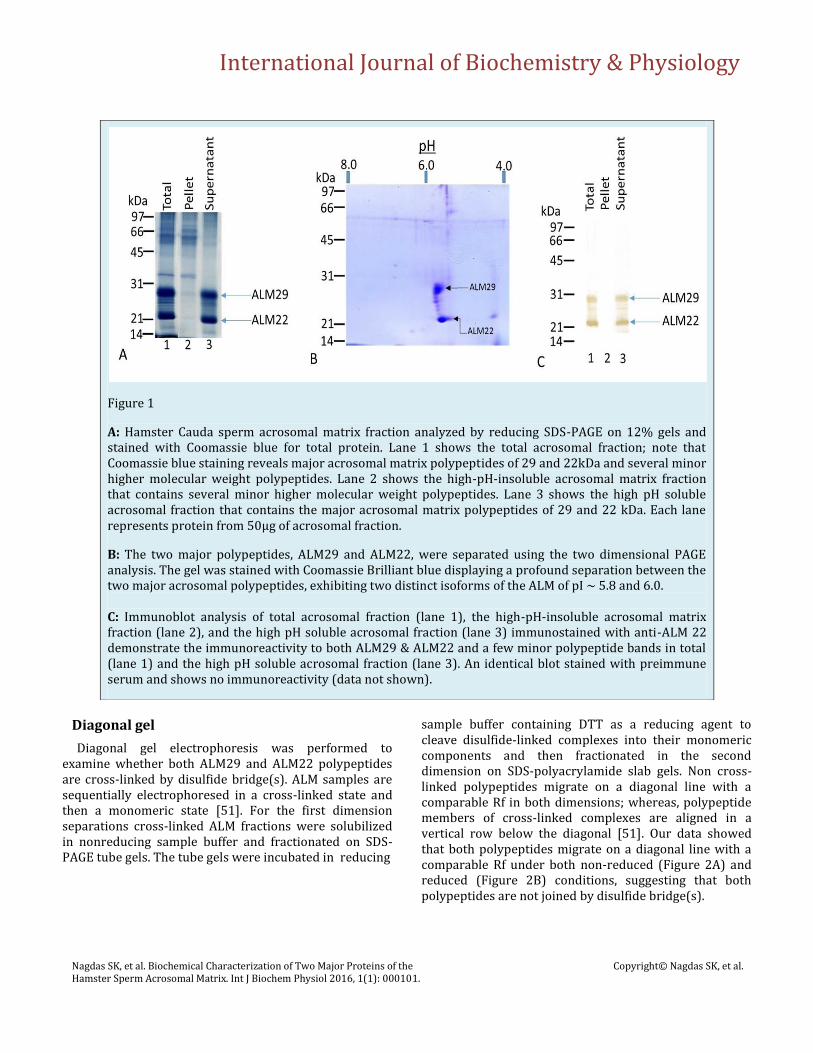
Figure 1
A: Hamster Cauda sperm acrosomal matrix fraction analyzed by reducing SDS-PAGE on 12% gels and stained with Coomassie blue for total protein. Lane 1 shows the total acrosomal fraction; note that Coomassie blue staining reveals major acrosomal matrix polypeptides of 29 and 22kDa and several minor higher molecular weight polypeptides. Lane 2 shows the high-pH-insoluble acrosomal matrix fraction that contains several minor higher molecular weight polypeptides. Lane 3 shows the high pH soluble acrosomal fraction that contains the major acrosomal matrix polypeptides of 29 and 22 kDa. Each lane represents protein from 50μg of acrosomal fraction.
B: The two major polypeptides, ALM29 and ALM22, were separated using the two dimensional PAGE analysis. The gel was stained with Coomassie Brilliant blue displaying a profound separation between the two major acrosomal polypeptides, exhibiting two distinct isoforms of the ALM of pI ~ 5.8 and 6.0. C: Immunoblot analysis of total acrosomal fraction (lane 1), the high-pH-insoluble acrosomal matrix fraction (lane 2), and the high pH soluble acrosomal fraction (lane 3) immunostained with anti-ALM 22 demonstrate the immunoreactivity to both ALM29 & ALM22 and a few minor polypeptide bands in total (lane 1) and the high pH soluble acrosomal fraction (lane 3). An identical blot stained with preimmune serum and shows no immunoreactivity (data not shown).

International Journal of Biochemistry & Physiology
Nagdas SK, et al. Biochemical Characterization of Two Major Proteins of the Hamster Sperm Acrosomal Matrix. Int J Biochem Physiol 2016, 1(1): 000101.
Copyright© Nagdas SK, et al.
Acrosin binding efficiency of V8-digested peptides of ALM29 and ALM22 polypeptides
Previously, we have shown that V8 digestion of ALM29 and ALM22 polypeptides generated an identical set of four polypeptide bands of 20kDa, 16kDa, 13kDa, and 12kDa suggesting a structural similarity of ALM29 and ALM22 polypeptides [47]. N-terminal amino acid sequence of the generated peptides demonstrated that ALM 29 and ALM22 polypeptides share sequence homology to each other [47]. In the current study, an identical pattern of V8-digested peptides of ALM 29 (Figure 3, lane 2) and ALM22 (Figure 3, lane 4) were obtained by Coomassie blue staining as we reported earlier [47]. To explore the antigenic relationship of V8-digested peptides of ALM29 and ALM22, an identical blot stained with anti-ALM22 polyclonal antibody exhibits that
all V8-digested peptides of ALM29 (lane 6) and ALM22 (lane 8) showed immunoreactive bands suggesting that all peptides are antigenically related family. An identical blot was subjected to examine the acrosin binding efficiency by blot-overlay assay. Interestingly, only native ALM29 polypeptide binds acrosin (lane 9), neither V8-digested peptides (20kDa, 16kDa, 13kDa, and 12kDa) of ALM29 (lane 10), nor ALM22 polypeptide (lane 11) and V8-digested peptides of ALM22 (lane 12) bind acrosin. This study suggests that only the native ALM29 polypeptide possesses acrosin binding activity.
Figure 2: Diagonal Gel Electrophoresis of ALM Complex (25μg protein) was performed to examine whether both ALM29 and ALM22 polypeptides are cross-linked by disulfide bridge(s). For the first dimension separations, cross-linked ALM fractions were solubilized in nonreducing sample buffer and fractionated on SDS-PAGE tube gels. The tube gels were incubated in reducing sample buffer containing DTT as a reducing agent to cleave disulfide-linked complexes into their monomeric components and then fractionated in the second dimension on SDS-polyacrylamide slab gels. A—Non-Reduced Conditions in both dimensions; B—Non-Reduced Conditions (1st Dimension) & Reduced Conditions (2nd Dimension).

International Journal of Biochemistry & Physiology
Biochemical Characterization of Two Major Proteins of the Hamster Sperm Acrosomal Matrix Int J Biochem Physiol
Alkaline phosphatase treatment of ALM29/ALM22 polypeptides
ALM 29 and ALM 22 polypeptides were solubilized by high pH extraction of ALM. High pH supernatant fraction was treated with alkaline phosphatase and then examined by Western Blot Analysis. Blots were stained with anti-ALM22. Immunoblot data (Figure 4, lane 1) shows the
control experiment displaying two major polypeptides. Lane 2 reflects the alkaline phosphatase treated ALM29 and ALM22. Treated ALM29 and ALM22 were reduced in size by approximately 3 kDa (Figure. 4, lane 2). This study concluded that both ALM29 and ALM22 are phosphorylated.
Figure 3: Peptide maps of ALM29 and ALM22 using V8 protease. SDS-PAGE purified ALM29 (lanes 1,2,5,6,9,10) and ALM22 (lanes 3,4,7,8,11,12) were incubated in the absence (lanes 1,3,5,7,9,11) or presence (lanes 2,4,6,8,10,12) of V8 protease and then fractionated by SDS-PAGE on 15% acrylamide gels, transferred to PVDF membranes. Lanes 1-4 stained with Coomassie Blue Dye and lanes 5-8 stained with anti-ALM22 antibody. Lanes 9-12 was subjected to blot overlay assay to examine the binding efficiency of acrosin to the V8 protease-generated peptides of ALM29 and ALM22 polypeptides.

International Journal of Biochemistry & Physiology
Nagdas SK, et al. Biochemical Characterization of Two Major Proteins of the Hamster Sperm Acrosomal Matrix. Int J Biochem Physiol 2016, 1(1): 000101.
Copyright© Nagdas SK, et al.
Demonstration of acrosin binding to dephosphorylated and native ALM29 polypeptide
A sedimentation assay was employed to determine the binding efficiency of dephosphorylated ALM29 to acrosin (Table-1). The dephosphorylated ALM29 polypeptide
revealed a noticeable reduction in acrosin (~50% reduction) binding in comparison to the native ALM29 polypeptide binding to acrosin. In contrast, alkaline phosphatase conjugated beads, used as control for specificity, did not bind acrosin (data not shown) demonstrating the specificity of binding of acrosin to the native and dephosphorylated ALM29 polypeptide. Our results suggest that phosphate groups play an important role in acrosin binding.
Table-1: Acrosin Binding to Native and Dephosphorylated ALM29 Polypeptide. The distribution of across in between the pellet and supernatant fractions is shown in Table-1. Approximately, 50% reduction in acrosin binding to the dephosphorylated ALM29 polypeptide was noted. Values represent mean ± SD of three experiments.
Discussion
The ALM complex remains associated with the hybrid membrane complex after the acrosome reaction [47]. Utilizing both biochemical and immunological approaches, we have demonstrated that ALM29 and ALM22 polypeptides are structurally related and appear to arise from a common 40kDa precursor protein expressed in round spermatids [47]. SP10 related acrosomal proteins are reported in several species including human, baboon, fox, mouse, and bovine spermatozoa [54-59]. Both monoclonal and polyclonal antibodies to human SP-10 significantly lowered in vitro fertilization rates of bovine oocytes by bovine spermatozoa [59]. The potential of utilizing SP-10 antigen as targets for immunocontraceptive vaccines has previously been suggested [60]. Recently, Foster and Gerton [61] reported a list of mouse knockout genes of acrosomal matrix and associated proteins. Since a knockout mouse line of SP-10 has not been generated, the molecular mechanisms of acrosomal protein SP-10 in mammalian fertilization is yet to be elucidated. In the
Figure 4: Both ALM29 and ALM22 polypeptides were stripped out of the ALM complex by high pH (pH-11) extraction. Both respective polypeptides were treated with alkaline phosphatase in 50mM Glycine-NaOH buffer, pH 11 for 4 hours at 37oC. A parallel ALM control was run without an enzyme. Both the control and enzyme-treated fractions were analyzed by Western blot analysis. Blots were immunostained with anti-ALM 22. Lane 1 shows the control experiment exhibiting the presence of two major polypeptides: ALM29 and ALM22. Lane 2 reveals the alkaline phosphatase treated ALM; there is a reduction in size (~3 kDa) of both the ALM29 & ALM22 polypeptides.
Native ALM29
Polypeptide
Dephosphorylated
ALM29 olypeptide
Pellet 90±8 45±5
Supernatant 10±2 55±6

International Journal of Biochemistry & Physiology
Nagdas SK, et al. Biochemical Characterization of Two Major Proteins of the Hamster Sperm Acrosomal Matrix. Int J Biochem Physiol 2016, 1(1): 000101.
Copyright© Nagdas SK, et al.
present study, we address the biochemical characterization and assembly of ALM29 and ALM22 polypeptides. In addition, we also elucidate the mechanism of binding efficiency of acrosin to ALM29 polypeptide. We have observed an identical architect of related proteins in the bovine sperm acrosomal matrix [20,62] reflecting the presence of a similar acrosomal distribution pattern of SP-10 related proteins in nonhuman spermatozoa. Recently, we have shown that proteomic identification of the OMC32 polypeptide (one of the bovine sperm acrosomal proteins), by MALDI-TOF-TOF analysis, yielded 2 peptides that matched the NCBI database sequence of acrosin-binding protein (Bos Taurus; GI: 194666681) [46]. Anti-OMC32 recognized an antigenically related family of polypeptides (OMCrpf polypeptides) in the 38-19kDa range with isoelectric points ranging between 4.0 and 5.1 which are moderately acidic [46]. Human sperm SP-10 peptides from 24-34kDa have isoelectric points of approximately 4.9; whereas, the isoelectric point 17.5kDa peptide is basic [55]. Our results displayed the isoelectric points of both ALM29 and ALM22 polypeptides ranging between 5.8 and 6.0 which are slightly acidic. Results of several species strongly elucidate that the isoelectric points of acosomal matrix polypeptides are slightly acidic which could be one of the factors for the acrosomal hydrolases/proteins binding to the acrosmal matrix complex. By cross-linking experiments using the homobifunctional cleavable cross-linkers, dithiobis [sulfosuccinimidyl propionate] (DTSSP), we demonstrate that the ALM29 and ALM22 polypeptides are not cross-linked by disulfide bond. It is possible that both the ALM29 and ALM22 polypeptides interact with each other by other types of interaction. Future study will address this issue. Our peptide mapping and blot overlay assay revealed that V8-digested peptides of the ALM29 and ALM22 polypeptides do not interact with acrosin; acrosin only binds to the intact ALM29 polypeptide-not the intact ALM22 polypeptide. N-terminal sequence of different forms of SP-10 (17.5-34kDa) by Edman degradation demonstrates that the processing of 34kDa to 17.5kDa occurs via N-terminal cleavage [63]. Since acrosin binds to intact ALM29 polypeptide only, it is suggested that the acrosin binding domain is present at the N-terminus end of ALM29 polypeptide probably at the 7kDa region of the N-terminus end of ALM29 polypeptide. When ALM29 is converted to the ALM22 polypeptide, due to the cleavage of peptide at the N-terminus, the acrosin binding domain is lost in ALM22 polypeptide.
Alkaline phosphatase treatment of both the ALM29 and ALM22 polypeptides exhibit ~3kDa reduction suggesting that both polypeptides are phosphorylated. Previously, Baker et al [64] reported sixty-eight rat sperm phosphopeptides during epididymal maturation. Out of sixty-eight, twenty-two polypeptides have been identified. Two of the proteins are acrosomal proteins; they are IZUMO1 and sperm acrosome membrane associated I proteins. By immunoblot analyses, they also showed the reduction of rat cauda sperm IZUMO1 by alkaline phosphatase treatment that also supports their phosphoproteomic analyses [64]. In the present study, we have also shown by sedimentation assay that there is a ~50% reduction of acrosin binding to the de-phosphorylated ALM29 polypeptide compared to the native ALM29 polypeptide. Our study suggests that the phosphate group of the ALM29 polypeptide is one of the components involved in acrosin binding. By peptide mapping study, we have also shown that the acrosin binding domain of the ALM29 polypeptide is present at 7kDa region of the peptide near the N-terminus end. It is assumed the phosphate group which is involved in the acrosin binding is present at the 7kDa region of ALM29 polypeptide near the N-terminus end. In conclusion, we hypothesize that during the acrosome reaction the enzymatically inactive acrosomal phosphatase is converted to active phosphatase which cleaves the phosphate group of the ALM29 polypeptide leading to the dissociation of ALM29-acrosin interaction; consequently the release of acrosin occurs from the hamster sperm acrosomal matrix complex. How does the activation of phosphatase occur? It may be possible that the acrosomal phosphatase becomes enzymatically active by phosphorylation via a signal transduction pathway. Alternatively, the conversion of inactive to active phosphatase occurs by other biochemical modification(s). Future studies will elucidate all these possible pathways.
Acknowledgements
Supported by NIH/NIGMS/ 1SC3GM096875 (Dr. Subir Nagdas), FSU NC-LSAMP Grant, and Benedict College NSF Grant #1436222
References
1. Eddy EM, O'Brien DA (1994) The Spermatozoon. In Knobil E, Neill JD (Eds) The Physiology of Reproduction, Raven Press New York, pp 29-77.

International Journal of Biochemistry & Physiology
Nagdas SK, et al. Biochemical Characterization of Two Major Proteins of the Hamster Sperm Acrosomal Matrix. Int J Biochem Physiol 2016, 1(1): 000101.
Copyright© Nagdas SK, et al.
2. Yanagimachi R (1994) Mammalian Fertilization. In Knobil E, Neill JD (Eds) The Physiology of Reproduction, Raven Press New York, pp 189-317.
3. Arboleda CE, Gerton GL (1987) Studies of three major proteases associated with guinea pig sperm acrosomes. J Exp Zool 244(2): 277-287.
4. O'Brien DA, Gerton GL, Eddy EM (1988) Acrosomal constituents identified with a monoclonal antibody are modified during late spermiogenesis in the mouse. Biol Reprod 38(4): 955-967.
5. Anakwe OO, Gerton GL (1990) Acrosome biogenesis begins during meiosis: evidence from the synthesis and distribution of an acrosomal glycoprotein, acrogranin, during guinea pig spermatogenesis. Biol Reprod 42(2): 317-328.
6. Yoshinaga K, Tanii I, Oh-oka T, Toshimori K (2001) Changes in distribution and molecular weight of the acrosomal protein acrin2 (MC41) during guinea pig spermiogenesis and epididymal maturation. Cell Tissue Res 303(2): 253-261.
7. Olson GE, Winfrey VP, Nagdas SK (1997) Temporal expression and localization of protein farnesyltransferase during spermiogenesis and posttesticular sprm maturation in the hamster. Mol Reprod Dev 48: 71-76.
8. Olson GE, Winfrey VP, Nagdas SK (2003) Structural modification of the hamster sperm acrosome during posttesticular development in the epididymis. Microsc Res Tech 61(1): 46-55.
9. Tulsiani DR, Nagdas SK, Skudlarek MD, Orgebin-Crist MC (1995) Rat sperm plasma membrane mannosidase: localization and evidence for proteolytic processing during epididymal maturation. Dev Biol 167(2): 584-595.
10. Olson GE, Winfrey VP, Ming B, Hardy DM, Nagdas SK (2004) Zonadhesin assembly into the hamster sperm acrosomal matrix occurs by distinct targeting strategies during spermiogenesis and maturation in the epididymis. Biol Reprod 71(4): 1128-1134.
11. Anakwe OO, Sharma S, Hardy DM, Gerton GL (1991) Guinea pig proacrosin is synthesized principally by round spermatids and contains O-linked as well as N-linked oligosaccharide side chains. Mol Reprod Dev 29(2): 172-179.
12. Nagdas SK, Skudlarek MD, Orgebin-Crist MC, Tulsiani DRP (1992) Biochemical alterations in the proacrosin-acrosin system during epididymal maturation of rat spermatozoa. J Androl 13(1): 36-43.
13. Cross NL (1998) Role of cholesterol in sperm capacitation. Biol Reprod 59(1): 7-11.
14. Visconti PE, Kopf GS (1998) Regulation of protein phosphorylation during sperm capacitation. Biol Reprod 59(1): 1-6.
15. de Lamirande E, Leclerc P, Gagnon C (1997) Capacitation as a regulatory event that primes spermatozoa for the acrosome reaction and fertilization. Mol Hum Reprod 3(3): 175-194.
16. Cohen-Dayag A, Eisenbach M (1994) Potential assays for sperm capacitation in mammals. Am J Physiol 267(5): C1167-1176.
17. Jin M, Fujiwara E, Kakiuchi Y, Okabe M, Satouh Y, et al. (2011) Most fertilizing mouse spermatozoa begin their acrosome reaction before contact with the zona pellucida during in vitro fertilization. Proc Natl Acad Sci USA 108(12): 4892-4896.
18. Meizel S (1984) The importance of hydrolytic enzymes to an exocytotic event, the mammalian sperm acrosome reaction. Biol Rev Camb Philos Soc 59(1): 125-157.
19. Kopf GS, Gerton GL (1991) The Mammalian Sperm Acrosome and the Acrosome Reaction. In Wassarman PM (Ed) Elements of Mammalian Fertilization, CRC Press: Boca Raton, Ann Arbor, Boston. pp 153-203.
20. NagDas SK, Winfrey VP, Olson GE (1996) Proacrosin-acrosomal matrix binding interactions in ejaculated bovine spermatozoa. Biol Reprod 54(1): 111-121.
21. NagDas SK, Winfrey VP, Olson GE (1996) Identification of hydrolase binding activities of the acrosomal matrix of hamster spermatozoa. Biol Reprod 55(6): 1405-1414.
22. Jones R, Brown CR (1987) Identification of a zona-binding protein from boar spermatozoa as proacrosin. Exp Cell Res 171(2): 503-508.
23. Stambaugh R, Brackett BG, Mastroianni L (1969) Inhibition of in vitro fertilization of rabbit ova by trypsin inhibitors. Biol Reprod 1(3): 223-227.

International Journal of Biochemistry & Physiology
Nagdas SK, et al. Biochemical Characterization of Two Major Proteins of the Hamster Sperm Acrosomal Matrix. Int J Biochem Physiol 2016, 1(1): 000101.
Copyright© Nagdas SK, et al.
24. Bhattacharyya AK, Goodpasture JC, Zaneveld LJ (1979) Acrosin of mouse spermatozoa. Am J Physiol 237(1): E40-44.
25. Baba T, Azuma S, Kashiwabara S, Toyoda Y (1994) Sperm from mice carrying a targeted mutation of the acrosin gene can penetrate the oocyte zona pellucida and effect fertilization. J Biol Chem 269(50): 31845-31849.
26. Yamagata K, Murayama K, Okabe M, Toshimori K, Nakanishi T, et al. (1998) Acrosin accelerates the dispersal of sperm acrosomal proteins during acrosome reaction. J Biol Chem 273(17): 10470-10474.
27. Miller DJ, Gong X, Shur BD (1993) Sperm require beta-N-acetylglucosaminidase to penetrate through the egg zona pellucida. Development 118(4): 1279-1289.
28. Takada M, Yonezawa N, Yoshizawa M, Noguchi S, Hatanaka Y, et al. (1994) pH-sensitive dissociation and association of beta-N-acetylhexosaminidase from boar sperm acrosome. Biol Reprod 50(4): 860-868.
29. Akruk SR, Farooqui AA, Williams WL, Srivastava PN (1979) Release of acrosomal enzymes following in vitro capacitation of ejaculated rabbit spermatozoa. Gamete Res 2(1): 1-13.
30. Fawcett DW (1975) The mammalian spermatozoon. Dev Biol 44(2): 394-436.
31. Phillips DM (1972) Substructure of the mammalian acrosome. J Ultrastruct Res 38(5): 591-604.
32. Westbrook-Case VA, Winfrey VP, Olson GE (1994) A domain-specific 50-kilodalton structural protein of the acrosomal matrix is processed and released during the acrosome reaction in guinea pig. Biol Reprod 51(1): 1-13.
33. Foster JA, Friday BB, Maulit MT, Blobel C, Winfrey VP, et al. (1997) AM67, a secretory component of the guinea pig sperm acrosomal matrix, is related to mouse sperm protein sp56 and the complement component 4-binding proteins. J Biol Chem 272: 12714-12722.
34. Noland TD, Friday BB, Maulit MT, Gerton GL (1994) The sperm acrosomal matrix contains a novel member of the pentaxin family of calcium-dependent binding proteins. J Biol Chem 269(51): 32607-32614.
35. Reid MS, Blobel CP (1994) Apexin, an acrosomal pentaxin. J Biol Chem 269(51): 32615-32620.
36. Westbrook-Case VA, Winfrey VP, Olson GE (1995) Sorting of the domain-specific acrosomal matrix protein AM50 during spermiogenesis in the guinea pig. Dev Biol 167(1): 338-349.
37. Olson GE, Winfrey VP, Davenport GR (1988) Characterization of matrix domains of the hamster acrosome. Biol Reprod 39(5): 1145-1158.
38. Hardy DM, Oda MN, Friend DS, Huang TT Jr (1991) A mechanism for differential release of acrosomal enzymes during the acrosome reaction. Biochem J 275(3): 759-766.
39. DiCarlantonio G, Talbot P (1988) Evidence for sequential deployment of secretory enzymes during the normal acrosome reaction of guinea pig sperm in vitro. Gamete Res 21(4): 425-438.
40. Green DP (1978) The activation of proteolysis in the acrosome reaction of guinea-pig sperm. J Cell Sci 32: 153-164.
41. Nuzzo NA, Anderson RA Jr, Zaneveld LJ (1990) Proacrosin activation and acrosin release during the guinea pig acrosome reaction. Mol Reprod Dev 25(1): 52-60.
42. Multamäki S, Suominen J (1976) Distribution and removal of the acrosin of bull spermatozoa. Int J Fertil (2): 69-81.
43. Srivastava PN, Munnell JF, Yang CH, Foley CW (1974) Sequential release of acrosomal membranes and acrosomal enzymes of ram spermatozoa. J Reprod Fertil 36(2): 363-372.
44. Noland TD, Davis LS, Olson GE (1989) Regulation of proacrosin conversion in isolated guinea pig sperm acrosomal apical segments. J Biol Chem 264(23): 13586-13590.
45. Hyatt H, Gwatkin RB (1988) Characterization of isolated acrosomal matrices from hamster spermatozoa. J Reprod Fertil 83(1): 419-429.
46. Nagdas SK, Smith L, Medina-Ortiz I, Hernandez-Encarnacion L, Raychoudhury S (2016) Identification of bovine sperm acrosomal proteins that interact with a 32-kDa acrosomal matrix protein. Molecular and Cellular Biochemistry 414(1-2): 153-169.

International Journal of Biochemistry & Physiology
Nagdas SK, et al. Biochemical Characterization of Two Major Proteins of the Hamster Sperm Acrosomal Matrix. Int J Biochem Physiol 2016, 1(1): 000101.
Copyright© Nagdas SK, et al.
47. Olson GE, Winfrey VP, NagDas SK (1998) Acrosome biogenesis in the hamster: Ultrastructurally distinct matrix regions are assembled from a common precursor polypeptide. Biol Reprod 58(2): 361-370.
48. Laemmli UK (1970) Cleavage of structural proteins during the assembly of the head of bacteriophage T4. Nature 227: 680-685.
49. Fairbanks G, Steck TL, Wallach DF (1971) Electrophoretic analysis of the major polypeptides of the human erythrocyte membrane. Biochemistry 10(13): 2606-2617.
50. Towbin H, Staehelin T, Gordon J (1979) Electrophoretic transfer of proteins from polyacrylamide gels to nitrocellulose sheets: procedure and some applications. Proc Natl Acad Sci USA 76(9): 4350-4354.
51. Wang K, Richards FM (1974) An approach to nearest neighbor analysis of membrane proteins. Application to the human erythrocyte membrane of a method employing cleavable cross-linkages. J Biol Chem 249(24): 8005-8018.
52. Bradford MM (1976) A rapid and sensitive method for the quantitation of microgram quantities of protein utilizing the principle of protein-dye binding. Anal Biochem 72(1-2): 248-254.
53. NagDas SK, Winfrey VP, Olson GE (2000) Identification of a hamster epididymal region-specific secretory glycoprotein that binds nonviable spermatozoa. Biol Reprod 63(5): 1428-1436.
54. Reddi PP, Naaby-Hansen S, Aguolnik I, Tsai JY, Silver LM, et al. (1995) Complementary deoxyribonucleic acid cloning and characterization of mSP-10: the mouse homologue of human acrosomal protein SP-10. Biol Reprod 53(4): 873-881.
55. Herr JC, Flickinger CJ, Homyk M, Klotz K, John E (1990) Biochemical and morphological characterization of the intra-acrosomal antigen SP-10 from human sperm. Biol Reprod 42(1): 181-193.
56. Herr JC, Wright RM, John E, Foster J, Kays T, et al. (1990) Identification of human acrosomal antigen SP-10 in primates and pigs. Biol Reprod 42(2): 377-382.
57. Freemerman AJ, Wright RM, Flickinger CJ, Herr JC (1993) Cloning and sequencing of baboon and cynomolgus monkey intra-acrosomal protein SP-10:
homology with human SP-10 and a mouse sperm antigen (MSA-63). Mol Reprod Dev 34(2): 140-148.
58. Beaton S, ten Have J, Cleary A, Bradley MP (1995) Cloning and partial characterization of the cDNA encoding the fox sperm protein FSA-Acr.1 with similarities to the SP-10 antigen. Mol Reprod Dev 40(2): 242-252.
59. Coonrod SA, Herr JC, Westhusin ME (1996) Inhibition of bovine fertilization in vitro by antibodies to SP-10. J Reprod Fertil 107(2): 287-297.
60. Freemerman AJ, Wright RM, Flickinger CJ, Herr JC (1994) Tissue specificity of the acrosomal protein SP-10: a contraceptive vaccine candidate molecule. Biol Reprod 50(3): 615-621.
61. Foster JA, Gerton GL2 (2016) The Acrosomal Matrix. Adv Anat Embryol Cell Biol 220: 15-33.
62. Olson GE, Winfrey VP, Neff JC, Lukas TJ, NagDas SK (1997) An antigenically related polypeptide family is a major structural constituent of a stable acrosomal matrix assembly in bovine spermatozoa. Biol Reprod 57(2): 325-334.
63. Herr JC, Klotz K, Shannon J, Wright RM, Flickinger CJ (1992) Purification and microsequencing of the intra-acrosomal protein SP-10. Evidence that SP-10 heterogeneity results from endoproteolytic processes. Biol Reprod 47(1): 11-20.
64. Baker MA, Hetherington L, Weinberg A, Naumovski N, Velkov T, et al. (2012) Analysis of phosphopeptide changes as spermatozoa acquire functional competence in the epididymis demonstrates changes in the post-translational modification of Izumo1. Journal of Proteome Research 11(11): 5252-5264.