
BIOCHEMICAL AND SPECTROSCOPIC … · saline; PCB, phycocyanobilin; ... the precursor of the...
36
1 Phycocyanobilin:Ferredoxin Oxidoreductase (PcyA) of Anabaena sp. PCC 7120 BIOCHEMICAL AND SPECTROSCOPIC CHARACTERIZATION Nicole Frankenberg and J. Clark Lagarias a,¶ a Section of Molecular and Cellular Biology, University of California at Davis, One Shields Avenue, Davis, California 95616 Running title: Phycocyanobilin:Ferredoxin Oxidoreductase ¶ To whom correspondence should be addressed: E-mail [email protected] phone: 1-530 752 1865 fax 1-530-752-3085 1 Abbreviations used: BR, bilirubin; BV, biliverdin; Cph1, cyanobacterial phytochrome 1; DHBV, dihydrobiliverdin; DMSO, dimethylsulfoxide; Fd, ferredoxin; Fldx, flavodoxin; FNR, ferredoxin-NADP + -oxidoreductase; GST, glutathione-S-transferase; PBS, phosphate buffered saline; PCB, phycocyanobilin; PcyA, phycocyanobilin: ferredoxin oxidoreductase; PEB, phycoerythrobilin; PΦB, phytochromobilin; TCA, trichloro acetic acid; TFA, trifluoro acetic acid Copyright 2003 by The American Society for Biochemistry and Molecular Biology, Inc. JBC Papers in Press. Published on January 3, 2003 as Manuscript M211643200 by guest on July 4, 2018 http://www.jbc.org/ Downloaded from
Transcript of BIOCHEMICAL AND SPECTROSCOPIC … · saline; PCB, phycocyanobilin; ... the precursor of the...

1
Phycocyanobilin:Ferredoxin Oxidoreductase (PcyA) of Anabaena sp. PCC 7120
BIOCHEMICAL AND SPECTROSCOPIC CHARACTERIZATION
Nicole Frankenberg and J. Clark Lagariasa,
a Section of Molecular and Cellular Biology, University of California at Davis, One Shields
Avenue, Davis, California 95616
Running title: Phycocyanobilin:Ferredoxin Oxidoreductase
¶ To whom correspondence should be addressed:
E-mail [email protected]
phone: 1-530 752 1865
fax 1-530-752-3085
1 Abbreviations used: BR, bilirubin; BV, biliverdin; Cph1, cyanobacterial phytochrome 1;
DHBV, dihydrobiliverdin; DMSO, dimethylsulfoxide; Fd, ferredoxin; Fldx, flavodoxin; FNR,
ferredoxin-NADP+-oxidoreductase; GST, glutathione-S-transferase; PBS, phosphate buffered
saline; PCB, phycocyanobilin; PcyA, phycocyanobilin: ferredoxin oxidoreductase; PEB,
phycoerythrobilin; PΦB, phytochromobilin; TCA, trichloro acetic acid; TFA, trifluoro acetic
acid
Copyright 2003 by The American Society for Biochemistry and Molecular Biology, Inc.
JBC Papers in Press. Published on January 3, 2003 as Manuscript M211643200 by guest on July 4, 2018
http://ww
w.jbc.org/
Dow
nloaded from

2
SUMMARY
In cyanobacteria, the biosynthesis of the phycobiliprotein and phytochrome chromophore
precursor phycocyanobilin is catalyzed by the ferredoxin-dependent enzyme PcyA that mediates
an atypical four-electron reduction of biliverdin IXα. Here we describe the expression, affinity
purification and biochemical characterization of recombinant PcyA from Anabaena sp.
PCC7120. A monomeric protein with a native Mr of 30,400 ± 5,000, recombinant PcyA forms a
tight and stable stoichiometric complex with its substrate biliverdin IXα. The enzyme exhibits a
strong preference for plant-type [2Fe-2S]-ferredoxins, however, flavodoxin can also serve as an
electron donor. HPLC analyses establish that catalysis proceeds via the two electron-reduced
intermediate 181,182-dihydrobiliverdin, indicating that exo-vinyl reduction preceeds A-ring
(endo-vinyl) reduction. Substrate specificity studies indicate that the arrangement of the A- and
D-ring substituents alters the positioning of the bilin substrate within the enzyme, profoundly
influencing the course of catalysis. Based on these observations and the apparent lack of a metal
or small molecule cofactor, a radical mechanism for biliverdin IXα reduction by
phycocyanobilin:ferredoxin oxidoreductase is envisaged.
by guest on July 4, 2018http://w
ww
.jbc.org/D
ownloaded from

3
INTRODUCTION
Phycocyanobilin (PCB1) is a linear tetrapyrrole (bilin) found in cyanobacteria, algae and
cryptomonads that functions as the direct precursor of the chromophores of the light-harvesting
phycobiliproteins and cyanobacterial/algal phytochromes (1,2). The PCB biosynthetic pathway
shares common intermediates with those of heme and chlorophyll to the level of protoporphyrin
IX whereupon the pathways diverge upon metalation with either iron or magnesium (1). All
phycobilins share the common intermediacy of biliverdin IXα (BV IXα) which is derived from
heme (1,3). BV IXα is the product of heme oxygenase-mediated cleavage of the heme
macrocycle which yields equimolar quantities of BV IXα, CO and iron. The metabolic fate of
BV IXα differs in mammals, cyanobacteria, and plants where BV IXα is metabolized by
different reductases with unique double bond specificities. In contrast with the mammalian
NAD(P)H-dependent BV IXα reductase (BVR), cyanobacteria and red algae possess ferredoxin-
dependent bilin reductases primarily for the synthesis of the linear tetrapyrrole precursors of their
phycobiliprotein light-harvesting antennae complexes, while evolutionarily related ferredoxin-
dependent bilin reductases are found in higher plants for the synthesis of the phytochrome
chromophore precursor phytochromobilin (PΦB) (3,4).
We recently documented that pcyA genes from cyanobacteria and oxyphotobacteria encode bilin
reductases which catalyze the ferredoxin-dependent reduction of BV IXα to 3Z-PCB - the bilin
precursor of their phycobiliprotein and phytochrome chromophores. Designated
phycocyanobilin:ferredoxin oxidoreductases (E.C. 1.3.7.5), PcyA enzymes are atypical bilin
reductases because they catalyze a four-electron reduction; all others catalyze two-electron
reductions (4). Formally two-electron reductions of vinyl substituents on the pyrrole A- and D-
rings of BV IXα, the sequence of reductions mediated by PcyA is presently unknown (see Figure
by guest on July 4, 2018http://w
ww
.jbc.org/D
ownloaded from

4
1). Neither of the two likely dihydrobiliverdin (DHBV) intermediates, 3Z-PΦB or 181, 182-
DHBV IXα, have yet been detected (4). Since 3Z-PΦB is an intermediate in the biosynthesis of
PCB, the precursor of the phytochrome chromophore in the green alga Mesotaenium
caldariorum (5), its intermediacy in the PcyA-mediated biosynthesis of PCB in cyanobacteria is
therefore a reasonable possibility. Previous studies have shown that the biosynthesis of PCB in
the red alga Cyanidium caldarium proceeds via phycoerythrobilin (PEB) as an intermediate
(Figure 1) (1). The enzyme(s) that catalyze the conversion of PEB to PCB have not yet been
identified; hence it is conceivable that PcyA might also mediate this conversion.
This study was undertaken to characterize the biochemical properties of a representative PcyA
enzyme from the filamentous cyanobacterium Anabaena sp. PCC 7120. The specific objectives
of these experiments were to identify the semi-reduced intermediate produced during the
catalysis of BV and to probe the bilin substrate specificity of this unusual ferredoxin-dependent
four-electron reductase. Based on these investigations, a chemical mechanism for PcyA-
mediated bilin reduction is proposed.
by guest on July 4, 2018http://w
ww
.jbc.org/D
ownloaded from

5
EXPERIMENTAL PROCEDURES
Reagents. Unless otherwise specified, all chemical reagents were American Chemical Society
grade or better. Glutathione agarose, spinach ferredoxin, Clostridium pasteurianum ferredoxin,
ferredoxin:NADP+ oxidoreductase (FNR) and size exclusion molecular weight markers (MW-
GF-200) were purchased from Sigma (St. Louis, MO). Restriction enzymes and Taq polymerase
were obtained from Gibco BRL (Cleveland, OH). HPLC-grade acetone, chloroform and 80%
formic acid were purchased from Fisher Scientific (Pittsburgh, PA). The expression vector
pGEX-6P-1 and PreScission™ protease were obtained from Amersham Biosciences
(Piscataway, NJ). Centricon-10 concentrator devices were purchased from Amicon (Beverly,
MA).
Bilin preparations. BV IXα, BV XIIIα, BV IIIα, PCB and PΦB preparations used as substrate
and/or HPLC standards were obtained as described previously (6,7). 181, 182-DHBV IXα was
synthesized by acid scrambling of a mixture of mesoBR IIIα and BR XIIIα followed by
oxidation with 2,3-dichloro-5,6-dicyanobenzoquinone (8). MesoBR IIIα was kindly provided by
Dr. D. A. Lightner (University of Nevada, Reno). BR XIIIα was prepared by acid scrambling of
commercially obtained BR (7,9).
Expression and purification of PcyA. Anabaena sp. PCC 7120 pcyA was cloned into the
Escherichia coli expression vector pGEX-6-P1 (Amersham Biosciences, Piscataway, NJ) to
produce pGEXpcyA (4). E. coli strain DH5α containing pGEXpcyA was induced to express
GST-PcyA which was purified according to instructions supplied by the manufacturer and
protocols described earlier (10). Proteolytic cleavage with the PreScission protease yielded the
native protein with the N-terminal amino acid extension GPLGSPEF and with the initiator
by guest on July 4, 2018http://w
ww
.jbc.org/D
ownloaded from

6
methionine residue changed to isoleucine. Purified PcyA protein concentration was estimated
from the absorbance at 280 nm using the calculated ε280nm of 29,726 M-1 cm-1(11).
Purification of recombinant reductants. Synechococcus sp. PCC 7002 ferredoxin and flavodoxin
clones, obtained from Dr. D. A. Bryant, were expressed and purified as described previously
(12,13). Expression and purification of putidaredoxin and putidaredoxin reductase, whose clones
were kindly provided by Dr. Paul Ortiz de Montellano, UCSF, were performed as describeda.
Flavodoxin was quantified by absorption at 467 nm and an absorption coefficient of ε467 nm 9,500
M-1 cm-1 (13), while putidaredoxin and putidaredoxin reductase were quantified by absorption at
454/415 nm, respectively using the absorption coefficients of ε454 nm 10,000 M-1 cm-1 and ε415 nm
11,100 M-1 cm-1.a
Standard bilin reductase activity assay. Assays for bilin reductase activity were performed as
described previously (10,14). Standard assays contained 1.5 µM PcyA, 4.8 µM ferredoxin and 5
µM BV IXα in 25 mM TES-KOH pH 7.5 (assay buffer) and were incubated for 30 min at 28 °C
under green safe-light unless otherwise specified. Following catalysis, bilins were isolated using
a C18 Sep-Pak (Waters) and evaporated to dryness in vacuo (4).
Direct HPLC analysis. Bilin reaction products were dissolved in 10 µl of DMSO, and diluted
with 200 µl of the HPLC mobile phase. Following brief centrifugation and filtration through a
0.45 µm PTFE syringe filter, bilins were resolved by reversed phase chromatography using an
Agilent Technologies 1100 Liquid Chromatograph. The HPLC column used for all of the
analyses was a 4.6 mm x 250 mm Phenomenex Ultracarb 5 µm ODS20 analytical column with a
4.6 mm x 30 mm guard column of the same material. The mobile phase consisted of acetone:20
mM formic acid (50:50 by volume) and the flow rate was 0.6 ml/min. Eluates were monitored at
a Nishida, C. and Ortiz de Montellano, P., personal communication.
by guest on July 4, 2018http://w
ww
.jbc.org/D
ownloaded from

7
650 nm, 560 nm and at 380 nm using an Agilent Technologies 1100 series diode array detector.
As needed, complete spectra were obtained for the peaks desired. Peak areas were quantitated
using Agilent Technologies Chemstation software.
Size exclusion chromatography. A Pharmacia Superdex 200 HR10/30 size exclusion column was
equilibrated in 50 mM TES-KOH buffer pH 7.5, containing 100 mM KCl and 10% (v/v) glycerol
(size exclusion chromatography buffer) at a flow rate of 0.4 ml/min. Standards with known Mr
(i.e. β-amylase 200,000, alcohol dehydrogenase 150,000, bovine serum albumin 66,000,
carbonic anhydrase 29,000 and cytochrome c 12,600) were applied to the column (100 µg) and
their elution volumes were determined spectroscopically. Anabaena sp. PcyA, PcyA:BV
(1:1::mol:mol), Fd:PcyA (2:1::mol:mol) and Fd:PcyA:BV (2:1:1::mol:mol:mol) were
chromatographed under identical conditions.
Glycerol gradient centrifugation. PcyA preparations (40 µg) were sedimented through a 2.5 ml
continuous 10-25% glycerol gradient in size exclusion chromatography buffer. A detailed
experimental procedure described previously was used for sedimentation coefficient
determination (15).
Spectroscopic analysis of biliverdin binding. Increasing amounts of PcyA were added to 5 µM
(final concentration) BV IXα, BV XIIIα or BV IIIα solutions in a final volume of 500 µl 25 mM
TES-KOH pH 7.5 buffer under green safe-light. After incubation for 30 min at room
temperature, absorbance spectra were recorded using a HP 8453 spectrophotometer.
Normalization of the spectra and spectral deconvolution was performed using Microsoft Excel.
To obtain bilin:PcyA dissociation constants, absorbance differences (∆A) at the λmax of each
bilin:PcyA complex were plotted as a function of PcyA concentration. Dissociation constants
by guest on July 4, 2018http://w
ww
.jbc.org/D
ownloaded from

8
were obtained by fitting this data to the hyperbolic equation ∆A = ∆Amax x [PcyA]/(Kapp +
[PcyA]) using DeltaGraph Pro 3.5 (DeltaPoint, Monterey, CA) where Kd = Kapp – 2.5 µM.
by guest on July 4, 2018http://w
ww
.jbc.org/D
ownloaded from

9
RESULTS
Expression and purification of recombinant phycocyanobilin:ferredoxin oxidoreductase. The
Anabaena sp. PCC 7120 pcyA gene was expressed using a tac promoter-driven N-terminal GST-
fusion expression system. Recombinant PcyA, obtained by ‘on-column’ proteolytic cleavage of
the GST fusion protein, was purified to ~90% homogeneity as shown in Figure 2A. ‘On-column’
cleavage was preferable to ‘in-solution’ proteolysis of the GST-PcyA fusion protein which led to
extensive protein precipitation and poor protein recovery. One liter bacterial cultures typically
yielded 3 mg of ‘on-column’ cleaved PcyA. All results presented here correspond to PcyA.
However, GST-PcyA preparations showed nearly identical catalytic properties (data not shown).
Determination of the native molecular mass of PcyA. The native molecular mass of PcyA was
determined using size exclusion chromatography and glycerol gradient sedimentation (Figure
2BC). A relative molecular weight of 30,400 ± 5,000 for PcyA was deduced with both methods,
which is in good agreement with the calculated molecular mass of 28,726 Daltons. Thus,
recombinant PcyA appears to be a monomeric enzyme. In order to determine whether PcyA can
form a stable complex with spinach ferredoxin, PcyA was incubated with a two-fold molar
excess of spinach Fd, and evaluated by both methods. Higher-order complex formation between
PcyA and Fd was not observed with either method under the conditions examined, nor did the
addition of two fold molar excess of BV IXα influence the result (data not shown).
PcyA lacks metal or small molecule cofactors. Purified recombinant PcyA was analyzed using
absorption spectroscopy for the presence of light absorbing cofactors such as hemes, flavins,
iron-sulfur clusters etc. Spectroscopic evidence for any of these cofactors was not obtained for
PcyA at concentrations as high as 5 mg/ml. In order to understand whether solvent accessible
metal ions are critical for activity (directly or indirectly as structural components), purified PcyA
by guest on July 4, 2018http://w
ww
.jbc.org/D
ownloaded from

10
was incubated with the metal chelators EDTA (10 mM), 1,10-phenanthroline and 2, 2’-dipyridyl
(5 mM each). After removal of the chelator from the protein by passing the mixture through a G-
25 desalting column (Amersham Biosciences), enzyme activity was determined. None of the
chelators had any inhibitory effect on the activity of PcyA (data not shown).
Reductant specificity of PcyA. The enzymes that mediate the reductive conversion of BV IXα to
phycobilins are all dependent on plant-type [2Fe-2S]-ferredoxins (4,14). For this reason, all
PcyA assays were performed in the presence of saturating levels of spinach ferredoxin and the
ferredoxin-reducing system consisting of spinach FNR and NADPH. Omission of any of these
components led to no PcyA activity (data not shown; (4)). To test whether other reductants can
serve as electron donors to PcyA, we tested recombinant Synechococcus sp. PCC 7002
ferredoxin and flavodoxin also in combination with spinach FNR. Other reductants tested
included C. pasteurianum ferredoxin, a 2[4Fe-4S]-ferredoxin, and a putidaredoxin/putidaredoxin
reductase system from Pseudomonas putida. As shown in Table I, maximum PcyA activity was
obtained with plant-type ferredoxin either from spinach or Synechococcus, while the FMN-
containing redox protein flavodoxin, less effectively supported PcyA catalysis. C. pasteurianum
ferredoxin and the putidaredoxin/putidaredoxin reductase system from P. putida were
considerably less active.
Bilin binding to PcyA. Bilin binding experiments demonstrated that PcyA forms a complex with
its bilin substrate which is stable through ultrafiltration, size exclusion chromatography and
dialysis. Spectrophotometric titration experiments with its natural substrate BV IXα and the two
analogs, BV XIIIα and BV IIIα is shown in Figure 3A. Upon binding to PcyA, significant blue-
shifts of the long wavelength absorption maxima were detected for all of the bilin analogs along
with an increase in molar absorption coefficient and the appearance of a shoulder at longer
by guest on July 4, 2018http://w
ww
.jbc.org/D
ownloaded from

11
wavelengths. The appearance of the shoulder was evident only for BV IXα and BV XIIIα, but
not BV IIIα (Figure 3B)- a result that was consistent with the ability of PcyA to metabolize these
two bilins (see later section). Figure 3C depicts replots of these absorption changes as a function
of increasing PcyA concentration. From hyperbolic curve fitting of this data, the equilibrium
dissociation constants were estimated to be 12.5, 14.5 and 4.5 µM for the respective IXα, XIIIα
and IIIα isomers under these experimental conditions. As a control, BV IXα binding
experiments were also performed using bovine carbonic anhydrase from the Sigma MW-GF-200
molecular weight kit. No spectral changes were observed with increasing carbonic anhydrase
concentration, indicating that the altered spectra reflected the formation of bilin:PcyA complexes
(Figure 3C).
PcyA’s two-electron reduced intermediate. The conversion of BV to PCB is a four-electron
reduction that formally consists of sequential two-electron reductions with the intermediacy of a
DHBV. As shown in Figure 1, the most likely candidates for this intermediate are 3Z-PΦB or
181, 182-DHBV IXα in which initial reduction occurs at the A- or D-rings of BV, respectively.
To identify the putative DHBV intermediate, time course experiments were performed. HPLC
analyses revealed the transient appearance of a new pigment during the course of catalysis
(Figure 4; Table II). This new pigment, which eluted earlier than 3Z-PCB (labeled I in Figure
4A), reached a maximum level within 10 minutes and disappeared after 30 minutes (Figure 4B).
Although pigment I eluted at the same retention time as 3E-PΦB, its absorption spectrum
differed from that of 3E-PΦB (Table II). The time course of the disappearance of BV IXα, the
appearance/disappearance of pigment I, and the appearance of the two isomers of PCB, shown in
Figure 4B, support the intermediacy of pigment I in the PcyA-mediated conversion of BV IXα
to PCB.
by guest on July 4, 2018http://w
ww
.jbc.org/D
ownloaded from
12
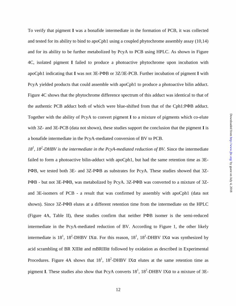
To verify that pigment I was a bonafide intermediate in the formation of PCB, it was collected
and tested for its ability to bind to apoCph1 using a coupled phytochrome assembly assay (10,14)
and for its ability to be further metabolized by PcyA to PCB using HPLC. As shown in Figure
4C, isolated pigment I failed to produce a photoactive phytochrome upon incubation with
apoCph1 indicating that I was not 3E-PΦB or 3Z/3E-PCB. Further incubation of pigment I with
PcyA yielded products that could assemble with apoCph1 to produce a photoactive bilin adduct.
Figure 4C shows that the phytochrome difference spectrum of this adduct was identical to that of
the authentic PCB adduct both of which were blue-shifted from that of the Cph1:PΦB adduct.
Together with the ability of PcyA to convert pigment I to a mixture of pigments which co-elute
with 3Z- and 3E-PCB (data not shown), these studies support the conclusion that the pigment I is
a bonafide intermediate in the PcyA-mediated conversion of BV to PCB.
181, 182-DHBV is the intermediate in the PcyA-mediated reduction of BV. Since the intermediate
failed to form a photoactive bilin-adduct with apoCph1, but had the same retention time as 3E-
PΦB, we tested both 3E- and 3Z-PΦB as substrates for PcyA. These studies showed that 3Z-
PΦB - but not 3E-PΦB, was metabolized by PcyA. 3Z-PΦB was converted to a mixture of 3Z-
and 3E-isomers of PCB - a result that was confirmed by assembly with apoCph1 (data not
shown). Since 3Z-PΦB elutes at a different retention time from the intermediate on the HPLC
(Figure 4A, Table II), these studies confirm that neither PΦB isomer is the semi-reduced
intermediate in the PcyA-mediated reduction of BV. According to Figure 1, the other likely
intermediate is 181, 182-DHBV IXα. For this reason, 181, 182-DHBV IXα was synthesized by
acid scrambling of BR XIIIα and mBRIIIα followed by oxidation as described in Experimental
Procedures. Figure 4A shows that 181, 182-DHBV IXα elutes at the same retention time as
pigment I. These studies also show that PcyA converts 181, 182-DHBV IXα to a mixture of 3E-
by guest on July 4, 2018http://w
ww
.jbc.org/D
ownloaded from

13
and 3Z-PCB thereby confirming the identity of the semi-reduced intermediate to be 181, 182-
DHBV IXα.
Bilin substrate specificity studies. Since the substrate analogs BV XIIIα and BV IIIα bind to
PcyA (see Figure 3), their ability to be metabolized by PcyA was also examined. As shown in
Figure 5A, BV XIIIα could be metabolized by PcyA to yield two products. Based on the relative
retention time of known bilins in our HPLC system and the absorbance spectra of the two
products (Figure 5B), we propose that BV XIIIα is converted by PcyA to the 3E- and 3Z-
isomers of isoPΦB (16). This hypothesis is also supported by the observation that both products
yield identical difference spectra upon incubation with apoCph1 (Figure 5C). By contrast with
the other two BV isomers, BV IIIα was not metabolized by PcyA. This result is interesting in
view of the observation that BV IIIα has the highest binding affinity for PcyA of the three BV
isomers (Figure 3C). The results of the bilin substrate specificity experiments are summarized in
Table III.
by guest on July 4, 2018http://w
ww
.jbc.org/D
ownloaded from

14
DISCUSSION
PcyA is a monomeric enzyme that forms a stable porphyrin-like complex with bilins. Among the
family of ferredoxin-dependent bilin reductases, phycocyanobilin:ferredoxin oxidoreductase
(PcyA) is unique in its ability to catalyze the four-electron reduction of BV IXα (4). Like oat
phytochromobilin synthase, a ferredoxin-dependent bilin reductase that converts BV IXα to PΦB
in plants (14), PcyA is a monomeric enzyme. BV binding neither promoted PcyA dimerization
nor oligomerization, suggesting that the distinct spectral properties of the three PcyA:BV
complexes studied here reflect the unique protein environment and conformation of the bound
bilin. The observed spectral features, i.e. long to short wavelength absorption ratio < 1, indicate
that bilins bind to PcyA in a cyclic, porphyrin-like configuration, as opposed to the more
extended configurations found in phytochromes and phycobiliproteins (17). This cyclic
configuration precludes simultaneous protonation of both B- and C-ring nitrogen atoms of the
bilin prosthetic group due to steric crowding (see Figure 6). This conclusion is further supported
by the observed lack of fluorescence of the PcyA:BV complex as efficient proton transfer
between H-bonded pyrrole rings would be expected to quench the excited state of the PcyA:BV
complex.
Interestingly, a long wavelength shoulder was detected in the spectra of the PcyA complexes of
BV IXα and BV XIIIα. Since this shoulder was not observed for BV IIIα, a non-metabolized
PcyA substrate analog, we speculate that this new absorption band corresponds to a distinct
bilin-PcyA interaction which reflects the bilin’s ability to be reduced (i.e. H-bonding,
protonation or aromatic π-π interaction).
PcyA prefers plant-type [2Fe-2S]-ferredoxins. Pioneering work by Beale and Cornejo (18) has
established that reduction of BV in the rhodophyte C. caldarium is Fd-mediated. This result was
by guest on July 4, 2018http://w
ww
.jbc.org/D
ownloaded from

15
later confirmed with the cloning of the bilin reductase family and the demonstration that all bilin
reductases are Fd-dependent enzymes (4,10). The present studies revealed that PcyA exhibits a
preference for plant-type [2Fe-2S]-Fds. Fldx, a two-electron acceptor which also can undergo
two successive low-potential single electron reductions (i.e. -413 mV at pH 7, 25 °C)(19), also
supported PcyA activity. Under the conditions tested here, the Fldx-dependent activity was
approximately 13% of that of Fd. More recently, we have been able to increase the Fldx-
mediated PcyA activity up to 50% of that of Fd by increasing the pH of the assay buffer to 8.5
and adding 100 mM KCl (Shih-Long Tu, unpublished data). Based on these results, it is
conceivable that Fldx can functionally substitute for Fd to drive PcyA activity under iron limiting
conditions in vivo (20).
The observation that the 2[4Fe-4S]-ferredoxin from C. pasteurianum poorly supports PcyA-
mediated BV reduction needs to be interpreted with the following caveats. The PcyA assay
requires a spinach FNR-containing ferredoxin reducing system which may be unable to
effectively reduce the C. pasteurianum Fd thereby limiting PcyA activity. Since spinach Fd and
C. pasteurianum Fd have the same redox potential around –420 mV, C. pasteurianum Fd
conceivably could support PcyA activity assuming the right reducing system is present. In this
regard, C. pasteurianum Fd serves as an electron acceptor in the anaerobic oxidation of pyruvate
(21) – the components of which may be able to drive C. pasteurianum Fd-dependent PcyA
activity.
The endogenous reductant for PcyA in E. coli may be flavodoxin. Recent studies reporting the
assembly of holophytochrome and holophycobiliproteins in E. coli (22-25), indicate that PcyA
can use naturally occurring reductants in living cells. E. coli cells possess several possible
reductants. E. coli Fd is an adrenodoxin-type [2Fe-2S] ferredoxin for which genetic analyses
by guest on July 4, 2018http://w
ww
.jbc.org/D
ownloaded from

16
have shown to perform an essential role in the maturation of various iron-sulfur proteins (26).
Indeed, E. coli Fd is more structurally related to the adrenodoxin-type ferredoxins, i.e. bovine
adrenodoxin (Adx) and P. putida putidaredoxin (Pdx), than to plant-type Fds (27). As such, E.
coli Fd likely functions as a component of the complex machinery responsible for the biogenesis
of Fe-S clusters. Based on the observation that the PcyA-mediated catalysis is poorly supported
by the putidaredoxin system (see Table I), we hypothesize that engineered PCB biosynthesis in
E. coli uses a different reducing system. Other than this Adx-type ferredoxin, the E. coli genome
possesses two Fldx genes and a flavorubredoxin gene (28). In the light of the data presented here,
we propose that the biosynthesis of PCB in E. coli is driven by one of the two Fldxs.
PcyA-mediated 18-vinyl reduction preceeds A-ring reduction. The identification of 181, 182-
DHBV IXα as an intermediate in the conversion of BV to PCB has established that D-ring exo-
vinyl reduction preceeds A-ring reduction. PcyA is therefore composed of two separate activities
mediated by a 181,182-DHBV:ferredoxin oxidoreductase and a PCB:ferredoxin oxidoreductase.
This double bond specificity of PcyA presumably ensures that PΦBs are never produced in
PcyA-containing cyanobacteria or red algae – the production of which might lead to miss-
incorporation of PΦBs into their phycobiliproteins. We speculate that PΦB-containing
phycobiliproteins would be more susceptible to photooxidative damage than the natural PCB-
containing antennae of these organisms due to the presence of the reactive exo-vinyl group on
the former. Evolution of PΦB-producing bilin reductases, such as HY2, would therefore prove a
selective disadvantage to these organisms – a selection pressure which would not apply to
terrestrial plants which lack phycobiliproteins. In this regard, it will be of interest to clone the
genes for these enzymes from the green alga M. caldariorum which mediate the conversion of
BV to PCB via the intermediacy of PΦB (5).
by guest on July 4, 2018http://w
ww
.jbc.org/D
ownloaded from

17
Through examination of substrate analogs which include the unnatural XIIIα and IIIα isomers of
BV and the A-ring reduced phytochromobilin isomers, 3Z- and 3E-PΦB, our studies have
provided insight into the catalytic specificity of PcyA. Of the two unnatural BV analogs, only
BV XIIIα was metabolized by PcyA yielding the two-electron reduced isoPΦB product (both
3Z- and 3E-isomers). Since BV XIIIα is symmetrical and lacks the exo-vinyl group found on BV
IXα, this result indicated that PcyA-mediated A-ring reduction can occur in the absence of exo-
vinyl reduction. The apparent lack of the reduction of the second endo-vinyl group of BV XIIIα
also indicated that isoPΦB is a poor PcyA substrate. By contrast to the IXα and XIIIα isomers,
BV IIIα was not metabolized by PcyA. This result was unexpected given the sequence of PcyA-
mediated vinyl reductions of BV IXα. In this regard, BV IIIα is symmetrical, possessing two
exo-vinyl groups, one or both of which should have been reduced by PcyA. Moreover, our
results indicate that BV IIIα binds to PcyA with the highest affinity of the three BV isomers
tested. These data indicate that bound BV IIIα is not properly oriented within the enzyme’s bilin
binding site for catalysis. Our studies show that 3Z-PΦB can be metabolized by PcyA, yielding a
mixture of PCB isomer products, while 3E-PΦB is not a substrate for PcyA. These data indicate
a strong influence of the geometry of the 3-ethylidene moiety on catalysis. Whether this is due to
a positioning defect or to a lack of binding of 3E-PΦB to PcyA remains to be determined. Taken
together, these studies suggest that proper substrate positioning/activation within the enzyme is a
prerequisite for catalysis.
A radical mechanism for bilin reduction by PcyA. Four major Fd-dependent enzymes have been
characterized to date - Fd:NADP+ oxidoreductase (FNR), Fd:nitrite oxidoreductase, glutamate
synthase and Fd:thioredoxin reductase (29). All of these enzymes contain redox-active cofactors
including FAD (FNR), iron-sulfur clusters (glutamate synthase, nitrite reductase, sulfite
by guest on July 4, 2018http://w
ww
.jbc.org/D
ownloaded from

18
reductase) and siroheme (nitrite reductase). By contrast with these Fd-dependent enzymes, PcyA
appears to lack a metal or flavin cofactor that can mediate single electron transfers. For these
reasons, we propose that the PcyA-mediated reduction of BV proceeds via bilin radical
intermediates as depicted in Figure 6.
Based upon the absorption spectrum of the PcyA:BV complex, the bilin substrate is depicted in a
cyclic conformation within the protein cavity (see Figure 6). The lack of photochromism of the
PcyA:BV complex can be rationalized by the binding of the terminal pyrrolinone A- and D-rings
into the protein matrix with its propionate side chains extending towards the solvent. This
porphyrin-like configuration not only would sterically prevent photoisomerization of the C5 and
C15 double bonds, but would also bury reactive radical intermediates within the protein matrix
thus minimizing side reactions with molecular oxygen. This substrate binding model is
consistent with the broad substrate specificity of the extended bilin reductase family, which
includes the enzyme RCCR that metabolizes a chlorophyll catabolite with monomethyl ester and
isocyclic ring substituents (30). In this regard, the hypothesis that the bilin reductase family may
have evolved from ancestors which metabolized (Mg)-porphyrins remains a intriguing
possibility.
As shown in Figure 6 (step 1), we envisage that bilin reduction occurs by binding of reduced Fd
to the PcyA:BV complex, followed by electron transfer to the bound bilin and proton transfer
from a protein residue labeled D1-H to generate a neutral radical shown in step 2. The benzylic
position would help to stabilize this radical by resonance within the extended tetrapyrrole
π−system, until a second electron and proton transfer, shown in steps 2 and 3, occurs to produce
the intermediate 181, 182-DHBV IXα. The hypothetical proton donors, D1-H and D2-H, could
either be carboxylic acids (i.e. asp or glu), sulfhydryls (i.e. cys), phenolics (i.e. tyr) or even
by guest on July 4, 2018http://w
ww
.jbc.org/D
ownloaded from
19
protonated nitrogen residues such as histidine or lysine. It is also possible that protons are
derived from bound water molecules which are protonated by appropriate protein residues. For
all of these protein residues except for histidine or lysine, proton transfer would be accompanied
by an increase in negative charge which would be a reasonable ‘driving force’ for the release of
product. Since PcyA kinetically reduces the intermediate 181, 182-DHBV IXα without its
release, we hypothesize that D1 and D2 are protonated histidine and/or lysine residues.
We propose that the subsequent reduction of the A-ring of 181, 182-DHBV IXα proceeds in a
similar fashion, generating another resonance-stabilized bilin radical intermediate shown in
Figure 6 (steps 4-6). For this transformation, we hypothesize that the proton donating residues
are carboxylic acids, sulfhydryls and/or phenolics which would generate negative charge within
the bilin pocket thereby promoting release of the PCB product (Figure 6, step 6). Experiments to
detect potential radical intermediates by electron spin resonance spectroscopy and to identify
putative proton donating residues within the PcyA polypeptide by site-directed mutagenesis are
in progress. With this experimental approach, we hope to elucidate the molecular basis for the
unique double bond reduction specificities of the different members of the extended bilin
reductase family (4) and ultimately to engineer novel specificity of this important family of
enzymes.
ACKNOWLEDGEMENT
The authors are grateful to Mr. James Partridge for synthesis of 181, 182-DHBV. We thank Drs.
D. A. Bryant and P. Ortiz de Montellano for providing clones and Dr. D. A. Lightner for the gift
of mBRIIIα. We thank Dr. Shih-Long Tu and Ms. Amanda Ellsmore for critical reading of the
manuscript. This work was supported by grant AMD-0103397 from United States Department of
by guest on July 4, 2018http://w
ww
.jbc.org/D
ownloaded from

20
Agriculture Competitive Research Grant Initiative (to J.C.L.), and by a fellowship from the
Deutsche Forschungsgemeinschaft to N.F.
by guest on July 4, 2018http://w
ww
.jbc.org/D
ownloaded from

21
REFERENCES
1. Beale, S. I. (1993) Chem. Rev. 93, 785-802
2. Hübschmann, T., Börner, T., Hartmann, E., and Lamparter, T. (2001) Eur. J. Biochem.
268, 2055-2063
3. Frankenberg, N., and Lagarias, J. C. (2003) in The Porphyrin Handbook (Kadish, K. M.,
Smith, K. M., and Guilard, R., eds) Vol. 13, Elsevier Science (USA)
4. Frankenberg, N., Mukougawa, K., Kohchi, T., and Lagarias, J. C. (2001) Plant Cell 13,
965-978
5. Wu, S.-H., McDowell, M. T., and Lagarias, J. C. (1997) J. Biol. Chem. 272, 25700-25705
6. Cornejo, J., Beale, S. I., Terry, M. J., and Lagarias, J. C. (1992) J. Biol. Chem. 267,
14790-14798
7. Elich, T. D., McDonagh, A. F., Palma, L. A., and Lagarias, J. C. (1989) J. Biol. Chem.
264, 183-189
8. Lightner, D. A., and Ma, J. S. (1985) J. Heterocyc. Chem. 21, 1005-1007
9. McDonagh, A. F. (1979) in The Porphyrins (Dolphin, D., ed) Vol. VI, pp. 293-491,
Academic Press, New York
10. Kohchi, T., Mukougawa, K., Frankenberg, N., Masuda, M., Yokota, A., and Lagarias, J.
C. (2001) Plant Cell 13, 425-436
11. Gill, S. C., and von Hippel, P. H. (1989) Anal. Biochem. 182, 319-326
12. Schluchter, W. M. (1994) Ph.D. Dissertation, The Pennsylvania State University,
University Park. PA
13. Zhao, J. D., Li, R. G., and Bryant, D. A. (1998) Anal. Biochem. 264, 263-270
14. McDowell, M. T., and Lagarias, J. C. (2001) Plant Physiol. 126, 1546-1554
by guest on July 4, 2018http://w
ww
.jbc.org/D
ownloaded from

22
15. Frankenberg, N., Heinz, D. W., and Jahn, D. (1999) Biochemistry 38, 13968-13975
16. Lindner, I. (2000) Ph.D. Dissertation, Gesamthochschule Duisburg, Duisburg, Germany
17. Falk, H. (1989) The Chemistry of Linear Oligopyrroles and Bile Pigments., Springer-
Verlag, Vienna
18. Beale, S. I., and Cornejo, J. (1991a) J. Biol. Chem. 266, 22328-22332
19. Yalloway, G. N., Mayhew, S. G., Malthouse, J. P. G., Gallagher, M. E., and Curley, G. P.
(1999) Biochemistry 38, 3753-3762
20. Straus, N. A. (1994) in The Molecular Biology of Cyanobacteria (Bryant, D. A., ed), pp.
731-750, Kluwer Academic Publishers, Dordrecht
21. Moulis, J. M., and Davasse, V. (1995) Biochemistry 34, 16781-16788
22. Gambetta, G. A., and Lagarias, J. C. (2001) Proc. Natl. Acad. Sci. U.S.A. 98, 10566-
10571
23. Landgraf, F. T., Forreiter, C., Pico, A. H., Lamparter, T., and Hughes, J. (2001) FEBS
Lett 508, 459-462
24. Tooley, A. J., Cai, Y. A., and Glazer, A. N. (2001) Proc. Natl. Acad. Sci. U.S.A. 98,
10560-10565
25. Tooley, A. J., and Glazer, A. N. (2002) J. Bacteriol. 184, 4666-4671
26. Lill, R., and Kispal, G. (2000) Trends Biochem Sci 25, 352-356
27. Kakuta, Y., Horio, T., Takahashi, Y., and Fukuyama, K. (2001) Biochemistry 40, 11007-
11012
28. Blattner, F. R., Plunkett, G., Bloch, C. A., Perna, N. T., Burland, V., Riley, M.,
ColladoVides, J., Glasner, J. D., Rode, C. K., Mayhew, G. F., Gregor, J., Davis, N. W.,
by guest on July 4, 2018http://w
ww
.jbc.org/D
ownloaded from

23
Kirkpatrick, H. A., Goeden, M. A., Rose, D. J., Mau, B., and Shao, Y. (1997) Science
277, 1453+
29. Knaff, D. B., and Hirasawa, M. (1991) Biochim. Biophys. Acta 1056, 93-125
30. Wüthrich, K. L., Bovet, L., Hunziker, P. E., Donnison, I. S., and Hörtensteiner, S. (2000)
Plant J 21, 189-198
by guest on July 4, 2018http://w
ww
.jbc.org/D
ownloaded from

24
FIGURE LEGENDS
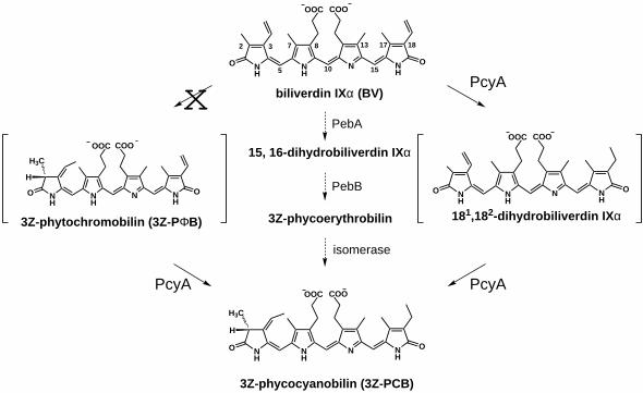
Figure 1. The reduction of biliverdin IXα by phycocyanobilin:ferredoxin oxidoreductase
(PcyA) can proceed via two possible intermediates. PcyA-mediated reduction of BV IXα
could result in the formation of 3Z-PΦB or 181,182-dihydrobiliverdin IXα as an intermediate.
Results presented here support the intermediacy of 181,182-dihydrobiliverdin IXα, however 3Z-
PΦB is also metabolized by PcyA (see text for details). An alternative pathway for the formation
of PCB in the red alga C. caldarium via the intermediacy of 15,16-dihydrobiliverdin IXα and
phycoerythrobilin is also shown in the center. This conversion is catalyzed by the bilin
reductases, PebA and PebB, and an as yet, unidentified isomerase (1,4).
Figure 2. Affinity purification of recombinant PcyA and determination of the molecular
mass. A, SDS-PAGE analysis of whole cell protein extracts before (lane 1) and after (lane 2)
induction with IPTG. Lane 3 shows the soluble fraction after induction; lane 4, recombinant
PcyA after “on column” cleavage and elution from glutathione agarose. Numbers on the right
indicate positions of molecular weight markers. B, Size exclusion chromatography using a
Superdex 200 HR10/30 column that had been calibrated with the following marker proteins: β-
amylase (βAM; 200 kDa), alcohol dehydrogenase (ADH; 150 kDa), bovine serum albumin
(BSA; 66 kDa), carbonic anhydrase (CA; 29kDa) and cytochrome c (CYTc; 12.4 kDa). The
elution positions of PcyA and ferredoxin are shown. C, Molecular mass determination by
glycerol gradient sedimentation. Sedimentation positions of marker protein are plotted against
known sedimentation coefficients. The sedimentation position of PcyA is indicated with an
arrow.
by guest on July 4, 2018http://w
ww
.jbc.org/D
ownloaded from

25
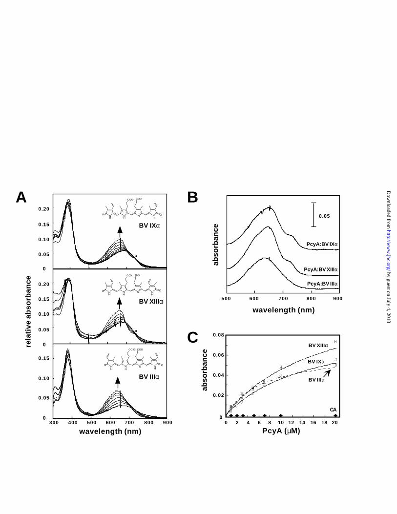
Figure 3. Binding of PcyA to BV measured by absorbance spectroscopy. A, Equilibrium
binding experiments of BV IXα (top panel), BV XIIIα (middle panel) and BV IIIα (bottom
panel) with increasing amounts of PcyA were performed as described under Experimental
Procedures. The direction of the spectral changes with increasing PcyA concentrations are
indicated by arrows. For the IXα and XIIIα isomers, PcyA:BV complex formation resulted in a
shoulder at longer wavelength (indicated by a black dot). B, Long wavelength absorption spectra
of 5 µM PcyA:BV complexes at saturating levels of PcyA (i.e. 20 µM PcyA). C, Absorbance
changes at the wavelength maxima of the PcyA:BV complexes (λmax) were plotted as a function
of PcyA concentration. Peak positions were 655 nm (BV IXα), 640 nm (BV IIIα) and 660 nm
(BV XIIIα). A control experiment used bovine carbonic anhydrase instead of PcyA is shown. All
data were fitted to a hyperbolic equation as described in Experimental Procedures. R2 values
were determined to be 0.995, 0.996 and 0.992 for the IIIα, IXα and XIIIα isomers, respectively.
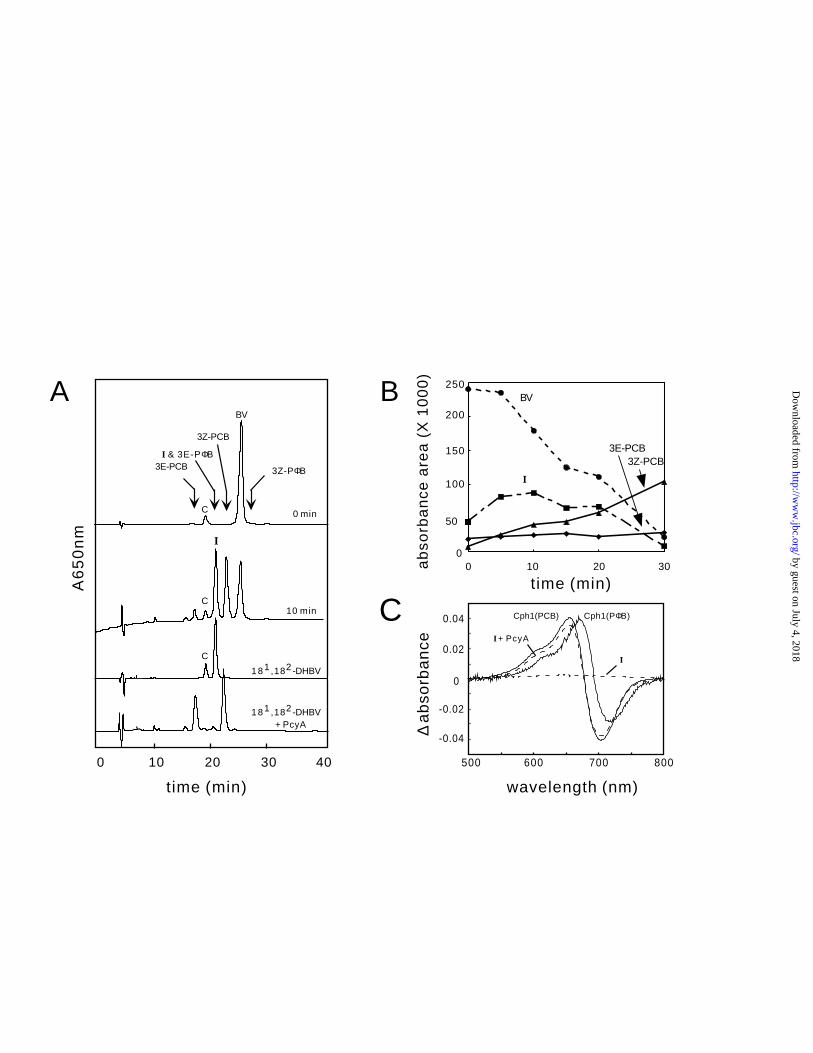
Figure 4. Identification of an intermediate in the PcyA-catalyzed reaction. A, HPLC profiles
of reactions products monitored at 650 nm were determined following the PcyA-mediated
reduction of BV IXα for 0 and 10 min (upper two profiles). In addition to 3Z- and 3E-PCB
products, an unknown pigment, labeled I, which co-elutes with the 3E-PΦB standard was
detected. Synthetic 181,182-dihydrobiliverdin IXα co-elutes with pigment I and can be converted
to a mixture of 3Z-and 3E-PCB (two bottom elution profiles). The peak labeled C corresponds to
a contaminant. B, The course of the reaction is plotted as peak areas as a function of reaction
time. Symbols used are BV (�); intermediate (�); 3E-PCB (�); 3Z-PCB (▲). C, Phytochrome
difference spectra were obtained following incubation of apoCph1 with pigment I before or after
metabolism with PcyA (labeled I and I + PcyA, respectively). Phytochrome difference spectra
by guest on July 4, 2018http://w
ww
.jbc.org/D
ownloaded from

26
of PCB- and PΦB-adducts of apoCph1, i.e. Cph1(PCB) and Cph1(PΦB), are shown for
comparison.
Figure 5. PcyA-mediated metabolism of BV-analogs. A, HPLC product profiles for BV
analogs after metabolism by PcyA for 30 min as described under Experimental Procedures.
Elution position of BV IXα, XIIIα and IIIα are indicated by arrows. B, Absorbance spectra of
BV XIIIα (dashed) and the two isoPΦB reaction products (solid lines) are shown; C,
Phytochrome difference spectra assay following assembly of apoCph1 with BV XIIIα before
(solid) and after PcyA-mediated catalysis (dashed) are shown. The difference spectra of the
PCB-adduct of apoCph1, i.e. Cph1(PCB), is shown for comparison.
Figure 6. Proposed radical mechanism for the PcyA-catalyzed reduction of BV IXα.
by guest on July 4, 2018http://w
ww
.jbc.org/D
ownloaded from
27
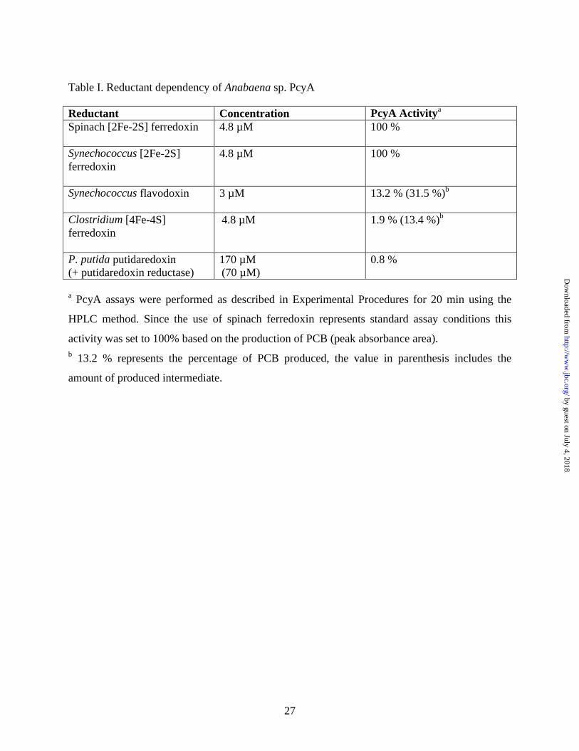
Table I. Reductant dependency of Anabaena sp. PcyA
Reductant Concentration PcyA Activitya Spinach [2Fe-2S] ferredoxin
4.8 µM 100 %
Synechococcus [2Fe-2S] ferredoxin
4.8 µM 100 %
Synechococcus flavodoxin
3 µM 13.2 % (31.5 %)b
Clostridium [4Fe-4S] ferredoxin
4.8 µM 1.9 % (13.4 %)b
P. putida putidaredoxin (+ putidaredoxin reductase)
170 µM (70 µM)
0.8 %
a PcyA assays were performed as described in Experimental Procedures for 20 min using the
HPLC method. Since the use of spinach ferredoxin represents standard assay conditions this
activity was set to 100% based on the production of PCB (peak absorbance area). b 13.2 % represents the percentage of PCB produced, the value in parenthesis includes the
amount of produced intermediate.
by guest on July 4, 2018http://w
ww
.jbc.org/D
ownloaded from

28
Table II. Reversed Phase HPLC Retention Times and Absorption Spectra Properties of Bilin Substrates and Productsa.
Retention Time (min)b
λmax1 (nm) λmax2 (nm) ratio (λmax2/λmax1)
BV IXα 23.2 376 668 0.35
BV XIIIα 18.3 374 656 0.46
BV IIIα 27.7 380 676 0.29
3Z-PΦB 24.1 372 646 0.4
3E-PΦB 19.0 380 654 0.5
Pigment Ic 19.0 368 656 0.44
181,182-DHBV 19.0 368 656 0.45
3Z-isoPΦB 19.3 368 638 0.52
3E-isoPΦB 14.7 374 648 0.56
3Z-PCB 20.9 362 636 0.46
3E-PCB 15.5 368 650 0.53
a Absorption maxima in HPLC mobile phase buffer (acetone:20 mM formic acid::50:50::v:v)
were determined with an Agilent Technologies 1100 Series diode array flow-through detector.
b Retention times (± 0.5 min) were determined using a C18 reversed phase HPLC system as described in Materials and Methods.
c Pigment I is the transient intermediate produced during metabolism of BV IXα by PcyA.
by guest on July 4, 2018http://w
ww
.jbc.org/D
ownloaded from
29
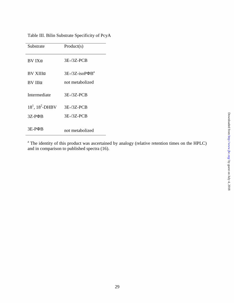
Table III. Bilin Substrate Specificity of PcyA
Substrate Product(s)
BV IXα
3E-/3Z-PCB
BV XIIIα
3E-/3Z-isoPΦBa
BV IIIα
not metabolized
Intermediate
3E-/3Z-PCB
181, 182-DHBV
3E-/3Z-PCB
3Z-PΦB
3E-/3Z-PCB
3E-PΦB
not metabolized
a The identity of this product was ascertained by analogy (relative retention times on the HPLC) and in comparison to published spectra (16).
by guest on July 4, 2018http://w
ww
.jbc.org/D
ownloaded from
biliverdin IXα (BV)
NH
ON NH
NH
O
OOC COO
2 3
5
7 8
10
13
15
17 18
NH
ON NH
NH
O
OOC COO
NH
H3C
ON NH
NH
O
OOC COO
H
3Z-phycocyanobilin (3Z-PCB)
NH
H3C
ON NH
NH
O
OOC COO
H
181,182-dihydrobiliverdin IXα3Z-phytochromobilin (3Z-PΦB)
15, 16-dihydrobiliverdin IXα
3Z-phycoerythrobilin
PebA
PebB
isomerase
x PcyA
PcyA PcyA
by guest on July 4, 2018http://w
ww
.jbc.org/D
ownloaded from

4
4.2
4.4
4.6
4.8
5
5.2
5.4
1.3 1.5 1.7 1.9 2.1 2.3
Ve/Vo
log
MW
βAM ADH
BSA
CA
CYTc
PcyAFd
0
1
2
3
4
5
6
7
8
1 2 3 4 5 6
fraction number
S2
0,w
(s
ec
x1
0-1
3)
PcyA BSA
CYTc
ADH
A
B
1 2 3 4 kDa
130907060
40
3020
15
C
by guest on July 4, 2018http://w
ww
.jbc.org/D
ownloaded from
0
0.05
0.10
0.15
0.20
0
0.05
0.10
0.15
0.20
0.05
0.10
0.15
300 400 500 600 700 800 900
wavelength (nm)
BV IXα
ON NH
NH
NH
O
COO- COO-
BV IIIα
NH
NH
NH
COO-
NO
CO O-
O
BV XIIIα
ON NH
NH
NH
O
COO- COO -
0
rela
tive
ab
sorb
ance
A
BB
B
BB
B
B
JJJ
J
J
J
J
H
H
H
H
H
H
BV IIIα
BV IXα
BV XIIIα
CA
0 2 4 6 8 10 12 14 16 18 20
0.02
0.04
0.06
0.08
PcyA (µM)
abso
rban
ce
0
C
B0.05
500 600 700 800 900
wavelength (nm)
PcyA:BV XIIIα
PcyA:BV IXα
PcyA:BV IIIα
abso
rban
ce
by guest on July 4, 2018http://w
ww
.jbc.org/D
ownloaded from
10 20 30
A6
50
nm
0
10 min
0 min
40
3Z-PΦB
3Z-PCB
3E-PCBI & 3E-PΦB
BV
C
C
C
A
1 8 1 ,182 -DHBV
1 8 1 ,182 -DHBV+ PcyA
time (min)
I0
0 10 20 30
BV
I
3E-PCB3Z-PCB
ab
sorb
an
ce a
rea
(X
10
00
)
B
-0.04
-0.02
0
0.02
0.04
500 600 700 800
Cph1(PΦB)Cph1(PCB)
I
C
50
100
150
200
250
I + PcyA
time (min)
wavelength (nm)
∆ a
bso
rba
nce
by guest on July 4, 2018http://w
ww
.jbc.org/D
ownloaded from

0
20
40
60
80
100
120
140
300 400 500 600 700 800
rela
tive
ab
sorb
an
ce
3Z-isoPΦB
BV XIIIα
3E-isoPΦB
BON N
HNH
NH
O
CO O- C OO-
H ....H3C
3Z/3E-isoPΦB
wavelength (nm)
-0.08
-0.06
-0.04
-0.02
0
0.02
0.04
0.06
0.08
500 600 700 800
wavelength (nm)
∆ a
bso
rba
nce
Cph1(PCB) BV XIIIα + PcyA
BV XIIIα
C
0 10 15 20 25 30 35 40
A6
50
nm BV-IIIα
BV XIIIα
BV IXα
BV IXα
BV IIIα
BV XIIIα
5
time (min)
A3E-isoPΦB 3Z-isoPΦB
3E-PCB 3Z-PCB
+PcyA
by guest on July 4, 2018http://w
ww
.jbc.org/D
ownloaded from

N N
COO-
HO O
N N
-OOC
H
H. . .
. . .
A
BC
D
BV IXα
D1-H+
H-D2+
++++ e-
Fd (red)
N N
COO-
HO O
N N
-OOC
H
H. . .
. . .
A
BC
D
.D1
++++ e-
Fd (red)
H-D2+
N N
COO-
HO O
N N
-OOC
H
H. . .
. . .
A
BC
D
++++
D2D1
181,182-DHBV
181,182-DHBV
N N
COO-
H
O O
N N
-OOC
H
H. . .
. . .
A
BC
D.
++++
H-D3
D4
D2D1
e-
Fd (red) 3Z-PCB
N N
COO-
H
O O
N N
-OOC
H
H. . .
. . .
A
BC
DH
++++
D2D1
D3-
D4-
release
1 2 3
4 5 6
N N
COO-
HO O
N N
-OOC
H
H. . .
. . .
A
BC
D
++++
D2D1
H-D3
H-D4
e-
Fd (red)
by guest on July 4, 2018 http://www.jbc.org/ Downloaded from

Nicole Frankenberg and J. Clark LagariasBiochemical and spectroscopic characterization
Phycocyanobilin:ferredoxin oxidoreductase (PcyA) of Anabaena sp. PCC 7120.
published online January 3, 2003J. Biol. Chem.
10.1074/jbc.M211643200Access the most updated version of this article at doi:
Alerts:
When a correction for this article is posted•
When this article is cited•
to choose from all of JBC's e-mail alertsClick here
by guest on July 4, 2018http://w
ww
.jbc.org/D
ownloaded from


