Monoclinic Paracetamol vs. Paracetamol-4,4'-Bipyridine Co ...

1979
National Cancer Institute
CARCINOGENESIS Technical Report Series No. 128
BIOASSAY OF 3,3'-DIMETHOXYBENZIDINE4,4'-DIISOCYANATE FOR POSSIBLE CARCINOGENICITY
CAS No. 91-93-0
NCI-CG-TR-128
U.S. DEPARTMENT OF HEALTH, EDUCATION, AND WELFARE Public Health Service National Institutes of Health

BIOASSAY OF
3,3'-DIMETHOXYBENZIDINE-4,4'-DIISOCYANATE
FOR POSSIBLE CARCINOGENICITY
Carcinogenesis Testing Program Division of Cancer Cause and Prevention
National Cancer Institute National Institutes of Health Bethesda, Maryland 20014
U.S. DEPARTMENT OF HEALTH, EDUCATION, AND WELFARE Public Health Service
National Institutes of Health
DHEW Publication No. (NIH) 79-1383


REPORT ON THE BIOASSAY OF 3,3'-DIMETHOXYBENZIDINE4,4 '-DIISOCYANATE FOR POSSIBLE CARCINOGENICITY
CARCINOGENESIS TESTING PROGRAM DIVISION OF CANCER CAUSE AND PREVENTION
NATIONAL CANCER INSTITUTE, NATIONAL INSTITUTES OF HEALTH
FOREWORD: This report presents the results of the bioassay of 3,3'dimethoxybenzidine-4,4'-diisocyanate conducted for the Carcinogenesis Testing Program, Division of Cancer Cause and Prevention, National Cancer Institute (NCI), National Institutes of Health, Bethesda, Maryland. This is one of a series of experiments designed to determine whether selected chemicals have the capacity to produce cancer in animals. Negative results, in which the test animals do not have a significantly greater incidence of cancer than control animals, do not necessarily mean the test chemical is not a carcinogen because the experiments are conducted under a limited set of circumstances. Positive results demonstrate that the test chemical is carcinogenic for animals under the conditions of the test and indicate a potential risk to man. The actual determination of the risk to man from animal carcinogens requires a wider analysis.
CONTRIBUTORS: This bioassay of 3,3'-dimethoxybenzidine-4,4'-diisocyanate was conducted by Litton Bionetics, Inc., Bethesda, Maryland, initially under direct contract to the NCI and currently under a subcontract to Tracer Jitco, Inc., prime contractor for the NCI Carcinogenesis Testing Program.
The experimental design was determined by the NCI Project Officers, Dr. N. P. Page (1,2), Dr. E. K. Weisburger (1) and Dr. J. H. Weisburger (1,3). The principal investigators for the contract were Dr. F. M. Garner (4,5) and Dr. B. M. Ulland (4,5). Mr. S. Johnson (4) was the coprincipal investigator for the contract. Animal treatment and observation were supervised by Mr. R. Cypher (4), Mr. D. S. Howard (4) and Mr. H. D. Thornett (4); Mr. H. Paulin (4) analyzed dosed feed mixtures. Ms. J. Blalock (4) was responsible for data collection and assembly.
Histopathologic examinations were performed by Dr. B. C. Zook (4) at Litton Bionetics, Inc., the pathology narratives were written by Dr. B. C. Zook (4), and the diagnoses included in this report represent the interpretation of this pathologist. Histopathology findings and reports were reviewed by Dr. R. L. Schueler (6).
iii

Compilation of individual animal survival, pathology, and summary tables was performed by EG&G Mason Research Institute (7); the statistical analysis was performed by Mr. W. W. Belew (8,9) and Mr. R. M. Helfand (8), using methods selected for the Carcinogenesis Testing Program by Dr. J. J. Gart (10).
This report was prepared at METREK, a Division of The MITRE Corporation (8) under the direction of the NCI. Those responsible for this report at METREK are the project coordinator, Dr. L. W. Thomas (8), task leader Ms. P. Walker (8), senior biologist Mr. M. Morse (8), biochemist Mr. S. C. Drill (8), chemist Dr. N. Zimmerman (8), and technical editor Ms. P. A. Miller (8). The final report was reviewad by members of the participating organizations.
The following other scientists at the National Cancer Institute were responsible for evaluating the bioassay experiment, interpreting the results, and reporting the findings: Dr. K. C. Chu (1), Dr. C. Cueto, Jr. (1), Dr. J. F. Douglas (1), Dr. D. G. Goodman (1,11), Dr. R. A. Griesemer (1), Dr. M. H. Levitt (1), Dr. H. A. Milman (1), Dr. T. W. Orme (1), Dr. R. A. Squire (1,12), Dr. S. F. Stinson (1), Dr. J. M. Ward (1), and Dr. C. E. Whitmire (1).
1. Carcinogenesis Testing Program, Division of Cancer Cause and Prevention, National Cancer Institute, National Institutes of Health, Bethesda, Maryland.
2. Now with the U.S. Environmental Protection Agency, 401 M Street S.W., Washington, D.C.
3. Now with the Naylor Dana Institute for Disease Prevention, American Health Foundation, Hammon House Road, Valhalla, New York.
4. Litton Bionetics, Inc., 5516 Nicholson Lane, Kensington, Maryland.
5. Now with Hazleton Laboratories America, Inc., 9200 Leesburg Turnpike, Vienna, Virginia.
6. Tracer Jitco, Inc., 1776 East Jefferson Street, Rockville, Maryland.
7. EG&G Mason Research Institute, 1530 East Jefferson Street, Rockville, Maryland.
8. The MITRE Corporation, METREK Division, 1820 Dolley Madison Boulevard, McLean, Virginia.
iv

9. Now with the Solar Energy Research Institute, Cole Boulevard, Golden, Colorado.
10. Mathematical Statistics and Applied Mathematics Section, Biometry Branch, Field Studies and Statistics Program, Division of Cancer Cause and Prevention, National Cancer Institute, National Institutes of Health, Bethesda, Maryland.
11. Now with Clement Associates, Inc., 1010 Wisconsin Avenue, N.W., Washington, D.C.
12. Now with the Division of Comparative Medicine, Johns Hopkins University, School of Medicine, Traylor Building, Baltimore, Maryland.


SUMMARY
A bioassay for the possible carcinogenicity of 3,3"-dimethoxybenzidine-4,4'-diisocyanate was conducted using Fischer 344 rats and B6C3F1 mice. 3,3"-Dimethoxybenzidine-4,4'-diisocyanate was administered, at either of two concentrations, to groups of 50 male and 50 female animals of each species, with the exception of 49 high dose female rats. The compound was administered in the feed with the exception of the first 22 weeks of the rat bioassay, when it was administered by gavage. Twenty animals of each sex ami species were placed on test as controls. During intubation the high and low dosages of 3,3'-dimethoxybenzidine-4,4'-diisocyanate administered to rats were 3000 and 1500 mg/kg, respectively, while the high and low concentrations administered in the feed to both rats and mice were 44,000 and 22,000 ppm, respectively. The compound was administered for a period of 78 weeks, followed by an observation period of 26 weeks for rats and 25 weeks for mice.
There was a significant positive association between the administration of 3,3'-dimethoxybenzidine--4,4'-diisocyanate and mortality in male and female rats, but not in male or female mice. Adequate numbers of animals in all groups survived sufficiently long to be at risk from late-developing tumors.
For both sexes of rats, there was a significant positive association between dosage and the incidence of leukemia and malignant lymphoma. There was a significantly higher incidence of neoplasms of the skin, excluding skin of the ear, when dosed ui'iie rats were compared to controls. There was a significant positive association between the dosages administered and the incidences of endometrial stromal polyps in female rats. None of the statistical tests for any site in male or female mice indicated a signifiesat positive association between compound administration and tumor incidence.
Under the conditions of this bioassay, administration of 3,3'dimethoxybenzidine-4,4'-diisocyanate was carcinogenic to Fischer 344 rats, causing neoplasms of the skin (excluding skin of the ear) in males, endometrial stromal polyps in females, and leukemia and malignant lymphoma in both sexes. The compound was also associated with the development of a combination of squamous-cell carcinomas and sebaceous adenocarcinomas of the Zymbal's gland and skin of the ear in rats of both sexes. There was no evidence for the carcinogenicity of the compound in B6C3F1 mice.
vli


TABLE OF CONTENTS
Page
I. INTRODUCTION 1
II. MATERIALS AND METHODS 4
A. Chemicals 4 B. Dosage Preparation 5 C. Dietary Preparation 5 D. Animals 6 E. Animal Maintenance 7 F. Selection of Initial Concentrations 9 G. Gastric Intubation 10 H. Experimental Design 10 I. Clinical and Histopathologic Examinations 14 J. Data Recording and Statistical Analyses 1
21 III. CHRONIC TESTING RESULTS: RATS
A. Body Weights and Clinical Observations 21 21B. Survival
C. Pathology 24
D. Statistical Analyses of Results 9
42 IV. CHRONIC TESTING RESULTS: MICE
A. Body Weights and Clinical Observations 42 B. Survival 42 C. Pathology 46
D. Statistical Analyses of Results 47
V. DISCUSSION 53
VI. BIBLIOGRAPHY 56
APPENDIX A SUMMARY OF THE INCIDENCE OF NEOPLASMS IN RATS TREATED WITH 3,3'-DIMETHOXYBENZIDINE4,4'-DIISOCYANATE A-l
APPENDIX B SUMMARY OF THE INCIDENCE OF NEOPLASMS IN MICE TREATED WITH 3,3'-DIMETHOXYBENZIDINE4,4'-DIISOCYANATE B-l
APPENDIX C SUMMARY OF THE INCIDENCE OF NONNEOPLASTIC LESIONS IN RATS TREATED WITH 3,3'-DIMETHOXYBENZ'IDINE-4,4'-DIISOCYANATE C-l
ix

TABLE OF CONTENTS (Concluded)
Page
APPENDIX D SUMMARY OF THE INCIDENCE OF NONNEOPLASTIC LESIONS IN MICE TREATED WITH 3,3'-DIMETHOXYBENZIDINE-4,4' -DUSOCYAMATE D-l
x

LIST OF ILLUSTRATIONS
Figure Number Page
1 CHEMICAL STRUCTURE OF 3,3'-DIMETHOXYBENZIDINE-4,4'-DIISOCYANATE 2
GROWTH CURVES FOR 3,3'-DIMETHOXYBENZIDINE4,4'-DIISOCYANATE CHRONIC STUDY RATS 22
SURVIVAL COMPARISONS OF 3,3'-DIMETHOXYBENZIDINE-4,4'-DIISOCYANATE CHRONIC STUDY RATS 23
GROWTH CURVES FOR 3,3'-DIMETHOXYBENZIDINE4,4'-DIISOCYANATE CHRONIC STUDY MICE 43
SURVIVAL PROBABLITY COMPARISONS OF 3,3'-DIMETHOXYBENZIDINE-4,4'-DIISOCYANATE CHRONIC STUDY MICE 44
PERCENT SURVIVAL OF 3,3'-DIMETHOXYBENZIDINE4,4'-DIISOCYANATE CHRONIC STUDY MICE 45
LIST OF TABLES
Table Number
DESIGN SUMMARY FOR FISCHER 344 RATS—3,31 -DIMETHOXYBENZIDINE-4,4'-DIISOCYANATE CHRONIC BIOASSAY 11
DESIGN SUMMARY FOR B6C3F1 MICE—3,3'-DIMETHOXYBENZIDINE-4,4'-DIISOCYANATE FEEDING EXPERIMENT 12
TIME-ADJUSTED ANALYSES OF THE INCIDENCE OF PRIMARY TUMORS AT SPECIFIC SITES IN MALE RATS TREATED WITH 3,3'-DIMETHOXYBENZIDINE4,4'-DIISOCYANATE 30
ANALYSES OF THE INCIDENCE OF PRIMARY TUMORS AT SPECIFIC SITES IN FEMALE RATS TREATED WITH 3,3'-DIMETHOXYBENZIDINE-4,4'-DIISOCYANATE 36
ANALYSES OF THE INCIDENCE OF PRIMARY TUMORS AT SPECIFIC SITES IN MALE MICE TREATED WITH 3,3'-DIMETHOXYBENZIDINE-4,4'-DIISOCYANATE 48
XI
LIST OF TABLES (Concluded)
Table Number Page
6 ANALYSES OF THE INCIDENCE OF PRIMARY TUMORS AT SPECIFIC SITES IN FEMALE MICE TREATED WITH 3,3'-DIMETHOXYBENZIDINE-4,4'-DIISOCYANATE 50
Al SUMMARY OF THE INCIDENCE OF NEOPLASMS IN MALE RATS TREATED WITH 3,3'-DIMETHOXYBENZIDINE-4, 4 '-DIISOCYANATE A-3
A2 SUMMARY OF THE INCIDENCE OF NEOPLASMS IN FEMALE RATS TREATED WITH 3,3'-DIMETHOXYBENZIDINE-4, 4'-DIISOCYANATE A-7
Bl SUMMARY OF THE INCIDENCE OF NEOPLASMS IN MALE MICE TREATED WITH 3,3'-DIMETHOXYBENZIDINE-4, 4 '-DIISOCYANATE B-3
B2 SUMMARY OF THE INCIDENCE OF NEOPLASMS IN FEMALE MICE TREATED WITH 3,3'-DIMETHOXYBENZIDINE-4, 4 '-DIISOCYANATE B-6
Cl SUMMARY OF THE INCIDENCE OF NONNEOPLASTIC LESIONS IN MALE RATS TREATED WITH 3,3'DIMETHOXYBENZIDINE-4,4'-DIISOCYANATE 0-3
C2 SUMMARY OF THE INCIDENCE OF NONNEOPLASTIC LESIONS IN FEMALE RATS TREATED WITH 3,3'-DIMETHOXYBENZIDINE-4,4'-DIISOCYANATE C-7
Dl SUMMARY OF THE INCIDENCE OF NONNEOPLASTIC LESIONS IN MALE MICE TREATED WITH 3,3'-DIMETHOXYBENZIDINE-4,4'-DIISOCYANATE D-3
D2 SUMMARY OF THE INCIDENCE OF NONNEOPLASTIC LESIONS IN FEMALE MICE TREATED WITH 3,3'DIMETHOXYBENZIDINE-4,4'-DIISOCYANATE D-7
XII

I. INTRODUCTION
3,3'-Dimethoxybenzidine-4,4'-diisocyanate (Figure 1) (NCI No.
C02175), a dimer of o-anisidine isocyanate, was selected for bioassay
by the National Cancer Institute because of the structural similarity
of this compound to 3,3'-dimethoxybenzidine, a carcinogen in Fischer
rats (Hadidian et al., 1968).
The Chemical Abstracts Service (CAS) Ninth Collective Index (1977)
name fcr this compound is 4,4'-diisocyanato-3,3'-dimethoxy-1,1'-bi
* phenyl. It is also called dianisidine diisocyanate; isocyanic acid
3,3'-dimethoxy-4,4'-biphenylene ester; 4,4'-diisocyanato-3,3'-dime
thoxybiphenyl; 3,3'-dimethoxybiphenylene-4,4'-diisocyanate; 3,3'-dime
thoxy-4, 4'-biphenyl diisocyanate; and 3,3'-dimethoxy-4,4'-biphenylene
diisocyanate.
Several patents have been issued or applied for describing poten
tial uses of 3,3'-dimethoxybenzidine-4,4'-diisocyanate. These include
synthesis of shock-absorbing polyurea-polyurethanes, which can be used
in railway car couplers (Chung, 1975); in situ manufacture of polyure
thane gaskets and seals for metal pail and drum covers, aerosol mount
ing cups, and metal bottoms of fiber drums (Grace, 1974); synthesis of
heat stable thermoplastic urethane rubbers, useful as gaskets, seals,
0-rings, and wire coatings (Bonk and Shah, 1975); formulation of pho
tocurable triazine-containing polyene-polythiol lacquers for steel
* The CAS registry number is 91-93-0.

N=C=0
CH3—0 0—CHs
FIGURE 1 CHEMICAL STRUCTURE OF 3,3'-DIMETHOXYBEIMZIDINE-4. 4'-DMSOCYANATE

cans (Guthrie and Rendulic, 1975); and manufacture of abrasion-
resistant antireflection coatings for color photographs (Chiklis,
1975); however, these uses appear to be purely experimental.
Specific production data for 3,3'-dimethoxybenzidine-4,4'-dLiso
cyanate are not available; however, exclusion of this compound from
the 1977 Directory of Chemical Producers, U.S.A. (Stanford Research
Institute, 1977) implies that it is not currently produced in commer
cial quantities (in excess of 1000 pounds or $1000 in value annually)
in the United States.
The potential for exposure to 3J3l-dimethoxybenzidine-4,4'-diiso
cyanate is limited to researchers, particularly those involved in the
experimental synthesis of elastomers and polymeric coatings.

II. MATERIALS AND METHODS
A. Chemicals
3,3'-Dimethoxybenzidine-4,4'-diisocyanate was purchased from
Upjohn Company, Kalamazoo, Michigan. Chemical analysis was performed
by Litton Bionetics, Inc., Kensington, Maryland. The experimentally
determined range in melting point was 113° to 116°C. No literature
value was found for comparison. Thin-layer chromatography (TLC) was
performed utilizing two solvent systems (i.e., anhydrous acetone and
ethyl acetate). Each plate was visualized with visible and ultra
violet light, iodine vapor, and ferricyanide-nitroprusside spray.
The plate developed with acetone showed one contaminant that was less
motile than the major spot, while the plate developed with ethyl ace
tate revealed two contaminants, one of lesser and one of greater mo
tility than the major spot. The results of infrared (IR) and nuclear
magnetic resonance (NMR) analyses were consistent with those expected
based on the structure of the compound. Ultraviolet/visible (UV/VIS)
spectrophotometry showed X at 310 and 285 nm with respective molar max
4 4extinction coefficients of 2.55 x 10 and 1.87 x 10 .
A second batch of the compound was purchased approximately 1.5
years later from the same manufacturer. The experimentally determined
range in melting point was 109.3° to 111.3°C. TLC was performed as
with the first batch. One impurity that was less motile than the
major spot was observed on each plate. The results of IR and NMR
analyses were consistent with those expected based on the structure
4
of the compound. UV/VIS analysis revealed \ at 307 and 280 nm max
A with respective molar extinction coefficients of 2.33 x 10 and
1.67 x 10⁴.
Throughout this report the term 3,3'-dimethoxybenzidine-4,4'
diisocyanate is used to represent this material.
B. Dosage Preparation
Fresh solutions of 3,3'-dimethoxybenzidine-4,4'-diisocyanate in
* steroid suspending vehicle (Litton Bionetics, Inc., Kensington, Mary
land) were prepared on each day that intubation was performed. Excess
portions of the mixtures were disposed of rather than stored. The
concentration of 3,3l-dimethoxybenzidine-4,4'-diisocyanate in steroid
suspending vehicle ranged from 15 to 30 percent.
C. Dietary Preparation
The basal laboratory diet for both dosed and control animals
consisted of Wayne Lab-Blox (Allied Mills, Inc., Chicago, Illinois).
3,3'-Dimethoxybenzidine-4,4'-diisocyanate was administered to the
dosed mice as a component of the diet throughout the period of chemi
cal administration, while it was administered to rats as a component
of the diet beginning with week 23 and continuing for the duration
of compound administration. (Rats were intubated with the chemical
for the first 22 weeks of the bioassay.)
* Steroid suspending vehicle = 9 gm NaCl, 5 gm carboxymethylcellulose,
~4 ml polysorbate 80, and 9 ml benzyl alcohol q.s. to 1 liter with distilled water.

The chemical was removed from its container and a proper amount
was blended with an aliquot of the ground feed using a mortar and
pestle. Once visual homogeneity was attained, the mixture was placed
in a 6 kg capacity Patterson-Kelley standard model twin-shell stain
less steel V-blender along with the remainder of the feed to be pre
pared. After 20 minutes of blending, the mixtures were placed in
double plastic bags and stored in the dark at 4°C. The mixture was
prepared once weekly.
Dosed feed preparations containing 23,000 and 46,000 ppm of 3,3'
dimethoxybenzidine-4,4'-diisocyanate were analyzed spectrophototnetri
cally. The mean result immediately after preparation was 95.2 _+ 4.2
percent of theoretical, including correction for the analytical method
of recovery used. After ten days, at ambient room temperature, the
mean result was 100.3 +_ 4.4 percent of theoretical, including correc
tion for the analytical method of recovery used.
D. Animals
Two animal epecies, rats and mice, were used in the carcinogeni
city bioassay. Fischer 344 rats and B6C3F1 mice were obtained through
contracts of the Division of Cancer Treatment, National Cancer Insti
tute. Rats were supplied by A. R. Schmidt, Madison, Wisconsin, and
Charles River Breeding Laboratories, Inc., Wilmington, Massachusetts.
All mice were supplied by Charles River Breeding Laboratories, Inc.

Rats and mice were approximately 4 weeks old when received. Upon
receipt, animals were examined for visible signs of disease or para
sites. Obviously ill or runted animals were culled. The remaining
animals were quarantined for 2 weeks prior to initiation of test.
Animals which did not manifest clinical signs of disease were placed
on test at this time. Animals were assigned to groups and distributed
among cages so that the average body weight per cage was approximately
equal for a given species and sex.
E. Animal Maintenance
All animals were housed by species in temperature- and humidity-
controlled rooms. The temperature range was 22° to 26°C and the
relative humidity was maintained between 45 and 55 percent. Incoming
air was filtered through HEPA filters (Flanders Filters, McLean, Vir
ginia) at a rate of 12 to 15 complete changes of room air per hour»
Fluorescent lighting was provided 8 hours per day (9:00 a.m. to 5:00
p.m.).
All rats were housed four per cage by sex and all mice were housed
five per cage by sex. Throughout the study dosed and control animals
of both species were housed in polycarbonate cages (Lab Products, Inc.,
Gartielc, New Jersey) suspended from aluminum racks. Racks were
fitted wjth a continuous piece of stainless steel mesh over which a
sheet of rilter paper was firmly secured. Filter paper was changed
at 2-week intervals, when the racks were sanitized* Clean cages and
®bedding were provided twice weekly. Ab-sorb-dri hardwood chip

bedding (Wilner Wood Products Company, Norway, Maine) was used in
polycarbonate cages for the entire bioassay.
Acidulated water (pH 2.5) was supplied to animals in water bot
tles filled by an automated metering device that was checked daily
for diluting accuracy. Water bottles were changed and washed twice
weekly, and sipper tubes were washed at weekly intervals. During the
period of chemical administration, dosed and control animals received
®treated or untreated Wayne Lab-Blox meal as appropriate. The feed
was supplied in hanging stainless steel hoppers which were refilled
three times per week and sanitized weekly. Food and water were
available ad libitum for both species.
All dosed and control rats were housed in a room with other rats
* receiving diets containing N-phenyl-p-phenylenediamine hydrochloride
(2198-59-6); acetylaminofluorene (53-96-3); and nitrilotriacetic acid
(139-13-9).
All dosed and control mice were housed in a room with mice re
ceiving diets containing EDTA trisodium salt. (150-38-9); diaminozide
(1596-84-5); N,N'-diethylthicurea (105-55-5); triphenyltin hydroxide
(76-87-9); carbromal (75-65-6); p-quinone dioxime (105-11-3); 4-amino
2-nitrophenol (119-34-6); other mice intubated with lithocholic acid
(434-13-9); and other mice receiving I.P. injections of methiodol
sodium (126-31-8).
* CAS registry numbers are given in parentheses.

F. Selection of Initial Concentrations
In order to establish the maximum tolerated concentrations of
3,3'-dimethoxybenzidine-4,4'-diisocyanate for administration to dosed
animals in the chronic study, subchronic toxicity tests were conducted
with both rats and mice. Animals of each species were distributed
among six groups, each consisting of five males and five females.
3,3'-Dimethoxybenzidine-4,4'-diisocyanate mixed with distilled water
was introduced by gavage to five of the six rat groups at dosages of
800, 1260, 2000, 3160, and 5000 mg/kg and to five of the six mouse
groups at dosages of 215, 464, 1000, 2150, and 4640 mg/kg. The sixth
group of each species served as a control, receiving only steroid
suspending vehicle by gavage. Intubation was performed five times
per week for 4 weeks, followed by a 2-week observation period to
detect any delayed toxicity. Individual body weights were recorded
weekly. At the end of the observation period, all survivors were
sacrificed and necropsied.
At the end of the subchronic test, mean body weight gain among
male rats dosed with 2000 mg/kg was 1 percent greater than the mean
body weight gain of their controls, while female rats receiving the
same dosage displayed a mean body weight gain 4 percent less than
that of their controls. At a dosage of 3160 rag/kg, the mean body
weight gain among male rats was 9 percent less than that of their
controls, while female rats receiving the same dosage displayed a
mean body weight gain 14 percent less than that of their controls.

The high concentration selected for administration to dosed rats in
the chronic bioassay was 3000 mg/kg.
At the end of the subchronic test, mean body weight gain among
male mice dosed with 4640 mg/kg was 4 percent less than the mean
body weight gain of their controls, while female mice receiving the
same dosage displayed a mean body weight gain 11 percent less than
that of their controls. The high concentration selected for admin
istration to dosed mice in the chronic bioassay was 44,000 ppm
(equivalent to 7040 mg/kg, assuming an average food consumption of
4 grams/day and an average body weight of 25 grams).
G. Gastric Intubation
Intubation was performed for 5 consecutive days per week on a
mg/kg body weight basis, utilizing the most recently observed group
mean body weight as a guide for determining the dose. All rats
were weighed and dosages adjusted once monthly, based on group mean
body weight. Thus, although the ratio of dose to weight remained
constant, the total dosage administered fluctuated with an increase
or decrease in group mean body weight. Rats of each sex within a
dosed group received the same dosage.
H. Experimental Design
The experimental design parameters for the chronic study (spe
cies, sex, group size, concentrations administered, duration of
treated and untreated observation periods, and the time-weighted
average concentrations) are summarized in Tables 1 and 2.
10
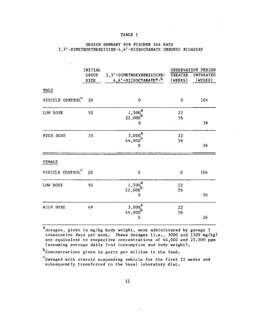
TABLE 1
DESIGN SUMMARY FOR FISCHER 344 RATS 3,3'-DIMETHOXYBENZIDINE-4,4l-DIISOCYANATE CHRONIC BIOASSAY
INITIAL OBSERVATION PERIOD GROUP 3,3'-DIMETHOXYBENZIDINE TREATED UNTREATED SIZE 4,4'-DIISOCYANATE3 » b (WEEKS) (WEEKS)
MALE
VEHICLE CONTROL 20 104
LOW DOSE 50 1,500* 22 22,000 56
0 26
HIGH DOSE 50 3,000* 22 44,000 56
0 26
FEMALE
VEHICLE CONTROL 20 104
LOW DOSE 50 1,500* 22 22,000 56
0 26
HIGH DOSE 49 3 000* 22 ' h 44,000 56
0 26
Dosages, given in rag/kg body weight, were administered by gavage 5 consecutive days per week. These dosages (i.e., 3000 and 1500 mg/kg) are equivalent to respective concentrations of 44,000 and 22,000 ppm (assuming average daily food consumption and body weight).
Concentrations given in parts per million in the feed,
Gavaged with steroid suspending vehicle for the first 22 weeks and subsequently transferred to the basal laboratory diet.
11
c

TABLE 2
DESIGN SUMMARY FOR B6C3F1 MICE 3,3'-DIMETHOXYBENZIDINE-4,4'-DIISOCYANATE FEEDING EXPERIMENT
MALE
INITIAL GROUP
SIZE
3,3' -DIMETHOXYBENZIDINE4,4'-DIISOCYANATE CONCENTRATION3
OBSERVATION PERIOD TREATED UNTREATED
(WEEKS) (WEEKS)
CONTROL 20 0 0 103
LOW DOSE 50 22,000 0
78 25
HIGH DOSE 50 44,000 0
78 25
FEMALE
CONTROL 20 0 0 103
LOW DOSE 50 22,000 0
78 25
HIGH DOSE 50 44,000 0
78 25
Concentrations given in parts per million.
12

The method of compound administration for the chronic bioassay
in rats, which was begun prior to the chronic bioassay in mice, was
initially intubation. Intubation was selected because it was be
lieved that the compound was not stable in feed. After determina
tion that the compound was stable in feed (as indicated on page 6)
the method of compound administration to rats was changed to dietary
and the mouse bioassay was initiated using dietary administration.
All rats were approximately 6 weeks old at the time the test was
initiated and were placed on test simultaneously. The dosages ini
tially administered to males and females were 3000 and 1500 rag/kg,
by gavage (equivalent to dietary concentrations of 44,000 and 22,000
ppm). Throughout this report those rats initially receiving the for
mer dosage are referred to as the high dose groups and those initially
receiving the latter dosage are referred to as the low dose groups.
All dosed rats were administered 3,3'-dimethoxybenzidine 4,4'-diiso
cyanate by gavage at the dosages indicated for 22 weeks. Beginning
in week 23, 3,3'-dimethoxybenzidine-4,4'-diisocyanate was mixed with
the feed and administered ad libitum in the diet. All dosed rats
were supplied feed containing 3,3'-dimethoxybenzidine-4,4'-dii8Ocya
nate for the remaining 56 weeks of compound administration. During
this 56-week period, the high and low concentrations administered
were 44,000 and 22,000 ppm, respectively. The dosed rats were
observed for a period of 26 weeks after compound administration
ceased.
13

All mice were approximately 6 weeks old at the time the test was
initiated and were placed on test simultaneously. The dietary con
centrations of 3,3'-dimethoxybenzidine-4,4'-diisocyanate administered
to both sexes were 44,000 and 22,000 ppm. Throughout this report
those mice receiving the former concentration are referred to as the
high dose groups and those receiving the latter concentration are
referred to as the low dose groups. Dosed mice were supplied with
feed containing 3,3!-dimethoxybenzidine-4,4'-diisocyanate for 78
weeks followed by a 25-week observation period.
I. Clinical and Histopathologic Examinations
Animals were weighed immediately prior to initiation of the ex
periment. From the first day, all animals were inspected twice daily
for mortality. Food consumption data were collected at monthly inter
vals from 20 percent of the animals in each group. Body weights of
rats were recorded once a week for the first 6 weeks, every 2 weeks
for the next 12 weeks, and once a month thereafter. Body weights of
mice were recorded once a week for the first 6 weeks, every 2 weeks
for the next 10 weeks and once a month for the remainder of the bio
assay.
All moribund animals or animals that developed large, palpable
masses that jeopardized their health were sacrificed. A necropsy
was performed on each animal regardless of whether it died, was sac
rificed when moribund, or was sacrificed at the end of the bioassay.
14

The animals were euthanized by carbon dioxide asphyxiation, and were
immediately necropsied. The histopathologic examination consisted of
gross and microscopic examination of all major tissues, organs, and
gross lesions taken from sacrificed animals and, whenever possible,
from animals found dead.
Tissues were preserved in a 10 percent neutral buffered formalin
solution, embedded in paraffin, sectioned, and stained with hematox
ylin and eosin prior to microscopic examination.
Slides were prepared from the following tissues: skin, subcuta
neous tissue, ear, lungs and bronchi, trachea, bone marrow, spleen,
lymph nodes, thymus, heart, salivary gland, liver, gallbladder (mice),
pancreas, esophagus, Zymbal's gland, stomach, small intestine, large
intestine, kidney, urinary bladder, pituitary, adrenal, thyroid,
parathyroid, testis, prostate, brain, uterus, mammary gland, and
ovary.
A few tissues were not examined for some animals, particularly
for those that died early. Also, some animals were missing, canni
balized, or judged to be in such an advanced state of autolysis as to
preclude histopathologic interpretation. Thus, the number of animals
for which particular organs, tissues, or lesions were examined micro
scopically varies and does not necessarily represent the number of
animals that were recorded in each group at the time that the test
was initiated.
15

J. Data Recording and Statistical Analyses
Pertinent data on this experiment have been recorded in an auto
matic data processing system, the Carcinogenesis Bioassay Data System
(Linhart et al., 1974). The data elements include descriptive infor
mation on the chemicals, animals, experimental design, clinical ob
servations, survival, body weight, and individual pathologic results,
as recommended by the International Union Against Cancer (Berenblum,
1969). Data tables were generated for verification of data transcrip
tion and for statistical review.
These data were analyzed using the statistical techniques de
scribed in this section. Those analyses of the experimental results
that bear on the possibility of carcinogenicity are discussed in the
statistical narrative sections.
Probabilities of survival were estimated by the product-limit
procedure of Kaplan and Meier (1958) and are presented in this report
in the form of graphs. Animals were statistically censored as of the
time that they died of other than natural causes or were found to be
missing; animals dying from natural causes were not statistically
censored. Statistical analyses for a possible dose-related effect
on survival used the method of Cox (1972) when testing two groups for
equality and used Tarone's (1975) extensions of Cox's methods when
testing a dose-related trend. One-tailed P-values have been reported
for all tests except the departure from linearity test, which is only
reported when its two-tailed P-value is less than 0.05.
16

The incidence of neoplastic or nonneoplastic lesions has been
given as the ratio of the number oi animals bearing such lesions at a
specific anatomic site (numerator) to the number of animals in which
that site was examined (denominator). In most instances, the denomi
nators included only those animals for which that site was examined
histologically. However, when macroscopic examination was required
to detect lesions prior to histologic sampling (e.g., skin or mammary
tumors), or when lesions could have appeared at multiple sites (e.g.,
lymphomas), the denominators consist of the numbers of animals necrop
sied.
The purpose of the statistical analyses of tumor incidence is to
determine whether animals receiving the test chemical developed a sig
nificantly higher proportion of tumors than did the control animals.
As a part of these analyses, the one-tailed Fisher exact test (Cox,
1970, pp. 48-52) was used to compare the tumor incidence of a control
group to that of a group of treated animals at each dose level. When
results for a number of treated groups, k, are compared simultaneously
with those for a control group, a correction to ensure an overall
significance level of 0.05 may be made. The Bonferroni inequality
(Miller, 1966, pp. 6-10) requires that the P-value for any comparison
be less than or equal to 0.05/k. In cases where this correction was
used, it is discussed in the narrative section. It is not, however,
presented in the tables, where the Fisher exact P-values are shown.
17

The Cochran-Armitage test for linear trend in proportions, with
continuity correction (Armitage, 1971, pp. 362-365), was also used
when appropriate0 Under the assumption of a linear trend, this test
determined if the slope of the dose-response curve is different from
zero at the one-tailed 0.05 level of significance. Unless otherwise
noted, the direction of the significant trend was a positive dose re
lationship. This method also provides a two-tailed test of departure
from linear trend.
A time-adjusted analysis was applied when numerous early deaths
resulted from causes that were not associated with the formation of
tumors» In this analysis, deaths that occurred before the first
tumor was observed were excluded by basing the statistical tests on
animals that survived at least 52 weeks, unless a tumor was found at
the anatomic site of interest before week 52. When such an early
tumor was found, comparisons were based exclusively on animals that
survived at least as long as the animal in which the first tumor was
founds, Once this reduced set of data was obtained, the standard pro
cedures for analyses of the incidence of tumors (Fisher exact tests,
Cocaran-Armitage tests, etc.) were followed.
When appropriate, life-table methods were used to analyze the
incidence of tumors. Curves of the proportions surviving without an
observed tumor were computed as in Saffiotti et al. (1972). The week
during which animals died naturally or were sacrificed was entered
as the time point of tumor observation. Cox's methods of comparing
18

these curves were used for two groups; Tarone's extension to testing
for linear trend was used for three groups. The statistical tests for
the incidence of tumors which used life-table methods were one-tailed
and, unless otherwise noted, in the direction of a positive dose
relationship. Significant departures from linearity (P < 0.05, two-
tailed test) were also noted.
The approximate 95 percent confidence interval for the relative
risk of each dosed group compared to its control was calculated from
the exact interval on the odds ratio (Gart, 1971). The relative risk
is defined as p /p where p is the true binomial probability of the t c t
incidence of a specific type of tumor in a treated group of animals
and p is the true probability of the spontaneous incidence of the c
same type of tumor in a control group. The hypothesis of equality
between the true proportion of a specific tumor in a treated group
and the proportion in a control group corresponds to a relative risk
of unity. Values in excess of unity represent the condition of a
larger proportion in the treated group than in the control.
The lower and upper limits of the confidence interval of the
relative risk have been included in the tables of statistical analy
ses. The interpretation of the limits is that in approximately 95
percent of a large number of identical experiments, the true ratio
of the risk in a treated group of animals to that in a control group
would be within the interval calculated from the experiment. When
the lower limit of the confidence interval is greater than one, it
19

can be inferred that a statistically significant result (a P < 0.025
one-tailed test when the control incidence is not zero, P < 0.050
when the control incidence is zero) has occurred. When the lower
limit is less than unity but the upper limit is greater than unity,
the lower limit indicates the absence of a significant result while
the upper limit indicates that there is a theoretical possibility
of the induction of tumors by the test chemical which could not be
detected under the conditions of this test.
20

III. CHRONIC TESTING RESULTS: RATS
A. Body Weights and Clinical Observations
Dose-related mean body weight depression was not apparent in
either male or female rats. There was, however, slight mean body
weight depression in dosed groups of both sexes when compared to
their respective controls (Figure 2).
No abnormal clinical signs were recorded.
B. Survival
The estimated probabilities of survival for male and female rats
in the control and 3,3'-dimethoxybenzidine-4,4'-diisocyanate-dosed
groups are shown in Figure 3. For male rats the Tarone test for a
positive association between dosage and mortality was significant
(P = 0.002). While the Tarone test was not significant for females,
the Cox tests comparing high dose to control and low dose to control
indicated a significantly higher mortality among dosed rats when com
pared with the control group.
There were adequate numbers of male rats at risk from late-
developing tumors as 80 percent (40/50) of the high dose, 88 percent
(44/50) of the low dose, and 90 percent (18/20) of the control group
survived on test more than 80 weeks. Thirty-two percent (16/50) of
the high dose, 44 percent (22/50) of the low dose and 75 percent
(15/20) of the control group survived on test until the termination
of the study.
21

750-r •750
600 -600
er o
450 h-450
LU
5
gsoo -300 CQ
150 -150
MALE RATS
I ~T 15 45 60 75 90 105 120
TIME ON TEST (WEEKS)
750
600 — -600
CC O
X
UJ
450 -450
D O m
r-300
150 — 150
FEMALE RATS
15 ~T~
30 ~T 45 CO 75
TIME ON TEST (WEEKS)
T 90 105 120
FIGURE 2 GROWTH CURVES FOR 3,3'-DIMETHOXYBENZIDINE-4,4'-DIISOCYANATE CHRONIC STUDY RATS
22

PR
OB
AB
ILIT
Y O
F S
UR
VIV
AL
P
RO
BA
BIL
ITY
OF
SU
RV
IVA
L
oo
pj>
o>
bo
ca
30
1
Tl
O
m
•o
m30
33
§
u
w
H
r! £
O
3
DO Z
}
ro
mO
J
s-
m
m
en
8
g-
O
I O
bo
b

Similarly, there were adequate numbers of female rats at risk
from late-developing tumors with 90 percent (45/50) of the high dose,
80 percent (40/50) of the low dose, and 95 percent (19/20) of the
control group surviving on test more than 80 weeks. At the termina
tion of the study, 42 percent (21/50) of the high dose, 42 percent
(21/50) of the low dose and 75 percent (15/20) of the control group
were alive on test.
C. Pathology
Histopathologic findings on neoplasms in rats are summarized in
Appendix A (Tables Al and A2); findings on nonneoplastic lesions are
summarized in Appendix C (Tables Cl and C2).
There were increased incidences of tumors of baso-squamous
epithelial origin in dosed male and female rats, when compared to
their respective controls. These tumors were commonly in the skin of
the head, inguinal area and back. The location of 28 tumors of the
head strongly suggested that these tumors arose from the Zymbal's
gland. The location and type of 17 tumors in the inguinal area sug
gested involvement of preputial glands. Grossly, they appeared as
irregular fungating or bulging ulcerated lesions measuring about 0.5
to 3.0 cm in greatest dimension.
Microscopically, the tumors thought to arise from the Zymbal's
gland were carcinomas of the sebaceous gland, squamous-cell carcino
mas, and a carcinosarcoma as summarized in the following table:
24

Low High Control Dose Dose
MALES
Number of Animals with '"ipsues Examined Histopatholofe^>..-lly (20) (50) (50)
ZYMBAL'S GLAND
Squamous-Cell Carcinoma 0 2(4%) 5(10%)
Sebaceous Adenocarcinoma 0 3(6%) 2(4%)
Careinosarcoma 0 0 0
FEMALES
Number of Animals with Tissues Examined Histopathologically (20) (50) (48)
ZYMBAL'S GLAND
Squamous-Cell Carcinoma 0 1(2%) 4(8%)
Sebaceous Adenocarcinoma 0 7(14%) 2(4%)
Carcinosarcoma 1(5%) 0 0
These and other tumors of baso-squamous origin were classified
according to the apparent direction of maturation. The auditory tu
mors had both sebaceous and squamous differentiation as did nearly
all other skin neoplasms discussed later. The Zymbal's gland tumors
appeared to have the most malignant potential of any of the skin tu
mors. Two invaded the skull and two others metastisized to the lung.
Tumors arising from preputial and clitoral glands (i.e., 0/20, 1/50
[2 percent], and 3/50 [6 percent] in the control, low dose, and high
25

dose males, respectively, and 0/20, 8/50 [16 percent], and 4/48 [8 per
cent] in the control, low dose, and high dose females, respectively)
were the most consistent of the skin tumors in that they mostly resem
bled sebaceous glands and as a rule had little squamous differentiation.
These tumors appeared to have little malignant potential and were there
fore regarded of low-grade malignancy. Additional skin tumors arose
from other locations as shown in the following table:
Low High Control Dose Dose
MALES
SKIN
Number of Animals with Tissues Examined Histopathologically (20) (50) (50)
Neoplasms NOS 0 1(2%) 0 Papilloma NOS 0 0 2(4%) Squamous-Cell Carcinoma 0 5(10%) 4(8%) Basal-Cell Tumor 0 4(8%) 5(10%) Trichoepithelioma 0 2(4%) 1(2%) Sebaceous Adenoma 0 2(4%) 0 Sebaceous Adenocarcinoma 0 1(2%) 0 Keratoacanthoma 1(5%) 4(8%) 5(10%) Carcinosarcoma 0 0 1(2%)
FEMALES
SKIN
Number of Animals with Tissues Examined Histopathologically (20) (50) (48)
Neoplasms NOS 0 0 1(2%) Papilloma NOS 0 0 0 Squamous-Cell Carcinoma 0 1(2%) 0 Basal-Cell Tumor 0 0 1(2%) Trichoepithelioma 0 1(2%) 0 Sebaceous Adenoma 0 0 1(2%) Sebaceous Adenocarcinoma 1(2%) 1(2%) 1(2%) Keratoacanthoma 0 0 0 Carcinos arcoma
26 1(2%) 0 0

The classification ot these tumors was seldom obvious. More often
than not, the tumors were mixtures of basal cells often with sebaceous
differentiation, squamous cells often forming keratin pearls and some
times mimicked hair follicles. The direction of differentiation was
obscured by maturation in two or more directions simultaneously. The
impression was that all such skin tumors arose from similar pleuropo
tent cells and that the path of differentiation is variable, thus they
should all be considered to have a malignant potential, however, low-
grade.
Squamous-cell carcinomas were characterized by down growth of
clusters of basal cells with individual cell and group keratinization,
often with many keratin pearls. In some, invasion of the stroma led
to a desmoplastic response. Sebaceous carcinomas were composed of
lobules of basal type epithelium maturing toward sebaceous cells and
often filling cystic spaces with "ghosts" of necrotic cells. Basal-
cell tumors formed lobules of typical basophilic basal cells in a
dense fibrous connective tissue. There were always some keratin
pearls and sebaceous cells. Keratoacanthomas arose from greatly
thickened surface epithelium and tended to form inverted cysts filled
with laminated keratin. The cyst wall was thick, complex and con
tained keratin whorls and sebaceous glands, but was generally sharply
circumscribed.
Leiikemias were common in dosed male (i.e., 0/20, 18/50 [36
percent], and 16/50 [32 percent] in the control, low dose, and high
27

dose, respectively) and dosed female (i.e., 1/20 [5 percent], 8/50
[16 percent], and 12/48 [25 percent] in the control, low dose, and
high dose, respectively) rats and were nearly absent in controls.
The leukemias usually involved the spleen, causing splenomegaly, and
less commonly involved the liver, giving it a slightly enlarged and
mottled appearance.
Microscopically, in addition to the spleen and liver, the pulmo
nary capillaries were often filled with leukemic cells and sometimes
the bone marrow sections contained leukemic cells. In all cases in
which the tissues were not autolyzed, the leukemia was classified as
of the undifferentiated type. The affected cells contained large
nucleoli with evenly distributed chromatin. The nuclei were round
with various degrees of indentation.
There was a spontaneous occurrence of a variety of other tumors
in both the control and dosed groups. These lesions were of the type,
incidence, and distribution often observed in aged Fischer 344 rats,
and, therefore, were not attributed to compound administration.
The usual nonneoplastic lesions were seen in rats of all groups
and were not considered to be compound-related.
The results of this pathologic examination indicated that
3,3'-dimethoxybenzidine-4,4'-diisocyanate caused skin and adnexal
tumors in male and female rats, and may also be associated with
leukemia in male and female rats.
28

D. Statistical Analyses of Results
The results of the statistical analyses of tumor incidence in
rats are summarized in Tables 3 and 4. The analysis is included for
every type of malignant tumor in either sex where at least two such
tumors were observed in at least one of the control or 3,3'-dime
thoxybenzidine-4,4'-diisocyanate-dosed groups and where such tumors
were observed in at least 5 percent of the group. Due to the early
mortality of a number of male rats the analyses for males have been
based solely upon those males surviving at least 52 weeks or, in the
event that the tumor of interest was observed earlier, at least as
long as the time at which the first tumor of interest was observed.
For male rats, the Cochran-Artnitage test indicated a significant
(P = 0.022) positive association between dose and the combined inci
dence of leukemia or malignant lymphomas. This result was supported
by significant Fisher exact test results for the low dose (P = 0.001)
and the high dose (P = 0.002) groups. For females, the Cochran-
Armitage test was also significant (P = 0.007) and was supported by
a significant (P = 0.016) Fisher exact test comparing high dose to
control. Based on these statistical results, the administration of
3,3'-dimethoxybenzidine-4,4"-diisocyanate was associated with the
increased combined incidence of leukemia or malignant lymphomas in
male and female Fischer 344 rats under the conditions of this bioas
say.
Also in male rats, the Fisher exact tests for the incidence of
skin (excluding skin of the ear) neoplasms (i.e., papilloma NOS,
29

TABLE 3
TIME-ADJUSTED ANALYSES OF THE INCIDENCE OF PRIMARY TUMORS AT a,f SPECIFIC SITES IN MALE RATS TREATED WITH 3,3'-DIMETHOXYBENZIDINE-4,4'-DIISOCYANATE
TOPOGRAPHY: MORPHOLOGY
Skin (Excluding Skin of Ear): Squamous-Cell Carcinoma13
P Values0
Relative Risk (Control) Lower Limit Upper Limit
Weeks to First Observed Tumor u> o
Skin (Excluding Skin of Ear): Sebaceous Adenoma or Sebaceous Adenocarcincma^
P Values0
Relative Risk (Control) Lower Limit Upper Limit
Weeks to First Observed Tumor
Skin (Excluding Skin of Ear): Papilloma NOS, Basal-Cell Tumor, Trichoepithelioma, Sebaceous Adenoma, or Keratoacanthomab
P Values0
Relative Risk (Control) Lower Limit Upper Limit
Weeks to First Observed Tumor
VEHICLE CONTROL
0/18(0.00)
N.S.
,
0/13(0.00)
N.S.
0/18(0.00)
N.S.
LOW DOSE
5/48(0.10)
N.S.
Infinite 0.496
Infinite
94
3/48(0.06)
N.S.
Infinite 0.236
Infinite
79
12/48(0.25)
P = 0.014
Infinite 1.445
Infinite
79
HIGH DOSE
4/46(0.09)
N.S.
Infinite 0.380
Infinite
82
0/46(0.00)
N.S.
11/46(0.24)
? = 0.018
Infinite 1.366
Infinite
60
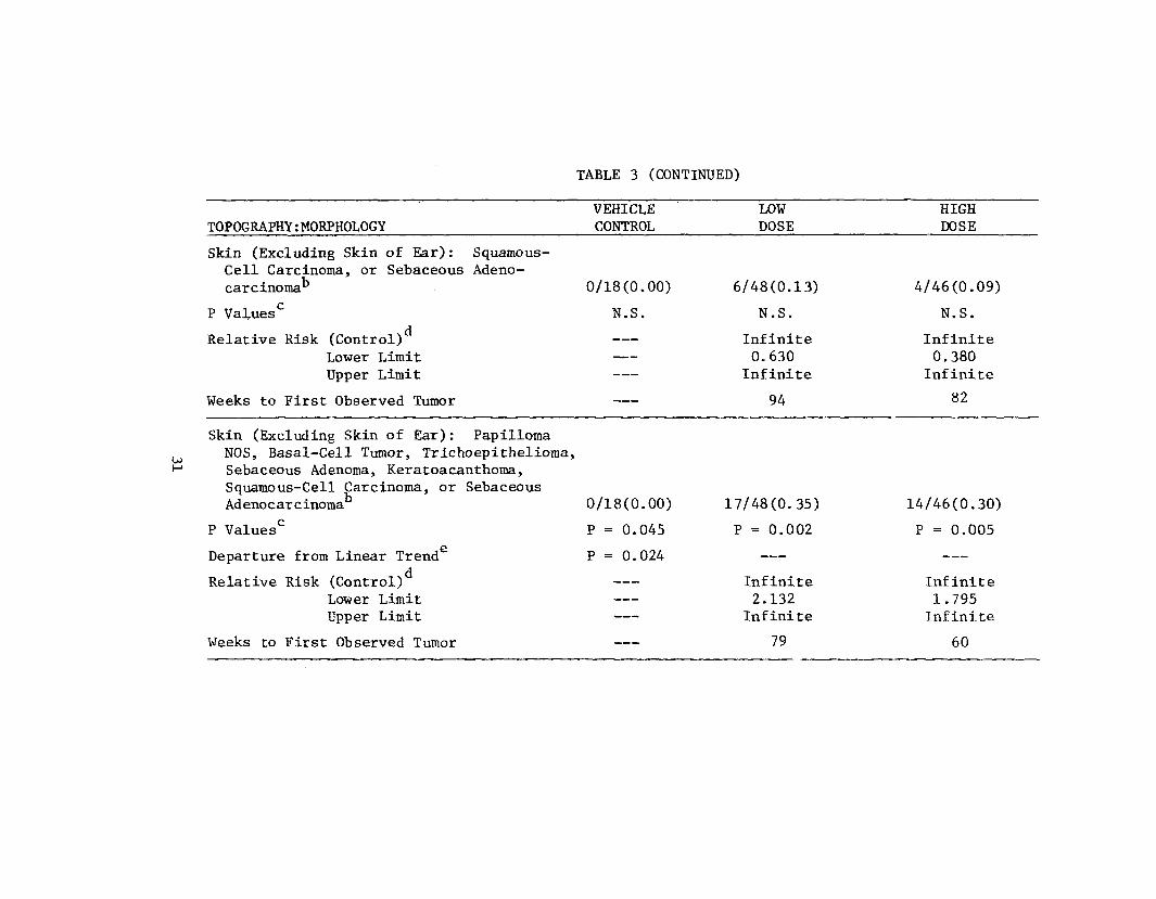
TOPOGRAPHY: MORPHOLOGY
Skin (Excluding Skin of Ear) : Squamous-Cell Carcinoma, or Sebaceous Adenocarcinomab
P Values0
Relative Risk (Control) Lower Limit Upper Limit
Weeks to First Observed Tumor
Skin (Excluding Skin of Ear) : Papilloma NOS, Basal-Cell Tumor, Trichoepithelioma, Sebaceous Adenoma, Keratoaeanthoma, Squamous-Cell Carcinoma, or Sebaceous Adenocarcinomab
P Values0
Departure from Linear Trend6
Relative Risk (Control) Lower Limit Upper Limit
Weeks to First Observed Tumor
TABLE 3 (CONTINUED)
VEHICLE CONTROL
0/18(0.00)
N.S.
0/18(0.00)
P = 0.045
P = 0.024
LOW DOSE
6/48(0.13)
N.S.
Infinite 0.630 Infinite
94
17/48(0.35)
P = 0.002
Infinite 2.132 Infinite
79
HIGH DOSE
4/46(0.09)
N.S.
Infinite 0.380 Infinite
82
14/46(0.30)
P = 0.005
Infinite 1.795 Infinite
60

___
TOPOGRAPHY : MORPHOLOGY
Lung: Alveolar /Bronchiolar Adenoma
P Values0
Departure from Linear Trend
Relative Risk (Control) Lower Limit Upper Limit
Weeks to First Observed Tumor
Hematopoietic System:•! Leukemia or
Malignant LymphomaD
S3 OJ
P Values0
Departure from Linear Trend
Relative Risk (Control)d
Lower Limit Upper Limit
Weeks to First Observed Tumor
Thyroid: C-Cell Adenoma or C-Cell Carcinoma
P Values0
Relative Risk (Control) Lower Limit Upper Limit
Weeks to First Observed Tumor
TABLE 3 (CONTINUED)
VEHICLE CONTROL
0/18(0.00)
N.S .
P = 0.002
0/18(0.00)
P = 0.022
P = 0.028
1/18(0.06)
N.S.
104
LOW DOSE
8/47(0.17)
N.S.
Infinite 0.919
Infinite
79
18/48(0.38)
P = 0.001
Infinite 2.270
Infinite
72
3/43(0.07)
N.S.
1.256 0.112
64.377
98
HIGH DOSE
0/43(0.00)
N.S .
16/46(0.35)
P = 0.002
Infinite 2.083
Infinite
80
0/42(0.00)
N.S.
0.000 0.000 7.981

TABLE 3 (CONTINUED)
TOPOGRAPHY: MORPHOLOGY VEHICLE CONTROL
Preputial Gland:carcinoma'5
P Values0
Sebaceous Adeno0/18(0.00)
N.S.
Relative Risk (Control) Lower Limit Upper Limit
Weeks to First Observed Tumor
OJ OJ
Testis: Interstitial -Cell Tumor
P Values0
Relative Risk (Control) Lower Limit Upper Limit
17/18(0.94)
N.S.
Weeks to First Observed Tumor 97
Liver: Hepatocellular Adenoma or Hepatocellular Carcinoma*5
P Values0
1/18(0.06)
N.S.
Relative Risk (Control) Lower Limit Upper Limit
Weeks to First Observed Tumor 104
LOW DOSE
1/48(0.02)
N.S.
Infinite 0.021 Infinite
97
39/48(0.81)
N.S.
0.860 0.793 1.139
77
8/48(0.17)
N.S.
3.000 0.457
129.949
104
HIGH DOSE
3/46(0.07)
N.S.
Infinite 0.246 Infinite
94
36/45(0.80)
N.S.
0.847 0.779 1.128
79
3/46(0.07)
N.S.
1.174 0.104 60.276
86

___
TABLE 3 (CONTINUED)
VEHICLE TOPOGRAPHY : MORPHOLOGY CONTROL
Pituitary: Chromophobe Adenoma ' 3/16(0.19)
P Values0 N.S .
Relative Risk (Control) Lower Limit Upper Limit
Weeks to First Observed Tumor 39
Adrenal: Pheochromocytoma 4/18(0.22)
P Values0 P = 0.003(N)
u> Relative Risk (Control) —_ -p- Lower Limit
Upper Limit
Weeks to First Observed Tumor 104
Zymbal's Gland, Ear, and Skin of Ear: Squamous-Cell Carcinoma or Sebaceous Adenocarcinoma'3 0/18(0.00)
P Values0 P = 0.040
Relative Risk (Control) Lower Limit Upper Limit
Weeks to First Observed Tumor
LOW DOSE
7/44(0.16)
N.S.
0.848 0.231 4.673
80
3/48(0.06)
N.S .
0.281 0.047 1.530
104
5/48(0.10)
N.S.
Infinite 0.496
Infinite
77
HIGH DOSE
2/34(0.06)
N .S .
0.314 0.029 2.516
69
0/45(0.00)
P = 0.005(N)
0.000 0.000 0.425
8/46(0.17)
N.S.
Infinite 0.939
Infinite
70

TABLE 3 (CONCLUDED)
VEHICLE LOW HIGH TOPOGRAPHY:MORPHOLOGY CONTROL DOSE DOSE
Zymbal's Gland, Ear, and Skin ofSquamous-Cell Carcinoma, SebacAdenocarcinoma, Keratoacanthoma,Neoplasm NOS, or Basal-Cell Tumor
Ear eous
, 1/18(0.06) 6/48(0.13) 9/46(0.20)
P Values0 N.S . N . S . N.S .
Relative Risk (Control) Lower Limit Upper Limit
2.250 0.308
101.140
3.522 0.556
150.403
Weeks to First Observed Tumor 104 77 70
Treated groups received doses of 1500 or 3000 mg/kg by gavage for 22 weeks followed by concentrations of 22,000 or 44,000 ppm in feed for 54 weeks.
Number of tumor-bearing animals/number of animals examined at site (proportion). c
The probability level for the Cochran-Armitage test is given beneath the incidence of tumors in the control group when P < 0.05; otherwise, not significant (N.S.) is indicated. The probability level for the Fisher exact test for the comparison of a treated group with the control group is given beneath the incidence of tumors in the treated group when P < 0.05; otherwise, not significant (N.S.) is indicated. For both Cochran-Armitage and Fisher exact tests a negative designation (N) indicates a lower incidence in the treated group(s) than in the control group.
The 95% confidence interval on the relative risk of the treated group to the control group. £
The probability level of the test for departure from linear trend is given beneath the control group when P < 0.05.
These analyses were based solely upon animals surviving at least 52 weeks, except for sites where the first tumor of interest was observed earlier than 52 weeks in any group of this sex and species, where the analyses were based upon all animals that survived until or past the date that the first tumor was observed.
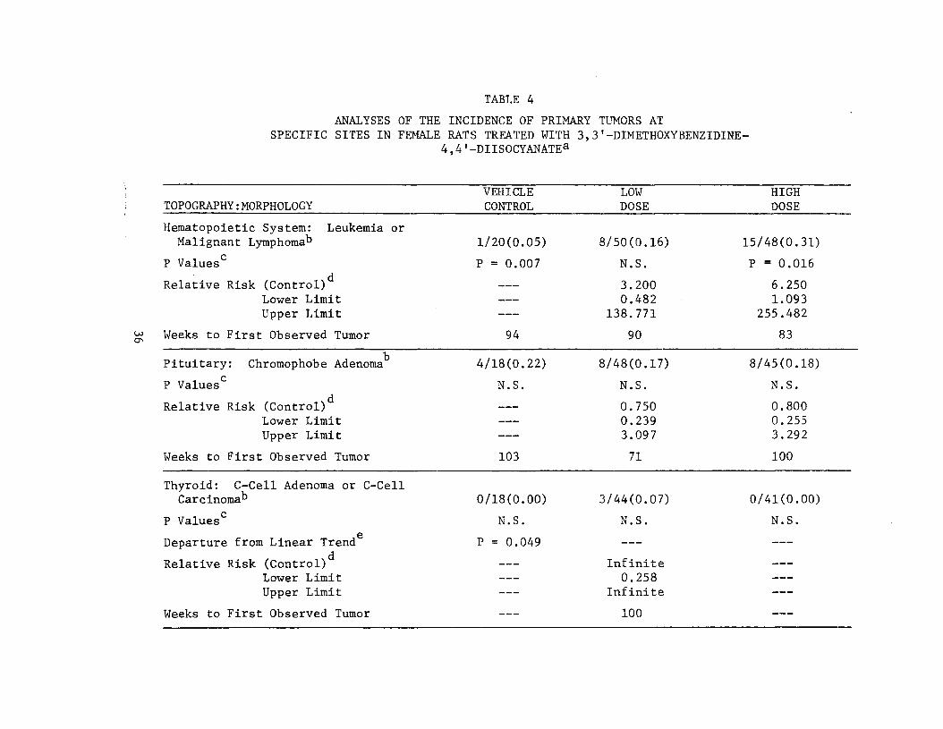
TABLE 4
ANALYSES OF THE INCIDENCE OF PRIMARY TUMORS AT SPECIFIC SITES IN FEMALE RATS TREATED WITH 3,3'-DIMETHOXYBENZIDINE
TOPOGRAPHY: MORPHOLOGY
Hematopoietic System: Leukemia or Malignant Lymphoma^
P Values0
Relative Risk (Control) Lower Limit Upper Limit
Weeks to First Observed Tumor
Pituitary: Chromophobe Adenoma
P Values0
Relative Risk (Control) Lower Limit Upper Limit
Weeks to First Observed Tumor
Thyroid: C-Cell Adenoma or C-Cell Carcinoma'3
P Values0
Departure from Linear Trend
Relative Risk (Control) Lower Limit Upper Limit
Weeks to First Observed Tumor
4,4'-DIISOCYANATEa
VEHICLE LOW HIGH CONTROL DOSE DOSE
1/20(0.05) 8/50(0.16) 15/48(0.31)
P = 0.007 N.S. P = 0.016
3.200 6.250 0.482 1.093
138.771 255.482
94 90 83
4/18(0.22) 8/48(0.17) 8/45(0.18)
N.S. N.S. N.S. ___
0.750 0.800 0.239 0.255 3.097 3.292
103 71 100
0/18(0.00) 3/44(0.07) 0/41(0.00)
N.S. N.S. N.S.
P = 0.049
Infinite 0.258
Infinite
100

TABLE 4 (CONTINUED)
TOPOGRAPHY: MORPHOLOGY VEHICLE CONTROL
Mammary Gland: Fibroadenoma, Adenoma NOS, or Adenocarcinoma NOS*5 0/20(0.00)
P Values0 N.S.
Relative Risk (Control) Lower Limit Upper Limit
___
Weeks to First Observed Tumor
Preputial Gland: Sebaceous Adenocarcinoma or Squamous-Cell Carcinoma*3 0/20(0.00)
P Values0 N.S.
Departure from Linear Trend
Relative Risk (Control) Lower Limit Upper Limit
P = 0.043 ___
Weeks to First Observed Tumor
Uterus: Endometrial Stromal Polyp 0/20(0.00)
P Values0
Relative Risk (Control) Lower Limit Upper Limit
P = 0.013 ___
Weeks to First Observed Tumor
LOW DOSE
5/50(0.10)
N.S.
Infinite 0.525
Infinite
103
8/50(0.16)
N.S.
Infinite 0.952
Infinite
71
5/48(0.10)
N.S.
Infinite 0.547
Infinite
74
HIGH DOSE
6/48(0.13)
N.S.
Infinite 0.695
Infinite
100
4/48(0.08)
N.S.
Infinite 0.402
Infinite
93
10/48(0.21)
P = 0.022
Infinite 1.292
Infinite
86

TABLE 4 (CONCLUDED)
VEHICLE LOW HIGH TOPOGRAPHY: MORPHOLOGY CONTROL DOSE DOSE
Zymbal's Gland: Squamous-Cell Carcinoma or Sebaceous Adenocarcinoma^ 0/20(0.00) 8/50(0.16) 6/48(0.13)
P Values0 N.S. N.S. N.S.
Relative Risk (Control) Infinite Infinite Lower Limit 0.952 0.695 Upper Limit Infinite Infinite
Weeks to First Observed Tumor 44 72
Zymbal's Gland and Skin of Ear: Squamous-Cell Carcinoma, Sebaceous
U) Adenocarcinoma or Trichoepitheliomab 0/20(0.00) 9/50(0.18) 6/48(0.13)oo
P Values0 N.S. P = 0.039 N.S.
Relative Risk (Control) Infinite Infinite Lower Limit 1.096 0.695 Upper Limit Infinite Infinite
Weeks to First Observed Tumor 44 72
Treated groups received doses of 1500 or 3000 mg/kg by gavage for 22 weeks followed by concentrations of 22,000 or 44,000 ppm in feed for 54 weeks.
Number of tumor-bearing animals/number of animals examined at site (proportion). s\
The probability level for the Cochran-Armitage test is given beneath the incidence of tumors in the control group when P < 0.05; otherwise, not significant (N.S.) is indicated. The probability level for the Fisher exact test for the comparison of a treated group with the control group is given beneath the incidence of tumors in the treated group when P < 0.05; otherwise, not significant (N.S.) is indicated. For both Cochran-Armitage and Fisher exact tests a negative designation (N) indicates a lower incidence in the treated group(s) than in the control group.
The 95% confidence interval on the relative risk of the treated group to the control group. P
The probability level of the test for departure from linear trend is given beneath the control group when P < 0.05.
basal-cell tumor, trichoepithelioma, sebaceous adenoma, keratoacan
thoma, squamous-cell carcinoma, or sebaceous adenocarcinoma) was
significant when comparing both the high dose to control (P = 0.005)
and the low dose to control (P = 0.002). The Cochran-Artnitage test
was significant (P = 0.045) with a significant (P = 0.024) departure
from linear trend due to the higher incidence in the low dose group
relative to the high dose group. Based on these statistical results,
the administration of 3,3'-dimethoxybenzidine-4,4'-diisocyanate was
associated with the increased incidence of skin neoplasms in male
Fischer 344 rats under the conditions of this bioassay.
In male rats the Cochran-Armitage test indicated a significant
(P = 0.040) positive association between dose and the incidence of
squamous-cell carcinomas or sebaceous adenocarcinomas of the Zymbal's
gland and the skin of the ear. While the Fisher exact tests did not
support these results, the historical control data for untreated male
Fischer 344 rats at this laboratory indicated only a 0.3 percent (1/300)
incidence of these tumors in untreated males, indicating the rarity of
occur ence of these tumors. In female rats, none of the statistical
tests ror these tumors at this site were significant under the iion
ferroni criterion. However, as with male rats, historical control
data for untreated female Fischer 344 rats at this laboratory indi
cate squamous-cell carcinomas and sebaceous adenocarcinomas of the
Zymbal's gland and skin of the ear to be rarely occurring, with 0 per
cent (0/298) incidence. Based upon these results the data suggest
that the administration of 3,3"-dimethoxybenzidine-4,41-diisocyanate
39

was associated with the increased incidence of squamous-cell car
cinomas or sebaceous adenocarcinomas of the Zymbal's gland and the
skin of the ear in male and female rats.
In female rats the Cochran-Armitage test indicated a significant
(P = 0.013) positive association between dose and the incidence of
endometrial stromal polyps. The high dose to control Fisher exact
test was also significant (P = 0.022). Based on these statistical
results the administration of 3,3'-dimethoxybenzidine-4,4'-diisocy
anate was associated with the increased incidence of endometrial
stromal polyps.
The Cochran-Armitage test and the Fisher exact test comparing
high dose to control indicated a significant negative association
between dose and the incidence of adrenal pheochromocytomas in male
rats. However, the historical data for this laboratory indicated an
incidence of 7 percent (20/294) in male control Fischer 344 rats as
compared to the higher 22 percent (4/18) observed in control male
rats in this bioassay.
Tn summary, the statistical findings were that the administra
tion of 3,3'-dimethoxybenzidine-4,41-diisocyanate was associated
with the increased incidence of leukemia or malignant lymphomas in
male and female rats, the incidence of skin neoplasms (excluding
skin of the ear) in male rats, and the incidence of endometrial
stromal polyps in female rats. The data also suggest that the inci
dence of squamous-cell carcinomas or sebaceous adenocarcinomas of
40

the Zymbal's gland and the skin of the ear was associated with
chemical administration to both sexes.
41
IV. CHRONIC TESTING RESULTS: MICE
A. Body Weights and Clinical Observations
Slight dose-related mean body weight depression was apparent in
male mice. Female mice evidenced slight mean body weight depression
in comparison to the control group (Figure 4).
No abnormal clinical signs were recorded.
B. Survival
The estimated probabilities of survival for male and female mice
in the control and 3,3'-dimethoxybenzidine-4,4'-diisocyanate-dosed
groups are shown in Figure 5. The Tarone test for association be
tween dosage and mortality was not significant for either male or
female mice.
The percentage of mice surviving on test is shown in Figure 6.
In the male mice, despite the disappearance of 1 control in week 22,
1 low dose mouse in week 38, 3 low dose mice in week 41, and 1 low
dose mouse in week 92, and the accidental sacrifice of 1 high dose
mouse in week 44, there were adequate numbers at risk from late-
developing tumors. Ninety-two percent (46/50) of the high dose,
80 percent (40/50) of the low dose, and 90 percent (18/20) of the
control group survived on test until the termination of the study.
With 62 percent (31/50) of the high dose, 72 percent (36/50) of
the low dose and 90 percent (18/20) of the control group surviving
on test until termination of the study, there were adequate numbers
42

50- -50
40- -40
cc ¥. 30H -30
g £20 -20
O 00
CONTROL
LOW DOSE 10— -10
MALE MICE HIGH DOSE
' I i \ r
15 30 45 60 90 105 120
TIME ON TEST (WEEKS)
50- -50
40- -40
VI
<
-30
X CD
>- 20- -20 O O CO
CONTROL
10 — —10 LOW DOSE
FEMALE MICE _ _ _ _ HIGH DOSE
!\ I 15 30 45 60 90 105 120
TIME ON TEST (WEEKS)
FIGURE 4 GROWTH CURVES FOR 3,3'-DIMETHOXYBENZIDINE-4,4'-DIISOCYANATE CHRONIC STUDY MICE
43

- -
— 1.0 LO _.........................*
^••- fr
t-0.8 0.8
-0.6 co 0.6u. O
-0.4 0.4
m O cc _ CONTROL a.
,,., LOW DOSE -0.2 0.2
MALE MICE . _ HIGH DOSE
0.0- 1 -0.0 I I I I ' I T \
15 30 45 60 75 90 105 120
TIME ON TEST (WEEKS)
— 1.0
-- " l , ' ...
_, 0.8- "~~U — 0.8
t£ cc
-0.6 w 0.6u. O
-
5 0.4- -0.4
CD 0 CC ••
a. i _^_i CONTROL
-0.2 0.2- LOW DOSE
FEMALEMICE _ — HIGH DOSE
00 0 0 i I i I i I i I i 1 i I i I i 0 15 30 45 60 75 90 105 120
TIME ON TEST (WEEKS)
FIGURE 5
SURVIVAL PROBABILITY COMPARISONS OF 3,3'-DIMETHOXYBENZIDINE-*,4'-DIISOCYANATE CHRONIC STUDY MICE
44

1.0- f—1.0
-0.8 0.8
-0.6 Sj 0.6W
O £ 0.4 H -0.4 a.
CONTROL.
-0.2 0.2- LOW DOSE
MALE MICE HIGH DOSE
0.0- 0.0 I \^ I ' I ' I \ ' \ '
15 30 45 60 75 90 105 120
TIME ON TEST (WEEKS)
— 1.0
— 0.8 0.8
-0.6 DC 0.6
V) I
LU O % 0.4 H -0.4
CONTROI
-0.2 0.2 LOW DOSE
FEMALE MICE HIGH OOSE
0.0- -0.0
15 30 45 60 75 90 105 120 TIME ON TEST (WEEKS)
FIGURE 6 PERCENT SURVIVAL OF 3,3'-DIMETHOXYBENZIOINE-4,4'-DIISOCYANATE CHRONIC STUDY MICE
45

of female mice at risk from late-developing tumors. Seven females
from the high dose group and 9 from the low dose group were missing
by week 28.
C. Pathology
Histopathologic findings on neoplasms in mice are summarized in
Appendix B (Tables Bl and B2); findings on nonneoplastic lesions are
summarized in Appendix D (Tables Dl and D2).
A variety of tumors occurred in both the control and dosed
groups. The type, incidence, and distribution of these lesions were
similar to those expected in aged B6C3F1 mice; therefore, these le
sions were considered to be spontaneous and not related to compound
administration.
A few neoplasms occurred only, or with a greater frequency, in
dosed groups as compared with controls. This may be noted in male
mice in which benign and malignant hepatocellular neoplasms were
found in 21 percent of the high dose mice compared to 5 percent of
controls.
A variety of nonneoplastic, inflammatory, degenerative or fiforo
tic lesions occurred randomly in all groups and none appeared to be
compound-related.
The results of this pathologic examination indicated that
3 ,3"-dimethoxybenzidine-4,4"-diisocyanate was not carcinogenic in
male or female B6C3F1 mice under the conditions of this bioassay.
46

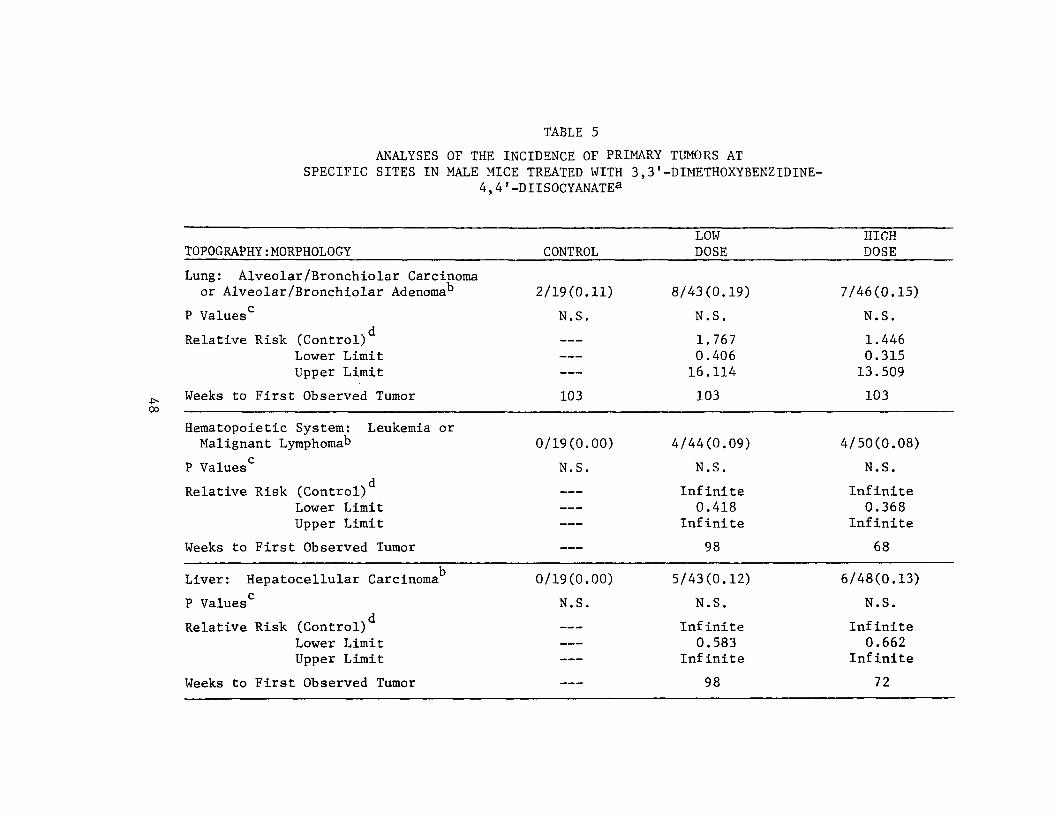
D. Statistical Analyses of Results
The results of the statistical analyses of tumor incidence in
mice are summarized in Tables 5 and 6. The analysis is included for
every type of malignant tumor in either sex where at least two such
tumors were observed in at least one of the control or 3,3'-dime
thoxybenzidine-4,4'-diisocyanate-dosed groups and where such tumors
were observed in at least 5 percent of the group.
None of the statistical tests for any site of either male or fe
male mice indicated a significant positive association between chemi
cal administration and tumor incidence. Based upon these statistical
results there was no evidence that 3,3'-dimethoxybenzidine-4,4'-diiso
cyanate was a carcinogen in B6C3F1 mice under the conditions of this
bioassay.
In male mice the Cochran-Armitage test and the Fisher exact test
comparing high dose to control indicated a significant negative asso
ciation between dose and the incidence of interstitial-cell tumors of
the testis. However, historical control data from the same laboratory
indicate an incidence of 2 percent (4/266) of these tumors in untreated
male B6C3F1 mice as compared with the 16 percent (3/19) in control male
mice in this bioassay.
To provide additional insight into the possible carcinogenicity
of this compound, 95 percent confidence intervals on the relative
risk have been estimated and entered in the tables based upon the
observed tumor incidence rates. In many of the intervals shown in
47
__ _
___
00
TABLE 5
ANALYSES OF THE INCIDENCE OF PRIMARY TUMORS AT SPECIFIC SITES IN MALE MICE TREATED WITH 3,3'-DIMETHOXYBENZIDINE
4,4'-DIISOCYANATEa
LOW TOPOGRAPHY: MORPHOLOGY CONTROL DOSE
Lung: Alveolar /Bronchiolar Carcinoma or Alveolar /Bronchiolar Adenoma" 2/19(0.11) 8/43(0.19)
P Values0 N.S. N.S.
Relative Risk (Control) 1.767 Lower Limit 0.406 Upper Limit 16.114
Weeks to First Observed Tumor 103 103
Hematopoietic System: Leukemia or Malignant Lymphomab 0/19(0.00) 4/44(0.09)
P Values0 N.S. N.S.
Relative Risk (Control) Infinite Lower Limit 0.418 Upper Limit Infinite
Weeks to First Observed Tumor 98
Liver: Hepatocellular Carcinoma 0/19(0.00) 5/43(0.12)
P Values0 N.S. N.S.
Relative Risk (Control) Infinite Lower Limit 0.583 Upper Limit Infinite
Weeks to First Observed Tumor 98
HIGH DOSE
7/46(0.15)
N.S.
1.446 0.315 13.509
103
4/50(0.08)
N.S.
Infinite 0.368
Infinite
68
6/48(0.13)
N.S.
Infinite 0.662
Infinite
72

TABLE 5 (CONCLUDED)
TOPOGRAPHY: MORPHOLOGY CONTROL LOW DOSE
HIGH DOSE
Liver: Hepatocellular Carcinoma cr Hepatocellular Adenoma" 1/19(0.05) 8/43(0.19) 10/48(0.21)
P Values0 N.S. N.S. N.S.
Relative Risk (Control) Lower Limit Upper Limit
3.535 0.537
152.566
3.958 0.639
167.483
Weeks to First Observed Tumor 103 98 72
Testis: Interstitial-Cell Tumor 3/19(0.16) 1/42(0.02) 0/48(0.00)
P Values0
Relative Risk (Control) Lower Limit Upper Limit
P = 0.007(N) ___
N.S.
0.151 0.003 1.760
P = 0.020(N)
0.000 0.000 0.651
Weeks to First Observed Tumor 103 103
Treated groups received doses of 22,000 or 44,000 ppm in feed.
Number of tumor-bearing animals/number of animals examined at site (proportion). ^
"The probability level for the Cochran-Armitage test is given beneath the incidence of tumors in the control group when P < 0.05; otherwise, not significant (N.S.) is indicated. The probability level for the Fisher exact test for the comparison of a treated group with the control group is given beneath the incidence of tumors in the treated group when P < 0.05; otherwise, not significant (N.S.) is indicated. For both Cochran-Armitage and Fisher exact tests a negative designation (N) indicates a lower incidence in the treated group(s) than in the control group.
The 95% confidence interval on the relative risk of the treated group to the control group.

TABLE 6
ANALYSES OF THE INCIDENCE OF PRIMARY TUMORS AT SPECIFIC SITES IN FEMALE MICE TREATED WITH 3,3'-DIMETHOXYBENZIDINE
TOPOGRAPHY : MORPHOLOGY
Lung: Alveolar /Bronchiolar Adenoma
P Values
Relative Risk (Control) Lower Limit Upper Limit
Ul Weeks to First Observed Tumor o
Hematopoietic System: Leukemia or Malignant Lymphoma^
P Values0
Relative Risk (Control) Lower Limit Upper Limit
Weeks to First Observed Tumor
4,4'-DIISOCYANATEa
LOW HIGH CONTROL DOSE DOSE
3/20(0.15) 2/41(0.05) 1/40(0.03)
N.S. N.S. N.S.
0.325 0.167 0.030 0.003 2.651 1.945
103 103 103
3/20(0.15) 9/41(0.22) 10/43(0.23)
N.S. N.S. N.S. ___
1.463 1.550 0.424 0.465 7.728 8.072
103 86 33

TABLE 6 (CONCLUDED)
o
Treated groups received doses of 22,000 or 44,000 ppm in feed.
Number of tumor-bearing animals/number of animals examined at site (proportion). Q
The probability level for the Cochran-Armitage test is given beneath the incidence of tumors in the control group when P < 0.05; otherwise, not significant (N.S.) is indicated. The probability level for the Fisher exact test for the comparison of a treated group with the control group is given beneath the incidence of tumors in the treated group when P < 0.05; otherwise, not significant (N.S.) is indicated. For both Cochran-Armitage and Fisher exact tests a negative designation (N) indicates a lower incidence in the treated group(s) than in the control group.
The 95% confidence interval on the relative risk of the treated group to the control group.

Tables 5 and 6, the value one is included; this indicates the absence
of statistically significant results. It should also be noted that
many of the confidence intervals have an upper limit greater than one,
indicating the theoretical possibility of tumor induction in mice by
3,3'-dimethoxybenzidine-4,4'-diisocyanate that could not be estab
lished under the conditions of this test.
52

V. DISCUSSION
There was a significant positive association between the dosages
of 3,3'-dimethoxybenzidine-4,4'-diisocyanate administered and mortal
ity in male rats. For female rats, mortality among the dosed groups
was significantly higher than that for the controls. There were no
significant positive associations between dosage and mortality among
male or female mice. Adequate numbers of animals in all groups sur
vived sufficiently long to be at risk from late-developing tumors.
Mean body weight depression was slight, but detectable, when dosed
male and female mice were compared to their respective controls,
indicating that the dosages administered to these animals may have
approximated the maximum tolerated dosages.
For both sexes of rats, there was a significant positive asso
ciation between dosage and the incidence of leukemia and malignant
lymphoma. The high dose to control and the low dose to control Fisher
exact comparisons for males and the high dose to control comparison
for females were also significant for the incidences of these tumors.
Excluding the skin of the ear in considering neoplasms of the skin
(i.e., a combination of papilloma NOS, basal-cell tumor, trichoepith
elioma, sebaceous adenoma, keratoacanthoma, squamous-cell carcinoma,
and sebaceous adenocarcinoma) the Fisher exact comparisons were sig
nificant for both dose levels in male rats. There was a significant
positive association between the dosages of the chemical administered
and the incidences in female rats of endometrial stromal polyps.
53
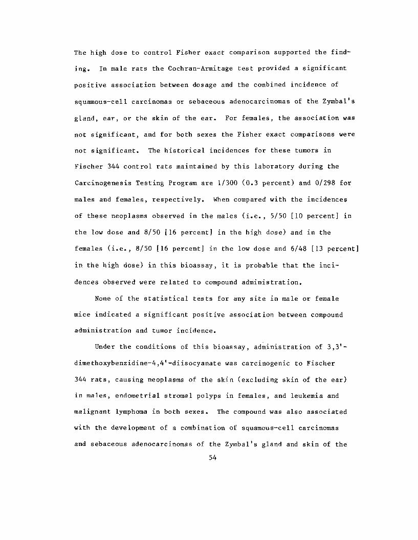
The high dose to control Fisher exact comparison supported the find
ing. In male rats the Cochran-Armitage test provided a significant
positive association between dosage and the combined incidence of
squamous-cell carcinomas or sebaceous adenocarcinomas of the Zymbal's
gland, ear, or the skin of the ear. For females, the association was
not significant, and for both sexes the Fisher exact comparisons were
not significant. The historical incidences for these tumors in
Fischer 344 control rats maintained by this laboratory during the
Carcinogenesis Testing Program are 1/300 (0.3 percent) and 0/298 for
males and females, respectively. When compared with the incidences
of these neoplasms observed in the males (i.e., 5/50 [10 percent] in
the low dose and 8/50 [16 percent] in the high dose) and in the
females (i.e., 8/50 [16 percent] in the low dose and 6/48 [13 percent]
in the high dose) in this bioassay, it is probable that the inci
dences observed were related to compound administration.
None of the statistical tests for any site in male or female
mice indicated a significant positive association between compound
administration and tumor incidence.
Under the conditions of this bioassay, administration of 3,3'
dimethoxybenzidine-4,4'-diisocyanate was carcinogenic to Fischer
344 rats, causing neoplasms of the skin (excluding skin of the ear)
in males, endometrial stromal polyps in females, and leukemia and
malignant lymphoma in both sexes. The compound was also associated
with the development of a combination of squamous-cell carcinomas
and sebaceous adenocarcinomas of the Zymbal's gland and skin of the
54

ear in rats of both sexes. There was no evidence for the carcino
genicity of the compound in B6C3F1 mice.
55

VI. BIBLIOGRAPHY
Armitage, P., Statistical Methods in Medical Research, Chapter 14. J. Wiley & Sons, New York, 1971.
Berenblum, I., editor, Carcinogenicity Testing. International Union Against Cancer, Technical Report Series, Vol. 2. International Union Against Cancer, Geneva, 1969.
Bonk, H.W., and T.M. Shah, "Polyurethanes from 3,3'-dimethyldiphenyl4,4'-diisocyanate Polyester Diols and BisChydroxethyl ether) of Hydroquinone." U.S. Patent 3,899,467 (Upjohn Co.), August 12, 1975; Chemical Abstracts 83. 194863r.
Chemical Abstracts Service, The Chemical Abstracts Service (CAS) Ninth Collective Index, Volumes 76-85, 1972-1976. American Chemical Society, Washington, D.C., 1977.
Chiklis, C.K., "Photographic Products Containing Antireflection Layer." U.S. Public Patent Application B 354,008 (Polaroid Corp.), January 28, 1975; Chemical Abstracts 83, 35653f.
Chung, D.A., "Polyurethane Shock Absorbers." Ger. Offen. 2,435,078 (Goodyear Tire and Rubber Co.), February 20, 1975; Chemical Abstracts 83, 44171z.
Cox, D.R., Analysis of Binary Data, Chapters 4 and 5. Methuen and Co., Ltd., London, 1970.
Cox, D.R., "Regression Models and Life-Tables." Journal of the Royal Statistical Society. Series "B" 34:187-220, 1972.
Gart, J.J., "The Comparison of Proportions: A Review of Significance Tests, Confidence Limits, and Adjustments for Stratification." International Statistical Institute Review 39:148-169, 1971.
Grace, W.R. and Co., "Polyurethane Gaskets." British Patent 1,374,745, November 20, 1974; Chemical Abstracts 83, 116347m.
Guthrie, J.L. and F.J. Rendulic, "Photocurable Triazine-Containing Polyene-Polythiol Lacquer Composition." U.S. Patent 3,900,594 (Grace, W.R., and Co.), August 19, 1975; Chemical Abstracts 83, 195438t.
56

Hadidian, Z., T.N. Frederickson, E.K. Weisburger, J.H. Weisburger, R.M. Glass, and S. Msntel, "Tests for Chemical Carcinogens. Report on the Activity of Derivatives of Aromatic Amines, Nitrosamines, Quinolines, Nitroalkanes, Amides, Epoxides, Aziridines, and Purine Antimetabolites." Journal of the National Cancer Institute 4_1_(4): 985-1025, 1968.
Kaplan, E.L., and P. Meier. "Nonparametric Estimation from Incomplete Observations." Journal of the American Statistical Association 53_:457-481, 19587"
Linhart, M.S., J.A. Cooper, R.L* Martin, N.P. Page, and J.A. Peters, "Carcinogenesis Bioassay Data System." Computers and Biomedical Research 7_: 230-248, 1974.
Miller, R.G., ^imul.tanepus Statistical Inference. McGraw-Hill Book Co. , New York," 1966.
Saffiottij U., R. Montesano, A.R. Sellakumar, F. Cefis, and D.G. Kaufman, "Respiratory Tract Carcinogenesis in Hamsters Induced by Different Numbers of Administration of Benzo (a) Pyrene and Ferric Oxide." Can£er_Researcli 32:1073-1079, 1972.
Stanford Research Institute, 1977 Directory of Chemical Producers, U.S_.A.. Menlo Park, California, 1977,,
Tarone, R.E., "Tests for Trend in Life-Table Analysis." Biometrika 62:679-682, 1975.
57


APPENDIX A
SUMMARY OF THE INCIDENCE OF NEOPLASMS IN RATS TREATED WITH 3,3'-DIMETHOXYBENZIDINE
4,4'-DIISOCYANAT'r


TABLE A1 SUMMARY OF THE INCIDENCE OF NEOPLASMS IN MALE RATS TREATED
WITH3,3'-DIMETHOXYBENZIDINE-4,4'-DIISOCYANATE
... ... , . _ ! . ,
CONTROL (VfH) LOS COSE 11-1095 11-1093
ANIMALS INITIALLY IN STUDY 20 50 ANIMALS NECROPSIED 20 50 ANIMALS EXJBINED HISTOPATHOLOGICALIY ** 20 50
INTEGUHENTAPY SYSTEM
*SKIN (20) (50) NEOPLASM, NOS 1 (2%) EAPILLOMA, NOS SQDAMOOS CELL CARCINOMA 5 (10X) EASAL-CELL TUNOE t (8%) TEICHOEPITHELIOHA 2 (IX) SEBACEOUS ADENOMA 2 (IX) SEBACEOUS ADENOCAECINOMA 1 (2*) KFPATOACANTHOMA 1 (5%) <t (8X) CARCIHOSAFCOMA
•SUBCUT TISSUE (20) (50) FIBROMA 1 <2X) FIBHOSARCOMA 1 (2X)
RESPIRATORY SYSTEM
*LUNG (20) (19)ALVEOLAB/B80NCHIOLAR ADENOMA a (16*) CAECINOSARCOMA, METASTATIC
HFMATOPOIETIC SYSTEM
*MtHTIPLE ORGANS (20) (50) MALIGNANT LYHPHOHA, NOS 1 12%)LEUKEMIA,DOS 12 (24*) UNDIFFERENTIATED LEUKEMIA 6 (12X)
tCERTICAL LYMPH NODE (17) (19) CARCINOSARCOMA, METASTATIC
CIRCULATORY SYSTEM
SQSI
* N U M B E R OF A N I M A L S SITH TISSUE E X A M I N E D MICROSCOPICALLY * N U M B E R OF A N I M A L S NECROPSIEP
"EXCLUDES PARTIALLY AUTOLYZED ANIMALS
A-3
HIGH DOSE 11-1091
50 50 50
(50)
2 (IX) 1 (8%) 5 (10X) 1 (2%)
5 (10*) 1 (2%)
(50)
(17)
1 (2*)
(50) 1 (2%) 7 <1«X) 9 (18X)
(12) <2X)

TABLE A1 (CONTINUED)
CONTROL (VEH) LOH DOSE HIGH EOSE 11-1095 11-1093 11-1091
DIGESTIVF SYSTEM
OLIVEP (20) (50) (50) HEPATOCELIULAR ADENOMA 1 (5%) 6 (12%) 2 <4X) HEPATOCELLOLAR CARCINOMA 2 («») 1 <2X)
•PANCR2AS (20) (19) (50) ACINAR-CELL ADENOMA 1 <2X)
*STCPIACH (20) (49) (48) PAPILLOMA, NOS 1 (2%)
ISflALI INTESTINE (20) (19) (49) ADENOMATODS POLYP, NOS 1 (2X)
tCOION (19) (47) (46) ADENOCARCINOHA, NOS 1 (2%)
DEINARY SYSTEM
IKIDNFY/PELVIS (20) (50) (49) TRANSITIONAL-CELL CARCINOMA 1 (5X)
ENDOCRINE SYSTEM
tPITUITABY (16) (45) (36) CHROMOPHOB? ADENOMA 3 (19X) 7 (16%) 2 (6%)
*APRENAL (20) (50) (49) PHEOCHROMOCYTOMA 1 (20%) 3 (6%)
•THYROID (19) (45) (46) FOLLICOLAR-CELL ADENOMA 1 (5%) FOLLICOLAR-CELL CARCINOMA 1 (2%) C-CELL ADENOMA 1 (5%) 1 (2%) C-CELL CARCINOMA 2 <4X) CYSTADENONS. NOS 1 (2%)
•PANCREATIC ISLETS (20) (49) (50) ISLET-CELI ADENOMA 1 (5%) 1 (2%)
REPRODUCTIVE SYSTEM
*PREP(JTIAL GLAND (20) (50) <5<M SEBACEOBS ADENOCARCINOMA 1 {2%) 3_J6il
i NUMBER OP ANIMALS HITH TISSUE EXAMINED MICROSCOPICALLY * NUMBER OF ANIMALS NECROPSIED
A-4

TABLE A1 (CONTINUED)
CONTROL (VEH) LOW DOSE HIGH COSE 11-1095 11 -1093 11-1091
tTESTIS (20) (50) (U9) INTEBSTITIAL-CELL TUBOB 17 (85%) 39 (78X) 36 (73*)
*EPIDIDYMIS (20) (50) (50) FIBFOSAECOMA, INVASIVE 1 (2X)
NFPVCUS SYSTEM
tBBAIN (19) (50) («7) SQDABOUS CELL CARCINOMA, INVASIV 1 (2%) GLIOBA, NOS 1 (2%) ASTROCYTOBA 1 (5*) CLIGODENDBOGLIOMA 1 (2*)
SPECIAL SENS! ORGANS
*EAF (20) (50) (50) SQUAMOUS CELL CARCINOMA 1 (2%)
*ZYMBAL'S GLAND (20) (50) (50) SQUAMOUS CELL CARCINOMA 2 (HX) 5 (10%) SEBACEOUS ADENOCARCINOHA 3 (6X) 2 (<»%)
HUSCOIOSKELETAL SYSTEM
NONS
BODY CAVITIES
*MEDtASTINUM (20) (50) (50) SAFCOMA, NOS 1 (2%)
*ABCO«INAL CAVITY (20) (50) (50) LIPOBA 1 (2«)
ALL OTHER SYSTEMS
*MULTIPLE ORGANS (20) (50) (50) CAFCINOSARCOMi
t NUBBER OF ANIMALS WITH TISSUE EXAMINED MICROSCOPICALLY * NOMBEF OF ANIMALS NECEOPSIFD
A-5

MESOTHELIOMA, NOS
ANIMAL DISPOSITION SUMMARY
ANIMALS INITIALLY IN STUDY NATURAL DEATHS MORIBUND SACRIFICE SCHEDULED SACRIFICE ACCIDENTALLY RULED TERMINAL SACRIFICE ANIMAL HISSING
INCLUDES AUTOLYZED ANIMALS
TUMOR SU M M A R Y
TOTAL ANIHALS WITH PRINARl TOBORS* TOTAL PRIMARY TKCOST
TOTAL ANIMALS WITH BENIGN TUMORS TOTAL BENIGN TUMORS
TOTAL ANIMALS WITH MALIGNANT TUMCRS TOTAL MALIGNANT TUMORS
TOTAL ANIMALS WITH SECONDAPY TUMORS* TOTAL SECONDARY TDMORS
TOTAL ANIHALS WITH TUMORS USC?PTAINBENIGN OR MALIGNANT
TOTAL UNCERTAIN TUMORS
TOTAL ANIMALS HITH TUMOHE UNCERTAINPBINAPY OF HETASTRTIC
TOTAL UNCERTAIN TUMORS
TABLE Al (CONCLUDED)
CONTROL (VEH)11-1095
2032
15
18 31
18 29
2 2
LOW DOSE HIGH DOSE 11-1093 11-1091
1 (2*)
50 50 16 12 12 22
US 16 119 95
U5 13 80 56
30 31 38 38
* PRIMARY TUMOSS: ALL TUMORS EXCEPT SECONDARY TUMORS * SECONDARY TUMORS: METASTATIC TUMORS OR TUMORS INVASIVE INTO AN ADJACENT ORGAN
A-6

TABLE A2 SUMMARY OF THE INCIDENCE OF NEOPLASMS IN FEMALE RATS TREATED
WITH3,3'-DIMETHOXYBENZIDINE-4,4'-DIISOCYANATE
CONTROL (VEH) 10* DOSE HIGH COSE 11-1096 11-109U 11-1092
ANIMALS INITIALLY IN STUDY 20 50 50 ANIHALS NECPOFSIED 20 50 US ANIMALS EXAMINED HISTOPATHOLOGICALIY ** 2" SO U8
I N T E G U B E N T A B Y S Y S T E M
» S K I N (20) (5P) NEOPLASM, NOS 1 (2%) SQUAHOOS CELL CABCINOBA 1 (2X) BASAL-CELL TUMOB 1 (2«) TBICHOEPITHELIOHA SEBACEOUS ADENOMA 1 <2X) SEBACEOUS ADENOCABCINOMA 1 (5«) 1 (2«) 1 (2%) CARCINOSABCOMA 1 (5%)
*SUBCDT TISSUE (20) (50) FTBBOSABCOWA 1 (2%) IIPOHA 1 (2X)
PESPIBATORY SYSTEM
*HASAL CAVITY (20) (50) (48) SQUAMOOS CELL CABCINOMA, INVASIV 1 (2X)
ILONG (20) («8) (US) SQUAMOUS CELL CAECIMOMA, HETASTA 1 (2X) ADENOCAHCIHOMA, NOS, METASTATIC 1 (2X) ALVEOLAR/EBONCHIOLAH ADENOMA 1 (5%) 2 (IX) 1 (2») SEBACEOUS ADENOCAECINOMA. METAST 1 (2%) TIBROSABCCMA, HFTASTATIC 1 (2X) CABCINOSABCOMA, HETASTATIC 1 (2X)
HEMATOPOIETIC SYSTEM
•MULTIPLE ORGANS (20) (50) (U8) MALIGNANT LYHPHONA, NOS 1 (2%)
6 (13V
* N U M B E R OF A N I M A L S SITH TISSUE E X A M I N E D MICBOSCOPICALlt * N U M B E R OP A N I M A L S NECROPSIED *«EXCLUDES PARTIALLY AUTOLYZED ANIMALS
A-7

TABLE A2 (CONTINUED)
UNDIFPEBENTIATED LEUKEMIA
tBONE MARROW LEUKEMIA, NOS
ISPLEEN UNDIFFERENTIATED LEUKEMIA
CONTROL (VEH)11-1096
1 (5*)
(19)
(20)
LOW DOSE 11-1091
3 <6X)
(15)
(18)
HIGH COSE 11-1092
6 (13*)
(15) 1 (2*)
(18) 1 (2%)
CIRCULATORY SYSTEM
NONE
DIGESTIVE SYSTEM
iLIVER HEPATOCELLULAR ADENOMA
fPANCREAS ADENOCARCINOMA, NOS
tSTCHACS SQUAMOOS CELL CARCINOMA
fSMALL INTESTINE CYSTADENOCARCINOMA, NOS
tILEUM ADENOMATOUS POLYP, NOS
tCOION ADENOCARCINOMA, NOS
(20)
(20)
(20)
(20)
(20)
(18)
(50)
(18) 1 (2*)
(50) 1 (2%)
(50)
(50)
(15) 1 (2*)
(18) 2 (1*)
(18)
(18) 1 (2%)
(18) 1 (2%)
(18) 1 (2*)
(17) 1 (2X)
DRINARY SYSTBB
tKIDNEY TUBULAR-CELL ADENOMA
(20) (19) 1 (2X)
(18)
ENDOCRINE SYSTEM
iPITUITARY NSO PLASM, NOS
(18) (18)
f NUMBER OF ANIMALS WITH TISSUE EXAMINED MICROSCOPICALLY * NUMBER OF ANIMALS NECROPSIED
("5) 1 .12*1 . . . _, ,
A-8

TABLE A2 (CONTINUED)
HIGH COSE 11-1092
8 (18%)
(47) 1 (2%)
(41)
(48) 1 (2%) 1 (2%) 1 (2%) 4 (8%)
(48)
4 (8%)
(48) 1 (2%)
(48)
1 (2%) 10 (21%)
(48)
1 (2%)
(48) 1 (2%)
CHROMOPHOBE ADENOMA
*ADRENAL CORTICAL ADENOMA FHFOCHROMOCYTOMA
•THYROID FOLLICOLAR-CELL ADENOMA C-CELL ADENOMA C-CELL CARCINOMA
REPRODUCTIVE SYSTEM
*MAMHARY GLAND ADENOMA, NOS ADENOCARCINOMA, NOS CYSTADENOHA, NOS FIBROADENOMA
*PHHPUTIAL GLAND SQOAMOOS CELL CARCINOMA SEBACEOOS ADENOCARCINOMA
*VAGINA PAPILLOMA, NOS
IUTF.ROS SQOAMOOS CELL CARCINOMA ADENOCARCINOMA, NOS IEIOMYOMA LEIOMYOSARCOMA INDONETRIAL STPONAL POLYP
*UT!PUS/!NDOMETRIU« CARCINOMA, NOS ADENOCAPCINOMA, NOS
•OVARY CYSTADENOCARCINOMA, NOS
NERVCOS SYSTEM
IBRAIN SEBACEOOS ADENOCARCIHONA, INYASI
CONTROL (VHH) 11-1096
* (22%)
(20)
1 (5%)
(18)
(20)
(20)
(20)
(20)
(20) 1 (5%)
(20)
(20)
LOR DOSE 11-109H
8 (17%)
(U9) 1 (2%) 1 (2%)
(UU) 1 (2%) 2 (5%) 1 (2%)
(50) 1 (2%)
5 (10%)
(50) 1 (2%) ^ (14%)
(50)
(48) 1 (2%) 1 (2%) 1 (2%)
5 (10%)
(48)
(U7)
(50)
* NDNBER OF ANIMALS »ITH TISSUE EXAMINED MICROSCOPICALLY * SOMBER OF ANIMALS NECROPSIED
A-9

EPENDYMONA
SPECIAL SENSE ORGANS
*ZYMBAL'S GLAND SQOANODS CELL CARCINOMA SEBACEOUS ADENOCARCINOMA CARCINOSARCOHA
MOSCULOSKELFTAl SYSTEM
NONE
BODY CAVITIES
NONF
ALL OTHER SYSTEMS
•MULTIPLE ORGANS ADEHOCAHCINONA, NOS, HETASTATIC CARCINOSARCOMA
ANIBAL DISPOSITION SUMMARY
ANIMALS INITIALLY IN STUDY NATURAL DEATHS MORIBUND SACRIFICE SCHEDULED SACRIFICE ACCIDENTALLY KILLED TERMINAL SACRIFICE ANIMAL MISSING ANIMAL DELETED (WRONG SEX)
9 INCLUDES_AUTQLTZEE ANIMALS
TABLE A2 (CONTINUED)
COHTROL (VEH) 11-1096
(20)
1 (5*)
(20)
15
LOW DOSB HIGH COSE 11-1094 11-1092
1 (2«)
(50) 1 (2*) t (8*) 7 2 <«*)
(50) (18) 1 (2*) 1 (2*)
50 50 9 16 20 12
21 21
1
I NUMBER OF ANIMALS HITH TISSOE EXAMINED MICROSCOPICALLY * NUMBER OF ANIMALS NECROPSIED
A-10

TUMOR SUMMARY
TOTAL ANIMALS WITH PRIMARY TDMOBS*TOTAL PRIMARY TUMORS
TOTAL ANIMALS WITH BENIGN TUMORSTOTAL BENIGN TUMORS
TOTAL ANIMALS WITH MALIGNANT TDMORSTOTAL MALIGNANT TUMORS
TOTAL ANIMALS WITH SECONDARY TUMORStTOTAL SECONDARY TUMORS
TOTAL ANIMALS WITH TUMORS UNCERTAINBENIGN OR MALIGNANT
TOTAL UNCERTAIN TUMORS
TOTAL ANIMALS WITH THMOHS UNCERTAINPRIMARY OR METASTATIC
TOTAL UNCERTAIN TUMORS
TABLE A2 (CONCLUDED)
CONTROL (VEH)11-1096
10 11
6 6
5 5
LOB DOSE HIGH EOSB 11-1091 11-1092
UO <t« 63 69
22 26 29 33
27 30 3M 3U
5 2 6 2
2 2
* PRIMARY TUMORS: ALL TUMORS EXCEPT SECONDARY TUMORS i SECONDARY TUMORS: METASTATIC TUMCRS OR TUMORS INVASIVE INTO AN ADJACENT ORGAN
A-ll


APPENDIX B
SUMMARY OF THE INCIDENCE OF NEOPLASMS IN MICE TREATED WITH 3,3'-DIMETHOXYBENZIDINE
4,4'-DIISOCYANATE


TABLE Bl SUMMARY OF THE INCIDENCE OF NEOPLASMS IN MALE MICE TREATED WITH
3,3'-DIMETHOXYBENZIDINE-4,4'-DIISOCYANATE
~ CONTROL (ONTR) 22-2095
LOB DOSE 22-2093
HIGH EOS? 22-2091
ANIMALS INITIALLY IN STUDY ANIMALS HISSING, ANIMALS NECROPSIED ANIMALS EXAMINED HISTOPATHOLOGIC ALLY
20 1
19 ** 19
50 5
I)U 1IM
50
50 50
I N T E G D M E N T A R Y S Y S T E M
* S U E C U T TISSUE (19) (UK) (50) LIPOMA 1 (2X)
RESPIRATORY SYSTEM
tLUNG (19) HEPATOCELLULAH CARCINOMA, METAST 2 (5X) ALVEOLAB/EHONCHIOLAR ADENOMA 1 (5%) 8 (19X) 7 (15*) ALVEOLAR/ERONCHIOLAR CARCINOMA 1 (5*)
HEBATOPOIETIC SYSTEM
*MULTIPLE ORGANS (19) (50) MALIGNANT LYMPHOHA, NOS 2 (5*) 1 (2*) MALIG.LYMPHCMA. HISTIOCYTIC TYPE 1 (2%) LEUKEMIA,NOS 1 (2«)
tSPIEEN (18) (37) (15) HEMANGIOSARCOMA 1 (2*)
*MESENTERIC L. NODE (18) (36) (38) MALIGNANT LYHPHOMA, NOS 1 (3*) MALIGNANT LYMPHOMA, BIXZB TYPE 1 (3K)
tAXILLARY LYMPH NODE (18) (36) (38) MALIG.LYBPHOMA, LYMPHOCYTIC TYPE
CIRCULATORY SYSTEM
•HEART (19) (U8) HEMANGIOSARCOHi L.J2S1.
t N U M B E R OP A N I M A L S WITH TISSUE E X A M I N E D MICROSCOPICALLY * N U M B E R OP A N I M A L S N E C R O P S I E D **EXCLUDES PARTIALLY AUTOLYZED ANIMALS
B-3

TABLE Bl (CONTINUED)
CONTROL (UNTR) LOW DOSE HIGH EOSE 22-2095 22-2093 22-2091
D I G E S T I V E S Y S T E M
*LIVEE (19) (43) (18) HEPATOCELLOLAR ADENOMA 1 (5%) 3 (7X) U (8*) HEPATOCELL'JLAF CARCINOMA 5 (12S) 6 (13%)
• STOMACH (15) (41) (49) JDENOPIATOnS POLYP, NOS 1 (7*)
URINARY SYSTEB
NOSE
ENDOCRINE SYSTEM
IAORENAL (13) (25) (36) CORTICAL ADENONA 1 (31)
BEPHCEtlCTIVE SYSTEM
tTESTIS (19) (42) (48) ISTEHSTITIAL-CELL TUHOR 3 (16«) 1 (2*)
NEEVCDS SYSTEH
NONE
SPECIAL SENS'? ORGANS
NON"
KDSCUIOSKELETAl SYSTEM
N3NE
BODY CAVITIES
NPJE. , , * NUMBER OF ANIHALS WITH TISSUE EXAMINED MICROSCOPICALLY * NOMBEP OF ANIMALS NECHOPSIED
B-4

TABLE Bl (CONCLUDED)
ALL CTHER SYST5MS
NONE
ANIMAL DISPOSITION SUMMARY
ANIMALS INITIALLY IN STUDYNATURAL DEATHSMORIBUND SACRIFICESCHEDULED SACRIFICE ACCIDENTALLY KILLEDTERMINAL SACRIFICEANIMAL MISSING
8 INCLUDES AUTOLYZED ANIMALS
TUMOE SUMMARY
TOTAL ANIMALS WITH PRIMARY TUMORS*TOTAL PRIMARY TUMOBS
TOTAL ANIMALS WITH BENIGN TUMORSTOTAL BENIGN TUMORS
TOTAL ANIMALS WITH MALIGNANT TUMC8STOTAL MALIGNANT TUMORS
TOTAL ANIMALS WITH SECONDARY TUMCRS*TOTAL SECONDARY TUMORS
TOTAL ANIMALS WITH TUMORS UNCERTAINBENIGN OR MALIGNANT
TOTAL UNCERTAIN TUMORS
TOTAL ANIMALS WITH TUMORS UNCERTAINPRIMARY OR METASTATIC
TOTAL UNCERTAIN TUMORS
CONTROL (UNTR)22-2095
20 1
18 1
7 7
6 6
1 1
LOW DOSE HIGH EOSE 22-2093 22-2091
50 50 2 2 3 1
1 »0 16
5
17 21 22 2<1
11 12 13 12
8 10 9 12
2 2
* PRIMARY TUMOHS: ALL TUMORS EXCEPT SECONDARY TUMORS # SECONDARY TUMORS: METASTATIC TUMORS OR TUMORS INVASIVE INTO AN ADJACENT ORGAN
B-5

TABLE B2 SUMMARY OF THE INCIDENCE OF NEOPLASMS IN FEMALE MICE TREATED
WITH 3,3'-DIMETHOXYBENZIDINE-4,4'-DlISOCYANATE
CONTROL(UNTR) LOW DOSE 22-2096 22-2094
ANIMALS INITIALLY IN STUDY 2"ANIMALS MISSINGANIMALS NECROPSIED 20ANIMALS EXAMINED HISTOPATHOLOGICALIY** 20
50 9
41 41
INTEGUMENTARY SYSTEM
*SUBCDT TISSUE (20) (1*1) SARCOMA, NOS
PFSPIBATORY SYSTEM
tLUNG (20) (1*1) ALVEOLAR/BRONCHIOLAR ADENOMA 3 (15X) 2 (5X)
HEMATOPOIETIC SYSTEM
'MULTIPLE ORGANS (20) (11) MALIGNANT LYMPHOMA, NOS 2 (10%) 5 (12*) MALIG.LYMPHOMA, LYHPHOCYTIC TYPE 1 (2X) MALIG.LYMPHOMA, HISTIOCYTIC TYEE LEUKEMIA, NOS 3 (7»)
tSPLEEN (18) (41) MALIGNANT LYMPHOMA, NOS
tMESENTERIC L. NODE (20) (40) MALIGNANT LYMPHOMA, NOS 1 (5%)
*DUCDENOH (20) C41) MALIG.LYHPHOHA, LYMPHOCYTIC TYPE
CIFCOLATOHY SYSTEM
NONE
DIGESTIVE SYSTEM
tSAIIVARY GLAND (14) (30)
I N U M B E R OP A N I M A L S W I T H TISSDE E X A M I N E D MICROSCOPICALLY * N O M B E F OF A N I M A L S NECROPSIED "EXCLUDES PARTIALLY AUTOLYZED ANIMALS
HIGH COSE 22-2092
50 7
43 42
(13) 1 (2X)
(40) 1 (3*)
(43) 3 <7X) 1 (2X) 3 (7X) 1 (2X)
(U1) 1 <2X)
(37)
(38) 1 (3X)
(40)
B-6
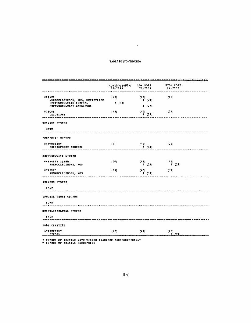
TABLE B2 (CONTINUED)
CONTROL(UNTR)22-2P96
LOW DOSE 22-2091
HISH EOS1 22-2092
tLIVERADENOCARCINOMA. NOS, METASTATICHEPATOCELLULAR ADENOMAHBPATOCELLULAR CARCINOMA
tCECUMLEIOSYOMA
(20)
1 (5*)
(19)
(«1) 1 (2X)
1 (2X)
C*0) 1 (3X)
(12)
(37)
URINARY SYSTEB
NONE
ENDOCRINE SYSTEB
»PITOITARYCHRONOPHOBE ADENORA
(8) (13) 1 (8X)
(25)
REPRCCDCTIVH SYSTEB
*NAH!IARY GLANDADENOCARCINOMA, NOS
tOTEROSADENOCARCINONA, NOS
(20)
(19)
(41) 1 (2X)
(40) 1 (3X)
(U3) 1 (2%)
(37)
NERVCDS SYSTEB
NONE
SPECIAL SENSE CRGANS
NONE
NDSCULOSKELETAL SYSTEB
NONE
BODY CAVITIES
•MESENTERY (20)
t NUMBER OF ANIMALS SITH TISSUE EXAMINED MICROSCOPICALLY * NUMBER OF ANIMALS NECROPSIED
(U3) 1
B-7

TABLE B2 (CONCLUDED)
CONTROL (UNTR) LOU DOSE HIGH DOSE 22-2096 22-2091 22-2092
ALL OTHER SYSTEMS
HONE
ANIMAL DISPOSITION SUMMARY
ANIMALS INITIALLY IN STUDY 20 50 50 NATURAL DEATHS 2 3 7 MORIBUND SACRIFICE 2 5 SCHEDULED SACRIFICE ACCIDENTALLY KILLED TERMINAL SACRIFICE 18 36 31 ANIMAL MISSING 9 7
a INCLUDES AtlTOLYZED ANIMALS
TUMOR SUMMARY
TOTAL ANIMALS KITH PRIMARY TUMORS* 6 13 13 TOTAL PRIMARY TUMORS 7 17 14
TOTAL ANIMALS WITH BENIGN TUHORS U 14 2 TOTAL BENIGN TUHORS U 4 2
TOTAL ANIMALS WITH MALIGNANT TUMORS 3 11 11 TOTAL MALIGNANT TUMORS 3 13 12
TOTAL ANIMALS WITH SECONDARY TUHCRSt 1 TOTAL SECONDARY TUMORS 1
TOTAL ANIMALS WITH TUMORS UNCERTAINBENIGN OR MALIGNANT
TOTAL UNCERTAIN TUMORS
TOTAL ANIMALS WITH TUMORS UNCERTAINPRIMARY OR HETASTATIC
TOTAL UNCERTAIN TUMORS
* PRIMARY TUMORS: ALL TUMORS EXCEPT SECONDAFY TDMORS * SECONDARY TDMOBS: METASTATIC TUMOBS OB TUMORS INVASIVE INTO AN ADJACENT ORGAN
B-8

APPENDIX C
SUMMARY OF THE INCIDENCE OF NONNEOPLASTICJ LESIONS IN RATS TREATED WITH 3,3'-DIMETHOXYBENZIDINE
4,4'-DIISOCYANATE


TABLEC1 SUMMARY OF THE INCIDENCE OF NONNEOPLASTIC LESIONS IN MALE RATS
TREATED WITH 3,3'-DIMETHOXYBENZIDINE-4,4'-DIISOCYANATE
CONTROL (VEH) LOB DOSE HIGH DOSE 11-1095 11-1093 11-1091
ANIMALS INITIALLY IV STUDY 20 50 50 ANIMALS NECROPSIED 20 50 50 ANIMALS EXAMINED HISTOPATHOLOGICSLLY** 20 50 50
I N T E G U M E N T A R Y S Y S T E M
N O N E
R E S P I R A T O R Y S Y S T E M
*TRACHEA (20) (149) (U6) INFLAMMATION, NOS 1 (5%)
ILUNG (20) (H9) («7) PNEUMONIA, ASPIRATION 2 (1*) INFLAMMATION, SOPPURATIVE 1 (2)1) ERONCHOPNEUMONIA, ACUTE 1 (2*) PNEUMONIA, CHRONIC MURINE 3 (15%) 7 (1K») 6 (13*) INFLAMMATION, CHRONIC 1 (2*) 1 (2%) INFLAMMATION, CHRONIC SUPPURATIV 1 (2*) HYPERPLASIA, ALVEOLAR EPITHELIUM 1 (2*)
ILUNG/ALVEOLI (20) (149) («7) PNEUMONIA, ASPIRATION 1 (2*)
HFMATOPOIETIC SYSTEM
ISP1EEN (19) (19) («9) HEMOSIDEHOSIS 2 (UX) 2 (1%) HYPERPLASIA, NODULAR 1 (2*) HEMATOPOIESIS 1 (2K) 1 (2*)
tMANDIBULAP L. NODE (17) (H9) (U2) HYPERPLASIA, PLASHA CELL 1 (2K)
tMESENTEHIC L. NODE (17) (49) (12) ____ HYPERPLASIA, LYHPHOID
t N U M B E R OF A N I M A L S WITH TISSUE E X A M I N E D M I C R O S C O P I C A L L Y * N U M B E R OF A N I M A L S NECROPSIED **EXCLUDES PARTIALLY AUTOLYZED ANIMALS
C-3

TABLE CI (CONTINUED)
HIGH DOSE 1 1-1091
(46)
(46)
4 (9*)
(50) 2 (4*) 1 (25) 1 ;2*j 4 (8*) 1 ( 2%) 1 (2*)
1 (2*)
(50) 1 (2*)
(50)
(50)
(48) 1 (2*)
(49) 1 (2%)
(49) 1 (2*)
(49j
CIRCULATORY SYSTEM
•HEART THROMBOSIS, NOS INFLAMMATION, CHRONIC
tflYCCASDIUM INFLAMMATION, CHRONIC FIB80SIS FIBROSIS, DIFFUSE DEGENERATION, NOS
DIGESTIVE SYSTiH
*LTVER NECROSIS, I'OCAL NECROSIS, DIFFUSE NFCROSIS, COAGOLATIVE KETAflORPHOSIS FATTY GLYCOGENIC CELL HYPERPLASIA, SODULAR HYPERPLASTiC NODCI.E HEMATOPOIESIS
*LIVER/HEPATOCYTES HYPERPLASIA, FOCAL
*BILE DUCT HYPEFPLftSIA, NOS
tPANCREAS HBROSIS, FOCAL
tSTOMACH HYPEBKERATOSI3
ISMALL INTESTINE OLCER, NOS
tPEYERS PATCH ABSCESS, NOS
IDUCDENUM UIC.E.S* NOS _
CONTROL (VEH) 1 1-1095
(19)
(19)
1 (31) 1 (5%) ? (11*)
(20)
(20)
(20) 1 (5!S)
(20)
(20)
(20)
(20)
(20)
LOB DOSE 11-1093
(48) 1 12%) 2 (U%)
(US) 2 C*%) 2 14*)
1 (2*)
(50) 2 CiS)
4 (8X)
1 (2%) 1 (?X)
(50)
(50)
(49) 3 (6*)
(49)
(49)
(49)
(49)
1 J2X1 _
t NUMBER OF ANIHALS KITH TISSOE EXAMINED MICROSCOPICALLY * NUMBER OF ANIMALS NECROPSIED
C-4

TABLE Cl (CONTINUED)
tLARGE INTESTINE NEBATODIASIS
ICOIOH HBBATODIASIS
URINARY SYSTEB
tKIDNEY HYD80NEPHROSIS INFLAHBATION, CHRONIC GRANULOBA, NOS EIGBENTATIOM, NCS
ENDOCRINE SYST5B
*PITOITARY CYST, NOS HYPEHPLASIA, FOCAL HYPERPLASIA, CHEOBOPHOBE-CEII
tADRBNAL IIPOIDOSIS
tADSENAL CORTEX HYPERPLASIA, NOS
tADRENAL BEDULLA FIBROSIS HYPERPLASIA, SOS
tTHYROID COLLOID CYST FOLLICDLAR CYST, NOS HYPERPLASIA, C-CELL
tPANCREATIC ISLETS HYPERPLASIA, HOS HYPEBPLASIA, ADENOBATOUS
BEPRCDOCTIVF SYSTIB
*BABBABY GLAND BULIILgCULAR CYST
CONTROL (VEH) 11-1095
(19) 7 (37H)
(19)
(20) 1 (5X)
13 (65X)
(16)
2 (13%)
(20)
(20)
(20)
2 (10X)
(19)
(20)
(20)
LOW DOSE HIGH DOSE 11-1093 11-1091
(U7) ("6) 1 (2%)
(U7) (I46) 14 (30*) 6 (13»)
(50) (49)
23 (U6X) 19 (39X) 1 <2X)
1 <2X)
(15) (36) 1 (2X) 1 (3X)
3 (7X)
(50) (<*9) 1 (2%)
(50) (U9) 1 <2X)
(50) (49) 1 (2%) 3 (6X)
(l»5) (1*6) 1 (2X) 1 (2X)
5 (11X) 1 (2%)
(19) (50) 1 <2X) 1 (2X)
(50) (50) LJ2X1
» NOBBER OF AHIBALS SITH TISSUE EXABINED BICEOSCOPICALLY » NDBBER OF ANIBALS NECROPSIED
C-5

1 ABLE Cl (CONCLUDED)
CONTROL (VEH) LOW DOSE HIGH COSE 11-1095 11-1093 11-1091
•PROSTATE (16) (46) INFLAHMATION, NOS 1 (2*) HYPERPLASIA, NOS 1 (2*) HYPERPLASIA. CYSTIC 3 (7«)
*SEHINAL VESICLE (20) (50) (50) CAST, NOS 1 (5X) INFLAMMATION, SOPPDRATIVE 1 (2X) HYPERPLASIA. CYSTIC
*TESTIS (20) (50) (49) ATEOPHY, NOS 4 (20K) 2 (4X)
SEBVCOS SYSTEM
*SBAIN (19) (50) (47) ATROPHY, NOS 2 (4«) ATROPHY, PRESSURE 1 (5«)
SPECIAL SENSE ORGANS
NONT
MDSCOLOSKELETAI SYSTEH
NONE
BODY CAVITIES
*INGDINAL REGION (20) (50) (50) K7CFOSIS, FAT 1 (2X)
ALL OTHER SYSTEHS
ADIPOSE TISSUE NECROSIS, FAT 1 2
SPECIAL MORPHOLOGY SDBHARY
AOTO/NECEOPSY/HISTO PERF 1
I NOH8ER OF ANIMALS WITH TISSHE EIAHINED MICROSCOPICALLY * NDHEER OP ANIMALS NBCROPSIED
HSAC: MORIBUND SACRIFICE OTHR: OTHER
C-6

TABLE C2 SUMMARY OF THE INCIDENCE OF NONNEOPIASTIC LESIONS IN FEMALE RATS
TREATED WITH 3,3'-DIMETHOXYBENZIDINE-4,4'-DIISOCYANATE
CONTROL (VEH)11-1096
LOW DOSE 11-1091
HIGH EOSE 11-1092
ANIMALS INITIALLY IN STUDY 20ANIMALS NECHOPSIED 20ANIMALS EXAMINED HISTOPATHOLOGICALIY** 20
50 50 50
50 18 18
INTEGUMENTARY SYSTEM
•SKIN (20) (50) (18) EPIDERMAL INCLUSION CYST 1 (5X)
*SUECUT TISSUE (20) (50) (18) HEMOBRHAGIC CYST 1 (2*)
R E S P I R A T O R Y S Y S T E M
*LARYNX (20) (50) (18) INFLAMMATION, CHRONIC 1 (2%)
•TRACHEA (20) (51) (17) INFLAMMATION, CHRONIC SUPPURATIV 1 (2«)
• LONG (20) (18) (18) PNEUMONIA, ASPIRATION 1 (2X) PNEUMONIA, CHRONIC HUBINE 7 (35X) 16 (33t) 11 (23%) HYPERPLASIA, ADENOMATOUS 1 (2%)
HEMATOPOIETIC SYSTEM
tBONE MARROW (19) (15) (15) HYPEBPLASIA, NOS 1 (2*)
iSPLEEN (20) (18) (18) HEHOSIDEROSIS 2 («*) 2 (1«) HYPEEPLASIA, HETICULOM CFLL 1 (211) HEMATOPOIESIS 7 (15») 1 (2%)
*CERVICAL LYMPH NODE (20) (15) (18)
* N U M B E R OF A N I M A L S KITH TISSUE E X A M I N E D M I C R O S C O P I C A L L Y * N U M B E R OF A N I M A L S NECROPSIFD **EXCLUDES PARTIALLY AUTOLYZED ANIMALS
C-7

TABLE C2 (CONTINUED)
—_ _ — —'———— ——
CONTROL (VEH) LOW DOSE HIGH DOSE 11-1096 11-109U 11-1092
CIRCULATORY SYSTEM
tHYCCARDIOB (18) (49) (US) FIBROSIS 1 (6*) 1 (2X) 2 (1*) FIBROSIS, FOCAL 1 (2*) DEGENERATION, NOS 1 (2*)
•DIGESTIVE SYSTIB
ILIVER (20) (50) (48) CYST, SOS 1 (2*) HEBOSRHAGIC CYST 1 (2*) NECROSIS, NOS 1 (5*) NECROSIS, FOCAL t (8*) NECROSIS, BIDZONAL 1 (2*) METAMORPHOSIS FATTY t (8*) « (8*) HYPERPLASIA, NODULAR 1 (2*) HYPERPtASIA, FOCAL 1 (2*)
tLIVBR/CENTRILOBULAR (20) (50) («8) BETABORPHOSIS FATTY 1 (2K)
*9IIE DDCT (20) (50) (»8) HYPERPLASIA, NOS 1 (2X)
*PANCBEAS (20) (K8) (48) FIBROSIS, FOCAL 2 (10*) 1 (2*) ATROPHY, FOCAL 1 (2*)
tSBAIL INTESTINE (20) (50) (18) BYPERPLASIA. LYBPHOID 1 (2*)
tLARG? INTESTINE (18) (US) <K7) NEKATODIASIS 2 (11*) « (9X) 2 («*)
IC010N (18) (15) (17) NEBATODIASIS 2 (11*) "» (9*) 3 <6X)
URINARY SYSTF.B
•KIDNEY (20) («9) (t8) INFLAHBATION. CHRONIC 5_JL25*J 5 JJ511 1U (29*)
* NOBBER OF ANIBALS WITH TISSOE EXABINED BICROSCOPICALLY * NOMBEB OF ANIMALS NECROPSIED
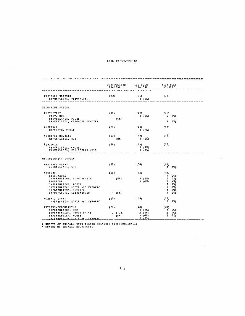
TABLE C2 (CONTINUED)
CONTROL (VIH) LOS DOSE 11-1096 11-1191*
SORINARY BLADDER (12) (36) HYPERPLASIA, FPITBELIAL 1 (3*)
ENDOCRINE SYSTEM
tPITUITARY (18) («8) CYST, NOS 1 (2*) HYPFBPLASIA, FOCAL 1 (6*) HYPERPL1SIA, CHROMOPHOBE-CELI
*ADRENAL (20) <«9) NECROSIS, FOCAL 1 (2*)
tADRENAL MEDULLA (20) (K9) HYPERPLASIA, SOS 1 (5*) 1 (2*)
*THYROID (18) (11) HYPERPLASIA, C-CELL 3 (7%) HYPERPLASIA, FOLIICULAR-CBLL 1 (2X)
REPRODUCTIVE SYSTEM
*MAM«ARY GLAND (20) (50) HYPERPLASIS, NOS
»UTERUS (20) (IB) HYDROHETRA INFLAMMATION, SDPPURATIVE 1 (5*) 1 (2*) EYOMETRA 3 (6X) INFLABMATIOS, ACDTE INFLAMMATION ACUTE AND CHRONIC INFLAMMATION. CHRONIC HYPERPLASIA, ADENONATODS 1 <5X)
ICERVIX UTERI (20) («8) INFLAMMATION ACUTE AND CHRONIC
IUT5ROS/ENDOHETRIDB (20) (18) INFLAMMATION, NOS 1 (2«) INFLAMMATION, SDPPUEATIVE 2 (10*) 2 (it*) INFLAMMATION, ACUTE 1 (5*) 3 <6X) IJiFLiMJJA^ION ACOTE AND CHFOJJIC 1 J2*L
t NOMBEB OF ANIMALS HITH TISSUE EXAMINED MICROSCOPICALLY * NUMBER OF ANISALS NECHOPSIED
HIGH DOSE 11-1092
(37)
(15) 2 (HX)
3 (7X)
(17)
(1*7)
(11)
(«8) 1 (2%)
(18) 1 (2X) 1 (2%) 2 («X) 1 (2%) 1 (2X) 1 (2X) 1 (2%)
(18) 1 (2X)
(18) 1 (2X) 2 (IS) 3 (6X)
. . . . . . .
C-9
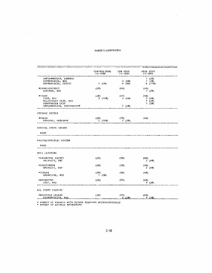
TABLE C2 (CONTINUED)
CONTROL (VEH) LOH DOSE HIGH COSE 11-1096 11-109U 11-1092
INFLAHHATICN, CH80NIC 1 (2*) HYPERPLASIA, NOS 2 («X) 1 <2X) HYPEBPLASIA, CYSTIC 1 (5X) « (3%) 6 (13*)
tOVARY/OVIDOCT (20) (US) (US) ABSCESS, NOS 1 (2*)
IOVAHY (20) (HI) (18) CYST, NOS 3 (15%) 2 (a*) 3 (6*) FOLLICULAR CYST, NOS 1 (2%) fAROVABIAN CYST 1 <2X) INFLAHMATION, SUPPORATIVE 1 (2X)
NEBVOUS SYSTEM
•BRAIN (20) (50) (U8) ATROPHY, PRESSURE 2 (10X) 1 12%)
SPECIAL SENSE CRGANS
NONE
WUSCOLOSKELETAL SYSTEH
NONE
BODY CAVITIES
•ABDOMINAL CAVITY (20) (50) (48) NECROSIS, FAT 2 (4X)
*PEBITONEOH (20) (50) (18) NECROSIS, FAT 2 (4X)
•PLEURA (20) (50) (48) GRANULOMA, NOS 1 (5X)
•MESENTERY (20) (50) (48) CYST, NOS 1 (2X)
ALL OTHFB SYSTEMS
•MULTIPLE OBGANS (20) (50) (48) PIGMENTATION. NOS 1 J2X1 1 J2X)
* NUMBER OF ANIMALS WITH TISSUE EXAMINED HICROSCOPICALLY * NUMBER OF ANIMALS NECROPSIED
C-10

TABLE C2 (CONCLUDED)
CONTROL (VEH) LOW DOSE HIGH EOSE 11-1096 11-1091 11-1092
ADIPOSE TISSUE STEATITIS NECROSIS. FAT
SPECIAL MORPHOLOGY SUHHARY
NO LESIOH REPORTED 2 1 AOTO/NECHOPSY/HISTO PERF 1 AOTOLYSIS/NO NECROPSY
HISS: BISSIHG TSAC: THRHINAL SACRIFICE HSAC: HOH1BDND SACRIFICE OTHR: OTHER
C-ll


APPENDIX D
SUMMARY OF THE INCIDENCE OF NONNEOPLASTIC LESIONS IN MICE TREATED WITH '3,3'-DIMETHOXYBENZIDINE
4,4'-DIISOCYANATE

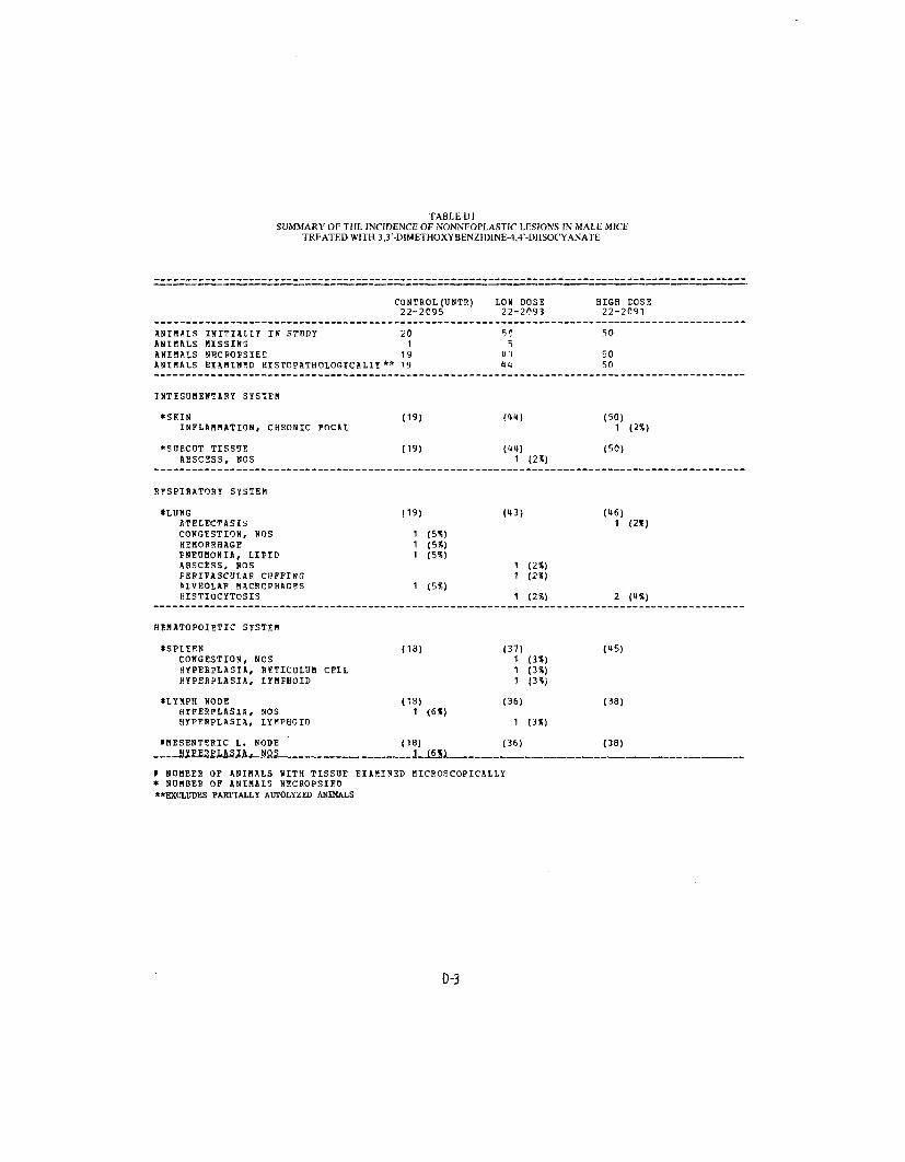
TABLE Dl SUMMARY OF THE INCIDENCE OF NONNEOPLASTIC LESIONS IN MALE MICE
TREATED WITH 3,3'-DIMETHOXYBENZIDINE-4,4'-DIISOCYANATE
CONTROL (UNTR)22-2095
ANIMALS INITIALLY IN STUDY ANIMALS MISSIN3 ANIMALS NECROPSIES
20 1
19 ANIMALS EXAMINED HISTOPATHOLOGICALIY ** 19
INTEGUMENTARY SYSTEM
*S(UN INFLAMMATION, CHRONIC FOCAL
*SUBCUT TISSUE ABSCESS, NOS
RESPIRATORY SYSTEM
ILUNG ATELECTASIS CONGESTION, NOS HEMORRHAGE PNEUMONIA, LIPIB ABSCESS, NOS PEBIVASCULAE CI3FFING SLVEOLAP MACRCPHAGES HISTIOCYTOSIS
HEMATOPOIETIC SYSTEM
tSPLEFN CONGESTION, NOS HYPE8PLASIA, RETICULUM CELL HYPERPLASIA, LYMPHOID
•LYMPH NODE HYPERPLASIA, NOS HYPEBPLASIA, LYMPHOID
*MESENTERIC L. NODE
t N U M B E R OF A N I H A L S K I T H TISSUE* N U M B E R OF A N I M A L S NECROPSIED "EXCLUDES PARTIALLY AOTOLYZED ANIMALS
(19)
(19)
(19)
1 (5X) 1 (5V 1 (5V
1 (5%)
(18)
(IS) 1 (6X)
(18)
E X A M I N E D M I C R O S C O P I C A L L Y
D-3
LOU DOSE HIGH tOSE 22-2093 22-2091
5? 50 5
u.-i 50 itl 50
"41) (50) 1 (2X)
(11) (50) 1 (2*)
(13) (16) 1 (2*)
1 (2*) 1 (2*)
1 (2%) 2 (IX)
(37) <U5) 1 (3V 1 (3X) 1 (3<J
(36) (38)
1 {3V
(36) (38)

TABLEDl (CONTINUED)
CONTROL (UNTR) 22-2095
LOW DOSE 22-2093
HYPERPLASIi, LYNFHOID
CIRCULATORY SYSTEM
IHEART (19) <«3) PERIARTERITIS 2 (5*) CALCIFICATION, FOCAL 1 (2*)
tMYOCARDIUM (19) (13) FIBROSIS 1 (2*)
DIGESTIVE SYSTEM
ILIVER (19) («3) IYBPHOCYTIC INFLAMMATORY INFILTR NECROSIS, FOCAL 2 (5*) METAMORPHOSIS FATTY 1 (5%) NUCLEAR ENLARGEMENT 1 (2X) HYPERPLASIA, NODULAR HYPERPLASIA, FOCAL
*BILE DUCT (19) (UK) CYST, NOS 1 (2%)
fPANCREAS (18) («1) INFLAMMATION, NECROTIZING ATROPHY, SOS 1 (2*)
tSTCMACH (15) (11)CYST, NOS 1 (2%) INFLAMMATION, NOS
ILARGE INTESTINE (18) (41)NEMATODIASIS 1 (6*) 1 (2%)
ICOION (18) (11)NEMATODIASIS 3 (17X) 3 (7X)
ORINBRY SYSTEM
tKIDNEY (19) (12) HYDBONEPjfgOSlS .__ i_J51L__
* NUMBER OF ANIMALS WITH TISSUE EXAMINED MICROSCOPICALLY * NUMBER OF ANIMALS NECROPSIEC
D-4
HIGH COSE 22-2091
1 <3X)
(«8)
(18)
<«8) 1 (2X) 1 (2*)
2 <1*) 1 (2%)
(50)
(38) 1 (3*)
(149)
1 (2*)
(»8)
(18)3 (6X)
<5P) ._. ._ , 2,, JUS) _ _ _ _ _ ._ ___ t

TABLE Dl (CONTINUED)
CONTBOL(UNTB) LOS DOSE HIGH DOSE 22-2095 22-2093 22-2091
•KIDNEY/CORTEX (19) <t2) (50) CYST, DOS 1 (2*)
ENDOCRINE SYSTEM
IADRENAL CORTEX 113) (25) (36) HYPERPLASIA, NOS 1 (8%)
• THYROID (13) (36) (35) COLLOID CYST 1 (8X) <3X) 2 FIBROSIS, FOCAL 1 (8*)
tPANCREATIC ISLETS (18) (41) (38) HYPEPPLASIA, NOS 1 (6«) 1 (3*)
REPRODUCTIVE SYSTEM
• N A U M A R Y G L A N C (19) ( H U ) (50) I N F L A P I B f T I O S , NOS 1 (5X)
*SEMINAL VESICLE (19) (14) (50) DILATATION/DUCTS 1 (2H)
NERVOUS SYSTEI"
•BRAIN (19) (t«) (50) CORPORA AMYLACEA 1 (5X) 1lt (32«) 9 (18X)
SPECIAL SENSE ORGANS
NONE
HUSCOI.OSKELETAL SYSTEM
NONE
BODY CAVITIES
NOSE
t NOMBEP OF ANIMALS WITH TISSUE EXAMINED MICROSCOPICALLY * NOMBEP OF ANIMALS NECBOPSIED
0-5

TABLE Dl (CONCLUDED)
CONTROL(DNTR) ICH COSE HIGH COSE 22-2095 22-2093 22-2091
ALL OTHER SYSTEMS
NONE
SPECIAL MORPHOLOGY SUMMARY
NO LESIOS DEPORTEDANIMAL MISSING/NO NECROPSYAUTO/NECROFSY/HISTO PERPAOTOLYSIS/HO HECROPSY
6 1
HISS: MISSINGMSAC: MORIBUND SACRIFICE
11 13 5
1 1
TSAC: TERMINAL SACRIFICE OTHR: OTHER
D-6
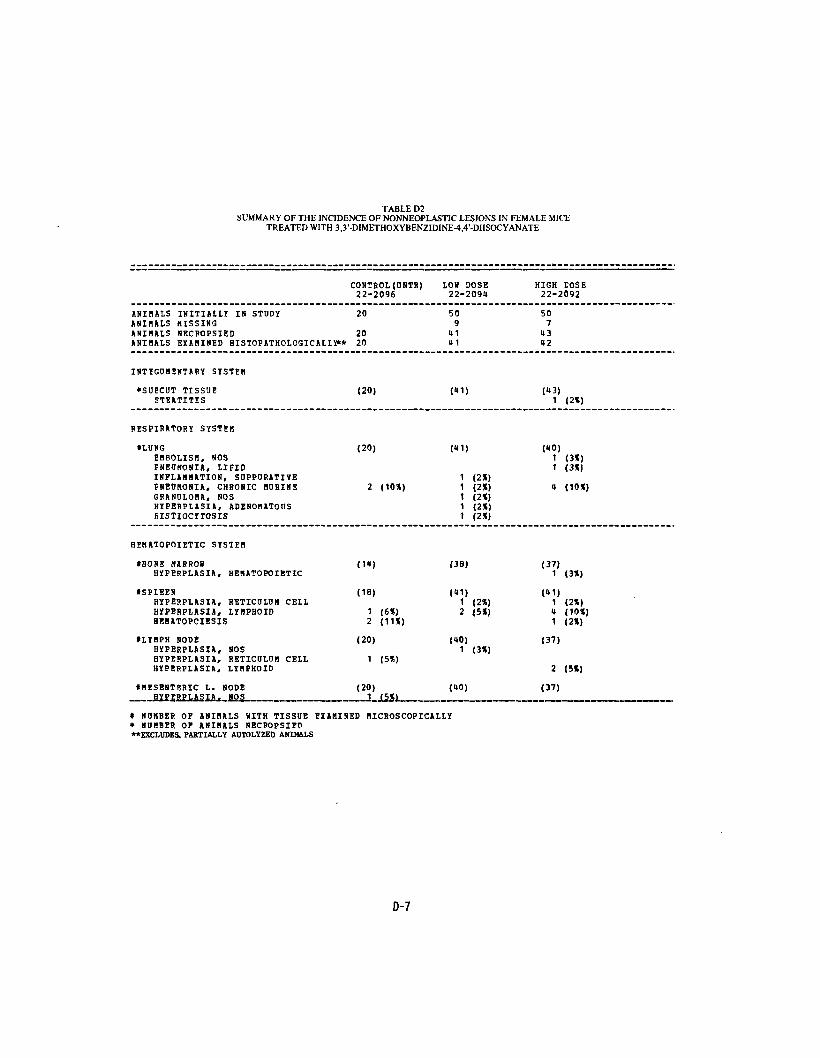
TABLE D2 SUMMARY OF THE INCIDENCE OF NONNEOPLASTIC LESIONS IN FEMALE MICE
TREATED WITH 3,3'-DIMETHOXYBENZIDINE^,4'-DHSOCYANATE
_— ___— I-"-'---™ 1 - —'—————— 1 • -.! -_•(••!•<•
CONTROL (UNTR) LOU DOSE HIGH EOSE 22-2096 22-209U 22-2092
ANIMALS INITIALLY IV STUDY 20 50 50 ANIMALS HISSING 9 7 ANIHALS NECSOPSIED 20 41 143 ANIMALS EXAMINED HISTOPATHOLOGICAIIY** 20 U1 12
INTEGUMENTARY SYSTEM
*SUECUT TISSUE (20) (41) (43) STEATITIS 1 (2%)
RESPIRATORY SYSTEM
• LUNG (20) («1) (10) EMBOLISM, NOS 1 (3*) PNEUMONIA, LIFID 1 (3») INFLAMMATION, SUPPURATIVE 1 (2*) PNEUMONIA, CHRONIC NURINE 2 (10*) 1 (2X) 4 (10*) GBANULONA, NOS 1 (2*) HYPESPLASIA, ADENOHATOUS 1 (2() HISTIOCYTOSIS 1 (2*)
HEMATOPOIETIC SYSTEM
«BOHE MARROW (1H) (38) (37) HYPERPLASIA, HEMATOPOIETIC 1 (3*)
ISPLEEN (18) (41) (11) HYPEHPLASIA, RETICOLDM CELL 1 (2*) 1 (2*) HYPERPLASIA, LYMPHOID 1 (6X) 2 (5*) 4 (10*) BEBATOPOIESIS 2 (11*) 1 (2*)
*LYMPH NODE (20) (40) (37) HYPERPLASIA, NOS 1 (3*) HYPERPLASIA, RETICOLOM CELL 1 (5*) HYPERPLASIA, LYHPROID 2 (5*)
*MESENTERIC L. NODE (20) (40) (37) NOS !_15JH
« N U M B E R OF A N I M A L S WITH TISSUE E X A M I N E D MICROSCOPICALLY * N U M B E R OF A N I M A L S NECROPSIEP "EXCLUDES. PARTIALLY AUTOLYZED ANIMALS
D-7

TABLE D2 (CONTINUED)
HIGH DOSE 22-2092
(12) 1 (8%)
(12)
1 (2%)
1 (2%)
(28)
(38)
(37) 1 (3*)
(37) 2 (5*)
(«2) 2 (5*) 1 (2«) 1 (2t)
1 <2E) 1 (2%)
(12) __ i 1251
tTHYKDS HYPERPLRSIA, IYMPH01D
CIRCULATORY SYSTEM
NON*
DIGESTIVE SYSIEH
*LIVER NECROSIS, FOCAL "ETAHORPHOSIS FATTY NUCLEAR ENLARGEMENT EFKATOPOIESIS
tPANCREAS LYMPHOCYTIC INFLAMMATORY INFILTR FIBBOSIS, FOCAL
tPEYERS PATCH HYPERPLASIA, LYMPHOID
tLARGE INTESTINE NE«ATODIASIS
ICOION NEHATODIASIS
URINARY SYSTEM
IKIDNF.Y HYDBONEPHROSIS GLOBEROLONEPHPITIS, NOS LYMPHOCYTIC INFLAMMATORY INFILTR INFLAMMATION. CHRONIC AMYLOID, NOS CALCINOSIS, NOS METAPLASIA. OSSEOUS
tKIENEY/NEDDLLA LYHPHOCYTJC INFLAMMATORY IHFILTR
CONTROL (ONTR) 27-2096
(20)
(19)
(20)
(19)
(19)
(19)
(19)
T-- -,,_ *.L -.. -..
L0» DOSE 22-209U
(»1) 1 (2«) 1 (2«) 1 (2%)
(39) 1 (3S) 1 (3X)
(11)1 <2X)
(«0) 1 (3*)
(UO)
(11)
1 (2X)
1 (2X)
1 (2*)
(11) t_
t NDMBER OF ANIMALS WITH TISSUE EXAMINED MICROSCOPICALLY * NUMBER OF AHIMALS NECROPSIED
0-8
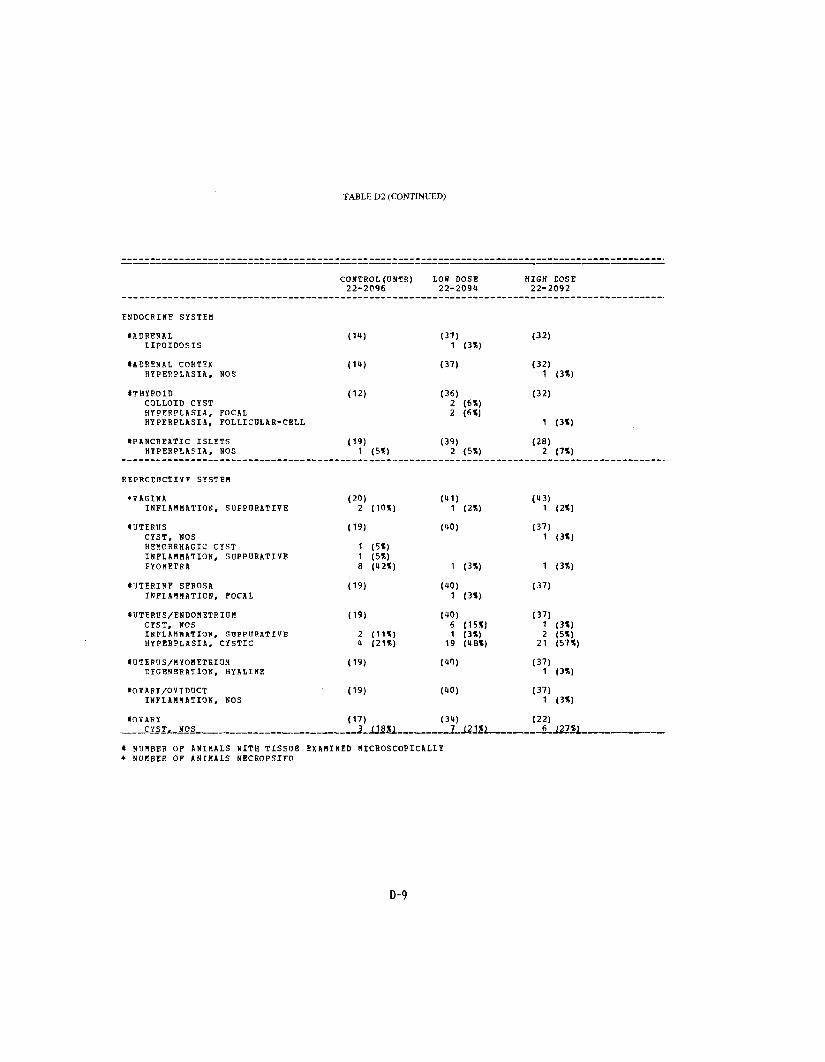
TABLE D2 (CONTINUED)
CONTROL (ONTR) LOW DOSS HIGH COSE 22-2096 22-209U 22-2092
ENDOCRINE SYSTEM
•ADRENAL (1t) (37) (32) IIPOIDOSIS 1 <3X)
•ADRENAL CORTEX HYPERPLASIA. NOS
(11) (37) (32) 1 (3%)
•THYROID (12) (36) (32) COLLOID CYST 2 (6X) HYPERPLASIA, FOCAL 2 (6X) HYPERPLASIA, FOLLICULAB-CELL 1 (3%)
fPAllCREATIC ISLETS (19) (39) (28) HIPERPLASIA, NOS 1 (5*) 2 (5%) 2 (7%)
EEPRCCDCTIVE SYSTEM
*VAGINA INFLAMMATION, SUPPOHATIVE
(20) 2 (10X)
(11) 1 (2X)
(13) 1 (2%)
•UTERUS (19) (tO) (37) CYST, NOS 1 (3%) HEMORRHAGIC CYST 1 (5*) INFLAMMATION, SUPPURATIVE 1 (5X) EYOHETRA 8 (K2%) 1 <3X) 1 (3%)
•UTIRINE SEROSA (19) (UO) (37) INFLAMMATION, FOCAL 1 (3X)
tUTERUS/ENDOMBTRIUH (19) (10) (37) CYST, NOS INFLAMMATION, SDPPtlSATIVE HYPERPLASIA, CYSTIC
2 (11*) U (21%)
6 (15%) 1 (3%)
19 (U8%)
1 (3%) 2 (5%)
21 (57%)
IUTIRUS/MYOMETRIOM EEGENERATION, HYALINE
(19) (1") (37) 1 (3%)
IOVARY/OVIDDCT (19) (10) (37) INFLAMMATION, NOS 1 (3%)
tOVARY CYST,. NOS _
(17) _ _ 3_JJ8X1 _
(31) _ 7 121*1_ _
(22) _ 6_J27%J
t NUMBER OF ANIMALS WITH TISSUE EXAMINED MICROSCOPICALLY * NUMBER OF ANIMALS NECROPSIED
D-9

TABLE D2 (CONCLUDED)
CONTROL(DNTB)22-2096
LOU DOSE 22-2091
HIGH EOSE 22-2092
FOLLICULAR CYST, NOSINFLAMMATION, SUEPURATIVEABSCESS, NOSI N F L A M M A T I O N , CHIiONICINFLAMMATION, GRANOLOMATOOS
1 (6%)
1 (3X)
1 (3X)
1 (3K)
1 (5X)
NERVOOS SYSTEM
tBHAINCOFFOPA AMYLACEA
(19) 2 (11%)
(39) 8 (21%)
(UO) 8 (20*)
SPECIAL SENSE ORGANS
NONF.
MOSCU10SKELETAL SYSTEM
*LIGAMENTABSCESS, NOS
(20) («1) 1 (2%)
(13)
BODY CAVITIES
*MESENTERYNECROSIS, FAT
' (20) («1) (13) 1 (21)
ALL CTHER SYSTEMS
"MULTIPLE ORGANSLYMPHOCYTIC INFLAMMATORY INFILTR
(20) 1 (5%)
(11) (13)
BROAD LIGAMENT ABSCESS, NOS 1
SPECIAL MORPHOLOGY SUMMARY
NO LESION REPORTEDANIMAL MISSING/NO NECROPSYAtJTO/N?CROPSY/NO HISTO
9 3
7 1
* NUMBER OF ANIMALS WITH TISSUE EXAMINED MICROSCOPICALLY * NUMBER OF ANIMALS SECBOPSIED
D-10

Review of the Bioassay of 3,3'-Dimethoxybenzidine-4,4'-Diisocyanate* for Carcinogenicity
by the Data Evaluation/Risk Assessment Subgroup of the Clearinghouse on Environmental Carcinogens
August 31, 1978
The Clearinghouse on Environmental Carcinogens was established in May, 1976, in compliance with DREW Committee Regulations and the Provisions of the Federal Advisory Committee Act. The purpose of the Clearinghouse is to advise the Director of the National Cancer Institute (NCI) on its bioassay program to identify and to evaluate chemical carcinogens in the environment to which humans may be exposed. The members of the Clearinghouse have been drawn from academia, industry, organized labor, public interest groups, State health officials, and quasi-public health and research organizations. Members have been selected on the basis of their experience in carcinogenesis or related fields and, collectively, provide expertise in chemistry, biochemistry, biostatistics, toxicology, pathology, and epidemiology. Representatives of various Governmental agencies participate as ad hoc members. The Data Evaluation/Risk Assessment Subgroup of the Clearinghouse is charged with the responsibility of providing a peer review of reports prepared on NCI-sponsored bioassays of chemicals studied for carcinogenicity. It is in this context that the below critique is given on the bioassay of 3,3'-Dimethoxybenzidine-4, 4'-Diisocyanate for carcinogenicity.
The primary reviewer said that 3,3'-Dimethoxybenzidine-4,4'-Diisocyanate induced a statistically significant increase in skin neoplasms and in leukemia in both sexes of treated rats, as well as endometrial stromal polyps in females. No evidence for the compound's carcinogenicity was found in treated mice. He noted that the compound was first administered by gavage and subsequently in the feed. The primary reviewer said that the study was acceptable and that the evidence was sufficient to conclude that 3,3'-Dimethoxybenzidine-4,4'-Diisocyanate posed a potential carcinogenic risk to man.
The secondary reviewer noted that the purity of 3,3'-Dimethoxybenzidine4,4'-Diisocyanate was not reported. Although he agreed that the skin tumors in male rats were treatment-related, he thought that the incidence was not sufficiently high in females to draw the same conclusion from a statistical basis. He suggested that the skin tumors may have resulted through direct skin contact with the compound. The secondary reviewer agreed that the leukemia and endometrial polyps in treated rats appeared to be treatment-related.
59

A motion was approved unanimously that the report on the bioassay of 3,3'-Dimethoxybenzidine-4,4'-Diisocyanate be accepted as written.
Members present were:
Arnold L. Brown (Chairman), University of Wisconsin Medical School Joseph Highland, Environmental Defense Fund Michael Shimkan, University of California at San Diego Louise Strong, University of Texas Health Sciences Center
Subsequent to this review, changes may have been made in the bioassay report either as a result of the review or other reasons. Thus, certain comments and criticisms reflected in the review may no longer be appropriate.
«r U.S. GOVERNMENT PRINTING OFFICE: 1978-281-217:3298
60


DHEW Publication No. (NIH) 79-1383


![6.5 [3,3]Sigmatropic Rearrangements The principles of orbial symmetry established that concerted [3,3] sigmatropic rearrangements are allowed processes.](https://static.fdocuments.us/doc/165x107/56649d095503460f949db954/65-33sigmatropic-rearrangements-the-principles-of-orbial-symmetry-established.jpg)