Bio Ilmiah
31
Introduction Pathology is a branch of the field of pathology which applies the principles of pathology to the legal profession. Forensic pathologists specialize in examining bodies and evidence such as body fluids and tissue samples for the purpose of gathering information which can be used in criminal investigation and court trials. They are sometimes known as medical examiners, coroners, or simply pathologists, depending on the region where they work. Some people think that the term “forensics” refers to forensic pathology, which generates considerable confusion when forensic accountants, forensic document examiners, forensic psychiatrists, and other forensics professionals are discussed. “Forensic” actually comes from the Latin forum, and it means “pertaining to a trial.” The field of forensics was developed in the 1800s, when people began applying scientific methods to criminal investigation and legal trials. The field of pathology in general is focused on the study of disease and its processes. A specialist in forensic pathology has training in this field, with additional skills which can be applied to the legal field. For example, he or she can examine a body to determine the cause of death, but the pathologist can also look for other clues and
-
Upload
charlene0126 -
Category
Documents
-
view
70 -
download
0
Transcript of Bio Ilmiah

Introduction
Pathology is a branch of the field of pathology which applies the principles of pathology
to the legal profession. Forensic pathologists specialize in examining bodies and evidence
such as body fluids and tissue samples for the purpose of gathering information which
can be used in criminal investigation and court trials. They are sometimes known as
medical examiners, coroners, or simply pathologists, depending on the region where they
work.
Some people think that the term “forensics” refers to forensic pathology, which generates
considerable confusion when forensic accountants, forensic document examiners,
forensic psychiatrists, and other forensics professionals are discussed. “Forensic” actually
comes from the Latin forum, and it means “pertaining to a trial.” The field of forensics
was developed in the 1800s, when people began applying scientific methods to criminal
investigation and legal trials.
The field of pathology in general is focused on the study of disease and its processes. A
specialist in forensic pathology has training in this field, with additional skills which can
be applied to the legal field. For example, he or she can examine a body to determine the
cause of death, but the pathologist can also look for other clues and information, such as
defensive marks on the hands which might indicate that the victim fought back while
being attacked.
Forensic pathology can involve the study of bodies, a process known as autopsy, or
analysis of samples taken at crime scenes or from the body. In cases where a complete
body is not available for examination, the forensic pathologist can gather information
from the available materials which can be used in investigation and eventual prosecution.
They can also examine samples of tissue and body fluids to look for toxins and signs
which can provide additional clues into the nature of the death, such as evidence that
someone was suffocated and then submerged to make the death look like a drowning.
In addition to being knowledgeable about human anatomy and pathology, a forensic
pathologist needs some additional skills. He or she must be able to collect evidence
properly and to maintain the chain of custody, ensuring that the evidence is not
compromised. It may also be necessary for forensic pathologists to testify on the witness
stand in some cases, and in some instances, a specialist in forensic pathology may be
retained by the defense for the purpose of refuting claims made by the specialist who
works for the government or the prosecution.
(http://www.wisegeek.com/what-is-forensic-pathology.htm)

Concept’s Definition
An autopsy (also known as a post-mortem examination or obduction) is the examination
of the body of a dead person and is performed primarily to determine the cause of death,
to identify or characterize the extent of disease states that the person may have had, or to
determine whether a particular medical or surgical treatment has been effective. In
academic institutions, autopsies sometimes are also requested for teaching and research
purposes. Forensic autopsies are autopsies with legal implications and are performed to
determine if death was an accident, homicide, suicide, or a natural event. The word
autopsy is derived from the Greek word autopsia: "to see with one's own eyes."
Autopsies are performed by pathologists; medical doctors who have received specialty
training in the diagnosis of diseases by the examination of body fluids and tissues.
(http://www.medicinenet.com/autopsy/article.htm )

Forensic autopsy
A forensic autopsy is used to determine the cause of death. Forensic science involves the
application of the sciences to answer questions of interest to the legal system. In United
States law, deaths are placed in one of five manners:
i)Natural
ii)Accident
iii)Homicide
iv)Suicide
v)Undetermined
In some jurisdictions, the Undetermined category may include deaths in absentia, such as
deaths at sea and missing persons declared dead in a court of law; in others, such deaths
are classified under "Other". But, medical examiners also attempt to determine the time
of death, the exact cause of death, and what, if anything, preceded the death, such as a
struggle. A forensic autopsy may include obtaining biological specimens from the
deceased for toxicological testing, including stomach contents. Toxicology tests may
reveal the presence of one or more chemical "poisons" (all chemicals, in sufficient
quantities, can be classified as a poison) and, the quantity of those chemicals. Because
post-mortem deterioration of the body, together with the gravitational pooling of bodily
fluids, will necessarily alter the bodily environment, toxicology tests may overestimate,
rather than underestimate, the quantity of the suspected chemical.
(http://www.relentlessdefense.com/autopsy.html )
Most states require the State medical examiner to complete an autopsy report and many
mandate that the autopsy be videotaped.
Following an in-depth examination of all the evidence, a medical examiner or coroner
will assign a manner of death as one of the five listed above, and detail the evidence on
the mechanism of the death.
(http://en.wikipedia.org/wiki/Autopsy#Forensic_autopsy )

Clinical autopsy
Clinical autopsies serve two major purposes. They are performed to gain more insight
into pathological processes and determine what factors contributed to a patient's death.
Autopsies are also performed to ensure the standard of care at hospitals. Autopsies can
yield insight into how patient deaths can be prevented in the future.
Within the United Kingdom, clinical autopsies can only be carried out with the consent of
the family of the deceased person as opposed to a medico-legal autopsy instructed by a
Coroner (England & Wales) or Procurator Fiscal (Scotland) to which the family cannot
object.
(http://en.wikipedia.org/wiki/Autopsy#Forensic_autopsy)
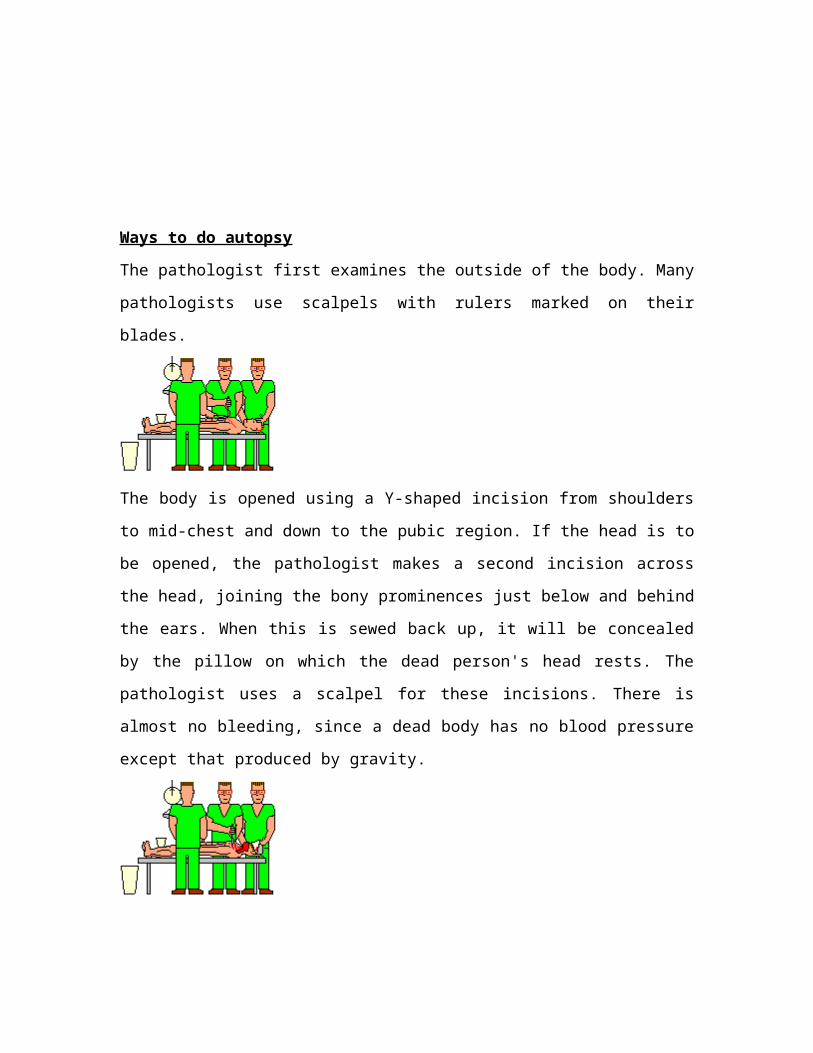
Ways to do autopsy
The pathologist first examines the outside of the body. Many pathologists use scalpels
with rulers marked on their blades.
The body is opened using a Y-shaped incision from shoulders to mid-chest and down to
the pubic region. If the head is to be opened, the pathologist makes a second incision
across the head, joining the bony prominences just below and behind the ears. When this
is sewed back up, it will be concealed by the pillow on which the dead person's head
rests. The pathologist uses a scalpel for these incisions. There is almost no bleeding, since
a dead body has no blood pressure except that produced by gravity.
The incisions are carried down to the skull, the rib cage and breastbone, and the cavity
that contains the organs of the abdomen. The scalp and the soft tissues in front of the
chest are then reflected back. Again, the pathologist looks around for any abnormalities.
Here, one pathologist is preparing to open the skull, using a special vibrating saw that
cuts bone but not soft tissue. This is an important safety feature.

Another pathologist is cutting the cartilages that join the ribs to the breastbone, in order to
be able to enter the chest cavity. This can be done using a scalpel, a saw, or a special
knife, depending on the pathologist's preferences and whether the cartilages have begun
to turn into bone, as they often do in older folks.
The third pathologist is exploring the abdominal cavity. The first dissection in the
abdomen is usually freeing up the large intestine. Some pathologists do this with a
scalpel, while others use scissors.
The skull vault is opened using two saw cuts, one in front, and one in back. These will
not show through the scalp when it is sewed back together.
When the breastbone and attached rib cartilages are removed, they are examined. Often
they are fractured during cardiopulmonary resuscitation.
Freeing up the intestine takes some time. The pathologist in this picture is cutting along
the attachment using a scalpel.
The top of the skull is removed, and the brain is very carefully cut free of its attachments
from inside the skull.
The chest organs, including the heart and lungs, are inspected. Sometimes the pathologist
takes blood from the heart to check for bacteria in the blood. For this, he/she uses a very
large hypodermic needle and syringe. The pathologist may also find something else that
will need to be sent to the microbiology lab to search for infection. Sometimes the
pathologist will send blood, urine, bile, or even the fluid of the eye or samples of brain
and/or liver for chemical study and to look for medicine, street drugs, alcohols, and/or
poisons.
Then the pathologist must decide in what order to perform the rest of the autopsy. The
choice will be based on a variety of considerations. This team will use the method of
Virchow, removing organs individually. After the intestines are mobilized, they may be
opened using special scissors.
Inspecting the brain often reveals surprises. A good pathologist takes some time to do
this.
The pathologist examines the heart, and generally the first step following its removal is
sectioning the coronary arteries that supply the heart with blood. There is often disease
here, even in people who believed their hearts were normal.

After any organ is removed, the pathologist will save a section in preservative solution.
Of course, if something looks abnormal, the pathologist will probably save more. The rest
of the organ goes into a biohazard bag, which is supported by a large plastic container.
The pathologist weighs the major solid organs (heart, lungs, brain, kidneys, liver, spleen,
sometimes others) on a grocer's scale. The smaller organs (thyroid, adrenals) get weighed
on a chemist's triple-beam balance.
The next step in this abdominal dissection will be exploring the bile ducts and then
freeing up the liver. Again, this pathologist has decided to use a scalpel.
After weighing the heart, the pathologist completes the dissection. There are a variety of
ways of doing this, and the choice will depend on the case. If the pathologist suspects a
heart attack, a long knife may be the best choice.
The liver has been removed. The pathologist has found something important. It appears
that this man had a fatty liver. It is too light, too orange, and a bit too big. Perhaps this
man had been drinking heavily for a while.

The pathologist has decided to remove the neck organs, large airways, and lungs in one
piece. This requires careful dissection. The pathologist always examines the neck very
carefully.
The liver in this case weighs much more than the normal 1400 gm.
The lungs are almost never normal at autopsy. These lungs are pink, because the dead
man was a non-smoker. The pathologist will inspect and feel them for areas of
pneumonia and other abnormalities.
The liver is cut at intervals of about a centimeter, using a long knife. This enables the
pathologist to examine its inner structure.

The pathologist weighs both lungs together, then each one separately. Afterwards, the
lungs may get inflated with fixative.
The rest of the team is continuing with the removal of the other organs. They have
decided to take the urinary system as one piece, and the digestive system down to the
small intestine as another single piece. This will require careful dissection.
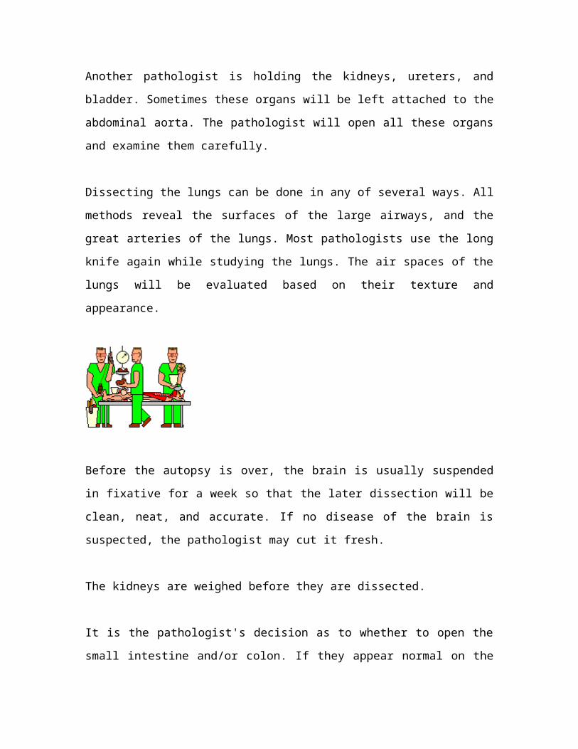
One pathologist is holding the esophagus, stomach, pancreas, duodenum, and spleen. He
will open these, and may save a portion of the gastric contents to check for poison.
Another pathologist is holding the kidneys, ureters, and bladder. Sometimes these organs
will be left attached to the abdominal aorta. The pathologist will open all these organs
and examine them carefully.
Dissecting the lungs can be done in any of several ways. All methods reveal the surfaces
of the large airways, and the great arteries of the lungs. Most pathologists use the long
knife again while studying the lungs. The air spaces of the lungs will be evaluated based
on their texture and appearance.

Before the autopsy is over, the brain is usually suspended in fixative for a week so that
the later dissection will be clean, neat, and accurate. If no disease of the brain is
suspected, the pathologist may cut it fresh.
The kidneys are weighed before they are dissected.
It is the pathologist's decision as to whether to open the small intestine and/or colon. If
they appear normal on the outside, there is seldom significant pathology on the inside. I
usually open them. The last pathologist is preparing the big needle and thread used to sew
up the body.
When the internal organs, have been examined, the pathologist may return all but the tiny
portions that have been saved to the body cavity. Or the organs may be cremated without
being returned. The appropriate laws, and the wishes of the family, are obeyed.
The breastbone and ribs are usually replaced in the body. The skull and trunk incisions
are sewed shut ("baseball stitch"). The body is washed and is then ready to go to the
funeral director.

These pathologists will submit the tissue they saved to the histology lab tomorrow, to be
made into microscopic slides. When these are ready, they will examine the sections, look
at the results of any lab work, and draw their final conclusions.
The only finding in this imaginary autopsy was fatty liver. There are several ways in
which heavy drinking, without any other disease, can kill a person. The pathologists will
rule each of these in or out, and will probably be able to give a single answer to the police
or family.
A final report is ready in a month or so. The glass slides and a few bits of tissue are kept
forever, so that other pathologists can review the work.
(http://www.pathguy.com/autopsy.htm)

Ways to determine the time of death(TOD)
1.Rigor Mortis
Rigor Mortis is the stiffening of the body after death because of a loss of Adenosine
Triphosphate (ATP) from the body's muscles. ATP is the substance that allows energy to
flow to the muscles and help them work and without this the muscles become stiff and
inflexible.
Rigor Mortis begins throughout the body at the same time but the body's smaller muscles
such as those in the face, neck, arms and shoulders are affected first and then the
subsequent muscles throughout the rest of the body; those which are larger in size, are
affected later.
Rigor normally appears within the body around two hours after the deceased has passed
away with as we have already mentioned, the facial and upper neck and shoulder muscles
first to visibly suffer from its effects. Many Scenes of Crime Officers (SOCO) have
reported that upon discovering the deceased that their face might have taken on what
looks to be a grimace; this is because the facial muscles have contracted as ATP drains
from them.
Once the contracting of all the body's muscles has taken place this state of Rigor -
technically referred to as the Rigid Stage - normally lasts anything from eight to twelve
hours after which time the body is completely stiff; this fixed state lasts for up to another
eighteen hours.
Contrary to common perception the process of Rigor Mortis actually does reverse and the
body returns to a flaccid state; the muscles losing their tightness in the reverse of how
they gained it: i.e.: those larger muscles that contracted last will lose their stiffness first
and return to their pre-Rigor condition.
Rigor Mortis is a good means of indicating time of death as is normally visible within the
first thirty-six to forty-eight hours after death; after which it leaves the body.
(http://www.csifanwiki.com/page/Forensic+Pathology)

2. Lividity
Lividity is also useful for this purpose. Lividity is the process through which the body's
blood supply will stop moving after the heart has stopped pumping it around the inside of
the deceased. What normally happens at this point is that the blood supply - or at least
any blood that remains within the corpse depending on the nature of their death - will
settle in direct response to gravity. For example an individual found lying on their
stomach would be found with all the blood from their back heading towards the ground.
Lividity also displays itself as a dark purple discolouration of the body and can also be
referred to as Livor Mortis or Post Mortem Hypostasis.
Any part of the body which has come into contact with a firm surface for a period of time
such as a floor or bench top will show signs of this during lividity as this impression
against the skin displays itself as an indentation surrounded by gravity-pulled blood.
It is worth noting that lividity begins to work through the deceased within thirty minutes
of their heart stopping and can last up to twelve hours. Only up to the first six hours of
death can lividity be altered by moving the body. After the six hour mark lividity is fixed
as blood vessels begin to break down within the body.
(http://www.csifanwiki.com/page/Forensic+Pathology)
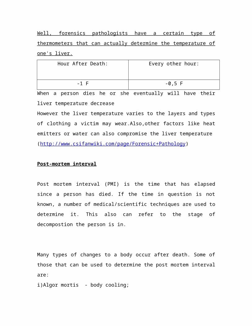
3.Liver Temperature
Well, forensics pathologists have a certain type of thermometers that can actually
determine the temperature of one's liver.
Hour After Death: Every other hour:
-1 F -0,5 F
When a person dies he or she eventually will have their liver temperature decrease
However the liver temperature varies to the layers and types of clothing a victim may
wear.Also,other factors like heat emitters or water can also compromise the liver
temperature
(http://www.csifanwiki.com/page/Forensic+Pathology)
Post-mortem interval
Post mortem interval (PMI) is the time that has elapsed since a person has died. If the
time in question is not known, a number of medical/scientific techniques are used to
determine it. This also can refer to the stage of decompostion the person is in.
Many types of changes to a body occur after death. Some of those that can be used to
determine the post mortem interval are:
i)Algor mortis - body cooling;
ii)Rigor mortis - stiffening of limbs;
iii)Forensic entomology - insect activity on the corpse;
iv)Vitreous humour changes - eye chemistry;
v)State of decomposition - autolysis (process of self digestion) and putrefaction (process
caused by bacteria found within the body)
A person who judges the time of death by the means of decomposition is privy to a
simple five stage process:
Stage 1: Initial Decay - This is basically where the body will stop producing antigens and
enzymes that are used to fight off bacteria located mainly in the lower intestine.
Stage 2: Putrefaction - Because the body no longer has a defense system in place the
bacteria grow and multiply by feeding off the body. They will begin to bring forth certain
gases, which in turn will give the dead body a sort of bloated look and will cause a rather
unpleasant odor.
Stage 3: Black Putrefaction - This stage will bring further discoloration to the body
(whether it be black, blue, purple, green etc.). It will also have an even more horrendous
odor as the gases caused by the bacteria begin to escape out of the body.
Stage 4: Butyric Fermentation - Where the internal organs begin to liquefy and the body
will begin to desiccate-or dry out.

Stage 5: Dry Rot - This is the slowest process out of the five stages. Basically, as the
name indicates, the body will slowly begin to dry out and ultimately skeletonize.
(http://en.wikipedia.org/wiki/Post-mortem_interval)

Autopsy’s Photo
Autopsy Photos of Lisa McPherson
Forensic entomologists identified over a hundred cockroach feeding sites on her body,
and three nationally prominent forensic pathologists opined that the manner of death was
"homicide". Pathologist Werner Spitz, M.D. wrote in his affidavit that "the insect bites
appearing in the autopsy photographs of Lisa McPherson, and in particular on her hands
and feet, are antemortem and peri-mortem/postmortem."
In other words, bugs were feeding on Lisa before she died, while she was dying and after
she had died. Perhaps it is for this reason that they found it necessary to bath a dead Lisa
McPherson before they drove her to a hospital 45 minutes away so she could see an
emergency room doctor who is a Scientologist, passing four other hospitals.
First released Lisa McPherson Autopsy pictures
1. Lisa 19, left hand, IV tubing
(credit : First released Lisa McPherson Autopsy pictures;http://www.xenu-
directory.net/mirrors/www.whyaretheydead.net/lisa_mcpherson/autopsy/index.html )

2. Lisa 20, right hand, close up, hip visible
(credit : First released Lisa McPherson Autopsy picture ; http://www.xenu-
directory.net/mirrors/www.whyaretheydead.net/lisa_mcpherson/autopsy/index.html )
3. Lisa 21, right forearm, medical tags
(credit : First released Lisa McPherson Autopsy pictures ; http://www.xenu-
directory.net/mirrors/www.whyaretheydead.net/lisa_mcpherson/autopsy/index.html )
4. Lisa 22, right forearm and hand, hip and tags visible
(credit : First released Lisa McPherson Autopsy pictures ; http://www.xenu-
directory.net/mirrors/www.whyaretheydead.net/lisa_mcpherson/autopsy/index.html )

5. Lisa 23, right fingers, close up
(credit : First released Lisa McPherson Autopsy pictures ; http://www.xenu-
directory.net/mirrors/www.whyaretheydead.net/lisa_mcpherson/autopsy/index.html )
6. Lisa 24, left forearm
(credit : First released Lisa McPherson Autopsy pictures ; http://www.xenu-
directory.net/mirrors/www.whyaretheydead.net/lisa_mcpherson/autopsy/index.html )
7. her back (note the blood pooled in her back as she was lying overnight)
( credit : Autopsy Photos of Lisa McPherson ;
http://www.lisamcpherson.org/images/autopsy/lisa9.jpg )
8. her right foot (coroner made some cuts)
(credit : Autopsy Photos of Lisa McPherson ;
http://www.lisamcpherson.org/images/autopsy/lisa32.jpg )
9. her legs from the left
(credit : Autopsy Photos of Lisa McPherson ;
http://www.lisamcpherson.org/images/autopsy/lisa14.JPG )
10. backs of her ankles
(credit : Autopsy Photos of Lisa McPherson ;
http://www.lisamcpherson.org/images/autopsy/lisa17.jpg )