Bio Factsheet - AMAZING WORLD OF SCIENCE WITH MR. GREEN · Bio Factsheet September 1997Number 1 The...
4
1 B io F actsheet September 1997 Number 1 The kidney: excretion and osmoregulation Kidneys have two main functions. 1. They are excretory organs, removing nitrogenous and other waste from the body. 2. They play an important part in maintaining a constant internal environment by helping to regulate pH, water and sodium ion concentrations in the blood and tissues. This Factsheet will focus on the role of the kidney in excretion and osmoregulation. Excretion Surplus nitrogen-containing compounds such as amino acids have to be broken down in the body because they are toxic and are then excreted as ammonia, urea or uric acid (Table 1). Table 1. Nitrogenous excretory products Renal corpuscle Proximal convoluted tubule Distal convoluted tubule Collecting duct Ascending limb of loop of Henle Descending limb of loop of Henle A B C Figure 2. The kidney nephron A- Ultrafiltration B- Selective reabsorption C- Osmoregulation Basic kidney structure The basic structure of the mammalian kidney is shown in Figure 1. Figure 1. Vertical section through mammalian kidney. Excretory product Source Urea Deamination of amino acids via the ornithine cycle in the liver Uric acid Deamination of purines (adenine and guanine) Ammonia Deamination of amino acids. Ammonia is secreted into the urine by cells in the kidney tubule Exam Hint - Don’t confuse urea and urine. Urea is made by the deamination of amino acids in the liver. Urine is the fluid produced by the kidneys. Each kidney contains a million coiled tubes called nephrons and it is in the nephron that urine formation occurs. Each nephron is divided into a number of distinct regions with particular functions labelled A, B, C (Figure 2). Cortex Medulla Protective capsule of adipose tissue Nephron (produces urine) Pyramid (where nephrons empty to renal pelvis) Positions of nephrons and collecting ducts Renal vein Renal artery Renal pelvis (cavity to receive urine) Ureter (to carry urine to bladder) Ammonia Urea Uric acid Solubility Very high High Very low Amount of water necessary to remove from body Very large Medium Very little Toxicity 0 4 8 Some important biological properties of these three substances are summarised in Table 2. Table 2. Biological properties of excretory products. From this you can work out that: • Freshwater fish excrete ammonia. Although it is very poisonous, fish are surrounded by large amounts of water so the ammonia can easily be diluted to safe levels. • Mammals excrete nitrogen mainly as urea. Urea requires more energy in the form of ATP for its production but is much less toxic than ammonia and fairly soluble. It therefore does not require large amounts of water to remove it from the body. • Birds excrete nitrogen mainly as uric acid. Flight demands a low body mass. Removing nitrogenous waste as uric acid means that large amounts of water are not required. Insects also excrete uric acid. As they are so small, they are very prone to water loss so it is important that they do not lose large amounts of water in excreting nitrogenous waste. Molecules of ATP needed High Medium Low
Transcript of Bio Factsheet - AMAZING WORLD OF SCIENCE WITH MR. GREEN · Bio Factsheet September 1997Number 1 The...

1
Bio FactsheetSeptember 1997 Number 1
The kidney: excretion and osmoregulationKidneys have two main functions.1. They are excretory organs, removing nitrogenous and other waste from the body.2. They play an important part in maintaining a constant internal environment by helping to regulate pH, water and sodium ion
concentrations in the blood and tissues. This Factsheet will focus on the role of the kidney in excretion and osmoregulation.
ExcretionSurplus nitrogen-containing compounds such as amino acids have to bebroken down in the body because they are toxic and are then excreted asammonia, urea or uric acid (Table 1).
Table 1. Nitrogenous excretory products
Renalcorpuscle
Proximalconvolutedtubule
Distalconvolutedtubule
Collecting duct
Ascending limbof loop of Henle
Descending limbof loop of Henle
A
B
C
Figure 2. The kidney nephron
A- UltrafiltrationB- Selective
reabsorptionC- Osmoregulation
Basic kidney structureThe basic structure of the mammalian kidney is shown in Figure 1.
Figure 1. Vertical section through mammalian kidney.
Excretory product Source
Urea Deamination of amino acids via the ornithinecycle in the liver
Uric acid Deamination of purines (adenine and guanine)
Ammonia Deamination of amino acids. Ammonia issecreted into the urine by cells in the kidneytubule
Exam Hint - Don’t confuse urea and urine. Urea is made by thedeamination of amino acids in the liver. Urine is the fluid produced bythe kidneys.
Each kidney contains a million coiled tubes called nephrons and it is in thenephron that urine formation occurs. Each nephron is divided into a numberof distinct regions with particular functions labelled A, B, C (Figure 2).
Cortex
MedullaProtective capsuleof adipose tissue
Nephron(produces urine)
Pyramid(wherenephrons emptyto renal pelvis)
Positionsof nephrons andcollecting ducts
Renal veinRenal artery
Renal pelvis(cavity toreceive urine)
Ureter (to carryurine to bladder)
Ammonia Urea Uric acid
Solubility Very high High Very low
Amount of water necessaryto remove from body
Very large Medium Very little
Toxicity
0 4 8
Some important biological properties of these three substances aresummarised in Table 2.
Table 2. Biological properties of excretory products.
From this you can work out that: • Freshwater fish excrete ammonia. Although it is very poisonous, fish
are surrounded by large amounts of water so the ammonia can easily bediluted to safe levels.
• Mammals excrete nitrogen mainly as urea. Urea requires more energyin the form of ATP for its production but is much less toxic thanammonia and fairly soluble. It therefore does not require large amountsof water to remove it from the body.
• Birds excrete nitrogen mainly as uric acid. Flight demands a low bodymass. Removing nitrogenous waste as uric acid means that large amountsof water are not required. Insects also excrete uric acid. As they are sosmall, they are very prone to water loss so it is important that they donot lose large amounts of water in excreting nitrogenous waste.
Molecules of ATP needed
High Medium Low
The kidney: excretion and osmoregulation Bio Factsheet
2
Figure 4.
The structure of the filter (Figure 4).Blood plasma is separated from the filtrate by two rows of cells, the liningcells of the capillary and the podocytes which make up the inner layer ofthe capsule. The capillaries have pores in their walls which the moleculesin the plasma are able to pass directly through. The small molecules thenpass through the basement membrane and once through this, they can passbetween the processes of the podocyte directly into the cavity of the renalcapsule. The actual filter is just the basement membrane and this is extremelythin.
B. Selective reabsorption in the proximal convoluted tubuleThe filtrate contains toxic substances such as urea which it is necessary toremove from the body but it also contains substances such as glucosewhich are required by the body. The function of the proximal convolutedtubule is to reabsorb these useful substances (Figure 5).
Figure 5. Reabsorption in the proximal convoluted tubule.
Microvilli
Fluid in tubule Cell liningproximalconvolutedtubule
Intercellularspaces
Capillary
1
2
2
3
Mitochondria
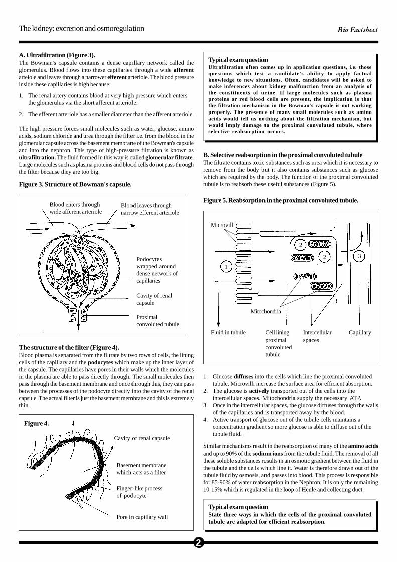
A. Ultrafiltration (Figure 3).The Bowman's capsule contains a dense capillary network called theglomerulus. Blood flows into these capillaries through a wide afferentarteiole and leaves through a narrower efferent arteriole. The blood pressureinside these capillaries is high because:
1. The renal artery contains blood at very high pressure which entersthe glomerulus via the short afferent arteriole.
2. The efferent arteriole has a smaller diameter than the afferent arteriole.
The high pressure forces small molecules such as water, glucose, aminoacids, sodium chloride and urea through the filter i.e. from the blood in theglomerular capsule across the basement membrane of the Bowman's capsuleand into the nephron. This type of high-pressure filtration is known asultrafiltration. The fluid formed in this way is called glomerular filtrate .Large molecules such as plasma proteins and blood cells do not pass throughthe filter because they are too big.
Cavity of renal capsule
Basement membranewhich acts as a filter
Finger-like processof podocyte
Pore in capillary wall
Figure 3. Structure of Bowman's capsule.
Blood enters throughwide afferent arteriole
Blood leaves throughnarrow efferent arteriole
Podocyteswrapped arounddense network ofcapillaries
Cavity of renalcapsule
Proximalconvoluted tubule
Typical exam questionUltrafiltration often comes up in application questions, i.e. thosequestions which test a candidate's ability to apply factualknowledge to new situations. Often, candidates will be asked tomake inferences about kidney malfunction from an analysis ofthe constituents of urine. If large molecules such as plasmaproteins or red blood cells are present, the implication is thatthe filtration mechanism in the Bowman's capsule is not workingproperly. The presence of many small molecules such as aminoacids would tell us nothing about the filtration mechanism, butwould imply damage to the proximal convoluted tubule, whereselective reabsorption occurs.
1. Glucose diffuses into the cells which line the proximal convolutedtubule. Microvilli increase the surface area for efficient absorption.
2. The glucose is actively transported out of the cells into theintercellular spaces. Mitochondria supply the necessary ATP.
3. Once in the intercellular spaces, the glucose diffuses through the wallsof the capillaries and is transported away by the blood.
4. Active transport of glucose out of the tubule cells maintains aconcentration gradient so more glucose is able to diffuse out of thetubule fluid.
Similar mechanisms result in the reabsorption of many of the amino acidsand up to 90% of the sodium ions from the tubule fluid. The removal of allthese soluble substances results in an osmotic gradient between the fluid inthe tubule and the cells which line it. Water is therefore drawn out of thetubule fluid by osmosis, and passes into blood. This process is responsiblefor 85-90% of water reabsorption in the Nephron. It is only the remaining10-15% which is regulated in the loop of Henle and collecting duct.
Typical exam questionState three ways in which the cells of the proximal convolutedtubule are adapted for efficient reabsorption.

The kidney: excretion and osmoregulation Bio Factsheet
3
C. Osmoregulation (Figure 6)The ability to produce concentrated urine is important in allowing terrestrialmammals to conserve water. The loop of Henle and the collecting ductform a system known as a countercurrent multiplier whose function isto remove water from the fluid in the tubule and produce a concentratedurine.
1. Na+ and Cl- ions are actively pumped out of the ascending limb. Theions accumulate in the interstitial fluid. This lowers the water potentialof the interstitial fluid. The tendency is for water to osmotically followthe Na+ and Cl- ions but it cannot since most of the ascending limb isimpermeable to water.
2. Water is drawn out of the descending limb and into the interstitialfluids by osmosis. This makes the fluid in the descending limb moreand more concentrated.
3. By the time the fluid in the descending limb has reached the bottom ofthe limb, it has lost a lot of water and is very concentrated. The fluidsurrounding the bottom of the loop - in the medulla of the kidney - isalso very concentrated because of the accumulation of Na+ and Cl- ions.The direction of the concentration gradient is shown by the arrow.
4. The fluid then enters the ascending limb. As it moves up the ascendinglimb, sodium ions are actively pumped out of it.
5. This makes the fluid at the top of the ascending limb very dilute again.
6. The fluid then empties into collecting ducts which pass through thevery concentrated medullary region.
7. Under the influence of the hormone ADH, the wall of the collectingduct becomes permeable to water which is therefore osmotically drawnout of the collecting duct and into the blood capillaries in the region.
8. By drawing water out of the fluid in the collecting duct, a veryconcentrated urine can be produced. By regulating the permeability ofthe collecting duct (via ADH), the amount of water in the blood andthe concentration of the urine can be controlled.
Exam Hint - The longer the loop of Henle, the more concentrated theurine that can be formed. A nephron from a frog or a toad doesn’t havea loop of Henle so these animals are unable to produce concentratedurine. Small mammals such as gerbils that live in deserts havenephrons with very long loops of Henle. These animals can produceextremely concentrated urine, thus reducing water loss from the body.This is often tested as an application question.
What controls ADH?The osmotic concentration of the blood is monitored by osmoreceptors inthe hypothalamus. Blood pressure is monitored by baroreceptors whichare widely dispersed throughout the circulatory system. Both types ofreceptor can send impulses to the posterior pituitary gland to start/stopADH release.
If too much water is lost from the body:1. The volume of blood plasma falls, its osmotic concentration therefore
increases and blood pressure falls.
2. Osmoreceptor and baroreceptors detect these changes and an impulseis sent to the posterior pituitary gland.
3. ADH is released.
4. ADH increases the permeability to water of the collecting duct andthe distal convoluted tubule.
5. More water is therefore drawn out of the collecting duct and distalconvoluted tubule back into the blood. This restores the volume andpressure of the blood and reduces its osmotic concentration.
6. The stimulus to the posterior pituitary is switched off.
If the osmotic concentration of the blood falls or if blood pressure increases,less ADH is released, less water is reabsorbed, resulting in a large volumeof dilute urine. This restores the osmotic concentration and pressure of theblood.
Typical exam questionIndividuals who are unable to produce ADH suffer from diabetesinsipidus. Describe the likely symptoms of this condition.
Na+
Cl-
H2O
Na+
Cl-
Distal convolutedtubule
H2O
H2O
H2OH
2O
H2O
H2O
Cortex Cortex
Col.duct
Medulla
Medulla
IncreasingNa+ Cl-
concentration concentrated urine
4
1
4
4
5
6
3
2
8
Interstitialfluid
7
Figure 6.

The kidney: excretion and osmoregulation Bio Factsheet
4
Practice questionsSemicolons indicate marking points.1. The diagram shows a cell from the proximal convoluted tubule in the
nephron.
B
Fluid in tubule Capillary
1
2
2
3
A
(a) State the name of the major process which occurs in region:(i) A (1 mark)(ii) B (1 mark)
(b) Suggest why some desert mammals have very long loopsof Henle (2 marks)
(a) Outline the function of:(i) Component A (2 marks)(ii) Component B (2 marks)
(b) Suggest explanations for each of the following:(i) The presence of glucose in the urine of a person who
has consumed large amounts of glucose (2 marks)(ii) The presence of protein in the urine of a person
suffering from high blood pressure. (2 marks)
2. (a) Outline the process of ultrafiltration (3 marks)
(b) The diameter of the efferent arteriole can be decreased by musclecontraction. Suggest what effect this would have on the processof ultrafiltration (2 marks)
3. The table shows the composition of fluids drawn from different regionsof a mammalian kidney.
Substance Plasma Glom. filtrate Urine
Glucose 0.08 0.07 0
Sodium 0.33 0.31 0.33
Urea 0.028 0.028 1.9
7 0 0
Concentration in fluid (g/100cm3)
(a) Suggest an explanation for the difference in composition of theplasma and urine (3 marks)
4. The diagram represents part of a mammalian kidney nephron.
A
Bowman'scapsule
B
descending limb
ascending limb
collectingduct
distalconvolutedtubule
afferentarteriole
efferent arteriole
interstitual fluid
Answers1. (a) (i) Provide ATP;
for active transport of glucose into intercellular spaces;
(ii) Provide large surface;for diffusion of glucose;
(b) (i) No. of glucose molecules exceeds No. of carriers in proximalcell;therefore unable to be actively transported;
(ii) High pressure forces proteins through basement membrane;pressure may damage membrane;
2. (a) High blood pressure;because of short afferent arteriole;narrow efferent arteriole;forces small molecules through basement membrane; (any 3)
(b) Increase it;by increasing pressure;
3. (a) All glucose reabsorbed in proximal convoluted tubule;Sodium concentration remains the same because active pumpingout of ions is balanced by corresponding water loss;Urea is concentrated through reabsorption of water in distalconvoluted tubule and collecting duct;
4. (a) (i) Ultrafiltration(ii) Selective reabsorption
(b) To conserve water;the longer the loop the more water can be reabsorbed;
Protein
Acknowledgements;Curriculum Press, Unit 305B, The Big Peg,120 Vyse Street, Birmingham. B18 6NFBio Factsheets may be copied free of charge by teaching staff or students,provided that their school is a registered subscriber.No part of these Factsheets may be reproduced, stored in a retrieval system, ortransmitted, in any other form or by any other means, without the priorpermission of the publisher. ISSN 1351-5136