BILIRUBIN METABOLISM & JAUNDICEvictorjtemple.com/Bilirubin Metabolism Jaundice PPP 9.pdf ·...
27
BILIRUBIN METABOLISM & JAUNDICE UNIVERSITY OF PNG SCHOOL OF MEDICINE AND HEALTH SCIENCES DISCIPLINE OF BIOCHEMISTRY & MOLECULAR BIOLOGY PBL MBBS II SEMINAR VJ Temple 1
Transcript of BILIRUBIN METABOLISM & JAUNDICEvictorjtemple.com/Bilirubin Metabolism Jaundice PPP 9.pdf ·...

BILIRUBIN METABOLISM & JAUNDICE
UNIVERSITY OF PNGSCHOOL OF MEDICINE AND HEALTH SCIENCES
DISCIPLINE OF BIOCHEMISTRY & MOLECULAR BIOLOGYPBL MBBS II SEMINAR
VJ Temple
1
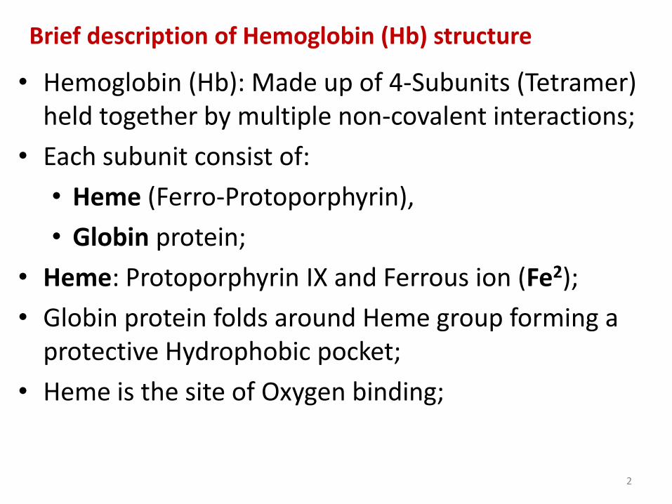
Brief description of Hemoglobin (Hb) structure
• Hemoglobin (Hb): Made up of 4-Subunits (Tetramer) held together by multiple non-covalent interactions;
• Each subunit consist of:
• Heme (Ferro-Protoporphyrin),
• Globin protein;
• Heme: Protoporphyrin IX and Ferrous ion (Fe2);
• Globin protein folds around Heme group forming a protective Hydrophobic pocket;
• Heme is the site of Oxygen binding;
2
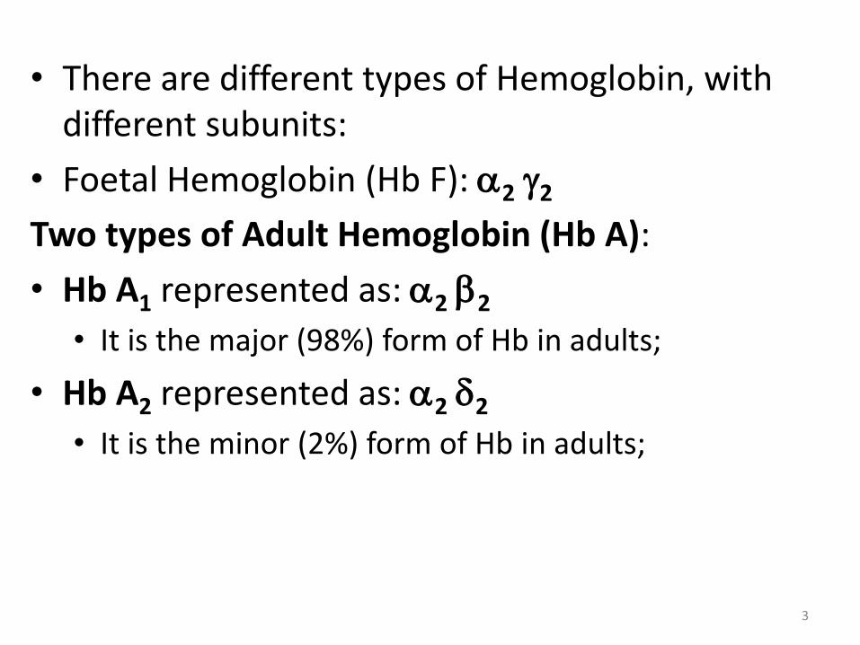
• There are different types of Hemoglobin, with different subunits:
• Foetal Hemoglobin (Hb F): 2 2
Two types of Adult Hemoglobin (Hb A):
• Hb A1 represented as: 2 2
• It is the major (98%) form of Hb in adults;
• Hb A2 represented as: 2 2
• It is the minor (2%) form of Hb in adults;
3
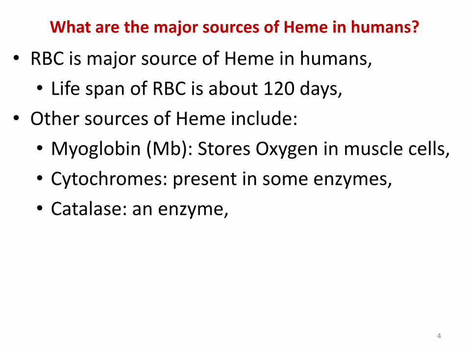
What are the major sources of Heme in humans?
• RBC is major source of Heme in humans,
• Life span of RBC is about 120 days,
• Other sources of Heme include:
• Myoglobin (Mb): Stores Oxygen in muscle cells,
• Cytochromes: present in some enzymes,
• Catalase: an enzyme,
4
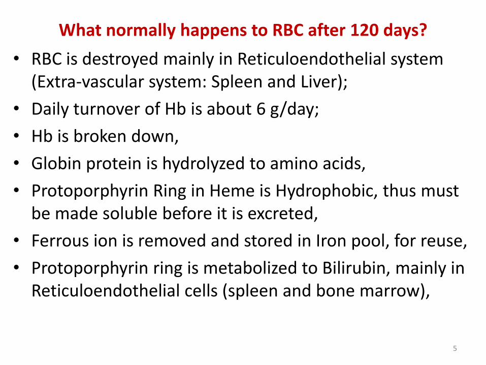
What normally happens to RBC after 120 days?
• RBC is destroyed mainly in Reticuloendothelial system (Extra-vascular system: Spleen and Liver);
• Daily turnover of Hb is about 6 g/day;
• Hb is broken down,
• Globin protein is hydrolyzed to amino acids,
• Protoporphyrin Ring in Heme is Hydrophobic, thus must be made soluble before it is excreted,
• Ferrous ion is removed and stored in Iron pool, for reuse,
• Protoporphyrin ring is metabolized to Bilirubin, mainly in Reticuloendothelial cells (spleen and bone marrow),
5
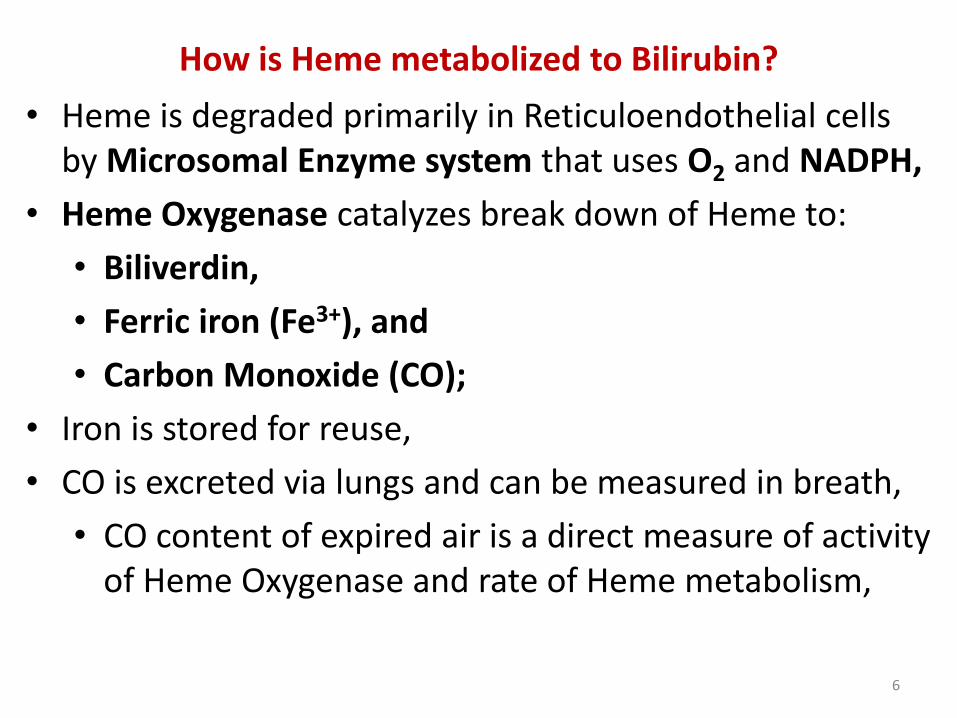
How is Heme metabolized to Bilirubin?
• Heme is degraded primarily in Reticuloendothelial cells by Microsomal Enzyme system that uses O2 and NADPH,
• Heme Oxygenase catalyzes break down of Heme to:
• Biliverdin,
• Ferric iron (Fe3+), and
• Carbon Monoxide (CO);
• Iron is stored for reuse,
• CO is excreted via lungs and can be measured in breath,
• CO content of expired air is a direct measure of activity of Heme Oxygenase and rate of Heme metabolism,
6
• Biliverdin is reduced to Unconjugated Bilirubin in reaction catalyzed by Biliverdin Reductase that requires NADPH, (Fig. 1)
• Unconjugated Bilirubin is Hydrophobic;
• It is transported in blood tightly bound to albumin,
• Presence of endogenous and exogenous binding competitors, like certain drugs, decreases the binding affinity of albumin for bilirubin,
• Small fraction of unconjugated bilirubin in plasma is not bound to albumin,
• Free unconjugated bilirubin can cross cell membranes, including blood-brain barrier, leading to Neuro-toxicity,
7
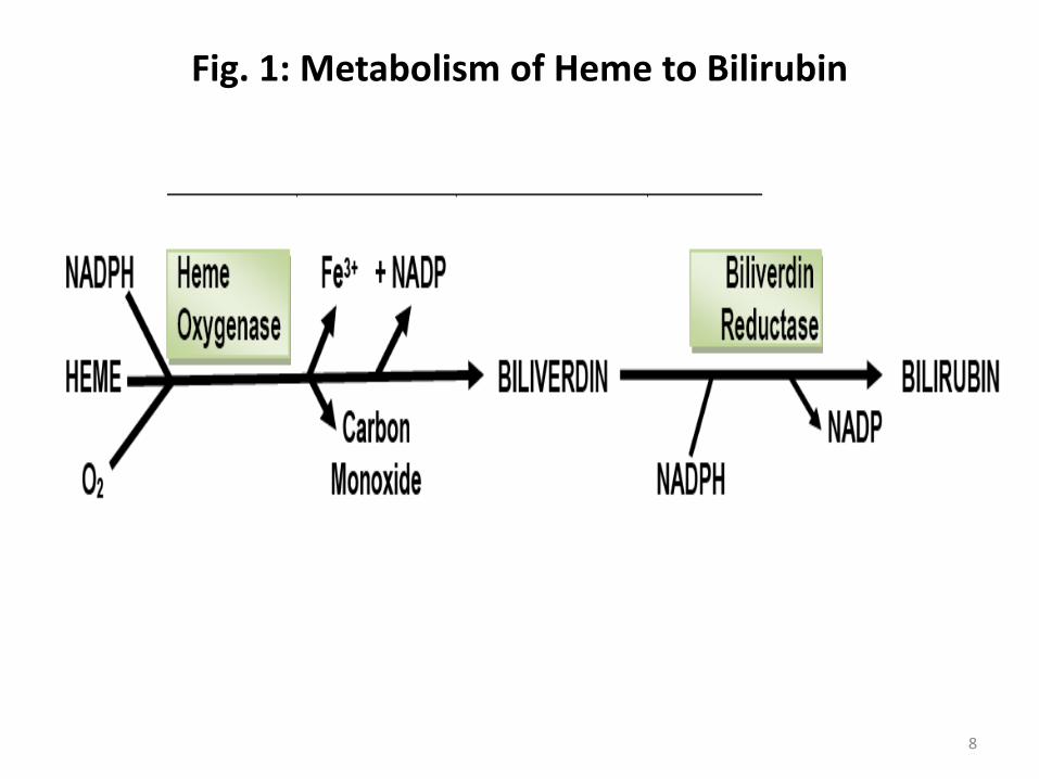
Fig. 1: Metabolism of Heme to Bilirubin
8
IMPORTANT TO NOTE
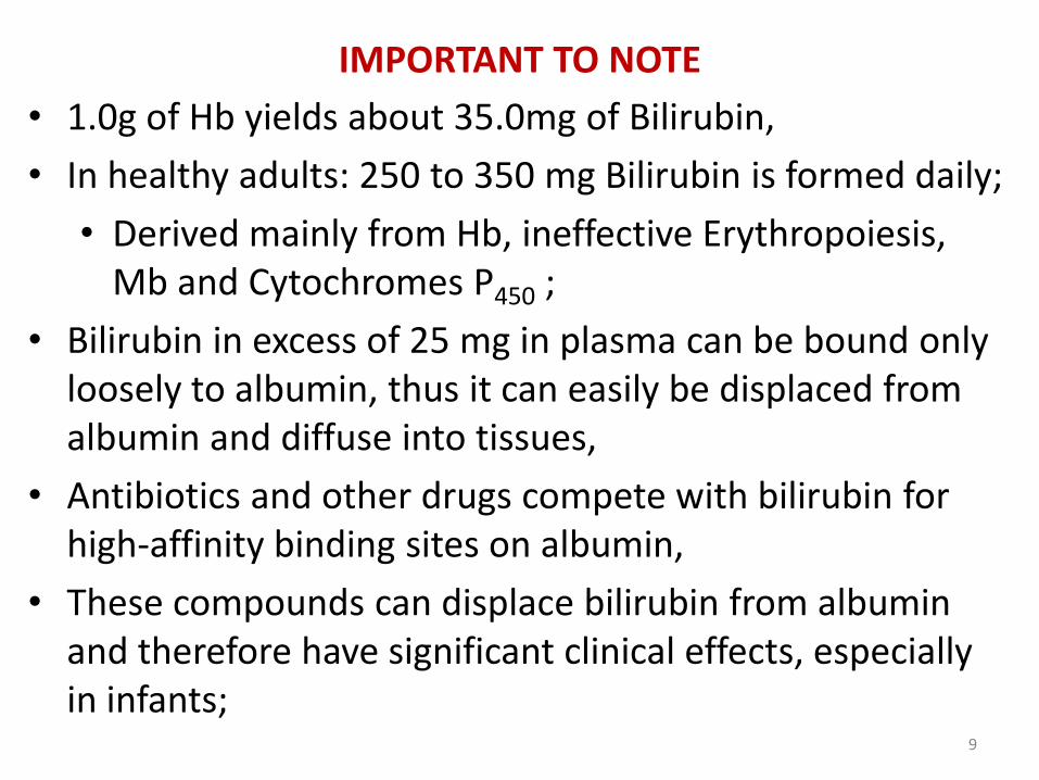
• 1.0g of Hb yields about 35.0mg of Bilirubin,
• In healthy adults: 250 to 350 mg Bilirubin is formed daily;
• Derived mainly from Hb, ineffective Erythropoiesis, Mb and Cytochromes P450 ;
• Bilirubin in excess of 25 mg in plasma can be bound only loosely to albumin, thus it can easily be displaced from albumin and diffuse into tissues,
• Antibiotics and other drugs compete with bilirubin for high-affinity binding sites on albumin,
• These compounds can displace bilirubin from albumin and therefore have significant clinical effects, especially in infants;
9
• Color of Biliverdin is Blue-Green,
• Color of Bilirubin is Yellow-Red,
• Change in color as Biliverdin is converted to Bilirubin is partly responsible for progressive changes in color of Hematoma, or Bruise, in which damaged tissue changes its color from:
• Initial Dark Blue to Red-Yellow,
• Finally to Yellow color before all the pigments are transported out of the affected tissue;
10
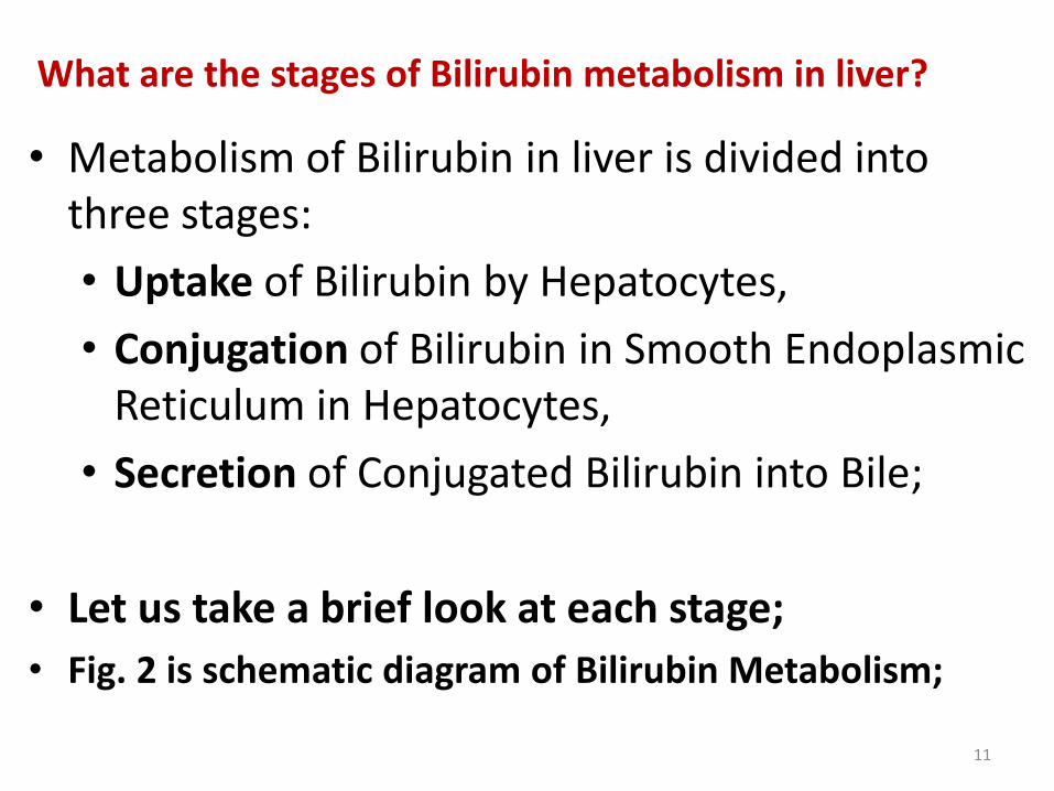
What are the stages of Bilirubin metabolism in liver?
• Metabolism of Bilirubin in liver is divided into three stages:
• Uptake of Bilirubin by Hepatocytes,
• Conjugation of Bilirubin in Smooth Endoplasmic Reticulum in Hepatocytes,
• Secretion of Conjugated Bilirubin into Bile;
• Let us take a brief look at each stage;
• Fig. 2 is schematic diagram of Bilirubin Metabolism;
11
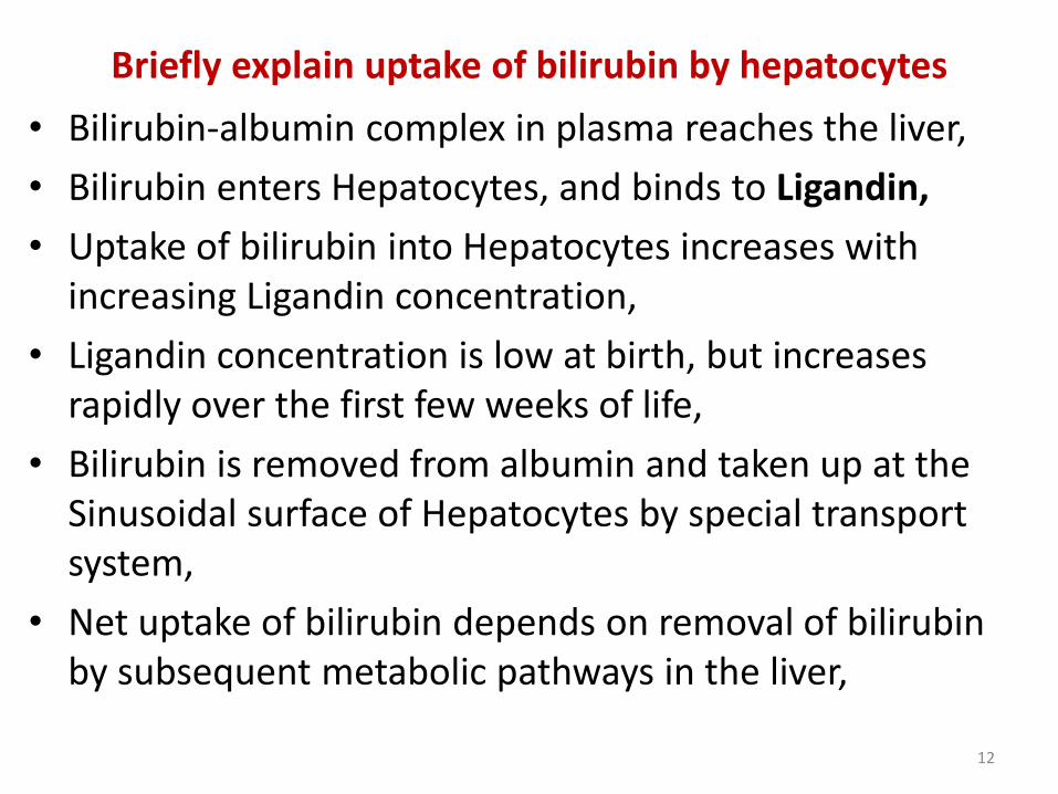
Briefly explain uptake of bilirubin by hepatocytes
• Bilirubin-albumin complex in plasma reaches the liver,
• Bilirubin enters Hepatocytes, and binds to Ligandin,
• Uptake of bilirubin into Hepatocytes increases with increasing Ligandin concentration,
• Ligandin concentration is low at birth, but increases rapidly over the first few weeks of life,
• Bilirubin is removed from albumin and taken up at the Sinusoidal surface of Hepatocytes by special transport system,
• Net uptake of bilirubin depends on removal of bilirubin by subsequent metabolic pathways in the liver,
12
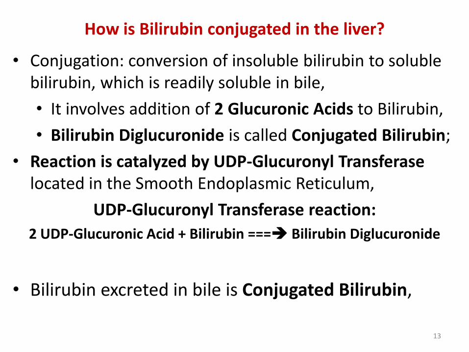
How is Bilirubin conjugated in the liver?
• Conjugation: conversion of insoluble bilirubin to soluble bilirubin, which is readily soluble in bile,
• It involves addition of 2 Glucuronic Acids to Bilirubin,
• Bilirubin Diglucuronide is called Conjugated Bilirubin;
• Reaction is catalyzed by UDP-Glucuronyl Transferaselocated in the Smooth Endoplasmic Reticulum,
UDP-Glucuronyl Transferase reaction:
2 UDP-Glucuronic Acid + Bilirubin === Bilirubin Diglucuronide
• Bilirubin excreted in bile is Conjugated Bilirubin,
13
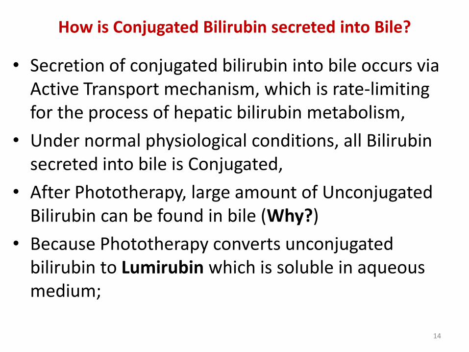
How is Conjugated Bilirubin secreted into Bile?
• Secretion of conjugated bilirubin into bile occurs via Active Transport mechanism, which is rate-limiting for the process of hepatic bilirubin metabolism,
• Under normal physiological conditions, all Bilirubin secreted into bile is Conjugated,
• After Phototherapy, large amount of Unconjugated Bilirubin can be found in bile (Why?)
• Because Phototherapy converts unconjugated bilirubin to Lumirubin which is soluble in aqueous medium;
14
• De-conjugation of conjugated bilirubin by beta-Glucuronidase located in brush border, can occur in Proximal Small Intestine,
• Unconjugated bilirubin formed can be reabsorbed into circulation, increasing total plasma unconjugated bilirubin level,
• Cycle of bilirubin Uptake, Conjugation, Excretion, De-conjugation, and Reabsorption is termed: Enterohepatic Circulation of Bilirubin;
• It occurs mainly in neonates;
15
How is Conjugated Bilirubin metabolized in Intestine?
• Bile containing conjugated bilirubin is released in GIT,
• Conjugated bilirubin may be De-conjugated by bacteria, resulting in Enterohepatic circulation of bilirubin,
• Fecal flora converts some conjugated bilirubin into Urobilinogens,
• Some urobilinogen is reabsorbed and re-excreted via liver to constitute Intra-hepatic Urobilinogen cycle,
• Some urobilinogen is excreted in the urine,
• Urobilinogen is excreted in feces and oxidized to Urobilin,
• Darkening of feces in air is due to oxidation of residual Urobilinogen to Urobilin,
16
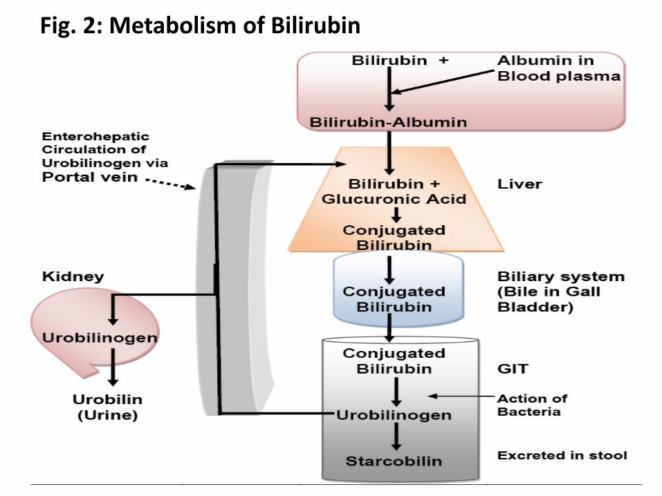
Fig. 2: Metabolism of Bilirubin
17
HYPERBILIRUBINEMIA
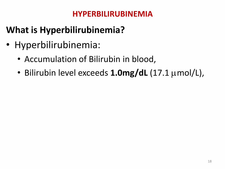
What is Hyperbilirubinemia?
• Hyperbilirubinemia:
• Accumulation of Bilirubin in blood,
• Bilirubin level exceeds 1.0mg/dL (17.1 mol/L),
18
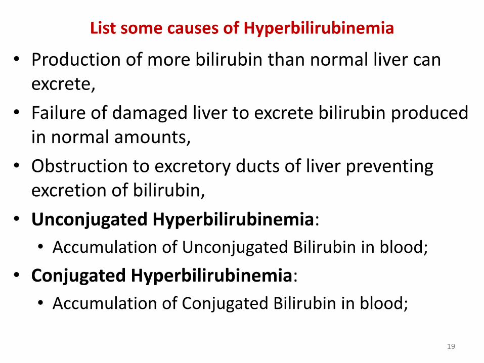
List some causes of Hyperbilirubinemia
• Production of more bilirubin than normal liver can excrete,
• Failure of damaged liver to excrete bilirubin produced in normal amounts,
• Obstruction to excretory ducts of liver preventing excretion of bilirubin,
• Unconjugated Hyperbilirubinemia:
• Accumulation of Unconjugated Bilirubin in blood;
• Conjugated Hyperbilirubinemia:
• Accumulation of Conjugated Bilirubin in blood;
19
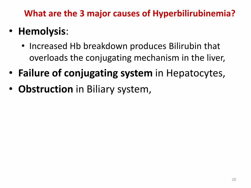
What are the 3 major causes of Hyperbilirubinemia?
• Hemolysis:
• Increased Hb breakdown produces Bilirubin that overloads the conjugating mechanism in the liver,
• Failure of conjugating system in Hepatocytes,
• Obstruction in Biliary system,
20
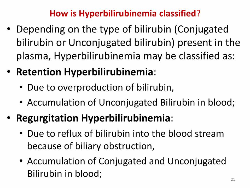
How is Hyperbilirubinemia classified?
• Depending on the type of bilirubin (Conjugated bilirubin or Unconjugated bilirubin) present in the plasma, Hyperbilirubinemia may be classified as:
• Retention Hyperbilirubinemia:
• Due to overproduction of bilirubin,
• Accumulation of Unconjugated Bilirubin in blood;
• Regurgitation Hyperbilirubinemia:
• Due to reflux of bilirubin into the blood stream because of biliary obstruction,
• Accumulation of Conjugated and Unconjugated Bilirubin in blood;
21
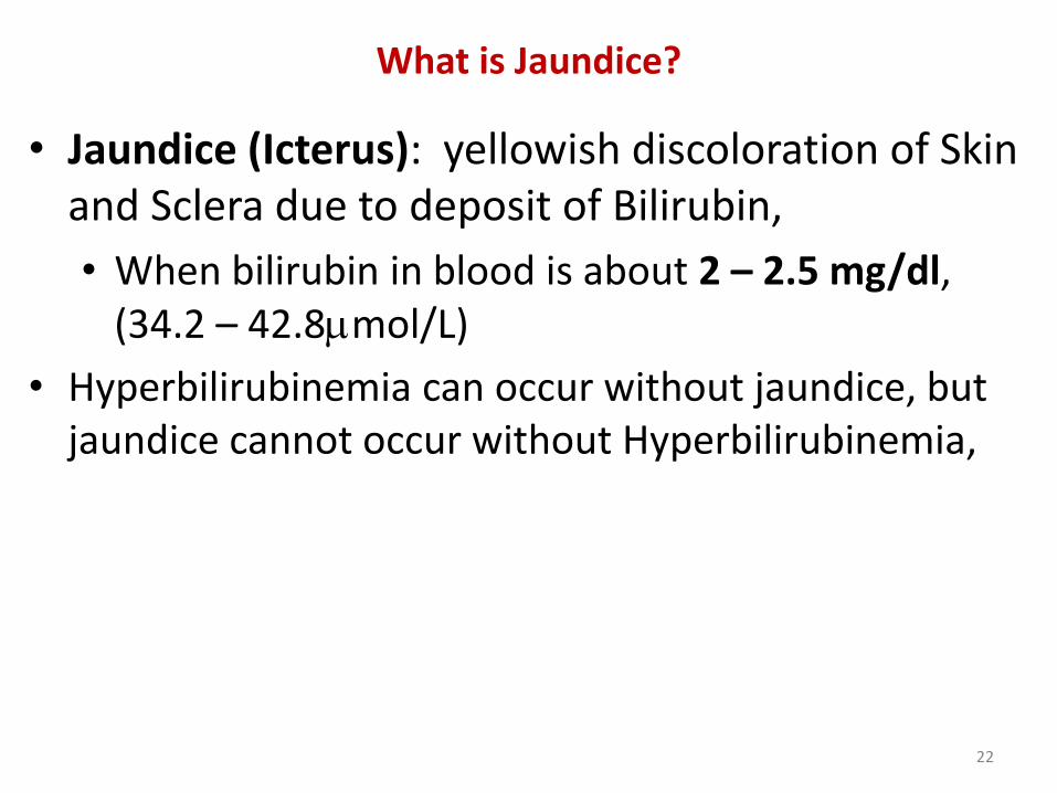
What is Jaundice?
• Jaundice (Icterus): yellowish discoloration of Skin and Sclera due to deposit of Bilirubin,
• When bilirubin in blood is about 2 – 2.5 mg/dl, (34.2 – 42.8mol/L)
• Hyperbilirubinemia can occur without jaundice, but jaundice cannot occur without Hyperbilirubinemia,
22
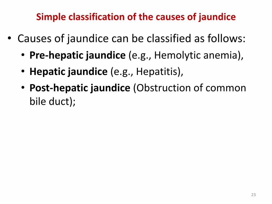
Simple classification of the causes of jaundice
• Causes of jaundice can be classified as follows:
• Pre-hepatic jaundice (e.g., Hemolytic anemia),
• Hepatic jaundice (e.g., Hepatitis),
• Post-hepatic jaundice (Obstruction of common bile duct);
23
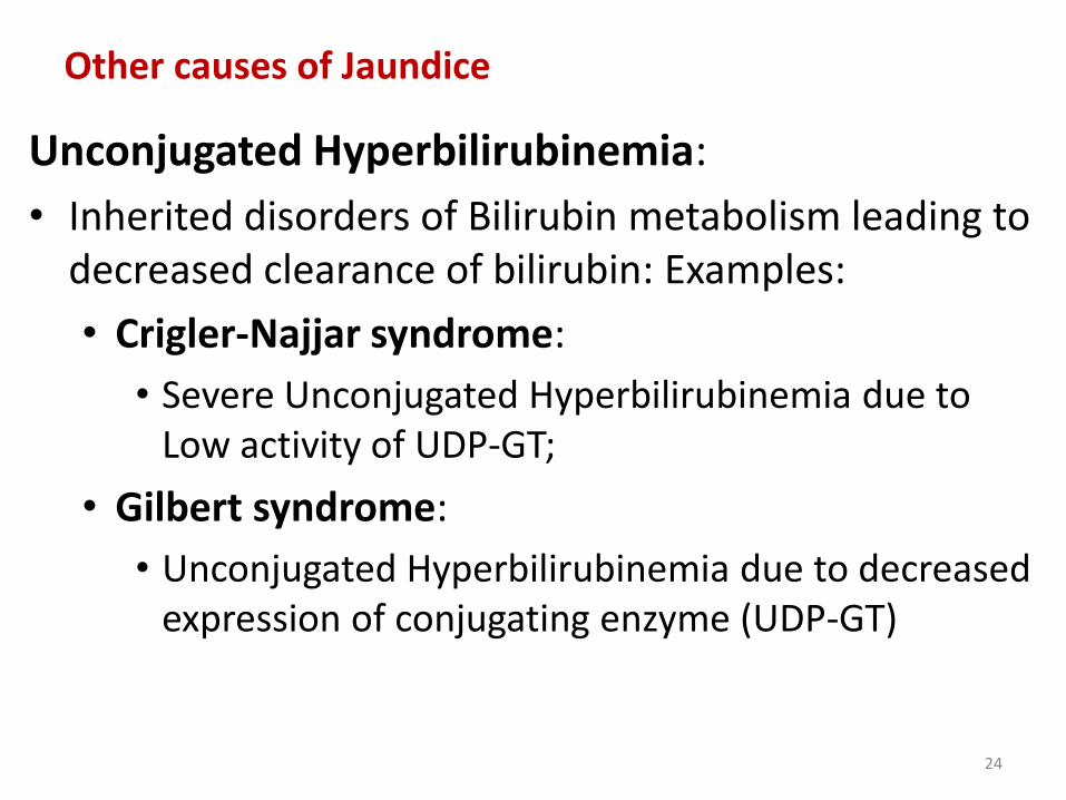
Other causes of Jaundice
Unconjugated Hyperbilirubinemia:
• Inherited disorders of Bilirubin metabolism leading to decreased clearance of bilirubin: Examples:
• Crigler-Najjar syndrome:
• Severe Unconjugated Hyperbilirubinemia due to Low activity of UDP-GT;
• Gilbert syndrome:
• Unconjugated Hyperbilirubinemia due to decreased expression of conjugating enzyme (UDP-GT)
24
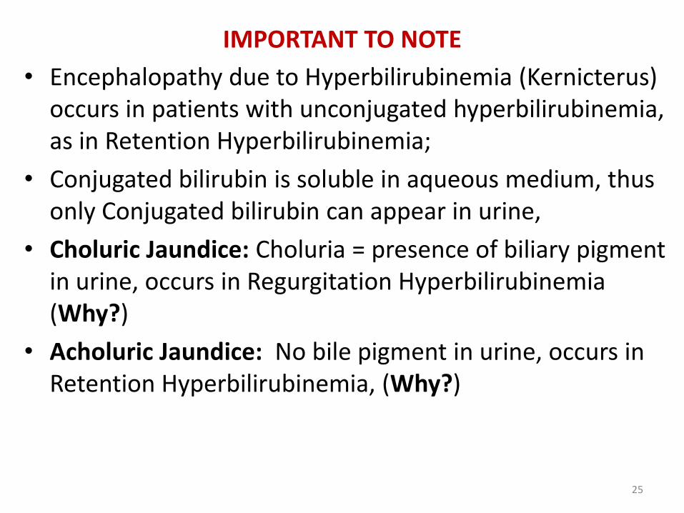
IMPORTANT TO NOTE
• Encephalopathy due to Hyperbilirubinemia (Kernicterus) occurs in patients with unconjugated hyperbilirubinemia, as in Retention Hyperbilirubinemia;
• Conjugated bilirubin is soluble in aqueous medium, thus only Conjugated bilirubin can appear in urine,
• Choluric Jaundice: Choluria = presence of biliary pigment in urine, occurs in Regurgitation Hyperbilirubinemia (Why?)
• Acholuric Jaundice: No bile pigment in urine, occurs in Retention Hyperbilirubinemia, (Why?)
25
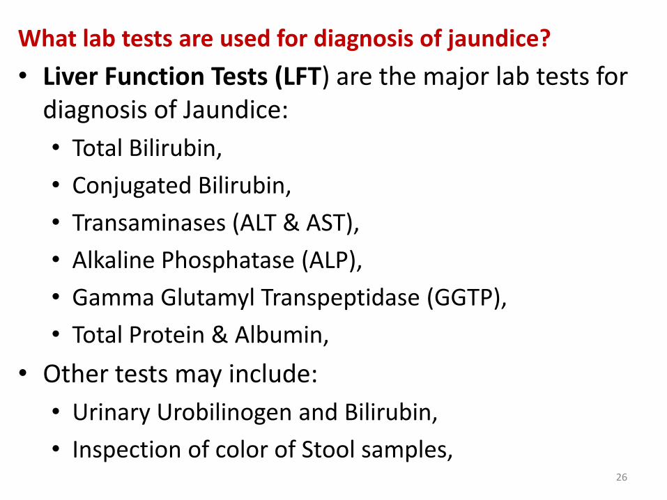
What lab tests are used for diagnosis of jaundice?
• Liver Function Tests (LFT) are the major lab tests for diagnosis of Jaundice:
• Total Bilirubin,
• Conjugated Bilirubin,
• Transaminases (ALT & AST),
• Alkaline Phosphatase (ALP),
• Gamma Glutamyl Transpeptidase (GGTP),
• Total Protein & Albumin,
• Other tests may include:
• Urinary Urobilinogen and Bilirubin,
• Inspection of color of Stool samples, 26
REFERENCES
• Textbook of Biochemistry, with clinical correlations, Ed. By T. M. Devlin, 4th Ed.
• Harper’s Illustrated Biochemistry 26th Edition; 2003; Ed. By R. K. Murray et. al.
• Biochemistry, By V. L. Davidson & D. B. Sittman. 3rd Edition.
• Hames BD, Hooper NM, JD Houghton; Instant Notes in Biochemistry, Bios Scientific Pub, Springer; UK.
• VJ Temple Biochemistry 1001: Review and Viva Voce Questions and Answers Approach; Sterling Publishers Private Limited, 2012, New Delhi-110 – 020.
• G Beckett, S Walker, P Rae & P Ashby. Lecture Notes: Clinical Biochemistry 7th
Ed. Blackwell Publishing, Australia 2008.
• http://www.emedicine.com/PED/topic2774.htm
• http://www.emedicine.com/med/topic1066.htm
• http://www.emedicine.com/PED/topic282.htm
•
27