Beyond the cell The cell biology of regeneration
10
The Rockefeller University Press J. Cell Biol. Vol. 196 No. 5 553–562 www.jcb.org/cgi/doi/10.1083/jcb.201105099 JCB 553 JCB: Review Correspondence to Phillip A. Newmark: [email protected] Abbreviations used in this paper: CREB, cAMP response element–binding protein; nAG, newt anterior gradient; RSK, ribosomal S6 kinase. 1 It should be noted that within 15 h of initiating his regenerative cycle, the Doctor is capable of robust appendage regeneration (e.g., the hand regeneration scene in the episode, “The Christmas Invasion”). Regeneration: Sometimes truth is stranger than (science) fiction Fans of Doctor Who, the quintessential British science fiction television series, have been able to enjoy the adventures of its main character (the Doctor) for over three decades. As a Time Lord from the planet Gallifrey, the Doctor can regenerate his body completely as he nears death. The Doctor’s regenerative abilities, however, are not without flaw: after he regenerates, he takes on different features and characteristics. This imper- fect process enables the series to continue with a new actor portraying the Doctor upon the departure of his predecessor. The Doctor’s slightly defective regenerative power is not only a boon for the show’s fans (and producers), but it is also a rare example of science fiction being outdone by the real world. On Earth, a wide range of actual organisms can regen- erate missing parts after injury. Unlike the fictional Doctor, these animals can rebuild new structures that are indistin- guishable from those they are replacing 1 . With the goal of un- derstanding the factors that enable some organisms, but not others, to restore missing structures, biologists have been studying regeneration for well over two centuries (Morgan, 1901; Lenhoff and Lenhoff, 1986). Although the Doctor’s regenerative abilities remain a mystery, recent research on various earthlings has started to reveal the mechanisms leading to regeneration of complex structures. During the past decade the application of molecular genetic techniques, including double-stranded RNA-mediated genetic interference (RNAi) and transgenesis, has revitalized studies of classical animal models of regeneration (Poss, 2010). Here we highlight a sample of recent work addressing some of the critical questions facing regeneration researchers: What sig- nals initiate regeneration? What is the nature of the cells that produce the regenerate? What role does the nervous system play in this process? The answers to these and related questions should help developmental biologists, tissue engineers, and cli- nicians to understand and one day overcome the limits on human regeneration. Back from the dead: Apoptosis and regeneration After amputation, local responses at the site of the wound play important roles in the initiation of regenerative processes. Wound healing (Gurtner et al., 2008), ion flux (Levin, 2009), and interactions between the wound epidermis and the underlying tissue appear critical for regenerative outgrowth. Recent work suggests that programmed cell death may play a role in trigger- ing regenerative responses in many different organisms, includ- ing Hydra (Bergmann and Steller, 2010). Hydra, a freshwater polyp belonging to the Phylum Cni- daria (which includes jellyfish, sea anemones, and corals), was the subject of the first scientific investigations of regeneration in animals (Lenhoff and Lenhoff, 1986). The Hydra body consists of ectodermal and endodermal cell layers separated by an extra- cellular matrix; neurons and interstitial cells (stem cells that produce neurons, gland cells, and germ cells, among other cell types) reside between these two layers. The animal is radially symmetric, and is polarized along the oral (head and tentacles)/ aboral (foot) axis (Fig. 1 A). Small fragments of Hydra tissue can regenerate a complete organism; even dissociated single cells can reaggregate, reestablish polarity, and form a new animal (Noda, 1971; Gierer et al., 1972). Regeneration of complex structures after injury requires dramatic changes in cellular behavior. Regenerating tis- sues initiate a program that includes diverse processes such as wound healing, cell death, dedifferentiation, and stem (or progenitor) cell proliferation; furthermore, newly regenerated tissues must integrate polarity and positional identity cues with preexisting body structures. Gene knock- down approaches and transgenesis-based lineage and functional analyses have been instrumental in deciphering various aspects of regenerative processes in diverse ani- mal models for studying regeneration. Beyond the cell The cell biology of regeneration Ryan S. King and Phillip A. Newmark Howard Hughes Medical Institute, Department of Cell and Developmental Biology, University of Illinois at Urbana-Champaign, Urbana, IL 61801 © 2012 King and Newmark This article is distributed under the terms of an Attribution– Noncommercial–Share Alike–No Mirror Sites license for the first six months after the pub- lication date (see http://www.rupress.org/terms). After six months it is available under a Creative Commons License (Attribution–Noncommercial–Share Alike 3.0 Unported license, as described at http://creativecommons.org/licenses/by-nc-sa/3.0/). THE JOURNAL OF CELL BIOLOGY Downloaded from http://rupress.org/jcb/article-pdf/196/5/553/1354773/jcb_201105099.pdf by guest on 09 December 2021
Transcript of Beyond the cell The cell biology of regeneration

The Rockefeller University PressJ. Cell Biol. Vol. 196 No. 5 553–562www.jcb.org/cgi/doi/10.1083/jcb.201105099 JCB 553
JCB: Review
Correspondence to Phillip A. Newmark: [email protected] used in this paper: CREB, cAMP response element–binding protein; nAG, newt anterior gradient; RSK, ribosomal S6 kinase.1It should be noted that within 15 h of initiating his regenerative cycle, the Doctor is capable of robust appendage regeneration (e.g., the hand regeneration scene in the episode, “The Christmas Invasion”).
Regeneration: Sometimes truth is stranger than (science) fictionFans of Doctor Who, the quintessential British science fiction television series, have been able to enjoy the adventures of its main character (the Doctor) for over three decades. As a Time Lord from the planet Gallifrey, the Doctor can regenerate his body completely as he nears death. The Doctor’s regenerative abilities, however, are not without flaw: after he regenerates, he takes on different features and characteristics. This imper-fect process enables the series to continue with a new actor portraying the Doctor upon the departure of his predecessor. The Doctor’s slightly defective regenerative power is not only a boon for the show’s fans (and producers), but it is also a rare example of science fiction being outdone by the real world. On Earth, a wide range of actual organisms can regen-erate missing parts after injury. Unlike the fictional Doctor, these animals can rebuild new structures that are indistin-guishable from those they are replacing1. With the goal of un-derstanding the factors that enable some organisms, but not others, to restore missing structures, biologists have been studying regeneration for well over two centuries (Morgan, 1901; Lenhoff and Lenhoff, 1986).
Although the Doctor’s regenerative abilities remain a mystery, recent research on various earthlings has started to reveal the mechanisms leading to regeneration of complex structures. During the past decade the application of molecular genetic techniques, including double-stranded RNA-mediated genetic interference (RNAi) and transgenesis, has revitalized studies of classical animal models of regeneration (Poss, 2010). Here we highlight a sample of recent work addressing some of the critical questions facing regeneration researchers: What sig-nals initiate regeneration? What is the nature of the cells that produce the regenerate? What role does the nervous system play in this process? The answers to these and related questions should help developmental biologists, tissue engineers, and cli-nicians to understand and one day overcome the limits on human regeneration.
Back from the dead: Apoptosis and regenerationAfter amputation, local responses at the site of the wound play important roles in the initiation of regenerative processes. Wound healing (Gurtner et al., 2008), ion flux (Levin, 2009), and interactions between the wound epidermis and the underlying tissue appear critical for regenerative outgrowth. Recent work suggests that programmed cell death may play a role in trigger-ing regenerative responses in many different organisms, includ-ing Hydra (Bergmann and Steller, 2010).
Hydra, a freshwater polyp belonging to the Phylum Cni-daria (which includes jellyfish, sea anemones, and corals), was the subject of the first scientific investigations of regeneration in animals (Lenhoff and Lenhoff, 1986). The Hydra body consists of ectodermal and endodermal cell layers separated by an extra-cellular matrix; neurons and interstitial cells (stem cells that produce neurons, gland cells, and germ cells, among other cell types) reside between these two layers. The animal is radially symmetric, and is polarized along the oral (head and tentacles)/aboral (foot) axis (Fig. 1 A). Small fragments of Hydra tissue can regenerate a complete organism; even dissociated single cells can reaggregate, reestablish polarity, and form a new animal (Noda, 1971; Gierer et al., 1972).
Regeneration of complex structures after injury requires dramatic changes in cellular behavior. Regenerating tis-sues initiate a program that includes diverse processes such as wound healing, cell death, dedifferentiation, and stem (or progenitor) cell proliferation; furthermore, newly regenerated tissues must integrate polarity and positional identity cues with preexisting body structures. Gene knock-down approaches and transgenesis-based lineage and functional analyses have been instrumental in deciphering various aspects of regenerative processes in diverse ani-mal models for studying regeneration.
Beyond the cell
The cell biology of regeneration
Ryan S. King and Phillip A. Newmark
Howard Hughes Medical Institute, Department of Cell and Developmental Biology, University of Illinois at Urbana-Champaign, Urbana, IL 61801
© 2012 King and Newmark This article is distributed under the terms of an Attribution–Noncommercial–Share Alike–No Mirror Sites license for the first six months after the pub-lication date (see http://www.rupress.org/terms). After six months it is available under a Creative Commons License (Attribution–Noncommercial–Share Alike 3.0 Unported license, as described at http://creativecommons.org/licenses/by-nc-sa/3.0/).
TH
EJ
OU
RN
AL
OF
CE
LL
BIO
LO
GY
Dow
nloaded from http://rupress.org/jcb/article-pdf/196/5/553/1354773/jcb_201105099.pdf by guest on 09 D
ecember 2021

JCB • VOLUME 196 • NUMBER 5 • 2012 554
the proliferative burst induced by apoptosis, whereas treatment with exogenous Wnt3 rescues proliferation when apoptosis is inhibited (Chera et al., 2009). The induction of apoptosis after injury appears to require the mitogen-activated protein kinase (MAPK) pathway, including ribosomal S6 kinase (RSK), and cAMP response element–binding protein (CREB). RNAi knockdown of RSK and CREB blocks apoptosis at the head-regenerating tip, as does treatment with U0126, a pharmacological inhibitor of MEK, the kinase that phosphorylates MAPK (Chera et al., 2011). In fragments that will regenerate a new head, the transcription factor CREB is phosphorylated by RSK within minutes of amputation (Fig. 1 A; Chera et al., 2011). Intriguingly, MAPK/CREB-induced apoptosis is not required in fragments
An apoptotic program is initiated asymmetrically after transection midway through the body column. The fragment that will regenerate a new head displays a robust apoptotic re-sponse near the wound site, whereas the fragment regenerating a new foot does not (Fig. 1 A; Chera et al., 2009). Inhibition of apoptosis blocks head regeneration, and activation of apoptosis at a foot-regenerating wound leads to ectopic head formation (Chera et al., 2009). Thus, induction of apoptosis is both neces-sary and sufficient for head regeneration. The apoptotic re-sponse stimulates a synchronous burst of proliferative activity in neighboring cells and leads to the establishment of a head organizer via secretion of Wnt3 ligand by apoptotic interstitial cells (Chera et al., 2009). RNAi knockdown of Wnt3 prevents
Figure 1. Increased apoptosis is associated with early phases of regeneration. (A) After mid-gastric bisection in Hydra, MAPK signaling leads to rapid acti-vation of the transcription factor CREB in fragments regenerating a head (blue cells). MAPK/CREB activity is required for stimulating a wave of apoptosis in interstitial cells near the site of injury (red cells). These apoptotic cells secrete Wnt3, inducing a zone of proliferation (green cells) below the region of apopto-sis. (B) Planarians and (C) Xenopus larval tails also show a rapid, localized increase in apoptosis after amputation. In Xenopus, like Hydra, apoptosis may provide important signals during early phases of regeneration; inhibiting apoptosis during the first 24 h post amputation (hpa) blocks regeneration.
Dow
nloaded from http://rupress.org/jcb/article-pdf/196/5/553/1354773/jcb_201105099.pdf by guest on 09 D
ecember 2021

555The cell biology of regeneration • King and Newmark
knockout of the p53 inhibitor Mdm2) is eventually compen-sated in adults by increased proliferation and expansion of the stem cell pool (Valentin-Vega et al., 2008). Apoptotic cells also contribute to homeostasis in epithelia by lipid-based signaling (sphingosine-1-phosphate) that triggers actomyosin contraction in the surrounding cells, leading to the extrusion of the dying cells (Gu et al., 2011). These observations suggest many poten-tial roles for dead and dying cells to alter cell behavior at sites of injury.
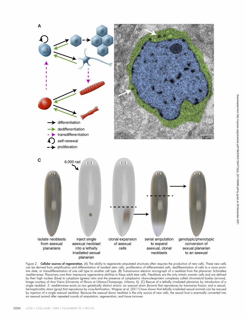
Conjuring up spare parts: Cellular sources of regenerationIn most regenerating organisms, replacing an amputated struc-ture requires the production of new cells. Therefore, one of the main functions of early signaling events after injury is to stimulate the production of additional cells that are capable of rebuilding lost structures. New cells coalesce near the site of injury, giving rise to a mass of undifferentiated cells called the regeneration blastema. Subsequent signals then regulate out-growth and patterning of the newly formed tissue. To understand how early signaling events initiate regeneration and stimulate blastema formation, it is crucial to identify the cells upon which these signals act. New cells can be generated in a variety of ways, including proliferation of a resident stem cell population, division of terminally differentiated cells, or dedifferentiation/transdifferentiation of mature cells to a stem cell–like precursor or another cell type (Fig. 2 A). The extent to which each mode is used varies between species and even across tissues within the same species.
Stem cells and transdifferentiation both contribute to the regenerative abilities of Hydra. These animals possess three distinct stem cell populations: ectodermal, interstitial, and endodermal stem cells (Galliot et al., 2006). Development of transgenesis in Hydra (Wittlieb et al., 2006) has enabled in vivo tracking of the stem cell lineages: for example, investigating the differentiation of interstitial stem cells and migration of their progeny in intact animals (Khalturin et al., 2007). Transgenesis has also aided the analysis of transdifferentiation. By express-ing eGFP in the zymogen gland cells (a derivative of the inter-stitial stem cells), Siebert et al. (2008) showed that these cells move up the body column and down-regulate the expression of a zymogen gland cell–specific marker. By using histology, elec-tron microscopy, and in situ hybridization with cell type–specific markers, they identified cells with characteristics of both zymogen gland cells and another cell type (granular mucous cells), suggesting that zymogen gland cells could convert to granular mucous cells as they were displaced up the body col-umn and entered the head region (Siebert et al., 2008). The abil-ity of Hydra cells to transdifferentiate permits these animals to regenerate even in the absence of cell proliferation (Cummings and Bode, 1984).
Freshwater planarians (another classical model for study-ing regeneration) owe their amazing regenerative abilities to stem cells. In these animals, a population of mesenchymal stem cells, called neoblasts, is the source of cells for regeneration (Baguñà et al., 1989). Neoblasts are the only dividing somatic cells and have been defined by their high nuclear/cytoplasmic
regenerating a foot, and cell proliferation is not up-regulated in foot regenerates. Rather, foot regeneration proceeds through cellular rearrangement and transdifferentiation (morphallactic regeneration). The exact nature of the signal that leads to asym-metric MAPK activation (and, thus, apoptosis) at the site of in-jury in head-regenerating fragments remains to be determined.
The apoptosis-induced compensatory proliferation (Fan and Bergmann, 2008) observed in Hydra may be a conserved mechanism for stimulating proper wound healing and regenera-tion. Apoptotic cells have been observed during early phases of regeneration in several animals that can regenerate missing tissues: planarians (Fig. 1 B), Xenopus (Fig. 1 C), and newts (Hwang et al., 2004; Vlaskalin et al., 2004; Tseng et al., 2007; Chera et al., 2009; Pellettieri et al., 2010). Although the require-ment for apoptosis during regeneration has not been addressed in planarians and newts, treatment of Xenopus larvae with cas-pase inhibitors during the first 24 h after amputation blocks tail regeneration (Tseng et al., 2007). As observed in Hydra, inhibi-tion of apoptosis impedes proliferation; in addition, nerves fail to extend appropriately toward the site of amputation. Because nerves release important regenerative signals (see penultimate section), it is unclear whether reduced proliferation results di-rectly from lack of signals derived from apoptotic cells, or indi-rectly from defective innervation. Furthermore, because these assays rely on caspase inhibition it is possible that nonapoptotic roles for caspases (Kuranaga and Miura, 2007) may be involved rather than apoptosis by itself.
Apoptotic cells have been shown to provide a number of signals that can regulate wound healing and regeneration. In Drosophila, the larval imaginal discs are capable of regenerat-ing: wing discs can produce appropriately sized adult wings after radiation-induced killing of over 50% of the cells (Haynie and Bryant, 1977). MAPK signaling through Jun kinase is im-portant both for initiating apoptosis and production of Wnt (Wg) and BMP (Dpp) mitogens (Pérez-Garijo et al., 2009; Bergantiños et al., 2010; Morata et al., 2011). The extent to which these fac-tors are required for stimulating compensatory proliferation re-mains unclear. After radiation-induced apoptosis, neither dpp nor wg were required (Pérez-Garijo et al., 2009). However, after ectopic expression of a pro-apoptotic gene, wg was required for the proliferative response (Smith-Bolton et al., 2009). Further-more, wg is also required for disc regeneration after surgical transection (Schubiger et al., 2010). Additional roles for apopto-sis have been reported in epidermal wound healing and liver regeneration in mouse. Caspase 3 and caspase 7 mutant mice have defects in both processes, and these mutants show reduced cell proliferation in these contexts (Li et al., 2010). Caspases 3 and 7 can activate Ca2+-independent phospholipase A2, leading to pro-duction of arachidonic acid and prostaglandin in apoptotic cells, the latter of which can stimulate proliferation (Li et al., 2010).
Injury-induced apoptotic signals are also required to main-tain tissue homeostasis. When cells of the adult Drosophila midgut are injured by toxins or induced to undergo apoptosis, intestinal enterocytes secrete the cytokine Unpaired, which stimulates proliferation of intestinal stem cells through activation of the Jak/Stat pathway (Jiang et al., 2009). Similarly, in the mouse intestine massive induction of apoptosis (via intestine-specific
Dow
nloaded from http://rupress.org/jcb/article-pdf/196/5/553/1354773/jcb_201105099.pdf by guest on 09 D
ecember 2021
JCB • VOLUME 196 • NUMBER 5 • 2012 556
Figure 2. Cellular sources of regeneration. (A) The ability to regenerate amputated structures often requires the production of new cells. These new cells can be derived from amplification and differentiation of resident stem cells, proliferation of differentiated cells, dedifferentiation of cells to a more primi-tive state, or transdifferentiation of one cell type to another cell type. (B) Transmission electron micrograph of a neoblast from the planarian Schmidtea mediterranea. Planarians owe their impressive regenerative abilities to these adult stem cells. Neoblasts are the only mitotic somatic cells and are defined by their high nuclear (blue) to cytoplasm (green) ratio and the presence of cytoplasmic ribonucleoprotein complexes called chromatoid bodies (arrows). Image courtesy of Ana Vieira (University of Illinois at Urbana-Champaign, Urbana, IL). (C) Rescue of a lethally irradiated planarian by introduction of a single neoblast. S. mediterranea exists as two genetically distinct strains: an asexual strain (brown) that reproduces by transverse fission; and a sexual, hermaphroditic strain (gray) that reproduces by cross-fertilization. Wagner et al. (2011) have shown that lethally irradiated sexual animals can be rescued by injection of a single asexual neoblast. Because the asexual donor neoblast is the only source of new cells, the sexual host is eventually converted into an asexual animal after repeated rounds of amputation, regeneration, and tissue turnover.
Dow
nloaded from http://rupress.org/jcb/article-pdf/196/5/553/1354773/jcb_201105099.pdf by guest on 09 D
ecember 2021

557The cell biology of regeneration • King and Newmark
the axolotl and newt remain open questions. In contrast, larval tail regeneration in Xenopus laevis appears to be driven largely by progenitor cells; in lineage-tracing experiments the extent of labeling of new muscle correlates with the amount of satellite cell labeling before amputation (Slack et al., 2004). Lineage tracing experiments in both axolotl and Xenopus indicate that multiple cell types contribute to formation of the blastema (Slack et al., 2004; Kragl et al., 2009). However, it is still un-clear whether these other tissues contribute cells through prolif-eration of progenitor cells or dedifferentiation of mature cells. Such an understanding will be crucial for deciphering the mechanisms by which early regenerative signals trigger blas-tema formation.
To thine own self be true: Cellular memory during regenerationIn addition to identifying the cellular sources of regeneration in different systems, researchers have been examining whether blas-temal cells are pluripotent, multipotent, or have more limited poten-tial. As discussed already, pluripotent neoblasts are the source of new cellular material that drives regeneration in planarians. However, cell proliferation is restricted largely to regions outside the blastema (Reddien and Sánchez Alvarado, 2004; Wenemoser and Reddien, 2010); thus, post-mitotic neoblast progeny are the predominant cells within the blastema that rebuild lost tissues. Eisenhoffer et al. (2008) have exploited the sensitivity of neo-blasts to -radiation to identify numerous neoblast as well as early and late neoblast progeny markers. Interestingly, not all early neoblast progeny express the same panel of markers (Fig. 3 A). Although this difference could reflect transient temporal altera-tions in gene expression in early neoblast progeny, it more likely indicates that these cells are in various early stages of lin-eage commitment and/or en route to different terminal cell types. This heterogeneity has also been suggested by gene expression profiling on individual neoblasts (and their progeny) isolated using fluorescence-activated cell sorting (Hayashi et al., 2010). A subset of radiation-sensitive cells with G2 DNA content and expressing various neoblast markers also expressed a marker of muscle differentiation, suggesting that not all lineage-committed neoblasts are post-mitotic (Hayashi et al., 2010). Consistent with this result, S phase or G2 neoblasts have been observed to express markers of differentiated cell types, including excretory and neu-ronal markers (Nishimura et al., 2011; Scimone et al., 2011).
In planarians, specification of blastema positional identity (i.e., as anterior or posterior) likely occurs early during regener-ation. Sengel (1960) removed anterior blastemas after 3 d of re-generation and cultured them as explants in vitro; these blastema explants produced only anterior tissues, including photorecep-tors and cephalic ganglia (Fig. 3 B; Sengel, 1960). By contrast, explanted posterior blastemas did not produce anterior tissues. Therefore, these early experiments suggested that specification of blastemas to produce heads or tails occurs during early stages of regeneration. This idea has been confirmed by recent mo-lecular studies showing rapid up-regulation of polarity signals (Petersen and Reddien, 2009, 2011; Gurley et al., 2010). Surpris-ingly, when anterior blastemas were co-cultured together with pos-terior blastemas, they were now able to produce small planarians
ratio and sensitivity to -radiation, as well as the expression of several post-transcriptional regulators and markers of prolifera-tion (Fig. 2 B; Baguñà et al., 1989; Newmark and Sánchez Alvarado, 2000; Orii et al., 2005; Reddien et al., 2005; Salvetti et al., 2005; Guo et al., 2006; Yoshida-Kashikawa et al., 2007; Rouhana et al., 2010). Classical experiments showed that in-jection of neoblasts could restore regenerative abilities and long-term viability to lethally irradiated planarians; furthermore, injection of neoblasts derived from a sexual planarian strain could “transform” lethally irradiated asexual individuals into sexuals (Baguñà et al., 1989). Because these injections used thousands of cells, neoblasts as a population have long been considered pluripotent. However, whether individual neoblasts are pluripotent or if there are subsets of lineage-committed neo-blasts has remained an open question. Recent work by Wagner et al. (2011) provides convincing evidence that a subset of neo-blasts is pluripotent. They injected single asexual donor neo-blasts into lethally irradiated sexual hosts (the irradiated sexual animals survive longer after irradiation than the asexuals do, permitting sufficient time for clonal expansion of the injected cells). Single neoblast injections were able to restore viability to the sexual animals and convert them to an asexual mode of reproduction. Restriction fragment length polymorphism and haplotype sequencing confirmed the genotypic conversion of the host to that of the donor strain (Fig. 2 C; Wagner et al., 2011). Characterizing this pluripotent subset of the neoblast population will be an important avenue for future research.
The source of regenerative cells in vertebrates varies be-tween tissues and organisms, and in some cases remains a mat-ter of continued debate. Many vertebrate tissues contain adult stem cells that play important roles in tissue turnover and home-ostasis. Nonetheless, division, dedifferentiation, and transdif-ferentiation of differentiated cells contribute to regeneration in several different contexts. For example, whereas liver progeni-tor cells appear to be major sources of new hepatocytes under conditions of extreme damage or chronic disease, restoration of liver mass after partial hepatectomy or mild liver injury is largely accomplished through proliferation of remaining hepa-tocytes (Riehle et al., 2011). Similarly, recent lineage-tracing experiments indicate that after damage to the zebrafish heart, existing cardiomyocytes undergo dedifferentiation and prolifer-ate to generate new cardiomyocytes for replacing lost heart mass (Jopling et al., 2010; Kikuchi et al., 2010). Pigmented epi-thelial cells in the newt dorsal iris can regenerate a new lens via transdifferentiation: these cells from the dorsal iris can dedif-ferentiate, reenter the cell cycle, and differentiate to produce new lens cells (Henry and Tsonis, 2010).
Dedifferentiation also contributes new cells during ap-pendage regeneration in Urodele amphibians (newts and axo-lotls). Near the site of amputation, syncytial skeletal myotubes fragment and produce mononucleate cells that reenter the cell cycle (Lo et al., 1993; Echeverri et al., 2001; Kumar et al., 2004). Skeletal muscle from adult newts also contains Pax7-positive muscle stem cells (satellite cells) that become activated and contribute to new tissues during regeneration (Morrison et al., 2006, 2010). The respective contributions of muscle dedifferen-tiation and satellite cell activation during limb regeneration in
Dow
nloaded from http://rupress.org/jcb/article-pdf/196/5/553/1354773/jcb_201105099.pdf by guest on 09 D
ecember 2021

JCB • VOLUME 196 • NUMBER 5 • 2012 558
than they would have formed otherwise. Alternatively, the blas-tema explants may have contained some neoblasts that responded to the juxtaposition of anterior and posterior tissues to generate distinct body structures.
Recent experiments using transgene-based lineage tracing suggest that vertebrate blastema cells can “remember” the tissue from which they are derived, and that their fates are largely re-stricted to forming similar tissues in the regenerate. For exam-ple, transgenesis in axolotl and Xenopus has been combined with embryonic grafting to specifically label various tissues, including muscle, Schwann cells, spinal cord, and dermis; this tissue-specific labeling enables the contribution of various tis-sues to be analyzed during regeneration (Fig. 4 A; Gargioli and Slack, 2004; Kragl et al., 2009). These experiments revealed that regenerated muscle tissue is derived solely from muscles present in the limb before amputation. However, in the axolotl, interconversion of dermis and cartilage was observed. Both of these tissues are derivatives of lateral plate mesoderm, suggest-ing that these tissues may dedifferentiate to produce progenitors restricted to lateral plate fates or that the dermis may contain an uncommitted stem cell population (Kragl et al., 2009).
Lineage restriction has also been observed during fin regeneration in zebrafish. Tu and Johnson (2011) have used transposon-based clonal analysis to examine the potency of vari-ous cell lineages during development, growth, and regeneration (Tu and Johnson, 2010, 2011). This method stably labels one to a few cells in the developing fin bud, enabling the progeny of single fin bud cells to be monitored. Examination of hundreds of animals indicated that fin bud cells are greatly restricted in developmental potential, only generating one to a few cell types in the adult fin (Tu and Johnson, 2011). As in axolotl and Xenopus, zebrafish caudal fin cells remain lineage committed during regeneration, and do not contribute to lineages other than that from which they are derived (Tu and Johnson, 2011). Similar results were obtained in studies of bone regeneration in the zebrafish fin. Osteoblasts near the amputation site down-regulate osteoblast differentiation markers, lose their differenti-ated morphology, proliferate, and give rise to new bone in the regenerate (Knopf et al., 2011). These results reveal that regen-eration in these contexts does not require dedifferentiation to a pluripotent state, and that the cells that make up the blastema are lineage restricted. These lineage-restricted progenitors even occupy distinct spatial domains in the blastema, indicating that the initial sorting and positioning of these cells may be crucial for producing a properly patterned appendage (Kragl et al., 2009; Tu and Johnson, 2011).
Although the lineage restrictions observed during limb regeneration (Kragl, et al., 2009) are similar to those observed during limb development (Pearse et al., 2007), there are dra-matic differences between development and regeneration. In addition to nerve dependence (see next section) and differences in scale of the structures being formed, limb regeneration dif-fers significantly from limb development in that some compo-nents of the proximo-distal axis of the limb are retained after amputation. To prevent duplication or deletion of limb structures, cells at the site of amputation must be able to determine their position along the proximo-distal axis and use this information to
containing head, central body, and tail structures (Fig. 3 B). These results suggest that, although neoblast progeny are speci-fied early during regeneration, they may retain some develop-mental plasticity; in response to altered position cues (e.g., juxtaposition of anterior and posterior tissues) they may be able to change their fates and generate different cell types and tissues
Figure 3. Neoblast progeny are specified during early phases of regen-eration yet retain developmental plasticity. (A) The planarian blastema is composed of a heterogeneous mixture of differentiating cells. The blas-tema is largely devoid of proliferating neoblasts, and instead is composed of cells expressing early and late neoblast progeny markers with distinct spatial distributions. Although some cells coexpress early (or late) neoblast progeny markers, other cells show distinct expression profiles for various early neoblast progeny markers, suggesting early neoblast progeny may be en route to committing to terminal cell types (adapted from Eisenhoffer et al., 2008). (B) 3-d anterior blastemas (top) are capable of forming ante-rior structures, including photoreceptors and cephalic ganglia, after being surgically isolated from intact tissue and cultured in vitro. Similarly, 3-d posterior blastemas (bottom) repigment and form muscle, but do not gener-ate anterior structures, suggesting that by 3 d of regeneration head or tail specification has occurred. When 3-d anterior and posterior blastemas are juxtaposed (middle) they can generate a planarian containing mid-body structures (including a pharynx), in addition to head and tail tissue. These results indicate that at least some of the post-mitotic neoblast progeny that compose the blastema are capable of altering their fates (adapted from Sengel, 1960).
Dow
nloaded from http://rupress.org/jcb/article-pdf/196/5/553/1354773/jcb_201105099.pdf by guest on 09 D
ecember 2021

559The cell biology of regeneration • King and Newmark
Based on its differential expression along the proximo-distal axis, da Silva et al. (2002) identified a cell surface protein, referred to as Prod1, that is expressed at high levels proximally and low levels distally. Treatment with anti-Prod1 antibodies blocked the encapsulation of distal blastema by proximal blastema tissues, sug-gesting that Prod1 plays a role in cell–cell interactions that mediate identity along the proximo-distal axis. Furthermore, when distal blastema cells overexpressing Prod1 were transplanted into prox-imal blastemas, they contributed to proximal rather than distal structures (Echeverri and Tanaka, 2005). The mechanisms by which differences in Prod1 expression along the proximo-distal axis translate into faithful regeneration of the limb are not com-pletely clear; however, Prod1 has been found to interact with a secreted protein called newt Anterior Gradient (nAG; see next section), providing a link between proximo-distal patterning and the role of innervation in regeneration (Kumar et al., 2007a).
regenerate only the structures that have been lost. Interestingly, when distal (wrist) blastemas are transplanted onto proximal (shoulder) blastemas, the distal blastema cells contribute only to distal structures, suggesting that they retain memory of their proximo-distal origin (Fig. 4 B; Echeverri and Tanaka, 2005). Additionally, when proximal and distal blastemas are co-cultured, the proximal blastema encapsulates the distal blastema, sug-gesting that the adhesive properties of cells differ along the proximo-distal axis (Nardi and Stocum, 1984; da Silva et al., 2002). Lineage tracing and examination of proximal (e.g., nuclear localized MEIS) and distal limb markers (e.g., hoxA13 expres-sion) indicate that although some cells (such as cartilage) retain positional identity, other cells (such as Schwann cells) do not (Kragl et al., 2009). Because of the important role positional identity plays in limb regeneration, it is critical to understand the sources that provide positional values along the limb axes.
Figure 4. Cellular memory during vertebrate limb regeneration. (A) Although the regeneration blastema appears to be a homogeneous mass of undif-ferentiated cells, lineage-tracing experiments in the axolotl limb, the Xenopus tail, and the zebrafish caudal fin indicate that blastema cells only contribute to tissues of similar developmental origin as that from which they are derived. For example, muscle (red) only gives rise to new muscle. Although dermis (gray) can give rise to new skeletal elements, these tissues are both lateral plate mesoderm derivatives, suggesting only limited dedifferentiation. Therefore, the blastema is composed of a heterogeneous mixture of lineage-restricted cells and is not a homogeneous population of multipotent cells. (B) Blastema cells retain their proximo-distal identity: distal amputations produce blastemas that only regenerate distal structures, whereas more proximal amputations produce blastemas that regenerate medial and distal structures. When cells from a distal blastema (green dots) are transplanted into a proximal blastema, they contribute only to distal structures; by contrast, cells from a proximal blastema (green dots) contribute to structures along the length of the proximo-distal axis. Overexpression of the cell surface protein Prod1 transforms distal blastema cells to more proximal fates.
Dow
nloaded from http://rupress.org/jcb/article-pdf/196/5/553/1354773/jcb_201105099.pdf by guest on 09 D
ecember 2021
JCB • VOLUME 196 • NUMBER 5 • 2012 560
an intriguing example of a molecule that has apparently evolved within a specific evolutionary lineage, the Urodele amphibians (Garza-Garcia et al., 2010). Thus, it will also be important for investigators to consider the roles of such taxon-specific genes in regeneration in diverse organisms. Ultimately, we need to know a great deal more about the cellular and molecular mecha-nisms operating during various regenerative processes to better understand the distribution of regenerative abilities throughout the metazoa.
Concluding thoughtsAs discussed in this Review, investigations of model organisms have begun to characterize the cellular sources of regeneration, define their potency, and identify molecules required for restor-ative events. Thus, there is now a great opportunity for cell bio-logical studies to link gene function to cellular behavior. For example, the cell biology of dedifferentiation is poorly understood: How is the complex cytoskeleton of cardiomyocytes or skeletal muscles reconfigured during the course of dedifferentiation to enable assembly of a mitotic spindle? How are mononucleate cells produced by syncytial myotubes? In addition, we need to understand how individual cell behaviors are integrated across tissues to permit a coordinated response to tissue loss. For ex-ample, the observation that tissues from proximal limb blastema encapsulate those from distal limb blastema suggests that cell–cell interactions are critical for proper sorting and organization within the blastema (Nardi and Stocum, 1984; da Silva et al., 2002). The nature of such differential adhesion remains to be determined in regenerating tissues. Similarly, it will be important to charac-terize how the extracellular environment is altered during re-generation, and how different extracellular matrices shape cell behaviors in the blastema (Calve et al., 2010). One of the long-term goals of regeneration research is to understand why humans have such limited regenerative potential and what, if anything, can be done to improve it. The knowledge gained from studying cell biological questions in model organisms should help drive such future efforts of regenerative medicine.
We thank Jim Collins, David Forsthoefel, Rachel Roberts-Galbraith, and the anonymous reviewers for helpful comments on the manuscript, as well as Ana Vieira for providing the electron micrograph for Figure 2. We extend our sincere apologies to the many colleagues whose work we were unable to cite due to space limitations.
Research in P.A. Newmark’s laboratory is supported by the National Institutes of Health (R01-HD043403) and the National Science Foundation (IOS-0744689). P.A. Newmark is an investigator of the Howard Hughes Medical Institute.
Submitted: 19 May 2011Accepted: 24 January 2012
ReferencesBaguñà, J., E. Saló, and C. Auladell. 1989. Regeneration and pattern formation
in planarians III. Evidence that neoblasts are totipotent stem cells and the source of blastema cells. Development. 107:77–86.
Bely, A.E. 2010. Evolutionary loss of animal regeneration: pattern and process. Integr. Comp. Biol. 50:515–527. http://dx.doi.org/10.1093/icb/icq118
Bergantiños, C., M. Corominas, and F. Serras. 2010. Cell death-induced regen-eration in wing imaginal discs requires JNK signalling. Development. 137:1169–1179. http://dx.doi.org/10.1242/dev.045559
Bergmann, A., and H. Steller. 2010. Apoptosis, stem cells, and tissue regenera-tion. Sci. Signal. 3:re8. http://dx.doi.org/10.1126/scisignal.3145re8
“Anything’s possible if you’ve got enough nerve”: Nerve dependence in regenerationThe above, out-of-context quote (Rowling, 2003) introduces the important role that innervation plays during regeneration in many organisms. For example, earthworms in which a por-tion of the ventral nerve cord was removed near the amputa-tion plane failed to regenerate new heads, whereas manipulations resulting in two anteriorly exposed nerve cords produced two heads (Morgan, 1901). In another annelid, the polychaete Spirographis spallanzani, deviation of the nerve cord toward the lateral body wall induced the formation of supernumerary structures: the anterior-directed cut nerve cord led to the forma-tion of an ectopic head, whereas the posterior-directed cut nerve cord led to the production of an ectopic tail (Kiortsis and Moraitou, 1965). The nervous system also appears to be required for arm regeneration in Echinoderms because removal of the radial nerve in the starfish prevents arm regeneration (Huet, 1975). In planarians, the nervous system is required for proper regenera-tion of the gonads and accessory reproductive organs. Collins et al. (2010) identified a single neuropeptide that mediates the effects of the nervous system on the postembryonic develop-ment and maintenance of the reproductive organs. Furthermore, the formation of ectopic cephalic outgrowths in planarians with improperly connected cephalic ganglia and ventral nerve cords suggests that discontinuities between the brain and nerve cords result in the production of signals that trigger proliferative out-growth (Cebrià and Newmark, 2007).
The role of innervation is best understood in the context of amphibian limb regeneration. Classical experiments by Singer showed that denervated limbs failed to regenerate, and that the amount of innervation was critical for proper regeneration (Carlson, 2007). Kumar et al. (2007b) identified the nAG pro-tein as a ligand for Prod1, the cell surface protein involved in proximo-distal patterning. During regeneration nAG is expressed in the Schwann cells of the nerve sheath and, later, in gland cells of the wound epidermis. Expression in both cell types is dependent on proper innervation. Remarkably, introduction of a vector encoding nAG into denervated limbs was able to rescue regeneration to the digit stage, thus restoring proximo-distal patterning. Experiments in which nAG was added to blastemal cells in culture revealed that nAG exerts its effects upon regen-eration by stimulating blastemal cell proliferation. The mecha-nism by which nAG stimulates blastema cell proliferation remains an open question. However, when Prod1 is expressed in cultured newt blastema cells, it leads to the activation of MMP9 expression and ERK signaling; it also interacts with the epider-mal growth factor receptor (Blassberg et al., 2011). It will be important to determine whether nAG plays a role in mediating these activities of Prod1.
Given the wide distribution of regenerative abilities throughout the animal kingdom, it has been postulated that some regenerative mechanisms have been conserved throughout evo-lution (Sánchez Alvarado and Tsonis, 2006; Brockes and Kumar, 2008; Bely, 2010; Poss, 2010). According to this view, lack of regenerative abilities reflects evolutionary loss. For good rea-sons, most investigators have focused their studies on broadly conserved developmental regulators. However, Prod1 provides
Dow
nloaded from http://rupress.org/jcb/article-pdf/196/5/553/1354773/jcb_201105099.pdf by guest on 09 D
ecember 2021

561The cell biology of regeneration • King and Newmark
Haynie, J.L., and P.J. Bryant. 1977. The effects of X-rays on the prolifera-tion dynamics of cells in the imaginal wing disc of Drosophila mela-nogaster. Rouxs Arch. Dev. Biol. 183:85–100. http://dx.doi.org/10.1007/ BF00848779
Henry, J.J., and P.A. Tsonis. 2010. Molecular and cellular aspects of amphibian lens regeneration. Prog. Retin. Eye Res. 29:543–555. http://dx.doi.org/10 .1016/j.preteyeres.2010.07.002
Huet, M. 1975. [Role of the nervous system during the regeneration of an arm in a starfish: Asterina gibbosa Penn. (Echinodermata, Asteriidae)]. J. Embryol. Exp. Morphol. 33:535–552.
Hwang, J.S., C. Kobayashi, K. Agata, K. Ikeo, and T. Gojobori. 2004. Detection of apoptosis during planarian regeneration by the expression of apoptosis- related genes and TUNEL assay. Gene. 333:15–25. http://dx.doi.org/ 10.1016/j.gene.2004.02.034
Jiang, H., P.H. Patel, A. Kohlmaier, M.O. Grenley, D.G. McEwen, and B.A. Edgar. 2009. Cytokine/Jak/Stat signaling mediates regeneration and ho-meostasis in the Drosophila midgut. Cell. 137:1343–1355. http://dx.doi .org/10.1016/j.cell.2009.05.014
Jopling, C., E. Sleep, M. Raya, M. Martí, A. Raya, and J.C. Izpisúa Belmonte. 2010. Zebrafish heart regeneration occurs by cardiomyocyte dedifferen-tiation and proliferation. Nature. 464:606–609. http://dx.doi.org/10.1038/ nature08899
Khalturin, K., F. Anton-Erxleben, S. Milde, C. Plötz, J. Wittlieb, G. Hemmrich, and T.C. Bosch. 2007. Transgenic stem cells in Hydra reveal an early evolution-ary origin for key elements controlling self-renewal and differentiation. Dev. Biol. 309:32–44. http://dx.doi.org/10.1016/j.ydbio.2007.06.013
Kikuchi, K., J.E. Holdway, A.A. Werdich, R.M. Anderson, Y. Fang, G.F. Egnaczyk, T. Evans, C.A. Macrae, D.Y. Stainier, and K.D. Poss. 2010. Primary contribution to zebrafish heart regeneration by gata4(+) cardio-myocytes. Nature. 464:601–605. http://dx.doi.org/10.1038/nature08804
Kiortsis, V., and M. Moraitou. 1965. Factors of regeneration in Spirographis spall-anzanii. In Regeneration in Animals and Related Problems. V. Kiortsis and H.A.L. Trampusch, editors. North Holland, Amsterdam. 250–261.
Knopf, F., C. Hammond, A. Chekuru, T. Kurth, S. Hans, C.W. Weber, G. Mahatma, S. Fisher, M. Brand, S. Schulte-Merker, and G. Weidinger. 2011. Bone regenerates via dedifferentiation of osteoblasts in the zebrafish fin. Dev. Cell. 20:713–724. http://dx.doi.org/10.1016/j.devcel.2011.04.014
Kragl, M., D. Knapp, E. Nacu, S. Khattak, M. Maden, H.H. Epperlein, and E.M. Tanaka. 2009. Cells keep a memory of their tissue origin during axolotl limb regeneration. Nature. 460:60–65. http://dx.doi.org/10.1038/ nature08152
Kumar, A., C.P. Velloso, Y. Imokawa, and J.P. Brockes. 2004. The regenera-tive plasticity of isolated urodele myofibers and its dependence on MSX1. PLoS Biol. 2:E218. http://dx.doi.org/10.1371/journal.pbio.0020218
Kumar, A., P.B. Gates, and J.P. Brockes. 2007a. Positional identity of adult stem cells in salamander limb regeneration. C. R. Biol. 330:485–490. http://dx.doi.org/10.1016/j.crvi.2007.01.006
Kumar, A., J.W. Godwin, P.B. Gates, A.A. Garza-Garcia, and J.P. Brockes. 2007b. Molecular basis for the nerve dependence of limb regeneration in an adult vertebrate. Science. 318:772–777. http://dx.doi.org/10.1126/ science.1147710
Kuranaga, E., and M. Miura. 2007. Nonapoptotic functions of caspases: caspases as regulatory molecules for immunity and cell-fate determination. Trends Cell Biol. 17:135–144. http://dx.doi.org/10.1016/j.tcb.2007.01.001
Lenhoff, S.G., and H.M. Lenhoff. 1986. Hydra and the Birth of Experimental Biology: Abraham Trembley’s Memoirs Concerning the Natural History of a Type of Freshwater Polyp with Arms Shaped like Horns. The Boxwood Press, Pacific Grove, CA. 192 pp.
Levin, M. 2009. Bioelectric mechanisms in regeneration: Unique aspects and future perspectives. Semin. Cell Dev. Biol. 20:543–556. http://dx.doi.org/ 10.1016/j.semcdb.2009.04.013
Li, F., Q. Huang, J. Chen, Y. Peng, D.R. Roop, J.S. Bedford, and C.Y. Li. 2010. Apoptotic cells activate the “phoenix rising” pathway to promote wound healing and tissue regeneration. Sci. Signal. 3:ra13. http://dx.doi .org/10.1126/scisignal.2000634
Lo, D.C., F. Allen, and J.P. Brockes. 1993. Reversal of muscle differentiation during urodele limb regeneration. Proc. Natl. Acad. Sci. USA. 90:7230–7234. http://dx.doi.org/10.1073/pnas.90.15.7230
Morata, G., E. Shlevkov, and A. Pérez-Garijo. 2011. Mitogenic signaling from apoptotic cells in Drosophila. Dev. Growth Differ. 53:168–176. http://dx.doi.org/10.1111/j.1440-169X.2010.01225.x
Morgan, T.H. 1901. Regeneration and Liability to Injury. Science. 14:235–248. http://dx.doi.org/10.1126/science.14.346.235
Morrison, J.I., S. Lööf, P. He, and A. Simon. 2006. Salamander limb regen-eration involves the activation of a multipotent skeletal muscle sat-ellite cell population. J. Cell Biol. 172:433–440. http://dx.doi.org/ 10.1083/jcb.200509011
Blassberg, R.A., A. Garza-Garcia, A. Janmohamed, P.B. Gates, and J.P. Brockes. 2011. Functional convergence of signalling by GPI-anchored and an-chorless forms of a salamander protein implicated in limb regeneration. J. Cell Sci. 124:47–56. http://dx.doi.org/10.1242/jcs.076331
Brockes, J.P., and A. Kumar. 2008. Comparative aspects of animal regeneration. Annu. Rev. Cell Dev. Biol. 24:525–549. http://dx.doi.org/10.1146/annurev .cellbio.24.110707.175336
Calve, S., S.J. Odelberg, and H.G. Simon. 2010. A transitional extracellular matrix instructs cell behavior during muscle regeneration. Dev. Biol. 344:259–271. http://dx.doi.org/10.1016/j.ydbio.2010.05.007
Carlson, B.M. 2007. Principles of Regenerative Biology. Academic Press, Burlington, MA. 400 pp.
Cebrià, F., and P.A. Newmark. 2007. Morphogenesis defects are associated with abnormal nervous system regeneration following roboA RNAi in planar-ians. Development. 134:833–837. http://dx.doi.org/10.1242/dev.02794
Chera, S., L. Ghila, K. Dobretz, Y. Wenger, C. Bauer, W. Buzgariu, J.C. Martinou, and B. Galliot. 2009. Apoptotic cells provide an unexpected source of Wnt3 signaling to drive hydra head regeneration. Dev. Cell. 17:279–289. http://dx.doi.org/10.1016/j.devcel.2009.07.014
Chera, S., L. Ghila, Y. Wenger, and B. Galliot. 2011. Injury-induced activation of the MAPK/CREB pathway triggers apoptosis-induced compensatory proliferation in hydra head regeneration. Dev. Growth Differ. 53:186–201. http://dx.doi.org/10.1111/j.1440-169X.2011.01250.x
Collins, J.J. III, X. Hou, E.V. Romanova, B.G. Lambrus, C.M. Miller, A. Saberi, J.V. Sweedler, and P.A. Newmark. 2010. Genome-wide analyses reveal a role for peptide hormones in planarian germline development. PLoS Biol. 8:e1000509. http://dx.doi.org/10.1371/journal.pbio.1000509
Cummings, S.G., and H.R. Bode. 1984. Head regeneration and polarity reversal in Hydra attenuata can occur in the absence of DNA synthesis. Rouxs Arch. Dev. Biol. 194:79–86. http://dx.doi.org/10.1007/BF00848347
da Silva, S.M., P.B. Gates, and J.P. Brockes. 2002. The newt ortholog of CD59 is implicated in proximodistal identity during amphibian limb regenera-tion. Dev. Cell. 3:547–555. http://dx.doi.org/10.1016/S1534-5807(02) 00288-5
Echeverri, K., and E.M. Tanaka. 2005. Proximodistal patterning during limb regeneration. Dev. Biol. 279:391–401. http://dx.doi.org/10.1016/j.ydbio .2004.12.029
Echeverri, K., J.D. Clarke, and E.M. Tanaka. 2001. In vivo imaging indicates muscle fiber dedifferentiation is a major contributor to the regenerating tail blas-tema. Dev. Biol. 236:151–164. http://dx.doi.org/10.1006/dbio.2001.0312
Eisenhoffer, G.T., H. Kang, and A. Sánchez Alvarado. 2008. Molecular analysis of stem cells and their descendants during cell turnover and regenera-tion in the planarian Schmidtea mediterranea. Cell Stem Cell. 3:327–339. http://dx.doi.org/10.1016/j.stem.2008.07.002
Fan, Y., and A. Bergmann. 2008. Apoptosis-induced compensatory prolifera-tion. The Cell is dead. Long live the Cell! Trends Cell Biol. 18:467–473. http://dx.doi.org/10.1016/j.tcb.2008.08.001
Galliot, B., M. Miljkovic-Licina, R. de Rosa, and S. Chera. 2006. Hydra, a niche for cell and developmental plasticity. Semin. Cell Dev. Biol. 17:492–502. http://dx.doi.org/10.1016/j.semcdb.2006.05.005
Gargioli, C., and J.M. Slack. 2004. Cell lineage tracing during Xenopus tail regeneration. Development. 131:2669–2679. http://dx.doi.org/10.1242/ dev.01155
Garza-Garcia, A.A., P.C. Driscoll, and J.P. Brockes. 2010. Evidence for the local evolution of mechanisms underlying limb regeneration in salamanders. Integr. Comp. Biol. 50:528–535. http://dx.doi.org/10.1093/icb/icq022
Gierer, A., S. Berking, H. Bode, C.N. David, K. Flick, G. Hansmann, H. Schaller, and E. Trenkner. 1972. Regeneration of hydra from reaggregated cells. Nat. New Biol. 239:98–101. http://dx.doi.org/10.1038/239098a0
Gu, Y., T. Forostyan, R. Sabbadini, and J. Rosenblatt. 2011. Epithelial cell ex-trusion requires the sphingosine-1-phosphate receptor 2 pathway. J. Cell Biol. 193:667–676. http://dx.doi.org/10.1083/jcb.201010075
Guo, T., A.H. Peters, and P.A. Newmark. 2006. A Bruno-like gene is required for stem cell maintenance in planarians. Dev. Cell. 11:159–169. http://dx.doi.org/10.1016/j.devcel.2006.06.004
Gurley, K.A., S.A. Elliott, O. Simakov, H.A. Schmidt, T.W. Holstein, and A. Sánchez Alvarado. 2010. Expression of secreted Wnt pathway compo-nents reveals unexpected complexity of the planarian amputation response. Dev. Biol. 347:24–39. http://dx.doi.org/10.1016/j.ydbio.2010.08.007
Gurtner, G.C., S. Werner, Y. Barrandon, and M.T. Longaker. 2008. Wound re-pair and regeneration. Nature. 453:314–321. http://dx.doi.org/10.1038/ nature07039
Hayashi, T., N. Shibata, R. Okumura, T. Kudome, O. Nishimura, H. Tarui, and K. Agata. 2010. Single-cell gene profiling of planarian stem cells using fluorescent activated cell sorting and its “index sorting” function for stem cell research. Dev. Growth Differ. 52:131–144. http://dx.doi.org/10.1111/j.1440-169X.2009.01157.x
Dow
nloaded from http://rupress.org/jcb/article-pdf/196/5/553/1354773/jcb_201105099.pdf by guest on 09 D
ecember 2021

JCB • VOLUME 196 • NUMBER 5 • 2012 562
and Myc. Dev. Cell. 16:797–809. http://dx.doi.org/10.1016/j.devcel .2009.04.015
Tseng, A.S., D.S. Adams, D. Qiu, P. Koustubhan, and M. Levin. 2007. Apoptosis is required during early stages of tail regeneration in Xenopus laevis. Dev. Biol. 301:62–69. http://dx.doi.org/10.1016/j.ydbio.2006.10.048
Tu, S., and S.L. Johnson. 2010. Clonal analyses reveal roles of organ found-ing stem cells, melanocyte stem cells and melanoblasts in establish-ment, growth and regeneration of the adult zebrafish fin. Development. 137:3931–3939. http://dx.doi.org/10.1242/dev.057075
Tu, S., and S.L. Johnson. 2011. Fate restriction in the growing and regenerating zebrafish fin. Dev. Cell. 20:725–732. http://dx.doi.org/10.1016/j.devcel.2011 .04.013
Valentin-Vega, Y.A., H. Okano, and G. Lozano. 2008. The intestinal epithe-lium compensates for p53-mediated cell death and guarantees organ-ismal survival. Cell Death Differ. 15:1772–1781. http://dx.doi.org/10 .1038/cdd.2008.109
Vlaskalin, T., C.J. Wong, and C. Tsilfidis. 2004. Growth and apoptosis during larval forelimb development and adult forelimb regeneration in the newt ( Notophthalmus viridescens). Dev. Genes Evol. 214:423–431. http://dx.doi.org/10.1007/s00427-004-0417-1
Wagner, D.E., I.E. Wang, and P.W. Reddien. 2011. Clonogenic neoblasts are pluripotent adult stem cells that underlie planarian regeneration. Science. 332:811–816. http://dx.doi.org/10.1126/science.1203983
Wenemoser, D., and P.W. Reddien. 2010. Planarian regeneration involves distinct stem cell responses to wounds and tissue absence. Dev. Biol. 344:979–991. http://dx.doi.org/10.1016/j.ydbio.2010.06.017
Wittlieb, J., K. Khalturin, J.U. Lohmann, F. Anton-Erxleben, and T.C. Bosch. 2006. Transgenic Hydra allow in vivo tracking of individual stem cells during morphogenesis. Proc. Natl. Acad. Sci. USA. 103:6208–6211. http://dx.doi.org/10.1073/pnas.0510163103
Yoshida-Kashikawa, M., N. Shibata, K. Takechi, and K. Agata. 2007. DjCBC-1, a conserved DEAD box RNA helicase of the RCK/p54/Me31B family, is a component of RNA-protein complexes in planarian stem cells and neurons. Dev. Dyn. 236:3436–3450. http://dx.doi.org/10.1002/dvdy.21375
Morrison, J.I., P. Borg, and A. Simon. 2010. Plasticity and recovery of skeletal muscle satellite cells during limb regeneration. FASEB J. 24:750–756. http://dx.doi.org/10.1096/fj.09-134825
Nardi, J.B., and D.L. Stocum. 1984. Surface properties of regenerating limb cells: Evidence for gradation along the proximodistal axis. Differentiation. 25:27–31. http://dx.doi.org/10.1111/j.1432-0436.1984.tb01334.x
Newmark, P.A., and A. Sánchez Alvarado. 2000. Bromodeoxyuridine specifi-cally labels the regenerative stem cells of planarians. Dev. Biol. 220:142–153. http://dx.doi.org/10.1006/dbio.2000.9645
Nishimura, K., T. Inoue, K. Yoshimoto, T. Taniguchi, Y. Kitamura, and K. Agata. 2011. Regeneration of dopaminergic neurons after 6-hydroxydopamine-induced lesion in planarian brain. J. Neurochem. 119:1217–1231. http://dx.doi.org/10.1111/j.1471-4159.2011.07518.x
Noda, K. 1971. Reconstitution of dissociated cells of Hydra. Zool Mag. 80:27–31.
Orii, H., T. Sakurai, and K. Watanabe. 2005. Distribution of the stem cells (neo-blasts) in the planarian Dugesia japonica. Dev. Genes Evol. 215:143–157. http://dx.doi.org/10.1007/s00427-004-0460-y
Pearse, R.V. II, P.J. Scherz, J.K. Campbell, and C.J. Tabin. 2007. A cellular lineage analysis of the chick limb bud. Dev. Biol. 310:388–400. http://dx.doi.org/10.1016/j.ydbio.2007.08.002
Pellettieri, J., P. Fitzgerald, S. Watanabe, J. Mancuso, D.R. Green, and A. Sánchez Alvarado. 2010. Cell death and tissue remodeling in planarian regeneration. Dev. Biol. 338:76–85. http://dx.doi.org/10.1016/j.ydbio.2009.09.015
Pérez-Garijo, A., E. Shlevkov, and G. Morata. 2009. The role of Dpp and Wg in compensatory proliferation and in the formation of hyperplas-tic overgrowths caused by apoptotic cells in the Drosophila wing disc. Development. 136:1169–1177. http://dx.doi.org/10.1242/dev.034017
Petersen, C.P., and P.W. Reddien. 2009. A wound-induced Wnt expression pro-gram controls planarian regeneration polarity. Proc. Natl. Acad. Sci. USA. 106:17061–17066. http://dx.doi.org/10.1073/pnas.0906823106
Petersen, C.P., and P.W. Reddien. 2011. Polarized notum activation at wounds inhibits Wnt function to promote planarian head regeneration. Science. 332:852–855. http://dx.doi.org/10.1126/science.1202143
Poss, K.D. 2010. Advances in understanding tissue regenerative capacity and mechanisms in animals. Nat. Rev. Genet. 11:710–722. http://dx.doi.org/ 10.1038/nrg2879
Reddien, P.W., and A. Sánchez Alvarado. 2004. Fundamentals of planar-ian regeneration. Annu. Rev. Cell Dev. Biol. 20:725–757. http://dx.doi .org/10.1146/annurev.cellbio.20.010403.095114
Reddien, P.W., N.J. Oviedo, J.R. Jennings, J.C. Jenkin, and A. Sánchez Alvarado. 2005. SMEDWI-2 is a PIWI-like protein that regulates planarian stem cells. Science. 310:1327–1330. http://dx.doi.org/10.1126/science.1116110
Riehle, K.J., Y.Y. Dan, J.S. Campbell, and N. Fausto. 2011. New concepts in liver regeneration. J. Gastroenterol. Hepatol. 26(Suppl 1):203–212. http://dx.doi.org/10.1111/j.1440-1746.2010.06539.x
Rouhana, L., N. Shibata, O. Nishimura, and K. Agata. 2010. Different require-ments for conserved post-transcriptional regulators in planarian regenera-tion and stem cell maintenance. Dev. Biol. 341:429–443. http://dx.doi .org/10.1016/j.ydbio.2010.02.037
Rowling, J.K. 2003. Harry Potter and the Order of the Phoenix. Scholastic Press, NY. 896 pp.
Salvetti, A., L. Rossi, A. Lena, R. Batistoni, P. Deri, G. Rainaldi, M.T. Locci, M. Evangelista, and V. Gremigni. 2005. DjPum, a homologue of Drosophila Pumilio, is essential to planarian stem cell maintenance. Development. 132:1863–1874. http://dx.doi.org/10.1242/dev.01785
Sánchez Alvarado, A., and P.A. Tsonis. 2006. Bridging the regeneration gap: genetic insights from diverse animal models. Nat. Rev. Genet. 7:873–884. http://dx.doi.org/10.1038/nrg1923
Schubiger, M., A. Sustar, and G. Schubiger. 2010. Regeneration and transdeter-mination: the role of wingless and its regulation. Dev. Biol. 347:315–324. http://dx.doi.org/10.1016/j.ydbio.2010.08.034
Scimone, M.L., M. Srivastava, G.W. Bell, and P.W. Reddien. 2011. A regulatory program for excretory system regeneration in planarians. Development. 138:4387–4398. http://dx.doi.org/10.1242/dev.068098
Sengel, C. 1960. Culture in vitro de blastèmes de régénération de Planaires. J. Embryol. Exp. Morphol. 8:468–476.
Siebert, S., F. Anton-Erxleben, and T.C. Bosch. 2008. Cell type complex-ity in the basal metazoan Hydra is maintained by both stem cell based mechanisms and transdifferentiation. Dev. Biol. 313:13–24. http://dx.doi .org/10.1016/j.ydbio.2007.09.007
Slack, J.M., C.W. Beck, C. Gargioli, and B. Christen. 2004. Cellular and molec-ular mechanisms of regeneration in Xenopus. Philos. Trans. R. Soc. Lond. B Biol. Sci. 359:745–751. http://dx.doi.org/10.1098/rstb.2004.1463
Smith-Bolton, R.K., M.I. Worley, H. Kanda, and I.K. Hariharan. 2009. Regenerative growth in Drosophila imaginal discs is regulated by Wingless
Dow
nloaded from http://rupress.org/jcb/article-pdf/196/5/553/1354773/jcb_201105099.pdf by guest on 09 D
ecember 2021