
Benign mesothelial cells in lymph nodes and lymphatic ... · PDF fileBenign mesothelial cells...
4
Summary. Intra-nodal mesothelial cells are assumed to be indicative of metastatic mesothelioma. The invasion of benign mesothelial cells into lymph nodes is an extraordinary complication of different (mostly inflammatory) disorders involving the serosal cavities. In a cirrhotic patient with recurrent ascites, this report describes the first case of mesothelial cell spreading into lymphatic vessels, coexisting with non-malignant inclusions of mesothelial cells in multiple abdominal lymph nodes. Key words: Intra-nodal mesothelial cells, Benign mesothelial implants, Cirrhosis, Ascites Introduction The histological differentiation between malignant mesothelioma and mesothelial hyperplasia is a major surgical pathology challenge, which basically relies on both morphology and immunohistochemical profiling. As in the present case, however, the clinico-pathological correlations remain of paramount importance to conclusively establish the diagnosis (Husain et al., 2013). Overall, the histological detection of mesothelial aggregates in lymph nodes and/or within the lumen of lymphatic vessels is considered indicative of malignant mesothelioma. In rare cases, however, intra-nodal non- neoplastic mesothelial cells have been documented even in inflammatory and neoplastic disorders involving the serosal cavities (mostly associated with pleural, pericardial and/or ascitic effusions) (Brooks et al., 1990; Clement et al., 1996; Parkash et al., 1999; Colebatch et al., 2001; Turner et al., 2003; Moonim et al., 2011; Peng et al., 2013). Benign mesothelial cell clusters have been described in the subcutaneous lymphatics of a cirrhotic patient (González-Navajas et al., 2007), but no cases of intra- nodal benign mesothelial cell spreading have ever been reported. This report describes the first case of coexisting lymphatic vessel invasion and nodal inclusions of benign mesothelial cells in a cirrhotic patient with recurrent ascites. The clinical, radiological, and histological features of this extremely rare complication of liver cirrhosis are outlined here. How a differential diagnosis between malignant and benign mesothelial proliferations was achieved is also discussed. Materials and methods Clinical and laboratory data Clinical and laboratory data were retrieved from the patient's clinical chart. The following clinical, radiological and laboratory parameters were assessed: (i) symptoms at presentation; (ii) physical examination at admission; (iii) cytochemical findings of the ascitic fluid; (iv) PET-TC findings (total body examination). Benign mesothelial cells in lymph nodes and lymphatic spaces associated with ascites Marco Pizzi 1 , Elisa Valentini 1 , Alessandra Galligioni 1 , Sonia Cesaro 1 , Patrizia Pontisso 2 , Gianfranco Da Dalt 3 and Massimo Rugge 1 1 Surgical Pathology and Cytopathology Unit, Department of Medicine-DIMED, University of Padova, 2 Clinica Medica 5, Department of Medicine-DIMED; University of Padova and 3 Clinica Chirugica 3, Department of Surgery, Oncology and Gastroenterology-DiSCOG, University of Padova, Padova, Italy Histol Histopathol (2016) 31: 747-750 http://www.hh.um.es Offprint requests to: Massimo Rugge, M.D., F.A.C.G., Surgical Pathology and Cytopathology Unit, Department of Medicine DIMED, University of Padova, Via A. Gabelli, 61 35121 Padova, Italy. e-mail: [email protected] DOI: 10.14670/HH-11-711 Histology and Histopathology From Cell Biology to Tissue Engineering
Transcript of Benign mesothelial cells in lymph nodes and lymphatic ... · PDF fileBenign mesothelial cells...

Summary. Intra-nodal mesothelial cells are assumed tobe indicative of metastatic mesothelioma. The invasionof benign mesothelial cells into lymph nodes is anextraordinary complication of different (mostlyinflammatory) disorders involving the serosal cavities.In a cirrhotic patient with recurrent ascites, this reportdescribes the first case of mesothelial cell spreading intolymphatic vessels, coexisting with non-malignantinclusions of mesothelial cells in multiple abdominallymph nodes.Key words: Intra-nodal mesothelial cells, Benignmesothelial implants, Cirrhosis, Ascites
Introduction
The histological differentiation between malignantmesothelioma and mesothelial hyperplasia is a majorsurgical pathology challenge, which basically relies onboth morphology and immunohistochemical profiling.As in the present case, however, the clinico-pathologicalcorrelations remain of paramount importance toconclusively establish the diagnosis (Husain et al.,2013).
Overall, the histological detection of mesothelialaggregates in lymph nodes and/or within the lumen of
lymphatic vessels is considered indicative of malignantmesothelioma. In rare cases, however, intra-nodal non-neoplastic mesothelial cells have been documented evenin inflammatory and neoplastic disorders involving theserosal cavities (mostly associated with pleural,pericardial and/or ascitic effusions) (Brooks et al., 1990;Clement et al., 1996; Parkash et al., 1999; Colebatch etal., 2001; Turner et al., 2003; Moonim et al., 2011; Penget al., 2013).
Benign mesothelial cell clusters have been describedin the subcutaneous lymphatics of a cirrhotic patient(González-Navajas et al., 2007), but no cases of intra-nodal benign mesothelial cell spreading have ever beenreported.
This report describes the first case of coexistinglymphatic vessel invasion and nodal inclusions of benignmesothelial cells in a cirrhotic patient with recurrentascites. The clinical, radiological, and histologicalfeatures of this extremely rare complication of livercirrhosis are outlined here. How a differential diagnosisbetween malignant and benign mesothelial proliferationswas achieved is also discussed.Materials and methods
Clinical and laboratory data
Clinical and laboratory data were retrieved from thepatient's clinical chart. The following clinical,radiological and laboratory parameters were assessed: (i)symptoms at presentation; (ii) physical examination atadmission; (iii) cytochemical findings of the asciticfluid; (iv) PET-TC findings (total body examination).
Benign mesothelial cells in lymph nodes and lymphatic spaces associated with ascitesMarco Pizzi1, Elisa Valentini1, Alessandra Galligioni1, Sonia Cesaro1, Patrizia Pontisso2, Gianfranco Da Dalt3 and Massimo Rugge11Surgical Pathology and Cytopathology Unit, Department of Medicine-DIMED, University of Padova, 2Clinica Medica 5, Department ofMedicine-DIMED; University of Padova and 3Clinica Chirugica 3, Department of Surgery, Oncology and Gastroenterology-DiSCOG,University of Padova, Padova, Italy
Histol Histopathol (2016) 31: 747-750
http://www.hh.um.es
Offprint requests to: Massimo Rugge, M.D., F.A.C.G., SurgicalPathology and Cytopathology Unit, Department of Medicine DIMED,University of Padova, Via A. Gabelli, 61 35121 Padova, Italy. e-mail:[email protected]: 10.14670/HH-11-711
Histology andHistopathology
From Cell Biology to Tissue Engineering

The patient's written informed consent was obtained,according to the institutional regulation for clinicalresearch.Histological evaluation
Formal-fixed, paraffin-embedded tissue sectionswere stained with Hematoxylin and Eosin (H&E) forhistological evaluation. Immunohistochemical analysiswas performed on representative tissue samples(peritoneal and lymph node tissue samples), using a fullyautomated platform (Bond-maX; Leica, Newcastle UponTyne, UK) (Pizzi et al., 2015). The following primaryantibodies were used: pan-cytokeratin (clone MNF116,Dako), Calretinin (polyclonal, Cell Marque), EMA(clone E29, Thermo Scientific), HMBE-1 (clone HMBE-1, Cell Marque), CK5/6 (clone DS/16B4, Invitrogen),WT-1 (clone BC.6F-H2, Biocare Medical), CK7 (cloneOV-TL 12/30, Cell Marque), CEA (clone CEA31, CellMarque), CK20 (KS20.8, Cell Marque), Desmin (cloneD33, Dako), p53 (clone D07, Cell Marque), S100(polyclonal, Genemed), TTF1 (clone 8G7G3/1,Genemed), p16 (clone E6H4, CINtec) (Table 1).Cytological and microbiologic evaluation of the asciticfluid
Ascitic fluid was submitted for both cytology andmicrobiology evaluations. Cytologic assessment wasperformed on Giemsa-stained smears. Microbiologictests included: i) standard bacterial cultures forpathogens commonly associated with spontaneousbacterial peritonitis (Escherichia coli, Staphylococcusaureus, and Klebsiella pneumoniae); ii) PCR analysis forthe detection of bacterial DNA, using the Mastermix 16SComplete kit (Molzym, Bremen, Germany), as describedby S. Krohn and colleagues (Krohn et al., 2014).Results
In January 2014, a 66-year-old Caucasian male with
an established history of exotoxic cirrhosis and ascites(two episodes in 2012 and 2013) presented at PadovaTeaching Hospital with symptoms of abdominaldiscomfort and dyspnea. Physical examination revealedmild hypotension, muco-cutaneous jaundice, andabdominal swelling. Cytochemical analysis of the asciticfluid uncovered a neutrophil count of 180 cells/mm3; themicrobiological assessment of the peritoneal fluid failed,however, to identify any bacterial infection (nocultural/molecular evidence of Escherichia coli,Staphylococcus aureus, Klebsiella pneumoniae or otherbacteria infections). The concurrent cytologicalassessment of the serous effusion featured clusters ofatypical mesothelial cells, “suspicious for abdominalmesothelioma” (Fig. 1A).
Both the cytological findings and the lack ofimprovement of clinical symptoms led to further clinicalinvestigations. A total body PET-CT scan excluded anythoracic and abdominal lesions suspicious for neoplasia.Laparoscopic exploration of the abdominal cavityuncovered peritoneal hyperemia with no evidence ofneoplastic lesions and/or intestinal perforation. Randombiopsies of the peritoneum and four enlarged mesentericlymph nodes were obtained and submitted forhistological evaluation.
The biopsy samples obtained from the peritonealsurface disclosed florid mesothelial hyperplasia and(low-grade) sub-serosal inflammation. Superficialmesothelial cells were prominent, and occasionallyenlarged with increased nuclear-to-cytoplasmic ratio;they did not feature, however, overt cytological atypia.None of the available biopsy samples showed“infiltrating” mesothelia in the sub-mesothelial fat tissuelayer. Occasional aggregates of mesothelial cells (seeimmunophenotyping results) were present in theenlarged sub-serosal lymphatic vessels. In 3 out of the 4mesenteric submitted lymph nodes, cohesive clusters ofmesothelial cells were also documented within both thesub-capsular and the medullary sinuses; these nodal“mesothelial implants” were not associated with nodalarchitecture derangement, desmoplastic reaction, or
748Benign mesothelial nodal inclusions is ascites
Table 1. Immunohistochemical profile of the intra-vascular/intranodal mesothelial cells
Antibody Antibody source Working dilution Immunostain
Calretinin Polyclonal (Cell Marque, Rocklin; CA-USA) 1:100 PositiveMNF-116 MNF116 (Dako, Glostrup; DENMARK) 1:200 PositiveEMA E29 (Thermo Scientific, Waltham; MA-USA) 1:200 PositiveHMBE-1 HBME-1 (Cell Marque, Rocklin; CA-USA) 1:100 PositiveCK5/6 DS/16B4 (Invitrogen, Milan; ITALY) 1:100 PositiveWT-1 BC.6F-H2 (Biocare Medical, Concord; CA-USA) 1:50 PositiveCK7 OV-TL 12/30 (Cell Marque, Rocklin CA-USA) 1:200 Weakly positiveCEA CEA31 (Cell Marque, Rocklin; CA-USA) 1:100 NegativeCK20 KS20.8 (Cell Marque, Rocklin CA-USA) 1:200 NegativeDesmin D33 (Dako, Glostrup; DENMARK) 1:50 Negativep53 D07 (Cell Marque, Rocklin; CA-USA) 1:50 NegativeS100 Polyclonal (Genemed, San Francisco; CA-USA) 1:50 NegativeTTF1 8G7G3/1 (Genemed, San Francisco; CA-USA) 1:50 Negativep16 E6H4 (CINtec, Tucson; AZ-USA) pre-diluted Weakly positive
angiogenic activity. Mesothelial cells were confinedwithin the nodal structure, with no invasion of the nodalcapsule (Fig. 1B-D).
At immunohistochemical profiling, both the intra-lymphatic and the intra-nodal cells consistently featuredCalretinin, MNF-116, EMA, CK7, HBME-1, CK5/6,WT-1, and scattered expression of p16 protein.Immunostaining for Desmin, p53, S100, and otherepithelial cell markers (CK20, TTF-1, CEA) wereconsistently negative (Table 1; Fig. 1E-H).
The histological features were considered consistentwith endo-lymphatic spreading of benign mesothelialcells, with nodal “implants” of benign mesothelia;coexisting mesothelial peritoneal hyperplasia, and low-grade subserosal inflammation were also reported.
A year later, the patient was referred to the samehospital for worsening of a preexisting inguinal herniaand the lesion was surgically treated. In view of theprevious suspicion of mesothelial malignancy, theabdominal cavity was carefully explored (laparoscopy),but no evidence of any gross lesion suspicious formalignancy was found.
At his last clinical examination (July 2015), thepatient did not refer any clinical signs/symptoms besidesthose expected in a cirrhotic patient.
Discussion
The spreading of mesothelial cells into lymphaticvessels and/or in lymph nodes is usually considered asevidence of malignancy; both these situations, however,may extraordinarily occur in either inflammatory, orneoplastic disorders involving serosal cavities (Brooks etal., 1990; Clement et al., 1996; Parkash et al., 1999;Colebatch et al., 2001; Turner et al., 2003; Moonim et al,2011; Peng et al., 2013). The single case associated withcirrhosis simply featured mesothelial vascularembolization (Suarez-Vilela and Izquierdo-Garcia,2000), and no cases have ever been described of nodalmesothelial benign implants.
The pathophysiology of this rare clinico-biologicalsituation is poorly understood. Electron microscopystudies demonstrate that normal subserosal lymphaticvessels are structured in terminal lacunae in closeproximity to the serosal surface. In view of this peculiaranatomic situation, any damage to the serosal barrier(due to both mechanical or inflammatory situations) maytheoretically permit mesothelial cells to gain access tothe lymphatic system, potentially resulting in benignmesothelial implants in lymph nodes (Wang, 1975;Brooks et al., 1990).
749Benign mesothelial nodal inclusions is ascites
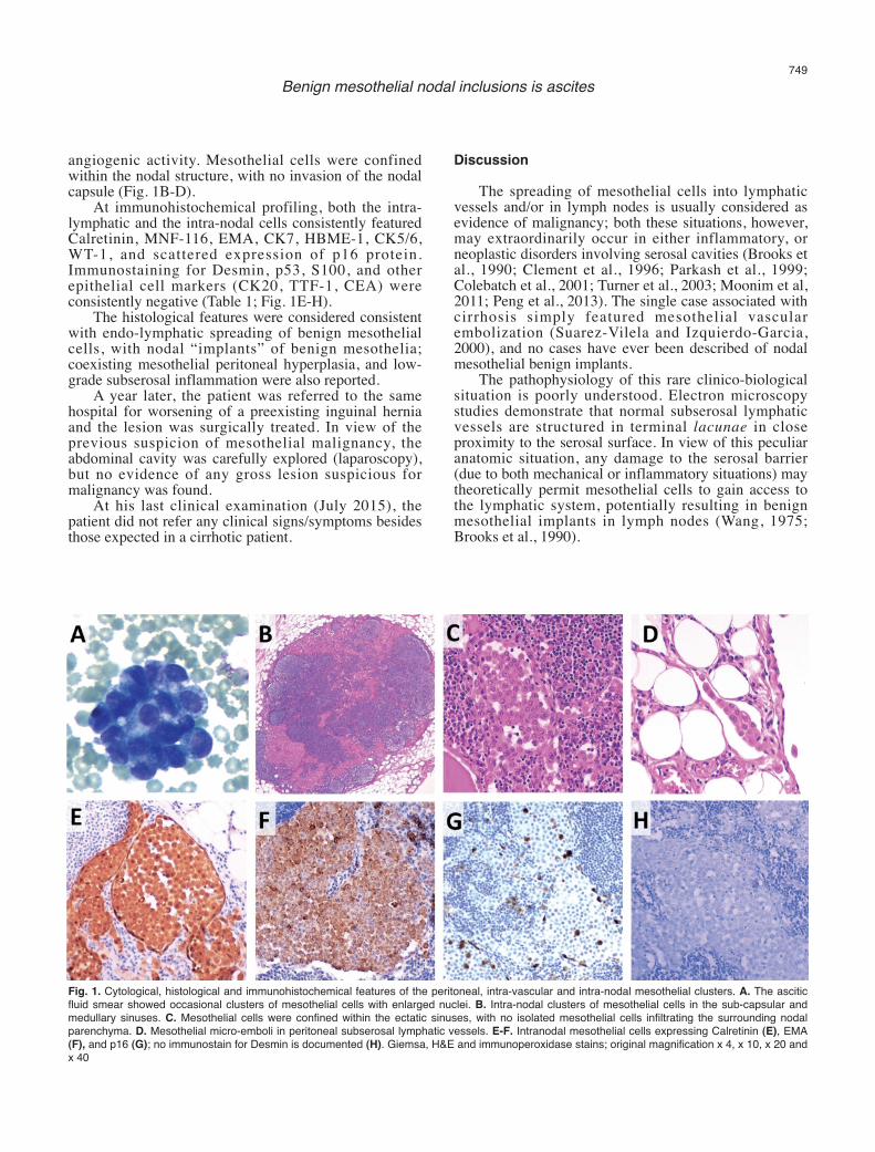
Fig. 1. Cytological, histological and immunohistochemical features of the peritoneal, intra-vascular and intra-nodal mesothelial clusters. A. The asciticfluid smear showed occasional clusters of mesothelial cells with enlarged nuclei. B. Intra-nodal clusters of mesothelial cells in the sub-capsular andmedullary sinuses. C. Mesothelial cells were confined within the ectatic sinuses, with no isolated mesothelial cells infiltrating the surrounding nodalparenchyma. D. Mesothelial micro-emboli in peritoneal subserosal lymphatic vessels. E-F. Intranodal mesothelial cells expressing Calretinin (E), EMA(F), and p16 (G); no immunostain for Desmin is documented (H). Giemsa, H&E and immunoperoxidase stains; original magnification x 4, x 10, x 20 andx 40

In the present case, the mesothelial profile of thenodal inclusions was consistently confirmed byimmunohistochemistry; additionally, the expression ofp16 protein argued against those p16 derangements,which have been mostly associated (at both genetic, andprotein level) with mesothelial malignancies (Chiosea etal., 2008; Takeda et al., 2010; Hwang et al., 2014).Hence, in spite of the alarming histological features ofthe “misplaced” mesothelia, the conclusive diagnosis ofnon-neoplastic disease was primarily based on the“benign phenotype” of the nodal implants: preservednodal architecture, lack of infiltrating growth pattern, theabsence of any angiogenic activity and desmoplasia(Attanoos et al., 2003; Mocanu et al., 2007; Hasteh et al.,2010; Husain et al., 2013). The non-malignant nature ofthe intra-vascular/intra-nodal mesothelial spreading hasbeen further supported by the long-term follow up,which consistently excluded any neoplastic disease, asreliably assessed by both PET-CT, and by twolaparoscopic inspections of the abdominal cavity.
We report the first case of a cirrhotic patient inwhom a benign mesothelial embolization to thelymphatic vessels coexisted with multiple nodalmesothelial implants. Both of these features should beincluded among the rare, non-neoplastic complicationsof recurrent ascites associated with cirrhosis.References
Attanoos R.L., Griff in A. and Gibbs A.R. (2003). The use ofimmunohistochemistry in distinguishing reactive from neoplasticmesothelium: A novel use for desmin and comparative evaluationwith epithelial membrane antigen, p53, platelet-derived growthfactor-receptor, P-glycoprotein and Bcl-2. Histopathology 43, 231-238.
Brooks J.S., LiVolsi V.A. and Pietra G.G. (1990). Mesothelial cellinclusions in mediastinal lymph nodes mimicking metastaticcarcinoma. Am. J. Clin. Pathol. 93, 741-748.
Chiosea S., Krasinskas A., Cagle P.T., Mitchell K.A., Zander D.S. andDacic S. (2008). Diagnostic importance of 9p21 homozygousdeletion in malignant mesotheliomas. Mod. Pathol. 21, 742-747.
Clement P.B., Young R.H., Oliva E., Sumner H.W. and Scully R.E.(1996). Hyperplastic mesothelial cells within abdominal lymphnodes: mimic of metastatic ovarian carcinoma and serous borderlinetumor--a report of two cases associated with ovarian neoplasms.Mod. Pathol. 9, 879-886.
Colebatch A., Clarkson A. and Gill A.J. (2011). Benign mesothelial cellsas confounders when cytokeratin immunohistochemistry is used insentinel lymph nodes. Hum. Pathol. 42, 1209-1210.
González-Navajas J.M., Francés R. and Such J. (2007). Bacterial DNAin patients with cirrhosis and sterile ascites. Its role as a marker ofbacterial translocation and prognostic tool. Rev. Esp. Enferm. Dig.99, 599-603.
Hasteh F., Lin G.Y., Weidner N. and Michael C.W. (2010). The use ofimmunohistochemistry to distinguish reactive mesothelial cells from
malignant mesothelioma in cytologic effusions. Cancer Cytopathol.118, 90-96.
Husain A.N., Colby T., Ordonez N., Krausz T., Attanoos R., BeasleyM.B., Borczuk A.C., Butnor K., Cagle P.T., Chirieac L.R., Churg A.,Dacic S., Fraire A., Galateau-Salle F., Gibbs A., Gown A., HammarS., Litzky L., Marchevsky A.M., Nicholson A.G., Roggli V., TravisW.D. and Wick M.; International Mesothelioma Interest Group.(2013). Guidelines for pathologic diagnosis of malignantmesothelioma: 2012 update of the consensus statement from theInternational Mesothelioma Interest Group. Arch. Pathol. Lab. Med.137, 647-667.
Hwang H., Tse C., Rodriguez S., Gown A. and Churg A. (2014). p16FISH deletion in surface epithelial mesothelial proliferations ispredictive of underlying invasive mesothelioma. Am. J. Surg. Pathol.38, 681-688.
Krohn S., Böhm S., Engelmann C., Hartmann J., Brodzinski A.,Chatzinotas A., Zeller K., Prywerek D., Fetzer I. and Berg T. (2014).Application of qualitative and quantitative real-time PCR, directsequencing, and terminal restriction fragment length polymorphismanalysis for detection and identification of polymicrobial 16S rRNAgenes in ascites. J. Clin. Microbiol. 52, 1754-1757.
Mocanu L., Cimpean A.M. and Raica M. (2007). Expression ofcytokeratin MNF116 and vimentin in pleural serous effusions. Rom.J. Morphol. Embryol. 48, 291-294.
Moonim M.T., Ng W.W. and Routledge T. (2011). Benign metastasizingmesothelial cells: a potential pitfall in mediastinal lymph nodes. J.Clin. Oncol. 29, 546-548.
Parkash V., Vidwans M. and Carter D. (1999). Benign mesothelial cellsin mediastinal lymph nodes. Am. J. Surg. Pathol. 23, 1264-1269.
Peng L., Shen Q., Liu X., Wang J., Shi S., Yu B. and Zhou X. (2013).Diffuse hyperplastic mesothelial cells in multiple lymph nodes: casereport with review of the literature. Int. J. Clin. Exp. Pathol. 6, 926-931.
Pizzi M., Piazza F., Agostinelli C., Fuligni F., Benvenuti P., Mandato E.,Casellato A., Rugge M., Semenzato G. and Pileri S.A. (2015).Protein kinase CK2 is widely expressed in follicular, Burkitt anddiffuse large B-cell lymphomas and propels malignant B-cell growth.Oncotarget 6, 6544-6552.
Suarez-Vilela D. and Izquierdo-Garcia F.M. (2000). Hyperplasticmesothelial cells in mediastinal lymph node sinuses. Arch. Pathol.Lab. Med. 124:1749.
Takeda M., Kasai T., Enomoto Y., Takano M., Morita K., Kadota E. andNonomura A. (2010). 9p21 deletion in the diagnosis of malignantmesothelioma, using fluorescence in situ hybridization analysis.Pathol. Int. 60, 395-399.
Turner R.R., Nora D.T., Trocha S.D. and Bilchik A.J. (2003). Colorectalcarcinoma nodal staging. Frequency and nature of cytokeratin-positive cells in sentinel and nonsentinel lymph nodes. Arch. Pathol.Lab. Med. 127, 673-679.
Wang N. (1975). The preformed stomas connecting the pleural cavityand the lymphatics in the parietal pleura. Am. Rev. Respir. Dis. 111,12-20.
Accepted December 23, 2015
750Benign mesothelial nodal inclusions is ascites