Benign Lesions of the Vocal Folds
35
Benign Lesions of the Vocal Folds Noah Meltzer, M.D. Zandy Hillel, M.D. December 14, 2007
Transcript of Benign Lesions of the Vocal Folds

Benign Lesions of theVocal Folds
Noah Meltzer, M.D.Zandy Hillel, M.D.
December 14, 2007

Learning Objectives
1) Review the presentation, pathophysiology, and stroboscopic exams of benign vocal fold lesions.
2) Participants will review case presentations of various vocal fold lesions.

Normal Anatomy

Histological Layers of Vocal Fold

Stratification Methods• Morphology: Location within the vocal
fold structure• Stroboscopy: Assessment of the
mucosal wave activity associated with the lesion(s)

Vocal Fold Polyp
– Unilateral or Bilateral– Presentation: hoarseness, increased effort– Exam: superior or inferior mucosal edge
• Translucent or Hemorrhagic
– Risk Factors: Smoking, Talkativeness– Pathophysiology: edema, vascular congestion,
venous stasis may cause polypoid changes.

Unilateral Mucoid Polyp

Hemorrhagic Polyp

Polyp with Reactive LesionReactive
LesionVocalFoldPolyp

Vocal Fold Polyp Management
– Strobe: Normal or Minimal Pathology– Voice Therapy: Voice reeducation– Stop smoking– Surgical therapy
• reduction w/mucosal sparing• Avoid vocal fold striping

Vocal Fold Cysts
• Unilateral or Bilateral• Mucus retention cysts & epidermoid cysts• Presentation
– Diplophonia, hoarseness, pain, fatigue • Pathophysiology
– Mucous gland duct plugged - Mucus retention– Keratin accumulation - Epidermoid

Subepithelial or Submucosal
Subepithelial Submucosal

Epidermoid Inclusion Cyst

Mucus Rentention Cyst

Management
1) Stroboscopy Essential• Significant reduction in mucosal wave
when lesion is submucosal • Minimally diminished mucosal wave when
subepithelial2) Direct Laryngoscopy
• Direct Visualization• Palpation• Rule out neoplasm

Treatment
• Medical: antireflux, hydration• Voice Therapy - minimally helpful• Surgery: cordotomy• Follow-up voice therapy is effective

Cordotomy

Cyst Removal through Cordotomy

Vocal Fold Nodules
– Bilateral & Fairly Symmetric– Epidemiology: Boys & Women– Presentation: breathy, weak, raspy voice
• Limited vocal range– Exam: Medial Surface of VFs– Pathophysiology: Vascular Congestion in
submucosa causes hyalinization of Reinke’s space & thickening of epithelium

Vocal Fold Nodules

VF Nodules

Management
• Strobe: no disturbance in mucosal waveIncomplete closure on glottic cycle
• Medical - hydration, reflux control• Behavioral - Voice Therapy effective• Surgical (persistant nodules w/
continued impaired voice)– Microdissection followed by voice rest &
speech therapy

Benign Glottic Lesions: IIStroboscopy Rounds
Noah Meltzer, MD12/14/07

Contact granulomaarrow points to cleft in granuloma from contralat arytenoid

Contact Granuloma
• Seen primarily in males (professional voice)• Other risk factors: GERD, chronic throat clearing,
?stress?• Trauma due to forceful adduction produces either
ulceration or granulation• Exam: Laryngoscopy will show a mass over vocal
process of arytenoid, possible contact cleft• Management: antireflux regimen, SLP,
?triamcinolone inh, bx after 3-4 mos med. Mgmt. (preserve the base/pedicle)

Pedunculated contact Granuloma A below cords B above C normal phonation due to pedunculated below
vocal surface

Postintubation Granuloma

Postintubation Granuloma
• Self-explanatory name• Risk factor: intubation/laryngeal
instrumentation• Diagnosis: history and endoscopy are
characteristic• Management: Antibiotics, SLP?,
triamcinolone inh?, SML excision preserving stalk?, mitomycin C postop?
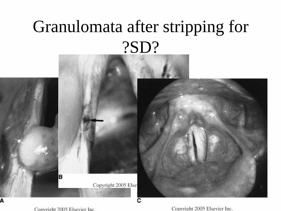
Granulomata after stripping for ?SD?

Post-surgical dysphonia
• Scarred/stiff TVC, height mismatch TVC• Avoided by conservative, meticulous
microsurgical technique• Hx: prior surgery• Exam: Strobe--stiff folds with minimal
mucosal wave, even at low frequencies• Management: voice-building for 1 yr. Then
surgery?

Surgical approach to adhesions

RRP
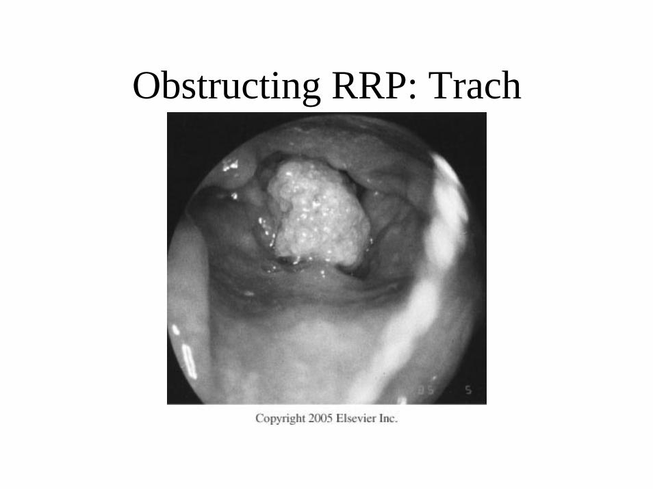
Obstructing RRP: Trach

RRP
• Squamous papillomata• The most common benign laryngeal neoplasm
(84% in a large series)• HPV (papova)• Risk factor: maternal HPV?• Bimodal presentation: juvenile (severe,
progressive), adult (limited, rarely progressive)• Mgmt: Shaver vs CO2 laser, ?interferon ?indole-
3-carbinol ?cidofovir