Behavioral/Systems/Cognitive ... · 12278 • J.Neurosci.,November19,2008 • 28(47):12274–12283...
10
Behavioral/Systems/Cognitive The Midbrain Periaqueductal Gray Control of Respiration Hari H. Subramanian, 1 Ron J. Balnave, 2 and Gert Holstege 1 1 Center for Uroneurology, University Medical Center Groningen, University of Groningen, 9700 RB Groningen, The Netherlands, and 2 School of Medical Sciences, University of Sydney, Lidcombe, New South Wales 1825, Australia The midbrain periaqueductal gray (PAG) organizes basic survival behavior, which includes respiration. How the PAG controls respira- tion is not known. We studied the PAG control of respiration by injecting D,L-homocysteic acid in the PAG in unanesthetized precollicu- larly decerebrated cats. Injections in different parts of the PAG caused different respiratory effects. Stimulation in the dorsomedial PAG induced slow and deep breathing and dyspnea. Stimulation in the dorsolateral PAG resulted in active breathing and tachypnea consistent with the respiratory changes during fright and flight. Stimulation in the medial part of lateral PAG caused inspiratory apneusis. Stimu- lation in lateral parts of the lateral and ventrolateral PAG produced respiratory changes associated with vocalization (mews, alternating mews and hisses, or hisses). D,L-Homocysteic acid injections in the caudal ventrolateral PAG induced irregular breathing. These results demonstrate that the PAG exerts a strong influence on respiration, suggesting that it serves as the behavioral modulator of breathing. Key words: midbrain; emotional breathing control; pattern generation; periaqueductal gray; brainstem; respiration Introduction Cell groups in the medullary and pontine lateral tegmental field are thought to control respiration (von Euler, 1983; Feldman, 1986; Long and Duffin, 1986). The inspiration-related cell groups differ from those related to expiration. Some of the inspiration- related cell groups contain premotor interneurons that project to the phrenic and external intercostal muscle motoneurons in the cervical and thoracic spinal cord, respectively (Holstege and Kuypers, 1982; Lipski et al., 1983; Duffin and Lipski, 1987; Hol- stege, 1991b). Expiration-related nuclei contain interneurons projecting to the internal intercostal and abdominal muscle mo- toneurons in the thoracic and upper-lumbar spinal cord (Hol- stege and Kuypers, 1982; Miller et al., 1989; Holstege, 1991b). Together, these premotor respiration-related centers regulate cy- clic breathing (or eupnea) on the basis of afferent information from the pulmonary mechanoreceptors (lung volume, stretch, and airway receptors) and from peripheral chemoreceptors in the carotid arteries measuring pCO 2 and pO 2 levels in the blood (von Euler, 1983; Feldman, 1986; Long and Duffin, 1986). Most of this information enters the brainstem via the vagal nerve. One might consider the caudal brainstem respiration-related cell groups as reflex centers that maintain, increase, or decrease the respiratory frequency (RF), depending on pulmonary and chemoreceptor afferent information. However, this reflex system is not sufficient for proper survival behavior. In case sudden events in the sur- rounding of the individual represent danger, fight or flight reac- tions might be necessary, which includes immediate rapid shal- low breathing. Other strong anxiety reactions necessitate gasping or vocalization. The caudal brainstem premotor respiration- related cell groups do not receive from vagal afferents informa- tion concerning the external environment. To adjust respiration to survive threatening events, a mediator is necessary that re- ceives visual, auditory, and somatosensory information and has access to respiration-related cell groups in the caudal brainstem. We hypothesize that this mediator is the midbrain periaqueduc- tal gray (PAG). The PAG, by way of its projections to the respiration-related cell groups in the caudal brainstem, can change the eupneic breathing pattern into respiratory function appropriate for the situation. However, studies about the PAG control of respiration in the cat do not exist except in the context of vocalization. In these studies (Magoun et al., 1937; Bandler and Carrive, 1988; Zhang et al., 1994; Nonaka et al., 1999; Nakazawa et al., 2000), the sound production was measured but not the respiratory function. In the rat, a few studies exist (Huang et al., 2000; Hayward et al., 2003, 2004; Zhang et al., 2005, 2007; Sub- ramanian et al., 2007, 2008), but they were limited to the dorsal PAG showing only an increase in respiratory frequency. The present study is the first that investigated the PAG effects on respiration by stimulation of different parts of the PAG. We stud- ied not only the changes in respiratory phase durations but also the intratracheal pressure and the diaphragmatic, intercostal, and abdominal muscle EMG patterns after stereotaxic chemical stim- ulation of different parts of the PAG. Materials and Methods Surgery. Approval for these studies was obtained from the Institutional Animal Care Ethics Committee of the University of Sydney. Eight cats (weighing between 2.2 and 4.5 kg) were used. They were anesthetized in a box filled with a mixture of isoflurane and oxygen delivered by a ven- tilation pump. After induction, the anesthesia was maintained through a face mask, and femoral arterial and venous catheters were inserted. Using the intravenous catheter, sodium thiopental (10 –20 mg/kg) was injected Received Sept. 2, 2008; revised Sept. 25, 2008; accepted Oct. 1, 2008. This work was supported by grants from the Faculty of Health Sciences, The University of Sydney (H.H.S.). H.H.S. was a recipient of an Australian National Health and Medical Research Council Neuroscience postgraduate scholar- ship and Balmain Hospital research assistantship. Correspondence should be addressed to Gert Holstege, Center for Uroneurology, University Medical Center Gro- ningen, University of Groningen, Postbus 30.001, 9700 RB Groningen, The Netherlands. E-mail: [email protected]. DOI:10.1523/JNEUROSCI.4168-08.2008 Copyright © 2008 Society for Neuroscience 0270-6474/08/2812274-10$15.00/0 12274 • The Journal of Neuroscience, November 19, 2008 • 28(47):12274 –12283
Transcript of Behavioral/Systems/Cognitive ... · 12278 • J.Neurosci.,November19,2008 • 28(47):12274–12283...
Behavioral/Systems/Cognitive
The Midbrain Periaqueductal Gray Control of Respiration
Hari H. Subramanian,1 Ron J. Balnave,2 and Gert Holstege1
1Center for Uroneurology, University Medical Center Groningen, University of Groningen, 9700 RB Groningen, The Netherlands, and 2School of MedicalSciences, University of Sydney, Lidcombe, New South Wales 1825, Australia
The midbrain periaqueductal gray (PAG) organizes basic survival behavior, which includes respiration. How the PAG controls respira-tion is not known. We studied the PAG control of respiration by injecting D,L-homocysteic acid in the PAG in unanesthetized precollicu-larly decerebrated cats. Injections in different parts of the PAG caused different respiratory effects. Stimulation in the dorsomedial PAGinduced slow and deep breathing and dyspnea. Stimulation in the dorsolateral PAG resulted in active breathing and tachypnea consistentwith the respiratory changes during fright and flight. Stimulation in the medial part of lateral PAG caused inspiratory apneusis. Stimu-lation in lateral parts of the lateral and ventrolateral PAG produced respiratory changes associated with vocalization (mews, alternatingmews and hisses, or hisses). D,L-Homocysteic acid injections in the caudal ventrolateral PAG induced irregular breathing. These resultsdemonstrate that the PAG exerts a strong influence on respiration, suggesting that it serves as the behavioral modulator of breathing.
Key words: midbrain; emotional breathing control; pattern generation; periaqueductal gray; brainstem; respiration
IntroductionCell groups in the medullary and pontine lateral tegmental fieldare thought to control respiration (von Euler, 1983; Feldman,1986; Long and Duffin, 1986). The inspiration-related cell groupsdiffer from those related to expiration. Some of the inspiration-related cell groups contain premotor interneurons that project tothe phrenic and external intercostal muscle motoneurons in thecervical and thoracic spinal cord, respectively (Holstege andKuypers, 1982; Lipski et al., 1983; Duffin and Lipski, 1987; Hol-stege, 1991b). Expiration-related nuclei contain interneuronsprojecting to the internal intercostal and abdominal muscle mo-toneurons in the thoracic and upper-lumbar spinal cord (Hol-stege and Kuypers, 1982; Miller et al., 1989; Holstege, 1991b).Together, these premotor respiration-related centers regulate cy-clic breathing (or eupnea) on the basis of afferent informationfrom the pulmonary mechanoreceptors (lung volume, stretch,and airway receptors) and from peripheral chemoreceptors in thecarotid arteries measuring pCO2 and pO2 levels in the blood (vonEuler, 1983; Feldman, 1986; Long and Duffin, 1986). Most of thisinformation enters the brainstem via the vagal nerve. One mightconsider the caudal brainstem respiration-related cell groups asreflex centers that maintain, increase, or decrease the respiratoryfrequency (RF), depending on pulmonary and chemoreceptorafferent information. However, this reflex system is not sufficientfor proper survival behavior. In case sudden events in the sur-rounding of the individual represent danger, fight or flight reac-
tions might be necessary, which includes immediate rapid shal-low breathing. Other strong anxiety reactions necessitate gaspingor vocalization. The caudal brainstem premotor respiration-related cell groups do not receive from vagal afferents informa-tion concerning the external environment. To adjust respirationto survive threatening events, a mediator is necessary that re-ceives visual, auditory, and somatosensory information and hasaccess to respiration-related cell groups in the caudal brainstem.We hypothesize that this mediator is the midbrain periaqueduc-tal gray (PAG). The PAG, by way of its projections to therespiration-related cell groups in the caudal brainstem, canchange the eupneic breathing pattern into respiratory functionappropriate for the situation. However, studies about the PAGcontrol of respiration in the cat do not exist except in the contextof vocalization. In these studies (Magoun et al., 1937; Bandler andCarrive, 1988; Zhang et al., 1994; Nonaka et al., 1999; Nakazawaet al., 2000), the sound production was measured but not therespiratory function. In the rat, a few studies exist (Huang et al.,2000; Hayward et al., 2003, 2004; Zhang et al., 2005, 2007; Sub-ramanian et al., 2007, 2008), but they were limited to the dorsalPAG showing only an increase in respiratory frequency. Thepresent study is the first that investigated the PAG effects onrespiration by stimulation of different parts of the PAG. We stud-ied not only the changes in respiratory phase durations but alsothe intratracheal pressure and the diaphragmatic, intercostal, andabdominal muscle EMG patterns after stereotaxic chemical stim-ulation of different parts of the PAG.
Materials and MethodsSurgery. Approval for these studies was obtained from the InstitutionalAnimal Care Ethics Committee of the University of Sydney. Eight cats(weighing between 2.2 and 4.5 kg) were used. They were anesthetized ina box filled with a mixture of isoflurane and oxygen delivered by a ven-tilation pump. After induction, the anesthesia was maintained through aface mask, and femoral arterial and venous catheters were inserted. Usingthe intravenous catheter, sodium thiopental (10 –20 mg/kg) was injected
Received Sept. 2, 2008; revised Sept. 25, 2008; accepted Oct. 1, 2008.This work was supported by grants from the Faculty of Health Sciences, The University of Sydney (H.H.S.). H.H.S.
was a recipient of an Australian National Health and Medical Research Council Neuroscience postgraduate scholar-ship and Balmain Hospital research assistantship.
Correspondence should be addressed to Gert Holstege, Center for Uroneurology, University Medical Center Gro-ningen, University of Groningen, Postbus 30.001, 9700 RB Groningen, The Netherlands. E-mail:[email protected].
DOI:10.1523/JNEUROSCI.4168-08.2008Copyright © 2008 Society for Neuroscience 0270-6474/08/2812274-10$15.00/0
12274 • The Journal of Neuroscience, November 19, 2008 • 28(47):12274 –12283

to enable the insertion of an endotracheal catheter, which was then usedfor the administration of isoflurane. The cat’s head was secured in astereotaxic frame with the body suspended from the frame by straps. Toallow access to the midbrain, two burr holes were drilled in the skull oneither side of the sagittal sinus. The dura was then incised and the medialpart of the cortex was suctioned. After ligation and removal of a portionof the sagittal sinus, precollicular decerebration was performed usingsuction diathermia, a surgical technique that uses pulsations of electricalenergy to generate heat and cauterize blood vessels to prevent excessivebleeding while suctioning brain tissue. Bleeding during decerebrationwas reduced by lowering the mean arterial pressure from 100 –130 to65–70 mmHg by raising the level of anesthesia. All brain tissue rostral tothe superior colliculus including the entire diencephalon was removed.After completion of decerebration, anesthesia was discontinued. The catstarted breathing spontaneously and the mean arterial pressure returnedto 100 –130 mmHg within 30–60 min. A thermostatic infrared lamp wasused to maintain the animal body temperature at 37.5–38.5°C. The end-tidalCO2 (measured by means of a Morgan 901 gas analyzer) was intermittentlymonitored. Fluid supplements were administered via the femoral intrave-nous catheter.
Chemical stimulation in the PAG. Excitatory amino acid [D,L-homocysteic acid (DLH); 200 mM; Sigma-Aldrich] microinjections wereused for stimulation in the PAG. For these injections, a pressure system(Picospritzer II; Parker Instrumentation) was connected to a single-barrel glass micropipette (tip diameter, 10 –30 �m). This micropipettewas inserted into different parts of the PAG guided by stereotaxic coor-dinates, because previous studies (Bandler and Carrive, 1988; Bandlerand Depaulis, 1991; Behbehani, 1995) have shown that different portionsof the PAG have different effects in the context of survival behavior. Theinjected volume of DLH was determined by using a precalibrated scale.To eliminate the effect of a previous injection, the interval between mi-croinjections was at least 25 min. Rhodamine beads were added to the
DLH solution for later histological identifica-tion of the injection sites. At the end of eachexperiment, the animal was deeply anesthetizedand perfused transcardially with 0.9% salinefollowed by 4% paraformaldehyde in phos-phate buffer, pH 7.2. After perfusion, the brainwas removed and stored in 4% formaldehydefor 2 h. It was then transferred to a 30% sucrose/formaldehyde mixture for at least 48 h to pre-vent ice crystal formation. The midbrain wascut on a freezing microtome into 50 �m coro-nal sections. The injection sites marked withrhodamine were identified using fluorescentmicroscopy and were represented on standarddrawings according to the stereotaxic atlas ofBerman (1968).
EMG recording. Unilateral EMG activity wasrecorded using bipolar Teflon-coated, multi-stranded stainless-steel electrodes, bared for 2mm at their tips. The electrodes were surgicallyimplanted in the crural diaphragm and in a se-lection of cricothyroid, internal intercostal, in-ternal and external abdominal oblique, and ge-nioglossus muscles. A maximum of four muscleEMGs could be recorded simultaneously andanalyzed during each PAG stimulation. A 19-gauge needle was inserted into the trachea andconnected to a differential pressure transducerto record the changes in tracheal pressure.Mean arterial pressure was recorded throughthe catheter placed in the femoral artery andobtained from the low-pass-filtered pressurepulses. The mouth of the cat was kept open, andvocalization was recorded with a microphoneplaced 10 cm from the mouth.
Data analysis. The bandpass-filtered (0.1–5kHz) respiratory and laryngeal muscle EMG ac-tivity as well as other signals such as tracheal
pressure, mean arterial pressure, and the sound of vocalization, wererecorded on the “Pulse Code Modulator” (PCM) (A. R. Vetter) andplayed back for analysis using the PCM/MacLab/Apple system and Chartsoftware (ADInstruments). The recording started at least 60 s before theadministration of DLH. After the injection, the recording continued forat least 5 min. Statview software running on an Apple computer was usedfor the computation of inspiratory time (Ti), expiratory time (Te), andRF as well as for statistical comparisons, which were performed usingANOVA. Significant differences between the mean values were detectedusing Scheffe’s least difference test. A probability of �0.05 was consid-ered significant. Chart records have been transformed into MacDraw Prosoftware for data representation.
Measurement of respiratory durations and frequency. The raw dia-phragm EMG signal was used for measurement of inspiratory (Ti) andexpiratory (Te) durations and RF. The duration from the onset of thediaphragm signal to its offset was defined as Ti. The duration between theoffset of the diaphragm and its subsequent onset was defined as Te. Theexact points of onset and offset of inspiration was determined via com-parison of the diaphragm EMG with internal intercostal EMG (Fig. 1 A)or tracheal pressure signal (Fig. 1 B).
ResultsSpontaneously breathing vagiintact precollicularly decerebratedcats (n � 8) had the following respiratory and cardiovascularparameters (control values expressed as mean � SE): Ti, 0.60 �0.05 s; Te, 0.90 � 0.05 s; RF, 40 � 4 breaths/min; blood pressure(BP), 105 � 5 mmHg; heart rate, 199 � 5 beats/min; arterialblood pH, 7.39 � 0.05; paCO2, 25.2 � 5.0 Torr; paO2, 110.5 �10.0 Torr. All animals had postinspiratory activity in the cruraldiaphragm but only during eupnea. The postinspiratory activity
Diaphragm EMG
Internal Intercostal EMG
1s
offset of inspiration/ onset of expiration
offset of expiration/ onset of inspiration
A
Te TiTi
post-inspiratory activity
Tracheal Pressure
1s
Ti Ti
post-inspiratory activity
Te
onset of inspiration
offset of expiration/ onset of inspiration
offset of inspiration/ onset of expiration
B
Diaphragm EMG
Figure 1. Illustration of Ti and Te measured from the raw diaphragm EMG signal. The diaphragm EMG activity is compared withinternal intercostal EMG (A) or tracheal pressure (B) while locating the start and fall of its activity.
Subramanian et al. • PAG Control of Respiration J. Neurosci., November 19, 2008 • 28(47):12274 –12283 • 12275
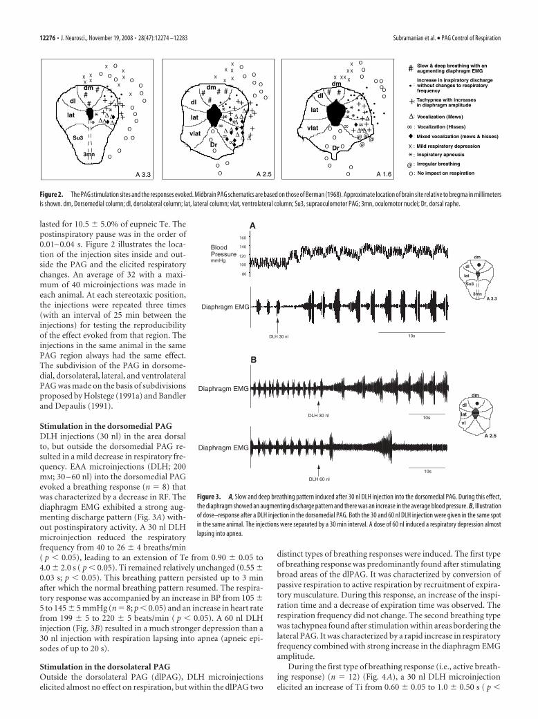
lasted for 10.5 � 5.0% of eupneic Te. Thepostinspiratory pause was in the order of0.01– 0.04 s. Figure 2 illustrates the loca-tion of the injection sites inside and out-side the PAG and the elicited respiratorychanges. An average of 32 with a maxi-mum of 40 microinjections was made ineach animal. At each stereotaxic position,the injections were repeated three times(with an interval of 25 min between theinjections) for testing the reproducibilityof the effect evoked from that region. Theinjections in the same animal in the samePAG region always had the same effect.The subdivision of the PAG in dorsome-dial, dorsolateral, lateral, and ventrolateralPAG was made on the basis of subdivisionsproposed by Holstege (1991a) and Bandlerand Depaulis (1991).
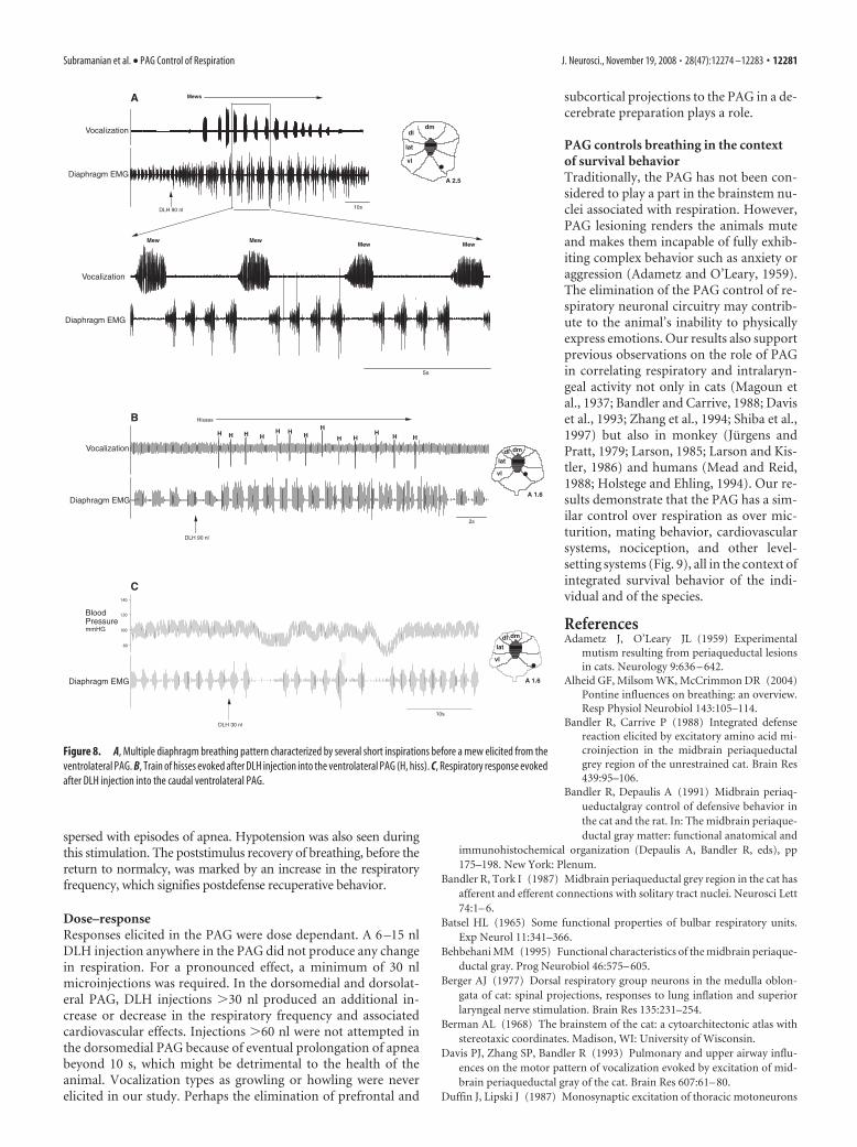
Stimulation in the dorsomedial PAGDLH injections (30 nl) in the area dorsalto, but outside the dorsomedial PAG re-sulted in a mild decrease in respiratory fre-quency. EAA microinjections (DLH; 200mM; 30 – 60 nl) into the dorsomedial PAGevoked a breathing response (n � 8) thatwas characterized by a decrease in RF. Thediaphragm EMG exhibited a strong aug-menting discharge pattern (Fig. 3A) with-out postinspiratory activity. A 30 nl DLHmicroinjection reduced the respiratoryfrequency from 40 to 26 � 4 breaths/min( p � 0.05), leading to an extension of Te from 0.90 � 0.05 to4.0 � 2.0 s ( p � 0.05). Ti remained relatively unchanged (0.55 �0.03 s; p � 0.05). This breathing pattern persisted up to 3 minafter which the normal breathing pattern resumed. The respira-tory response was accompanied by an increase in BP from 105 �5 to 145 � 5 mmHg (n � 8; p � 0.05) and an increase in heart ratefrom 199 � 5 to 220 � 5 beats/min ( p � 0.05). A 60 nl DLHinjection (Fig. 3B) resulted in a much stronger depression than a30 nl injection with respiration lapsing into apnea (apneic epi-sodes of up to 20 s).
Stimulation in the dorsolateral PAGOutside the dorsolateral PAG (dlPAG), DLH microinjectionselicited almost no effect on respiration, but within the dlPAG two
distinct types of breathing responses were induced. The first typeof breathing response was predominantly found after stimulatingbroad areas of the dlPAG. It was characterized by conversion ofpassive respiration to active respiration by recruitment of expira-tory musculature. During this response, an increase of the inspi-ration time and a decrease of expiration time was observed. Therespiration frequency did not change. The second breathing typewas tachypnea found after stimulation within areas bordering thelateral PAG. It was characterized by a rapid increase in respiratoryfrequency combined with strong increase in the diaphragm EMGamplitude.
During the first type of breathing response (i.e., active breath-ing response) (n � 12) (Fig. 4A), a 30 nl DLH microinjectionelicited an increase of Ti from 0.60 � 0.05 to 1.0 � 0.50 s ( p �
# #dm
dl
Dr
++
+++
• ••• • •
##
#
lat
vlat
++
++
XX X
X X
A 2.5
X
++
•***
OO O
O
O OO O
OOO
O
O
O
A 1.6
dm
dl
lat
vlat
Dr
#•
#••
+ +
•
+
+• ++•
+
X XXX X
X
••
•• •••
••
X
•***
* •
@@@@
OO O
O
O OO
O O
OOO
O OOOO
• :Increase in inspiratory discharge without changes to respiratory frequency
+: Tachypnea with increases in diaphragm amplitude
: Vocalization (Mews)
#: Slow & deep breathing with an augmenting diaphragm EMG
: Mixed vocalization (mews & hisses)
: No impact on respiration
: Vocalization (Hisses)
X : Mild respiratory depression
* : Inspiratory apneusis
: Irregular breathing@OA 3.3
dm##
+++
• •
X
XX X
+
•
++
••
+
X
X
XX
#
X
++dl
lat **
*
OO
O O
OO
OO
OOO
O
OO O
O
Su3
3mn
Figure 2. The PAG stimulation sites and the responses evoked. Midbrain PAG schematics are based on those of Berman (1968). Approximate location of brain site relative to bregma in millimetersis shown. dm, Dorsomedial column; dl, dorsolateral column; lat, lateral column; vlat, ventrolateral column; Su3, supraoculomotor PAG; 3mn, oculomotor nuclei; Dr, dorsal raphe.
DLH 30 nl
Diaphragm EMG
10s
B
DLH 60 nl
Diaphragm EMG
10s
A 2.5
dm
dl
lat
vl
•
A 3.3
dm
dl
lat
Su3
3mn
•Blood PressuremmHg
Diaphragm EMG
120
160
80
100
140
DLH 30 nl 10s
A
Figure 3. A, Slow and deep breathing pattern induced after 30 nl DLH injection into the dorsomedial PAG. During this effect,the diaphragm showed an augmenting discharge pattern and there was an increase in the average blood pressure. B, Illustrationof dose–response after a DLH injection in the dorsomedial PAG. Both the 30 and 60 nl DLH injection were given in the same spotin the same animal. The injections were separated by a 30 min interval. A dose of 60 nl induced a respiratory depression almostlapsing into apnea.
12276 • J. Neurosci., November 19, 2008 • 28(47):12274 –12283 Subramanian et al. • PAG Control of Respiration

0.05) and a decrease of Te from 0.90 � 0.05 to 0.55 � 0.03 s ( p �0.05), leaving the respiratory frequency relatively unchanged(39 � 2 breaths/min; p � 0.05). In addition, a strong increase ofthe diaphragm EMG amplitude, blood pressure [105 � 4 to120 � 2 mmHg ( p � 0.05)], and heart rate [199 � 5 to 220 � 5beats/min ( p � 0.05)] was observed. The diaphragm did notexhibit any postinspiratory activity. There was also recruitmentof the external abdominal oblique muscle.
During the second type of breathing response (i.e., tachypnea)(n � 5) (Fig. 4B), a 30 nl DLH injection elicited a rapid rise in therespiratory frequency from 40 � 2 to 84 � 5 breaths/min (n � 6;p � 0.05), caused by a decrease of both Ti (0.25 � 0.02 s; p � 0.05)and Te (0.50 � 0.03 s; p � 0.05). There was a strong increase of
the diaphragm EMG amplitude, but nopostinspiratory activity. This tachypneawas accompanied by a blood pressure in-crease from 105 � 5 to 150 � 5 mmHg(n � 6; p � 0.05) and a heart rate increasefrom 199 � 5 to 250 � 10 beats/min (n �6; p � 0.05).
Stimulation in the lateral PAGEAA microinjections in the lateral PAG(A3.3–A2.5) evoked three types of breath-ing responses: (1) tachypnea (n � 9) sim-ilar to the one observed stimulating thedorsolateral PAG (see above), (2) inspira-tory apneusis (n � 9; p � 0.05), and (3)respiratory changes in the context of vo-calization (n � 13; p � 0.05).
The second effect, inspiratory apneusis(Fig. 5A), was obtained from especially themedial part of the lateral PAG and wascharacterized by prolongation of inspira-tion up to 8 s after a 30 nl DLH injection.No postinspiratory activity was seen in thediaphragm EMG and no cardiovascularchanges were observed.
The third type of respiratory changeproduced by 30 nl DLH injections in thelateral PAG was a transient vocal responselasting 2–5 s. Larger injections (90 nl) elic-ited much longer vocal episodes (20 –120s). The main type of vocalization elicitedfrom the lateral PAG (n � 9) was mews,consisting of recruitment of genioglossus,cricothyroid (Fig. 5B), external abdominaloblique (Fig. 6A), and internal intercostaland internal abdominal oblique muscles(Fig. 6B). The contraction of these mus-cles caused an increase in the tracheal pres-sure of up to 40 cm H2O leading to vocal-ization (Fig. 6C). The vocalization effectevoked from the lateral PAG representedin Figures 5 and 6 are from different DLH(90 nl) injections.
During mew episodes, the respiratoryfrequency strongly decreased from 40 � 3to 26 � 5 breaths/min (n � 10; p � 0.05).The Ti showed a small reduction from0.60 � 0.05 to 0.50 � 0.01 s ( p � 0.05),and the diaphragm did not exhibit postin-spiratory activity. The Te was prolonged,
from 0.9 � 0.05 s to an average of 2.0 � 0.50 s ( p � 0.05 vs), witha maximum expiratory vocal time of 5 s.
Stimulation in the ventrolateral PAGStimulation in the ventrolateral PAG produced a mixed vocalresponse (n � 8) consisting of alternating long mews and shorthisses accompanied by a so-called “double diaphragm” breathingpattern (Fig. 7A). The sequence was generally mew, inspiration,hiss, inspiration, mew, inspiration, hiss, etc. A hiss was character-ized by small changes in tracheal pressure (up to 6 cm H2O) andsmall recruitments of the external oblique abdominal muscle(Fig. 7B). The Ti preceding the hiss was generally longer than ineupnea, whereas the Te during the hiss was shorter. During the
A 2.5
dmdl
lat
vl
•
Diaphragm EMG
DLH 30nl
B
100
120
140
160
80
10s
1s
Diaphragm EMG
Blood PressuremmHg
A
Blood PressuremmHg
80
100
120
140
160
Diaphragm EMG
A 3.3
dmdl
lat
Su3
3mn
•External abdominal oblique EMG
DLH 30 nl10s
External abdominal oblique EMG
Diaphragm EMG
5s
Figure 4. A, Injection of DLH in the dorsolateral PAG elicited an increase in the inspiratory duration, decrease in the expiratoryduration, but no change in the respiratory frequency. B, In another case, DLH injection in the dorsolateral PAG resulted intachypnea characterized by a decrease in both inspiratory and expiratory durations and consequent increase in the respiratoryfrequency. In both effects, an increase in the diaphragm amplitude can be seen.
Subramanian et al. • PAG Control of Respiration J. Neurosci., November 19, 2008 • 28(47):12274 –12283 • 12277

entire mixed vocal episode, Ti increasedfrom 0.60 � 0.05 to 0.70 � 0.05 s ( p �0.05), Te decreased from 0.90 � 0.05 to0.50 � 0.05 s ( p � 0.05), and the respira-tory frequency decreased considerablyfrom 40 � 3 to 12 � 3 breaths/min ( p �0.05). In most cases (n � 8; p � 0.05), thismixed vocalization episode was precededby tachypnea (Fig. 7C), which started �10s after a 90 nl DLH injection and contin-ued until the start of vocalization (�30 safter DLH injection). This tachypneic epi-sode was similar to that observed afterDLH injection in the dorsolateral and lat-eral PAG. During this tachypnea, a posi-tive shift in the baseline of the cricothyroidmuscle EMG was seen (Fig. 7C).
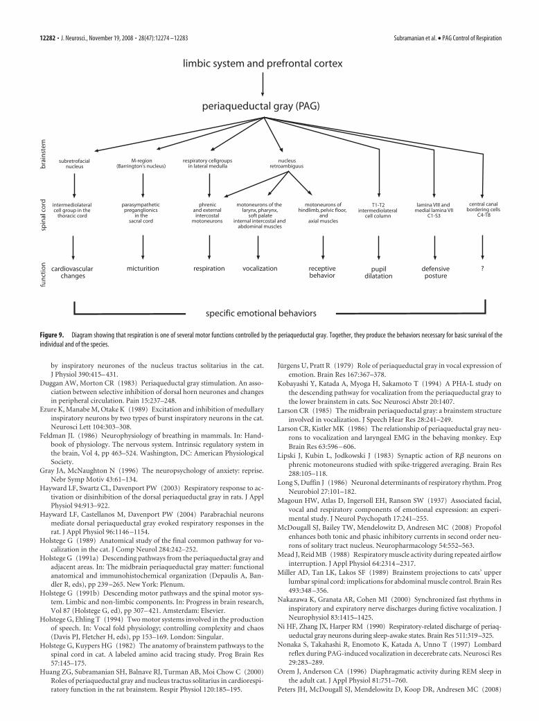
In three cases ( p � 0.05), the DLHinjection generated a “multiple dia-phragm” breathing pattern (Fig. 8 A)consisting of two to four short inspirationsbefore a mew, but without any hisses.
In six cases, a train of only hisses wasevoked ( p � 0.05) (Fig. 8B) from the me-dial part of the ventrolateral PAG. Duringsuch a train, the respiratory frequency in-creased from 40 � 3 to 63 � 3 breaths/min( p � 0.05) because of a reduction of Tefrom 0.90 � 0.05 to 0.40 � 0.02 s ( p �0.05), with Ti remaining the same (0.60 �0.05 s; p � 0.05).
In four cases, stimulation of the caudalventrolateral PAG elicited a respiratory re-sponse characterized by irregular breath-ing interspersed by episodes of apnea andno vocalization (Fig. 8C). The apneas ex-tended up to 5 s. The diaphragm EMGdiffered from burst to burst during theeffect period but did not exhibit anypostinspiratory activity. There was asmall increase in respiratory frequencyfrom 40 � 2 to 48 � 2 breaths/min ( p �0.05). This respiratory response was ac-companied by a decrease in blood pres-sure [from 105 � 5 to 85 � 6 mmHg (n � 4; p � 0.05)] andheart rate [from 199 � 5 to 160 � 5 beats/min ( p � 0.05)].
DiscussionThe present investigation in the cat provides the first demonstra-tion that different regions of the PAG have different effects onbreathing. Our data demonstrate that the PAG is able to converteupneic breathing into a breathing pattern that fits the particularbehavior necessary for survival in specific circumstances. Threeother studies in the cat (Zhang et al., 1994; Nonaka et al., 1999;Nakazawa et al., 2000) examined the role of the PAG in vocaliza-tion or fictive vocalization but not respiration. Moreover, none ofthese studies explores stereotaxically the influence of differentareas of the PAG on respiratory function. Subramanian et al.(2007, 2008) studied the PAG-induced changes to caudal medul-lary respiration-related neuronal activity in the rat, and Huang etal. (2000) also in the rat investigated the role of the PAG oncardiorespiratory control, and subsequently Hayward et al.(2003, 2004) and Zhang et al. (2005, 2007) examined the PAG-
induced change to respiratory frequency. However, all thesestudies in the rat focused on the dorsal PAG and reported onlyexcitatory effects. In the present study in the cat, we show that notonly excitatory but also inhibitory effects can be evoked from thePAG. Another major problem is that the rat studies used anesthe-tized preparations. Anesthesia is a major problem in studyingrespiration, because it suppresses the respiratory responses thatcan be evoked by stimulation in various brainstem areas (Batsel,1965; McDougall et al., 2008; Peters et al., 2008). It means thatchemical activation in anesthetized animals never provides con-clusive results. Because our data have been obtained in decere-brate, unanesthetized cats, they closely resemble the effects thatwill be observed after PAG activation in unanesthetized animalsor humans, albeit that the influence from higher brain levels onthe brainstem is missing.
Midbrain– brainstem respiratory pathwaysThe PAG has no direct connections with any motoneuronal cellgroup in brainstem and spinal cord. To achieve effects on motor
A
Blood PressuremmHg
100
110
90
Diaphragm EMG
10sDLH 30nl
Inspiratory apneusis
A 2.5
dmdl
lat
vl•
Genioglossus EMG
Cricothyroid EMG
Blood PressuremmHg
Diaphragm EMG
Vocalization
100110
90
DLH 90 nl
Mews
10s
A 2.5
dmdl
lat
vl•
B
BPmmHg
Vocalization
Cricothyroid EMG
Diaphragm EMG
100
90
110
2s
Mew Mew Mew Mew
Genioglossus EMG
Figure 5. A, A 30 nl DLH injection in the medial part of the lateral PAG resulted in inspiratory apneusis. B, Mews were the resultof injection of 90 nl DLH in the lateral PAG. The expansion illustrates the precise vocalization effect in three muscles.
12278 • J. Neurosci., November 19, 2008 • 28(47):12274 –12283 Subramanian et al. • PAG Control of Respiration

output, it uses premotor interneurons involved in coordina-tion of motor output. Also for producing changes in breath-ing, the PAG uses respiration-related premotor interneuronsin the brainstem. In the cat, it projects to the parabrachial andKolliker–Fuse nuclei (Holstege, 1991a), the retrofacial nu-cleus (Kobayashi et al., 1994; Sakamoto et al., 1996), the soli-tary nucleus (NTS) (Bandler and Tork, 1987), and the nucleusretroambiguus (NRA) (Holstege, 1989). The pontine respira-tory groups maintain direct projections to the phrenic nucleus(Holstege and Kuypers, 1982) and have been proposed to playan important role in the modulation of respiratory reflexes,such as phase-switching (Alheid et al., 2004). The retrofacialregion in the rostral medulla contains inspiratory neurons thatproject directly to the phrenic motoneurons (Holstege and
Kuypers, 1982), but neurons in this re-gion also excite other bulbospinal andpropriobulbar inspiratory cells (Ezure etal., 1989). The nucleus of the solitarytract in the cat contains inspiratory neu-rons (von Baumgarten and Kanzow,1958; Berger, 1977) that drive thephrenic (Lipski et al., 1983) and externalintercostal motoneurons through directconnections (Holstege and Kuypers,1982; Duffin and Lipski, 1987). Cells inthe rostral part of the nucleus retroam-biguus play a role in inspiration and sev-eral other motor output systems by wayof their projections to the nucleus am-biguus (Holstege, 1989), the phrenic nu-cleus, and the external intercostal mo-toneurons (Holstege and Kuypers, 1982;Miller et al., 1989). Neurons in the caudalNRA, however, play a role in expiration andmany other motor functions via their pro-jections to abdominal, internal intercostal,pelvic floor, and several other motoneuro-nal cell groups (Holstege, 1991b). In short,the PAG uses the respiration-related cellgroups to integrate respiration in the con-text of organizing specific survival behav-ior. In the rat, stimulation in the dorsalPAG has been shown to result in phasicexcitation of lateral parabrachialinspiration-related neurons (Hayward etal., 2004) and in both phasic and tonic ac-tivation of inspiration- and expiration-related neurons in the ventrolateral me-dulla (Subramanian et al., 2008) andnucleus tractus solitarius (Subramanianet al., 2007). The involvement of�-adrenoceptors within the NTS mediat-ing the PAG-induced respiratory and car-diovascular effect has also been previouslyreported in the rat (Huang et al., 2000;Subramanian et al., 2007). Obviously,the basic behavioral repertoire in thePAG is further evolved in cat than in rat;however, in the cat, there are no electro-physiological data presently available onthe PAG effects on the brainstemrespiration-related nuclei.
Dorsomedial PAG stimulation produces dyspneaThe dorsomedial PAG is strongly implicated in the mediation offear and anxiety and associated defensive responses (Bandler andCarrive, 1988; Bandler and Depaulis, 1991). The slow and deepbreathing as we elicited in the dorsomedial PAG may fit such a fearreaction. The expiratory apneusis we also observed corresponds withDuggan and Morton (1983), who found that, in anesthetized cats,stimulation in the dorsal PAG increases expiratory CO2.
Dorsolateral PAG produces increased respiratory effortBoth defensive and aversive responses are elicited by stimulat-ing the dorsolateral PAG. This includes threat and confronta-tional behaviors as well as flight response (Bandler and De-paulis, 1991). For such confrontational behaviors, active
External abdominal oblique EMG
Diaphragm EMG
10s
DLH 90 nl
A 2.5
dmdl
lat
vl •
Vocalization
MewsA
Internal intercostal EMG
A 2.5
dmdl
lat
vl •
Internal abdominal oblique EMG
DLH 90 nl10s
Vocalization
MewsB
Vocalization
Mews
A 2.5
dmdl
lat
vl •
10s
0
20
40
60
-20
-40
-60
Tracheal Pressurecm H 02
DLH 90 nl
C
Mew Mew Mew Mew
0
60
-60
2s
Vocalization
Tracheal Pressurecm H 02
Figure 6. Recruitment of external abdominal oblique muscles (A), internal intercostal and internal oblique muscles (B), andincrease in the tracheal pressure (C) during lateral PAG induced vocalization.
Subramanian et al. • PAG Control of Respiration J. Neurosci., November 19, 2008 • 28(47):12274 –12283 • 12279

expiration and increased respiratoryeffort are required. A flight reactionrequires an immediate increase of therespiratory frequency, effort, and oxy-gen sufficiency. The diaphragm am-plitude increases twofold within twobreaths after the onset of response.This sudden and large increase signi-fies an abrupt increase in respiratoryeffort required to escape or confront adangerous situation. This sudden risein diaphragm amplitude also pro-duces an increase in the functional re-sidual capacity necessary for higheroxygen level that is required for aflight or a fight. Thus, the conversionof passive breathing to active breath-ing we elicited in the dorsolateral PAGfits its role in appropriating defensiveor aversive behavior.
Lateral PAG inducesinspiratory apneusisAnother important finding is the in-spiratory apneusis induced by stimula-tion in the lateral PAG. According tovon Euler (1983), inspiratory apneusis isalso present after bilateral lesions in theparabrachial nuclei, which led to theconcept that the parabrachial nucleiconstitute the pneumotaxic center inthe brainstem. The present results sug-gest that the lateral PAG has access tothis pneumotaxic breathing circuitry, be-cause stimulation in the medial part of thelateral PAG inhibits the inspiratory off-switch function, permitting inspiration tocontinue for longer periods of time. Thelateral PAG may also play a role in the ex-pansion of inspiratory time observed incats during rapid eye movement sleep (Niet al., 1990; Orem and Anderson, 1996).
Respiratory changes duringvocal behaviorVocalization is an important tool in sur-vival behavior. To produce vocalization,the animal needs sufficient air volumeand air pressure (Davis et al., 1993) forwhich the PAG controls expiration aswell as inspiration, probably via its projections to the NRA. Thisexplains why stimulation in the lateral PAG resulted not only in asmall decrease in Ti before a mew but also that during this Ti largeamplitude spikes appeared in the diaphragm EMG. These spikesproduce the increase of lung volume necessary for vocalization.Vocalization also requires expiratory facilitation. The present re-sults show that the PAG induces an expiratory drive by recruitingthe internal intercostal and internal and external abdominaloblique muscles before a mew. According to Shiba et al. (1997) inthe cat, expiration-related neurons and other “silent” cells in thenucleus retroambiguus as well as neurons in the reticular forma-tion medially adjoining the NRA are activated by the PAG andcould play an important role in shaping the vocal expiratory out-
flow. The large increases in the tracheal pressure (up to 40 cmH2O) demonstrate that the PAG integrates the expiratory andlaryngeal activity required for the generation of vocalization.
Caudal ventrolateral PAG induces irregular breathingStimulation in the caudal ventrolateral PAG produces freezingand immobility (Bandler and Carrive, 1988; Bandler and Depaulis,1991). A strong freezing reaction is regarded as a salient index of fearor anxiety in humans (Gray and McNaughton, 1996). The respira-tory effect after stimulation in the caudal ventrolateral PAG corre-sponds with freezing behavior. This was characterized by a momen-tary increase in respiratory frequency followed by respiratorydepression and an increase in the diaphragmatic EMG signal inter-
2s
short expiration
long expiration
Diaphragm EMGA 2.5
dmdl
lat
vl •
Mew
Hiss
Mew
Hiss
Mew
Vocalization
A
Vocalization
Tracheal Pressurecm H 0
2
Diaphragm EMG
External abdominal oblique EMG
Mew
HissMew
Hiss
1s
60
O
B
A 2.5
dmdl
lat
vl •
Cricothyroid EMG
Diaphragm EMG
DLH 90 nl
Tachypnea
10s
MewMew
MewMew
Hiss HissC
A 2.5
dmdl
lat
vl •
Figure 7. A, Double-diaphragm breathing pattern associated with mixed vocalization (mews and hisses) elicited from the ven-trolateral PAG. B, Small changes to tracheal pressure and a small recruitment of external abdominal oblique EMG during a hiss. C,Tachypnea before the onset of mixed vocalization. Note the tonic activation of the cricothyroid muscle during tachypnea. IntegratedEMG time constant, 100 ms.
12280 • J. Neurosci., November 19, 2008 • 28(47):12274 –12283 Subramanian et al. • PAG Control of Respiration
spersed with episodes of apnea. Hypotension was also seen duringthis stimulation. The poststimulus recovery of breathing, before thereturn to normalcy, was marked by an increase in the respiratoryfrequency, which signifies postdefense recuperative behavior.
Dose–responseResponses elicited in the PAG were dose dependant. A 6 –15 nlDLH injection anywhere in the PAG did not produce any changein respiration. For a pronounced effect, a minimum of 30 nlmicroinjections was required. In the dorsomedial and dorsolat-eral PAG, DLH injections �30 nl produced an additional in-crease or decrease in the respiratory frequency and associatedcardiovascular effects. Injections �60 nl were not attempted inthe dorsomedial PAG because of eventual prolongation of apneabeyond 10 s, which might be detrimental to the health of theanimal. Vocalization types as growling or howling were neverelicited in our study. Perhaps the elimination of prefrontal and
subcortical projections to the PAG in a de-cerebrate preparation plays a role.
PAG controls breathing in the contextof survival behaviorTraditionally, the PAG has not been con-sidered to play a part in the brainstem nu-clei associated with respiration. However,PAG lesioning renders the animals muteand makes them incapable of fully exhib-iting complex behavior such as anxiety oraggression (Adametz and O’Leary, 1959).The elimination of the PAG control of re-spiratory neuronal circuitry may contrib-ute to the animal’s inability to physicallyexpress emotions. Our results also supportprevious observations on the role of PAGin correlating respiratory and intralaryn-geal activity not only in cats (Magoun etal., 1937; Bandler and Carrive, 1988; Daviset al., 1993; Zhang et al., 1994; Shiba et al.,1997) but also in monkey (Jurgens andPratt, 1979; Larson, 1985; Larson and Kis-tler, 1986) and humans (Mead and Reid,1988; Holstege and Ehling, 1994). Our re-sults demonstrate that the PAG has a sim-ilar control over respiration as over mic-turition, mating behavior, cardiovascularsystems, nociception, and other level-setting systems (Fig. 9), all in the context ofintegrated survival behavior of the indi-vidual and of the species.
ReferencesAdametz J, O’Leary JL (1959) Experimental
mutism resulting from periaqueductal lesionsin cats. Neurology 9:636 – 642.
Alheid GF, Milsom WK, McCrimmon DR (2004)Pontine influences on breathing: an overview.Resp Physiol Neurobiol 143:105–114.
Bandler R, Carrive P (1988) Integrated defensereaction elicited by excitatory amino acid mi-croinjection in the midbrain periaqueductalgrey region of the unrestrained cat. Brain Res439:95–106.
Bandler R, Depaulis A (1991) Midbrain periaq-ueductalgray control of defensive behavior inthe cat and the rat. In: The midbrain periaque-ductal gray matter: functional anatomical and
immunohistochemical organization (Depaulis A, Bandler R, eds), pp175–198. New York: Plenum.
Bandler R, Tork I (1987) Midbrain periaqueductal grey region in the cat hasafferent and efferent connections with solitary tract nuclei. Neurosci Lett74:1– 6.
Batsel HL (1965) Some functional properties of bulbar respiratory units.Exp Neurol 11:341–366.
Behbehani MM (1995) Functional characteristics of the midbrain periaque-ductal gray. Prog Neurobiol 46:575– 605.
Berger AJ (1977) Dorsal respiratory group neurons in the medulla oblon-gata of cat: spinal projections, responses to lung inflation and superiorlaryngeal nerve stimulation. Brain Res 135:231–254.
Berman AL (1968) The brainstem of the cat: a cytoarchitectonic atlas withstereotaxic coordinates. Madison, WI: University of Wisconsin.
Davis PJ, Zhang SP, Bandler R (1993) Pulmonary and upper airway influ-ences on the motor pattern of vocalization evoked by excitation of mid-brain periaqueductal gray of the cat. Brain Res 607:61– 80.
Duffin J, Lipski J (1987) Monosynaptic excitation of thoracic motoneurons
80
100
120
140
BloodPressuremmHG
Diaphragm EMG
DLH 30 nl
10s
A 1.6
dmdl
lat
vl •
C
Vocalization
H H H HH H H
H
H HH
H H
Diaphragm EMG
2s
DLH 90 nl
A 1.6
dmdl
lat
vl •
B
5s
Mew
Diaphragm EMG
Vocalization
MewMew Mew
A
Vocalization
Mews
10sDLH 90 nl
Diaphragm EMGA 2.5
dmdl
lat
vl •
Hisses
Figure 8. A, Multiple diaphragm breathing pattern characterized by several short inspirations before a mew elicited from theventrolateral PAG. B, Train of hisses evoked after DLH injection into the ventrolateral PAG (H, hiss). C, Respiratory response evokedafter DLH injection into the caudal ventrolateral PAG.
Subramanian et al. • PAG Control of Respiration J. Neurosci., November 19, 2008 • 28(47):12274 –12283 • 12281
by inspiratory neurones of the nucleus tractus solitarius in the cat.J Physiol 390:415– 431.
Duggan AW, Morton CR (1983) Periaqueductal gray stimulation. An asso-ciation between selective inhibition of dorsal horn neurones and changesin peripheral circulation. Pain 15:237–248.
Ezure K, Manabe M, Otake K (1989) Excitation and inhibition of medullaryinspiratory neurons by two types of burst inspiratory neurons in the cat.Neurosci Lett 104:303–308.
Feldman JL (1986) Neurophysiology of breathing in mammals. In: Hand-book of physiology. The nervous system. Intrinsic regulatory system inthe brain, Vol 4, pp 463–524. Washington, DC: American PhysiologicalSociety.
Gray JA, McNaughton N (1996) The neuropsychology of anxiety: reprise.Nebr Symp Motiv 43:61–134.
Hayward LF, Swartz CL, Davenport PW (2003) Respiratory response to ac-tivation or disinhibition of the dorsal periaqueductal gray in rats. J ApplPhysiol 94:913–922.
Hayward LF, Castellanos M, Davenport PW (2004) Parabrachial neuronsmediate dorsal periaqueductal gray evoked respiratory responses in therat. J Appl Physiol 96:1146 –1154.
Holstege G (1989) Anatomical study of the final common pathway for vo-calization in the cat. J Comp Neurol 284:242–252.
Holstege G (1991a) Descending pathways from the periaqueductal gray andadjacent areas. In: The midbrain periaqueductal gray matter: functionalanatomical and immunohistochemical organization (Depaulis A, Ban-dler R, eds), pp 239 –265. New York: Plenum.
Holstege G (1991b) Descending motor pathways and the spinal motor sys-tem. Limbic and non-limbic components. In: Progress in brain research,Vol 87 (Holstege G, ed), pp 307– 421. Amsterdam: Elsevier.
Holstege G, Ehling T (1994) Two motor systems involved in the productionof speech. In: Vocal fold physiology; controlling complexity and chaos(Davis PJ, Fletcher H, eds), pp 153–169. London: Singular.
Holstege G, Kuypers HG (1982) The anatomy of brainstem pathways to thespinal cord in cat. A labeled amino acid tracing study. Prog Brain Res57:145–175.
Huang ZG, Subramanian SH, Balnave RJ, Turman AB, Moi Chow C (2000)Roles of periaqueductal gray and nucleus tractus solitarius in cardiorespi-ratory function in the rat brainstem. Respir Physiol 120:185–195.
Jurgens U, Pratt R (1979) Role of periaqueductal gray in vocal expression ofemotion. Brain Res 167:367–378.
Kobayashi Y, Katada A, Myoga H, Sakamoto T (1994) A PHA-L study onthe descending pathway for vocalization from the periaqueductal gray tothe lower brainstem in cats. Soc Neurosci Abstr 20:1407.
Larson CR (1985) The midbrain periaqueductal gray: a brainstem structureinvolved in vocalization. J Speech Hear Res 28:241–249.
Larson CR, Kistler MK (1986) The relationship of periaqueductal gray neu-rons to vocalization and laryngeal EMG in the behaving monkey. ExpBrain Res 63:596 – 606.
Lipski J, Kubin L, Jodkowski J (1983) Synaptic action of R� neurons onphrenic motoneurons studied with spike-triggered averaging. Brain Res288:105–118.
Long S, Duffin J (1986) Neuronal determinants of respiratory rhythm. ProgNeurobiol 27:101–182.
Magoun HW, Atlas D, Ingersoll EH, Ranson SW (1937) Associated facial,vocal and respiratory components of emotional expression: an experi-mental study. J Neurol Psychopath 17:241–255.
McDougall SJ, Bailey TW, Mendelowitz D, Andresen MC (2008) Propofolenhances both tonic and phasic inhibitory currents in second order neu-rons of solitary tract nucleus. Neuropharmacology 54:552–563.
Mead J, Reid MB (1988) Respiratory muscle activity during repeated airflowinterruption. J Appl Physiol 64:2314 –2317.
Miller AD, Tan LK, Lakos SF (1989) Brainstem projections to cats’ upperlumbar spinal cord: implications for abdominal muscle control. Brain Res493:348 –356.
Nakazawa K, Granata AR, Cohen MI (2000) Synchronized fast rhythms ininspiratory and expiratory nerve discharges during fictive vocalization. JNeurophysiol 83:1415–1425.
Ni HF, Zhang JX, Harper RM (1990) Respiratory-related discharge of periaq-ueductal gray neurons during sleep-awake states. Brain Res 511:319–325.
Nonaka S, Takahashi R, Enomoto K, Katada A, Unno T (1997) Lombardreflex during PAG-induced vocalization in decerebrate cats. Neurosci Res29:283–289.
Orem J, Anderson CA (1996) Diaphragmatic activity during REM sleep inthe adult cat. J Appl Physiol 81:751–760.
Peters JH, McDougall SJ, Mendelowitz D, Koop DR, Andresen MC (2008)
T1-T2 intermediolateral
cell column
pupildilatation
limbic system and prefrontal cortex
nucleusretroambiguus
lamina VIII and medial lamina VII
C1-S3
motoneurons ofhindlimb, pelvic floor,
andaxial muscles
motoneurons of thelarynx, pharynx,
soft palateinternal intercostal and
abdominal muscles
parasympatheticpreganglionics
in thesacral cord
defensiveposture
M-region(Barrington's nucleus)
micturition
phrenicand external
intercostalmotoneurons
respiratory cellgroupsin lateral medulla
respiration vocalization
central canalbordering cells
C4-T8
?receptivebehavior
specific emotional behaviors
intermediolateralcell group in the
thoracic cord
subretrofacialnucleus
cardiovascularchanges
periaqueductal gray (PAG)
Figure 9. Diagram showing that respiration is one of several motor functions controlled by the periaqueductal gray. Together, they produce the behaviors necessary for basic survival of theindividual and of the species.
12282 • J. Neurosci., November 19, 2008 • 28(47):12274 –12283 Subramanian et al. • PAG Control of Respiration

Isofluorane differentially modulates inhibitory and excitatory synaptictransmission to the solitary nucleus. Anesthesiology 108:675– 683.
Sakamoto T, Nonaka S, Katada T (1996) Control of respiratory muscles duringspeech and vocalization. In: Neural control of respiratory muscles (MillerAD, Bianchi AL, Bishop BP, eds), pp 249–258. Boca Raton, FL: CRC.
Shiba K, Umezaki T, Zheng Y, Miller AD (1997) The nucleus retroambig-ualis controls laryngeal muscle activity during vocalization in the cat. ExpBrain Res 115:513–519.
Subramanian HH, Chow CM, Balnave RJ (2007) Identification of differenttypes of respiratory neurones in the dorsal brainstem nucleus tractussolitarius of the rat. Brain Res 1141:119 –132.
Subramanian H, Huang ZG, Balnave R (2008) Responses of brainstem re-spiratory neurons to activation of midbrain periaqueductal gray in the rat.Adv Exp Med Biol 605:377–381.
von Baumgarten R, Kanzow E (1958) The interaction of two types of in-spiratory neurons in the region of the tractus solitarius of the cat. Arch ItalBiol 96:361–373.
von Euler C (1983) On the central pattern generator for the basic breathingrhythmicity. J Appl Physiol 55:1647–1659.
Zhang SP, Davis PJ, Bandler R, Carrive P (1994) Brain stem integration ofvocalization: role of midbrain periaqueductal gray. J Neurophysiol72:1337–1356.
Zhang W, Hayward LF, Davenport PW (2005) Respiratory muscle re-sponses elicited by dorsal periaqueductal gray stimulation in rats. Am JPhysiol Regul Integr Comp Physiol 289:R1338 –R1347.
Zhang W, Hayward LF, Davenport PW (2007) Respiratory responses elic-ited by rostral versus caudal dorsal periaqueductal gray stimulation inrats. Auton Neurosci 134:45–54.
Subramanian et al. • PAG Control of Respiration J. Neurosci., November 19, 2008 • 28(47):12274 –12283 • 12283


![5 ESSENTIAL FACTORS TO CONSIDER WHEN IMPLEMENTING …12274]_5... · 5 ESSENTIAL FACTORS TO CONSIDER WHEN IMPLEMENTING PROFIBUS/PROFINET ... Best installation practices Segregate bus](https://static.fdocuments.us/doc/165x107/5e9d3090ed2b597feb1d3ae7/5-essential-factors-to-consider-when-implementing-122745-5-essential-factors.jpg)