Behavioral and neural effects of intra-striatal infusion of anti-streptococcal antibodies in rats
14
Behavioral and neural effects of intra-striatal infusion of anti-streptococcal antibodies in rats Dafna Lotan a , Itai Benhar b , Kathy Alvarez c , Adita Mascaro-Blanco c , Lior Brimberg a,d , Dan Frenkel e , Madeleine W. Cunningham c,1 , Daphna Joel a,⇑,1 a School of Psychological Sciences and Sagol School of Neuroscience, Tel Aviv University, Tel Aviv, Israel b Department of Molecular Microbiology and Biotechnology, George S. Wise Faculty of Life Sciences, Tel Aviv University, Tel Aviv, Israel c Department of Microbiology and Immunology, University of Oklahoma Health Sciences Center, Oklahoma City, OK, USA d The Feinstein Institute for Medical Research, Center for Autoimmune and Musculoskeletal Diseases, Manhasset, NY, USA e Department of Neurobiology, George S. Wise Faculty of Life Sciences, Tel Aviv University, Tel Aviv, Israel article info Article history: Received 4 November 2013 Received in revised form 7 February 2014 Accepted 12 February 2014 Available online 20 February 2014 Keywords: Streptococcus group A (GAS) Sydenham’s chorea (SC) Pediatric autoimmune neuropsychiatric disorders associated with streptococcus (PANDAS) Dopamine Serotonin Animal model abstract Group A b-hemolytic streptococcal (GAS) infection is associated with a spectrum of neuropsychiatric dis- orders. The leading hypothesis regarding this association proposes that a GAS infection induces the pro- duction of auto-antibodies, which cross-react with neuronal determinants in the brain through the process of molecular mimicry. We have recently shown that exposure of rats to GAS antigen leads to the production of anti-neuronal antibodies concomitant with the development of behavioral alterations. The present study tested the causal role of the antibodies by assessing the behavior of naïve rats follow- ing passive transfer of purified antibodies from GAS-exposed rats. Immunoglobulin G (IgG) purified from the sera of GAS-exposed rats was infused directly into the striatum of naïve rats over a 21-day period. Their behavior in the induced-grooming, marble burying, food manipulation and beam walking assays was compared to that of naïve rats infused with IgG purified from adjuvant-exposed rats as well as of naïve rats. The pattern of in vivo antibody deposition in rat brain was evaluated using immunofluores- cence and colocalization. Infusion of IgG from GAS-exposed rats to naïve rats led to behavioral and motor alterations partially mimicking those seen in GAS-exposed rats. IgG from GAS-exposed rats reacted with D1 and D2 dopamine receptors and 5HT-2A and 5HT-2C serotonin receptors in vitro. In vivo, IgG deposits in the striatum of infused rats colocalized with specific brain proteins such as dopamine receptors, the serotonin transporter and other neuronal proteins. Our results demonstrate the potential pathogenic role of autoantibodies produced following exposure to GAS in the induction of behavioral and motor altera- tions, and support a causal role for autoantibodies in GAS-related neuropsychiatric disorders. Ó 2014 Elsevier Inc. All rights reserved. 1. Introduction Group A b-hemolytic streptococcal (GAS) infection is associated with a spectrum of neurological and neuropsychiatric disorders, including Sydenham’s chorea (SC), pediatric autoimmune neuro- psychiatric disorders associated with streptococcus (PANDAS), obsessive–compulsive disorder (OCD), and Tourette’s syndrome (TS) (Dale, 2005; Husby et al., 1976; Peterson et al., 2000; Swedo et al., 1998, 1994; Taranta and Stollerman, 1956). SC is the classical post-streptococcal neurological disorder occurring weeks to months after GAS infection, and is characterized by involuntary movements and neuropsychiatric disturbances, including obses- sive–compulsive symptoms, tics, and emotional lability (Mar- ques-Dias et al., 1997; Swedo et al., 1993). The leading hypothesis regarding the relationship between GAS infection and neuropsychiatric disorders proposes that a GAS infec- tion induces the production of autoantibodies, which cross-react with neuronal determinants in the brain (especially in the basal ganglia, prefrontal cortex and thalamus) through the process of molecular mimicry (Bonthius and Karacay, 2003; Bronze and Dale, 1993; Church et al., 2002; Cunningham, 2000, 2012; Husby et al., 1976; Kirvan et al., 2003; Pavone et al., 2004; Swedo et al., 1994). Five criteria should be met to establish a disorder as an autoan- tibody-mediated autoimmune disorder: (A) presence of autoanti- bodies in the serum or cerebrospinal fluid (CSF); (B) a therapeutic effect of plasma exchange; (C) presence of antibodies http://dx.doi.org/10.1016/j.bbi.2014.02.009 0889-1591/Ó 2014 Elsevier Inc. All rights reserved. ⇑ Corresponding author. Tel.: +972 3 6408996; fax: +972 3 6409547. E-mail address: [email protected] (D. Joel). 1 M.W.C. and D.J. are co-senior authors. Brain, Behavior, and Immunity 38 (2014) 249–262 Contents lists available at ScienceDirect Brain, Behavior, and Immunity journal homepage: www.elsevier.com/locate/ybrbi
Transcript of Behavioral and neural effects of intra-striatal infusion of anti-streptococcal antibodies in rats
Brain, Behavior, and Immunity 38 (2014) 249–262
Contents lists available at ScienceDirect
Brain, Behavior, and Immunity
journal homepage: www.elsevier .com/locate /ybrbi
Behavioral and neural effects of intra-striatal infusionof anti-streptococcal antibodies in rats
http://dx.doi.org/10.1016/j.bbi.2014.02.0090889-1591/� 2014 Elsevier Inc. All rights reserved.
⇑ Corresponding author. Tel.: +972 3 6408996; fax: +972 3 6409547.E-mail address: [email protected] (D. Joel).
1 M.W.C. and D.J. are co-senior authors.
Dafna Lotan a, Itai Benhar b, Kathy Alvarez c, Adita Mascaro-Blanco c, Lior Brimberg a,d, Dan Frenkel e,Madeleine W. Cunningham c,1, Daphna Joel a,⇑,1
a School of Psychological Sciences and Sagol School of Neuroscience, Tel Aviv University, Tel Aviv, Israelb Department of Molecular Microbiology and Biotechnology, George S. Wise Faculty of Life Sciences, Tel Aviv University, Tel Aviv, Israelc Department of Microbiology and Immunology, University of Oklahoma Health Sciences Center, Oklahoma City, OK, USAd The Feinstein Institute for Medical Research, Center for Autoimmune and Musculoskeletal Diseases, Manhasset, NY, USAe Department of Neurobiology, George S. Wise Faculty of Life Sciences, Tel Aviv University, Tel Aviv, Israel
a r t i c l e i n f o a b s t r a c t
Article history:Received 4 November 2013Received in revised form 7 February 2014Accepted 12 February 2014Available online 20 February 2014
Keywords:Streptococcus group A (GAS)Sydenham’s chorea (SC)Pediatric autoimmune neuropsychiatricdisorders associated with streptococcus(PANDAS)DopamineSerotoninAnimal model
Group A b-hemolytic streptococcal (GAS) infection is associated with a spectrum of neuropsychiatric dis-orders. The leading hypothesis regarding this association proposes that a GAS infection induces the pro-duction of auto-antibodies, which cross-react with neuronal determinants in the brain through theprocess of molecular mimicry. We have recently shown that exposure of rats to GAS antigen leads tothe production of anti-neuronal antibodies concomitant with the development of behavioral alterations.The present study tested the causal role of the antibodies by assessing the behavior of naïve rats follow-ing passive transfer of purified antibodies from GAS-exposed rats. Immunoglobulin G (IgG) purified fromthe sera of GAS-exposed rats was infused directly into the striatum of naïve rats over a 21-day period.Their behavior in the induced-grooming, marble burying, food manipulation and beam walking assayswas compared to that of naïve rats infused with IgG purified from adjuvant-exposed rats as well as ofnaïve rats. The pattern of in vivo antibody deposition in rat brain was evaluated using immunofluores-cence and colocalization. Infusion of IgG from GAS-exposed rats to naïve rats led to behavioral and motoralterations partially mimicking those seen in GAS-exposed rats. IgG from GAS-exposed rats reacted withD1 and D2 dopamine receptors and 5HT-2A and 5HT-2C serotonin receptors in vitro. In vivo, IgG depositsin the striatum of infused rats colocalized with specific brain proteins such as dopamine receptors, theserotonin transporter and other neuronal proteins. Our results demonstrate the potential pathogenic roleof autoantibodies produced following exposure to GAS in the induction of behavioral and motor altera-tions, and support a causal role for autoantibodies in GAS-related neuropsychiatric disorders.
� 2014 Elsevier Inc. All rights reserved.
1. Introduction
Group A b-hemolytic streptococcal (GAS) infection is associatedwith a spectrum of neurological and neuropsychiatric disorders,including Sydenham’s chorea (SC), pediatric autoimmune neuro-psychiatric disorders associated with streptococcus (PANDAS),obsessive–compulsive disorder (OCD), and Tourette’s syndrome(TS) (Dale, 2005; Husby et al., 1976; Peterson et al., 2000; Swedoet al., 1998, 1994; Taranta and Stollerman, 1956). SC is the classicalpost-streptococcal neurological disorder occurring weeks tomonths after GAS infection, and is characterized by involuntary
movements and neuropsychiatric disturbances, including obses-sive–compulsive symptoms, tics, and emotional lability (Mar-ques-Dias et al., 1997; Swedo et al., 1993).
The leading hypothesis regarding the relationship between GASinfection and neuropsychiatric disorders proposes that a GAS infec-tion induces the production of autoantibodies, which cross-reactwith neuronal determinants in the brain (especially in the basalganglia, prefrontal cortex and thalamus) through the process ofmolecular mimicry (Bonthius and Karacay, 2003; Bronze and Dale,1993; Church et al., 2002; Cunningham, 2000, 2012; Husby et al.,1976; Kirvan et al., 2003; Pavone et al., 2004; Swedo et al., 1994).
Five criteria should be met to establish a disorder as an autoan-tibody-mediated autoimmune disorder: (A) presence of autoanti-bodies in the serum or cerebrospinal fluid (CSF); (B) atherapeutic effect of plasma exchange; (C) presence of antibodies

250 D. Lotan et al. / Brain, Behavior, and Immunity 38 (2014) 249–262
at the tissue involved in the disorder pathogenesis; (D) inductionof symptoms by immunizing with the antigen in an animal model;and (E) induction of symptoms by passive transfer of antibodies toanimals (Archelos and Hartung, 2000). Several studies reported thepresence of autoantibodies in the sera and CSF of GAS-related neu-ropsychiatric disorders patients (Bronze and Dale, 1993; Churchet al., 2002; Gause et al., 2009; Husby et al., 1976; Kotby et al.,1998; Singer et al., 1998), and plasma exchange treatment hasbeen reported to alleviate symptoms in these patients (Garveyet al., 2005; Perlmutter et al., 1999), fulfilling criteria A and B.We (Brimberg et al., 2012) and others (Hoffman et al., 2004) havefound that exposure of rodents to GAS antigen leads to behavioralalterations, fulfilling Criterion D. Others (Ben-Pazi et al., 2012;Doyle et al., 2012; Hallett et al., 2000; Loiselle et al., 2004; Singeret al., 2005; Taylor et al., 2002; Yaddanapudi et al., 2010) have at-tempted to demonstrate that passive transfer of Immunoglobulin G(IgG) alters behavior (Criterion E). Some have been successful(Doyle et al., 2012; Hallett et al., 2000; Taylor et al., 2002; Yad-danapudi et al., 2010) while others have not (Ben-Pazi et al.,2012; Loiselle et al., 2004; Singer et al., 2005). The aim of the pres-ent study was to extend our previous findings (Brimberg et al.,2012) and test whether intra-striatal passive transfer of antibodiesto animals would lead to the induction of a behavioral syndromesimilar to those reported (Criterion E).
We have recently shown that exposure of male Lewis rats toGAS antigen led to a syndrome which resembles behavioral, phar-macological, immunological and neural characteristics of GAS-re-lated neuropsychiatric disorders (Brimberg et al., 2012).Behaviorally, GAS-exposed rats showed increased compulsive-likebehavior and motor disturbances, similar to the neuropsychiatricsymptoms found in GAS-related neuropsychiatric disorders.Moreover, the abnormal behaviors in GAS-exposed rats wereattenuated by pharmacological treatments used to treat the cor-responding symptoms in human patients (i.e., a selective seroto-nin reuptake inhibitor (SSRI) and a D2 blocker, respectively).Immunologically, IgG was found in the striatum, prefrontal cortex(PFC) and thalamus of GAS-exposed rats (Brimberg et al., 2012),corresponding to the brain regions implicated in GAS-relatedneuropsychiatric disorders (Barsottini et al., 2002; Citak et al.,2004; Dilenge et al., 1999; Giedd et al., 2000; Huyser et al.,2009). IgG in sera obtained from GAS-exposed rats demonstratedstrong immunoreactivity with D1 and D2 dopamine receptorsand activated calcium/calmodulin-dependent protein kinase IIsignaling, as has previously been found for IgG in sera obtainedfrom SC and PANDAS patients (Kirvan et al., 2003, 2006). In addi-tion, we have found alterations in dopamine and glutamate levelsin the medial frontal cortex and basal ganglia of GAS-exposedrats (Brimberg et al., 2012), further supporting a functional effectof the autoantibodies.
The aim of the present study was to directly test the role of theautoantibodies in inducing disease-like symptoms and to furthertest the involvement of the dopaminergic and the serotonergic sys-tems in the induction of behavioral alterations. To this end, IgGpurified from the sera of GAS-exposed rats and from adjuvant-ex-posed rats was infused directly into the striatum of naïve rats(GAS-I and Control-I rats, respectively) and the behavior of infusedrats as well as of naïve rats was assessed (for more details seeFig. 1). Our results show that infusion of IgG from GAS-exposedrats leads to behavioral and motor alterations partially mimickingthose seen in GAS-exposed rats. In addition, we show for the firsttime that IgG from GAS-exposed rats reacts with 5HT-2A and5HT-2C serotonin receptors in vitro. In vivo, IgG deposits in the stri-atum of infused rats colocalized with specific brain proteins suchas dopamine receptors, the serotonin transporter and other neuro-nal proteins.
Materials and methods
Animals
Male Lewis rats (Harlan, Jerusalem, Israel), 5 weeks old, werehoused in groups of 2–3 per cage under a reversed 12-h light–darkcycle (lights on at 1900–0700 h) with ad libitum food and water.Rats were weighed twice a week. All experimental protocols werecarried out according to the guidelines of the Institutional AnimalCare and Use Committee of Tel-Aviv University.
GAS-exposure
Streptococcus pyogenesM protein type 18 (Manfraedo) was obtained from Dr. Allon
Moses (Hadassah University Medical Center, Jerusalem, Israel),and grown as previously described (Brimberg et al., 2012). In short,streptococci were grown in 400 ml Todd-Hewitt broth (HyLab,Rehovot, Israel) overnight at 37 �C with rocking at 250 rpm. Thenext morning, the cells were collected by centrifugation at5000 rpm for 15 min at 4 �C. The cell pellets (in �1.5 g) were storedfrozen at �20 �C until used.
Mutanolysin-extracted GAS antigenA whole cell digest of M type 18 Streptococcus pyogenes was pre-
pared as previously described (Brimberg et al., 2012). In short, cellpellets were suspended in phosphate-buffered saline (PBS) con-taining mutanolysin (Sigma–Aldrich, Rehovot, Israel). Followingincubation at 37 �C for 2 h with rocking, the digest was further dis-rupted by sonication (Microson ultrasonic cell disruptor, Plainveiw,NY). The insoluble material was removed by centrifugation at12,000 rpm (�25,000g) for 30 min at 4 �C. Protein concentrationin the supernatant was determined using the Coomasie-Plus Brad-ford reagent (PIERCE) according to the supplier recommendations.The supernatant was dialyzed extensively against water (10,000MWCO, Sigma–Aldrich, Rehovot, Israel) then lyophilized and thepowder was stored at �70 �C.
Exposure of donor rats to GAS antigenThe exposure protocol followed Brimberg et al. (2012). Twenty
eight rats were handled for 2 min daily for 4 days before the begin-ning of the exposure protocol. The first exposure was done at5 weeks of age. Before each injection, rats were lightly anaesthe-tized with Isoflorene (VetMarket, Petach Tikva, Israel). Each rat inthe GAS group was immunized subcutaneously with 200 ll of1:1 emulsion of PBS containing 1.2 mg of the GAS antigen andComplete Freund’s adjuvant (CFA, Sigma–Aldrich, Rehovot, Israel)supplemented with 4 mg/ml of heat-killed mycobacteria H37RA(Difco Laboratories, Detroit, MI). In order to increase the perme-ability of the blood brain barrier (Linthicum et al., 1982) rats havereceived an intraperitoneal injection of 1010 heat-killed Bordetellapertussis (Bioport, Lansing, MI, USA) as an additional adjuvant. Twoboosts were introduced two and four weeks following the firstexposure. Each rat was boosted with 200 ll 1:1 emulsion of incom-plete Freund’s adjuvant (IFA, Sigma–Aldrich): PBS containing1.2 mg of the GAS antigen. Control animals were injected withPBS and adjuvants only. Behavioral testing began when the ratswere 11 weeks old.
Preparation of pooled GAS and control donor IgGRats were euthanized and blood was collected. After clotting
and centrifugation, serum was collected and stored at �70 �C. Pro-tein L resin (Genscript, USA) was packed in a polypropylene col-umn (1 ml) and equilibrated with 10 ml of wash buffer (20 mMNa2HPO4, 0.15 M NaCl, pH 8.0). Sera samples were filtered by pass-

Fig. 1. Time line and behavioral and immunological procedures included in the study assessing the effects of GAS-exposure (upper half) and in the study assessing the effectsof IgG infusion (lower half). 5HT-2AR = 5HT-2A serotonin receptor; 5HT-2CR = 5HT-2C serotonin receptor; D1R = D1 dopamine receptor; D2R = D2 dopamine receptor;GFAP = glial fibrillary acidic protein; NeuN = neuronal nuclei; SERT = serotonin transporter; WB = Western blot.
D. Lotan et al. / Brain, Behavior, and Immunity 38 (2014) 249–262 251
ing them through a 0.45 lm filter, and loaded onto the column. Thecolumn was washed with wash buffer. Total IgG was eluted with10–15 ml elution buffer (0.1 M glycine, pH 2.5), and neutralizedto pH 7.4 with neutralization buffer (1 M Tris–HCl, pH 8.5). Sam-ples were dialyzed extensively against PBS (10,000 MWCO, Sig-ma–Aldrich, Rehovot, Israel). Prior to microinfusion, IgG wassterilized by filtration, pooled and brought to a concentration of2 mg/ml. IgG concentration was determined by a NanoDrop Spec-trophotometer (Thermo Scientific).
Intra-striatal infusion of IgGFourteen Lewis rats, 6 weeks old, were anesthetized with an
intraperitoneal injection of ketamine and xylazine. Bilateral osmo-tic pump connector, 28 gauge, stainless steel cannulae (PlasticOne,USA), were implanted into the dorsolateral striatum at the follow-ing coordinates (Paxinos and Watson, 1998): 0.9 mm anterior tobregma, ±3.6 mm lateral to the midline, and 4.9 mm ventral tothe skull. The cannulae were connected to an osmotic pump (AlzetCorp, Palo Alto, CA), containing 200 ll of IgG (2 mg/ml) purifiedfrom the sera of either GAS-exposed rats (GAS-I group, n = 8) orcontrol rats (Control-I group, n = 6), through a sterile polyethylenetube (containing sterile PBS). The choice of IgG concentration wasbased on previous studies in which IgG purified from PANDAS, SCor TS patients was infused into the striatum using osmotic pumps(Doyle et al., 2012; Hallett et al., 2000; Taylor et al., 2002). IgG wasmicro-infused bilaterally for 21 days, at a rate of 0.5 ll/h. A thirdgroup of control rats were anesthetized but were not implantedwith cannulae (naïve group, n = 8). Behavioral testing began 5 daysfollowing cannula implantation.
Behavioral assessment
A 22-h food restriction schedule with water freely available wasinitiated at age 9 weeks for the GAS exposure experiment, and3 days after surgery for the intra-striatal IgG infusion experiment.
Food manipulationRats’ ability to manipulate a food pellet was assessed as previ-
ously described (Ayalon et al., 2004; Brimberg et al., 2012). Inshort, one day following a 10 min exposure to a 38 � 21 cm Plex-iglas observation box (habituation), food-deprived rats wereplaced in the box, and 5 min later several Purina rat food pellets(approximately 1 g each) were introduced into the box. Rats weregiven 10 min to consume the food pellet, and their ability tomanipulate it was rated independently by two observers, accord-ing to the scale of Kolb and Holmes (1983): (0) Unable to manip-ulate the food pellet with the forepaws; (1) holds the food pelletagainst the floor with its forepaws when it eats; (2) eats the foodpellet from the floor and sometimes hold the food pellet in itsforepaws; (3) picks the food pellet up in its forepaws and par-tially eats it but drops the food pellet before it is all consumed;and (4) sits up on its hind paws and holds the food pellet in itsfront paws until it is finished. Half scores were given when theanimal behavior was in-between scale definitions. The observerrating behavior was blind to the animal condition.
Beam walkingMotor coordination and balance were assessed by measuring
the ability of rats to traverse a 1 m long beam with a width of 5,2.5 or 1.5 cm (Brimberg et al., 2012; Carter et al., 1999; Urakawaet al., 2007). The beam was placed 1 m above the floor, with oneend mounted on a narrow support and the other end attached tothe rat’s home cage. On Day 1 each rat received three training trialson the 5 cm wide beam. On Day 2, after two training trials on the5 cm beam, the time it took the rat to traverse this beam and thenumber of foot slips were recorded. Immediately after, rats weregiven 2 training trials on the 2.5 and 1.5 cm wide beams and thetime it took each rat to traverse these beams on the third trialand the number of foot slips were recorded. The rater was blindto the animal condition.
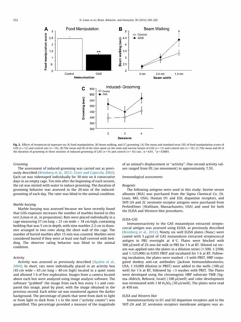
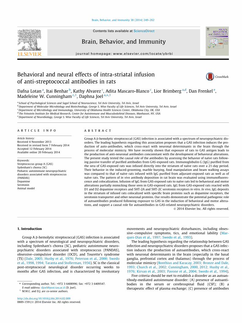
Fig. 2. Effects of streptococcal exposure on (A) food manipulation, (B) beam walking, and (C) grooming. (A) The mean and standard error (SE) of food manipulation scores ofGAS (n = 12) and control rats (n = 16). (B) The mean and SE of the time spent on the wide and narrow beams of GAS (n = 12) and control rats (n = 16). (C) The mean and SE ofthe duration of grooming in three sessions of induced-grooming of GAS (n = 9) and control (n = 16) rats. ⁄p < 0.01, ⁄⁄p < 0.0001.
252 D. Lotan et al. / Brain, Behavior, and Immunity 38 (2014) 249–262
GroomingThe assessment of induced-grooming was carried out as previ-
ously described (Brimberg et al., 2012; Greer and Capecchi, 2002).Each rat was videotaped individually for 30 min on 4 consecutivedays in an empty cage. Ten min after the beginning of each session,the rat was misted with water to induce grooming. The duration ofgrooming behavior was assessed in the 20 min of the induced-grooming of each day. The rater was blind to the animal condition.
Marble buryingMarble burying was assessed because we have recently found
that GAS-exposure increases the number of marbles buried in thistest (Lotan et al., in preparation). Rats were placed individually in acage measuring 37 cm long � 21 cm wide � 18 cm high, containingbedding that was 5 cm in depth, with nine marbles 2.3 cm in diam-eter arranged in two rows along the short wall of the cage. Thenumber of buried marbles after 15 min was counted. Marbles wereconsidered buried if they were at least one-half covered with bed-ding. The observer rating behavior was blind to the animalcondition.
ActivityActivity was assessed as previously described (Ayalon et al.,
2004). In short, rats were individually placed in an activity box(45 cm wide � 65 cm long � 40 cm high) located in a quiet roomand allowed 1 h of free exploration. Images from a camera locatedabove each box were analyzed using image analysis software. Thesoftware ‘‘grabbed’’ the image from each box every 1 s and com-pared this image, pixel by pixel, with the image obtained in theprevious second. Each white rat was monitored against the darkerbackground. The percentage of pixels that went from dark to lightor from light to dark from 1 s to the next (‘‘activity counts’’) wasquantified. This percentage provided a measure of the magnitude
of an animal’s displacement or ‘‘activity’’. One-second activity val-ues ranged from 0% (no movement) to approximately 7.5%.
Immunological assessments
ReagentsThe following antigens were used in this study: bovine serum
albumin (BSA) was purchased from the Sigma Chemical Co. (St.Louis, MO, USA). Human D1 and D2L dopamine receptors, and5HT-2A and 2C serotonin receptor antigens were purchased fromPerkinElmer (Waltham, Massachusetts, USA) and used for boththe ELISA and Western blot procedures.
ELISA-GASImmunoreactivity to the GAS mutanolysin extracted strepto-
coccal antigen was assessed using ELISA, as previously described(Brimberg et al., 2012). Ninety six well ELISA plates (Nunc) werecoated with 5 lg/ml of GAS mutanolysin extracted streptococcalantigen in PBS overnight at 4 �C. Plates were blocked with300 ll/well of 2% non-fat milk in PBS for 1 h at RT. Diluted rat ser-um was applied onto the plates in a dilution series (1:500, 1:2500,and 1:125,000) in 0.05% PBST and incubated for 1 h at RT. Follow-ing incubation, the plates were washed �3 with PBST. HRP conju-gated donkey anti-rat antibodies (Jackson Immunolaboratories,USA, 1:10,000 dilution in PBST) were added to the wells (100 ll/well) for 1 h at RT, followed by �3 washes with PBST. The Plateswere developed using the chromogenic HRP substrate TMB (Sig-ma–Aldrich, Rehovot, Israel) (100 ll/well) and color developmentwas terminated with 1 M H2SO4 (50 ll/well). The plates were readat 450 nm.
ELISA and Western blotImmunoreactivity to D1 and D2 dopamine receptors and to the
5HT-2A and 2C serotonin receptors membrane antigens was as-

Fig. 3. Effects of group A streptococcal (GAS) exposure on immunoreactivity of rat serum (IgG) with GAS antigen, dopamine receptors and serotonin receptors in the ELISA(A–E) and Western blot (F-I). ELISA results for anti-GAS serum IgG reactivity with: (A) GAS mutanolysin extracted antigen (GAS, n = 12; control, n = 10), (B) D1 dopaminereceptor (GAS, n = 11; control, n = 10), (C) D2 dopamine receptor (GAS, n = 13; control, n = 10), (D) 5HT-2A serotonin receptor (GAS, n = 12; control, n = 10), and (E) 5HT-2Cserotonin receptor (GAS, n = 12; control, n = 10), of sera taken from GAS and control rats, in comparison to reactivity with BSA. Western blot of pooled sera (IgG) reactivityfrom GAS-exposed rats compared with sera (IgG) from adjuvant-exposed control rats with: (F) D1 dopamine receptor, (G) D2 dopamine receptor, (H) 5HT-2A serotoninreceptor, and (I) 5HT-2C serotonin receptor. D1-PC = anti-D1 receptor positive control; D2-PC = anti-D2 receptor positive control; 5HT-2A-PC = anti-5HT-2A receptor positivecontrol; 5HT-2C-PC = anti-5HT-2C receptor positive control. ⁄p < 0.01, ⁄⁄p < 0.005, ⁄⁄⁄p < 0.0001.
D. Lotan et al. / Brain, Behavior, and Immunity 38 (2014) 249–262 253
sessed using ELISA and western blot analysis of sera obtained fromGAS-exposed rats:
ELISA. Ninety six well Immunolon 4 microtiter plates (DynatechLaboratories, Chantilly, VA) were coated with 10 lg/ml of eachantigen (D1, D2L, and 5HT-2A and 2C receptors membrane anti-gens; BSA served as a negative control protein) in 0.015 M carbon-ate buffer (pH 9.6) overnight at 4 �C. Plates were blocked with 1%BSA in PBS for 1 h. Diluted rat serum (50 ll/well) was applied ontothe plates in a dilution series and incubated overnight at 4 �C in 1%BSA in PBS. The next day, the plates were washed �3 with PBST.Alkaline phosphatase-conjugated rabbit anti-rat secondary anti-body specific to IgG (Sigma Chemical Co.) was added to the platesdiluted 1:1000 in 1% BSA–PBS. Plates were further incubated for1 h at room temperature and washed with PBST. Plates were devel-oped with 1 mg/ml p-nitrophenyl phosphate colorimetric sub-strate (Sigma Chemical Co.) and the optical density wasdetermined at 405 nm in a Opsys MR microplate reader (DynexTechnologies, Chantilly, VA). For the titers of antibodies against
the serotonin receptors 5HT-2A and 2C, the following dilutionswere performed: 1:200, 1:400, 1:800, 1:1600, 1:3200, 1:6400;D1: 1:300, 1:600; 1:1200, 1:4800, 1:9600; D2: 1:320, 1:640;1:1280, 1:5120, 1:10,240; BSA: 1:200, 1:400, 1:800, 1:1600. Titerswere determined as the last dilution resulting in an OD of 0.1.
Western blot. D1 and D2 dopamine receptors (Perkin Elmer) wereloaded as 10 lg per lane, and 5HT-2A and 2C serotonin receptors(abcam) were loaded as 1 lg per lane in the SDS PAGE gel. Antigenswere separated on a 7–10% SDS–PAGE gel using a 5% stacking geland separated antigens were electro-transferred onto the nitrocel-lulose membrane. Membranes were blocked overnight at 4 �C in 5%nonfat milk in Tris-Buffered Saline Tween-20 (TBST). To determinethe immunoreactivity of the sera to blotted proteins, individuallanes were separated into strips and incubated with a pool of serafrom the control or GAS-exposed rats, diluted in PBST (1:150) over-night at 4 �C. Rabbit anti-D1 receptor sera were diluted 1:1000(R&D systems); rabbit anti-D2 receptor sera were diluted 1:400(R&D systems); rabbit anti-5HT-2A receptor sera were diluted

Fig. 4. Effects of passive transfer of IgG from GAS-exposed and control rats to the striatum of naïve rats on (A) food manipulation, (B) beam walking, (C) grooming, and (D)marble burying. (A) The mean and standard error (SE) of food manipulation scores of rats infused with IgG extracted from GAS-exposed rats (GAS-I group, n = 8), rats infusedwith IgG extracted from control rats (Control-I group, n = 6) and naïve rats (n = 8). (B) The mean and SE of the time spent on the wide and narrow beams of GAS-I rats (n = 8),Control-I rats (n = 6) and naïve rats (n = 8). (C) The mean and SE of the duration of induced-grooming on three sessions of GAS-I rats (n = 8), Control-I rats (n = 5) and naïve rats(n = 8). (D) The mean and SE of the number of marbles buried by GAS-I rats (n = 8), control rats (n = 5) and naïve rats (n = 8). ⁄p < 0.05, ⁄⁄p < 0.01.
254 D. Lotan et al. / Brain, Behavior, and Immunity 38 (2014) 249–262
1:1000 (abcam); rabbit anti-5HT-2C receptor sera were diluted1:500 (abcam); all dilutions were used based on manufacturerguidelines. The strips were washed in PBST and incubated withHRP-conjugated donkey anti-rat antibodies or with HRP-conju-gated goat anti-rabbit antibodies (Jackson Immunolaboratories,USA) at dilutions of 1:5000. Blots were detected using the en-hanced chemiluminescence (ECL) Plus Western detection kit(Amersham Pharmacia Biotech). The bands developed from theimmunoblot were compared to the relative position of separatedproteins on Coomassie blue-stained immobilized nitrocellulosestrips. The relative molecular weight of proteins was determinedusing pre-stained molecular weight protein standards (Bio-Rad).
Preparation of tissue sectionsRats were overdosed with 100 mg/kg sodium pentobarbital, i.p,
and perfused intracardially with cold perfusion buffer. Brains wereremoved and placed in 4% paraformaldehyde over night, afterwhich they were cryoprotected in 30% sucrose solution for at least48 h. For immunostaining, brains were sectioned in the coronalplane at 16 lm for detecting IgG deposits, and sections weremounted on gelatin-coated slides. Slides were stored at �70 �C.
Immunostaining for IgG deposits and double stainingTo assess IgG deposits in the brain, 16 lm sections were incu-
bated in PBS until reaching room temperature. Sections were thenheated in citric acid (pH 6, 10 mM) for 5 min and were treated with0.5% Triton X-100 for 3 min. The sections were blocked (2% BSA
solution, 10% normal horse serum and 0.25% Triton X-100 in PBS)for 1 h in room temperature. Sections were then incubated for1 h with fluorescent Alexa 488 goat anti rat secondary antibody(1:250, Molecular Probes, Eugene, OR, USA) diluted in blockingsolution. Following Immunostaining for IgG deposits, the sectionswere incubated overnight at 4 �C with one of the following markerslabeled with mouse anti-neuron-selective NeuN (anti-neuronalnuclei, 1 : 250; Millipore), rabbit anti-Glial fibrillary acidic protein(GFAP, 1:400, Sigma), rabbit anti anti-D2 dopamine receptor(1:100, Millipore), rabbit anti-D1 dopamine receptor (1:300, Milli-pore), and rabbit anti-serotonin transporter (1:400, Millipore). Sec-tions were washed three times with PBS, and incubated withfluorescent Alexa 594 goat anti-mouse or anti-rabbit antibodies(1:250; molecular Probes) for 1 h at room temperature. Followingimmunostaining, sections were coverslipped with mounting mediawith DAPI (Vector Laboratories, Burlingame, USA) to counterstainthe nuclei. The imaging was carried out using a fluorescencemicroscopy (Nikon Eclipse 80i) and a NIS imaging software(Nikon).
Statistical analysis
We used two-sample Student’s t-tests to compare means of twoindependent groups. When more groups were compared, we usedanalysis of variance (ANOVA; the specific factors for each analysisare given in the Results section) together with post hoc LSD analy-sis (the specific analyses are detailed in the Results section). We

Fig. 5. Representative immunofluorescence images showing IgG deposition in the striatum of a GAS-I rat (A, D, G), Control-I rat (B, E, H) and naïve rat (C, F, I). (A–C) Labelingin the dorso-lateral striatum (DLS, the injection site); (D–F) labeling in the dorso-medial striatum (DMS); (G–I) labeling in the ventral striatum (VS). Tissue sections wereincubated with anti-rat alexa antibody 488 for visualization of IgG deposition; Blue signal indicates nuclear counterstaining (DAPI). 10� microscope objective; scalebar = 200 lm. (For interpretation of the references to colour in this figure legend, the reader is referred to the web version of this article.)
D. Lotan et al. / Brain, Behavior, and Immunity 38 (2014) 249–262 255
considered values as significant when p < 0.05. All data are pre-sented as means ± SEM.
Results
Effects of exposure to GAS antigen
Altered behavior in Lewis rats exposed to GAS antigenFig. 2A–C presents the behavior of GAS-exposed and control
(adjuvant-exposed) rats in the food manipulation, beam walkingand induced-grooming assays. GAS-exposed rats were impairedin manipulating food (Fig. 2A, t(26) = 5.726, p < 0.0001), and in tra-versing the narrow (1.5 cm) but not the wider (2.5 and 5.0 cm)beams, as evident in a longer time to traverse the beam (Fig. 2B,Exposure � Beam Width mixed ANOVA: Exposure:F(1,26) = 3.484, p = 0.0733; Beam Width: F(2,52) = 20.814,p < 0.0001; Exposure � Beam Width interaction: F(2,52) = 2.479,p = 0.0936; see Fig. 2B for the results of the post hoc comparisons).There were no differences between the groups in the number offoot slips (data not shown, p’s > 0.3). In addition, GAS rats exhibitedincreased grooming in the induced-grooming assay (Fig. 2C, Expo-sure � Session mixed ANOVA: Exposure: F(1,23) = 5.145, p < 0.05;Session: F(2,46)=2.818, p = 0.0701; Exposure � Session interaction:F(2,46) = 0.548, p = 0.5821). GAS rats did not have any gross indica-tions of skin lesions, nor did they show loss of hair, which couldindicate an increase in spontaneous grooming.
Sera from GAS-exposed rats reacted with dopamine and serotoninreceptors in the ELISA and Western immunoblot
As expected, IgG antibodies in sera of GAS-exposed rats weresignificantly elevated against the GAS cell wall antigen, the immu-nogen (Fig. 3A, F(1,20) = 99.53, p < 0.0001). In addition, we found inthe ELISA (Fig. 3B and C) and confirmed in the Western blot (Fig. 3Fand G) that the streptococcal exposure led to induction of antibod-ies against the dopamine receptors D1 and D2, and that this reac-tivity was specific when compared to BSA (Fig. 3B, D1:Exposure � Protein mixed ANOVA: Exposure: F(1,38) = 5.519,p < 0.05; Protein: F(1,38) = 187.845, p < 0.0001; Exposure � Proteininteraction: F(1,38) = 3.095, p = 0.0866; Fig. 3C, D2: Expo-sure � Protein mixed ANOVA: Exposure: F(1,40) = 3.137,p = 0.082; Protein: F(1,40) = 97.616, p < 0.0001; Exposure � Proteininteraction: F(1,40) = 3.137, p = 0.082, see Fig. 3B and C for the re-sults of the post hoc analysis). In addition to replicating our previ-ous findings (Brimberg et al., 2012), we have found in the ELISA(Fig. 3D and E) and confirmed in the Western blot (Fig. 3H and I),elevated antibodies against the 5HT-2A and the 5HT-2C serotoninreceptors. This reactivity was specific when compared to BSA(Fig. 3D, 5HT-2A: Exposure � Protein mixed ANOVA: Exposure:F(1,39) = 12.232, p < 0.005; Protein: F(1,39) = 133.090, p < 0.0001;Exposure � Protein interaction: F(1,39) = 3.919, p = 0.0548;Fig. 3E, 5HT-2C: Exposure � Protein mixed ANOVA: Exposure:F(1,38) = 8.197, p < 0.01; Protein: F(1,38) = 125.484, p < 0.0001;Exposure � Protein interaction: F(1,38) = 8.197, p < 0.01, seeFig. 3D and E for the results of the post hoc analysis).

Fig. 6. Representative immunofluorescence images taken from the dorso-lateral striatum (DLS, the injection site) of a GAS-I rat (A–B, E–F, I–J, M–N, Q–R), and a control-I rat(C–D, G–H, K–L, O–P, S–T) showing labeling with markers for neurons (NeuN, A–D), D1 dopamine receptor (E–H), D2 dopamine receptor (I–L), serotonin transporter (SERT, M–P), and astrocytes (GFAP, Q–T), and colocalization of these markers with IgG deposition (B, D, F, H, J, L, N, P, R, T). Tissue sections were incubated with anti-rat alexa antibody488 for visualization of IgG deposition; IgG against NeuN was labeled with anti-mouse alexa antibody 594; D1 and D2 dopamine receptors, SERT and GFAP were labeled withanti-rabbit alexa antibody 594. Blue signal indicates nuclear counterstaining (DAPI). 20� microscope objective; scale bar = 100 lm. The arrows point to colocalizationbetween the infused IgG and the relevant marker. (For interpretation of the references to colour in this figure legend, the reader is referred to the web version of this article.)
256 D. Lotan et al. / Brain, Behavior, and Immunity 38 (2014) 249–262
Effects of intra-striatal passive transfer of purified total IgG from seraof GAS-exposed rats
Increased compulsive-like behavior and motor deficits in recipient ratsFig. 4 presents the behavior of naïve rats, of rats intra-striatally
infused with total IgG purified from GAS-exposed rats (GAS-I), and
of rats intra-striatally infused with total IgG purified from adju-vant-exposed rats (Control-I) in the food manipulation (Fig. 4A),beam walking (Fig. 4B), induced-grooming (Fig. 4C) and marble-burying assays (Fig. 4D). There were no significant differencesbetween the three groups in the time spent grooming in the in-duced-grooming test (Fig. 4C, Condition � Session mixed ANOVA:

Fig. 7. Representative immunofluorescence images showing IgG deposition in the striatum of a GAS-I rat (A–C), Control-I rat (D–F) and naïve rat (G-I) and double labelingwith a marker for neurons (NeuN). Figures A, D and G display labeling of rat IgG; Figures B, E and H display NeuN labeling; Figures C, F and I display colocalization of the ratsIgG and NeuN. Tissue sections were incubated with anti-rat alexa antibody 488 for visualization of IgG deposition; IgG against NeuN was labeled with anti-mouse alexaantibody 594. Blue signal indicates nuclear counterstaining (DAPI). 40�microscope objective; scale bar = 25 lm. The arrows point to colocalization between the infused IgGand NeuN. (For interpretation of the references to colour in this figure legend, the reader is referred to the web version of this article.)
D. Lotan et al. / Brain, Behavior, and Immunity 38 (2014) 249–262 257
all p’s > 0.4), and both the GAS-I and Control-I groups were im-paired in manipulating food compared to the naïve group(Fig. 4A, Condition � Session mixed ANOVA: Condition:F(2,19) = 11.132, p < 0.0005; Session: F(4,38) = 2.008, p = 0.1482;Condition � Session interaction: F(4,38) = 4.723, p < 0.005, seeFig. 4A for the results of the post hoc analysis), suggesting a non-specific effect of the intra-striatal infusion. In contrast, the GAS-Igroup performed significantly different from the Control-I andnaïve groups in the beam walking and marble burying assays. Spe-cifically, on the last session of beam walking, GAS-I rats requiredmore time to traverse the narrow (1.5 cm) but not the wider (2.5and 5 cm) beams, compared to the naïve and Control-I groupswhich performed similarly (Fig. 4B). This difference reflects theshortening of the time to traverse the narrow beam over sessionsin the naïve and Control-I groups, which was not evident in theGAS-I group (Fig. 4B, Condition� Beam Width � Session mixedANOVA: Condition: F(2,19) = 2.64, p = 0.0974; Beam Width:F(2,38) = 97.423, p < 0.0001; Session, F(2,38) = 19.641, p < 0.0001;Condition � Beam Width, F(4,38) = 3.411, p < 0.05; Condi-tion � Session: F(4,38) = 3.808, p < 0.05; Session � Beam Width:F(4,76) = 0.802, p = 0.5277; Condition � Beam Width � Session:F(8,76) = 0.765, p = 0.6342, see Fig. 4B for the results of the posthoc analysis). A similar pattern of results was evident in the num-ber of falls (data not shown). In addition, rats in the GAS-I groupburied more marbles compared to the Control-I and naïve groups,which did not differ (Fig. 4D, F(2,18) = 5.250, p < 0.05, see Fig. 4Dfor the results of the post hoc analysis). The increased burying inthe GAS-I group was not a result of a non-selective increase inbehavioral output, as no significant differences were found be-
tween the three groups in activity level (in fact, the naïve grouptended to be more active, ANOVA: F(2,19) = 3.432, p = 0.0534, datanot shown).
Immunofluorescence of IgG deposits and colocalization with specificbrain proteins
Using an immunohistochemical analysis, we assessed the pat-tern of in vivo IgG reactivity in the striatum. We have found IgGlabeling in neurons in the striatum of GAS-I rats, in comparisonto a much weaker IgG pattern in the Control-I rats. No IgG was ob-served in the striatum of naïve rats (Fig. 5). DAPI staining of cell nu-clei overlaid the IgG reactivity in the figures.
Double immunolabeling shows that IgG clusters in GAS-I ratscolocalized with NeuN (labeling neurons, Figs. 6B and 7C), withthe D1 (Figs. 6F and 8C) and D2 (Figs. 6I and 9C) dopamine recep-tors and with the serotonin transporter (Figs. 6N and 10C). Muchless staining was observed in the Control-I rat brains (Figs. 6D, H,L, P and 7F, 8F, 9F and 10F). Interestingly, double immunolabelingof GFAP (labeling astrocytes) and IgG (Fig. 6Q–T) revealed thatastrocytes encircled the IgG clusters in the striatum of GAS-I rats(Figs. 6R and 11C), whereas this pattern was not observed in Con-trol-I rats (Figs. 6T and 11F).
Discussion
Our study evaluated the role of antibodies produced followingexposure of rats to GAS in the induction of neuropsychiatric abnor-malities. The first part of the study replicated and extended ourprevious findings on the behavioral and biological alterations fol-

Fig. 8. Representative immunofluorescence images showing IgG deposition in the striatum of a GAS-I rat (A–C), Control-I rat (D–F) and naïve rat (G–I) and double labelingwith anti-D1 dopamine receptor antibody. Figures A, D and G display labeling of rat IgG; Figures B, E and H display labeling of the D1 dopamine receptor; Figures C, F and Idisplay colocalization of the rats IgG and the D1 dopamine receptor. Tissue sections were incubated with anti-rat alexa antibody 488 for visualization of IgG deposition; IgGagainst the D1 dopamine receptor was labeled with anti-rabbit alexa antibody 594. Blue signal indicates nuclear counterstaining (DAPI). 40� microscope objective; scalebar = 25 lm. The arrows point to colocalization between the infused IgG and D1. (For interpretation of the references to colour in this figure legend, the reader is referred tothe web version of this article.)
258 D. Lotan et al. / Brain, Behavior, and Immunity 38 (2014) 249–262
lowing exposure of rats to GAS extract (Brimberg et al., 2012; Coxet al., 2013). Behaviorally, GAS-exposed rats demonstrated in-creased grooming in the induced-grooming assay, suggestive ofcompulsive-like behavior, and were impaired in the food manipu-lation and beam walking tasks, suggesting impairments in fine mo-tor control and gait, respectively. It should be noted, however, thatthe increased time it took GAS rats to traverse the narrow but notthe wider beams in the beam-walking assay, may reflect increasedanxiety of GAS rats rather than an impairment in gait. While wehave previously found that GAS rats bury more marbles (Lotanet al., unpublished observations), a behavior which has been sug-gested to reflect both increased compulsive-like and anxiety-likebehavior (for review see, Albelda and Joel, 2012), we did not finda significant difference between GAS and control rats in the pro-portion of time spent in the open arms of a plus-maze (p = 0.7,Brimberg et al., 2012), a commonly used test of anxiety-like behav-ior. Immunologically, sera from GAS-exposed rats reacted with theD1 and D2 dopamine receptors, as we have previously reported(Brimberg et al., 2012). A novel finding of the present study is thatsera from GAS-exposed rats also reacted with the 5HT-2A and 2Cserotonin receptors. It should be noted that sera from adjuvant-ex-posed control rats exhibited background reactivity against the fourreceptors in the ELISA, but the higher receptor specific reactivity ofsera IgG from GAS rats was confirmed by Western blots, where theadjuvant control rat sera was negative and the anti-GAS sera recog-
nized receptor specific bands. The higher background in the controlsera in the ELISA but not in the Western blot may be expected forthese G protein-coupled receptors, because only in the Westernblot are these receptors completely separated from the membrane(Baragli et al., 2007; Renart et al., 1979; Reynolds and Tanford,1970; Towbin et al., 1979). The higher background in the ELISAmay therefore reflect either lower avidity antibodies in the controlsera that react with the receptor–membrane complex, or immunecomplexes from the immunization that may also create nonspecificbinding. Our conclusion is supported by the lack of significant dif-ferences between reactivity of sera from adjuvant-exposed ratsand naïve rats against D1 and D2 dopamine receptors in the ELISA(p’s > 0.14, Brimberg et al., unpublished observations) and by thepresent findings of very low binding of IgG in the striatum of ratsthat were infused with IgG from adjuvant-exposed rats comparedwith the IgG binding in the striatum of rats that were infused withIgG from GAS-exposed rats.
In the second part of our study, antibodies purified from GAS-exposed rats and infused into the striatum of naïve rats (GAS-I)were found to replicate some of the behavioral effects of exposureto GAS. Specifically, on the last session of testing, GAS-I rats wereimpaired in traversing a narrow, but not a wide, beam, comparedto naïve rats and to rats infused with antibodies purified fromadjuvant-treated control rats (Control-I). The specificity of theimpairment to the narrow beam, seen also in GAS-exposed rats

Fig. 9. Representative immunofluorescence images showing IgG deposition in the striatum of a GAS-I rat (A–C), Control-I rat (D–F) and naïve rat (G–I) and double labelingwith anti-D2 dopamine receptor antibody. Figures A, D and G display labeling of rat IgG; Figures B, E and H display labeling of the D2 dopamine receptor; Figures C, F and Idisplay colocalization of the rats IgG and the D2 dopamine receptor. Tissue sections were incubated with anti-rat alexa antibody 488 for visualization of IgG deposition; IgGagainst the D2 dopamine receptor was labeled with anti-rabbit alexa antibody 594. Blue signal indicates nuclear counterstaining (DAPI). 40� microscope objective; scalebar = 25 lm. The arrows point to colocalization between the infused IgG and D2. (For interpretation of the references to colour in this figure legend, the reader is referred tothe web version of this article.)
Fig. 10. Representative immunofluorescence images showing IgG deposition in the striatum of a GAS-I rat (A–C), Control-I rat (D–F) and naïve rat (G–I) and double labelingwith anti-serotonin transporter (SERT) antibody. Figures A, D and G display labeling of rat IgG; Figures B, E and H display labeling of SERT; Figures C, F and I displaycolocalization of the rats IgG and SERT. Tissue sections were incubated with anti-rat alexa antibody 488 for visualization of IgG deposition; IgG against the SERT was labeledwith anti-rabbit alexa antibody 594. Blue signal indicates nuclear counterstaining (DAPI). 40� microscope objective; scale bar = 25 lm. The arrows point to colocalizationbetween the infused IgG and SERT. (For interpretation of the references to colour in this figure legend, the reader is referred to the web version of this article.)
D. Lotan et al. / Brain, Behavior, and Immunity 38 (2014) 249–262 259
in the present and previous (Brimberg et al., 2012) studies, sug-gests that the effect of anti-GAS antibodies was specific to gait,while sparing motivation and gross motor control. However, as de-tailed above, the increased time to traverse the narrow beam may
have reflected increased anxiety of GAS-I rats rather than a motorimpairment. Because the difference between the GAS-I and the twocontrol groups was due to the shortening of the time to traversethe narrow beam in the latter groups, it is possible that the differ-

Fig. 11. Representative immunofluorescence images showing IgG deposition in the striatum of a GAS-I rat (A–C), Control-I rat (D–F) and naïve rat (G–I) and double labelingwith a marker for astrocytes (GFAP). Figures A, D and G display labeling of rat IgG; Figures B, E and H display GFAP labeling; Figures C, F and I display colocalization of the ratsIgG and GFAP. Figures Tissue sections were incubated with anti-rat alexa antibody 488 for visualization of IgG deposition; IgG against GFAP was labeled with anti-rabbit alexaantibody 594. Blue signal indicates nuclear counterstaining (DAPI). 40�microscope objective; scale bar = 25 lm. (For interpretation of the references to colour in this figurelegend, the reader is referred to the web version of this article.)
260 D. Lotan et al. / Brain, Behavior, and Immunity 38 (2014) 249–262
ence reflects impaired motor learning of GAS-I rats. Alternatively,this difference may reflect the gradual development of behavioralalteration in GAS-I rats following the accumulation of antibodies,which counteracted the effects of training. A selective and specificeffect of IgG infusion was also seen in the marble burying assay.GAS-I rats buried more marbles compared to Control-I and naïverats, and this increased burying could not be attributed to a non-specific increase in activity level, because the latter was not signif-icantly different between the three groups (in fact, the two infusedgroups tended to be less active than the naïve group). As we haverecently found increased marble burying also in GAS-exposed rats(Lotan et al., in preparation), these results suggest that anti-GASantibodies caused a specific increase in marble burying. GAS-I ratsdid not show, however, increased grooming in the induced-groom-ing assay, in contrast to GAS-exposed rats in the present and pre-vious (Brimberg et al., 2012) studies. In addition, although GAS-Irats were impaired in manipulating food compared to naïve rats,so were Control-I rats, suggesting a non-specific effect of the in-tra-striatal infusion in this task. One of the limitations of our studyis that antibodies were passively transferred intra-striatally whichin itself may have led to some non-specific effects due either tomechanical disruption of striatal functioning following infusionof fluids, or an effect of the infusion of IgG, as Control-I rats werealso infused with antibodies (against the adjuvant mycobacteriainjected into the rats from which sera were collected). Taken to-gether, our new evidence demonstrates that intra-striatal infusionof antibodies (IgG) purified from GAS-exposed rats mimics part ofthe behavioral syndrome that is induced by exposing rats to GASantigen, and that some of these behavioral effects are specific toanti-GAS antibodies.
Our results are in line with previous studies, which reported theemergence of stereotypic behaviors and motor abnormalitiesfollowing infusion of either sera or IgG purified from sera of SC,PANDAS or TS patients to the striatum of naïve rats (Doyle et al.,2012; Hallett et al., 2000; Taylor et al., 2002). Yet, for reasons cur-rently unknown, other studies that used a similar approach failedto induce behavioral alterations (Ben-Pazi et al., 2012; Loiselleet al., 2004; Singer et al., 2005). See (Doyle et al., 2012; Singeret al., 2005) for a discussion of possible causes for the inconsistentresults in these studies. Our results are also in line with the findingthat systemic passive transfer of anti-streptococcal sera from GAS-exposed mice to naïve mice (concomitant with Lipopolysaccha-rides (LPS) administration to break the blood brain barrier) re-sulted in the development of motor and behavioral disturbancessimilar to those seen in the GAS-exposed mice (Yaddanapudiet al., 2010). Taken together with our present results, these collec-tive studies support a role for antibodies in the induction of behav-ioral alterations.
Our results further suggest a role for antibodies that target pro-teins involved in the functioning of the dopaminergic and seroto-nergic systems. Specifically, we have found anti-D1 and D2dopamine receptor antibodies, as well as anti-5HT-2A and 2C sero-tonin receptor antibodies in the sera of GAS-exposed rats. Severallines of evidence point to the involvement of these neurotransmit-ter systems in the pathophysiology of GAS-related neuropsychiat-ric disorders. Dysfunction of the dopaminergic system has beenimplicated in motor disturbances (e.g., Parkinson’s disease, Hun-tington’s disease, and Alzheimer’s disease (Jahanshahi et al.,2013; Michel et al., 2013; Reeves et al., 2010) as well as in OCD(Denys et al., 2004; Hesse et al., 2005; Nikolaus et al., 2010; Olveret al., 2009), and dysfunction of the serotonergic system has been

D. Lotan et al. / Brain, Behavior, and Immunity 38 (2014) 249–262 261
implicated in OCD and anxiety disorders (Flaisher-Grinberg et al.,2008; Hesse et al., 2005, 2011; Nikolaus et al., 2010; Perani et al.,2008). Dopaminergic and serotonergic drugs are used to treat mo-tor and psychiatric symptoms, respectively, in patients affected byGAS-related neuropsychiatric disorders (Demiroren et al., 2007;Gabbay and Coffey, 2003; Moretti et al., 2008; Murphy et al.,2010; Swedo and Grant, 2005; Swedo et al., 1993). Finally, immu-noreactivity to different components of the serotonergic system,such as serotonin and serotonin receptors, has been reported insera from panic disorder and rheumatoid arthritis patients (Coplanet al., 1999; Maes et al., 2013; Schott et al., 2003; Tanaka et al.,2003), and sera from SC and PANDAS patients reacted with D1and D2 receptors (Brimberg et al., 2012; Cox et al., 2013; Daleet al., 2012). Interestingly, Dale et al. (2012) have recently foundantibodies against surface D2 receptors in the sera of patients suf-fering from basal ganglia encephalitis, which is characterized bymotor (including, parkinsonism, dystonia and chorea), and psychi-atric symptoms (including, emotional lability, attention deficit andpsychosis), which are also found in GAS-related neuropsychiatricdisorders.
The pattern of IgG deposition in the striatum of GAS-I rats alsosupports the hypothesis that antibodies that target the dopaminer-gic and the serotonergic systems play a functional role in theinduction of behavioral alterations. Specifically, IgG purified fromGAS-exposed rats and infused into the striatum of naïve rats werecolocalized with neurons, with cells expressing D1 dopaminereceptors, with cells expressing D2 dopamine receptors and withcells expressing the serotonin transporter. Astrocytes encircledclusters of neurons containing IgG.
Colocalization of IgG with neurons in the striatum is in accor-dance with previous studies showing that antibodies found in seraof GAS-related neuropsychiatric disorders, such as SC, PANDAS,OCD, and TS, reacted with basal-ganglia neurons (Church et al.,2002; Cox et al., 2013; Gause et al., 2009; Husby et al., 1976; Kies-sling et al., 1993; Kotby et al., 1998; Martino et al., 2011; Singeret al., 1998). There is less data regarding a possible role of astro-cytes in GAS-related neuropsychiatric disorders. Kingston andGlynn (1971) found a cross-reaction of sera taken from rabbits pre-viously exposed to GAS with astrocytes. It is possible that astro-cytes encircled the antibody neuronal cell cluster in order todelimit their effect or remove them from the brain tissue.
Our results suggest a potential pathogenic role of autoantibod-ies produced following exposure to GAS in the induction of behav-ioral alterations, reminiscent of the outcomes of exposure to GAS inboth patients and rats, thus establishing the fifth condition re-quired to establish a disorder as an antibody-mediated autoim-mune disorder. Our evidence illustrates induction of symptomsby intra-striatal passive transfer of IgG to animals. Additional stud-ies are required in order to assess the exact mechanisms by whichthe autoantibodies produced in GAS-related neuropsychiatric dis-orders are pathogenic.
Financial disclosures
M.W.C. is Chief Scientific Officer with a financial interest inMoleculera Labs which provides diagnostic testing of children withautoimmune neuropsychiatric and movement disorders. Molecul-era Labs resides at the University of Oklahoma Health SciencesCenter and Presbyterian Health Foundation Research Park in Okla-homa City, OK. M.W.C. has research funding from National Insti-tutes of Health (Grant No. HL56267), from the National Institutesof Mental Health (Bench to Bedside Grant) and from OklahomaCenter for the Advancement of Science and Technology. D.J. has re-search funding from the Israel Science Foundation (Grant No. 341/07). All other authors assert that none has any commercial or
financial involvements that might present an appearance of a con-flict of interest in connection with the submitted manuscript.
Acknowledgments
We thank Dr. Dan Frenkel and his laboratory members, HilitLevy-Barazany, Dorit Trudler and Dorit Farfara (Department ofNeurobiology, Tel-Aviv University, Israel) for their assistance inthe immunohistochemical analyses. This research was supportedby the Israel Science Foundation (Grant No. 341/07) to D.J. and Na-tional Institutes of Health Grant HL56267, Bench to Bedside Grantfrom the National Institutes of Mental Health, and the OklahomaCenter for the Advancement of Science and Technology Grant toM.W.C.
References
Albelda, N., Joel, D., 2012. Current animal models of obsessive compulsive disorder:an update. Neuroscience 211, 83–106.
Archelos, J.J., Hartung, H.P., 2000. Pathogenetic role of autoantibodies inneurological diseases. Trends Neurosci. 23, 317–327.
Ayalon, L., Doron, R., Weiner, I., Joel, D., 2004. Amelioration of behavioral deficits ina rat model of Huntington’s disease by an excitotoxic lesion to the globuspallidus. Exp. Neurol. 186, 46–58.
Baragli, A., Alturaihi, H., Watt, H.L., Abdallah, A., Kumar, U., 2007.Heterooligomerization of human dopamine receptor 2 and somatostatinreceptor 2 – co-immunoprecipitation and fluorescence resonance energytransfer analysis. Cell. Signal. 19, 2304–2316.
Barsottini, O.G., Ferraz, H.B., Seviliano, M.M., Barbieri, A., 2002. Brain SPECT imagingin Sydenham’s chorea. Braz. J. Med. Biol. Res. 35, 431–436.
Ben-Pazi, H., Sadan, O., Offen, D., 2012. Striatal microinjection of Sydenham choreaantibodies: using a rat model to examine the dopamine hypothesis. J. Mol.Neurosci. 46, 162–166.
Bonthius, D.J., Karacay, B., 2003. Sydenham’s chorea: not gone and not forgotten.Semin. Pediatr. Neurol. 10, 11–19.
Brimberg, L., Benhar, I., Mascaro-Blanco, A., Alvarez, K., Lotan, D., Winter, C., Klein, J.,Moses, A.E., Somnier, F.E., Leckman, J.F., Swedo, S.E., Cunningham, M.W., Joel, D.,2012. Behavioral, pharmacological, and immunological abnormalities afterstreptococcal exposure: a novel rat model of Sydenham chorea and relatedneuropsychiatric disorders. Neuropsychopharmacology 37, 2076–2087.
Bronze, M.S., Dale, J.B., 1993. Epitopes of streptococcal M proteins that evokeantibodies that cross-react with human brain. J. Immunol. 151, 2820–2828.
Carter, R.J., Lione, L.A., Humby, T., Mangiarini, L., Mahal, A., Bates, G.P., Dunnett, S.B.,Morton, A.J., 1999. Characterization of progressive motor deficits in micetransgenic for the human Huntington’s disease mutation. J. Neurosci. 19, 3248–3257.
Church, A.J., Cardoso, F., Dale, R.C., Lees, A.J., Thompson, E.J., Giovannoni, G., 2002.Anti-basal ganglia antibodies in acute and persistent Sydenham’s chorea.Neurology 59, 227–231.
Citak, E.C., Gucuyener, K., Karabacak, N.I., Serdaroglu, A., Okuyaz, C., Aydin, K., 2004.Functional brain imaging in Sydenham’s chorea and streptococcal tic disorders.J. Child Neurol. 19, 387–390.
Coplan, J.D., Tamir, H., Calaprice, D., DeJesus, M., de la Nuez, M., Pine, D., Papp, L.A.,Klein, D.F., Gorman, J.M., 1999. Plasma anti-serotonin and serotonin anti-idiotypic antibodies are elevated in panic disorder. Neuropsychopharmacology20, 386–391.
Cox, C.J., Sharma, M., Leckman, J.F., Zuccolo, J., Zuccolo, A., Kovoor, A., Swedo, S.E.,Cunningham, M.W., 2013. Brain human monoclonal autoantibody fromSydenham chorea targets dopaminergic neurons in transgenic mice andsignals dopamine D2 receptor: implications in human disease. J. Immunol.191, 5524–5541.
Cunningham, M.W., 2000. Pathogenesis of group A streptococcal infections. Clin.Microbiol. Rev. 13, 470–511.
Cunningham, M.W., 2012. Streptococcus and rheumatic fever. Curr. Opin.Rheumatol. 24, 408–416.
Dale, R.C., 2005. Post-streptococcal autoimmune disorders of the central nervoussystem. Dev. Med. Child Neurol. 47, 785–791.
Dale, R.C., Merheb, V., Pillai, S., Wang, D., Cantrill, L., Murphy, T.K., Ben-Pazi, H.,Varadkar, S., Aumann, T.D., Horne, M.K., Church, A.J., Fath, T., Brilot, F., 2012.Antibodies to surface dopamine-2 receptor in autoimmune movement andpsychiatric disorders. Brain 135, 3453–3468.
Demiroren, K., Yavuz, H., Cam, L., Oran, B., Karaaslan, S., Demiroren, S., 2007.Sydenham’s chorea: a clinical follow-up of 65 patients. J. Child Neurol. 22, 550–554.
Denys, D., van der Wee, N., Janssen, J., De Geus, F., Westenberg, H.G., 2004. Low levelof dopaminergic D2 receptor binding in obsessive–compulsive disorder. Biol.Psychiatry 55, 1041–1045.
Dilenge, M.E., Shevell, M.I., Dinh, L., 1999. Restricted unilateral Sydenham’s chorea:reversible contralateral striatal hypermetabolism demonstrated on singlephoton emission computed tomographic scanning. J. Child Neurol. 14, 509–513.

262 D. Lotan et al. / Brain, Behavior, and Immunity 38 (2014) 249–262
Doyle, F., Cardoso, F., Lopes, L., Mendes, M., Dias, F., Cruz, L., Tavares, R., Camargos,A., Carneiro, M., Dias-Lopes, C., Chavez-Olortegui, C., 2012. Infusion ofSydenham’s chorea antibodies in striatum with up-regulated dopaminergicreceptors: a pilot study to investigate the potential of SC antibodies to increasedopaminergic activity. Neurosci. Lett. 523, 186–189.
Flaisher-Grinberg, S., Klavir, O., Joel, D., 2008. The role of 5-HT2A and 5-HT2Creceptors in the signal attenuation rat model of obsessive–compulsive disorder.Int. J. Neuropsychopharmacol. 11, 811–825.
Gabbay, V., Coffey, B., 2003. Obsessive–compulsive disorder, Tourette’s disorder, orpediatric autoimmune neuropsychiatric disorders associated withStreptococcus in an adolescent? Diagnostic and therapeutic challenges. J.Child Adolesc. Psychopharmacol. 13, 209–212.
Garvey, M.A., Snider, L.A., Leitman, S.F., Werden, R., Swedo, S.E., 2005. Treatment ofSydenham’s chorea with intravenous immunoglobulin, plasma exchange, orprednisone. J. Child Neurol. 20, 424–429.
Gause, C., Morris, C., Vernekar, S., Pardo-Villamizar, C., Grados, M.A., Singer, H.S.,2009. Antineuronal antibodies in OCD: comparisons in children with OCD-only,OCD+chronic tics and OCD+PANDAS. J. Neuroimmunol. 214, 118–124.
Giedd, J.N., Rapoport, J.L., Garvey, M.A., Perlmutter, S., Swedo, S.E., 2000. MRIassessment of children with obsessive–compulsive disorder or tics associatedwith streptococcal infection. Am. J. Psychiatry 157, 281–283.
Greer, J.M., Capecchi, M.R., 2002. Hoxb8 is required for normal grooming behaviorin mice. Neuron 33, 23–34.
Hallett, J.J., Harling-Berg, C.J., Knopf, P.M., Stopa, E.G., Kiessling, L.S., 2000. Anti-striatal antibodies in Tourette syndrome cause neuronal dysfunction. J.Neuroimmunol. 111, 195–202.
Hesse, S., Muller, U., Lincke, T., Barthel, H., Villmann, T., Angermeyer, M.C., Sabri, O.,Stengler-Wenzke, K., 2005. Serotonin and dopamine transporter imaging inpatients with obsessive–compulsive disorder. Psychiatry Res. 140, 63–72.
Hesse, S., Stengler, K., Regenthal, R., Patt, M., Becker, G.A., Franke, A., Knupfer, H.,Meyer, P.M., Luthardt, J., Jahn, I., Lobsien, D., Heinke, W., Brust, P., Hegerl, U.,Sabri, O., 2011. The serotonin transporter availability in untreated early-onsetand late-onset patients with obsessive–compulsive disorder. Int. J.Neuropsychopharmacol. 14, 606–617.
Hoffman, K.L., Hornig, M., Yaddanapudi, K., Jabado, O., Lipkin, W.I., 2004. A murinemodel for neuropsychiatric disorders associated with group A beta-hemolyticstreptococcal infection. J. Neurosci. 24, 1780–1791.
Husby, G., van de Rijn, I., Zabriskie, J.B., Abdin, Z.H., Williams Jr., R.C., 1976.Antibodies reacting with cytoplasm of subthalamic and caudate nuclei neuronsin chorea and acute rheumatic fever. J. Exp. Med. 144, 1094–1110.
Huyser, C., Veltman, D.J., de Haan, E., Boer, F., 2009. Paediatric obsessive–compulsive disorder, a neurodevelopmental disorder? Evidence fromneuroimaging. Neurosci. Biobehav. Rev. 33, 818–830.
Jahanshahi, A., Vlamings, R., van Roon-Mom, W.M., Faull, R.L., Waldvogel, H.J.,Janssen, M.L., Yakkioui, Y., Zeef, D.H., Kocabicak, E., Steinbusch, H.W., Temel, Y.,2013. Changes in brainstem serotonergic and dopaminergic cell populations inexperimental and clinical Huntington’s disease. Neuroscience 238, 71–81.
Kiessling, L.S., Marcotte, A.C., Culpepper, L., 1993. Antineuronal antibodies inmovement disorders. Pediatrics 92, 39–43.
Kingston, D., Glynn, L.E., 1971. A cross-reaction between Str. pyogenes and humanfibroblasts, endothelial cells and astrocytes. Immunology 21, 1003–1016.
Kirvan, C.A., Swedo, S.E., Heuser, J.S., Cunningham, M.W., 2003. Mimicry andautoantibody-mediated neuronal cell signaling in Sydenham chorea. Nat. Med.9, 914–920.
Kirvan, C.A., Swedo, S.E., Snider, L.A., Cunningham, M.W., 2006. Antibody-mediatedneuronal cell signaling in behavior and movement disorders. J. Neuroimmunol.179, 173–179.
Kolb, B., Holmes, C., 1983. Neonatal motor cortex lesions in the rat: absence ofsparing of motor behaviors and impaired spatial learning concurrent withabnormal cerebral morphogenesis. Behav. Neurosci. 97, 697–709.
Kotby, A.A., El Badawy, N., El Sokkary, S., Moawad, H., El Shawarby, M., 1998.Antineuronal antibodies in rheumatic chorea. Clin. Diagn. Lab. Immunol. 5,836–839.
Linthicum, D.S., Munoz, J.J., Blaskett, A., 1982. Acute experimental autoimmuneencephalomyelitis in mice. I. Adjuvant action of Bordetella pertussis is due tovasoactive amine sensitization and increased vascular permeability of thecentral nervous system. Cell. Immunol. 73, 299–310.
Loiselle, C.R., Lee, O., Moran, T.H., Singer, H.S., 2004. Striatal microinfusion ofTourette syndrome and PANDAS sera: failure to induce behavioral changes.Mov. Disord. 19, 390–396.
Maes, M., Ringel, K., Kubera, M., Anderson, G., Morris, G., Galecki, P., Geffard, M.,2013. In myalgic encephalomyelitis/chronic fatigue syndrome, increasedautoimmune activity against 5-HT is associated with immuno-inflammatorypathways and bacterial translocation. J. Affect. Disord. 150 (2), 223–230.
Marques-Dias, M.J., Mercadante, M.T., Tucker, D., Lombroso, P., 1997. Sydenham’schorea. Psychiatr. Clin. North Am. 20, 809–820.
Martino, D., Chiarotti, F., Buttiglione, M., Cardona, F., Creti, R., Nardocci, N., Orefici,G., Veneselli, E., Rizzo, R., 2011. The relationship between group A streptococcalinfections and Tourette syndrome: a study on a large service-based cohort. Dev.Med. Child Neurol. 53, 951–957.
Michel, P.P., Toulorge, D., Guerreiro, S., Hirsch, E.C., 2013. Specific needs ofdopamine neurons for stimulation in order to survive: implication forParkinson disease. FASEB J. 27 (9), 3414–3423.
Moretti, G., Pasquini, M., Mandarelli, G., Tarsitani, L., Biondi, M., 2008. What everypsychiatrist should know about PANDAS: a review. Clin. Pract. Epidemiol. Ment.Health 4, 13.
Murphy, T.K., Kurlan, R., Leckman, J., 2010. The immunobiology of Tourette’sdisorder, pediatric autoimmune neuropsychiatric disorders associated withStreptococcus, and related disorders: a way forward. J. Child Adolesc.Psychopharmacol. 20, 317–331.
Nikolaus, S., Antke, C., Beu, M., Muller, H.W., 2010. Cortical GABA, striatal dopamineand midbrain serotonin as the key players in compulsive and anxiety disorders–results from in vivo imaging studies. Rev. Neurosci. 21, 119–139.
Olver, J.S., O’Keefe, G., Jones, G.R., Burrows, G.D., Tochon-Danguy, H.J., Ackermann,U., Scott, A., Norman, T.R., 2009. Dopamine D1 receptor binding in the striatumof patients with obsessive–compulsive disorder. J. Affect. Disord. 114, 321–326.
Pavone, P., Bianchini, R., Parano, E., Incorpora, G., Rizzo, R., Mazzone, L., Trifiletti,R.R., 2004. Anti-brain antibodies in PANDAS versus uncomplicatedstreptococcal infection. Pediatr. Neurol. 30, 107–110.
Paxinos, G., Watson, C., 1998. The Rat Brain in Stereotaxic Coordinates. AcademicPress, San Diego.
Perani, D., Garibotto, V., Gorini, A., Moresco, R.M., Henin, M., Panzacchi, A.,Matarrese, M., Carpinelli, A., Bellodi, L., Fazio, F., 2008. In vivo PET study of5HT(2A) serotonin and D(2) dopamine dysfunction in drug-naive obsessive–compulsive disorder. Neuroimage 42, 306–314.
Perlmutter, S.J., Leitman, S.F., Garvey, M.A., Hamburger, S., Feldman, E., Leonard,H.L., Swedo, S.E., 1999. Therapeutic plasma exchange and intravenousimmunoglobulin for obsessive–compulsive disorder and tic disorders inchildhood. Lancet 354, 1153–1158.
Peterson, B.S., Leckman, J.F., Tucker, D., Scahill, L., Staib, L., Zhang, H., King, R., Cohen,D.J., Gore, J.C., Lombroso, P., 2000. Preliminary findings of antistreptococcalantibody titers and basal ganglia volumes in tic, obsessive–compulsive, andattention deficit/hyperactivity disorders. Arch. Gen. Psychiatry 57, 364–372.
Reeves, S., Mehta, M., Howard, R., Grasby, P., Brown, R., 2010. The dopaminergicbasis of cognitive and motor performance in Alzheimer’s disease. Neurobiol.Dis. 37, 477–482.
Renart, J., Reiser, J., Stark, G.R., 1979. Transfer of proteins from gels todiazobenzyloxymethyl-paper and detection with antisera – method forstudying antibody specificity and antigen structure. Proc. Natl. Acad. Sci. U. S.A. 76, 3116–3120.
Reynolds, J.A., Tanford, C., 1970. Binding of dodecyl sulfate to proteins at highbinding ratios – possible implications for state of proteins in biologicalmembranes. Proc. Natl. Acad. Sci. U. S. A. 66, 1002-&.
Schott, K., Schaefer, J.E., Richartz, E., Batra, A., Eusterschulte, B., Klein, R., Berg, P.A.,Bartels, M., Mann, K., Buchkremer, G., 2003. Autoantibodies to serotonin inserum of patients with psychiatric disorders. Psychiatry Res. 121, 51–57.
Singer, H.S., Giuliano, J.D., Hansen, B.H., Hallett, J.J., Laurino, J.P., Benson, M.,Kiessling, L.S., 1998. Antibodies against human putamen in children withTourette syndrome. Neurology 50, 1618–1624.
Singer, H.S., Mink, J.W., Loiselle, C.R., Burke, K.A., Ruchkina, I., Morshed, S., Parveen,S., Leckman, J.F., Hallett, J.J., Lombroso, P.J., 2005. Microinfusion of antineuronalantibodies into rodent striatum: failure to differentiate between elevated andlow titers. J. Neuroimmunol. 163, 8–14.
Swedo, S.E., Grant, P.J., 2005. Annotation: PANDAS: a model for human autoimmunedisease. J. Child Psychol. Psychiatry 46, 227–234.
Swedo, S.E., Leonard, H.L., Garvey, M., Mittleman, B., Allen, A.J., Perlmutter, S.,Lougee, L., Dow, S., Zamkoff, J., Dubbert, B.K., 1998. Pediatric autoimmuneneuropsychiatric disorders associated with streptococcal infections: clinicaldescription of the first 50 cases. Am. J. Psychiatry 155, 264–271.
Swedo, S.E., Leonard, H.L., Kiessling, L.S., 1994. Speculations on antineuronalantibody-mediated neuropsychiatric disorders of childhood. Pediatrics 93, 323–326.
Swedo, S.E., Leonard, H.L., Schapiro, M.B., Casey, B.J., Mannheim, G.B., Lenane, M.C.,Rettew, D.C., 1993. Sydenham’s chorea: physical and psychological symptomsof St Vitus dance. Pediatrics 91, 706–713.
Tanaka, S., Matsunaga, H., Kimura, M., Tatsumi, K., Hidaka, Y., Takano, T., Uema, T.,Takeda, M., Amino, N., 2003. Autoantibodies against four kinds ofneurotransmitter receptors in psychiatric disorders. J. Neuroimmunol. 141,155–164.
Taranta, A., Stollerman, G.H., 1956. The relationship of Sydenham’s chorea toinfection with group A streptococci. Am. J. Med. 20, 170–175.
Taylor, J.R., Morshed, S.A., Parveen, S., Mercadante, M.T., Scahill, L., Peterson, B.S.,King, R.A., Leckman, J.F., Lombroso, P.J., 2002. An animal model of Tourette’ssyndrome. Am. J. Psychiatry 159, 657–660.
Towbin, H., Staehelin, T., Gordon, J., 1979. Electrophoretic transfer of proteins frompolyacrylamide gels to nitrocellulose sheets – procedure and some applications.Proc. Natl. Acad. Sci. U. S. A. 76, 4350–4354.
Urakawa, S., Hida, H., Masuda, T., Misumi, S., Kim, T.S., Nishino, H., 2007.Environmental enrichment brings a beneficial effect on beam walking andenhances the migration of doublecortin-positive cells following striatal lesionsin rats. Neuroscience 144, 920–933.
Yaddanapudi, K., Hornig, M., Serge, R., De Miranda, J., Baghban, A., Villar, G., Lipkin,W.I., 2010. Passive transfer of streptococcus-induced antibodies reproducesbehavioral disturbances in a mouse model of pediatric autoimmuneneuropsychiatric disorders associated with streptococcal infection. Mol.Psychiatry 15, 712–726.

![[18F]Fluorodopa PETshows striatal dopaminergic dysfunction ...](https://static.fdocuments.us/doc/165x107/628e71a806be7c7a267428b6/18ffluorodopa-petshows-striatal-dopaminergic-dysfunction-.jpg)