Bedside Diagnosis for Infectious Diseases...Bedside Diagnosis for Infectious Diseases Outline Useful...
49
Sasisopin Kiertiburanakul, MD, MHS Associate Professor Division of Infectious Diseases Department of Medicine Faculty of Medicine Ramathibodi Hospital Mahidol University RCPT Rotating, Prae (Feb 8, 2012) Bedside Diagnosis for Infectious Diseases
Transcript of Bedside Diagnosis for Infectious Diseases...Bedside Diagnosis for Infectious Diseases Outline Useful...
Sasisopin Kiertiburanakul, MD, MHS Associate Professor
Division of Infectious Diseases
Department of Medicine
Faculty of Medicine Ramathibodi Hospital
Mahidol University
RCPT Rotating, Prae (Feb 8, 2012)
Bedside Diagnosis for
Infectious Diseases
Outline
Useful bedside methods
Basic and advance laboratory tests
Cases presentation
Spot diagnosis

Evaluation of Infectious Disease Case
Define if infection occur
History
Travel and exposure history
Social and family history
Physical examination
Laboratory investigations
Basic
Advance

Patients’ Evaluation
Who: Which kind of patient has infection?
Determine immunosuppressive status
When: Is an environmental factor influence the
disease occurrence?
Where: Which part of body?
What: What kind of the organism?
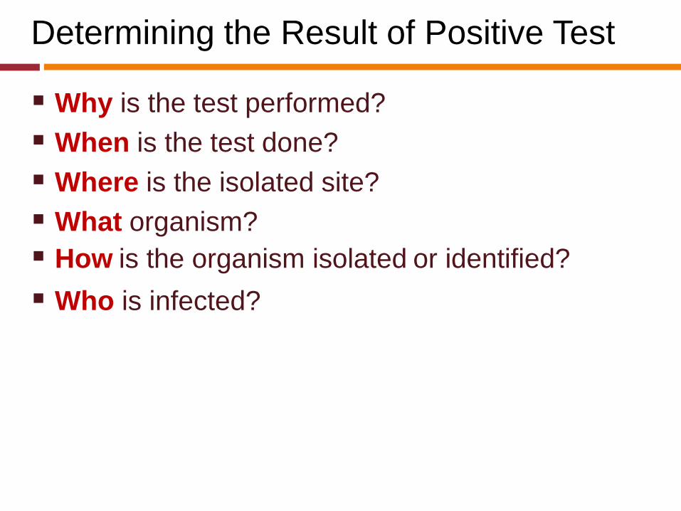
Determining the Result of Positive Test
Why is the test performed?
When is the test done?
Where is the isolated site?
What organism?
How is the organism isolated or identified?
Who is infected?

Non-specific signs
Lymphadenopathy
Hepatomegaly
Splenomegaly
……………
Acute fever
Non-specific
symptoms
Myalgia
Nausea/vomiting
……….
Systemic
infection
Acute Febrile Illness

Bacterial Infection by Site
Mouth Peptococcus Peptostreptococcus Actinomyces
Skin/Soft Tissue S. aureus S. pyogenes S. epidermidis Pasteurella
Bone and Joint S. aureus S. epidermidis Streptococci N. gonorrhoeae Gram-negative rods
Abdomen E. coli, Proteus Klebsiella Enterococcus Bacteroides sp.
Urinary Tract E. coli, Proteus Klebsiella Enterococcus Staph saprophyticus
Upper Respiratory S. pneumoniae H. influenzae M. catarrhalis S. pyogenes
Lower Respiratory Community S. pneumoniae H. influenzae K. pneumoniae Legionella pneumophila Mycoplasma, Chlamydia
Lower Respiratory Hospital K. pneumoniae P. aeruginosa Enterobacter sp. Serratia sp. S. aureus
Meningitis S. pneumoniae N. meningitidis H. influenza Group B Strep E. coli Listeria

Case Presentation

Case 1
PF: ชายไทยค อาย 65 ป อาชพ ขาราชการบ านาญ ภมล าเนา กรงเทพมหานคร
CC: ไข 3 วนกอนมารพ. ถกสงตวมาจากรพ. เอกชน PI: 7 วนกอนมารพ. ออนเพลย รบประทานอาหารไดนอย ไมมไข ไมไอ ไมเจบคอ ไมปวดทอง ถายปสสาวะและอจจาระปกต ไปรพ.แหงหนงไดยารกษากระเพาะอาหารอกเสบ อาการไมดขน
3 วนกอนมารพ. มไขสงหนาวสน คลนไส อาเจยน ปวดทองบรเวณลนป นอนรกษาทรพ. น 5 วน

Past and Personal History
No contact with person who had flu-liked symptoms
2 weeks PTA, he traveled to the orchid garden
in Chiang Rai and stayed there for 3-4 days
Hypertension and hyperlipidemia 15 years
Enalapril, simvastatin, gemfibrozil

Investigations
CBC: Hct 39.7%, WBC 4,700 (N 80, L 12, Atyp L
1, Band 7), Plt 75,000/mm3
BUN 45, Cr 2.3 mg/dL
Liver function test
AST 155 , ALT 88 , ALP 270, GGT 265 U/L
TB 1.5, DB 0.8 mg/dL, TP 7.3, alb 3.8 g/L

What is the Most Likely Diagnosis?
A. Dengue (hemorrhagic) fever
B. Rickettsioses
C. Leptospirosis
D. Malaria
E. Bacterial sepsis
(Gram negative/positive)
65 years, acute fever with systemic symptoms
History travel to Chiang Rai
Lab: thrombocytopenia, AKI, abnormal LFT

Further Investigations
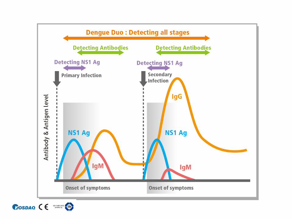
Dengue Rapid Test
IgM: positive
IgG: negative
NS1: negative
U/S upper abdomen
Normal thin wall GB with
gall stones
Small pericholecystic fluid
Diagnosis: Severe dengue infection
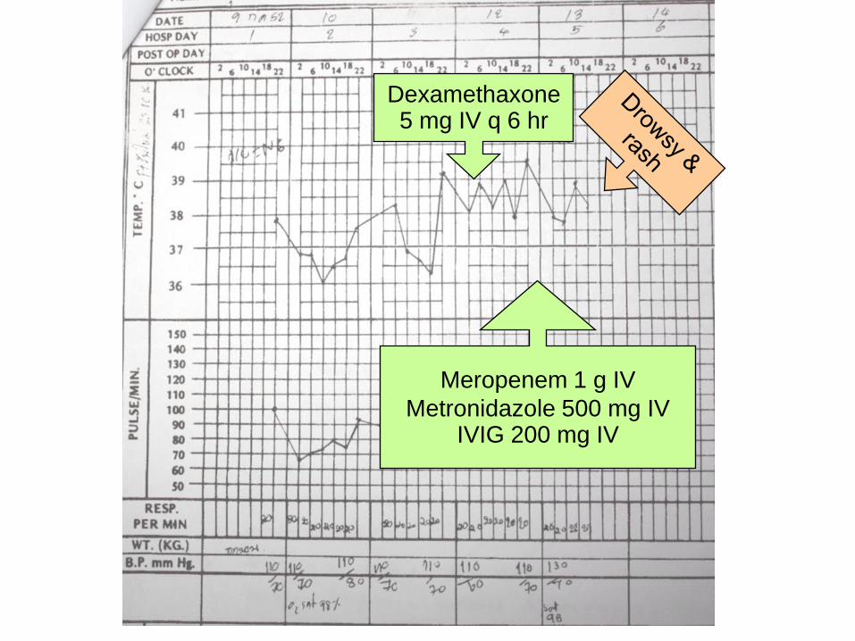
Dexamethaxone 5 mg IV q 6 hr
Meropenem 1 g IV
Metronidazole 500 mg IV IVIG 200 mg IV

Physical Examination at Rama
VS: T 37.8ºC , BP 150/90 mmHg, PR 89/min,
RR 20/min
GA: drowsy, not pale, mild jaundice, mild puffy
eyelids, no injected conjunctiva
LN: not palpable
Abdomen: mild tenderness at epigastrium,
liver 2 FB below RCM
Neurology: drowsy, no stiffness of neck
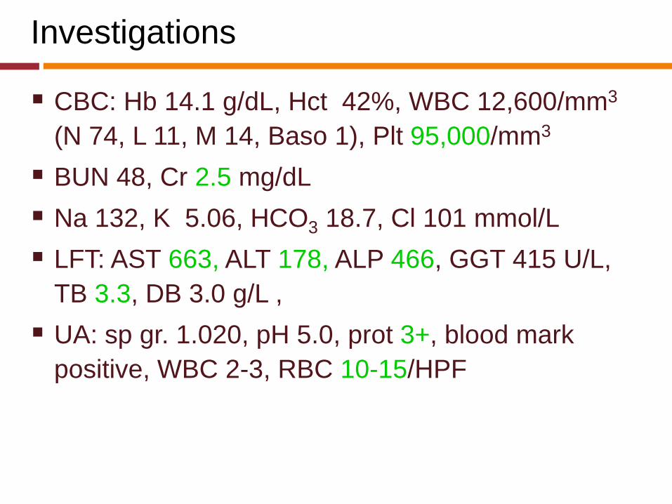
Investigations
CBC: Hb 14.1 g/dL, Hct 42%, WBC 12,600/mm3
(N 74, L 11, M 14, Baso 1), Plt 95,000/mm3
BUN 48, Cr 2.5 mg/dL
Na 132, K 5.06, HCO3 18.7, Cl 101 mmol/L
LFT: AST 663, ALT 178, ALP 466, GGT 415 U/L,
TB 3.3, DB 3.0 g/L ,
UA: sp gr. 1.020, pH 5.0, prot 3+, blood mark
positive, WBC 2-3, RBC 10-15/HPF
Which Investigations for Confirmation?
A. Weil-Felix Test
B. Widal Test
C. Dengue titer ELISA
D. IFA for rickettsioses
E. Leptospira titer
F. Hemo culture
G. Malarial film
H. None of the above
Eschar Lesions
Scrub typhus
Rickettsialpox
Other mite- or tick-borne rickettsiosis
Ecthyma: pseudomonas infection
Anthrax
Skin infection/pyoderma
Ecthyma/ulcerating impetigo
Vasculitis
Warfarin induced subcutaneous necrosis
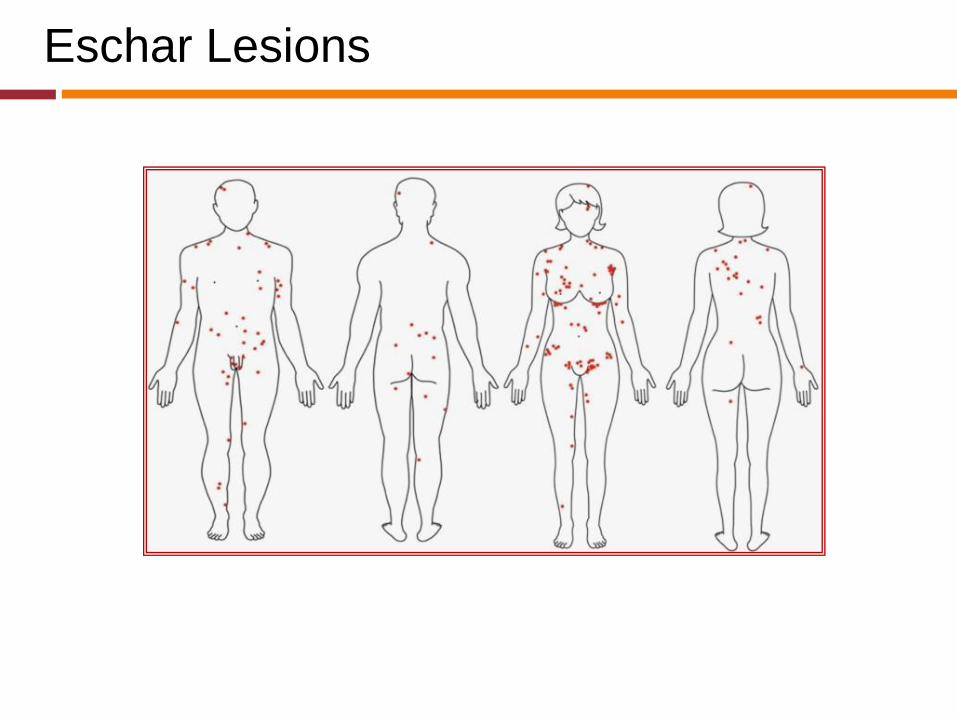
Eschar Lesions
Case 2: 56 Years Old Male
Government officer, Nakornprathom
CC: fever and jaundice 4 days
PI: 10 days low-grade fever, myalgia, low back
pain
4 days high-grade fever, increased pain, and
jaundice
PH: alcoholic drinking, smoking

Physical Examination
VS: T 39oC, RR 28/min, PR 120/min, BP 90/60
mmHg
HEENT: not pale, mild jaundice
Abdomen: soft, not tender, liver 3 FB below
RCM, span 15 cm, spleen just palpable, no
ascitis
Stigmata of chronic liver disease
Ext: Rt. shoulder marked tender if passive
movement
Rt. lower back: tender ~L5–S1

Investigations
CBC: Hct 35%, WBC 10,100/mm3 (N 87%, L
7%, M 5%, B 1%), plt 331,000/mm3
UA: pH 6.0, prot 3+, glu 3+, ketone slightly
positive, WBC 0-1, RBC 3-5/HPF
BUN/Cr: 14/1 mg/dl
LFT: AST 107, ALT 115, ALP 1418 u/L, alb 22.0 G/L, TB 3.4, DB 2.9 mg/dl

Provisional Diagnosis?
A. Cholangitis
B. Cholycystitis
C. Liver abscess
D. Psoas abscess
E. Acute pyelonephritis
56 years, fever and jaundice 4 days
PE: stigmata of chronic liver disease, hepatosplenomegaly,
shoulder and back pain
Lab: abnormal UA and LFT
Which Organism?
A. E. coli
B. K. pneumoniae
C. B. pseudomallei
D. P. aeruginosa
E. Acinetobacter spp.

Gram-Negative Aerobes
COCCI
M. catarrhalis
N. gonorrhoeae
N. meningitidis
H. influenzae
BACILLI
E. coli, Klebsiella spp.
Enterobacter spp.,
Citrobacter spp.
Proteus spp., Serratia
spp.
Salmonella spp.,
Shigella spp.
Acinetobacter spp.
Helicobacter spp.
Pseudomonas spp.
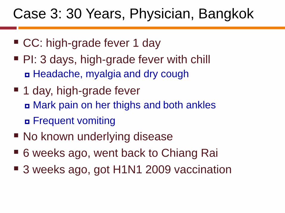
Case 3: 30 Years, Physician, Bangkok
CC: high-grade fever 1 day
PI: 3 days, high-grade fever with chill
Headache, myalgia and dry cough
1 day, high-grade fever
Mark pain on her thighs and both ankles
Frequent vomiting
No known underlying disease
6 weeks ago, went back to Chiang Rai
3 weeks ago, got H1N1 2009 vaccination

Physical Examinations
V/S: T 39.2ºC, BP 100/60 mmHg
GA: looked sick but good consciousness, not
pale, no jaundice
Extremities: mild tenderness at both thighs
Skin: no rash, no eschar
Neuro: alert, good orientation
Sharp optic disc, both
Pupil Rt 4 mm RTL, Lt 3 mm RTL
Mild limit ROM (80%) of Rt MR, SR & IR
Terminal stiffness of neck: positive
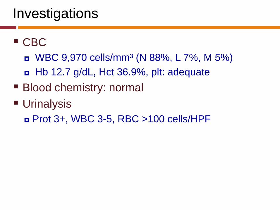
Investigations
CBC
WBC 9,970 cells/mm³ (N 88%, L 7%, M 5%)
Hb 12.7 g/dL, Hct 36.9%, plt: adequate
Blood chemistry: normal
Urinalysis
Prot 3+, WBC 3-5, RBC >100 cells/HPF
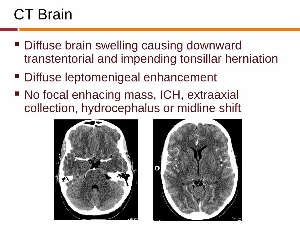
CT Brain
Diffuse brain swelling causing downward transtentorial and impending tonsillar herniation
Diffuse leptomenigeal enhancement
No focal enhacing mass, ICH, extraaxial collection, hydrocephalus or midline shift

Investigations
CBC
WBC 9,970 cells/mm³ (N 88%, L 7%, M 5%)
Hb 12.7 g/dL, Hct 36.9%, plt: adequate
Lumber puncture
OP/CP 37/37 cmH2O
Slightly turbid CSF
WBC 390 cells/mm3
PMN 87%, monocyte 13%
Protein 315, glucose 9 mg/dL
G/S: numerous PMN, no organism seen

What is the Causative Agent?
A. S. pneumoniae
B. M. tuberculosis
C. E. coli
D. N. meningitidis
E. H. influenzae
F. Not all of the above
OP/CP 37/37 cmH2O
Slightly turbid CSF
WBC 390 cells/mm3 (N 87%, monocyte 13%)
Protein 315, glucose 9 mg/dL

Acute Meningitis: Causative Agents
Characteristic Dexa Placebo
Nguyen TH et al. N Engl J Med 2007;357:2431-40. http://emedicine.medscape.com/article/784389-overview*
S. pneumoniae (30-50%), H. influenzae (1-3%), N. meningitidis (10-35%), gram-negative bacilli (1-10%), staphylococci (5-15%),
streptococci (5%), and Listeria spp. (5%)*
Spot Diagnosis
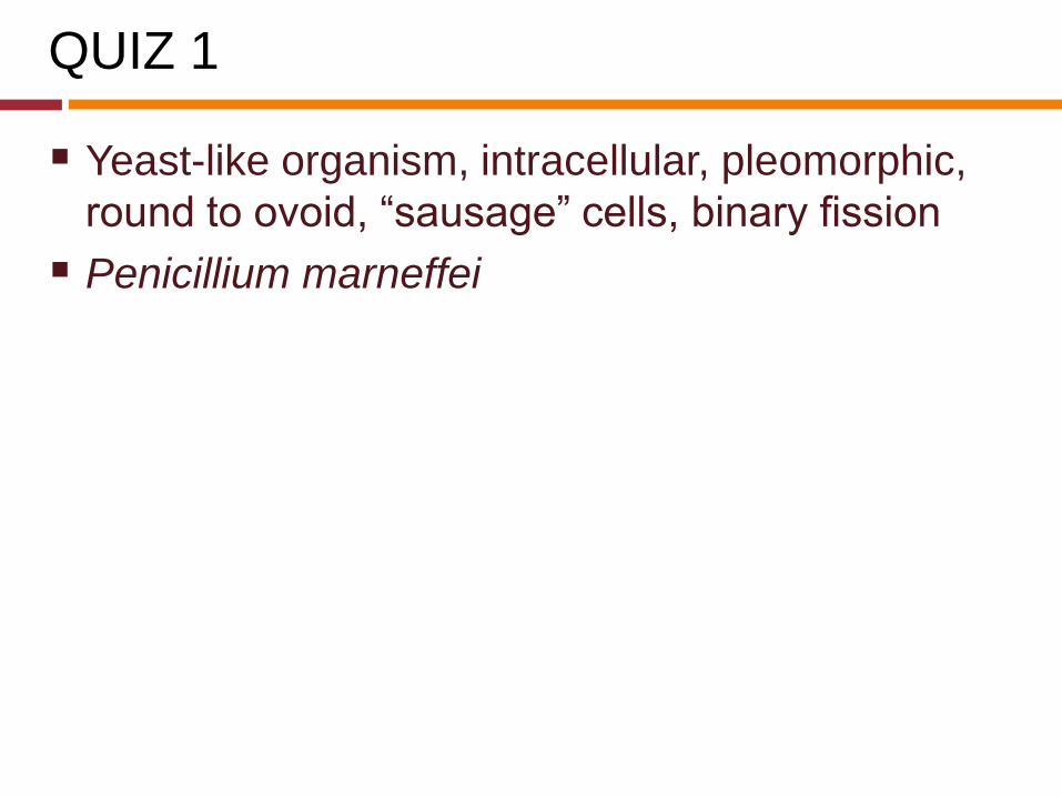
QUIZ 1
Yeast-like organism, intracellular, pleomorphic,
round to ovoid, “sausage” cells, binary fission
Penicillium marneffei
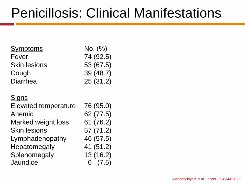
Penicillosis: Clinical Manifestations
Supparatpinyo K et al. Lancet 1994;344:110-3.
Symptoms No. (%)
Fever 74 (92.5)
Skin lesions 53 (67.5)
Cough 39 (48.7)
Diarrhea 25 (31.2)
Signs
Elevated temperature 76 (95.0)
Anemic 62 (77.5)
Marked weight loss 61 (76.2)
Skin lesions 57 (71.2)
Lymphadenopathy 46 (57.5)
Hepatomegaly 41 (51.2)
Splenomegaly 13 (16.2) Jaundice 6 (7.5)

Subacute-Prolonged Fever and Skin Lesions
Papulonecrotic lesion
Cryptococcosis
Penicillosis
Histoplasmosis
Mollusgum contagiosum
Eosinophilic pustular folliculitis
Prurigo nodularis
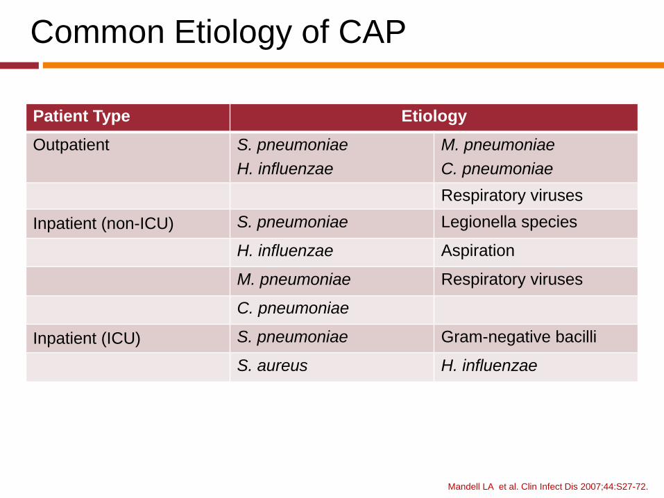
Common Etiology of CAP
Mandell LA et al. Clin Infect Dis 2007;44:S27-72.
Patient Type Etiology
Outpatient S. pneumoniae
H. influenzae M. pneumoniae
C. pneumoniae
Respiratory viruses
Inpatient (non-ICU) S. pneumoniae Legionella species
H. influenzae Aspiration
M. pneumoniae Respiratory viruses
C. pneumoniae
Inpatient (ICU) S. pneumoniae Gram-negative bacilli
S. aureus H. influenzae
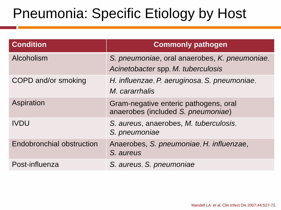
Pneumonia: Specific Etiology by Host
Mandell LA et al. Clin Infect Dis 2007;44:S27-72.
Condition Commonly pathogen
Alcoholism S. pneumoniae, oral anaerobes, K. pneumoniae,
Acinetobacter spp, M. tuberculosis
COPD and/or smoking H. influenzae, P. aeruginosa, S. pneumoniae,
M. cararrhalis
Aspiration Gram-negative enteric pathogens, oral anaerobes (included S. pneumoniae)
IVDU S. aureus, anaerobes, M. tuberculosis,
S. pneumoniae
Endobronchial obstruction Anaerobes, S. pneumoniae, H. influenzae,
S. aureus
Post-influenza S. aureus, S. pneumoniae

QUIZ 3
18-year-old man
High-graded fever and headache for 2 days
Stiffness of neck
Lumbar puncture was preformed and sent for
Gram Stain
QUIZ 3
Gram-positive, lancet-shaped cocci with capsule
Acute bacterial meningitis
S. pneumoniae

Gram-Positive Aerobes
COCCI
Clusters:
staphylococci
Pairs: S. pneumoniae
Chains: group and
viridans streptococci
Pairs and chains:
Enterococcus spp.
BACILLI
Bacillus spp.
Corynebacterium spp.
Listeria
monocytogenes
Nocardia spp.

Sizes of Helminth Eggs

BAL Cytology
Large round encapsulated yeasts
Budding yeast with negatively stained halos and
narrow necked buds

Cryptococcus spp.
Anti-HIV
Serum cryptococcal
antigen
Culture for fungus
Lumbar puncture

Data in Thailand: Ramathibodi Hosp.
40 cases of HIV negative, 1987-2003
Mean age: 49 (range 16-83) years
73% female
65% had associated underlying conditions
Kiertiburanakul S et al. Int J Infect Dis 2006;10:72-8.
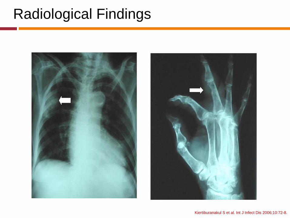
Radiological Findings
Kiertiburanakul S et al. Int J Infect Dis 2006;10:72-8.
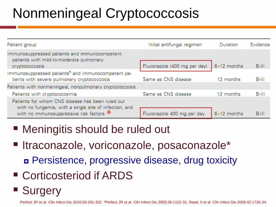
Nonmeningeal Cryptococcosis
Meningitis should be ruled out
Itraconazole, voriconazole, posaconazole*
Persistence, progressive disease, drug toxicity
Corticosteriod if ARDS
Surgery Perfect JR et al. Clin Infect Dis 2010;50:291-322. *Perfect JR et al. Clin Infect Dis 2003;36:1122-31. Raad, II et al. Clin Infect Dis 2006;42:1726-34.
*

Summary
Evaluation of patient who presents with infection
required
Detailed history and physical examination
Recognition of abnormal symptoms and signs
Diagnosis of infection and organism required
Experience bedside staining
Knowledge of basic laboratory interpretation
If any, high-tech or advanced tests may be needed