BBF RFC 100: Standard for Synthesis of Customized Peptides ...
16
RFC 100 Synthesis of Customized Non-Ribosomal Peptides - 1 - BBF RFC 100: Standard for Synthesis of Customized Peptides by Non-Ribosomal Peptide Synthetases Ralf Beer, Tania Christiansen, Konrad Herbst, Nikos Ignatiadis, Ilia Kats, Nils Kurzawa, Johanna Meichsner, Sophie Rabe, Anja Riedel, Joshua Sachs, Julia Schessner, Florian Schmidt, Philipp Walch, Lorenz Adlung, Katharina Genreith, Fanny Georgi, Tim Heinemann, Hannah Meyer, Dominik Niopek, Barbara Di Ventura, Roland Eils 2013-10-04 1 Purpose The purpose of this RFC is to introduce a standardized framework for the engineering of customizable non-ribosomal peptide synthetases (NRPS) and their application for in vivo and in vitro synthesis of short non-ribosomal peptides (NRPs) of user-defined sequence and structure. 2 Relation to other BBF RFCs BBF RFC100 updates RFC57 describing the “Assembly of BioBricks by the Gibson method”, which was proposed by the iGEM team Cambridge 2010. Specifically, RFC100 adopts the Gibson assembly strategy proposed by RFC57 for cloning of large bacterial NRPS expression constructs. BBF RFC100 also updates RFC99 describing the “HiCT: High Throughput Protocols for CPE Cloning and Transformation”, which was proposed by the iGEM team Heidelberg 2013. 3 Copyright Notice Copyright (C) The BioBricks Foundation (2013). All Rights Reserved. 4 Introduction to the Synthesis of Customized Peptides by Non-Ribosomal Peptide Synthetases 4.1 State of the Art in Peptide Synthesis Solid-phase synthesis of peptides was pioneered by Robert Bruce Merrifield in 1963 [Merrifield, 1963] and still represents the state of the art chemical method for manufacturing custom small peptides. It enables the stepwise assembly of peptides from single amino acids, also allowing for incorporation of non-essential amino acids into peptide chains. Although solid state peptide synthesis is nowadays standardized and automated, the method has an important limitation: it is expensive when applied for industry-scale production of synthetic peptides. Recombinant peptide synthesis represents an interesting alternative to chemical peptide synthesis, as it is easily scalable once the production is up and running. However, this approach is currently restricted to peptides composed mostly of proteinogenic amino acids. Furthermore, developing an efficient peptide production strain or establishing a corresponding in vitro translation systems is laborious and thus, also costly and time consuming. As consequence of these limitations, the interest of industry to invest into the development of peptide-based applications including peptide drugs has been rather low in the past, calling for alternative peptide production methods which are cost-efficient, automatable, scalable and fast. 4.2 Non-Ribosomal Peptide Synthesis
Transcript of BBF RFC 100: Standard for Synthesis of Customized Peptides ...
RFC 100 Synthesis of Customized Non-Ribosomal Peptides
- 1 -
BBF RFC 100: Standard for Synthesis of Customized Peptides by Non-Ribosomal Peptide Synthetases
Ralf Beer, Tania Christiansen, Konrad Herbst, Nikos Ignatiadis, Ilia Kats, Nils Kurzawa, Johanna Meichsner, Sophie Rabe, Anja Riedel, Joshua Sachs, Julia Schessner, Florian Schmidt, Philipp Walch, Lorenz Adlung, Katharina Genreith, Fanny Georgi, Tim Heinemann, Hannah Meyer, Dominik Niopek, Barbara Di Ventura, Roland Eils
2013-10-04 1 Purpose The purpose of this RFC is to introduce a standardized framework for the engineering of customizable non-ribosomal peptide synthetases (NRPS) and their application for in vivo and in vitro synthesis of short non-ribosomal peptides (NRPs) of user-defined sequence and structure. 2 Relation to other BBF RFCs BBF RFC100 updates RFC57 describing the “Assembly of BioBricks by the Gibson method”, which was proposed by the iGEM team Cambridge 2010. Specifically, RFC100 adopts the Gibson assembly strategy proposed by RFC57 for cloning of large bacterial NRPS expression constructs. BBF RFC100 also updates RFC99 describing the “HiCT: High Throughput Protocols for CPE Cloning and Transformation”, which was proposed by the iGEM team Heidelberg 2013. 3 Copyright Notice Copyright (C) The BioBricks Foundation (2013). All Rights Reserved. 4 Introduction to the Synthesis of Customized Peptides by
Non-Ribosomal Peptide Synthetases 4.1 State of the Art in Peptide Synthesis Solid-phase synthesis of peptides was pioneered by Robert Bruce Merrifield in 1963 [Merrifield, 1963] and still represents the state of the art chemical method for manufacturing custom small peptides. It enables the stepwise assembly of peptides from single amino acids, also allowing for incorporation of non-essential amino acids into peptide chains. Although solid state peptide synthesis is nowadays standardized and automated, the method has an important limitation: it is expensive when applied for industry-scale production of synthetic peptides. Recombinant peptide synthesis represents an interesting alternative to chemical peptide synthesis, as it is easily scalable once the production is up and running. However, this approach is currently restricted to peptides composed mostly of proteinogenic amino acids. Furthermore, developing an efficient peptide production strain or establishing a corresponding in vitro translation systems is laborious and thus, also costly and time consuming. As consequence of these limitations, the interest of industry to invest into the development of peptide-based applications including peptide drugs has been rather low in the past, calling for alternative peptide production methods which are cost-efficient, automatable, scalable and fast. 4.2 Non-Ribosomal Peptide Synthesis

RFC 100 Synthesis of Customized Non-Ribosomal Peptides
- 2 -
Non-ribosomal peptides (NRPs) are secondary metabolites produced by microorganisms, e.g. bacteria and fungi, where they have various functions ranging from antibiotics to pigments and chelators of toxic metal ions. Notably, non-ribosomal peptide synthesis does not require mRNA to direct the sequence of amino acid monomers incorporated into the growing peptide chain. Instead, NRPSs are organized in distinct functional subunits, the so-called NRPS modules. During NRP synthesis, every NRPS module recognizes one specific amino acid substrate and catalyzes the formation of a peptide bond between the amino acid substrate and the nascent peptide chain, which is handed over from the previous NRPS module. In contrast to ribosomal peptide and protein synthesis, NRPSs can incorporate proteinogenic as well as non-proteinogenic amino acids substrates (about 500 different amino acid building blocks are currently known) into NRPs. Moreover, NRPs may be linear as well as circularized. This leads to an enormous variability of NRPs explaining their diverse functions in nature. Every NRPS module can again be subdivided into different functional domains. A new amino acid is first adenylated by the A-domain and then bound to the T-domain (thiolation domain) via a thioester bond. The C-domain catalyzes the condensation of an existing peptide chain - which is bound to the T-domain of the previous module - and the amino acid of the next module. The T-domain itself does not exhibit any substrate specificity but is just a carrier domain to keep the peptide attached to the NRPS module complex. The core of every T-domain is a conserved 4’-phosphopanthetheinylated (4’-PPT) serine. The 4’-PPT residue is added by a 4’-Phosphopanthetheinyl-transferase (PPTase), which brings the NRPS apo-enzyme to its active holo-form. NRPSs are known to have a modular architecture, with single modules being specific for a specific amino acid as described above. The single modules can be connected covalently to form large multi-module-proteins. However, single modules can also associate via protein-protein interactions, mostly via so called communication domains. We and others have shown, that NRPS modules can be exchanged and even entirely shuffled in order to create new NRPSs synthesizing novel peptides [iGEM Team Heidelberg 2013, 2013.igem.org/Team:Heidelberg; Mootz et al. 2000; Nguyen et al. 2006, Stachelhaus et al. 1998, Finking & Marahiel 2004]. These custom NRPS may include (1) modules derived from a single NRPS pathway, (2) modules derived from different NRPS pathways from a single species, (3) modules derived from NRPS pathways from different species and (4) entirely synthetic modules composed of domains of varying origin. It was furthermore found to be generally possible to exchange domains comprised in a module for another domain conferring the same or similar functionality, emphasizing the modular architecture of NRPSs on several levels. Thus, NRPSs represent highly versatile and flexible platform for the in vivo synthesis of completely synthetic peptides. 4.3 A Standardized Framework for the engineering of customized NRPSs Here we propose a standardized framework for the engineering of customized NRPSs catalyzing the synthesis of user-defined NRPs. The framework consists of (1) the NRPSDesigner, a software tool for the in silico design of user-defined NRPSs, (2) a platform for standardized cloning and expression of NRPSs in different bacterial hosts and (3) a quality control procedure for the validation of NRP production (Figure 1). Following this framework enables a user to easily engineer customized NRPSs and thereby produce synthetic NRPs within a minimal amount of time and in a cost-efficient and easily scalable manner. The user may provide any custom NRP sequence as input for the NRPS designer (Figure 1.1), which is a completely automated software for the in silico design of synthetic NRPSs

RFC 100 Synthesis of Customized Non-Ribosomal Peptides
- 3 -
(for description of the NRPSDesigner buildup see Appendix A). The software is based on a manually curated database of NRPS modules, each of which represents a unit responsible for incorporating a specific amino acid building block into the corresponding NRP chain. Moreover, the user can also add novel modules not present in the database or completely synthetic modules, thereby enhancing the range of possible amino acid substrates and modifications available for the corresponding NRPs. Additionally, the NRPSDesigner offers the possibility to tag the custom NRPs with the blue NRPS pigment indigoidine. The user may add an engineered indigoidine synthetase [2013.igem.org/Team:Heidelberg/Project/Indigoidine] module as last module to the corresponding custom NRPS in the NRPSDesigner leading to a blue coloring of the NRP. Labeling NRPs with indigoidine has several advantages, as it enables
1) a direct visualization of successful cloning, NRPS expression and NRP production by the custom NRPS (colonies turn blue on plates)
2) an easy qualitative analysis of the NRP produced by thin layer chromatography (TLC) avoiding the need for Mass Spectrometry (MassSpec) analysis
3) a direct quantification of NRP productivity of different clones in a simple, photometric measurement
4) an easy purification of indigoidine-tagged peptides (e.g. by HPCL collecting fractions with absorption maxima at 600 nm)
After finalizing the NRPS design the user is provided with a set of Gibson primers and corresponding PCR template BioBricks (note: if there are no BioBricks available, the DNA of the NRPS module donor organism is used as template). Cloning is performed by applying a Gibson assembly strategy optimized for cloning of large expression cassettes (Figure 1.2) (note: a single NRPS module is about 1000 amino acids long). The so obtained vector can then directly be transformed into the host strain carrying a PPTase expression cassette of choice in order to start in vivo NRPS production. In order to validate NRPS functionality and successful NRP expression (Figure 1.3), the user must perform a qualitative control analysis either by MassSpec (for untagged NRPs; note: requires purification of the corresponding peptides) or by a photometric absorption measurement followed by TLC (for indigoidine-tagged NRPs). In the following section, we list detailed protocols that should enable the synthetic biology community to adopt and use the framework described in this RFC. We furthermore introduce concepts to the extension of the framework (section 6) towards high-throughput production of synthetic NRPs and combinatorial NRP libraries and in vitro synthesis of NRPs.
RFC 100 Synthesis of Customized Non-Ribosomal Peptides
- 4 -
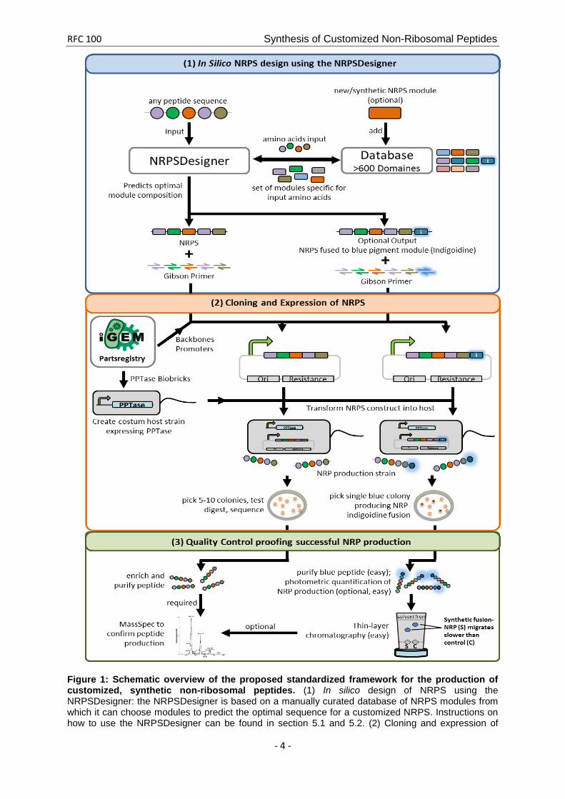
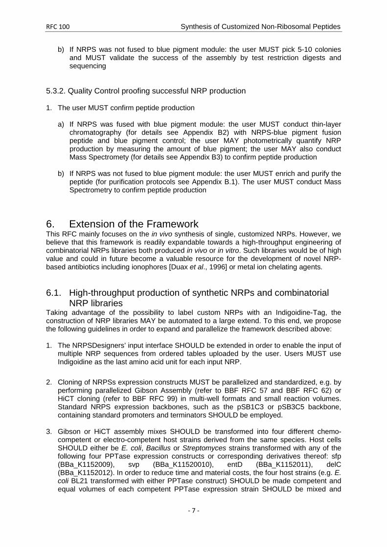
Figure 1: Schematic overview of the proposed standardized framework for the production of customized, synthetic non-ribosomal peptides. (1) In silico design of NRPS using the NRPSDesigner: the NRPSDesigner is based on a manually curated database of NRPS modules from which it can choose modules to predict the optimal sequence for a customized NRPS. Instructions on how to use the NRPSDesigner can be found in section 5.1 and 5.2. (2) Cloning and expression of
RFC 100 Synthesis of Customized Non-Ribosomal Peptides
- 5 -
NRPS: the NRPSDesigner outputs a Gibson cloning strategy for the assembly of the desired NRPS. The transformation of the Gibson-assembled NRPS constructs in host strains carrying BioBrick PPTase plasmids will allow for the expression of the customized NRP. Protocols on how to clone and and express NRPS construct are described in section 5.3.1. (3) Quality control proofing successful NRP production: validation strategies for the expression of the NRP depend on the design of the NRP as chosen by the user. Easy validation by screening for a blue phenotype of the host strains and expression control by TLC is enabled if the users choose to incorporate an indigoidine-tag in step (1) and (2). Detailed description of how to validate the expression of the custom NRP is provided in section 5.3.2. 5 Protocols for the Synthesis of Customized Peptides by engineered Non-Ribosomal Peptide Synthetases The user SHOULD use the NRPSDesigner for the in silico design of user-defined NRPSs (section 5.1). In addition, the user MAY extend the database on which the NRPSDesigner relies by adding new domains of defined substrate specificity (section 5.2). For the in vivo production of custom NRPs and the subsequent quality control, the users SHOULD follow the instructions given in section 5.3. 5.1 The NRPSDesigner: a comprehensive software guide to optimize
non-ribosomal peptide synthesis For the prediction of customized NRPSs by the NRPSDesigner the user MUST follow the procedure described below: 1. The user MUST enter the amino acids that constitute the desired peptide.
2. The user MUST choose between L- and D- conformation and MAY include possible
modifications (e.g. oxidation or methylation).
3. The user also MAY choose to add an indigoidine tag, which eases detection and quantification of the expression of the desired peptide.
4. Upon peptide entries by the user, the NRPSDesigner determines required modules and domains and computes the best-fitting combination. The software output to the user includes a suggestion for an assembly strategy that is based on Gibson cloning.
5. The user SHOULD consider suggested primer sequences though he MAY adapt the
primer-binding regions, which can be easily customized with the NRPSDesigner.
6. The user SHOULD use Gibson cloning for the assembly of the fragments as described in the BBF RFC 57.
5.2 Extending the database: Sharing your expertise in NRPS with the
synthetic biology community. Besides the design of novel, synthetic non-ribosomal peptides, the user MAY also extend the underlying database of the NRPSDesigner in order to share knowledge about NRPS domains and modules. Herefore, 1. the user MUST register with a username and a valid email address;
RFC 100 Synthesis of Customized Non-Ribosomal Peptides
- 6 -
2. the user MUST enter i) the DNA sequence in FASTA format or as plain text, ii) the respective source (i.e. a native organism or a BioBrick plasmid) and iii) the gene name;
3. the user SHOULD add a description for his entry;
4. the user SHOULD annotate the synthetic pathway if the sequence it originated from is
annotated. Upon fulfillment of all required entries, the NRPSDesigner predicts domains and their specificity of the putative NRPS using Hidden-Markov-Models (HMMs). Subsequently,
5. the user MAY define personal recommendations for domain boundaries, e.g. based on
multiple sequence alignments that are also offered by the NRPSDesigner;
6. the user MAY add a custom description of each domain;
7. the user MUST submit the final data form.
5.3 Working with NRPS: Creating custom NRPSs for in vivo NRP
synthesis. 5.3.1. Cloning and Expression of NRPS 1. Expression of NRPS construct MUST be conducted in host strains expressing the
following PPTase expression constructs or corresponding derivates thereof: sfp (BBa_K1152009), svp (BBa_K11520010), entD (BBa_K1152011), delC (BBa_K1152012. Therefore, four different chemo-competent or electro-competent host strains derived from the same species carrying each a single PPTase construct MUST be created. Host cells SHOULD either be E. coli, Bacillus or Streptomyces strains. In order to reduce time and material costs, the four host strains (e.g. E. coli BL21 transformed with either PPTase construct) SHOULD be made competent and equal volumes of each competent PPTase expression strain SHOULD be mixed and aliquoted afterwards. Thereby transformation into the four host strains MAY be performed in a single vial.
2. For amplification of the fragments, the user SHOULD use the primers proposed by the NRPSDesigner and MUST use a backbone with an antibiotic resistance marker other than that present on the PPTase-encoding plasmid.
3. The assembly of the NRPS plasmid MUST be done by Gibson [BBF RFC 57] or CPE cloning [BBF RFC 99], depending on the size and number of fragments to assemble. CPE Cloning MAY be used for up to four fragments, Gibson Cloning MUST be used for more than four fragments and NRPS expression constructs above 10 kbp in size. After cloning, product MAY be purified by isopropanol precipitation.
4. Transformations of the custom host strain with the synthesized NRPS MAY be conducted
with the cloning mixture or the purified assembly product. 5. NRPS-transformed custom host strains MUST be selected for the presence of both
constructs by two-fold antibiotic selection. 6. Selection for positive colonies:
a) If NRPS was fused with blue pigment module: the user MUST pick a single blue colony
RFC 100 Synthesis of Customized Non-Ribosomal Peptides
- 7 -
b) If NRPS was not fused to blue pigment module: the user MUST pick 5-10 colonies and MUST validate the success of the assembly by test restriction digests and sequencing
5.3.2. Quality Control proofing successful NRP production 1. The user MUST confirm peptide production
a) If NRPS was fused with blue pigment module: the user MUST conduct thin-layer
chromatography (for details see Appendix B2) with NRPS-blue pigment fusion peptide and blue pigment control; the user MAY photometrically quantify NRP production by measuring the amount of blue pigment; the user MAY also conduct Mass Spectromety (for details see Appendix B3) to confirm peptide production
b) If NRPS was not fused to blue pigment module: the user MUST enrich and purify the peptide (for purification protocols see Appendix B.1). The user MUST conduct Mass Spectrometry to confirm peptide production
6. Extension of the Framework This RFC mainly focuses on the in vivo synthesis of single, customized NRPs. However, we believe that this framework is readily expandable towards a high-throughput engineering of combinatorial NRPs libraries both produced in vivo or in vitro. Such libraries would be of high value and could in future become a valuable resource for the development of novel NRP-based antibiotics including ionophores [Duax et al., 1996] or metal ion chelating agents. 6.1. High-throughput production of synthetic NRPs and combinatorial
NRP libraries Taking advantage of the possibility to label custom NRPs with an Indigoidine-Tag, the construction of NRP libraries MAY be automated to a large extend. To this end, we propose the following guidelines in order to expand and parallelize the framework described above: 1. The NRPSDesigners’ input interface SHOULD be extended in order to enable the input of
multiple NRP sequences from ordered tables uploaded by the user. Users MUST use Indigoidine as the last amino acid unit for each input NRP.
2. Cloning of NRPSs expression constructs MUST be parallelized and standardized, e.g. by
performing parallelized Gibson Assembly (refer to BBF RFC 57 and BBF RFC 62) or HiCT cloning (refer to BBF RFC 99) in multi-well formats and small reaction volumes. Standard NRPS expression backbones, such as the pSB1C3 or pSB3C5 backbone, containing standard promoters and terminators SHOULD be employed.
3. Gibson or HiCT assembly mixes SHOULD be transformed into four different chemo-
competent or electro-competent host strains derived from the same species. Host cells SHOULD either be E. coli, Bacillus or Streptomyces strains transformed with any of the following four PPTase expression constructs or corresponding derivatives thereof: sfp (BBa_K1152009), svp (BBa_K11520010), entD (BBa_K1152011), delC (BBa_K1152012). In order to reduce time and material costs, the four host strains (e.g. E. coli BL21 transformed with either PPTase construct) SHOULD be made competent and equal volumes of each competent PPTase expression strain SHOULD be mixed and
RFC 100 Synthesis of Customized Non-Ribosomal Peptides
- 8 -
aliquoted afterwards, preferably directly into a multiwell format. Thereby transformation into the four host strains MAY be performed in a single vial.
4. Transformation mixes MUST be spread on agar plates containing appropriate growth
medium (this MAY be LB-Broth or 2-YT for E. coli) with corresponding antibiotics and incubated for 12-96 h at 21-37°C.
5. Picking of blue colonies (coloring refers to successful NRP expression) and inoculation of
liquid cultures SHOULD be performed by a cloning robot 24-72 h after transformation. White colonies MUST NOT be picked. The robot SHOULD pick at least three clones per construct and SHOULD inoculate liquid cultures preferably in small scale in a multi-well plate format. This format MAY be a 96-well format with 200 µl culture volume.
6. Liquid cultures should be grown at 30°C for 12-48 h and coloring of the cultures in the
different wells SHOULD be monitored by photo-absorption measurements (see Appendix B.4) in order to identify highly productive strains.
7. 180 µl of liquid cultures should be used for NRP purification (see Appendix B.1) which
MAY again be performed in small scale in a multi-well format or also automatized in case larger scales would be required. 5 µl 50% glycerol should be added to each well containing the remaining 20 µl of the liquid cultures and the multi-well plate should be stored at - 80°C.
8. NRP functionality tests MAY be performed directly in multi-well format in order to identify
the NRP candidates showing the required function. 9. Thin Layer Chromatography (TLC) should be performed for all NRP samples positive in
the functional assays. An indigoidine-only control MUST be loaded in parallel with all NRP samples onto the TLC. NRPs showing a slower migration compared to the indigoidine-only control SHOULD be considered positive.
10. Positive NRP samples from point 8 MAY be analyzed further by MassSpec (see
Appendix B.3) in order to confirm the correct peptide sequence. 11. Production of NRP candidates of interest MAY subsequently be up scaled in vivo or even
performed in vitro (see section 6.2) and purification of NRPs MAY be done by liquid chromatography (e.g. HPLC) collecting the fractions that MUST show highest absorbance at 600 nm.
Note: The indigoidine module MAY be removed from NRPSs if labeling of the corresponding NRPs is no longer desired.
6.2 In vitro NRPs Synthesis NRPSs may also be purified from their bacterial host and used for in vitro synthesis of NRPs. This might simplify the purification of NRPs and yield products of high purity. In addition, in vitro NRP synthesis can easily be optimized by varying buffer and incubation conditions.
1. NRPSs SHOULD be tagged C-terminally with affinity tags enabling the purification of the corresponding NRPS. Suitable tags MAY be the His6-, Myc-, or Flag-Tag.
RFC 100 Synthesis of Customized Non-Ribosomal Peptides
- 9 -
2. NRPSs MUST be purified applying any affinity-based purification protocol well known in the arts.
3. NRPSs MUST be incubated under appropriate buffer conditions and in presence of all
amino acid substrates to be incorporated into the corresponding NRP. Detailed protocols describing in vitro NRP synthesis can be found elsewhere [Mootz et al. 2000].
4. NRPs SHOULD be purified (see Appendix B.1) following in vitro synthesis. 5. If the produced NRP is labeled with indigoidine, the NRP should be analyzed by TLC as
described in Appendix B.2. If the produced NRP is not labeled with indigoidine, the NRP should be purified and analyzed by MassSpec as described in Appendix B.3.
7. In vitro NRP production MAY be further optimized by varying single or multiple
parameters influencing NRP synthesis. These MAY include but are not limited to the buffer conditions, NRPS(s) concentration(s), amino acid substrate concentrations or incubation temperature and time.
7. Author's contact information
• Ralf Beer: [email protected] • Tania Christiansen: [email protected] • Konrad Herbst: [email protected] • Nikolaus Ignatiadis: [email protected] • Ilia Kats: [email protected] • Nils Kurzawa: [email protected] • Johanna Meichsner: [email protected] • Sophie Rabe: [email protected] • Anja Riedel: [email protected] • Joshua Sachs: [email protected] • Julia Schessner: [email protected] • Florian Schmidt: [email protected] • Philipp Walch: [email protected] • Lorenz Adlung: [email protected] • Katharina Genreith: [email protected] • Fanny Georgi: [email protected] • Tim Heinemann: [email protected] • Hannah Meyer: [email protected] • Dominik Niopek: [email protected] • Barbara Di Ventura: [email protected] • Roland Eils: [email protected]
8. References Ansari MZ, Yadav G, Gokhale RS, Mohanty D (2004) NRPS-PKS: a knowledge-based resource for analysis of NRPS/PKS megasynthases. Nucleic acids research 32: 405-413. Merrifield RB (1963): Solid Phase Peptide Synthesis. I. The Synthesis of a Tetrapeptide. J. Am. Chem. Soc. 85(14):2149-54
RFC 100 Synthesis of Customized Non-Ribosomal Peptides
- 10 -
Duax WL, Griffin JF, Langs DA, Smith GD, Grochulski P, Pletnev V, Ivanov V (1996) Molecular structure and mechanisms of action of cyclic and linear ion transport antibiotics. Biopolymers 40(1):141-55. Mootz HD, Schwarzer D, Marahiel MA. (2000) Construction of hybrid peptide synthetases by module and domain fusions. PNAS 97:5848–5853 Myers JA, Curtis BS, Curtis WR (2013) Improving accuracy of cell and chromophore concentration measurements using optical density. BMC Biophysics 6:4 Nguyen KT, Ritz D, Gu JQ, Alexander D, Chu M, Miao V, Brian P, Baltz RH. (2006) Combinatorial biosynthesis of novel antibiotics related to daptomycin. PNAS 14;103(46):17462-7 Stachelhaus T, Mootz HD, Bergendahl V, Marahiel MA. (1998) Peptide bond formation in nonribosomal peptide biosynthesis. Catalytic role of the condensation domain. Journal of biological chemistry 273(35):22773-81 Finking R & Marahiel MA. (2004) Biosynthesis of Non-Ribosomal Peptides. Annual Reviews Mibrobiology 58:453-88
Appendix
Appendix A. Buildup of the NRPSDesigner and Graphical User Interface
The NRPSDesigner facilitates the prediction and synthesis of non-ribosomal synthetases (NRPS) which will catalyze the customized assembly of non-ribosomal peptides. The software itself is split into a command-line executable comprising the core algorithm and a web-interface providing easy access to the functionality. The NRPSDesigner contains three main functions: i) Data storage, ii) in silico NRPS prediction and iii) design of an NRPS synthesis strategy.
i) The manually curated database of the NRPSDesigner stores information of about 600 NRPS domains, their DNA coding sequences and respective substrate specificities (database entries mainly retrieved from [Ansari et al., 2004]). Users are also able to enter new domains and pathways into the database by providing the coding sequence of their NRPS. Additionally, the database can also be extended with curated content using automated domain prediction based on Hidden Markov Models.
ii) The NRPSDesigner uses the stored information from its database to predict the optimal domain sequence that is able to produce a user-defined NRP. The prediction algorithm is based on the phylogenetic distances of the host organisms from which the database entries were derived: combinations of domains from closely related organisms have a higher probability to form a functional NRPS than domain combinations involving organisms which are far apart on the taxonomic tree. The taxonomic distance is currently determined from lineage information available in the NCBI taxonomy database.
iii) To facilitate the transition from in silico NRPS design towards expression of the desired NRPS, the Gibthon iGEM software tool of Cambridge 2010 for Gibson primer construction was embedded into the web-interface of the NRPSDesigner.
RFC 100 Synthesis of Customized Non-Ribosomal Peptides
- 11 -
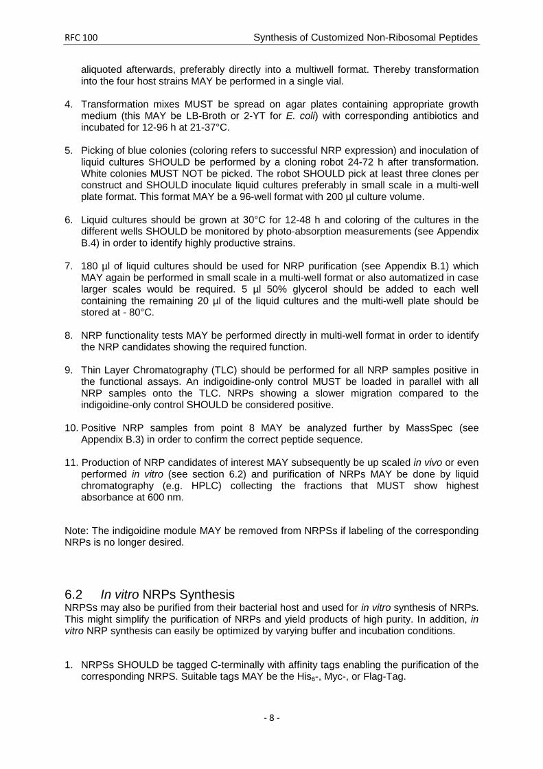
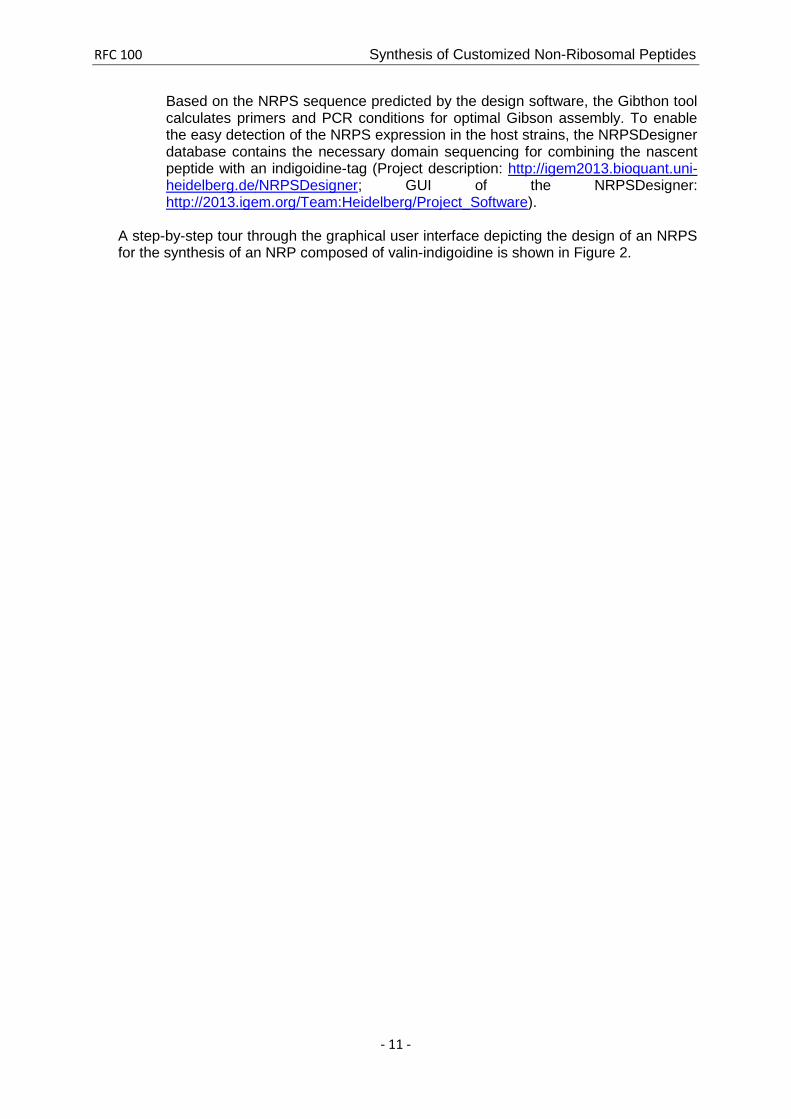
Based on the NRPS sequence predicted by the design software, the Gibthon tool calculates primers and PCR conditions for optimal Gibson assembly. To enable the easy detection of the NRPS expression in the host strains, the NRPSDesigner database contains the necessary domain sequencing for combining the nascent peptide with an indigoidine-tag (Project description: http://igem2013.bioquant.uni-heidelberg.de/NRPSDesigner; GUI of the NRPSDesigner: http://2013.igem.org/Team:Heidelberg/Project_Software).
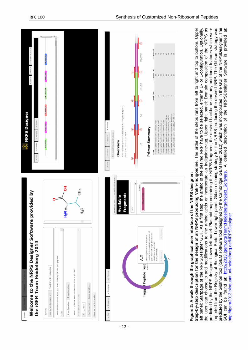
A step-by-step tour through the graphical user interface depicting the design of an NRPS for the synthesis of an NRP composed of valin-indigoidine is shown in Figure 2.
RFC 100 Synthesis of Customized Non-Ribosomal Peptides
- 12 -
Figu
re 2
: A w
alk
thro
ugh
the
grap
hica
l use
r int
erfa
ce o
f the
NR
PS d
esig
ner:
St
ep-b
y-st
ep d
escr
iptio
n fo
r th
e de
sign
of a
n N
RPS
pro
duci
ng V
alin
-Indi
goid
ine.
The
ord
er o
f the
ste
ps r
uns
from
left
to r
ight
and
top
to b
otto
m. U
pper
le
ft pa
nel:
Star
tpag
e of
the
NR
PSD
esig
ner
GU
T. A
s a
first
ste
p, th
e am
ino
of th
e de
sire
d N
RP
have
to b
e se
lect
ed, e
ither
in D
- or
L-c
onfig
urat
ion.
Opt
iona
lly,
the
user
can
cho
ose
to a
dd m
odifi
catio
ns t
o th
e am
ino
acid
s or
inc
orpo
rate
an
Indi
goid
ine-
tag.
Upp
er r
ight
pan
el:
Dom
ain
com
posi
tion
of t
he N
RP
S as
pr
edic
ted
by th
e N
RPS
des
igne
r. Lo
wer
left
pane
l: Pl
asm
id m
ap c
onta
inin
g th
e N
RPS
frag
men
t, th
e de
sire
d ba
ckbo
ne a
nd a
ny a
dditi
onal
feat
ures
whi
ch w
ere
impo
rted
from
the
Reg
istry
of B
iolo
gica
l Par
ts. L
ower
rig
ht p
anel
: Gib
son
clon
ing
stra
tegy
for
the
NR
PS
prod
ucin
g th
e de
sire
d N
RP.
The
Gib
son
stra
tegy
was
pr
edic
ted
by th
e G
ibth
on to
ol (i
GE
M s
oftw
are
tool
des
igne
d by
the
Cam
brid
ge iG
EM te
am 2
010)
whi
ch w
as in
corp
orat
ed in
the
GU
I of t
he N
RP
SDes
igne
r. Th
e G
UI
can
be f
ound
at:
http
://20
13.ig
em.o
rg/T
eam
:Hei
delb
erg/
Proj
ect_
Sof
twar
e. A
det
aile
d de
scrip
tion
of t
he N
RP
SDes
igne
r S
oftw
are
is p
rovi
ded
at:
http
://ig
em20
13.b
ioqu
ant.u
ni-h
eide
lber
g.de
/NR
PSD
esig
ner
RFC 100 Synthesis of Customized Non-Ribosomal Peptides
- 13 -
Appendix B. Supplementary Protocols B1. Peptide Purification
a) Purification of NRPs tagged with indigoidine for expression control by TLC 1 ml of IPTG-induced, blue culture is spun down at full speed (14,000 rpm) for 20 minutes, washed in 1 ml of methanol and centrifuged once more for 5 minutes at 14,000 rpm. Methanol is discarded and samples are allowed to dried. Lastly, dried samples are dissolved in 200-400 µl DMSO.
Note: For evaluation of expression over time this protocol is not recommended, as it is not suitable for quantification.
b) Purification of NRPs lacking an indigoidine tag for expression control by
MassSpec
3 ml of LB-medium with appropriate antibiotic are inoculated and incubated at 37°C overnight. At OD600 = ~0.6, culture is spun down for 30 minutes at 3750 rpm and 4°C, supernatant is discarded and pellet washed with M9 minimal media. Samples are centrifuged once more and resuspended in 50 ml M9 minimal media. Following an incubation time of 12 hours (OD600=~0.6) cells are induced with 1 mM IPTG.
After another 12 hours, samples are spun down for 45 minutes at 3750 rpm and 4°C, and supernatant is separated from pellet:
The Pellet is resolved in 500 µl 1x PBS and subsequently cells are lysed by ultra-sonification (3x20 s on ice) and the sample is centrifuged. Supernatant is transferred to 1.5 ml tubes and frozen in liquid nitrogen. The sample is ready for mass spectrometry.
Note: In order to remove aqueous and ionic residues, it is recommended to lyophilize the purified sample before subjection to MassSpec. Lyophilization is carried out for 12 hours at ~-45°C and ~0,1 mbar. The lyophilisate is resuspended in 500 µl methanol, vortexed and spun down. Supernatant is transferred to a 1.5 ml tube and frozen in liquid nitrogen. The sample is ready for mass spectrometry.
Optionally: For evaluation over time, 7.5 ml culture are taken at several time points (e.g. 8.5 h, 12 h, 24 h and 48 h) and purified as described above.
B2. Thin Layer Chromatography TLC is carried out on silica-gel as immobile phase and dichloromethane as mobile phase. For the procedure, a 50 ml beaker is filled with ~15 ml dichloromethane and allowed to stand for about 10 minutes to let the dichloromethane-vapor fill the beaker. As Indigoidine is light-sensitive, the beaker has to be covered with aluminium foil in order to prevent direct light irradiation. Sample purified according to Appendix B1a) as well as an indigoidine-only control (purified in analogy to the sample) are spotted

RFC 100 Synthesis of Customized Non-Ribosomal Peptides
- 14 -
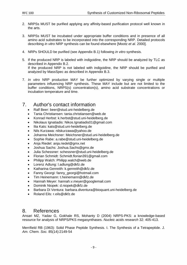
approximately 0.5 to 1 cm above the lower edge of a TLC plate coated with silica-gel. The TLC plate is placed in the beaker and TLC is allowed to run until the solvent front is at two thirds of the TLC plate. NRPs showing a slower migration compared to the indigoidine-only control are considered positive (compare to Figure 3)
B3. Recommended approach for the quantification of indigoidine production using OD measurement.
Detecting the amount of the NRP expressed by the bacterial host strain is desirable. By tagging the NRP with indigoidine, the amount of the fusion peptide can be determined by quantifying the amount of blue pigment present in the cells. As the amount of blue pigment is proportional to the amount of the NRP of interest, a method for the quantification of the blue pigment will yield information about the expression of the NRP. Quantification of the pure indigoidine pigment can be easily achieved by optical density (OD) measurements at its maximum wavelength of about 590 nm.
In cellular culture, indigoidine quantification by OD measurements is impaired. Cellular density of liquid cultures is standardly measured as the optical density (OD) at a wave length of 600 nm, i. e. the absorption peak of indigoidine interferes with the measurement of cell
Figure 3: Validation of the expression of an indigoidine-tagged NRP by thin layer chromatography. The purified Valin-Indigoidine sample and the Indigoidine-only control can clearly be separated by TLC (TLC plate coated with silica; running buffer: dichloromethane). The slower migration of the NRP compared to the indigoidine-only control serves as expression proof of the NRP.
RFC 100 Synthesis of Customized Non-Ribosomal Peptides
- 15 -
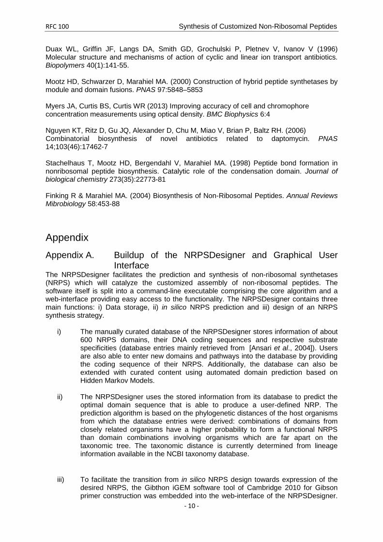
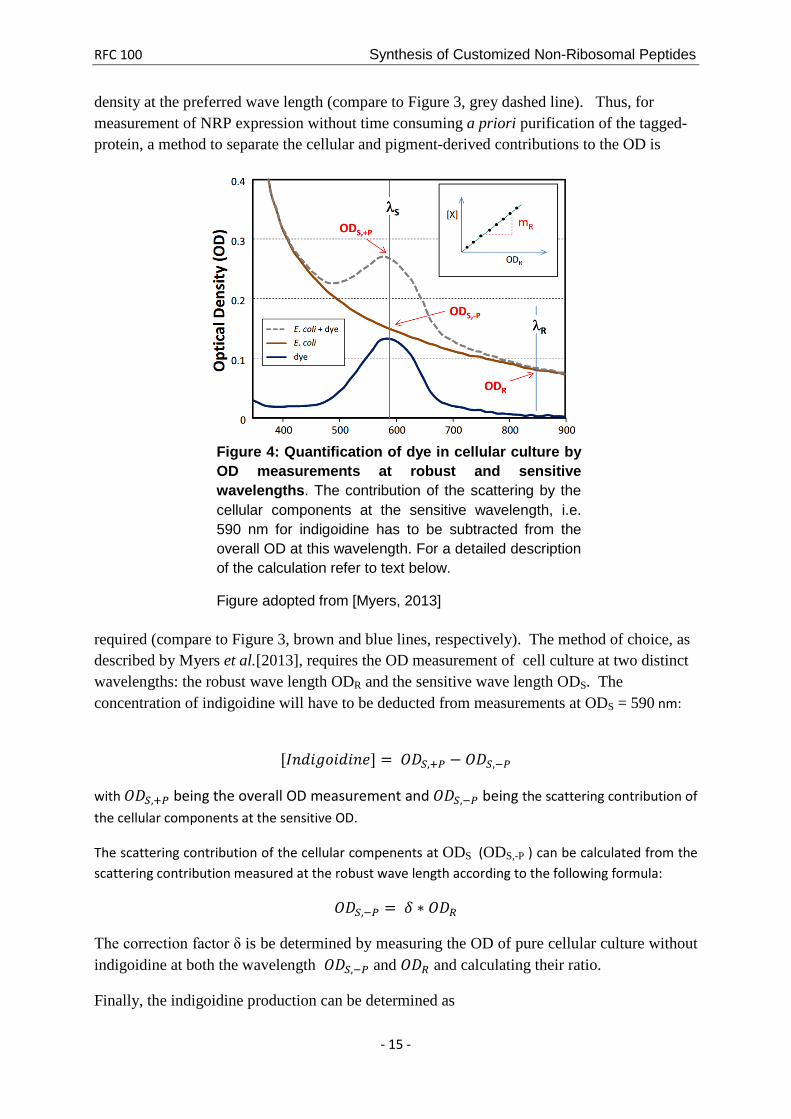
density at the preferred wave length (compare to Figure 3, grey dashed line). Thus, for measurement of NRP expression without time consuming a priori purification of the tagged-protein, a method to separate the cellular and pigment-derived contributions to the OD is
required (compare to Figure 3, brown and blue lines, respectively). The method of choice, as described by Myers et al.[2013], requires the OD measurement of cell culture at two distinct wavelengths: the robust wave length ODR and the sensitive wave length ODS. The concentration of indigoidine will have to be deducted from measurements at ODS = 590 nm:
[𝐼𝑛𝑑𝑖𝑔𝑜𝑖𝑑𝑖𝑛𝑒] = 𝑂𝐷𝑆,+𝑃 − 𝑂𝐷𝑆,−𝑃
with 𝑂𝐷𝑆,+𝑃 being the overall OD measurement and 𝑂𝐷𝑆,−𝑃 being the scattering contribution of the cellular components at the sensitive OD.
The scattering contribution of the cellular compenents at ODS (ODS,-P ) can be calculated from the scattering contribution measured at the robust wave length according to the following formula:
𝑂𝐷𝑆,−𝑃 = 𝛿 ∗ 𝑂𝐷𝑅
The correction factor δ is be determined by measuring the OD of pure cellular culture without indigoidine at both the wavelength 𝑂𝐷𝑆,−𝑃 and 𝑂𝐷𝑅 and calculating their ratio.
Finally, the indigoidine production can be determined as
Figure 4: Quantification of dye in cellular culture by OD measurements at robust and sensitive wavelengths. The contribution of the scattering by the cellular components at the sensitive wavelength, i.e. 590 nm for indigoidine has to be subtracted from the overall OD at this wavelength. For a detailed description of the calculation refer to text below.
Figure adopted from [Myers, 2013]

RFC 100 Synthesis of Customized Non-Ribosomal Peptides
- 16 -
[𝐼𝑛𝑑𝑖𝑔𝑜𝑖𝑑𝑖𝑛𝑒] = 𝑂𝐷𝑆,+𝑃 − 𝛿 ∗ 𝑂𝐷𝑅
For the calculation of the cellular component when measuring indigoidine producing liquid cell cultures, OD measurement at 800 nm as robust wavelength is recommended. By the approach described above, quantitative observation of the indigoidine production in a liquid culture over time as well as the indigoidine production in relation to the cell growth can be conducted.
Background correction i. e. the contribution of the culture medium to the OD measurement is achieved by subtracting the mean of pure culture medium replicates from all OD values measured.
Instructions and suggestions on how to prepare OD measurements of multiple cell cultures to quantify indigoidine production in a parallelized manner are described below.
96-well plates are prepared with 100 μl LB-medium/well containing appropriate antibiotics) and each well is inoculated with single colonies (in duplicates) from plates positive for the transformation experiments i.e. from plates with blue colonies. Two sets of negative controls are also inoculated on the plate: First, pure medium serving as the baseline for background correction for the OD measurements. Second, transformation controls accounting for potential differences in cell growth due to expression of proteins contained on the plasmids, i.e. the antibitotic resistance gene and IndC. In this set of controls, the plasmids coding for the IndC-constructs carry a randomly generated sequence instead of the T-domain. A second 96 well plate es prepared with 180 µl LB-medium/well for the measurement itself. The 96-well plate containing the pre-cultures of the co-transformed colonies is inoculated for 24 hours at 37°C. Subsequently, 20 µl of the pre-culture are transferred to the measurement plate. The absorbance of the bacterial cultures is measured at critical wavelengths of 590 and 800 nm. For time-resolved measurement of indigoidine, hence NRP expression, the OD of each well is measured every 30 min for 30 hours at 30°C in a Tecan infinite M200 plate reader.