basmala - docshare02.docshare.tipsdocshare02.docshare.tips/files/26964/269648324.pdfbasmala.tk...
288
basmala.tk
Transcript of basmala - docshare02.docshare.tipsdocshare02.docshare.tips/files/26964/269648324.pdfbasmala.tk...
basm
ala.tk
basm
ala.tk
STEP BY STEP®
SQUINT SURGERY
basm
ala.tk

basm
ala.tk
STEP BY STEP®
SQUINT SURGERY
Editor
Prasad Walimbe MS DNB FAEH FPO (USA)
Pediatric Ophthalmologist and Squint SpecialistConsultant
Jehangir Hospital and Deenanath Mangeshkar HospitalPune, Maharashtra, India
Walimbe Eye ClinicAranyeshwar Park, Sahakarnagar
Pune, Maharashtra, India
Foreword
Burton J Kushner

basm
ala.tk
Published by
Jaypee Brothers Medical Publishers (P) Ltd
Corporate Office4838/24 Ansari Road, Daryaganj, New Delhi - 110002, IndiaPhone: +91-11-43574357, Fax: +91-11-43574314
Offices in India• Ahmedabad, e-mail: [email protected]
• Bengaluru, e-mail: [email protected]
• Chennai, e-mail: [email protected]
• Delhi, e-mail: [email protected]
• Hyderabad, e-mail: [email protected]
• Kochi, e-mail: [email protected]
• Kolkata, e-mail: [email protected]
• Lucknow, e-mail: [email protected]
• Mumbai, e-mail: [email protected]
• Nagpur, e-mail: [email protected]
Overseas Offices• North America Office, USA, Ph: 001-636-6279734, e-mail: [email protected]
[email protected]• Central America Office, Panama City, Panama, Ph: 001-507-317-0160,
e-mail: [email protected], Website: www.jphmedical.com• Europe Office, UK, Ph: +44 (0) 2031708910, e-mail: [email protected]
Step by Step® Squint Surgery
© 2011, Jaypee Brothers Medical Publishers
All rights reserved. No part of this publication should be reproduced, stored in a retrieval system,or transmitted in any form or by any means: electronic, mechanical, photocopying, recording, orotherwise, without the prior written permission of the editor and the publisher.
This book has been published in good faith that the material provided by contributors isoriginal. Every effort is made to ensure accuracy of material, but the publisher, printer andeditor will not be held responsible for any inadvertent error (s). In case of any dispute, all legalmatters are to be settled under Delhi jurisdiction only.
First Edition: 2011
ISBN 978-93-5025-196-6
Typeset at JPBMP typesetting unit
Printed at Ajanta Offset

basm
ala.tk
Dedicated to
Department of Pediatric Ophthalmology and StrabismusAravind Eye Hospital
Madurai, Tamil Nadu, India
Where the mind is without fear and the head is held high;
Where knowledge is free;
Where the world has not been broken up into fragments
by narrow domestic walls;
Where words come out from the depth of truth;
Where tireless striving stretches its arms towards perfection;
Where the clear stream of reason has not lost its way
into the dreary desert of dead habit;
Where the mind is led forward by thee into ever widening thought and action,
Into that heaven of freedom, my father let my country awake.
Rabindranath Tagore(Gitanjali)

basm
ala.tk
basm
ala.tk
CONTRIBUTORS
A Ravichandar MDConsultant AnesthesiologistAravind Eye HospitalMadurai, Tamil Nadu, IndiaEmail: [email protected]
Arun Samprathi DOMS DNB FRCS (Edinburgh)Pediatric Ophthalmologist and SquintSpecialist, Samprathi Eye Hospital andSquint CentreBengaluru, Karnataka, IndiaEmail: [email protected]
Kalpana Narendran MS DNBChiefDepartment of Pediatric Ophthalmologyand StrabismusAravind Eye HospitalCoimbatore, Tamil Nadu, IndiaEmail: [email protected]
Mihir Kothari MS DNB FPOSDiploma in Pediatric Ophthalmologyand Strabismus (USA)DirectorJyotirmay Eye Clinic and PediatricLow Vision CenterKhopat, Thane (West), Maharashtra, IndiaEmail: [email protected]

basm
ala.tk
viii Squint Surgery
Milind Killedar MS DNB FPOSPediatric Ophthalmologist andStrabismologistSangli, Maharashtra, IndiaEmail: [email protected]
P Vijayalakshmi MSChiefDepartment of Pediatric Ophthalmologyand Adult StrabismusAravind Eye HospitalMadurai, Tamil Nadu, IndiaEmail: [email protected]
Prasad Walimbe MS DNB FAEH FPO (USA)Pediatric Ophthalmologist andSquint SpecialistConsultantJehangir Hospital and DeenanathMangeshkar Hospital Pune, Maharashtra, IndiaWalimbe Eye Clinic, Aranyeshwar Park, SahakarnagarPune, Maharashtra, IndiaEmail: [email protected]@gmail.com
R Muralidhar MD DNB FRCSAssociate ConsultantDepartment of Pediatric Ophthalmology andAdult StrabismusAravind Eye HospitalMadurai, Tamil Nadu, IndiaEmail: [email protected]
basm
ala.tk
Ramesh Murthy MS DNBAxis Eye FoundationPanjagutta, HyderabadAndhra Pradesh, IndiaEmail: [email protected]
S Ramakrishnan MSMedical OfficerDepartment of Pediatric Ophthalmologyand StrabismusAravind Eye HospitalCoimbatore, Tamil Nadu, IndiaEmail: [email protected]
Stephen P Kraft MD FRCSCStaff OphthalmologistProfessorThe Hospital for Sick Children andUniversity Health Network, 555 UniversityAvenue, Toronto, Ontario, CanadaM5G 1X8ProfessorDepartment of Ophthalmology and Vision SciencesFaculty of Medicine, University of TorontoToronto, Ontario, CanadaEmail: [email protected]
Sumita Agarkar MS DNBDeputy DirectorDepartment of Pediatric OphthalmologySankara NethralayaChennai, Tamil Nadu, IndiaEmail: [email protected]
ixContributors

basm
ala.tk
basm
ala.tk
FOREWORD
This book, Step by Step Squint Surgery, editedby Dr Prasad Walimbe is a real jewel. Asthe title suggests, it provides a step-by-stepapproach to the common and not socommon problems one encounters in thesurgical management of patients withstrabismus. It is straightforward, practical,full of useful pearls, and easy-to-read.Nestled between the front and back covers,in orderly fashion, are chapters dealing with thepreoperative, intraoperative, and postoperative challengesthe strabismus surgeons face. I think it belongs in a readilyaccessible spot on the bookshelf of the strabismus specialists,ophthalmology residents, and every comprehensiveophthalmologist who has the opportunity to treat strabismussurgically. They will certainly want to consult it frequently.I congratulate the authors on this worthwhile publication.
Burton J Kushner MDJohn W and Helen Doolittle Professor
Director, Pediatric Ophthalmology andAdult Strabismus
Department of Ophthalmology andVisual Sciences
University of WisconsinMadison, WI, USA

basm
ala.tk

basm
ala.tk
PREFACE
The history of medicine in ancient times goes back toremote antiquity—somewhere between 4000 and 900BC.1,2 The period between the seventh and first centuryBC saw an immense change in thinking process across theancient world: Greece, China, Mesopotamia and India.3
In each of these widely separated centers of civilization,there was evidence of an advance in speculative thoughtof medicine.4 Since then, the world has seen sea changesin the field of medicine with its new branches,superspecialties, drugs, techniques, surgeries, philosophiesand research.
The history of modern squint surgery starts from theend of eighteenth century.5 The first surgical trials consistedof performing myotomies of medial rectus. By the end ofnineteenth century, surgical treatment of exodeviations wasestablished. During twentieth century, progress achievedin anesthesiology, quality of suture materials and othertechniques; further refined the squint surgery.5 Over thepast decade, there has been a rapid evolution and advancesin all ophthalmic superspecialties including strabismology.
Despite the easy availability of extensive resourcesonline to the ophthalmologists, accessing this ocean ofinformation can be time-consuming and often confusing.Given the complexity and quantity of clinical knowledgerequired to correctly identify and surgically treat ocularmotility disorders, a quick and practical reference bookwith step-by-step explanation of various techniques ofsquint surgery represents an invaluable resource to the busygeneral ophthalmologists as well as postgraduate students.

basm
ala.tk
xiv Squint Surgery
There are very few quality books which teach us practicallyhow to do the basic steps in strabismus surgery. I feel thisbook will fill that void.
The purpose of this book is to present basic principlesand technique of squint surgery in a step-by-step and easy-to-understand manner.
The squint surgery is a combination of dexterity,knowledge, judgment and experience which is gleanedover many years of practice. In this book, the stalwarts instrabismology have privileged us with their experience,which is definitely carried over in the contents of thisexcellent guide.
Theoretical learning of squint surgery must always besupported by a positive practical training and this bookaims to help in giving that guidance. This book is writtenin a clear, logical and structured format with manyillustrations, diagrams, photographs and is also coupledwith high quality surgical video footage.
This book is divided into four sections, viz. preoperativeconsiderations, preferred surgical techniques, postoperativeconsiderations and recent advances; in which highlyregarded and expert strabismologists have presented theirstrategic thinking in this field and described the nuances ofsquint surgery in a simple yet very effective, step-by-stepand lucid approach.
I have been privileged to have the opportunity of first-hand studying all the thoughtful chapters fromdistinguished faculties in strabismology and I envy myselffor that. I have gone through several proofreadings,debated on scientific layout of manuscripts and finallyreceiving, an eagerly awaited scholarly gift—the finalprinted version of Step by Step Squint Surgery—the laborhas been very satisfying!
basm
ala.tk
xvContents
I sincerely hope that this book befriends your bookshelfand serves as a ready reckoner for general ophthal-mologists, orthoptists, optometrists, residents in ophthal-mology and squint specialists alike.
I also hope, the coming decade ushers in symbioticadvances in all specialties of medicine includingophthalmology and especially strabismology, whereophthalmologists will work for unity, achieve fusion andcreate a third dimension in spite of two disparate views—not only for the eyes but for the society as a whole!
Any suggestions from readers regarding any matter theybelieve could improve our future editions are welcomed.
Prasad Walimbe
References1. Muthu C. A Short Review of the History of Ancient Hindu
Medicine. Proc R Soc Med 1913;1:177.2. Sarma PJ. Hindu Medicine and its Antiquity. Ann Med Hist
1931;3:318.3. Royale JF. An Essay on the Antiquity of Hindu Medicine.
Wiliam H Allen & Co, London, 1837.4. Thorwald J. Science and Secrets of Early Medicine. Harcourt,
Brace & World Inc. New York, 1963 p 194.5. Remy C, Aracil P. History of Strabismus Surgery. J Fr
Ophthalmol 1984;7(6-7):493-8.
xvPreface

basm
ala.tk

basm
ala.tk
ACKNOWLEDGMENTS
This book is the result of tremendous efforts of all authors,who are doyens in the field of strabismology and whodedicated their precious time for this endeavor. I am greatlyindebted to all of them.
The authors’ royalty from the sale of this book goes toPoor Children Spectacle Fund, Department of PediatricOphthalmology and Strabismus; Aravind Eye Hospital, Madurai,Tamil Nadu, India.
I would like to acknowledge all respected authors:Dr P Vijayalakshmi, Dr Stephen P Kraft, Dr Kalpana Narendran,Dr Sumita Agarkar, Dr A Ravichandar, Dr Milind Killedar,Dr Arun Samprathi, Dr Mihir Kothari and Dr Ramesh Murthy;who kindly consented not to take their author’sremuneration for this noble cause.
I specially thank Dr Stephen P Kraft, Dr P Vijayalakshmiand Dr Kalpana Narendran, who in spite of their busyschedules, submitted their chapters before deadline.
I am immensely grateful to my teacher Dr P Vijayalakshmi,who considered me worthy for this job.
I express my heartfelt gratitude to my teacherDr Burton J Kushner, who always encourages and supportsme in all my ventures in this field.
I gratefully acknowledge M/s Jaypee Brothers MedicalPublishers (P) Ltd, New Delhi, India and especiallyMr Ramesh (Mumbai Branch) for their cooperation in theentire project.
I also wish to thank my wife Dr Tejaswini, my childrenAtharva and Ramaa and my parents for their continuingsupport.

basm
ala.tk

basm
ala.tk
CONTENTS
Section 1PREOPERATIVE CONSIDERATIONS
1. Anesthesia for Squint Surgery ................................ 3A RavichandarPreoperative Anesthetic Evaluation 5Starvation and Premedication 7Choice of Anesthesia 7
2. Strabismus: Anatomical Pearls ............................. 19Sumita AgarkarPalpebral Fissure 20Conjunctiva 20Tenon’s Capsule 22Muscles 25Sclera 32
3. Strabismus Evaluation ........................................... 35Sumita AgarkarHistory 36Visual Acuity (VA) 37Sensory Tests 43Measurement of Deviation 47Ductions/Versions 57Special Tests 58Cycloplegic Refraction 60Fundus Examination 61
basm
ala.tk
xx Squint Surgery
4. Counseling for Squint Surgery ............................. 63Milind KilledarPreoperative Counseling 64Postoperative Counseling 68
Section 2PREFERRED SURGICAL TECHNIQUES
5. Instruments for Strabismus Surgery .................... 73Arun SamprathiMandatory Instruments 74Optional Instruments 77Miscellaneous Instruments 80Light Source 80Pulse Oximeter 81
6. Principles of Strabismus Surgery ......................... 83P VijayalakshmiImportance of Good Preoperative Evaluation 84Age of the Patient at Surgery 85Bilateral Symmetrical Surgery 86Single Muscle Surgery 87Muscle Surgery 88Anesthesia 91Sutures and Needles 91On the Table 91Special Considerations for Paretic Strabismus 92Preventing Complications 93Postoperative Care and Follow-up 95
basm
ala.tk
xxiContents
7. Horizontal Muscle Surgery for Strabismus .......... 99P Vijayalakshmi, R MuralidharMagnification 100Preparatory Steps 100Conjunctival Incision 100Exposure of the Muscle 101Weakening Procedures 103Strengthening Procedures 108Steps in Isolating Horizontal Recti and Recession 111Steps in Resection 119
8. Vertical and Oblique Muscle Surgery ................ 125Kalpana Narendran, S RamakrishnanVertical Rectus Recession 127Oblique Muscle Surgery 129Myotomy 132Myectomy 133Recession 134Anteriorization 135Superior Oblique Procedures 136Tenotomy and Tenectomy 136Posterior Tenectomy of the Superior Oblique 137Superior Oblique Expander 138Superior Oblique Tuck 141
9. Strabismus Surgery in Special Situations .......... 143Kalpana NarendranRestrictive Strabismus 154Surgery in Congenital Fibrosis Syndrome 160Surgery after Scleral Buckling Procedure 162Strabismus in High Myopia 166
basm
ala.tk
xxii Squint Surgery
10. Squint Surgery with Adjustable Sutures ............. 169Mihir KothariHalf Bow Tie Method (Video 8) 170Sliding Noose Method (Video 9) 172
11. Botulinum Toxin in Squint ................................. 177Ramesh MurthyPharmacology 178Preparations 178Mechanism of Action 179Technique of Injection 179Contraindications to the Use of
Botulinum Toxin 180Clinical Applications 181Cranial Nerve Palsies 181Vertical Strabismus 182Paradoxical Diplopia 183Sensory Strabismus 183Nystagmus 183Dissociated Vertical Deviation 183Ophthalmoplegia 184Horizontal Strabismus 184Esotropia 184Exotropia 185
Section 3POSTOPERATIVE CONSIDERATIONS
12. Squint Postoperative Follow-up andTreatment ...................................................... 191
Milind KilledarFirst Postoperative Day 192Postoperative Drug Regime 193

basm
ala.tk
xxiiiContents
13. Complications in Squint Surgery ........................ 195Prasad WalimbeIntraoperative Complications 196Postoperative Complications 206Anesthetic Complications 210
Section 4RECENT ADVANCES
14. What is New in Strabismus?................................ 215Stephen P KraftBasic Sciences and Anatomy 216Diagnostic Testing 222Medical Therapy 230Surgical Treatment 235
Index ............................................................................ 259
basm
ala.tk
PREOPERATIVECONSIDERATIONS
1

basm
ala.tk

basm
ala.tk
1
ANESTHESIA FORSQUINT SURGERY
A Ravichandar

basm
ala.tk
4 Squint Surgery
Anesthesia for ophthalmic surgery can be said to have hadits origin when Karl Koller, an Austrian Ophthalmologistused cocaine to deaden sensation in the cornea. The anes-thetic drugs and techniques available in the early periodof anesthesia were not very satisfactory from ophthalmo-logic view point. Straining associated with postoperativenausea and vomiting and airway compromise were ram-pant complications of general anesthesia which sometimeswere severe enough to undo the benefits of the wellexecuted surgical procedure. However, modern anestheticdrugs and techniques have introduced an element of safetyand are considerably free from complications that hinderpostsurgical well-being.
Ophthalmic surgery has witnessed an explosive deve-lopment in its scope with the introduction of laser andother modern electronic gadgets; the range of patientswho are subjected to this type of surgery encompasses allage groups – from the very young to the very old. This hasimplications for the anesthetist who must be proficient inthe knowledge of factors concerning ophthalmic surgeryas well as the myriad medical conditions which affect theconduct of anesthesia as well as recovery and safety.
It is always difficult to decide the age at which localanesthesia can be preferred over general anesthesia.Atkinson suggested that all patients younger than 10 yearsundergo general anesthesia and patients older than 65years receive local anesthesia because of their increasedmedical risk due to coexisting medical conditions. VonNoorden indicated that apprehensive or nervous patientsand those undergoing re-do surgery, surgery on the in-ferior rectus muscle as a result of thyroid disease andsurgery on the muscles of both eyes should have generalanesthesia. However, no hard and fast rule can be laid downand the decision has to be made individually based on
basm
ala.tk
5Anesthesia for Squint Surgery
the condition of each case. As a general rule, long proce-dures, extensive or destructive procedures like eviscera-tion or enucleation should be done on general anesthesia.The safety and comfort of the patient are paramount andthe anesthetist’s/surgeon’s personal preferences should notstand in the way of choosing an appropriate technique forthe patient.
PREOPERATIVE ANESTHETIC EVALUATION
Every surgery, however trivial, should be preceded by ananesthetic evaluation in order to identify medical prob-lems that may compromise the outcome and also anes-thetic problems which may interfere with the conduct ofanesthesia. Parents of the pediatric patients are generallyvery anxious since the vision and future of their offspringhang in a balance. The opportunity to interact with themis well spent in explaining the risks associated with theprocedure and anesthesia, if there are any. It is always pru-dent to give a moderate and realistic picture rather than arosy and vivid scenario to avoid disappointment later ifunexpected developments occur.
Presence of clear respiratory system is mandatory forsmooth induction and maintenance of anesthesia. In thecase of children with running nose, purulent discharge,productive cough and fever, it is better to defer anesthe-sia. However, the child with running nose due to incessantcrying in the unfamiliar hospital environment can be takenfor anesthesia with due caution. If the patient gives thehistory of infectious diseases like Chickenpox it is betterto take up the patient for surgery after 3 weeks.
Medications for asthma, seizure, and other comorbidconditions should be continued perioperatively unlessadvice to the contrary is given by the anesthetist.

basm
ala.tk
6 Squint Surgery
Juvenile diabetes patients present special problems.Insulin therapy is the rule with blood sugar levels taken ascontrol. It is important to avoid hypoglycemia as well asketoacidosis. Prolonged fasting is to be avoided and gen-erally these patients are taken early in the list.
Congenital anomalies could be multiple and very oftenstrabismus and Down’s syndrome may coexist. Down’ssyndrome is associated with increased incidence of C1-C2instability. If detected, excessive extension of the neckshould be avoided, fiberoptic laryngoscopes and laryngealmask airway should be ready for intubation. Cerebral palsychildren are prone to recurrent seizures and since theyoften have increased incidence of gastroesophageal reflux,longer preoperative starvation is recommended for thesepatients. Craniofacial anomalies and airway abnormalitiesmay also be present and special gadgets may be necessaryfor airway management.
Preoperative Laboratory Test
Minimum investigations in normal healthy childreninclude baseline hemoglobin and hematocrit values to ruleout occult anemia. Urine is examined for sugar. All asth-matic patients should have a preoperative chest X-ray.
Further investigations may be required if coexistingdisease is present. This may include a cardiological evalu-ation in children suffering from congenital heart disease.In juvenile diabetics, HbA1c level estimation gives abetter idea of glycemic control than random blood sugarestimation. Preoperative blood sugar and urine for acetoneare done on the day of surgery.
basm
ala.tk
7Anesthesia for Squint Surgery
STARVATION AND PREMEDICATION
Preoperative starvation is mandatory for all patients un-dergoing surgery under general anesthesia. It is safe togive clear fluids up to 3 hours before induction, and thishelps increase the child’s comfort. It is mandatory to with-hold solids for 6 hours before surgery. The importance ofpreoperative starvation and the sequence of anesthesia areexplained to the parents and an informed consent is takenfrom them before surgery.
Anesthesia often begins with the administration of asedative/hypnotic/narcotic drugs as premedication. Chil-dren presenting for surgery are found to be crying andstruggling when separated from parents. Premedicationpaves the way for smooth transfer of the child to the oper-ating room and also ensures smooth induction.
Nowadays, with the shift towards day-care-procedures,premedication for pediatric patients is often omitted.
A vagolytic component is generally added to the seda-tive which ensures reduction of salivary secretions and pre-vents intraoperative oculocardiac reflex.
Inj. atropine (0.01 mg/kg) or glycopyrrolate (0.005 mg/kg) with Inj. midazolam (0.05 mg/kg) is given 30 mt beforesurgery. Oral midazolam in the dose of 0.5 to 0.75 mg/kgin flavored and palatable liquid is a better alternative toinjectable premedication in anxious patients. The intra-venous anticholinergic given just before induction as analternative to intramuscular injection, is equally effectivein preventing oculocardiac reflex.
CHOICE OF ANESTHESIA
General anesthesiaLocal anesthesia
basm
ala.tk
8 Squint Surgery
General Anesthesia
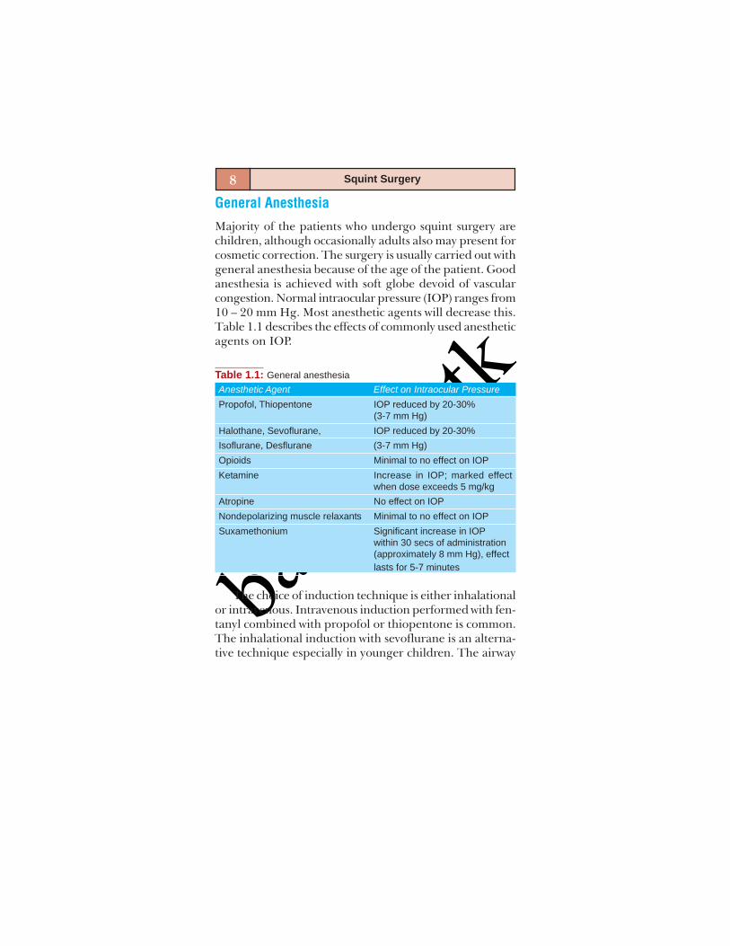
Majority of the patients who undergo squint surgery arechildren, although occasionally adults also may present forcosmetic correction. The surgery is usually carried out withgeneral anesthesia because of the age of the patient. Goodanesthesia is achieved with soft globe devoid of vascularcongestion. Normal intraocular pressure (IOP) ranges from10 – 20 mm Hg. Most anesthetic agents will decrease this.Table 1.1 describes the effects of commonly used anestheticagents on IOP.
Table 1.1: General anesthesiaAnesthetic Agent Effect on Intraocular PressurePropofol, Thiopentone IOP reduced by 20-30%
(3-7 mm Hg)Halothane, Sevoflurane, IOP reduced by 20-30%Isoflurane, Desflurane (3-7 mm Hg)Opioids Minimal to no effect on IOPKetamine Increase in IOP; marked effect
when dose exceeds 5 mg/kgAtropine No effect on IOPNondepolarizing muscle relaxants Minimal to no effect on IOPSuxamethonium Significant increase in IOP
within 30 secs of administration(approximately 8 mm Hg), effectlasts for 5-7 minutes
The choice of induction technique is either inhalationalor intravenous. Intravenous induction performed with fen-tanyl combined with propofol or thiopentone is common.The inhalational induction with sevoflurane is an alterna-tive technique especially in younger children. The airway
basm
ala.tk
9Anesthesia for Squint Surgery
is best managed by intubation with paralysis and controlledventilation. Access to the airway will be restricted duringthe surgery so it is important to secure the tracheal tubefirmly. A preformed RAE tubes or a reinforced flexible tra-cheal tubes are preferable. Nowadays the reinforced LMA(Laryngeal Mask Airway) are used for most of the eye pro-cedures. The advantages are reduced coughing at the endof the surgery and controlled ventilation with the use ofmuscle relaxants.
The nondepolarizing agents such as vecuronium arenormally preferred over suxamethonium for two reasons.Firstly, patients who have been given suxamethonium havea prolonged increase in the extraocular muscle tone, whichinterferes with the FDT (Foreced Duction Test). This effectof suxamethonium lasts roughly 15-20 minutes Secondly,patients undergoing correction of strabismus may be atincreased risk of developing malignant hyperthermia.Anesthesia is usually maintained with oxygen, N2O andSevoflurane/Propofol. As with induction, the choice ofmaintenance technique rests largely on the preferences ofthe anesthetist. Where halothane is used there is anincreased risk of dysrhythmias, particularly where eyepreparations containing adrenaline are used, and in thepresence of hypercapnia; isoflurane or sevoflurane maybe preferable.
Propofol has advantages in reducing the risk of post-operative nausea and vomiting (PONV) since propofol hasantiemetic effects. At the end of surgery, the nondepolarizingmuscle relaxant effect is reversed with neostigmine andglycopyrrolate. The use of glycopyrrolate is associated witha more stable cardiovascular system, fewer arrhythmias andsuperior control of oropharyngeal secretions at the timeof reversal.

basm
ala.tk
10 Squint Surgery
Anesthetic Challenges in Squint Surgery
Intraocular pressure: Anesthetic maneuvers like laryngo-scopy and intubation, straining and coughing duringinduction and bucking on the tube by inadequatelyparalyzed patient will have the effect of causing an increasein the intraocular pressure. Attenuation of this effect isessential to obtain a soft globe. This effect may beattenuated by a dose of lidocaine 1 mg/kg 3 minutes priorto intubation or extubation. Use of the LMA permitssmoother induction and emergence from anesthesia andhas much less effect on IOP.
Unobstructed ventilation and lowering the PaCO2 bymoderate hyperventilation during anesthesia and a slighthead up tilt ensure reduction in intraocular pressure andprevent venous congestion of the globe.
Hypoxia and hypercapnia both increase IOP andshould be scrupulously avoided.Oculocardiac reflex: Initially, described in 1908 by BernardAschner and Giuseppe Dagnini, the oculocardiac reflex iselicited by the pressure on the globe and by traction onthe conjunctiva, orbital structures and extraocular muscles,especially the medial rectus. It is a trigeminovagal reflex.It is characterized by sinus bradycardia, nodal rhythm,ectopic beats or sinus arrest. Afferent pathway is via thelong and short ciliary nerves to the ciliary ganglion termi-nating in trigeminal sensory nucleus in the floor of the4th ventricle. Efferent pathway is from the motor nucleusof vagus nerve via cardiac depressor nerve of 10th cranialnerve, which ends in myocardium. Pressure, torsion or pull-ing of extraocular muscle may elicit this reflex. Successiveprovocations decrease the reflex sensitivity.
Intraoperative management depends upon the severityof the reflex. The importance lies in its early recognition.
basm
ala.tk
11Anesthesia for Squint Surgery
On realizing this event the surgeon should relax the musclehook immediately. The bradycardia resolves almostimmediately after the stimulus has been removed. If itpersists the patient should be given intravenous atropine(0.15 mg/kg). In cases where the response to this treatmentis poor, a retrobulbar lignocaine is recommended to blockthe afferent loop. Sevoflurane is less likely to provoke thereflex than halothane; it is also less likely with deepanesthesia compared to light anesthesia. The incidence ofsignificant bradycardia is doubled if the carbon dioxidelevel is high, so controlled ventilation should be preferredover spontaneous breathing.Nausea and vomiting: PONV is most common after squintsurgery. The incidence of PONV after strabismus surgeryvaries from 46 to 88%. Most children have some pain aftereye surgery and should be given analgesics without anopioid as it induces emesis. Prophylactic antiemetic in thecombination of Inj. ondansetron 0.15 mg/kg with Inj.dexamethasone 100 ug/kg may be given along withpremedication.Postoperative pain: Even though most of the eye procedureshave mild to moderate pain, squint surgeries have moderatepain which require stronger intraoperative and postoperativeanalgesics. These include paracetamol, NSAID, intravenousfentanyl, and peribulbar or sub-Tenon’s block beforeemergence from the anesthesia.Others: Extubation should be considered in the deeperplane to avoid coughing and bucking in pediatric patients.
Malignant hyperthermia a rare complication is some-times associated with strabismus patients; it is commonlytriggered by inhalational agents such as halothane andsuccinylcholine.
basm
ala.tk
12 Squint Surgery
Local Anesthesia
Local anesthesia has become popular because of the in-creased use of adjustable sutures and the shift to day careprocedures. Lignocaine 2 to 4% and Bupivacaine 0.25 to0.75% are the commonly used anesthetic agents. It is oftenmixed with Epinephrine, 1:100,000 (or) Hyaluronidase.The choice depends on the anticipated duration of surgery.Adverse reaction can happen when the drug is accidentallyinjected into intravascular compartment, dural spreadthrough the optic nerve sheath and drug sensitivity.Methods aimed at reducing these complications includeproper techniques with careful positioning of the needle,aspiration before every injection and the use of a test dose.Direct injection into muscle can cause muscle necrosis. Theinferior oblique, inferior rectus and medial rectus are mostfrequently involved. This can occur in sub-Tenon’s injectionwhere the local anesthetic pools around the muscle. Therisk is reduced by the addition of hyaluronidase.
The choices of local anesthetic techniques are:Retrobulbar anesthesiaPeribulbar anesthesiaSub-Tenon’s anesthesiaTopical anesthesia
Retrobulbar Anesthesia
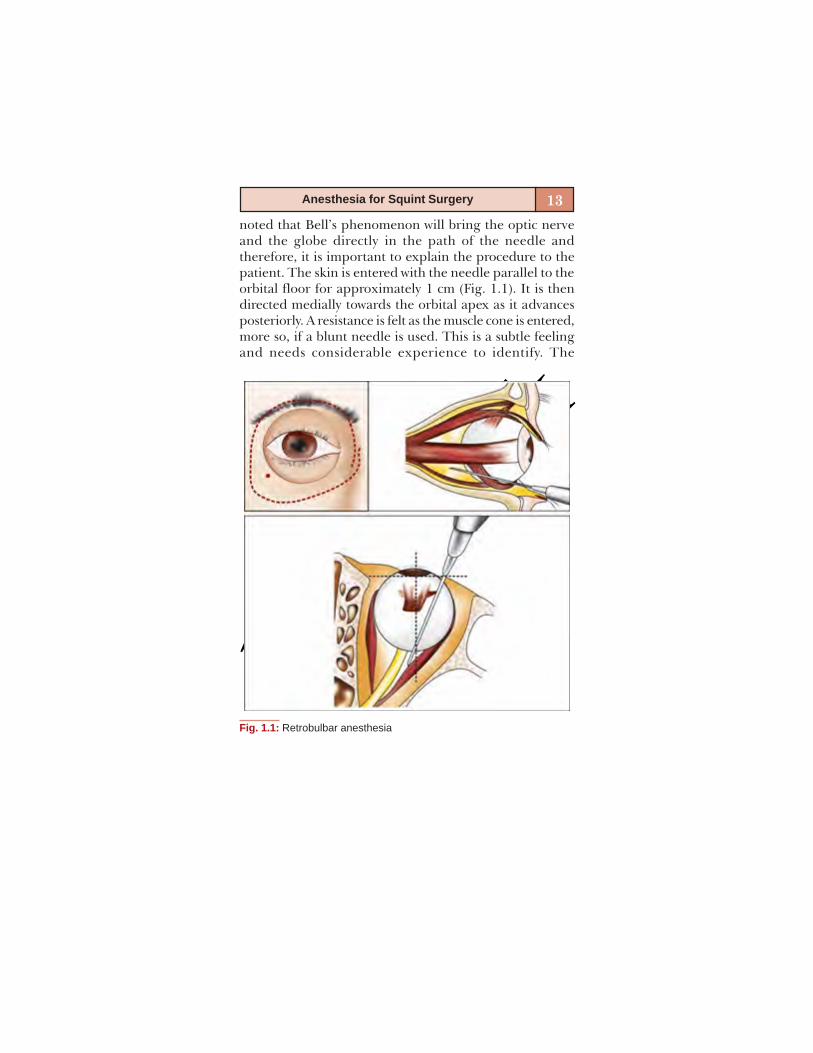
The technique involves instillation of anesthetic solutioninto the intraconal space. A 1.5 inch, 25 gauge needle isused. Blunt tipped needles such as the Atkinson’s needleare preferred as the risk of globe perforation is minimal.After skin preparation, the inferior orbital margin ispalpated to identify the junction between the outer 1/3rdand inner 2/3rd. The patient is asked to gaze straight ahead.This keeps the optic nerve out of Harm’s way. It should be
basm
ala.tk
13Anesthesia for Squint Surgery
noted that Bell’s phenomenon will bring the optic nerveand the globe directly in the path of the needle andtherefore, it is important to explain the procedure to thepatient. The skin is entered with the needle parallel to theorbital floor for approximately 1 cm (Fig. 1.1). It is thendirected medially towards the orbital apex as it advancesposteriorly. A resistance is felt as the muscle cone is entered,more so, if a blunt needle is used. This is a subtle feelingand needs considerable experience to identify. The
Fig. 1.1: Retrobulbar anesthesia

basm
ala.tk
14 Squint Surgery
injection is now given slowly. Usually 2-4 ml of solution isused. The needle is then withdrawn slowly and pressure isgiven on the globe over closed lids.
This technique achieves deep orbital anesthesia andakinesia and blocks the potential oculocardiac reflex witha minimal amount of anesthetic agent. The block effect isachieved in approximately 5 minutes. However, adjustmentof sutures cannot be done at the earliest as the return offull extraocular motility takes long time, and this techniquehas increased the incidence of associated morbidity.
Peribulbar Anesthesia
Here the anesthetic solution is injected in the extraconalspace. A 0.5 inch, 26 gauge disposable needle is preferred.More volume of anesthetic is needed and this technique isconsidered less effective than retrobulbar anesthesia. Theinjections are given at two sites. The first injection is givenin the inferotemporal quadrant at the junction of the lateral1/3rd and the medial 2/3rds. The needle is inserted parallelto the infraorbital margin along its entire length and maybe canted upwards once the equator of the globe is crossed.No attempt is made to enter the muscle cone however.Gentle sideways movement may be done to identifyengagement of the globe if any. Approximately 4-5 ml ofsolution is injected. The second injection is given in thesuperonasal quadrant at the junction of the lateral 2/3rdand medial 1/3rds. Again gentle sideways movement maybe done to identify any accidental perforation. Once theneedle is withdrawn, gentle pressure is given over the closedlids. The complications associated with this technique areless; but the risk of globe perforation exists, however it isless than with retrobulbar anesthesia. The block effect takeslittle longer than retrobulbar anesthesia, approximately10 minutes. Pupillary dilatation indicates that adequate

basm
ala.tk
15Anesthesia for Squint Surgery
anesthesia has been achieved, as the drug has diffused tothe ciliary ganglion.
Sub-Tenon’s Anesthesia
Sub-Tenon’s anesthetic technique is safe, when comparedto the other anesthetic techniques for strabismus surgeryperformed under local anesthesia (Fig. 1.2). The opticnerve function is not altered, which helps rapid visualrecovery for the patient and earlier adjustment ofadjustable sutures. The operating eye is prepared anddraped, topical Proparacaine 0.5% is applied on theconjunctiva. The conjunctival and Tenon’s incisions aremade; these incisions can be the same for the intended
Fig. 1.2: Sub-Tenon’s anesthesia
basm
ala.tk
16 Squint Surgery
strabismus surgery. Local anesthetic is injected into thesub-Tenon’s space through an irrigation syringe. The onsetof block is as fast as retrobulbar anesthesia, and the blockis usually performed in the course of surgery.
Topical Anesthesia
Strabismus surgery may be performed in select adultpatients under topical anesthesia alone. Success dependson the experience of the surgeon and careful patientcounseling and selection. This technique offersconsiderable advantage to the surgeon as single stageadjustment can be done right after the conclusion of thesurgery. In addition, this technique obviates all thecomplications described associated with local anesthesia.
Various drugs including 4% lignocaine, amethocaine,and proparacaine have been used. Lignocaine Jelly 2%has been reported to give superior analgesia. Thelignocaine jelly is applied to fill the fornices at least 20minutes prior to the procedure. The application is repeatedprior to the commencement. The patient is asked to gazeaway from the muscle being operated to gain maximumview. It is important to avoid excessive traction on themuscle throughout the procedure as this can cause severepain. The services of an experienced assistant will beinvaluable at this juncture. It is better to avoid operatingon the obliques and the superior rectus with topicalanesthesia. It is also better to use complete regional/generalanesthesia for resurgeries and complicated strabismusprocedures. The surgery otherwise proceeds as it normallywould. Should the patient report pain at any point? thesurgeon should not hesitate to supplement with topicalproparacaine/sub-Tenon’s lignocaine. It is important toremember that excessive supplementation can cause
basm
ala.tk
17Anesthesia for Squint Surgery
corneal toxicity and alter the contractility of the musclewhich may require the adjustment procedure to be delayed.The jelly is washed with saline at the end of the procedure.Supplementation of oral analgesics to the patients at theconclusion of the surgery is often helpful.
It is possible to augment topical anesthesia with theaid of sub-Tenon’s instillation of anesthetic solution or withintravenous sedation. In monitored sedation, patient isgiven Inj. Fentanyl 1microgram/kg with the continuousinfusion of Inj. Propofol at the dose of 6 to 8 mg/kg/hrthrough the infusion pump. The patient is monitored withpulse oxymeter and blood pressure.
Monitored sedation is not a substitute for good patientcounseling/selection. It is also not intended to replace gen-eral/topical anesthesia and only serves to make thesurgeon’s job a little easier. The speedy recovery affordedby the newer sedatives, facilitate early adjustment. Thepatient should be made to understand that he/she will begiven mild sedation and that his/her cooperation is cru-cial to the success of the procedure.
CONCLUSION
Eye is a delicate and sensitive organ. Consequently, anyophthalmic intervention including anesthesia has to be veryrefined and skilfully administered. Faulty maneuvers duringanesthesia may negate the benefits expected from surgery.
Careful attention to IOP and vascularity is essential;smooth extubation without coughing or straining is of para-mount importance. Complete elimination/considerableattenuation of PONV is strongly indicated and the anes-thetic technique and drugs administered should facilitatethe achievement of this ideal. Adequate pain relief withdrugs which do not induce PONV is indicated.

basm
ala.tk
18 Squint Surgery
In short, ophthalmic anesthesia should not only enableeventless surgery but also should aid and facilitate quickfunctional recovery postoperatively.
BIBLIOGRAPHY1. Anesthesia for correction of strabismus – issue 17(2003) Article
9;P 1.2. Anesthesia for pediatric eye surgery- Anesthesia tutorial of
the week 144-2009- Dr. Grant Stuart, Great Ormond StreetHospital, London, UK.
3. Greenbaum Ocular Anesthesia—Anesthesia for Eye MuscleSurgery; pp 125-150.
4. Ophthalmology Clinics of North America 2006;19(2):269-78.
5. Wylie and Churchill-Davidson’s “A Practice of Anesthesia”,6th edn, pp 1265-81.
basm
ala.tk
2
STRABISMUSANATOMICAL PEARLS
Sumita Agarkar

basm
ala.tk
20 Squint Surgery
A clear understanding of the anatomy including the extra-ocular muscles, periocular fascia and orbit is a pre-requisiteto successful strabismus surgery. The conjunctiva, anteriorand posterior Tenon’s capsule and muscle sheath play animportant part in movement of the globe. These structuresperform a passive role in initiation of movement but theyplay an active role in restriction of ocular movements.
The anatomical pearls are described under the follow-ing headlines:
Palpebral fissureConjunctivaTenon’s capsuleMusclesSclera
PALPEBRAL FISSURE
The dimensions of palpebral fissure increase nearly 50%in width (from 18 mm in newborn to 28 in adult) and 20%in height (from 8 to 10 mm) between infancy andadulthood. Importance of fissure size lies in choosing theright sized speculum for adequate exposure during surgery.Further, the shape of palpebral fissure (mongoloid,antimongoloid) causes the appearance of patterns insquints (A or V pattern) and it should be considered duringsurgical planning.
CONJUNCTIVA
The bulbar conjunctiva loosely covers the anterior part ofthe globe from the fornices above and below and fromthe canthi medially and laterally. It becomes fused withthe anterior Tenon’s capsule and sclera at the limbus.

basm
ala.tk
21Strabismus: Anatomical Pearls
Conjunctiva is thick in infancy and childhood and becomesthin and more friable in adulthood. This friability of con-junctiva precludes the use of fornicial incision in elderly.
Important surgical landmarks are:a : Plica semilunarisb : Carunclec : Fusion with underlying structuresd : Fat padCare should be taken to keep the position of plica and
caruncle undisturbed while incising and repairing conjunc-tiva. Plica should not be placed laterally making it moreobvious as an unsightly mass in the palpebral fissure.
Anterior to inferior fornix a fat pad is present thatextends to within 12-14 mm of limbus. It is situated beneaththe conjunctiva and its posterior condensations and isoutside both the layers of Tenon’s capsule. Atransconjunctival incision made medially or laterally shouldbe made posterior to the line of attachment of posteriorTenon’s capsule or at least 8 mm from the limbus but anteriorto inferior fat pad or no more than 12 mm from the limbus.
During extraocular muscle surgery incisions should belimited to the bulbar conjunctiva and should not extendto the forniceal or palpebral conjunctiva as this causesunnecessary bleeding and serves no purpose. The parks’cul-de-sac incision or fornix incision of extraocular musclesis actually made in the bulbar conjunctiva.
When a prior surgery has left the conjunctiva reddenedand unsightly scarred which limits its mobility, the con-junctiva should be recessed with or without removal oftissue. If the sclera is left uncovered it becomes quicklycovered with epithelium with out the need for any grafts.
basm
ala.tk
22 Squint Surgery
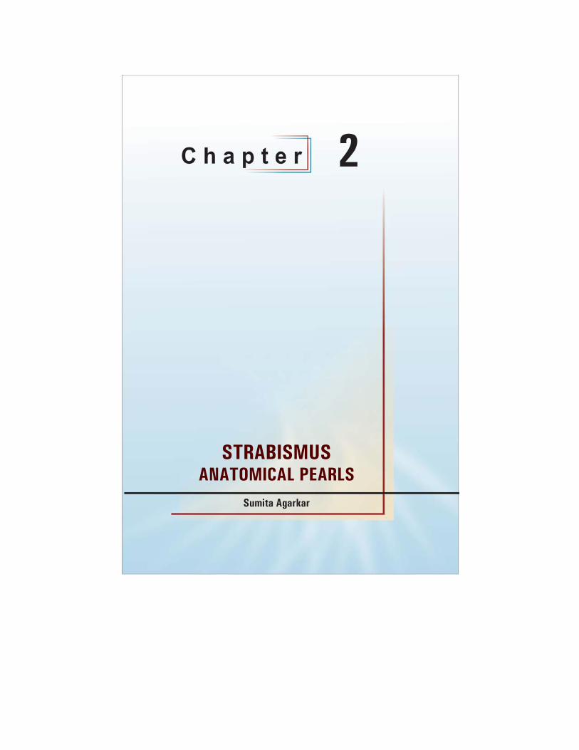
TENON’S CAPSULE (FIGS 2.1 AND 2.2)
Fig. 2.1: Tenon’s capsule. (1) Limbal fusion of conjunctiva and anteriorTenon’s capsule, (2) Potential space between anterior Tenon’scapsule and sclera, (3) Muscle in its sheath (posterior Tenon’scapsule), (4) Postinsertional muscle footplates, (Courtesy: Atlasof Strabismus Surgery by Eugene Helveston)
Fig. 2.2: Coronal section of Fig. 2.1 at X: (1) Conjunctiva, (2) AnteriorTenon’s capsule, (3) Muscle sheath, (4) Extraocular muscle,(5) Intermuscular membrane, (6) Sclera. (Courtesy: Atlas of Stra-bismus Surgery by Eugene Helveston)
basm
ala.tk
23Strabismus: Anatomical Pearls
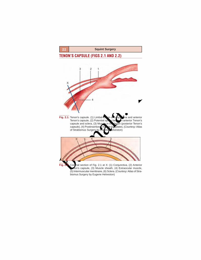
It is a thick structure with definite body and substancein childhood and becomes fibrillar and friable in old age.It is divided into anterior and posterior parts. AnteriorTenon’s is considered to be the vestigial capsulopalpebralhead of rectus muscles. It overlies the anterior half of rectusmuscles, intermuscular spaces and fuses with conjunctivaat the limbus.
Posterior Tenon’s capsule is composed of fibrous sheathof rectus muscles together with intramuscular septum. TheTenon’s capsule gets reflected on to the extra capsularportion of EOM for up to 12 mm backwards forming themuscular sheath. The muscle sheaths of the 4 recti musclesare connected by fibrous membrane known as intermuscu-lar membrane, closely relating to each other. The blendingof sheaths of IR and IO and its extensions on either side tosheaths of MR and LR form a suspending hammock ‘sus-pensory ligament of lockwood’ supporting the eyeball.
Fibrous attachments between the inner surface ofanterior Tenon’s capsule and the outer muscle sheath arecalled check ligaments. Other attachments between outersurface of anterior Tenon’s capsule and the orbital rimmedially and laterally are also called check ligaments.
Clinical Importance
The connective tissue bands between recti and adja-cent oblique muscle helps in locating the lost or slippedmuscle. The absence of such fascial connections aroundmedial rectus makes its retrieval extremely difficult iflost or slipped. Further a lost muscle slips into its sleeveonce detached, so locating the muscle sleeve/sheathforms first step to locate the muscle.
basm
ala.tk
24 Squint Surgery
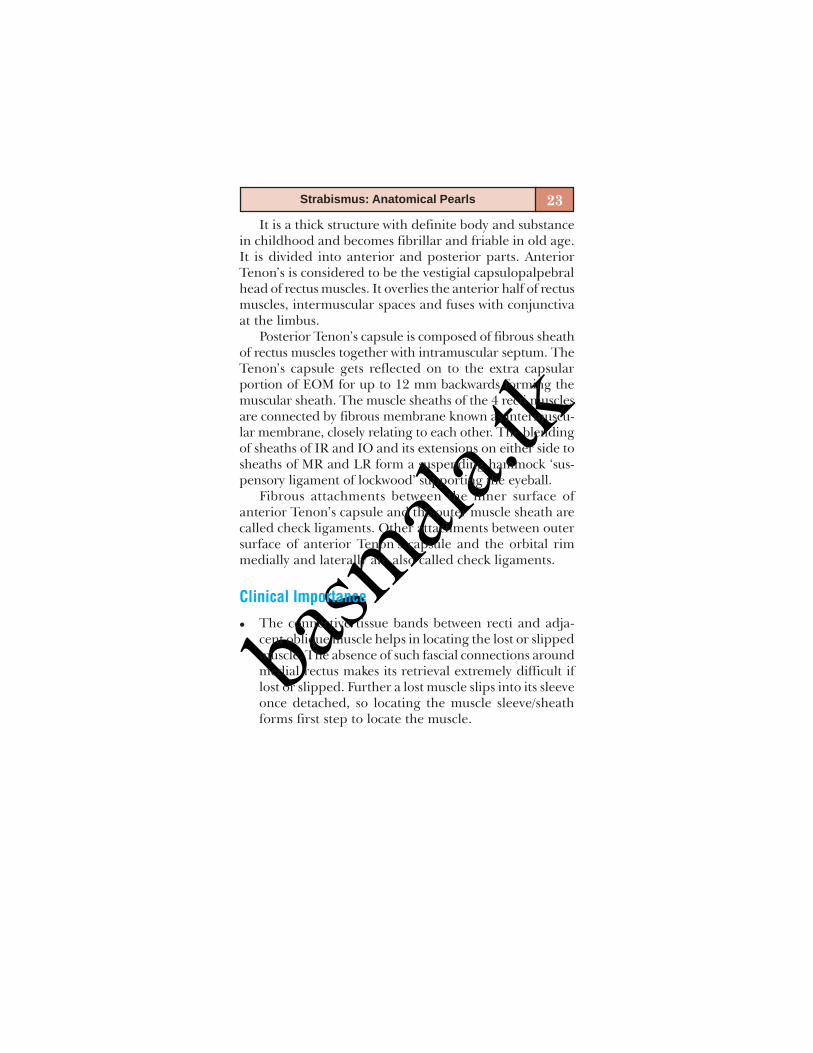
The clearing of anterior Tenon’s capsule off the tendoninsertion is needed in order to prevent the slippedmuscle. The surgeon may inadvertently secure the an-terior Tenon’s capsule with sutures instead of the ten-don fibres if it is not cleared off, thus allowing unse-cured muscle to slip posteriorly.Posterior Tenon’s capsule barrier keeps orbital fat sepa-rated from the globe and EOM. The violation of thisbarrier results in fat prolapse through torn Tenon’s cap-sule and scars to the sclera or EOM, i.e. fat adherencesyndrome. This can be prevented by carefully dissect-ing close to muscle belly or sclera and thus not disturb-ing posterior Tenon’s integrity (Fig. 2.3).
Fig. 2.3: Insertion of posterior Tenon’s capsule at the level of rectusmuscle insertions. (Courtesy: Atlas of Strabismus Surgery byEugene Helveston)

basm
ala.tk
25Strabismus: Anatomical Pearls
The check ligaments connect superior and inferior rectito the levator muscle and lower lid retractors respec-tively. A recession or resection of vertical rectus musclesrequires removal of these ligaments in order to avoidlid fissure changes after surgery.
MUSCLES (FIGS 2.4 AND 2.5)
Fig. 2.4: Actions of extraocular muscles—right eye. (Courtesy: SurgicalTechniques in Ophthalmology-Strabismus Surgery, Jaypee-Highlights)

basm
ala.tk
26 Squint Surgery
Horizontal Rectus Muscles
Medial Rectus
Originates from the annulus of Zinn at the orbital apexand inserts 5.5 mm from the limbus. Just above the musclelies the ophthalmic artery, the superior ophthalmic veinand the nasociliary nerve.
Its sole action in primary position is adduction.
Surgical Points
The vertical action can be brought into play during sur-gery by transposing the muscle insertion up or down toproduce elevating or depressing effect respectively.
Fig. 2.5: Location and width of extraocular muscle insertions in mm
basm
ala.tk
27Strabismus: Anatomical Pearls
Medial rectus has no fascial attachments to othermuscles and can therefore retract to apex of the orbit oncesevered from the globe.
The insertion of MR varies from 3-6 mm and this vari-ability of insertion makes it a poor landmark for measure-ment during recession. Thus, the use of limbus as the pointof reference for recession of MR as suggested by Helvestonet al.
Medial rectus is a tight muscle and has limited contactwith the globe. This shorter arc of contact makes it ideal tobe weakened by faden procedure. The terminal tendinousportion is only 4 mm long. Resections >4 mm cause morebleeding and if greater than 6 mm it will result in limitedabduction, narrowing of the palpebral fissure and retrac-tion of the globe. Similarly, recessions more than 6 mmwill cause limited adduction.
Lateral Rectus
This muscle lies adjacent to lateral orbital wall and runs atan angle of 56° from the medial rectus and visual axis. Theinsertion is 6.9 mm from the limbus and the primary ac-tion is abduction. Just above the muscle lies lacrimal arteryand nerve.
Surgical Points
Recession >7 mm can limit abduction where as resectionscan limit adduction if greater than 8 mm.
Attachments between the lower border of lateral rectusand inferior oblique should be freed during resection toprevent damage to inferior oblique or anterior draggingof muscle.
basm
ala.tk
28 Squint Surgery
Vertical Rectus Muscles
The vertical recti run in line with the orbital axis and areinserted in front of the equator. They form an angle of 23°with the visual axis.
Superior Rectus
Originates from the upper part of the annulus of zinn andinserts 7.7 mm from the equator. The primary action iselevation and the secondary actions are adduction andintorsion.
Surgical Points
Recessions more than 5 mm limit upgaze. In recessing themuscle, there are fascial attachments between the muscleand superior oblique, which must be divided before theprocedure to be effective. Resections of 5 mm or more canlimit downgaze.
Embryonically the superior rectus stems from the samemesoderm as the levator muscle which lies above and runsparallel to the superior rectus.Its fascial connections tolevator are to be dissected to avoid lid fissure changes assaid earlier.
Inferior Rectus
Originates at the inferior part of Annulus of Zinn andinserts 6.5 mm behind inferior limbus.
The primary action is depression and secondary actionsbeing adduction and extorsion.

basm
ala.tk
29Strabismus: Anatomical Pearls
Surgical Points
The capsule is connected to inferior oblique, the lid struc-tures through check ligaments and through attachmentsto lockwood’s ligament all of which must be freed duringrecession to limit lower lid retraction.
Recessions greater than 5 mm can limit downgaze. Re-sections greater than 5 mm can draw up the lid narrowingthe palpebral fissure.
Spiral of Tilaux
The imaginary line joining the insertions of four recti. Itis an important anatomical landmark when performingsurgery.
Oblique Muscles
Inferior Oblique
Originates at the medial end of inferior orbital rim at theouter crest of lacrimal fossa- proceeds temporally and pos-teriorly at an angle of 50° beneath inferior rectus (Fig. 2.6).
Inserts beneath inferior border of lateral rectus 12 mmfrom the insertion of inferior rectus. The posterior extentof insertion lies 2 mm below and lateral to macula.
Surgical Points
The inferior vortex vein leaves the sclera 8 mm posteriorto the inferior rectus insertion along its temporal borderloops just posterior to the inferior oblique and must beavoided.
Blood vessels that supply inferior oblique will notcontribute to the blood supply of anterior segment.
basm
ala.tk
30 Squint Surgery
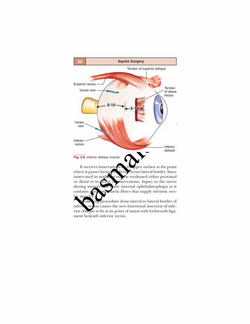
Fig. 2.6: Inferior oblique muscle
It receives innervations on its upper surface at the pointwhere it passes beneath inferior rectus lateral border. Sinceinnervated in middle it may be weakened either proximalor distal to its point of innervations. Injury to the nerveduring surgery can cause internal ophthalmoplegia as itcontains parasympathetic fibres that supply intrinsic ocu-lar muscles.
Weakening procedure done lateral to lateral border ofinferior rectus causes the new functional insertion of infe-rior oblique to be at its point of union with lockwoods liga-ment beneath inferior rectus.

basm
ala.tk
31Strabismus: Anatomical Pearls
Superior Oblique
The superior oblique muscle has a muscular part and atendinous part both of which are 30 mm long. Originatesat annulus of Zinn, becomes tendinous 10 mm posteriorto trochlea, then it passes posteriorly and temporallybeneath superior rectus to insert near lateral border ofsuperior rectus. The average anterior point of insertion is13 mm from the imbus. The SO tendon between trochleaand medial border of superior rectus is 3 mm in diameterand covered by dense fascia which may cause difficulty inidentifying it (Fig. 2.7).
The insertion of superior oblique is variable. Insertionnear medial border of superior rectus, absence of superior
Fig. 2.7: Superior oblique tendon

basm
ala.tk
32 Squint Surgery
oblique tendon also reported. Helveston has classified theanatomical variations of SO into four types.
Whitnall’s ligament (superior transverse ligament) andsuperior oblique tendon have common fascial attachmentsnear the trochlea. If it is weakened inadvertently whilehooking superior oblique tendon, can lead to ptosis of nasalportion of upper lid. So, it is safer to hook the same underdirect vision and preferably at its insertion.
The site of superotemporal vortex vein is 8 mm behindthe equator and close to the insertion of superior obliqueand it must be avoided during surgery.
Blood Supply
Anterior ciliary arteries travel in the four rectus muscles.Each of the rectus muscle has got two anterior ciliaryarteries with the exception of the lateral rectus which hasone. As a general rule not more than two rectus musclesshould be detached at one operation as it can lead to seg-mental iris atrophy in older individuals (Fig. 2.8).
The four vortex veins are located behind the equator afew millimetres on each side of the superior rectus andinferior rectus muscles. Every effort must be made to avoidsevering a vortex vein. If a vortex vein is cut pressure shouldbe applied to prevent bleeding. Cautery should be avoided.
SCLERA (FIG. 2.9)
The thickness of the sclera varies depending on the siteat the limbus—0.8 mmanterior to rectus muscle insertion—0.6 mmposterior to rectus muscle insertion—0.3 mmat equator—0.5 mmat posterior pole—1 mm thick.

basm
ala.tk
33Strabismus: Anatomical Pearls
Fig. 2.9: Thickness of sclera at different sites. (Courtesy: EugeneHelveston)
Fig. 2.8: Anterior ciliary blood vesels. Cross-section relationship of vesselsto sclera (S), muscle and intermuscular septum (M), Tenon’s fascia(T) and conjunctiva (C). (Courtesy: Complications in OphthalmicSurgery)

basm
ala.tk

basm
ala.tk
3
STRABISMUSEVALUATION
Sumita Agarkar

basm
ala.tk
36 Squint Surgery
The goals of a strabismus evaluation are:To establish a cause for strabismus, i.e. whether infan-tile esotropia, restrictive or paralytic causes.To assess binocular sensory status.To measure the deviation.To diagnose amblyopia.Thus, an evaluation which is done with these goals in
mind will prevent lengthy examination which often resultsin an uncooperative patient and a confused clinician. It isequally true that despite a focused evaluation, patient’sstrabismus may refuse to fall in a specific category.
The order for examination of a patient with strabismusis:
HistoryVisual acuitySensory testsMeasurement of deviationDuctions and versionsSpecial testsCycloplegic refractionFundus examination
HISTORY
A detailed history is a must before you start examining thepatient. History taking should be based on patient’s chiefcomplaints like squinting, diplopia or asthenopia. Durationof squinting, age of onset of squint, any apparentprecipitating factors like trauma or febrile episodes orcerebrovascular accident should be asked for.
Past history should include history of patching, spec-tacle wear, any history of trauma, any history of previoussurgery for strabismus, cataract, retinal detachment, blow
basm
ala.tk
37Strabismus Evaluation
out fracture, glaucoma implant or any periocular surgerylike sinus surgery or neurosurgery.
Birth history should include – age of gestation, birthweight and any significant events in antenatal and postna-tal period. There should be a mention of developmentalmilestones if it is normal or delayed.
Family history is extremely important in cases of certainhereditary forms of strabismus and response of other familymembers to surgery will give a clue to patient’s response tosurgery.
Leading questions should be asked about associatedneurological signs and symptoms like seizures, ataxia,muscle weakness, fatigue, ptosis, etc.
VISUAL ACUITY (VA)
This is one of the most important parts of the examina-tion. It can often be a test of patience for the examiner ifthe patient happens to be a child. Visually a newborn has aVA of 6/240 which increases to 6/90 by 1st month. By ageof 4-6 months VA varies between 6/18-6/6 and by 3 years itshould be 6/6. Behavior of child can often give a clue tovisual acuity especially in extremely young children. Bythe age of one month most infants turn eyes and head atlight source and can track light source horizontally.Preverbal children: In preverbal children visual acuity canbe assessed in following ways:
Fixation Pattern
Fixation should be central, steady and maintained. Unsteadyor wandering or eccentric fixation usually indicates poorvisual acuity. In a patient with strabismus, fixation preference
basm
ala.tk
38 Squint Surgery
for a particular eye also indicates poor visual acuity in theother eye.
Fixation preference can also be tested by using a verti-cal prism. In this test, we place a vertical prism of 15PDbase up or base down in front of one eye, which induces avertical strabismus and look for refixational movement.For example, if we place the prism base down in front ofthe right eye and there is a refixational movement in up-ward direction, then it indicates right eye is fixing but italso indicates that left eye is not fixing. This test is usefulto detect amblyopia in straight eyes, or those in whom angleof strabismus is small.
Optokinetic Nystagmus (OKN)
It can be used to assess visual acuity in very young children.Highest spatial frequency which produces a response canbe quantified to give visual acuity. It has limitations in thesense, that target must be presented in a rigidlystandardized condition which is difficult in clinical settings.Moreover, OKN response has a sensory and a motorcomponent so an infant with a normal sensory system maystill have abnormal OKN, if a motor problem exists.Another fallacy of the test is that OKN response may benormal in cases of cortical blindness.
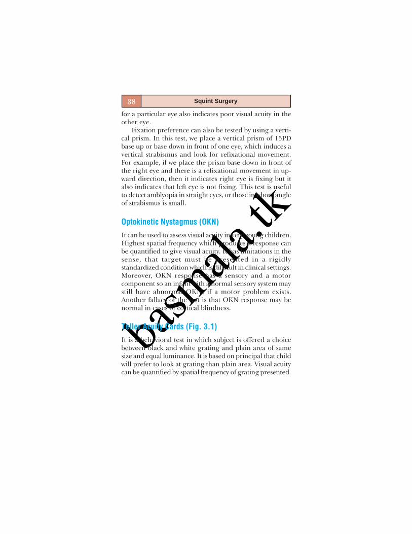
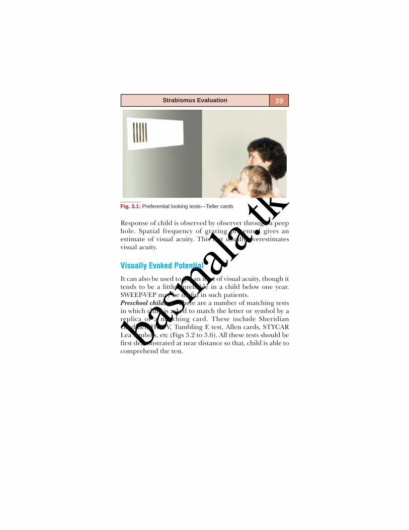
Teller Acuity Cards (Fig. 3.1)
It is a behavioral test in which subject is offered a choicebetween black and white grating and plain area of samesize and equal luminance. It is based on principal that childwill prefer to look at grating than plain area. Visual acuitycan be quantified by spatial frequency of grating presented.
basm
ala.tk
39Strabismus Evaluation
Response of child is observed by observer through a peephole. Spatial frequency of grating presented gives anestimate of visual acuity. This test usually overestimatesvisual acuity.
Visually Evoked Potential
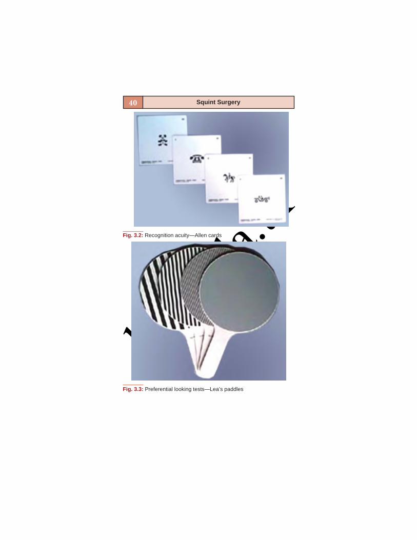
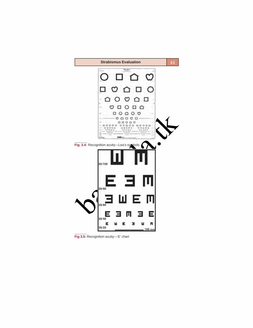
It can also be used to get an idea of visual acuity, though ittends to be a little unreliable in a child below one year.SWEEP-VEP may be useful in such patients.Preschool children: There are a number of matching testsin which child is asked to match the letter or symbol by areplica or a matching card. These include SheridianGardiner, HOTV, Tumbling E test, Allen cards, STYCARLea symbols, etc (Figs 3.2 to 3.6). All these tests should befirst demonstrated at near distance so that, child is able tocomprehend the test.
Fig. 3.1: Preferential looking tests—Teller cards
basm
ala.tk
40 Squint Surgery
Fig. 3.3: Preferential looking tests—Lea’s paddles
Fig. 3.2: Recognition acuity—Allen cards
basm
ala.tk
41Strabismus Evaluation
Fig. 3.4: Recognition acuity—Lea’s symbols
Fig 3.5: Recognition acuity—‘E’ chart

basm
ala.tk
42 Squint Surgery
Older children: VA can be tested using Snellen’s chart ornear vision test.Visual acuity in nystagmus: It is difficult to assess monocu-lar vision in a patient with nystagmus. Because occluderplaced over one eye often worsens the nystagmus andcauses decline of VA so while recording VA, we have toprovide some peripheral binocular clues to preventworsening of nystagmus, at the same time permit monocu-lar assessment of vision. It can be done by remote occlusion,i.e. occluder is placed at some distance in front of one eyeor we can use high plus lenses to check the other eye. Neu-tral density filters can also be used. Binocular VA shouldalso be recorded because that is often better than monocu-lar VA.
Fig. 3.6: Sheridan-Gardiner test

basm
ala.tk
43Strabismus Evaluation
SENSORY TESTS
It is an integral part of strabismus evaluation. It is donewith patient wearing full refractive correction. It includes:1. Tests for stereopsis2. Tests for retinal correspondence3. Tests for suppression.
Tests for Stereopsis
Tests for stereopsis usually incorporate two essentialfeatures: They dissociate eyes, i.e. each eye is presentedwith a separate field of view and each of the two viewsmust contain elements imaged on corresponding retinalareas. Stereopsis must be noted for near as well as fordistance. Distance stereoacuity often gives a betterindication of control of intermittent squints.Near stereoacuity (Fig. 3.7): It is assessed by followingmethods:
Titmus stereo testRandot stereograms/TNOLang’s testFrisby test
Fig. 3.7: Sensory tests—stereoacuity
basm
ala.tk
44 Squint Surgery
Titmus stereo test: There are vectograph cards which dis-sociate eyes optically. Patient needs to wear polaroid glassesto appreciate stereo images. It is simple and can be usedin clinical setting. Only disadvantage is that this test hasmonocular clues, it tends to overestimate stereopsis.Randot stereograms: These stereograms are devoid ofmonocular clues. Patient needs to use polaroid glass. TNOtest is also based on random dot but patient needs to wearred green glasses to get stereoscopic effect.Lang’s test: It is useful in children who refuse to wear red,green or polaroid spectacles. It is based on pantographicpresentation of random dot pattern. Eyes are dissociatedthrough the cylindrical elements imprinted on surfacelamination of card.Frisby test: It can be used for assess stereopsis for near. Italso does not require special spectacle.Distance stereoacuity: It can be measured by AmericanOptical Vectograph which uses polarized glasses. MentorB Vat system is new addition to assess stereopsis for dis-tance. It is a computerized system in which liquid crystalbinocular glasses are provided which are connected to amicroprocessor. Each eye is presented with disparate im-ages at a high frequency. So that stereopsis is achieved.
Tests for Retinal Correspondence
Retinal correspondence is ability of sensory system toappreciate the perceived direction of fovea in each eye,relative to the other eye. The two eyes have correspondingretinal elements that have a common visual direction. Forexample, both foveas share the straight ahead direction.
Normal retinal correspondence (NRC) is seen in straighteyes or when subjective and objective angles of deviation
basm
ala.tk
45Strabismus Evaluation
are same. In anomalous retinal correspondence(ARC),fovea of deviating eye loses common visual direction withthe fovea of fixing eye, and fovea of fixing eye shares acommon visual direction with a peripheral retinal elementof the deviating eye. Anomalous retinal correspondencecan be harmonious or unharmonious. Retinal correspon-dence can be assessed by using:
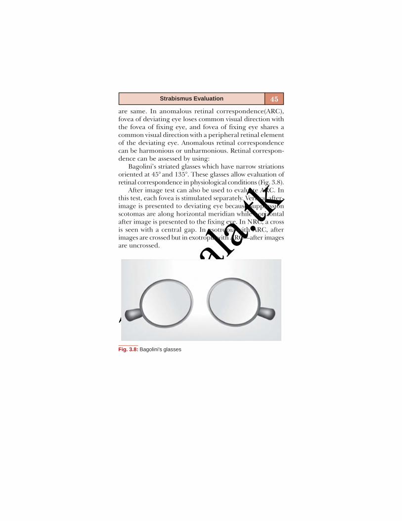
Bagolini’s striated glasses which have narrow striationsoriented at 45° and 135°. These glasses allow evaluation ofretinal correspondence in physiological conditions (Fig. 3.8).
After image test can also be used to evaluate ARC. Inthis test, each fovea is stimulated separately. Vertical after-image is presented to deviating eye because suppressionscotomas are along horizontal meridian while horizontalafter image is presented to the fixing eye. In NRC, a crossis seen with a central gap. In esotropia with ARC, afterimages are crossed but in exotropia with ARC—after imagesare uncrossed.
Fig. 3.8: Bagolini’s glasses

basm
ala.tk
46 Squint Surgery
Major amblyoscope can also be used to assess retinalcorrespondence. If subjective and objective angle ofdeviation are same, it indicates NRC (Fig. 3.9).
Worth 4 dot test can also be used to assess anomalousretinal correspondence. If patient reports four lights inpresence of manifest deviation, it indicates anomalousretinal correspondence.
Tests for Suppression
Suppression is alteration of visual sensation resulting frominhibition of one eye’s images from reaching conscious-ness. It is a sensory adaptation to avoid diplopia. It is apurely binocular phenomenon. Suppression can be cen-tral or peripheral, monocular or alternating, facultativeor obligatory.
Fig. 3.9: Major amblyoscope—Synoptophore
basm
ala.tk
47Strabismus Evaluation
MEASUREMENT OF DEVIATION
Worth 4 Dot Test (Fig. 3.10)
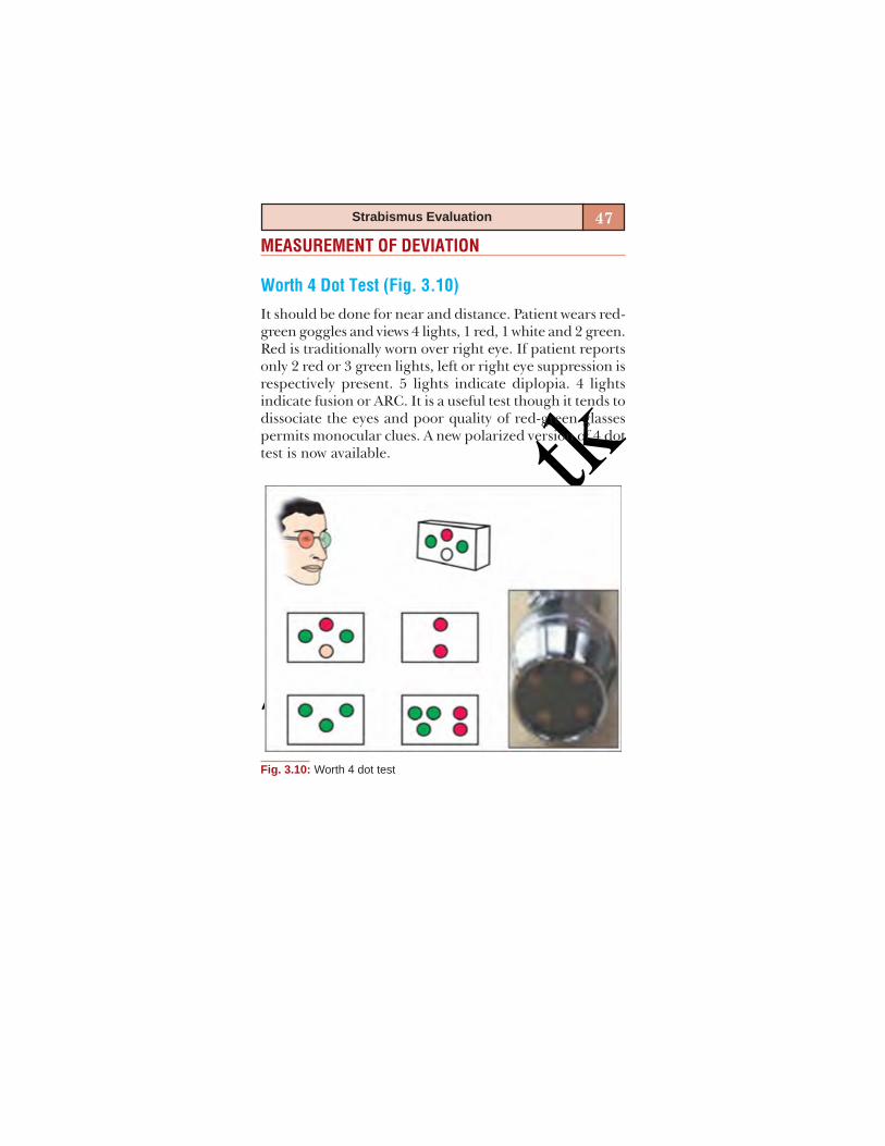
It should be done for near and distance. Patient wears red-green goggles and views 4 lights, 1 red, 1 white and 2 green.Red is traditionally worn over right eye. If patient reportsonly 2 red or 3 green lights, left or right eye suppression isrespectively present. 5 lights indicate diplopia. 4 lightsindicate fusion or ARC. It is a useful test though it tends todissociate the eyes and poor quality of red-green glassespermits monocular clues. A new polarized version of 4 dottest is now available.
Fig. 3.10: Worth 4 dot test

basm
ala.tk
48 Squint Surgery
4 Prism Diopter Baseout Test
This test demonstrates small foveal suppression scotomasassociated with microtropias. In a person with bifovealfixation if a 4 prism diopter base- out prism is placed infront of right eye, right eye will move nasally and left eyetemporally followed by a refixational movement in left eye.(i.e. eye without prism). If a suppression scotoma exists inright eye, the placing of prism will not elicit any movementin either eye. While, if we place the prism in front of lefteye, right eye will show initial conjugate saccade towardsapex of prism but there will be no refixational movements.
Bagolini’s glasses can also detect suppression. Patientwill perceive only 1 line.
Motor Testing
Head Posture (Fig. 3.11)
Comitant heterotropias usually have a normal headposture. Abnormal head posture is seen in incomitantsquints, A/V patterns, nystagmus or in some strabismic
Fig. 3.11: Compensatory head posture— Face turn, head tilt, chin up/down or combination of the above
basm
ala.tk
49Strabismus Evaluation
entities like Duane’s or Brown’s syndrome. Anomalous headposture is adopted either to avoid diplopia or achievebinocularity. Abnormal head posture can take form of faceturns or head tilts. There can be a chin elevation ordepression. However, nonocular causes of anomalous headposture like hearing loss, psychogenic, torticollis shouldalso be kept in mind.
Position of Lids
Position of lids and palpebral fissure should also be exam-ined. Any ptosis or pseudoptosis should be differentiatedand recorded.
Binocular Motor Functions
There are four types of ocular alignment tests.Corneal light reflex testsCover testsDissimilar image testsDissimilar target tests.
Corneal Light Reflex Tests
These tests are useful to assess ocular alignment in patientswith poor cooperation and those patients who have poorfixation.
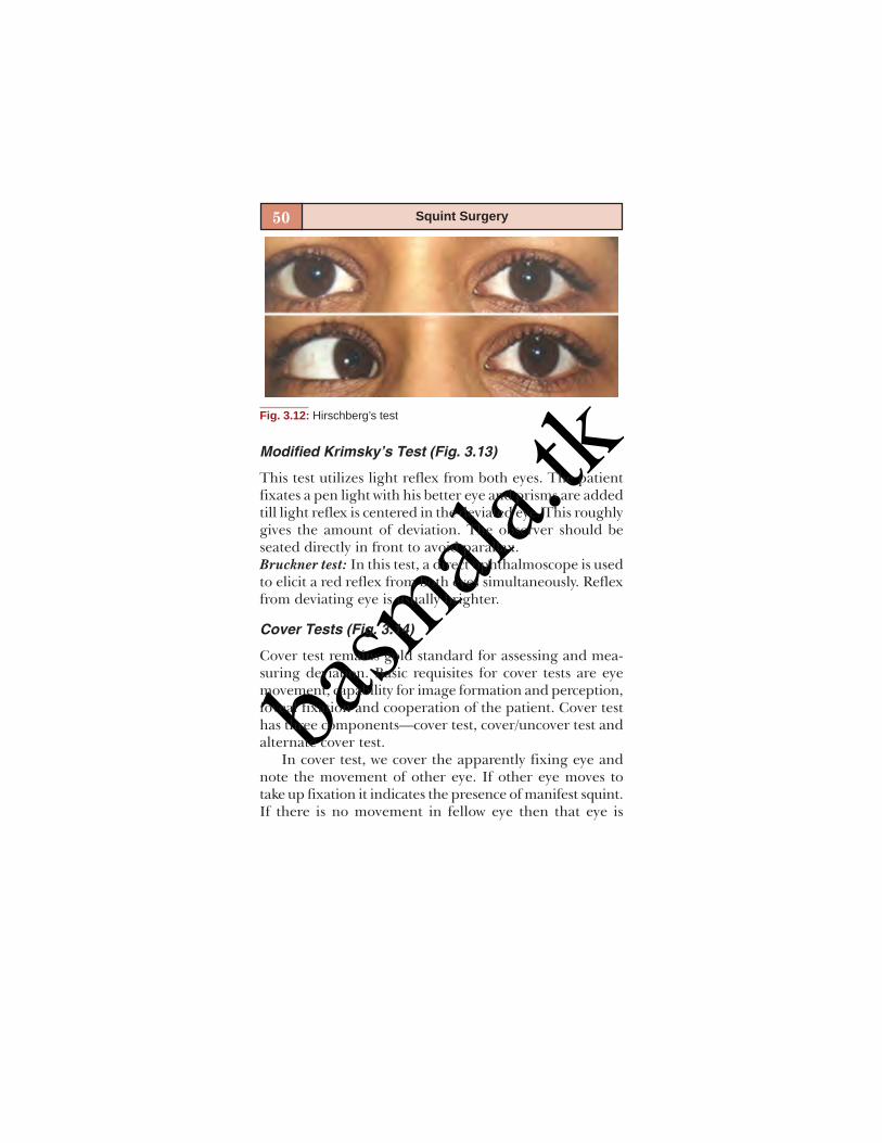
Hirschberg’s Test (Fig. 3.12)
This test is based on premise that 1 mm shift in light reflexfrom the center is equal to 7° of deviation of visual axis.Therefore, a light reflex at pupillary margin which is 2 mmaway from center corresponds to 15° deviation. In mid irisregion it is equal to 30° and at limbus it corresponds to 45°deviation.
basm
ala.tk
50 Squint Surgery
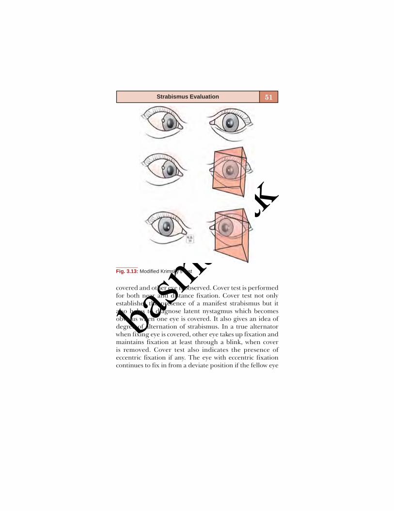
Modified Krimsky’s Test (Fig. 3.13)
This test utilizes light reflex from both eyes. The patientfixates a pen light with his better eye and prisms are addedtill light reflex is centered in the deviated eye. This roughlygives the amount of deviation. The observer should beseated directly in front to avoid parallax.Bruckner test: In this test, a direct ophthalmoscope is usedto elicit a red reflex from both eyes simultaneously. Reflexfrom deviating eye is usually brighter.
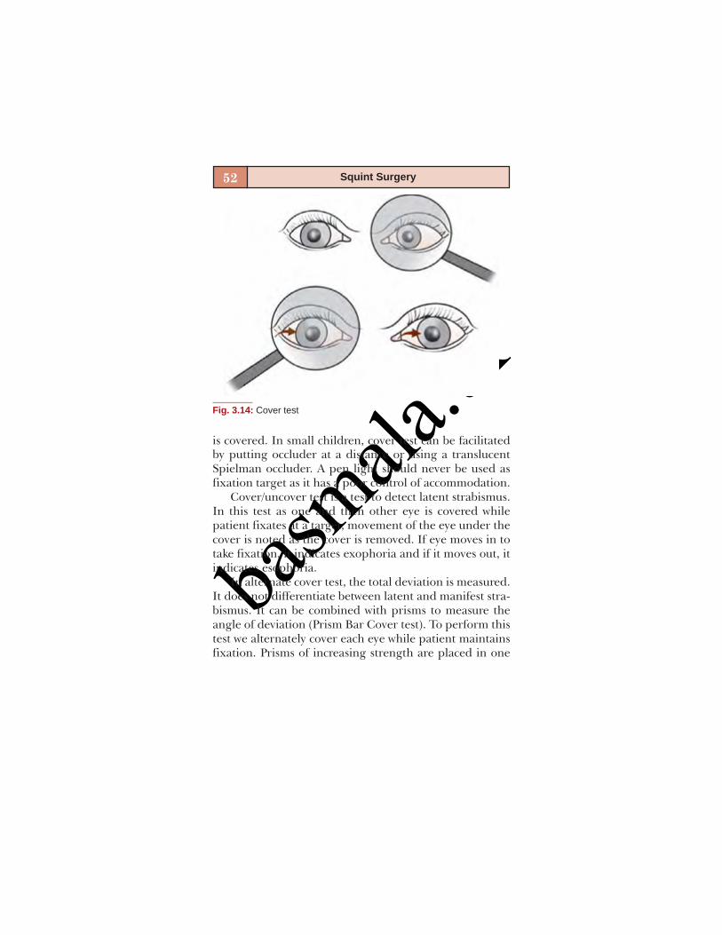
Cover Tests (Fig. 3.14)
Cover test remains gold standard for assessing and mea-suring deviation. Basic requisites for cover tests are eyemovement, capability for image formation and perception,foveal fixation and cooperation of the patient. Cover testhas three components—cover test, cover/uncover test andalternate cover test.
In cover test, we cover the apparently fixing eye andnote the movement of other eye. If other eye moves totake up fixation it indicates the presence of manifest squint.If there is no movement in fellow eye then that eye is
Fig. 3.12: Hirschberg’s test
basm
ala.tk
51Strabismus Evaluation
covered and other eye is observed. Cover test is performedfor both near and distance fixation. Cover test not onlyestablishes the presence of a manifest strabismus but italso helps to diagnose latent nystagmus which becomesobvious when one eye is covered. It also gives an idea ofdegree of alternation of strabismus. In a true alternatorwhen fixing eye is covered, other eye takes up fixation andmaintains fixation at least through a blink, when coveris removed. Cover test also indicates the presence ofeccentric fixation if any. The eye with eccentric fixationcontinues to fix in from a deviate position if the fellow eye
Fig. 3.13: Modified Krimsky’s test
basm
ala.tk
52 Squint Surgery
is covered. In small children, cover test can be facilitatedby putting occluder at a distance or using a translucentSpielman occluder. A pen light should never be used asfixation target as it has a poor control of accommodation.
Cover/uncover test is a test to detect latent strabismus.In this test as one and then other eye is covered whilepatient fixates at a target, movement of the eye under thecover is noted as the cover is removed. If eye moves in totake fixation, it indicates exophoria and if it moves out, itindicates esophoria.
In alternate cover test, the total deviation is measured.It does not differentiate between latent and manifest stra-bismus. It can be combined with prisms to measure theangle of deviation (Prism Bar Cover test). To perform thistest we alternately cover each eye while patient maintainsfixation. Prisms of increasing strength are placed in one
Fig. 3.14: Cover test

basm
ala.tk
53Strabismus Evaluation
eye with apex oriented towards direction of squint (i.e. basein for exotropia) till no redressal movement is elicited. Thisis also called as simultaneous prism and cover test. Ifvertical and horizontal squint exist together then firsthorizontal and then vertical angle is measured.
The amount of prism required to offset all redressalmovements is the angle of deviation. This test is also donefor both near and distance. Small accommodative fixationtargets should be used for near and 6/9 vision acuity linefor distance fixation. Pen light should never be used.
It is necessary to dissociate eyes before measuring theangle so cover should be placed alternately few times andpatient should not be allowed to regain fusion and com-pensatory mechanisms to check the deviation. If patienthas a refractive error, the angle should be measured bothwith and without glasses for near and distance. It is im-portant to remember that high plus lenses decrease andminus lenses increase the measured deviation. Prisms usedfor this test can be loose prisms or prism bar. Glass prismshould be held in Prentice position and plastic prisms infrontal plane position. Wrong positioning can give rightto wrong measurements. While measuring large angles itis preferable to divide prism in both eyes. Stacking ofprisms often produces errors, specially, if a low power prismis added to a high powered one.
Dissimilar Image Tests
These are tests based on diplopia principal. The diplopiais produced by presenting two dissimilar images. Diplopiacan be spontaneous as in case of paralytic squint or has tobe elicited, if there is suppression or ARC as in case ofcomitant squint. In these tests we determine subjectivelocalization of a single object imaged on fovea of one eye

basm
ala.tk
54 Squint Surgery
and extrafoveal point in other eye. Distance of doubleimage can be crossed as in exotropia or uncrossed as inesotropia. The different tests are:
Red filter testMaddox rodDouble Maddox rod test.
Red Filter Test
In red glass test, a red filter is placed over fixating eye todifferentiate two visual fields. Patient fixates over a smalllight and reports whether red image is crossed or un-crossed, up or down. Red glass should be dark enough todifferentiate as well as dissociate the eyes. Sometimes, avertical prism may be added to the red filter to appreciatediplopia better.
Maddox rod is a lens which consists of a series of paral-lel cylinders which convert a point source of light to a streakwhich is oriented perpendicular to that in Maddox rod.Maddox rod also dissociates the eyes. One eye perceives apoint of light while the other sees a red vertical line, ifMaddox rod is placed horizontally. If the line bisects thelight there is orthophoria, if it is on right side of whitelight, there is esodeviation and if it is on left side anexodeviation is present. This test cannot differentiatebetween heterophoria and tropia. Maddox rod is alsotraditionally worn over right eye.
It can be used to detect horizontal as well as verticaldeviations. To measure vertical deviation, cylinders areplaced vertically, to measure the angle, prisms are placedwith apex in direction of deviation till line crosses the light.Maddox rod can also be used to measure torsion. It isplaced in vertical direction. Patient is allowed to rotate thelens till one becomes horizontally straight. Degree oftorsion can be read off the trial frame.
basm
ala.tk
55Strabismus Evaluation
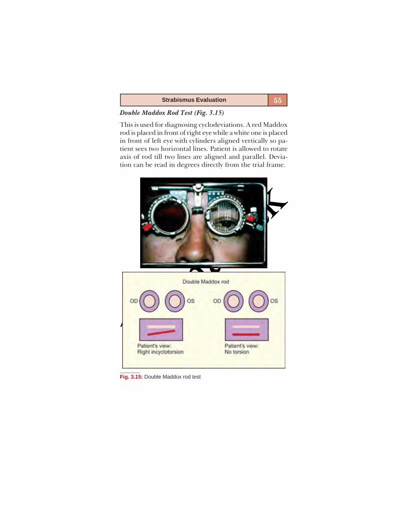
Double Maddox Rod Test (Fig. 3.15)
This is used for diagnosing cyclodeviations. A red Maddoxrod is placed in front of right eye while a white one is placedin front of left eye with cylinders aligned vertically so pa-tient sees two horizontal lines. Patient is allowed to rotateaxis of rod till two lines are aligned and parallel. Devia-tion can be read in degrees directly from the trial frame.
Fig. 3.15: Double Maddox rod test

basm
ala.tk
56 Squint Surgery
Dissimilar Target Tests
They are based on haploscopic principle, i.e. patient’sresponse to dissimilar images created by each eye viewinga different target. These tests are invaluable for incomitantsquints. The only prerequisite for this test is the presenceof NRC. The deviation is measured with first one eye fixingand then other. The common tests done are:
Hess’ Screening (Fig. 3.16)
This test utilizes red-green goggles and a screen that has ared light in 8 inner and 16 outer positions and a green slitprojection. Patient sits at 50 cm and is asked to put greenslit over each of red dot. Goggles are then reversed to recordsecondary deviation. A polarized version of Hess’ screenis also available.
Fig. 3.16: Hess’ screening

basm
ala.tk
57Strabismus Evaluation
Lancaster red/green test: This test also has a screen withsquares. Patient wears red green goggles and sits at 2meters. Examiner projects red slit on screen while patienttries to coincide with green slit in his hand. Goggles arethen reversed to record secondary deviation.Major amblyoscope: It projects dissimilar target which pa-tient is asked to superimpose. The deviation can be readdirectly off the scale.
DUCTIONS/VERSIONS (FIG. 3.17)
There are three types of ocular movements. Ductions aremonocular pursuit movements. Versions are binocularpursuit movements while vergences are binocularmovements where eyes move in opposite directions. Tocheck for ocular motility, patient fixates at a small fixationtarget and it is moved in the diagnostic positions of gaze,i.e. up right, right, down right, up, down, up left, left, downleft and primary position. Ductions are first tested andthen versions. Any overaction or underaction of muscleare noted, any A or V pattern should also be noted. Tomeasure motility- Kastenbaum’s limbus test of motility canbe used. This test is done by holding a transparent ruler infront of eye and if you want to measure abduction, positionof nasal limbus is marked on ruler in primary position andon maximum abduction, the difference is noted in mm.Similar way adduction, elevation and depression can benoted. Normal values for abduction, adduction and depres-sion are 10 mm and for elevation it is 5-7 mm. Clinicallyspeaking, in normal adduction an imaginary vertical linethrough lower lacrimal punctum should coincide with inner1/3 and outer 2/3 of cornea. If more cornea is hidden, thereis excessive adduction and if sclera remains visible it is

basm
ala.tk
58 Squint Surgery
defective. In normal abduction, corneal limbus shouldtouch outer canthus, if limbus passes that point abductionis excessive and vice-versa.
Fusional vergences amplitudes should be noted usingsynoptophore or prisms.
It is useful to measure near point of convergence andAC/A ratio in selected cases of strabismus.
SPECIAL TESTS
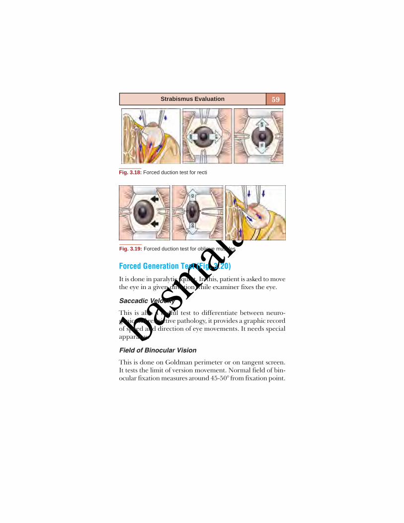
Forced Duction Test (Figs 3.18 and 3.19)
This is done to differentiate between restrictive and para-lytic strabismus. Indications for forced duction test areincomitant squints, thyroid ophthalmopathy, blow out frac-ture, Duane’s and Brown’s syndrome. To perform this test,topical anesthetic drops are applied. Eye is passively movedwith forceps in direction of limitation of movement. Passivemovement is possible in neurogenic pathology while it isnot possible in restrictive pathology.
Fig. 3.17: Extraocular movement charting
basm
ala.tk
59Strabismus Evaluation
Fig. 3.18: Forced duction test for recti
Fig. 3.19: Forced duction test for oblique muscles
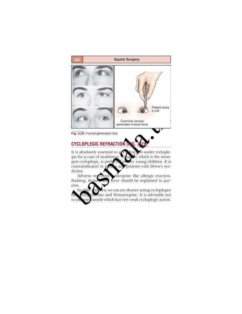
Forced Generation Test (Fig. 3.20)
It is done in paralytic squint. In this, patient is asked to movethe eye in a given direction while examiner fixes the eye.
Saccadic Velocity
This is also a useful test to differentiate between neuro-genic and restrictive pathology, it provides a graphic recordof speed and direction of eye movements. It needs specialapparatus.
Field of Binocular Vision
This is done on Goldman perimeter or on tangent screen.It tests the limit of version movement. Normal field of bin-ocular fixation measures around 45-50° from fixation point.
basm
ala.tk
60 Squint Surgery
CYCLOPLEGIC REFRACTION (FIG. 3.21)
It is absolutely essential to get refraction under cyclople-gia for a case of strabismus. Atropine, which is the stron-gest cycloplegic, is preferred in very young children. It iscontraindicated in infants and patients with Down’s syn-drome.
Adverse reactions to atropine like allergic reaction,flushing, dryness and fever should be explained to par-ents.
In older children, we can use shorter acting cycloplegicslike cyclopentolate and Homatropine. It is advisable notto use tropicamide which has very weak cycloplegic action.
Fig. 3.20: Forced generation test
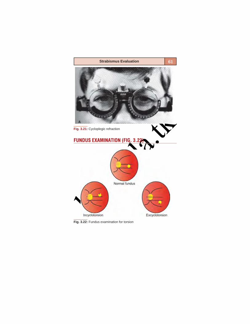
basm
ala.tk
61Strabismus Evaluation
FUNDUS EXAMINATION (FIG. 3.22)
Fig. 3.21: Cycloplegic refraction
Fig. 3.22: Fundus examination for torsion
basm
ala.tk
62 Squint Surgery
Fundus examination is last but not the least part of evalu-ation of strabismus. Many cases of sensory esotropia andexotropia are associated with disorders of optic nerve andretina.
Similarly, fundus examination can also give informa-tion about oblique overaction which produces shift of fovealreflex which is visible on indirect ophthalmoscopy.
BIBLIOGRAPHY1. Binocular vision and ocular motility by G Von Noorden.2. Clinical Strabismus Management – Rosenbaum, Santiago WB
Saunders and Co. 1999;pp3-52.3. Pediatric Ophthalmology and Strabismus. (AAO Section 6)
1999-2000;pp 56-72.4. Kenneth Wright. Textbook of Pediatric Ophthalmology:
Section 3, chapter 10,11pp(139-177).

basm
ala.tk
4
COUNSELING FOR SQUINTSURGERYMilind Killedar
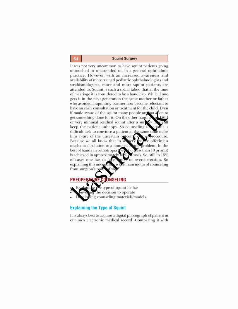
basm
ala.tk
64 Squint Surgery
It was not very uncommon to have squint patients goinguntouched or unattended to, in a general ophthalmicpractice. However, with an increased awareness andavailability of more trained pediatric ophthalmologists andstrabismologists, more and more squint patients areattended to. Squint is such a social taboo that at the timeof marriage it is considered to be a handicap. While if onegets it in the next generation the same mother or fatherwho avoided a squinting partner now become reluctant tohave an early consultation or treatment for the child. Evenif made aware of the squint many people are not keen toget something done for it. On the other hand, a fine DVDor very minimal residual squint after a surgery may stillkeep the patient unhappy. So counseling for squint isdifficult task to convince a patient at the same time makehim aware of the uncertain outcome of the procedure.Because we all know that in surgery we are offering amechanical solution to a nonmechanical problem. In thebest of hands an orthotropia (deviation less than 10 prisms)is achieved in approximately 85% of cases. So, still in 15%of cases one has to face under or overcorrection. Soexplaining this uncertainty is the main motto of counselingfrom surgeon’s point of view.
PREOPERATIVE COUNSELING
Explaining the type of squint he hasConveying the decision to operateDeveloping counseling materials/models.
Explaining the Type of Squint
It is always best to acquire a digital photograph of patient inour own electronic medical record. Comparing it with
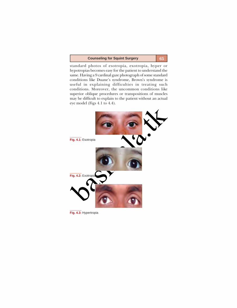
basm
ala.tk
65Counseling for Squint Surgery
standard photos of esotropia, exotropia, hyper orhypotropias becomes easy for the patient to understand thesame. Having a 9 cardinal gaze photograph of some standardconditions like Duane’s syndrome, Brown’s syndrome isuseful in explaining difficulties in treating suchconditions. Moreover, the uncommon conditions likesuperior oblique procedures or transpositions of musclesmay be difficult to explain to the patient without an actualeye model (Figs 4.1 to 4.4).
Fig. 4.3: Hypertropia
Fig. 4.1: Esotropia
Fig. 4.2: Exotropia

basm
ala.tk
66 Squint Surgery
Conveying Decision to Operate
Next important part is to convey the patient which eyeneeds to be operated and how many muscles to beoperated. It is better to explain him that it is a balance oftotal 12 muscles of two eyes that make a perfect balance.At this juncture, it is easy to explain the charges per musclewhich is easy to understand. This also makes it easy toexplain the additional need of operating extramuscle incase of under or overcorrection. Also to explain him if weare planning surgery in two parts (right eye first and thenleft eye after a gap of 2 months). This gives opportunity tomeasure smaller angles more accurately than larger angles.If an adjustable surgery is planned it is better to explainthe next day procedure first hand, otherwise it could betaken as unwarranted additional procedure. Usually, thismuch knowledge is sufficient for the patient to understandthe need for surgery.
As regards the surgical technique if it is fornix basedincision, it is useful to give emphasis to it as suture lesssurgery as some surgeons do it. Moreover, the postoperative
Fig. 4.4: Brown’s syndrome
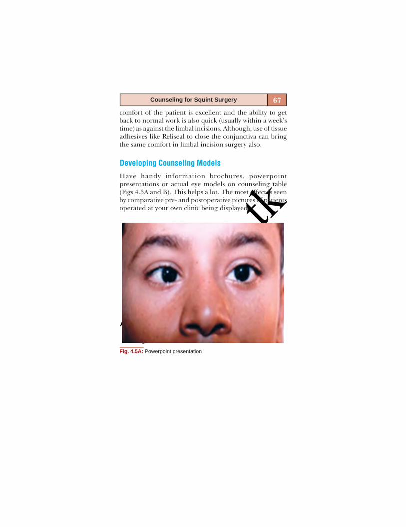
basm
ala.tk
67Counseling for Squint Surgery
comfort of the patient is excellent and the ability to getback to normal work is also quick (usually within a week’stime) as against the limbal incisions. Although, use of tissueadhesives like Reliseal to close the conjunctiva can bringthe same comfort in limbal incision surgery also.
Developing Counseling Models
Have handy information brochures, powerpointpresentations or actual eye models on counseling table(Figs 4.5A and B). This helps a lot. The most effect is seenby comparative pre- and postoperative pictures of patientsoperated at your own clinic being displayed.
Fig. 4.5A: Powerpoint presentation
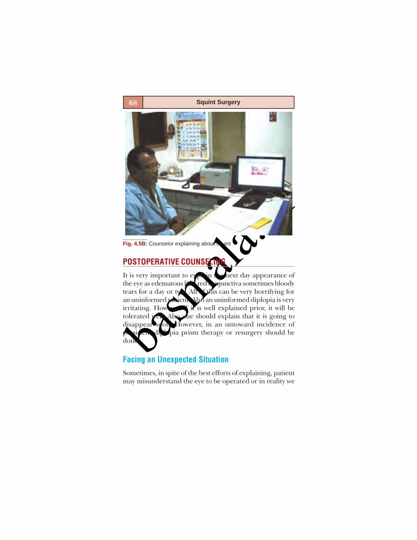
basm
ala.tk
68 Squint Surgery
POSTOPERATIVE COUNSELING
It is very important to explain the next day appearance ofthe eye as edematous lids, red conjunctiva sometimes bloodytears for a day or two. All of this can be very horrifying foran uninformed patient. Also an uninformed diplopia is veryirritating. However, if it is well explained prior, it will betolerated well. Also one should explain that it is going todisappear soon. However, in an untoward incidence ofpersistent diplopia prism therapy or resurgery should bedone.
Facing an Unexpected Situation
Sometimes, in spite of the best efforts of explaining, patientmay misunderstand the eye to be operated or in reality we
Fig. 4.5B: Counselor explaining about squint
basm
ala.tk
69Counseling for Squint Surgery
may operate wrong eye (according to patient). We know asfar as it is comitant squint results will not vary and there isno need to panic. However, as soon as it is brought to ournotice, patients’ relatives should be calmly counseled thatthe change in eye was purposeful and taken after carefulexamination under anesthesia (e.g. FDT). Although, itmight not be truth. However, if it was incomitant squint orwrong surgery, i.e. recess instead of resect or vice a versathen it is better to explain the facts and plan a redoimmediately.
Most of the book mentioned complications in squintsurgery like malignant hyperthermia, panophthalmitis areso rare that one may not see a single case in the lifetime. Sosuch, things need not be touched in routine pre- orpostoperative counseling as it may cause unnecessary worries.However, if it occurs, it can be explained as very rarely occurredcomplication and should be energetically managed. Thecompetency of a surgeon is not only to do a surgery greatelybut also to manage complications efficiently.
Lastly, it is a very good practice to get a follow-upphotograph of patient at one month and show him thecomparison between the preoperative and postoperativephotograph.

basm
ala.tk

basm
ala.tk
PREFERRED SURGICALTECHNIQUES
2

basm
ala.tk
basm
ala.tk
5
INSTRUMENTS FORSTRABISMUS SURGERY
Arun Samprathi

basm
ala.tk
74 Squint Surgery
Strabismology surgery is one of the cheapest subspecialtiesin ophthalmology, whether it is in the outpatient or theoperation theater. Minimal instruments are necessary forstrabismus surgery, which relies more on technique andsurgeon’s expertise than on instrumentation. A basiccataract set available in any OT is sufficient to perform ahorizontal muscle surgery. Though, there are advances insurgical techniques in strabismus surgery, theinstrumentation has not changed much in the last fewdecades.
We can classify the instruments as mandatory instru-ments and optional instruments.
MANDATORY INSTRUMENTS
Muscle Hooks
Muscle hooks are the most important part of a strabismusinstrument set. Standard muscle hooks have smooth,rounded tip, which do not damage the muscle when pass-ing under it. These hooks are useful for initial hooking ofthe muscle (Fig. 5.1).
Fig. 5.1: Muscle hooks

basm
ala.tk
75Instruments for Strabismus Surgery
Calipers (Castroviejo Caliper)
Calipers are very crucial for measuring the exact distancefor recession or resection of a muscle. A standard caliperavailable in the cataract set is most commonly used(Fig. 5.2).
Westcott’s Scissors
The curved and blunt tips allow easy dissection of theTenon’s capsule from the sclera with little risk of perforat-ing the scleral wall (Fig. 5.3).
Needle Holder
Good pair of needle holders is essential for performing asafe strabismus surgery. Taking bytes at correct depth is im-portant to avoid scleral perforation (Fig. 5.4).
Fig. 5.2: Castroviejo caliper
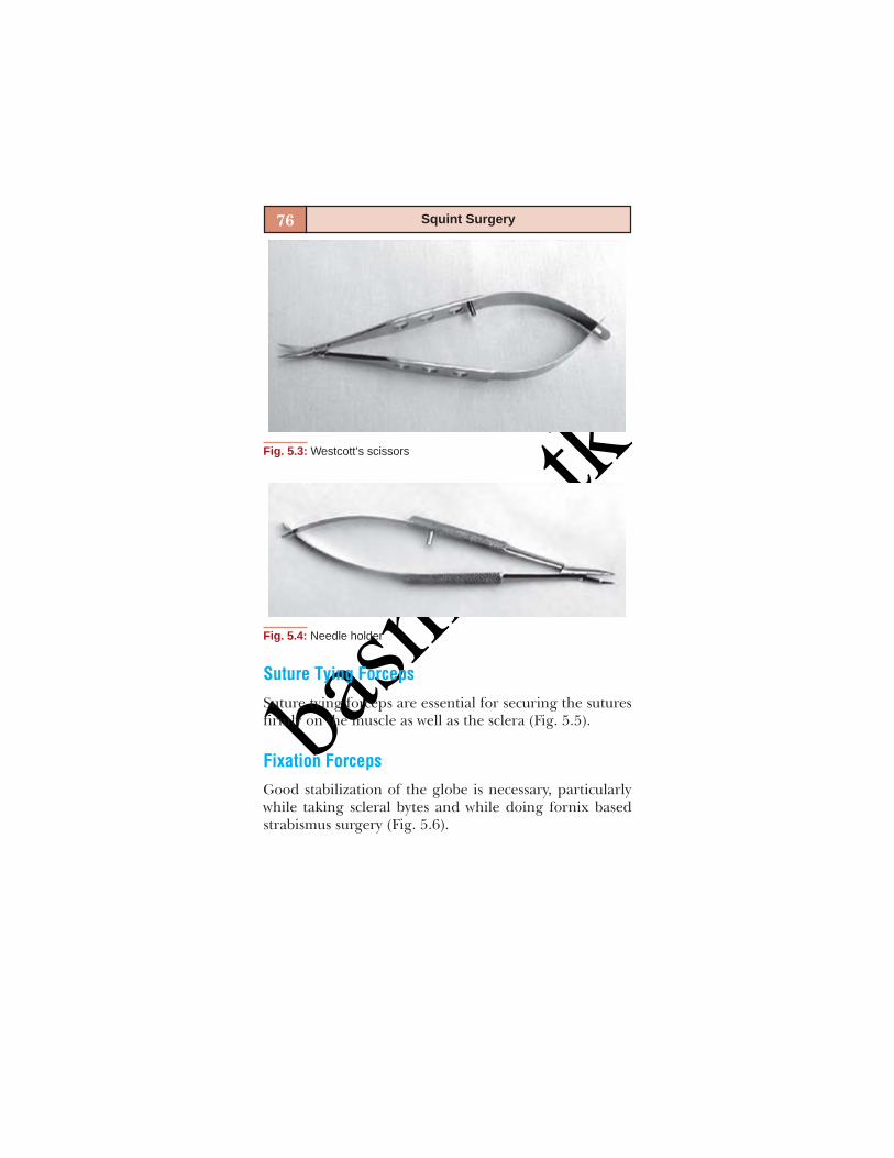
basm
ala.tk
76 Squint Surgery
Fig. 5.4: Needle holder
Fig. 5.3: Westcott’s scissors
Suture Tying Forceps
Suture tying forceps are essential for securing the suturesfirmly on the muscle as well as the sclera (Fig. 5.5).
Fixation Forceps
Good stabilization of the globe is necessary, particularlywhile taking scleral bytes and while doing fornix basedstrabismus surgery (Fig. 5.6).

basm
ala.tk
77Instruments for Strabismus Surgery
Fig. 5.5: Suture tying forceps
Fig. 5.6: Fixation forceps
OPTIONAL INSTRUMENTS
Modified Muscle Hooks
Jameson muscle hooks have a small projection at the tipto hold the muscle for passing the sutures and tying them.

basm
ala.tk
78 Squint Surgery
Chavassee hooks have an undulated horizontal surface,so that it allows for passing the central suture and thentying them at either edges of the muscle (Figs 5.7A and B).
Barbie Retractors
Surgery on the extraocular muscles needs good exposure,particularly in children with narrow palpebral apertures,re-surgeries, restrictive strabismus, etc. Barbie retractorsare of great value to get a good exposure, without damag-ing the tissues. Barbie retractors could be small, mediumor large depending on the needs (Fig. 5.8).
Bull Dog Clamps
Bull dog clamps are useful to hold the sutures separately.So that they do not get mixed-up while tying.
Muscle Clamps
Resections of muscles can be done with the help of muscleclamps to hold the muscle. Muscle clamps will prevent themuscle from slipping and retracting back into the orbit(lost muscle), which is a serious complication (Fig. 5.9).
Scleral Rulers
Special scleral rulers are available, which have the samecurve as the sclera. These rulers are useful when measur-ing recessions from the limbus or for posterior fixationsutures.

basm
ala.tk
79Instruments for Strabismus Surgery
Figs 5.7A and B: (A) Jameson muscle hooks (B) Chavassee hook
Fig. 5.8: Barbie retractor
Fig. 5.9: Muscle clamps
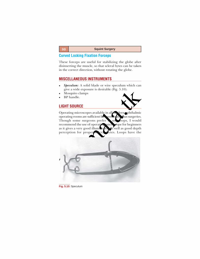
basm
ala.tk
80 Squint Surgery
Curved Locking Fixation Forceps
These forceps are useful for stabilizing the globe afterdisinserting the muscle, so that scleral bytes can be takenin the correct direction, without rotating the globe.
MISCELLANEOUS INSTRUMENTS
Speculum: A solid blade or wire speculum which cangive a wide exposure is desirable (Fig. 5.10).Mosquito clampsBP handle.
LIGHT SOURCE
Operating microscopes available in all modern ophthalmicoperating rooms are sufficient for most strabismus surgeries.Though some surgeons prefer to use loops, I wouldrecommend the use of operating microscope for beginnersas it gives a very good illumination as well as good depthperception for proper scleral bytes. Loops have the
Fig. 5.10: Speculum

basm
ala.tk
81Instruments for Strabismus Surgery
advantage of allowing free movement of the surgeonparticularly while operating on the obliques or operatingdeep inside the orbits.
PULSE OXIMETER
Monitoring the pulse constantly throughout the procedureis extremely essential. Bradycardia due to oculo-cardiac reflexis a dreaded complication of strabismus surgery and needsimmediate recognition and prompt management. Hence,strabismus surgery should never be attempted without a pulseoximeter and an anesthetist to monitor the patient.
CONCLUSION
Strabismus surgery can be performed with minimal instru-mentation available in all ophthalmic operating rooms(Fig. 5.11).
Fig. 5.11: Instruments for strabismus surgery

basm
ala.tk
basm
ala.tk
6
PRINCIPLES OFSTRABISMUS SURGERY
P Vijayalakshmi
basm
ala.tk
84 Squint Surgery
INTRODUCTION
Strabismus surgery aims to improve the alignment of theeyes to achieve or preserve binocular single vision apartfrom providing a good cosmetic result. Sometimes it isaimed at correcting the head posture thereby increasingthe binocular field of vision. It is mandatory that all patientsare examined in detail, refractive errors are correctedadequately and amblyopia, if any was treated with fullenthusiasm before planning for surgical correction ofstrabismus. We try to give a few guidelines on decisionmaking and surgical principles in this chapter.
IMPORTANCE OF GOOD PREOPERATIVE EVALUATION
A very careful and detailed preoperative work-up is man-datory to the successful outcome of strabismus surgery.Readers are advised to read the concerned chapters forthe details on this. Few points of clinical importance arestressed here.
Sometimes the presenting complaint may be anabnormal headposture, which could be so simple aspreference to fix with one eye (amblyopia), or complicatedlike a congenital superior oblique palsy, congenitalelevation deficiency, restrictive strabismus like Duane’s,Brown’s or fibrosis syndromes, nystagmus and nystagmusblockade syndrome. All efforts should be done to diagnosethe etiology of head posture including estimation ofbinocularity and the treatment is directed towardsimproving it. The next important point is to know underand overactions of both recti and oblique muscles whichwill actually indicate the muscles to be operated. Forexample, in cases of pattern deviations and in DVDassociated with oblique muscle overactions, that should be
basm
ala.tk
85Principles of Strabismus Surgery
weakened unless indicated otherwise.1 The most difficultin this part is estimating the amount of deviation whichwill tell us how many muscles to be operated. The choiceof surgical procedure depends upon the relationship ofnear and distance deviation, lateral incomitance, patterndeviations and presence of amblyopia, stressing again theimportance of detailed measurements in all requiredpositions of gaze by using a prism cover test with a correcttarget to fixate. The surgery should be deferred until thesurgeon is convinced that he has atleast two high qualitymeasurements not differing more than 5 to 10 prismdiopters each especially in all patients.
The binocular single vision with corrective prisms andstereopsis if possible should be estimated in every case, topredict the possible postoperative outcome. Delayed mile-stones if it is present should be given it’s due importance.2
The other associated findings like age of the patient, fissurechanges, other rarely associated syndromes should also berecognized.
AGE OF THE PATIENT AT SURGERY
Patients less than two years of age tend to have smallerglobes and the surgery tends to give more effect for eachmillimeter of muscle recessed/resected. The generalconsensus is that the strabismus surgery should not be donein children less than 4 months of age.3,4
Few authors claim better sensory outcome when it isperformed 3 to 4 months of age. They also agree that thedeviation must be constant and reproducible with repeatedexaminations before undertaking the surgery (RosenbaumBook). Various clinical trials have proved that there arebetter chances to gain binocularity, fusion with bifovealfixation, if it is performed below the age of 2 years.5 One

basm
ala.tk
86 Squint Surgery
study reports that 41% of infants whose eyes were alignedwithin 8 prism diopters in the first 16 months of life hadthe restoration of random dot stereopsis years later, thatwhose eyes were aligned at the age of 12 months atleast49% achieved stereopsis of finer grade.6,7 Still some amountof peripheral fusion may be possible even when the surgeryis done in later years especially in acquired deviations.8 Itis wise to delay the surgery in cases with globaldevelopmental delay, in situations where the measurementof the deviation is not satisfactory, variable deviations,dense amblyopes and reluctant parents.
BILATERAL SYMMETRICAL SURGERY
Surgery should aim to restore symmetry and alignment tothe maximum possible extent. Some authors believe thatthis can be achieved only by symmetrical surgeries andthey advocate this unless otherwise indicated. However, abilateral medial rectus recession is commonly done forinfantile esotropia with cross fixation, near deviation morethan distance, (convergence excess type) V pattern withno oblique dysfunction and in accommodative esodeviation.Bilateral lateral rectus recessions would be of value indivergence excess exotropia, intermittent exodeviations, Vpattern deviations. These bilateral recessions can becombined with shifting of muscles up or down or withbilateral inferior oblique weakening depending upon thepattern deviations.1 Bilateral resections are rarely done,e.g. a bimedial resection may be useful in exotropias ofpure convergence weakness and bilateral lateral rectusresections in large A pattern esotropias with no inferioroblique dysfunction.9

basm
ala.tk
87Principles of Strabismus Surgery
Unilateral Recess/Resect (R/R) Procedure
Recessions/resections result in a greater effect in thedirection of the action of the muscle and this can be usedto the surgeon’s advantage. R and R on the same eye tendsto correct the same amount of deviation both for distanceand near. Hence, this procedure is indicated when thepatient was already treated for amblyopia, definitedominance of one eye, parents feel the deviation presentin only one eye, when the deviation is secondary to thepathology in the eye (sensory strabismus). In cases of verylarge deviations, where more than 2 muscles surgery isindicated. Recess resect in one eye will be a better optionfor making the decision on the residual deviation ratherthan doing a symmetrical procedure two times.
SINGLE MUSCLE SURGERY
Single muscle recession has a more value in small deviationsand is more useful in vertical muscle surgery. In horizontaldeviations, especially in esotropias a single medial rectusrecession may be indicated when the deviation is less than20 prism diopters.10 This could be either a primarydeviation, with or without an accommodative component,or residual deviations after a first operation in the felloweye or in Duanes retraction syndrome with a small faceturn. However, a caution should be kept in mind that largerecession of a single muscle may give rise to incomitancein the field of action of the muscle, sometimes resulting indouble vision in that field. So a careful assessment of thepostoperative effects on side gaze measurements arenecessary preoperatively and it is better to limit the numbermaximum to 6 mm recession. Single lateral rectus recessionis rarely indicated except in small intermittent exophoria
basm
ala.tk
88 Squint Surgery
or a tropia. Inferior rectus recession especially in smallvertical deviations may be useful and a superior rectusrecession can be done in unilateral DVD.11
Single muscle resection again has a limited role andmay be indicated in small residue, a pattern with no obliquedysfunction. Resection of a single muscle has lesserdisadvantages than a recession as far as incomitant resultsare concerned.
MUSCLE SURGERY
When the deviation is large and the intention is a maxi-mum correction in one sitting a recess resect in one eye canbe combined with other eye recession especially in esotrpias.Overcorrections are known and caution is advised.12
Influence of Muscle Surgery on Palpabral Fissure
It is important to remember that medial rectus recessionsresult in a widening of the palpebral fissure and resectionsnarrow it. Careful consideration is needed for a goodoutcome. Inferior oblique anteropositioning can also nar-row the palpebral fissure and also result in a bulge in thelower lid. inferior rectus recession may produce a lowerlid lag.13 The patient should be warned of these potentialside effects.
Amount of Correction
The main aim is to correct the maximum deviation in onesitting, and distribute the surgery to 2 muscles wheneverindicated. Getting the numbers right is not that simple. Theaverage values are 4.5 pd per mm for the medial rectus and3 pd per mm for the lateral rectus. The limits for recession
basm
ala.tk
89Principles of Strabismus Surgery
are 6 mm for the medial rectus and 8 mm for the lateralrectus.14 Minimal recession for lateral rectus is4 mm and for the medial rectus is 3 mm. According toPratt-Johnson15 the effect of surgery, recession orresections on previously unoperated horizontal muscle is2.5 prism diopter correction per mm and this increases withthe amount of surgery done. At 7 mm one tends to get3 prisms diopter and at 10 mm it is 5 mm. This gives thepredicted effect in about 85% of his patients meaning thatreoperation may still be necessary in 15% of the patients.
In cases of inferior rectus recession one mm recessioncorrects 3 prism diopters in primary position whereas thatwill be 5 prism diopters in downgaze. This should becarefully considered in preoperative decision making.Pratt-Johnson also reports that a large recess resect can bedone occasionally in large unilateral deviations and heobserved that the initial weak ductions get better over timeas the muscle gets readjusted and also admits that thisis unpredictable. Surgery on a tight muscle primarily andresurgeries like advancements and resections will have moreeffect than expected. For that reason an adjustable suturetechnique may be used in this situation.
There are many tables published14,16 and one can followthem initially for a few cases till he or she reviews his ownpatients and formulates the surgical dosage.
Effect of Axial Length and Refractive Error onMuscle Surgery
Response to Strabismus surgery does correlate significantlyand inversely with axial length. This correlation may notbe clinically very important, given the much strongerinfluence of preoperative deviation.17

basm
ala.tk
90 Squint Surgery
For a better outcome each case should be individual-ized and the final decision should be made dependingupon the age of the patient, amount of deviation, pres-ence of amblyopia, presence of refractive errors and finallythe patient’s expectations. A checklist prior to taking thepatient for surgery may be helpful for the beginner.
Vision, refraction and cycloplegic refractionHas appropriate optical correction been given?Has amblyopia treatment been completed?Likely compliance with postoperative patching, glassesand follow-upMeasurement in relevant directions of gaze, measure-ment for distance and near with and without glassesWas the patient cooperative for the measurements? Areyou satisfied or would you rather delay the surgery untilaccurate measurements are obtained?Tests for binocularity and stereopsisPrognosis for achievement of binocular single visionafter surgeryOcular movements carefully recorded with attentionto over and underactionsIs the surgical plan adequate to tackle incomitance, Aand V patterns and oblique overactions?Comprehensive anterior and posterior segment evalu-ationAttention to systemic risk factors especially towardsadministration of general anesthesiaAttention to other factors like patient cooperation forlocal/topical anesthesiaPatient consent form duly signed with clarification ofdoubtsPreoperative checks to ensure the right eye and theright patient is being operated upon.
basm
ala.tk
91Principles of Strabismus Surgery
ANESTHESIA
We routinely use general anesthesia for all patients lessthan 18 years of age. For older patients, local (retrobulbar/peribulbar) or topical anesthesia (Lignocaine jelly andproparacaine drops with or without systemic augmenta-tion with Fentanyl and Propofol) is used with heart ratebeing monitored throughout the surgery. The techniqueto be used depends on the consensus between the patient,surgeon and the anesthetist. Topical anesthesia may beavoided for oblique muscles. The eyes may straighten outunder general anesthesia and it is important that thesurgeon should not change his/her decision based on this.
SUTURES AND NEEDLES
6-0 vicryl sutures are preferred for the routine recess resectprocedures on the muscles. Non absorbable sutures likeProlene 5-0 and merseilene are used in Muscle transposi-tions, muscle surgery after RD surgery with buckle,resurgeries, and globe fixation procedures. 8-0 vicryl is usedto close the conjunctiva. Always spatulated needles arerecommended for muscle surgery.
ON THE TABLE
The conjunctival sac is cleaned with povidone iodine alongwith the lids and brows. The eye to be operated is confirmedbefore draping. Forced duction test is performed on alleyes as a first step, since a tight muscle will give rise tobetter correction on its recession when compared to anormal muscle and an adjustable suture can be placed insuch situations where the results are unpredictable.
basm
ala.tk
92 Squint Surgery
SPECIAL CONSIDERATIONS FORPARETIC STRABISMUS
The surgeon must remember that in cases of pareticdeviations, if he is operating on the paretic eye he mustcorrect for the primary deviation, but if he is operating onthe normal eye, the secondary deviation must be takeninto account.
Weakening either the antagonist or the yoke muscle tothe paretic muscle is physiologically sound and should resultin the best improvement when all fields of gaze is takenunto consideration. Which muscle to be chosen dependsupon the predictability of results. For example in an inferiorrectus paresis, it is easy to operate on the direct antagonistsuperior rectus with precision than operating on the othereye superior oblique (yoke muscle) with less precision.Strengthening the paralysed muscle depends whether theparalyzed muscle is able to exert any force (as can bedetermined by analysis of saccadic velocity, the globe crossingthe midline and the forced generation test). If yes, a routinerecess/resect procedure will do. Any restriction caused bycontracture of the antagonist (tested by the forced ductiontest) has to be relieved. If no active force is generated,recessing the antagonist muscle may be of limited value asthe muscle in the absence of opposing force eventually takesup the slack and the strabismus recurs. Similarly, resectinga completely paralyzed muscle is of little value as the musclein the absence of any active contraction eventually stretchesout. A transposition procedure as described in later chaptersis of value in these conditions. This generates a constantforce. When attempting to look in the field of the paralyzedmuscle, the antagonist relaxes and the constant force pullsthe globe to some distance, thereby expanding the binocularfield of single vision. Third nerve palsy leaves us with little

basm
ala.tk
93Principles of Strabismus Surgery
options as most of the muscles are affected. If the globe isable to cross the midline, a recess/resect procedure on thehorizontal recti improves alignment. If not, the lateral rectusmay need to be inactivated by suturing it to the orbitalperiosteum and the medial rectus resected.18 Another optionis to perform a large recession of the lateral rectus and anchorthe globe to the medial palpebral ligament or the periosteumof the medial orbital wall.19 It is important to rememberthat alignment can be achieved only in the primary position.In cases of spread of comitance the recess resect procedurecan be chosen. The use of adjustable sutures play a greatrole in treating such patients.
In cases of 6th nerve palsy, the decision of surgical pro-cedure again depends upon the ability of the globe toabduct. The same principles hold good here also. Eitherthe recession of medial rectus. alone or in combinationwith resection of the lateral rectus if that is functioning. Ifnot a Jensen’s procedure is the choice. For 4th nerve palsyrefer the chapter on vertical strabismus.
PREVENTING COMPLICATIONS
Strabismus surgery is generally rated as a safe surgery withminimal or no complications that can be done by anyonewith residency training in ophthalmology. I agree with thefact that strabismus surgical techniques per se is not thatdifficult to learn and one can avoid surgical complications.Keeping in mind that this a very sensitive cosmetic surgeryapart from its sensory outcome, with high expectations frompatient’s side and considering that resurgeries are difficultand unpredictable, a proper decision making is mandatoryand that needs definitely an experience which has its ownlearning curve.

basm
ala.tk
94 Squint Surgery
Careful preoperative evaluation, selection of theappropriate procedure and judicious use of adjustabletechniques as detailed in the preceding sections and in thechapters to come goes a long way in minimizingovercorrections and undercorrections. To ensure patientsatisfaction, the parents/patient should be involved in thedecision making process regarding the eye to be operated,choice of anesthesia, etc. Even though the procedure isextraocular, postoperative infection is still a possible entity.Hence, it is important that aseptic precautions of the samedegree as for any other ocular surgery be employed.Meticulous surgical technique reduces the chance of muscleslippage and globe perforation. Traction on the extraocularmuscles should be kept to a minimum for two reasons. Oneit avoids the oculocardiac reflex and two aggressive tractionin the elderly can lead to the muscle snapping called thepulled in two syndrome (PITS).20 For resurgeries, if there isan option to operate on the virgin muscle, that should bepreferred rather than on the muscles previously operatedon. Anterior segment ischemia can be any surgeon’snightmare. It is discussed in detail in a later chapter. Theuse of a fornix incision, limiting the number of extraocularmuscles operated on a single eye (the obliques do not matter,but under no circumstance should all the four recti bedetached from the globe even in staged procedures), vesselsparing procedures, attention to systemic atherosclerotic riskfactors, avoidance of surgery on two adjacent recti tend tominimize this dreaded complication.
For essentially one eyed patients (e.g. other eye withsevere amblyopia, macular pathology, etc.), perform themaximum possible surgery in the affected eye, and usuallydo not advocate surgery on the good eye unless otherwiseindicated.

basm
ala.tk
95Principles of Strabismus Surgery
Last but not the least, the systemic condition of thepatient should receive due attention. The surgeon shouldwork in close coordination with the anesthetist to ensuresafe surgery.
POSTOPERATIVE CARE AND FOLLOW-UP
At conclusion of surgery, a drop of antibiotic steroid com-bination is instilled in the conjunctival sac and the eyebandaged. The bandage is removed the following day andwe recommend rest for 14 days. If both eyes have beenoperated, the bandage on one eye is removed an hour aftersurgery and that on the other eye removed the next day.The patient is discharged on a steroid antibiotic combina-tion with instruction to use it for a month (low potencysteroids like fluorometholone or loteprednol are preferred.For fornix incision a shorter course for a week or two issufficient). On the first postoperative day, the eyes areexamined mainly for the alignment, congestion,conjuntival opposition and for any gross pathology.
Review is timed, a month after the surgery. Carefulexamination is done to note any sutural allergic reactions,healing of the wound in general, the alignment of the eyes,estimation of visual acuity, binocular vision, fusion andstereopsis, fundus examination with refraction whenindicated. Any small refractive correction is given espe-cially if there is a residual deviation. Amblyopia therapy ifneeded can be resumed as early as two weeks after thesurgery. Further follow-ups are planned 6 months therafterupto 8 years when the results said to be getting stabilized.Special follow-ups are given for whom the amblyopiatreatment is advocated, or prisms are prescribed.
basm
ala.tk
96 Squint Surgery
To conclude, an adequate knowledge on the visiondevelopment and amblyopia forms the basis for anophthalmologist to become a strabismus surgeon, and toachieve this, the teaching and training should beincorporated into our postgraduate training in an adequateamount and this learning should be continued further inpractice. A lot of reading, strict adherence to surgicalprinciples, careful planning, interaction with peers andcolleagues, using telemedicine will increase the individual’sconfidence over time and will contribute in the making ofa successful strabismus surgeon.
REFERENCES1. A and V Pattern Deviations. In:Julio Prieto-Diaz, Carlos
Souza-Dias editors, Strabimsus. Butterworth Heinemann247-88.
2. Habot-Wilner Z, Spierer A, Glovinsky J, Wygnanski-Jaffe T.Bilateral medial rectus muscle recession: Results in childrenwith developmental delay compared with normally devel-oped children. J AAPOS 2006;10(2):150-4.
3. Wright KW, Edelman PM, McVey JH, Lin M. High-gradestereoacuity after early surgery for congenital esotropia. ArchOphthalmol 1994;112:913-9.
4. Esotropia. In: Pradeep Sharma (Ed), Strabismus Simplified.Modern Publishers, New Delhi 1999;87-97.
5. Ing MR. Early surgical alignment for congenital esotropia.Trans Am Ophthalmol Sci 1981;79:625-63.
6. Birch EE, Stager DR, Berry P, et al. Prospective assessmentof acuity and stereopsis in ambloyopic infantile esotropesfollowing early surgery. Invest Ophthalmol Vis Sci1990;31:758.
7. Birch EE, Stager Dr, Everett ME: Random dot stereoacuityfollowing surgical correction of infantile esotropia. J PediatrOphthalmol Strabismus 1995;32:231.

basm
ala.tk
97Principles of Strabismus Surgery
8. Lal G, Holmes JM. Postoperative Stereoacuity FollowingRealignment for Chronic Acquired Strabismus in Adults.J AAPOS 2002;6:233-7.
9. Santiago AP, Ing MR, Kushner BJ, Rosenbaum AL. IntermittentExotropia. In: Rosenbaum AL, Santiago AP (Eds), ClinicalStrabismus Management-principles and surgical techniques.WB Saunders Company. Philadelphia 1999;163-75.
10. Stack RR, Burley CD, Bedggood A, Elder MJ. UnilateralVersus Bilateral Medial Rectus Recession. J AAPOS2003;7:263-7.
11. Santiago AP, Rosenbaum AL. Dissociated vertical deviation.In: Pediatric Ophthalmology and Strabismus. Section 6.American Academy of Ophthalmology San Francisco 2004-2005;237-48.
12. Minkoff OV, Donahue SP. Three-Muscle surgery for Infan-tile Esotropia in children younger than age 2 years. J PediatrOphthalmol Strabismus 2005;42:144-8.
13. Kushner BJ. The Effect of Anterior Transposition of theInferior Oblique Muscle on the Palpebral Fissure. ArchOphthalmol 2000;118:1542-6.
14. Surgical Management.In: Pradeep Sharma (Ed), StrabismusSimplified. Modern Publishers, New Delhi 1999;177-206.
15. John A, Pratt-Johnson. Management of Strabismus andAmblyopia: A Practical Guide. Chapter 16; pp 207-26.
16. Surgery of the Extraocular Muscles. In: Pediatric Ophthal-mology and Strabismus. Section 6. American Academy ofOphthalmology San Francisco 2004-2005;165-85.
17. Gillies WE, Hughes A. Results in 50 cases of strabismus aftergraduated surgery designed by A scan Ultrasonography. BrJ Ophthalmol 1984;68:790-5.
18. Velez FG, Thacker N, Britt MT, Alcorn D, Foster S,Rosenbaum AL. Rectus Muscle Orbital Wall Fixation: Areversible profound weakening procedure. J AAPOS2004;8:473-80.

basm
ala.tk
98 Squint Surgery
19. Srivastava KK, Sundaresh K, Vijayalakshmi P. A new surgicaltechnique for ocular fixation in congenital third nerve palsy.J AAPOS 2004;8(4):371-7.
20. Siegel LM, Lozano MJ, Santiago AP, Rosenbaum AL.Adjustable and Nonadjustable Recession and ResectionTechniques. In: Rosenbaum AL, Santiago AP (Eds), ClinicalStrabismus Management-principles and surgical techniques.WB Saunders Company. Philadelphia 1999;435-8.
basm
ala.tk
7
HORIZONTALMUSCLE SURGERYFOR STRABISMUS
P Vijayalakshmi, R Muralidhar
basm
ala.tk
100 Squint Surgery
INTRODUCTION
Surgery on the horizontal recti is the simplest of squintsurgeries and the novice surgeon usually commences histraining by beginning with them. There are numerousvariations in the technique, but the basic principles remainthe same. We try to give some practical pearls in learningthese techniques and have made a sincere attempt to keepthe contents as simple as possible.
MAGNIFICATION
Magnification is recommended either in the form of loupesor microscopes. As almost all surgeons in India practicecataract surgery under microscope, using the microscopefor strabismus procedures will make it more practical. Weroutinely use the surgical microscope for all our surgeries.
PREPARATORY STEPS
The eye or the eyes are cleaned with povidone iodine afterconfirming the eye to be operated. Some surgeons preferto use phenylephrine hydrochloride on the table to inducevascular constriction which we are not practicing. Forcedduction testing is done to identify any restrictivecomponent. A traction suture can be used to pull the globeand expose the area of incision.
CONJUNCTIVAL INCISION
The Swan incision, where the incision is made at theinsertion of the muscle is not practiced widely. The surgeonhas the choice of either the limbal or the fornix incision.The fornix-based incision needs a more precise surgical
basm
ala.tk
101Horizontal Muscle Surgery for Strabismus
technique and needs a good assistance throughout thesurgery. The limbal incision is our preferred choice andwill be dealt in detail. The limbal incision gives the bestexposure of the two with almost a less learning curve andis recommended for beginners. Some rotation of the globeoccurs during general anesthesia and the surgeon shouldexert caution to avoid operating on the wrong muscle. It isusually possible to see the muscle through the conjunctivawith the speculum in place. Useful clues include the ciliaryvessels and a darker appearance compared to thesurrounding. This can be used to guide the placement ofthe incision.
After cleaning and draping, the surgeon grasps theconjunctiva at the limbus with a toothed forceps at anidentified location (5 o’ clock position) and a small incisionis made with Wescott’s scissors. The conjunctiva along withTenon’s is now separated from the sclera upto the muscleinsertion by passing the closed scissors from below upwardsbefore the incision is extended concentric to the limbusfor two to three clock hours, e.g. starting from 5 o’ clockposition and ending at 2 o’ clock position in case of surgeryon medial rectus. Two radial incisions are madeapproximately for 5 mm starting from the ends of theconcentric incisions. Precaution is to be taken to note anybig vessel crossing the line of incision. If it is not possibleto avoid cutting the vessel, one can use cautery either beforeor after cutting the vessel. Inappropriate location of theincision can result in muscle injury with bleeding or anugly scar postoperatively.
EXPOSURE OF THE MUSCLE
The conjunctiva is then retracted back and theintermuscular septum is buttonholed by blunt dissection
basm
ala.tk
102 Squint Surgery
with Wescott’s scissors to expose bare sclera. Once the sclerais exposed, the muscle is engaged with a gentle sweep of ablunt tipped hook like the Von Graefe’s or Jameson’s. Themovement should be smooth and any resistance in apreviously unoperated muscle means that the surgeon isin the wrong plane of hooking or hooking only part of themuscle. Two hooks passed from either side can be used toensure complete hooking. Two hooks placed one behindthe other can also be used to give the surgeon room forpassing sutures and disinsertion. Some surgeons use a hookwith a groove (the Wright’s grooved hook) for tight muscles.
The intermuscular septum is now cut on either side.The check ligaments should be dissected carefully by acombination of blunt and sharp dissection. Care shouldbe taken to preserve the muscle sheath as cutting the sheathresults in bleeding and more postoperative adhesions. Thecuts on the intermuscular septum and the check ligamentsshould be done under direct visualization with small snips.The incision on the check ligaments should be made closeto the muscle, taking care not to cut the muscle, or cut themuscle fascia.
The next step is to pass another hook a little posteriorto the first one and a gentle sweep is made with a hookbetween the muscle and the sclera to detach any posterioradhesions to a desired distance. Too much posterior sweepmay result in bleeding. The retraction of the conjunctivafor better exposure of the muscle can be done either withthe serrated edge forceps or a muscle hook, Desmarre’sretractor may be required very rarely, The conjunctivashould only be retracted and not pulled up as this willcause tenting up of the vessels. Care should be taken notto cut these. The distance to which dissection is carriedout is usually 6 mm for the medial rectus and 8 mm for thelateral rectus (may be a little more for resections). For
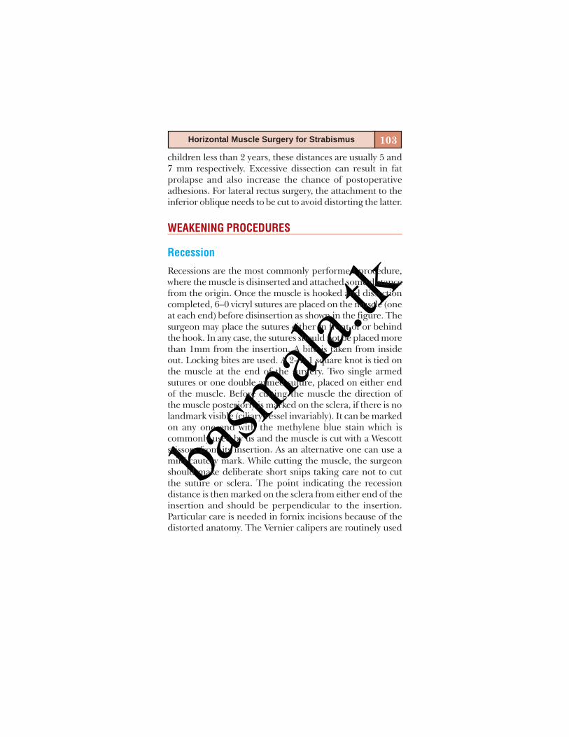
basm
ala.tk
103Horizontal Muscle Surgery for Strabismus
children less than 2 years, these distances are usually 5 and7 mm respectively. Excessive dissection can result in fatprolapse and also increase the chance of postoperativeadhesions. For lateral rectus surgery, the attachment to theinferior oblique needs to be cut to avoid distorting the latter.
WEAKENING PROCEDURES
Recession
Recessions are the most commonly performed procedure,where the muscle is disinserted and attached some distancefrom the origin. Once the muscle is hooked and dissectioncompleted, 6–0 vicryl sutures are placed on the muscle (oneat each end) before disinsertion as shown in the figure. Thesurgeon may place the sutures either in front of or behindthe hook. In any case, the sutures should not be placed morethan 1mm from the insertion. A bite is taken from insideout. Locking bites are used. A 2–1–1 square knot is tied onthe muscle at the end of the surgery. Two single armedsutures or one double armed suture, placed on either endof the muscle. Before cutting the muscle the direction ofthe muscle posteriorly is marked on the sclera, if there is nolandmark visible (ciliary vessel invariably). It can be markedon any one end with the methylene blue stain which iscommonly used by us and the muscle is cut with a Wescottscissors from its insertion. As an alternative one can use amild cautery mark. While cutting the muscle, the surgeonshould make deliberate short snips taking care not to cutthe suture or sclera. The point indicating the recessiondistance is then marked on the sclera from either end of theinsertion and should be perpendicular to the insertion.Particular care is needed in fornix incisions because of thedistorted anatomy. The Vernier calipers are routinely used
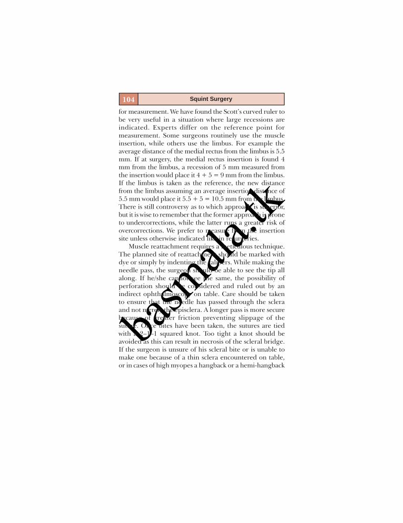
basm
ala.tk
104 Squint Surgery
for measurement. We have found the Scott’s curved ruler tobe very useful in a situation where large recessions areindicated. Experts differ on the reference point formeasurement. Some surgeons routinely use the muscleinsertion, while others use the limbus. For example theaverage distance of the medial rectus from the limbus is 5.5mm. If at surgery, the medial rectus insertion is found 4mm from the limbus, a recession of 5 mm measured fromthe insertion would place it 4 + 5 = 9 mm from the limbus.If the limbus is taken as the reference, the new distancefrom the limbus assuming an average insertion distance of5.5 mm would place it 5.5 + 5 = 10.5 mm from the limbus.There is still controversy as to which approach is superior,but it is wise to remember that the former approach is proneto undercorrections, while the latter runs a greater risk ofovercorrections. We prefer to measure from the insertionsite unless otherwise indicated like in resurgeries.
Muscle reattachment requires a meticulous technique.The planned site of reattachment should be marked withdye or simply by indenting the calipers. While making theneedle pass, the surgeon should be able to see the tip allalong. If he/she cannot see the same, the possibility ofperforation should be considered and ruled out by anindirect ophthalmoscopy on table. Care should be takento ensure that the needle has passed through the scleraand not merely the episclera. A longer pass is more securebecause of greater friction preventing slippage of thesuture. Once bites have been taken, the sutures are tiedwith a 2–1–1 squared knot. Too tight a knot should beavoided as this can result in necrosis of the scleral bridge.If the surgeon is unsure of his scleral bite or is unable tomake one because of a thin sclera encountered on table,or in cases of high myopes a hangback or a hemi-hangback
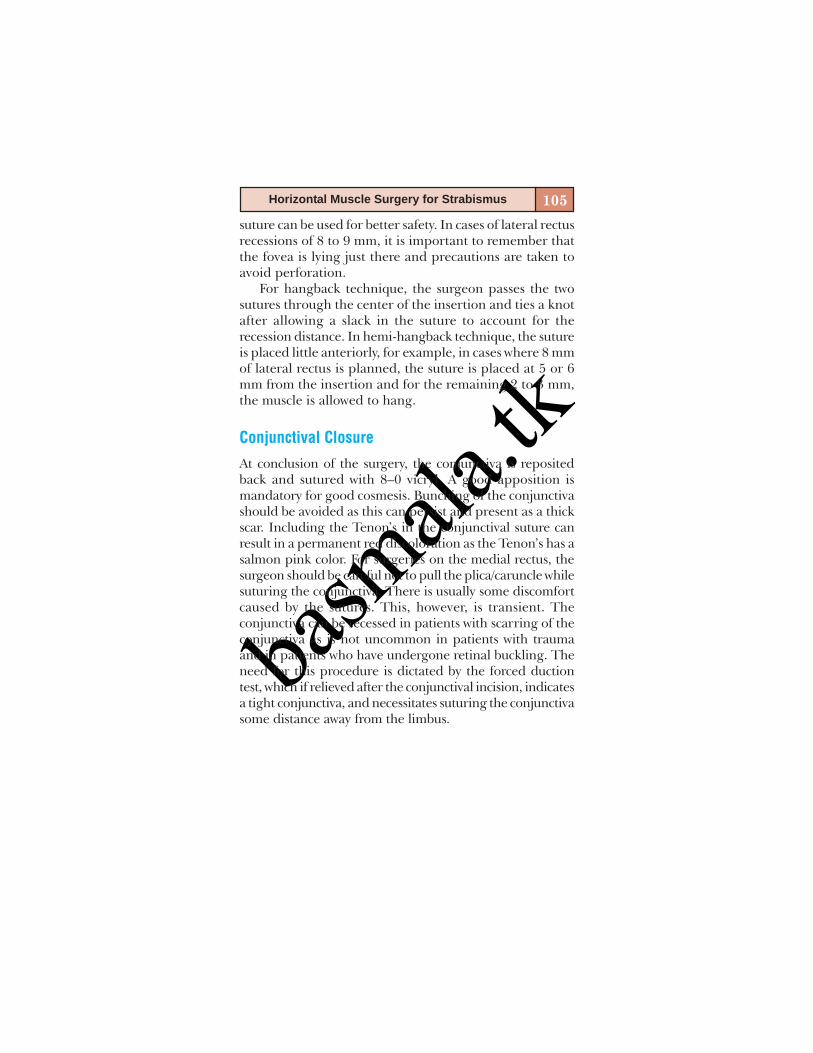
basm
ala.tk
105Horizontal Muscle Surgery for Strabismus
suture can be used for better safety. In cases of lateral rectusrecessions of 8 to 9 mm, it is important to remember thatthe fovea is lying just there and precautions are taken toavoid perforation.
For hangback technique, the surgeon passes the twosutures through the center of the insertion and ties a knotafter allowing a slack in the suture to account for therecession distance. In hemi-hangback technique, the sutureis placed little anteriorly, for example, in cases where 8 mmof lateral rectus is planned, the suture is placed at 5 or 6mm from the insertion and for the remaining 2 to 3 mm,the muscle is allowed to hang.
Conjunctival Closure
At conclusion of the surgery, the conjunctiva is repositedback and sutured with 8–0 vicryl. A good apposition ismandatory for good cosmesis. Bunching of the conjunctivashould be avoided as this can persist and present as a thickscar. Including the Tenon’s in the conjunctival suture canresult in a permanent red discoloration as the Tenon’s has asalmon pink color. For surgeries on the medial rectus, thesurgeon should be careful not to pull the plica/caruncle whilesuturing the conjunctiva. There is usually some discomfortcaused by the sutures. This, however, is transient. Theconjunctiva can be recessed in patients with scarring of theconjunctiva as is not uncommon in patients with traumaand in patients who have undergone retinal buckling. Theneed for this procedure is dictated by the forced ductiontest, which if relieved after the conjunctival incision, indicatesa tight conjunctiva, and necessitates suturing the conjunctivasome distance away from the limbus.

basm
ala.tk
106 Squint Surgery
Exposure of the Muscle with Fornix Incision
To expose the medial rectus a traction suture is placedadjacent to the limbus in the inferonasal quadrant. Theglobe is now pulled in the opposite direction and an inci-sion made 8–10 mm away from the limbus with a Wescott’sscissors. The intermuscular septum is now cut to exposebare sclera. The muscle is hooked with a von Graefe hook,which is then exchanged for a Green’s hook. The Green’shook is ideally suited for this purpose. The hook has aprojection which prevents the muscle from falling out. Themuscle is now prolapsed out of the insicion with the aid ofa Steven’s hook. The intermuscular septum is now button-holed to bare the tip of the Green’s hook and is movedinto the button hole. Dissection of the intermuscular sep-tum and the Check ligaments is as for the limbal incision.At conclusion of the surgery, the incision on the conjunc-tiva is opposed with a cotton bud or forceps. Inferior inci-sions can be left unsutured. Incisions placed in the supe-rior quadrant usually require a suture or two to avoid pro-lapse of the Tenon’s tissue.
Other Weakening Procedures
Free tenotomies have been given up for the most part ex-cept for certain special situations like strabismus fixusconvergens and on extremely tight muscles. Z tenotomiesof the horizontal recti are rarely practiced these days. Theyhave been reported to be useful in undercorrectionsfollowing nystagmus surgery.
Faden Operation
The faden operation is done by suturing the muscle to thesclera with a non-absorbable suture at a specified distance

basm
ala.tk
107Horizontal Muscle Surgery for Strabismus
(12–14 mm for the medial rectus, 14–16 mm for the verticalrecti measured from the insertion). This weakens themuscle in the direction of action of the muscle but doesnot affect primary position deviation. Faden on the lateralrectus is not very effective because of a larger arc of contactand is usually reserved for the medial rectus. This is ofvalue in some conditions like nonaccommodative esotropiawith a high AC/A ratio where the patient may beorthophoric for distance, but shows a large esotropia fornear. The amount of dissection of the check ligaments andthe intermuscular septum is much more than for routinerecessions and should be adequate to make an easyplacement of the suture. A Desmarre’s/Barbie retractor isvery useful for this purpose. This can be either combinedwith recession or done alone.Faden suture without recession: The muscle is hooked,dissection completed and the faden distance measuredfrom the limbus with the aid of a Scott ruler. A 5–0 polyestersuture is passed through the muscle on either side toinclude at least 25% of the muscle, then the needle is passedthrough the sclera and the suture tied tightly.Faden suture with recession: The muscle is hooked anddissected completely. The faden distance is marked on themuscle (calculated as faden distance measured from muscleinsertion minus recession distance). The muscle isdisinserted and a Desmarre’s retractor used to retract themuscle. The faden distance is marked on the sclera and asingle 5–0 polyester suture passed through the sclera at thisdistance. It is then brought out through the muscle at thedistance marked on the muscle. Recession proceeds as usual.After anchoring the muscle to the sclera, the faden suture istied. For further details, readers are advised to refer anytextbook on strabismus.

basm
ala.tk
108 Squint Surgery
Inactivation of the lateral rectus has been tried for thirdnerve palsies. The lateral rectus is secured to the orbitalperiosteum with the help of nonabsorbable 5–0 polyestersuture. The orbital periosteum is exposed by blunt dissec-tion a few millimeters posterior to the lateral orbital rim.Medial rectus resections after this procedure may be effec-tive in correcting the deviation in primary position as thereis no opposing force now.
STRENGTHENING PROCEDURES
Resections are by far the most commonly done procedureto strengthen the muscle. Briefly the procedure consists ofisolating the muscle, excising a portion of the musclecomprising primarily of the tendinous portion (startingfrom the insertion) and suturing the remaining muscle tothe insertion in effect tightening the muscle. 6–0 vicryl isused for this procedure. After isolating the muscle, theresection distance is marked with calipers and the suturespassed from inside out (one at each end as shown in figure).The sutures are tied with a squared 2–1–1 knot. Whilemarking the resection distance with calipers, care shouldbe taken not to stretch the muscle, which results in lessthan intended resection. While passing the sutures, 50%of the muscle is included in the suture. The two suturesare passed at either end of the insertion and tied.Redundant muscle tissue is then excised with Wescottscissors, again taking care not to cut the sutures. Any sagein the center should be corrected by passing another suturein the center. For lateral rectus resections, it is importantas mentioned earlier to severe the attachment to theinferior oblique else a J shaped deformity with resultantvertical deviations will result. In cases of medial rectusresections, the anatomy of the plica should be preserved,

basm
ala.tk
109Horizontal Muscle Surgery for Strabismus
otherwise will result in an unacceptable cosmetic appearancepostoperatively.
Muscle advancement can be employed for previouslyrecessed muscles. Generally it is held that each millimeterof advancement gives the same effect as resection. Non-absorbable sutures can be used if the surgeon fears muscleslippage. All measurements are taken from the limbus.
To sum up, horizontal rectus muscle surgery is a rela-tively simple and straightforward surgery. There are a fewpoints to watch out other than the complications detailedin the earlier chapter. It is preferable for the surgeon tolocate the muscle before placing the conjunctival incisionespecially for patients operated under general anesthesia.This avoids accidental muscle injury. Dissection shouldalways be under direct visualization. For the lateral rectusdissection should be meticulous along the inferior borderto avoid distorting inferior oblique anatomy. For reattach-ing the lateral rectus in recessions at distances of 8–10 mm,it is useful to remember that the fovea is located directlybeneath and perforations in this area can be disastrous.The vortex veins are rarely encountered in horizontal rec-tus surgery. Muscle slippage is rare but needs to be watchedout for. The medial rectus is the most common muscle toslip and the most difficult to retrieve. This is because of itsshort arc of contact and lack of attachments to the obliquemuscles. Meticulous attention to principles and techniqueusually results in a gratifying outcome.
BIBLIOGRAPHY1. Eugene M. Helveston. Surgical Management of Strabismus.
Wayenborgh Publishing, 2007.2. Julio Prieto-Diaz, Carlos Souza-Dias (Eds), Strabimsus.
Butterworth Heinemann 247-88.

basm
ala.tk
110 Squint Surgery
3. Kenneth W Wright (Ed). Colour Atlas of Strabismus Surgery.Springer Science + business LCC. New York, 2007.
4. Pradeep Sharma (Ed), Strabismus Simplified. ModernPublishers, New Delhi 1999;87-97.
5. Rosenbaum AL, Santiago AP (Eds), Clinical StrabismusManagement-Principles and Surgical Techniques. WBSaunders Company. Philadelphia, 1999.
6. Velez FG, Thacker N, Britt MT, Alcorn D, Foster S, RosenbaumAL. Rectus Muscle Orbital Wall Fixation: A reversible profoundweakening procedure. J AAPOS 2004;8:473-80.

basm
ala.tkSTEPS IN ISOLATING
HORIZONTAL RECTI AND RECESSION
Department of Pediatric OphthalmologyAravind Eye Hospital
Madurai, Tamil Nadu, India
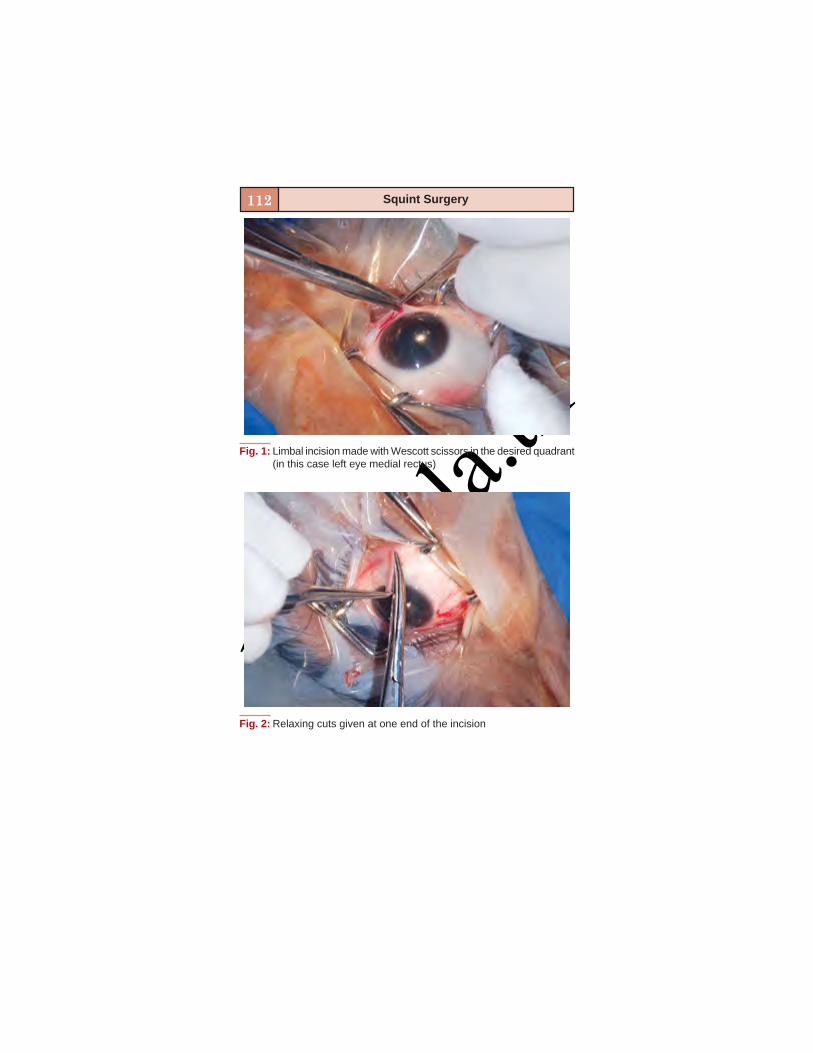
basm
ala.tk
112 Squint Surgery
Fig. 1: Limbal incision made with Wescott scissors in the desired quadrant(in this case left eye medial rectus)
Fig. 2: Relaxing cuts given at one end of the incision
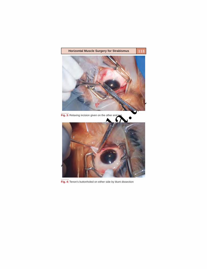
basm
ala.tk
113Horizontal Muscle Surgery for Strabismus
Fig. 3: Relaxing incision given on the other end too
Fig. 4: Tenon’s buttonholed on either side by blunt dissection
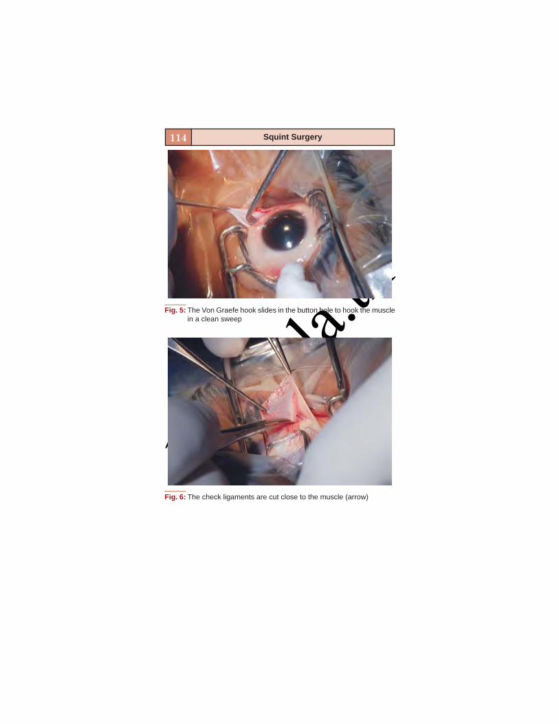
basm
ala.tk
114 Squint Surgery
Fig. 5: The Von Graefe hook slides in the button hole to hook the musclein a clean sweep
Fig. 6: The check ligaments are cut close to the muscle (arrow)
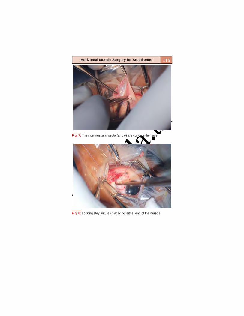
basm
ala.tk
115Horizontal Muscle Surgery for Strabismus
Fig. 7: The intermuscular septa (arrow) are cut on either side
Fig. 8: Locking stay sutures placed on either end of the muscle
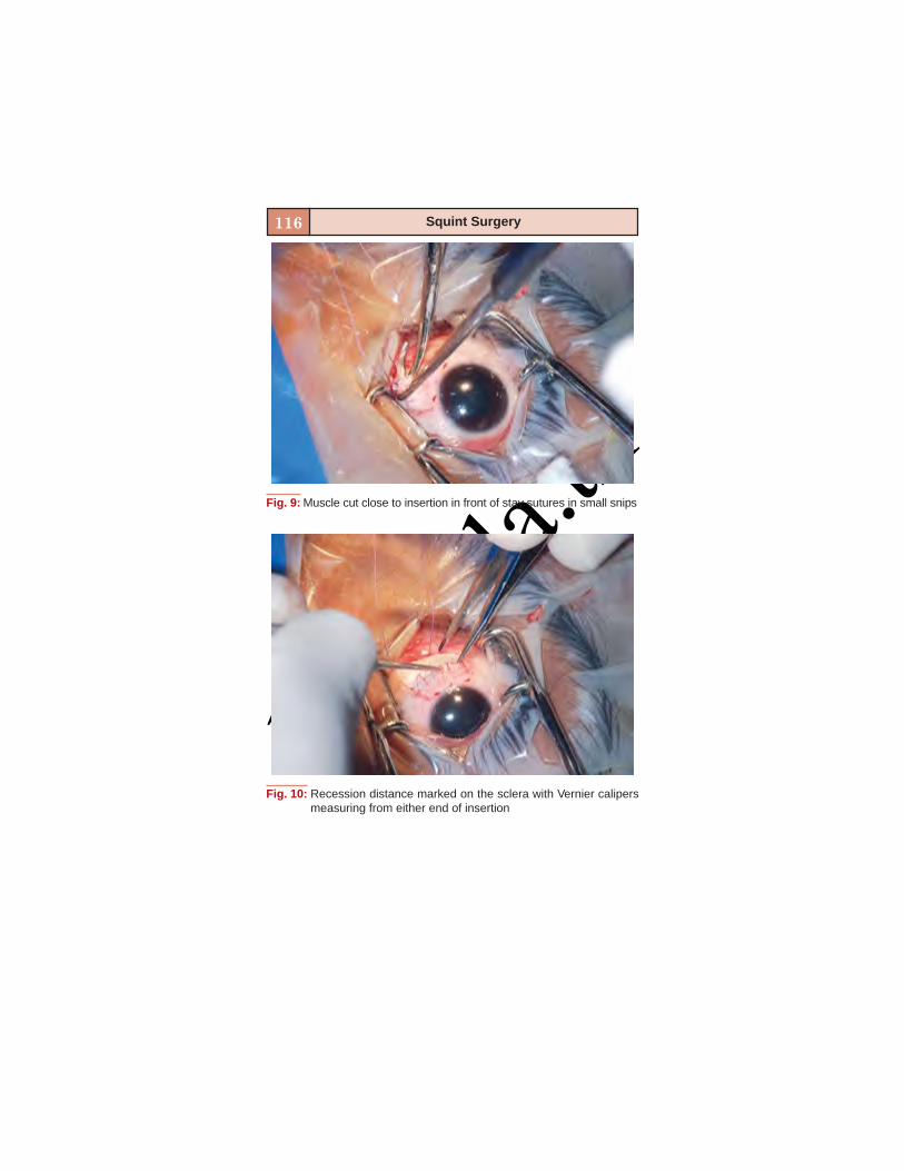
basm
ala.tk
116 Squint Surgery
Fig. 9: Muscle cut close to insertion in front of stay sutures in small snips
Fig. 10: Recession distance marked on the sclera with Vernier calipersmeasuring from either end of insertion
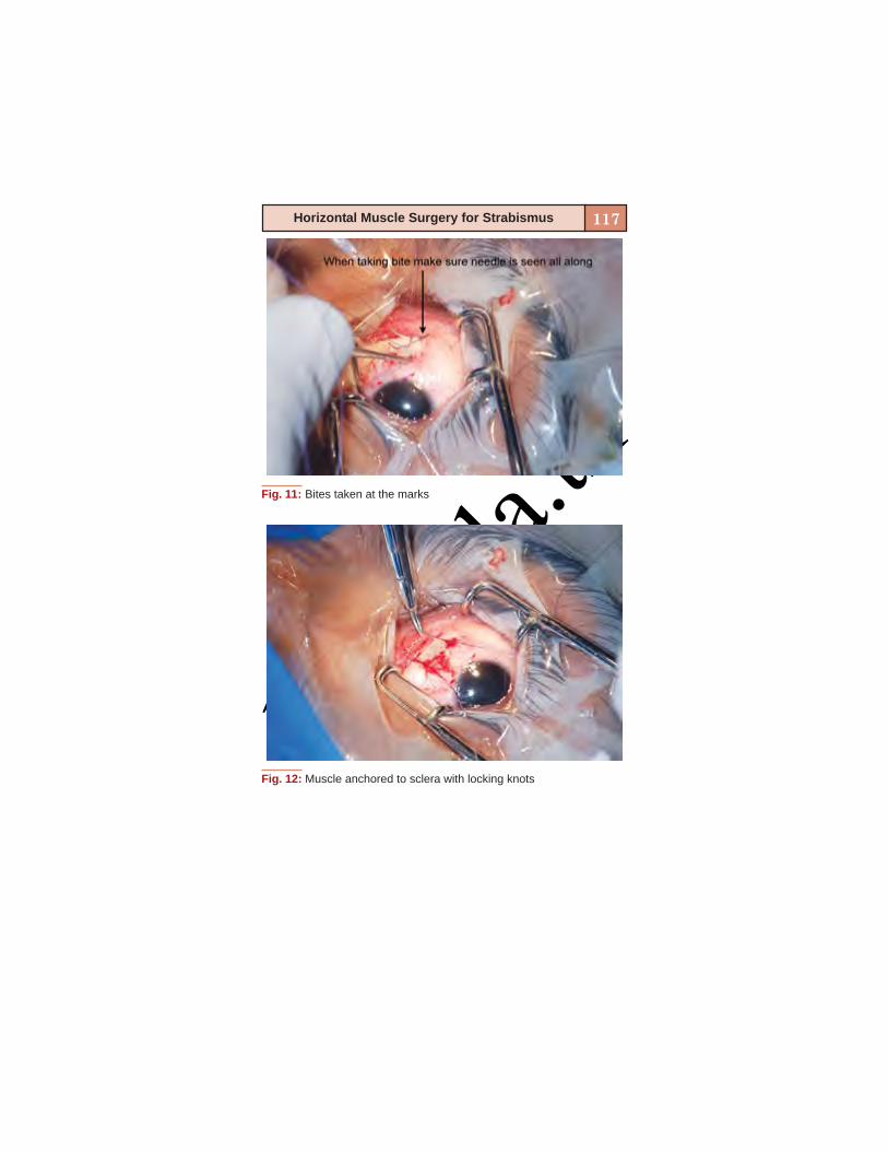
basm
ala.tk
117Horizontal Muscle Surgery for Strabismus
Fig. 11: Bites taken at the marks
Fig. 12: Muscle anchored to sclera with locking knots
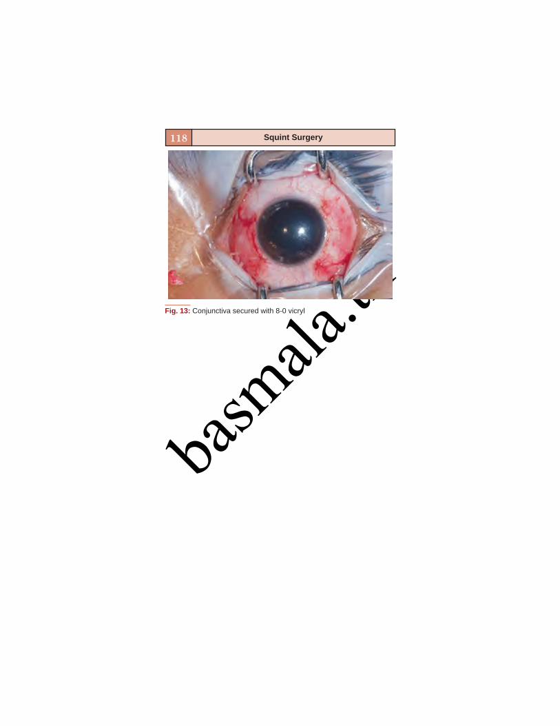
basm
ala.tk
118 Squint Surgery
Fig. 13: Conjunctiva secured with 8-0 vicryl

basm
ala.tkSTEPS IN RESECTION
Department of Pediatric OphthalmologyAravind Eye Hospital
Madurai, Tamil Nadu, India
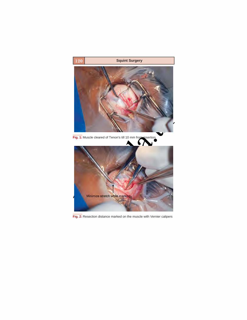
basm
ala.tk
120 Squint Surgery
Fig. 1: Muscle cleared of Tenon’s till 10 mm from insertion
Fig. 2: Resection distance marked on the muscle with Vernier calipers

basm
ala.tk
121Horizontal Muscle Surgery for Strabismus
Fig. 3: Locking bites taken and muscle secured at resection distance
Fig. 4: Muscle secured at either end with 6-0 vicryl
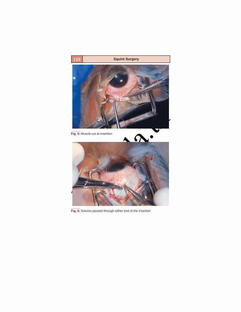
basm
ala.tk
122 Squint Surgery
Fig. 5: Muscle cut at insertion
Fig. 6: Sutures passed through either end of the insertion

basm
ala.tk
123Horizontal Muscle Surgery for Strabismus
Fig. 7: 2-1-1 reversing knots placed at either end
Fig. 8: Redundant muscle tissue (arrow) excised
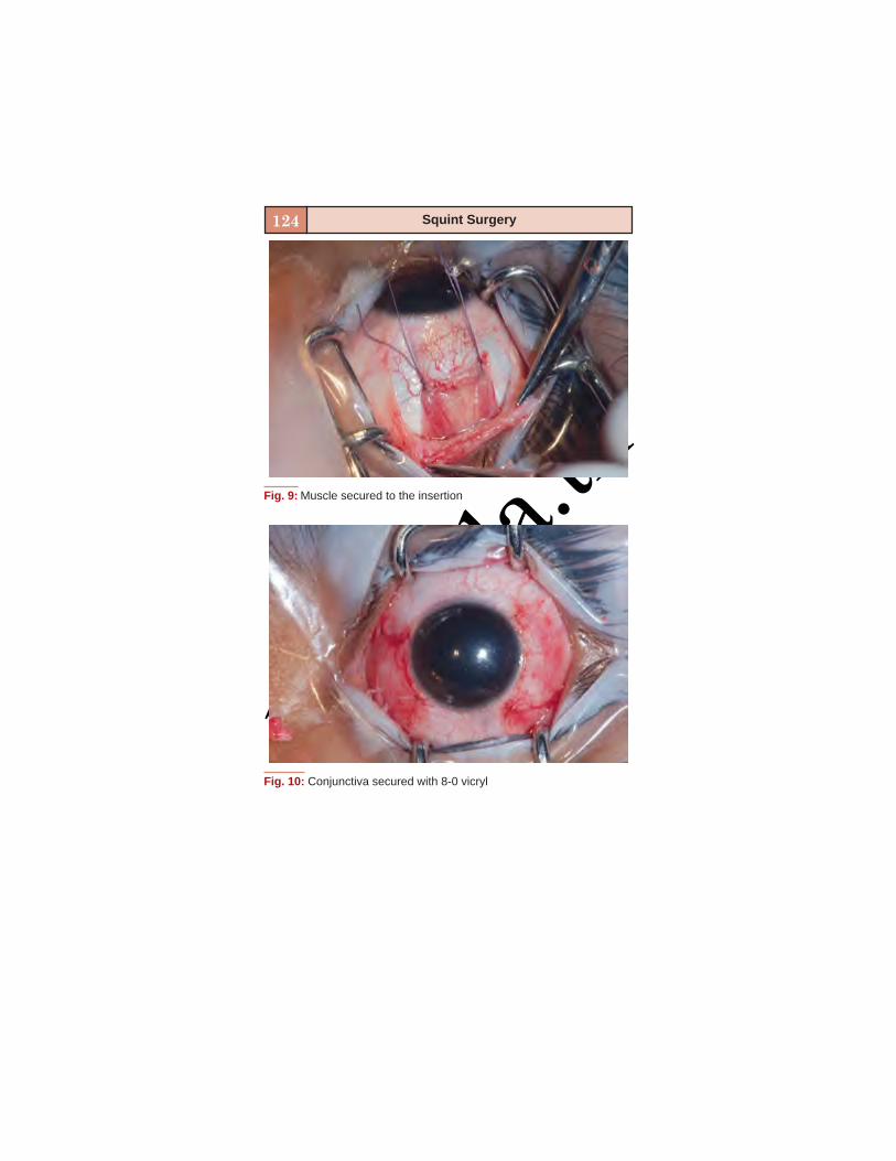
basm
ala.tk
124 Squint Surgery
Fig. 9: Muscle secured to the insertion
Fig. 10: Conjunctiva secured with 8-0 vicryl
basm
ala.tk
8
VERTICAL AND OBLIQUEMUSCLE SURGERY
Kalpana Narendran, S Ramakrishnan
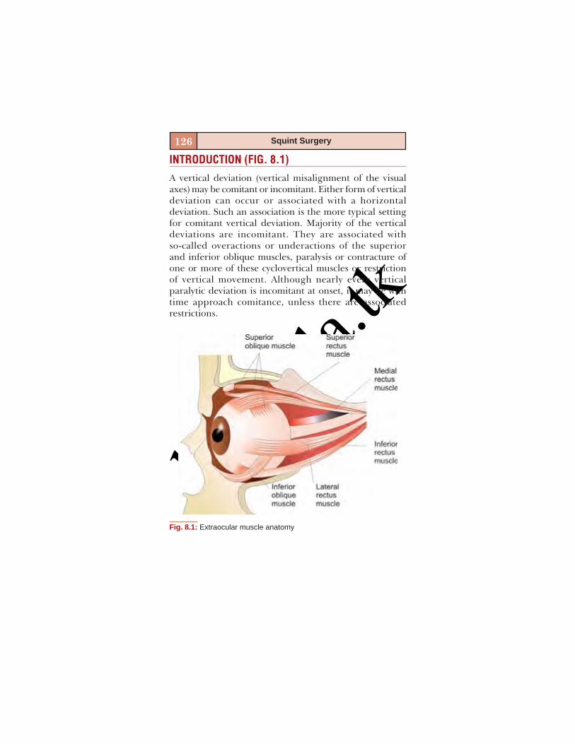
basm
ala.tk
126 Squint Surgery
INTRODUCTION (FIG. 8.1)
A vertical deviation (vertical misalignment of the visualaxes) may be comitant or incomitant. Either form of verticaldeviation can occur or associated with a horizontaldeviation. Such an association is the more typical settingfor comitant vertical deviation. Majority of the verticaldeviations are incomitant. They are associated withso-called overactions or underactions of the superiorand inferior oblique muscles, paralysis or contracture ofone or more of these cyclovertical muscles or restrictionof vertical movement. Although nearly every verticalparalytic deviation is incomitant at onset, it may be withtime approach comitance, unless there are associatedrestrictions.
Fig. 8.1: Extraocular muscle anatomy
basm
ala.tk
127Vertical and Oblique Muscle Surgery
A vertical deviation is described according to thedirection of the vertically deviating non-fixing eye. In casesof alternate fixation with equal dominance, the deviation isnamed for the usually hyperdeviating eye in ordinary visualcircumstances.
This chapter deals with the various surgical proceduresinvolved in tackling the vertical and oblique muscles.
VERTICAL RECTUS RECESSION
Superior Rectus
Indications
Dissociated vertical deviationSuperior oblique palsy with muscle sequelae.
Technique
The superior rectus is attached to the levator palpebraesuperioris above and the superior oblique below by meansof facial connections. Improper dissection can result inupper lid retraction due to the pull caused by the superiorrectus. A proper dissection of the upper lid attachmentsfrom the superior rectus is very important to avoid upperlid retraction. Dissection should not include the Tenon’scapsule lest it can cause fat adherence to the muscle andresult in restricted movements postoperatively.
Inferior Rectus
Indications
Monocular elevation deficitSuperior oblique palsy with muscle sequelae.

basm
ala.tk
128 Squint Surgery
Technique
Inferior rectus muscle is attached to the lower eyelid bymeans of facial attachments. The lower lid retractors, alsoknown as the capsulopalpebral system, interconnect theinferior rectus, inferior oblique and the lower eyelid. Thisis a continuous musculotendinous tissue which arises fromthe inferior rectus posteriorly to the inferior oblique muscleand then continues anteriorly to get inserted to the lowertarsal plate. Lockwood’s ligament, which surrounds theinferior oblique muscle, connects the tarsus to the inferiorrectus. Inferior rectus recession can lead to lower lidretraction due to pull on the Lockwood’s ligament. Posteriordissection for 10 to 15 mm along the inferior surface of theinferior rectus muscle can help minimize postoperative lowereyelid retraction. But this technique can result in fatadherence to the muscle which can restrict movements.Disinsertion of the lower lid retractors is another very goodoption to tackle this problem.
Lower lid retractor disinsertion is a very simpleprocedure. The lower lid is everted using a Desmarresretractor after passing a 4-0 silk stay suture through the lowerlid margin. The lower lid retractors insert at the inferiorborder of the lower tarsal plate. They can be removed usinga coagulation mode of a Colorado needle. Pass the Coloradoneedle 1 to 2 mm below the inferior border of the lowertarsus to burn through the conjunctiva and the underlyinglower lid retractors. This has to be done for the entire breadthof the tarsal plate.
Complications
Fat adherence to the muscle can result in restrictedmovements. Care has to be taken to avoid dissecting theTenon’s capsule to avoid this complication. Other usual
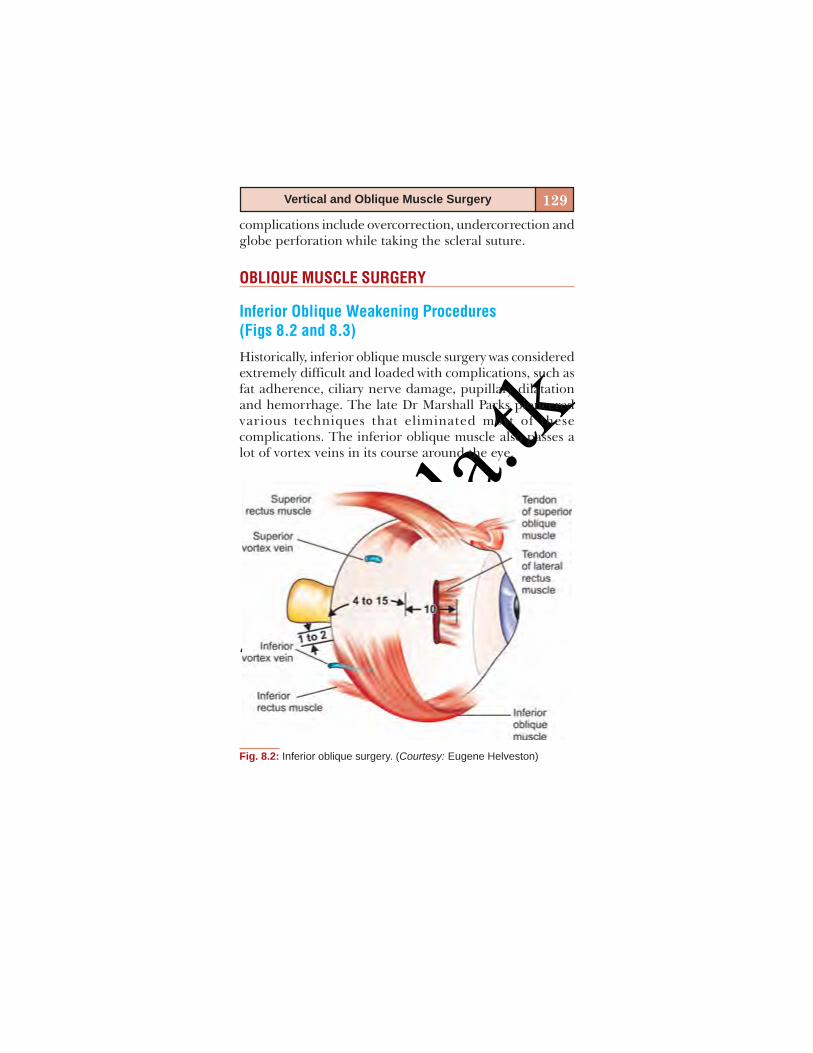
basm
ala.tk
129Vertical and Oblique Muscle Surgery
complications include overcorrection, undercorrection andglobe perforation while taking the scleral suture.
OBLIQUE MUSCLE SURGERY
Inferior Oblique Weakening Procedures(Figs 8.2 and 8.3)
Historically, inferior oblique muscle surgery was consideredextremely difficult and loaded with complications, such asfat adherence, ciliary nerve damage, pupillary dilatationand hemorrhage. The late Dr Marshall Parks pioneeredvarious techniques that eliminated most of thesecomplications. The inferior oblique muscle also passes alot of vortex veins in its course around the eye.
Fig. 8.2: Inferior oblique surgery. (Courtesy: Eugene Helveston)
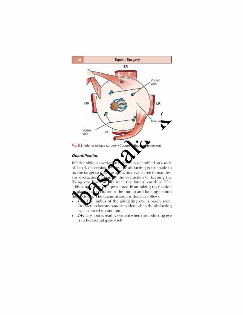
basm
ala.tk
130 Squint Surgery
Fig. 8.3: Inferior oblique surgery. (Courtesy: Eugene Helveston)
Quantification
Inferior oblique overaction is clinically quantified on a scaleof 1 to 4, on version testing, the abducting eye is made tofix the target so that the adducting eye is free to manifestany overactions. Quantify the overaction by keeping thefixing eye horizontally near the lateral canthus. Theadducting eye can be prevented from taking up fixationby placing an occluder or the thumb and looking behindthe occluder. The quantification is done as follows:
1+: The limbus of the adducting eye is barely seen.Overaction becomes more evident when the abductingeye is moved up and out.2+: Upshoot is readily evident when the abducting eyeis in horizontal gaze itself.
basm
ala.tk
131Vertical and Oblique Muscle Surgery
3+: Severe upshoot where the inferior sclera of theadducting eye is visible when the abducting eye is inhorizontal gaze position.4+: Very severe upshoot where the adducting eye movesupwards as the abducting eye is moved laterally. Inaddition to the upshoot, the adducting eye also showssome abduction as it goes into the field of action of theinferior oblique.Inferior oblique overaction produces a V-pattern – Y –
subtype, with most of the divergence occurring from primaryposition to upgaze. The final quantification is based on thecombined characteristics of the upshoot and the amount ofV-pattern.
Indications for Surgery
More than 2+ overactions1+ overaction with significant V-patternPrimary inferior oblique overaction is usually bilateral
and almost always requires bilateral surgery. Bilateral surgeryshould be performed even in patients with asymmetricoveractions. Unilateral surgery can be considered only inpatients with unilateral amblyopia, wherein the sound eyetakes up fixation always and will not manifest an upshoot.
Contraindications
Patients with V-pattern strabismus without inferior obliqueoveraction should be managed by muscle transpositionprocedures. Apparent overactions may be seen in patientswith craniofacial syndromes, but orbital and other anatomicabnormalities are usually evident radiographically.

basm
ala.tk
132 Squint Surgery
Technique
Capturing the Muscle
The incision is made in the bulbar conjunctiva and inter-muscular septum in the inferotemporal quadrant 7 mm to9 mm from the limbus, which provides an excellentapproach to the muscle and causes minimal postoperativediscomfort.
The inferotemporal limbus is grasped with a toothedforceps by an assistant to rotate the globe in extremeelevation and adduction. The conjunctival incision is made8 mm from the limbus in the inferotemporal quadrant.Tenon’s fascia is cut perpendicular to the conjunctivalincision. The lateral rectus is secured with a muscle hook.A flat iris spatula is used under direct visualization to dissectthe facial attachments of the inferior oblique muscle tothe globe. A muscle hook is positioned under the belly ofthe inferior oblique muscle. Care must be taken to avoidinjury to the vortex vein, which may result in extensivebleeding. The muscle is then freed from the surroundingfat with Wescott scissors. Connective tissue attachments tothe lateral rectus are freed. Adhesion between the inferioroblique and sclera are freed. When isolating the inferioroblique insertion area, the surgeon must be aware of thepossibility of multiple congenital insertions. The inferioroblique is attached very close to the area of macula andthe papillomacular fibers of the retina. Surgeons shouldtherefore, exercise extreme caution to prevent damage tothe underlying sclera in this region.
MYOTOMY
After the muscle is isolated, the surgeon simply incises thetendon at its insertion and allows the muscle to retract.
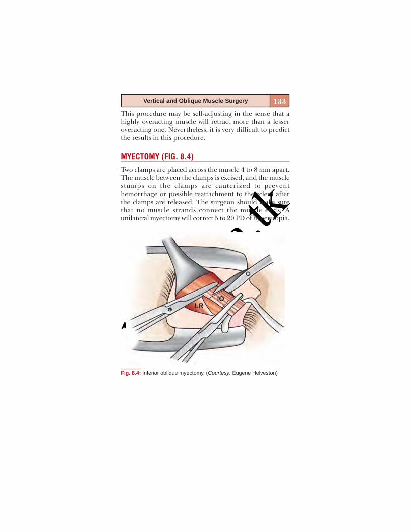
basm
ala.tk
133Vertical and Oblique Muscle Surgery
This procedure may be self-adjusting in the sense that ahighly overacting muscle will retract more than a lesseroveracting one. Nevertheless, it is very difficult to predictthe results in this procedure.
MYECTOMY (FIG. 8.4)
Two clamps are placed across the muscle 4 to 8 mm apart.The muscle between the clamps is excised, and the musclestumps on the clamps are cauterized to preventhemorrhage or possible reattachment to the sclera afterthe clamps are released. The surgeon should make surethat no muscle strands connect the muscle ends. Aunilateral myectomy will correct 5 to 20 PD of hypertropia.
Fig. 8.4: Inferior oblique myectomy. (Courtesy: Eugene Helveston)

basm
ala.tk
134 Squint Surgery
Reoperation of the muscle may be difficult after myectomywith cauterization, because relocating the muscle may bedifficult.
RECESSION (FIG. 8.5)
In a true recession, the entire muscle insertion is recededalong the anatomic course of the muscle and reattachedto the sclera with one or more sutures. This definition isextended in case of the inferior oblique muscle. Amaximum recession (12 to 14 mm) of the inferior obliquemuscle places its anterior border 4 mm behind thetemporal insertion site of the inferior rectus muscle. Caremust be taken to avoid injury to the vortex vein.
Graded recessions ranging from 6 to 12 mm are donefor greater or lesser degrees of inferior oblique overactions.The 8 mm recession of Parks is in truth not a pure recession.Scheie-Parks point is a point 2 mm lateral and 3 mmposterior from the lateral point of inferior rectus insertion.Placing the inferior oblique insertion at this point resultsin anteriorization by 1 to 1.5 mm. Probably, due to thisreason, the Parks recession, actually is a more powerfulweakening procedure than the 12 mm recession.
Fig. 8.5: Inferior oblique recession. (Courtesy: Pradeep Sharma)
basm
ala.tk
135Vertical and Oblique Muscle Surgery
ANTERIORIZATION
The anteriorization is achieved by placing the inferioroblique insertion anywhere anterior to its natural course.In practice, the rectus muscle is used for reference.
Pure anteriorization involves placing the inferioroblique muscle insertion just below the lower border ofthe lateral rectus muscle. This procedure is useful in casesof mild to moderate overactions.
In the more common procedure, the inferior rectus issecured with a muscle hook after isolating the inferioroblique. The anterior fibers of the inferior oblique aresutured to the sclera temporal to the inferior rectusinsertion. The posterior fibers are then bunched up towardsthe anterior fibers, using a needle passed through the scleraabout 1mm temporal to the first needle pass. If the newinsertion of the inferior oblique is spread out laterally, thereis an increased chance of restricting elevation.
Complications
Fat adherence syndrome: Excessive removal of adiposetissue can result in hemorrhage and subsequent scarringin the inferotemporal quadrant. This scarring can pull theeye downward or restrict upgaze. This may result inunilateral hypotropia. This complication is more commonafter myectomy than other procedures.
Excessive hemorrhage from the highly vascularized IOmuscle can rarely lead to proptosis due to bleed into theorbit. Damage to the vortex vein can also contribute to thehemorrhage.
Undercorrection and overcorrection can occur as withany squint surgery.
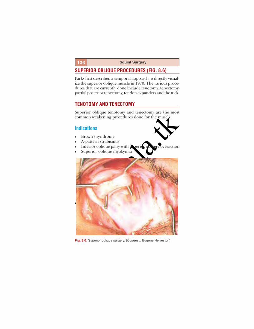
basm
ala.tk
136 Squint Surgery
SUPERIOR OBLIQUE PROCEDURES (FIG. 8.6)Parks first described a temporal approach to directly visual-ize the superior oblique muscle in 1970. The various proce-dures that are currently done include tenotomy, tenectomy,partial posterior tenectomy, tendon expanders and the tuck.
TENOTOMY AND TENECTOMYSuperior oblique tenotomy and tenectomy are the mostcommon weakening procedures done for the muscle.
Indications
Brown’s syndromeA-pattern strabismusInferior oblique palsy with superior oblique overactionSuperior oblique myokymia
Fig. 8.6: Superior oblique surgery. (Courtesy: Eugene Helveston)

basm
ala.tk
137Vertical and Oblique Muscle Surgery
The procedure can correct up to 20 PD of A-pattern inprimary gaze. Patients with bifoveal fixation may developtorsional diplopia after this procedure. It should not beperformed in patients with both superior and inferioroblique overaction.
Technique
The conjunctiva is incised in the superonasal quadrant8 mm posterior to the limbus. The incision is enlarged6 mm circumferentially. The superior oblique can bevisualized by placing a muscle hook on the posterior partof the incision. Care must be taken to avoid injury to thevortex vein. Under direct visualization, the superior obliquetendon is either divided (tenotomy) or a portion is removed(tenectomy). The complete width of the tendon should becut. Incomplete procedure may not yield the desiredresults. Forced duction testing is done to confirm theabsence of restriction in Brown’s syndrome.
Complications
Iatrogenic superior oblique palsy is a commonlyencountered complication. It has been reported in 30 to85 percent of cases. Intractable diplopia caused sometimesdue to this may be alleviated only by rejoining the cut endsusing a silicon spacer.
POSTERIOR TENECTOMY OFTHE SUPERIOR OBLIQUE
The anterior fibers of the superior oblique are responsiblefor the torsion and the posterior fibers are responsible fordepression and abduction. This procedure selectively

basm
ala.tk
138 Squint Surgery
weakens the depression and abduction effects of thesuperior oblique while preserving its torsional effects. Thiswas first described by Prieto-Diaz in 1976.
Indications
Moderate A-pattern strabismus (up to 20 PD) withsuperior oblique overaction.Incomitant dissociated vertical deviation with superioroblique overaction and A-pattern.
Technique
The globe is rotated downwards with the help of a staysuture. An incision is made in the superotemporal quadrantof the bulbar conjunctiva 8 mm posterior to the limbusand extended circumferentially by 6 mm. The superiorrectus muscle is secured with a muscle hook. The posteriorlip of conjunctiva is retracted with a Desmarres retractor.The superior oblique tendon is isolated with a muscle hookunder direct visualization at its insertion. The lid speculumis then removed to improve the field of visualization. Careis taken to include the entire width of the tendon. Thesuperior oblique fibers are then cut at its insertion leavingbehind the anterior 2 mm. A quadrilateral portion of theposterior muscle tendon including at least 10 mm of lengthis then cut and removed. The conjunctiva is closed withinterrupted 6-0 vicryl sutures.
SUPERIOR OBLIQUE EXPANDER (FIG. 8.7)
This procedure, first described by Wright, involves expand-ing the superior oblique tendon using a silicon spacer. Theweakening effect is graded by varying the length of theexpander.
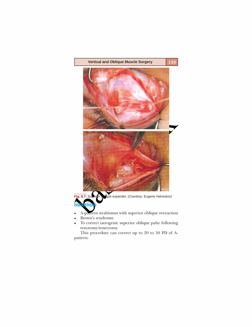
basm
ala.tk
139Vertical and Oblique Muscle Surgery
Indications
A-pattern strabismus with superior oblique overactionBrown’s syndromeTo correct iatrogenic superior oblique palsy followingtenotomy/tenectomy.This procedure can correct up to 20 to 50 PD of A-
pattern.
Fig. 8.7: Superior oblique expander. (Courtesy: Eugene Helveston)

basm
ala.tk
140 Squint Surgery
Technique
The globe is rotated downwards with a stay suture. A con-junctival incision is made in the superotemporal quadrant8 mm posterior to the limbus and extended 6 mmcircumferentially. The superior rectus muscle is isolatedon a muscle hook. The opening in the conjunctiva isreflected on the open end of the muscle hook nasally. Thesuperior oblique tendon is identified on the nasal borderof the superior rectus. The nasal intermuscular septumshould be preserved maximum possible, which will form asheath over the expander.
A double armed 6-0 non-absorbable suture is woventhrough the tendon and locked on each end 1 to 2 mmnasal to the superior rectus border. A similar suture is woven2 mm from the first and the superior oblique tendon is cutin between the sutures. Care must be taken to cut the entirewidth of the tendon.
A silicon band used for scleral buckling surgery is soakedin antibiotic solution, rinsed and trimmed according to thedesired length. The length is decided according to theamount of overaction or A-pattern. A 6 to 7 mm expanderis recommended for Brown syndrome. Each non-absorbablesuture is passed through the entire thickness of the siliconband. The sutures are tied on the band tightly securing thecut ends of the superior oblique tendon to each end of theband. The conjunctiva is closed with interrupted 6-0 vicrylsutures.
This procedure can be very successful and cause leastcomplications if performed skillfully.
basm
ala.tk
141Vertical and Oblique Muscle Surgery
SUPERIOR OBLIQUE TUCK
This procedure is indicated in cases of superior obliquepalsy, but its efficacy in complete palsies is questionable. Itis recommended in cases of incomplete palsies.
Technique
A lid speculum is inserted and the globe rotated down witha stay suture. A conjunctival incision is made similar to theabove procedures. The superior oblique tendon is exposedthrough a temporal approach. Care must be taken to in-clude the entire width of the tendon on the lateral borderof the superior rectus.
A marking suture can be used to tag the entire width ofthe tendon. The desired amount of tuck is measured witha caliper. The tuck is done temporal to the superior rectusby suturing the proximal and distal points of the tendonusing non-absorbable sutures. The conjunctiva is closedwith interrupted 6-0 vicryl sutures.
Iatrogenic Brown syndrome is the most common com-plication following superior oblique tuck, occurring in 17%of patients. Excessive restriction or intolerable torsionaldiplopia should be treated in the immediate postopera-tive period by releasing the tuck.
BIBLIOGRAPHY1. Binocular Vision and Ocular Motility Theory and Management
of Strabismus: GK Von Noorden 5th edn.2. Clinical Strabismus Management Principles and Surgical
techniques. Arthur L Rosenbaum, AP Santiago 1999 (Eds).3. Color Atlas of Strabismus Surgery: Kenneth Wright.

basm
ala.tk

basm
ala.tk
9
STRABISMUS SURGERYIN SPECIAL SITUATIONS
Kalpana Narendran
basm
ala.tk
144 Squint Surgery
INTRODUCTION
This chapter will elucidate in brief the evaluation and sur-gical management of special situations in strabismus. Thetopics covered would include:
Nonresolving paralytic strabismus—3,4,6 nerve palsyRestrictive strabismus—surgery in:– Duane’s syndrome– Brown’s syndrome– Moebius syndrome– Congenital fibrosis syndrome– Dysthyroid ophthalmopathy– Postscleral buckling– Blow out fracture.Other special situations:– Strabismus after local anesthetic injection– Strabismus in high myopia.
Nonresolving Paralytic Strabismus
The need to intervene in a case of long-standing and non-resolving paralytic strabismus is to:
Alleviate diplopiaTreat abnormal head postureTo treat strabismusTo increase the binocular diplopia free field of vision.Apart from routine preoperative evaluation including
assessment of visual acuity, detailed orthoptic work-up(motor and sensory), anterior segment and fundi evaluation,a few other important tests required include:
Diplopia charting: Provides a record of separation ofdiplopic images in the 9 gazes, shows areas of singlevision and diplopia, Crossed/uncrossed diplopic images,presence of image tilt.
basm
ala.tk
145Strabismus Surgery in Special Situations
Hess charting: Invaluable in assessing progress, planningtreatment and evaluating results. It shows underactingand overacting muscles and muscle sequelae. This is apermanent record and reveals even small limitation ofmovement at 30 degrees.Forced duction test: Used to distinguish between defectivemovement of an eye from mechanical restriction andthat due to weakness of the muscle primarily responsiblefor the movement of the eye to that gaze position.Active force generation test: Used to distinguish betweenpartial palsy and complete paralysis of muscle and alsohelps in assessing muscle recovery.Binocular field of vision.Evaluation of ocular torsion by objective and subjec-tive methods.Detailed neurological evaluation: Especially importantin deteriorating or progressive condition, younger pa-tient, presence of pupil involvement, aberrant regen-eration, nystagmus or any other neurological signs/symptoms.
Pearls before Planning Surgery
Determine the stability of ocular deviation for at least6 months.Repeat:– 9-gaze deviations– Diplopia charting– Hess/Lees charting– Binocular fields of fixation.At 6 weekly or bimonthly intervals, till the time twoconsecutive visits show no progression or change.Rule out central fusion disruption before attemptingsurgical correction to avoid intractable diplopia post-operatively especially in post-traumatic nerve palsies.
basm
ala.tk
146 Squint Surgery
Surgery in Sixth Nerve Palsy
The surgical plans would vary based on whether it is a caseof partial palsy or complete sixth nerve paralysis. Alwaysremember to do forced duction test to identify medialrectus muscle contracture before, during and after thesurgical procedure.
Partial Paralysis
In partial palsy preoperative force generation test wouldshow some amount of residual lateral rectus function.
Medial rectus recessionLateral rectus resectionCombined recess-resect procedure (R&R)This is the most commonly done procedure.R&R and contralateral medial rectus recessionThis is done if the deviation in primary gaze is more
than 40 PD.
Total Paralysis
The surgical goal in a case of total sixth nerve palsy is totry to create an abducting force and to increase the bin-ocular diplopia free field of vision. A muscle transpositionprocedure would be the surgery of choice.These include:
Partial vertical recti transposition to insertion of lateralrectus (Hummelscheim) + Medial rectus weakening byrecession or botulinum toxin (Figs 9.1A and B).
This includes splitting of the vertical recti muscles,cutting the temporal halves and attachment near thelateral rectus muscle insertion.Full tendon vertical recti transposition + Medial rectusweakening.
The entire superior and inferior recti are cut at theirinsertion and reattached near the insertion of lateralrectus (Figs 9.2A and B).
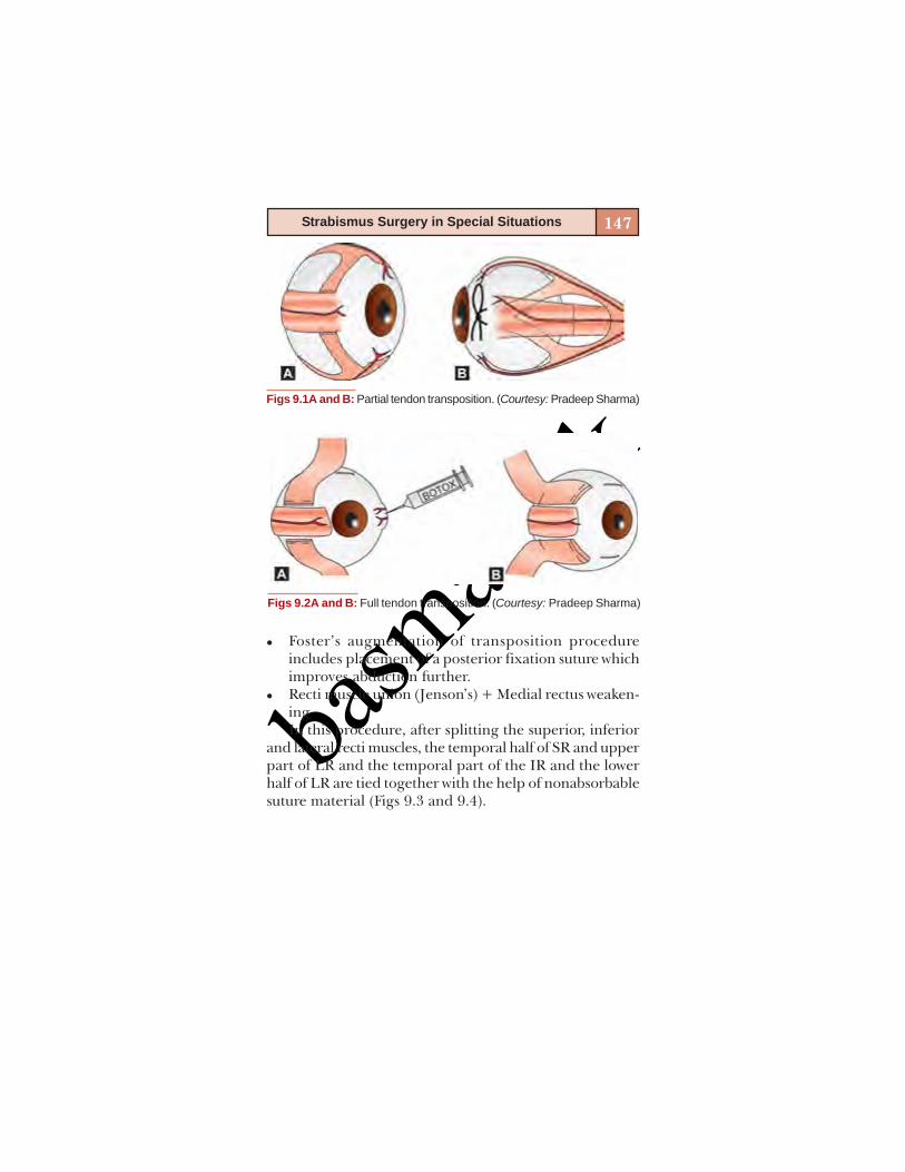
basm
ala.tk
147Strabismus Surgery in Special Situations
Figs 9.1A and B: Partial tendon transposition. (Courtesy: Pradeep Sharma)
Figs 9.2A and B: Full tendon transposition. (Courtesy: Pradeep Sharma)
Foster’s augmentation of transposition procedureincludes placement of a posterior fixation suture whichimproves abduction further.Recti muscle union (Jenson’s) + Medial rectus weaken-ingIn this procedure, after splitting the superior, inferior
and lateral recti muscles, the temporal half of SR and upperpart of LR and the temporal part of the IR and the lowerhalf of LR are tied together with the help of nonabsorbablesuture material (Figs 9.3 and 9.4).
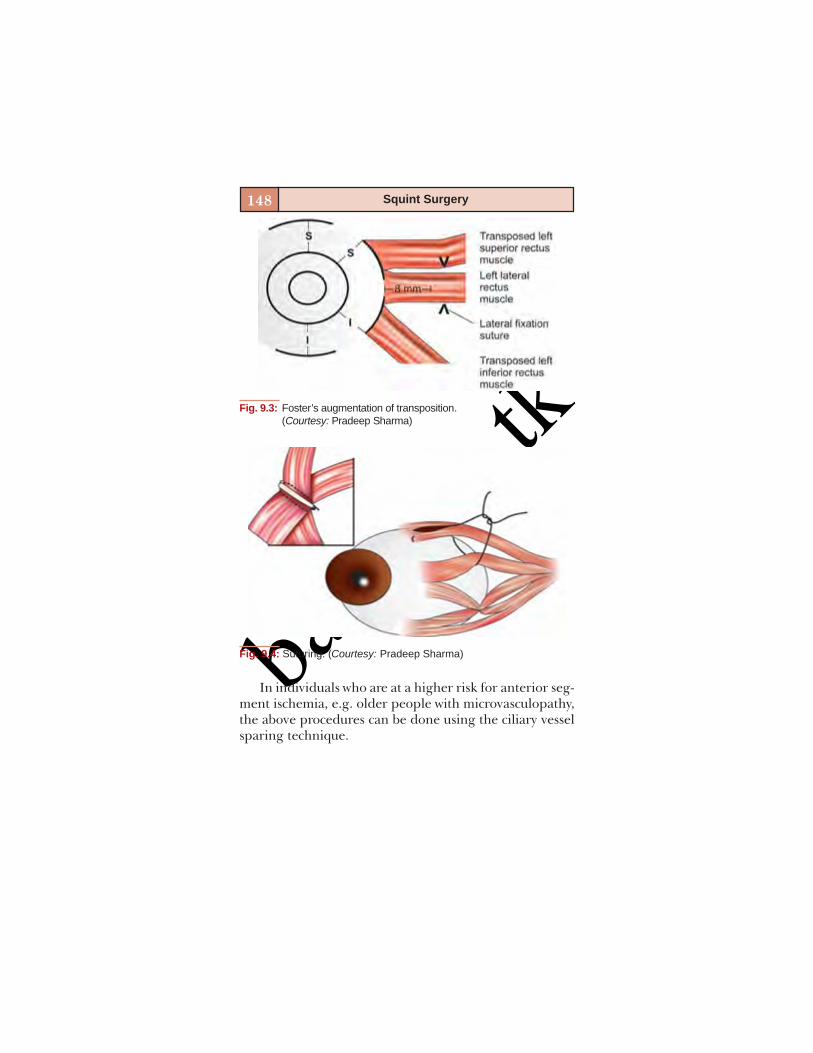
basm
ala.tk
148 Squint Surgery
Fig. 9.3: Foster’s augmentation of transposition.(Courtesy: Pradeep Sharma)
Fig. 9.4: Suturing. (Courtesy: Pradeep Sharma)
In individuals who are at a higher risk for anterior seg-ment ischemia, e.g. older people with microvasculopathy,the above procedures can be done using the ciliary vesselsparing technique.
basm
ala.tk
149Strabismus Surgery in Special Situations
Surgery in Third Nerve Palsy
Partial Third Nerve Palsy
The goals of surgery include good motor alignment, torelieve diplopia and to broaden the field of binocular singlevision.
Isolated muscle involvement: The paretic muscle withresidual function is resected and its antagonist isrecessed, preferably with adjustable suture.Multiple muscle involvement: Management strategydepends on which muscle action is more affected, e.g.In paralysis of inferior division of third nerve, Knappssuggested transposition of superior rectus to medialrectus and lateral rectus to inferior rectus. Tenetomyof the superior oblique may be included if there issignificant intorsion.
Total Third Nerve Paralysis
The goal of surgery is realignment in primary position. Asno significant area of single binocular vision is achieved,surgery is mainly performed in cases of congenital thirdnerve palsy with amblyopia, to achieve better cosmesis.Aquired third nerve paralysis is better left untreated asthere is a high-risk of intractable diplopia following surgery.
Various Surgical Strategies(Figs 9.5 and 9.6)
Fixate the globe (MR muscle insertion) to MPL inser-tion at anterior lacrimal crest using nonabsorbable 5-0polyester sutures + large LR recession in congenitaltotal 3rd nerve palsy (P Sharma et al, JAAPOS 2006, PVijayalakshmi et al, JAAPOS 2004).Superior oblique tendon resection and transposition tothe area between MR and SR insertion (Figs 9.5A and B).

basm
ala.tk
150 Squint Surgery
Figs 9.5A and B: Superior oblique tendon. Surgical strategy for total 3rdnerve palsy. (Courtesy: Clinical Strabismus ManagementRosenbaum)
Hemi-hangback recession of LR and resection of MR.Depending on the magnitude of vertical deviation, theinsertions of the horizontal rectus muscles were movedupward, alone or in combination with hemi-hangbackrecession IR (Courtesy: Köse S, Uretmen O, Pamukçu K.Strabismus 2001;9(1):1-8)
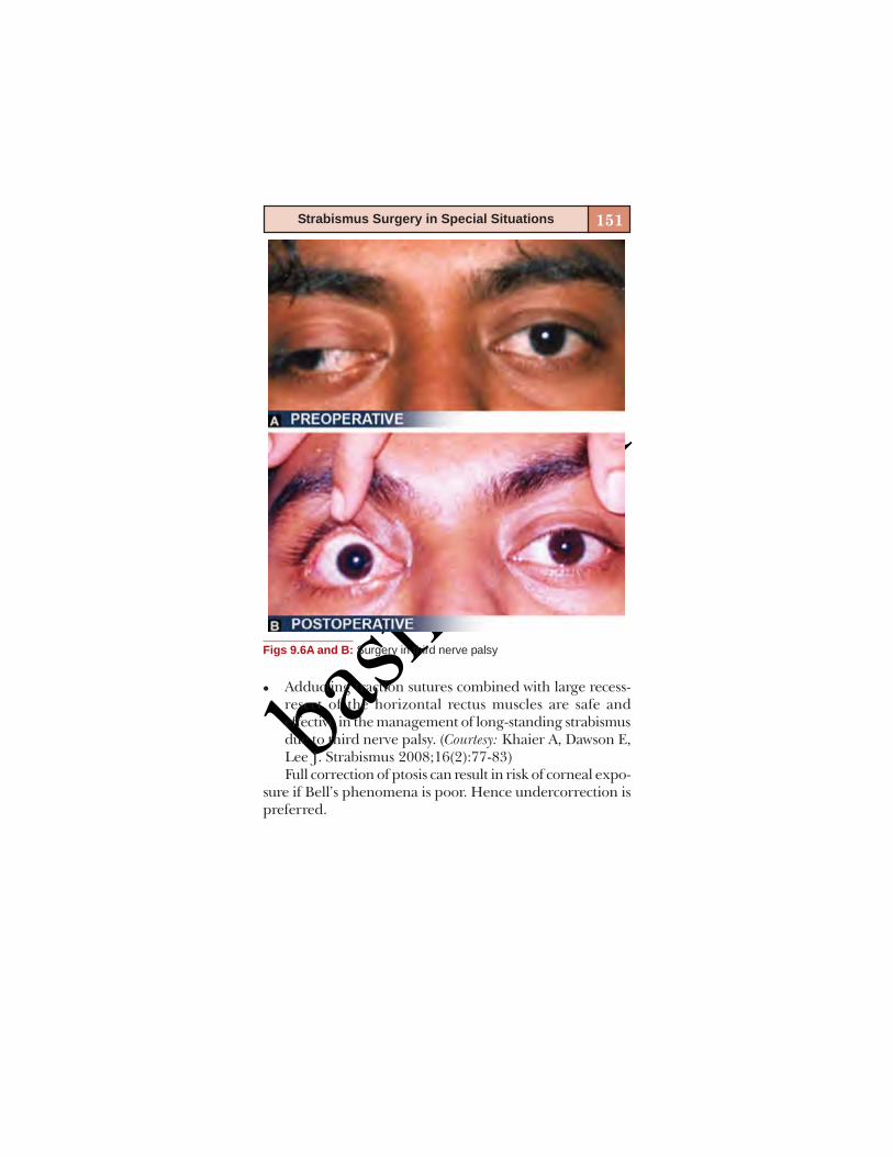
basm
ala.tk
151Strabismus Surgery in Special Situations
Figs 9.6A and B: Surgery in third nerve palsy
Adducting traction sutures combined with large recess-resect of the horizontal rectus muscles are safe andeffective in the management of long-standing strabismusdue to third nerve palsy. (Courtesy: Khaier A, Dawson E,Lee J. Strabismus 2008;16(2):77-83)Full correction of ptosis can result in risk of corneal expo-
sure if Bell’s phenomena is poor. Hence undercorrection ispreferred.

basm
ala.tk
152 Squint Surgery
Surgery for Fourth Nerve Palsy(Figs 9.7A and B and Flow Chart 9.1)In the case of bilateral fourth nerve paralysis, most com-monly seen following closed head trauma
If extorsion < 10°, then inferior oblique recession onone or both sides and bilateral inferior rectus recession(Fig 9.7A).If extorsion > 10°, bilateral Harada Ito procedure whichis splitting and advancement of the anterior half of thesuperior oblique tendon (Fig. 9.7B).
Figs 9.7A and B: Surgery in fourth nerve palsy. (Courtesy: ClinicalStrabismus Management, Rosenbaum)

basm
ala.tk
153Strabismus Surgery in Special Situations
Flow
Cha
rt 9
.1:S
urge
ry in
four
th n
erve
pal
sy (C
ourte
sy: C
linic
al S
trabi
smus
Man
agem
ent,
Ros
enba
um)

basm
ala.tk
154 Squint Surgery
RESTRICTIVE STRABISMUS
Surgery in Duane’s Syndrome
Indications for Surgery
Significant misalignment in primary positionSignificant head posture with poor cosmesis (Fig. 9.8)
Fig. 9.8: Compensatory head posture in DRS
Fig. 9.9: Narrowing of palpebral fissure in DRS
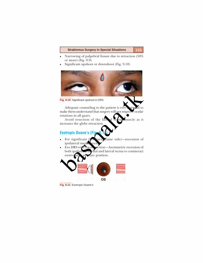
basm
ala.tk
155Strabismus Surgery in Special Situations
Narrowing of palpebral fissure due to retraction (50%or more) (Fig. 9.9)Significant upshoot or downshoot (Fig. 9.10)
Fig. 9.10: Significant upshoot in DRS
Adequate counseling to the patient is very essential tomake them understand that surgery will not improve ocularrotations in all gazes.
Avoid resection of the lateral rectus muscle as itincreases the globe retraction.
Esotropic Duane’s (Fig. 9.11)
For significant face turn (same side)—recession ofipsilateral medial rectus.Eso DRS with cocontraction—Asymmetric recession ofboth ipsilateral medial and lateral rectus to counteractesotropia in primary position.
Fig. 9.11: Esotropic Duane’s

basm
ala.tk
156 Squint Surgery
Transposition of vertical recti to the insertion of lateralrectus has been tried by few authors (Fig. 9.12).
Fig. 9.12: Transposition of vertical recti
For large angle esotropia—bimedial recession.Posterior fixation suture on the normal medial rectusmay diminish adduction innervation slightly in theinvolved eye making the esodeviation in the involvedside less (Fig. 9.13).
Fig. 9.13: Posterior fixation suture
Exotropic Duane’s (Fig. 9.14)
For significant face turn (opposite side)—recession ofipsilateral lateral rectus
Also corrects moderate globe retraction, mild upshoot/downshoot
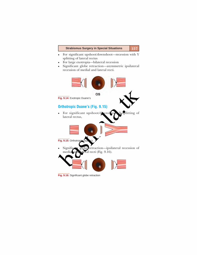
basm
ala.tk
157Strabismus Surgery in Special Situations
For significant upshoot/downshoot—recession with Ysplitting of lateral rectusFor large exotropia—bilateral recessionSignificant globe retraction—asymmetric ipsilateralrecession of medial and lateral recti.
Fig. 9.14: Exotropic Duane’s
Orthotropic Duane’s (Fig. 9.15)
For significant upshoot/downshoot—Y-splitting oflateral rectus,
Fig. 9.15: Orthotropic Duane’s
Significant globe retraction—ipsilateral recession ofmedial and lateral recti (Fig. 9.16).
Fig. 9.16: Significant globe retraction
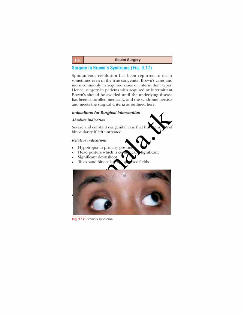
basm
ala.tk
158 Squint Surgery
Surgery in Brown’s Syndrome (Fig. 9.17)
Spontaneous resolution has been reported to occursometimes even in the true congenital Brown’s cases andmore commonly in acquired cases or intermittent types.Hence, surgery in patients with acquired or intermittentBrown’s should be avoided until the underlying diseasehas been controlled medically, and the syndrome persistsand meets the surgical criteria as outlined here.
Indications for Surgical Intervention
Absolute indication
Severe and constant congenital case that threatens loss ofbinocularity if left untreated.
Relative indications
Hypotropia in primary positionHead posture which is cosmetically significantSignificant downshootTo expand binocular diplopia free fields.
Fig. 9.17: Brown’s syndrome

basm
ala.tk
159Strabismus Surgery in Special Situations
Surgical Techniques
Initially tenectomy and tenotomy of the superioroblique tendon were the procedure of choice. This wasdone using a superonasal conjunctival incision, isolatingthe superior oblique tendon and cutting it in tenotomyor cutting a length of it in tenectomy.Parks recommended recession of the inferior obliquemuscle at the same sitting to avoid overaction of thesame postoperatively.To reduce the risk of postoperative superior obliquepalsy, Wright introduced the controlled weakeningprocedure in which a segment of silicone band is suturedbetween the cut ends of the tenotomized muscle. Thelength of the silicone band can be chosen based on theamount of weakening desired.The most common complication following surgery for
Brown’s syndrome is iatrogenic superior oblique palsy.
Surgery in Moebius Syndrome (Fig. 9.18)
This congenital condition is characterized by bilateralabducens nerve palsy with facial diplegia, along with cran-iofacial, skeletal, cardiac and muscular anomalies.
The surgical management in this condition is complexand multifactorial. In many cases optimal postoperativealignment may be possible only in primary gaze.
Perform forced duction test to identify a tight medialrectus muscle.
If FDT is negative, then bilateral vertical recti transpo-sition to lateral rectus is adequateIf FDT is positive, then vertical recti transposition tolateral rectus is followed by staged medial rectus weak-ening.

basm
ala.tk
160 Squint Surgery
Problems Encountered
Preoperative evaluation: Epicanthal folds, difficulty inspeechAnesthesia—difficult intubationHypoplasia, aplasia, fibrous bands or abnormal muscleinsertionsPoor postoperative wound healing due to lagophthalmosand inadequate blink mechanism.
SURGERY IN CONGENITAL FIBROSIS SYNDROME
Harley’s Classification
Generalized fibrosisFibrosis of the inferior rectus with blepharoptosisStrabismus fixusVertical retraction syndromeCongenital unilateral fibrosis.
Fig. 9.18: Moebius syndrome

basm
ala.tk
161Strabismus Surgery in Special Situations
Surgical Goals
Alleviate chin-up postureAlign eye in primary positionRemove obstacles to visual axis in primary gaze.
Procedure
Large recessions of the extraocular muscles are preferredwith conjunctival recession. Forced duction test should bedone both pre- and intraoperatively to assess if the restrictionhas been adequately removed.
Surgery in Thyroid Ophthalmopathy (Figs 9.19 A and B)This occurs more frequently in middle aged women, andocular myopathy does not necessarily reflect the state ofthyroid activity. Hypotropia with limited elevation is themost common motility abnormality seen.
Goal of Surgery
To align the eyes in primary gaze and reading positionwith some increase in the range of rotation.
Pre-requisites
Deviation should remain constant for at least 6 months.Surgery should be delayed in the presence of anteriorinflammatory signs.Orbital decompression, if needed, should be done priorto strabismus surgery.
Technique
Intraoperative FDT should be done before and after surgery.Care should be exercised while hooking a tight inferior rectusto avoid scleral perforation. Conjunctival recession can beadded. Adjustable sutures can be used. Avoid resections.

basm
ala.tk
162 Squint Surgery
Complications
Over and undercorrections, lower lid retraction, late slip-page of inferior rectus.
SURGERY AFTER SCLERAL BUCKLING PROCEDURE
Mechanisms for Heterotropia
Mechanical Factors
AdhesionsBuckle effect.
Figs 9.19A and B: Thyroid ophthalmopathy
basm
ala.tk
163Strabismus Surgery in Special Situations
Anatomical Factors
Nerve damageMacular Ectopia/Gliosis.
Sensory Factors
Breakdown infusionSensory deprivationAniseikoniaAnisometropia.
Muscle Factors
Muscle ischemiaDirect TraumaDisinsertionMuscle slip.
Preoperative Considerations
FDT—to analyze degree of muscle restriction presentContribution of buckle—if identified as a major factorcontributing to strabismus, then decision to remove partof it should be made in consultation with the retinasurgeon.
Approach to Surgery
A limbal conjunctival incision is preferred for better expo-sure. Adhesions are meticulously released. Only enoughbuckle is excised to allow the muscle to reattach to theglobe. Conjunctiva may be recessed at the ends if signifi-cant restriction is present. Adjustable suture techniquesmay be used.

basm
ala.tk
164 Squint Surgery
Complications
RedetachmentImplant migration and extrusionPersistent dipopia.
Surgery after Blowout Fracture(Figs 9.20A and B and Flow Chart 9.2)
Flow chart 9.2: Surgery after blowout fracture
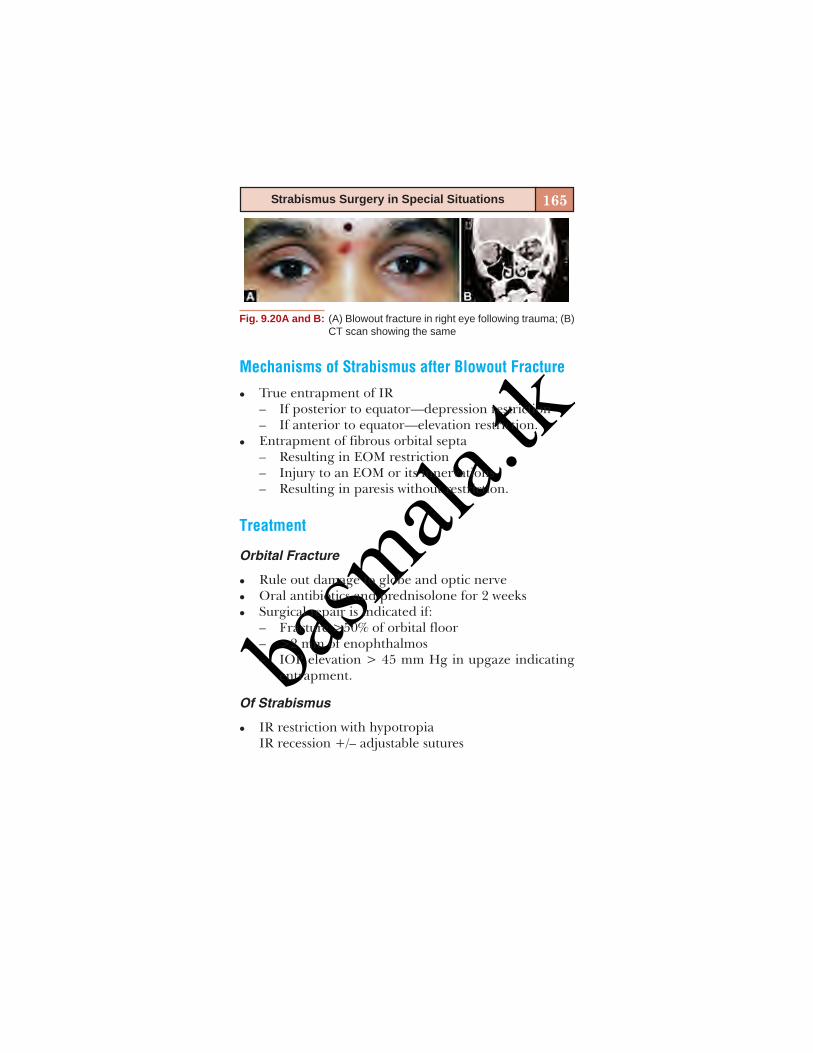
basm
ala.tk
165Strabismus Surgery in Special Situations
Mechanisms of Strabismus after Blowout Fracture
True entrapment of IR– If posterior to equator—depression restriction– If anterior to equator—elevation restriction.Entrapment of fibrous orbital septa– Resulting in EOM restriction– Injury to an EOM or its innervations– Resulting in paresis without restriction.
Treatment
Orbital Fracture
Rule out damage to globe and optic nerveOral antibiotics and prednisolone for 2 weeksSurgical repair is indicated if:– Fracture >50% of orbital floor– >2 mm of enophthalmos– IOP elevation > 45 mm Hg in upgaze indicating
entrapment.
Of Strabismus
IR restriction with hypotropiaIR recession +/– adjustable sutures
Fig. 9.20A and B: (A) Blowout fracture in right eye following trauma; (B)CT scan showing the same

basm
ala.tk
166 Squint Surgery
IR palsy:– If partial—IR resection or SR recession– If complete—horizontal recti transpositionIR palsy + restrictionComplex surgical managementMR entrapmentAs for isolated IR restriction.
Strabismus following Injection of Local Anesthetic
The inferior rectus is the most commonly affected muscle.Immediately following injection of local anesthetic, thereis a transcient period of muscle palsy followed by musclefibrosis.
Treatment
Wait for spontaneous resolution of diplopia for 6 to8 weeksIf not resolving then consider surgery to allievatediplopia.Surgical plan would be to recess the overacting infe-
rior rectus muscle—1 mm of recession would correct 3PDof hypotropia.
STRABISMUS IN HIGH MYOPIA
Bilateral Myopia and Esotropia
This happens as the lateral rectus muscle is displaced in-feriorly due to progressive superotemporal elongation ofthe globe. The altered muscle path can be seen on MRI.
basm
ala.tk
167Strabismus Surgery in Special Situations
Treatment
Large recession of medial rectus and to transpose the lat-eral rectus superiorly by suturing them to the sclera at theequator.
Heavy Eye Syndrome
Very high myopia with severe axial length elongation thatcauses esotropia and hypotropia with limitation of abduc-tion and elevation due to displacement of lateral rectusinferiorly and superior rectus nasally.
Treatment
To move the displaced muscles back to their originallocation. The lateral rectus is transposed superiorly andsuperior rectus temporally and united at the equator witha nonabsorbable suture.
BIBLIOGRAPHY1. Binocular Vision and Ocular Motility Theory and Manage-
ment of Strabismus: GK Von Noorden 5th edition.2. Clinical Strabismus Management Principles and Surgical tech-
niques: Rosenbaum AL, AP Santiago (Eds), 1999.3. Color Atlas of Strabismus Surgery: Kenneth Wright.4. Strabismus Simplified: Pradeep Sharma.

basm
ala.tk

basm
ala.tk
10
SQUINT SURGERY WITHADJUSTABLE SUTURES
Mihir Kothari
basm
ala.tk
170 Squint Surgery
A strabismus surgeon aims to achieve well aligned eyeswith good comitance, centralized and expanded field ofbinocular single vision, highest grade of binocular fusionand stereoacuity, symmetrical palpebral apertures, cor-rected head posture and full ocular movements by per-forming minimum number of squint surgeries and leastpostoperative discomfort. To achieve the target angle ofalignment in the immediate postoperative period, an ad-justable suture technique is helpful. Although the adjust-able suture technique was described by Harms way back in1941,1 it was in 1975 that it became popular after its de-scription in English literature by Jampolsky.2 Despite of afew reports questioning the superiority of the adjustablesuture surgery over the conventional surgery as a routine,reoperation rates have been reduced from 19-35% to 4-10%with the adjustable sutures.3-5 Currently, we use this tech-nique routinely for every patient regardless of the cause ofthe squint and the age of the patient. With adjustment ofsutures, we can alter up to 23PD deviation in the postop-erative period.6
(1) Half bow tie method2 (Video 8) and (2) Sliding noosemethod7 (Video 9) are the two popular methods of adjust-able suture squint surgery. Except for the final steps offixation of the muscle to the sclera, the technique of theconventional/nonadjustable suture technique and adjust-able suture technique remain the same. We will describethe final steps of the muscle refixation and the adjustmentprocedure.
HALF BOW TIE METHOD2 (VIDEO 8)
The needles are passed angulated at 30-45° to the mid-line from the insertional stump so that they exit closeto each other.
basm
ala.tk
171Squint Surgery with Adjustable Sutures
Care is taken to avoid incorporating any fibrous/Tenon’stissue to allow smooth postoperative adjustment.The muscle sutures are held tight by the surgeon andthe assistant holds the sutures with a nontoothed for-ceps at a desired distance measured from the scleralexit with the caliper.A half bow tie knot is placed over a double overhandtie.The muscle is then allowed to fall back.The measurements are confirmed and the sutures aretrimmed leaving 2-3 cm of length to allow adjustmentof the suture later.Conjunctiva is closed leaving a loop with a loose 8–0Vicryl suture to be tied tight after adjustment.An interrupted “bucket handle suture” is created thatwould be used for the adjustment.The sutures are tucked under the conjunctiva or left inthe fornix or taped to the skin.Antibiotic drops are instilled and eye pad is placed.
Postoperative Adjustment of the Sutures
This is performed on the alert and cooperative patientafter 4-24 hours preferably in the operation theaterunder all the aseptic precautions. In case of a child, itis performed as a single step procedure under generalanesthesia (preferably sevoflurane).Local anesthesia can be achieved with topical 0.5%proparacaine HCl/2% lignocaine.The assistant rotates the eye by holding the buckethandle suture and asking the patient to look in thedirection opposite to the muscle action.The muscle sutures are identified and held by the sur-geon.

basm
ala.tk
172 Squint Surgery
If an adjustment is not required, the loop of the bowtie is pulled to square off the knot without opening theknot.If an adjustment is required, the loop is untied as if itwere a shoelace. This will leave the double overhandthrow visible. To advance the muscle, the surgeon pullsup the muscle and will cinch down the knot at a newdistance, measured by the assistant where he/she holdsthe suture with a blunt forceps.The slip knot (half bow tie) is tied again and the covertest is performed once again.Approximately 2-3PD correction is obtained for 1 mmadjustment.This process can be repeated as many times as neces-sary to reach the desired target of correction.If the muscle needs to be further recessed, the over-hand knot is advanced to a new distance, measured bythe caliper and the muscle suture held by the assistantwith a blunt forceps.The slip knot (half bow tie) is tied again and the covertest is performed once again.Approximately 2-3PD correction is obtained for 1 mmadjustment.This process can be repeated as many times as neces-sary to reach the desired target of correction.At the end of the adjustment the loop of the bow tie ispulled to square off the knot and loose conjunctivalsuture is tied.
SLIDING NOOSE METHOD7 (VIDEO 9)
This is the favored and the recommended method foradjustable suture squint surgery. The adjustment can beperformed in the OPD with much higher safety and much
basm
ala.tk
173Squint Surgery with Adjustable Sutures
more efficiently than the half bow tie method. If a patientdoes not need the adjustment of the sutures, the suturescan be left alone without squaring off the knot without thefear of knot slippage.
There is no loose suture over the conjunctiva, there isno bucket handle suture and also the muscle suture istrimmed short, which stays under the conjunctiva; the pa-tient is very comfortable postoperatively.
The technique of sliding noose adjustable suture prac-ticed by us is as follows:
The needles are passed angulated at 30-45° to the mid-line from the insertional stump so that they exit cross-ing the midline or cross each other in the midline (Crosssword technique).Care is taken to avoid incorporating any fibrous/Tenon’stissue to allow smooth postoperative adjustment.The muscle sutures are held tight by the surgeon and asingle over hand knot is tied as shown. Or the assistantholds the sutures with a nontoothed forceps at a desireddistance, measured 5 mm ahead of the desired recessionfrom the scleral exit of the sutures with the caliper.While the assistant holds the muscle suture tight, anoose is tied around the muscle suture at the desireddistance by the double over throw and then squaringoff the knot. One more throw is taken and noose istightened after confirming the distance with the caliperto avoid unintended sliding of the noose.The ends of the noose are trimmed with both the armsof the sutures cut at equal length.The muscle is then allowed to fall back.The measurements are confirmed and the musclesutures are trimmed leaving one arm of the musclesuture slightly longer for the later identification.

basm
ala.tk
174 Squint Surgery
The sutures are tucked under the conjunctiva and theconjunctival incision sutured.Antibiotic drops are instilled and eye pad is placed.
Postoperative Adjustment of the Sutures
This is performed on the alert and cooperative patientafter 4-24 hours preferably in the examination roomunder all the aseptic precautions. In case of children,one time adjustment is performed under general an-esthesia (sevoflurane).Local anesthesia can be achieved with topical 0.5%proparacaine HCl/2% lignocaine.The patient to asked to look in the direction oppositeto the muscle action.The muscle sutures are identified from under the con-junctiva and pulled out (under slit lamp/binocular loop)and held by the surgeon.If the recession needs to be reduced, the sliding nooseis pushed down while the muscle suture is pulled for-ward (caution the patient that it will hurt a bit) and ifthe recession needs to be increased, the noose is pulledout by the desired amount and the patient is instructedto look in the direction of the muscle for the augmen-tation to take its effect.Approximately 2-3PD correction is obtained for 1 mmadjustment and the cover test is performed.This process can be repeated as many times as neces-sary to reach the desired target of correction.Once the desired correction is reached, the sutures aresimply buried under the conjunctiva and the antibioticeyedrops are instilled.

basm
ala.tk
175Squint Surgery with Adjustable Sutures
Additional Tips
If adjustable sutures are used in children – one timeadjustment is performed under general anesthesia,preferably using sevoflurane anesthesia which is aug-mented with topical anesthetic, preferably proparacaineHCl 0.5%.Instillation of HPMC (viscoelastic) eyedrops makessuture surface smooth and hence the adjustments easy.Excessive pulling of the muscle or jerky movementsshould be avoided for the fear of inciting pain or pro-voking the oculocardiac reflex.Avoid holding of the sutures with the toothed forceps –to prevent suture damage and the breakage of thesutures.Cheese wiring of the scleral bites should be avoided byholding the sutures with correct tension and in the rightdirection.Usually never required, however, an emergency traycontaining atropine injection should be available forthe patients who develop oculocardiac reflex. This ismore commonly seen in anxious patients, athletic malesand patients who have a history of motion sickness. Insuch a situation, injection atropine given intramuscu-larly half an hour prior to the adjustment is helpful.Adjustable suture technique is best reserved for therecessions. It can be applied to resections and trans-positions.Advancement of muscle is usually easier but more pain-ful than increasing the recession, which is less painfulbut more difficult.
basm
ala.tk
176 Squint Surgery
CONCLUSION
Adjustable suture technique of squint surgery requires afew modifications in the final steps of a squint surgery. Itprovides the surgeon a capability to change the alignmentpostoperatively in order to achieve targeted alignment.This technique of surgery is easy to learn and master andprovides superior results for immediate postoperativeocular alignment.
REFERENCES1. Harms H. Ein Vorlagerungsverfahren mit nachtregrlicher
Dosierungs moglichkeit. Klin Monastbl Augenheilkd1941;104:728.
2. Jampolsky A. Strabismus reoperation techniques. Trans AmAcad Ophthalmol Otolarngol 1975;79:704.
3. Karus DJ, Bullock JD. Treatment of thyroid ocular myopathywith adjustable and nonadjustable suture strabismus surgery.Trans Am Soc 1993;91:67.
4. Metz HS, Lerner H. Adjustable suture strabismus surgery. AmOphthalmol 1979;11:1593.
5. Wisnicki HJ, Repka MX, Guyton DL. Reoperation rate inadjustable strabismus surgery. J Pediatr Ophthalmol Strabis-mus 1988;25:112.
6. Rosenbaum AL. Metz HS, Carlson M, et al. Adjustable rectusmuscle recession surgery: A follow-up study. Arch Ophthalmol1977;95:817.
7. Jampolsky A. Current techniques of adjustable strabismussurgery. Am J Ophthalmol 1979;88:406.

basm
ala.tk
11
BOTULINUM TOXIN INSQUINTRamesh Murthy

basm
ala.tk
178 Squint Surgery
INTRODUCTION
Botulinum toxin has been described as the ‘most poisonouspoison’, though now it is one of the most common drugsused in the world especially for cosmetic indications.Clostridium Botulinum which produces the toxin was firstisolated by Van Ermengem in 1895.1 This organism is agram-positive, spore forming, anaerobic rod commonlyfound on plants, in soil, water and the intestinal tracts ofanimals and fish. The clinical picture of Botulism wasdescribed by Justinus Kerner in 1822. The concept ofinjecting pharmacological agents into extraocular musclesto paralyze them was first perceived by Dr Conrad Behrens.In 1973, Dr Alan Scott injected the extraocular muscles ofrhesus monkeys with different agents including botulinumtoxin and subsequently demonstrated the utility of thistoxin in humans.2 Currently botulinum toxin hasapplications in neurology, dermatology, gastroenterology,orthopedics, otorhinolaryngology and ophthalmology.
PHARMACOLOGY
Clostridium Botulinum produces eight different exotoxins(A, B, C1, C2, D, E, F and G).3 Type A is the most potentand frequently used. These are single polypeptide chainsof about 150 kDa weight with an L chain and a H chainjoined by disulfide bonds.
PREPARATIONS
There are three commonly available commercialpreparations – Botox (Allergan Inc, USA), Dysport (IpsenPharmaceuticals, France), both of which are type A andMyobloc which is a type B toxin. Type A toxin is the most
basm
ala.tk
179Botulinum Toxin in Squint
potent and is the one currently in vogue. It has the longestduration of action and has a favorable ratio between thebiologically active and inactive neurotoxin.
MECHANISM OF ACTION
This toxin acts at four different sites in the body: theneuromuscular junction, autonomic ganglia, postganglionicparasympathetic nerve endings and postganglionicsympathetic nerve endings which release acetylcholine (Ach).
At the neuromuscular junction botulinum toxin causesmuscle paralysis. Normally the muscle contraction occursdue to acetylcholine vesicles released into the synaptic cleft.Botulinum toxin when injected, binds to the nerve termi-nal gets internalized and prevents the release of the ace-tylcholine vesicles by the inhibition of SNARE proteins.About 8 weeks after the injection, new nerve terminalsprouts emerge and extend towards the muscle surface,reinnervating the muscle.4
The classic rationale given for the use of botulinum totreat strabismus is to induce a temporary paralysis of anormal or overacting muscle, which allows shortening andcontracture of the under acting or paretic antagonist ofthe injected muscle. This in turn produces a change in thealignment of the eyes towards normal with persistence afterresolution of the botulinum toxin induced paralysis.
TECHNIQUE OF INJECTION
The toxin is available as a 100 unit or 50 unit vial. Recon-stitution is performed with sterile non-preserved 0.9%saline prior to injection. The toxin concentration dependson the volume of diluent used. The dose used inthe extraocular muscles varies between 1-5 units. The
basm
ala.tk
180 Squint Surgery
solution once reconstituted should be clear, colorless andfree of particulate matter and should be stored in a refrig-erator at 4° C until use. The reconstituted toxin is drawninto a tuberculin syringe and injected with a fine gaugeneedle (30 G or 32 G). While it has been mentioned thatthe toxin needs to be used within 4 hours of injection, thevial once reconstituted can be used upto 4 weeks.
The injection can be given under electromyographicguidance or under direct visualization. After instilling topicalanesthetic agents, the injection needle is passed throughthe conjunctiva into the muscle on the orbital side afterturning the eye into the field of action of the muscle to beinjected. If using EMG guidance, a needle electrodeconnected to a tuberculin syringe is used. The electro-myographic signal is used as a guide to advance the needleto a point within the muscle and about 2.5 cm to its insertion.The toxin is then injected. If unavailable, direct injectioninto the muscle can be performed by exposing the musclewith a conjunctival incision.
CONTRAINDICATIONS TO THE USE OFBOTULINUM TOXIN
1. Neuromuscular junction disorders:a. Myasthenia gravis.b. Eaton-Lambert syndrome.
2. Peripheral motor neuron disease:a. Amyotrophic lateral sclerosis.b. Motor neuropathy.
3. Pregnancy.4. Areas of active infection.5. Known hypersensitivity to any ingredient in the for-
mulation.
basm
ala.tk
181Botulinum Toxin in Squint
CLINICAL APPLICATIONS
The main advantage of botulinum toxin is it can beperformed as an outpatient procedure in adults and canreduce misalignment. The current indications in squintinclude:
Paralytic strabismus.Postoperative consecutive strabismus.Augmentation of transposition effect.Active thyroid orbitopathy.Cyclic esotropia.Previous multiple surgeries.Strabismus less than 40 prism diopters.Patients who are not suitable for surgery.In cases like restrictive squints, large angle deviations
and over recessed muscles botulinum toxin is ineffective.While the risks are low, problems include multiple repeatedinjections, temporary improvement, diplopia and or sen-sory disorientation and temporary overcorrection due tounderaction of paralyzed muscle. Globe injury, retrobul-bar hemorrhage and changes in eyelid position can occurwith periocular injection. Spread of the drug beyond themuscle can lead to partial weakness of the orbicularis andlevator muscle and can cause lagophthalmos, ptosis andimpairment of Bell’s reflex.
CRANIAL NERVE PALSIES
Acute acquired nerve palsies may improve over time, butchronic palsies are more resistant to cure. Botulinum toxincan prevent muscle contracture in these cases.5 Childrenwith nerve palsy who do not develop a compensatory headposture may develop amblyopia and lose stereopsis in afew weeks. In cases of sixth nerve palsy botulinum toxininjection can restore fusion and prevent medial rectus

basm
ala.tk
182 Squint Surgery
contracture. In some cases of sixth nerve palsies, somesurgeons prefer to perform transposition of the verticalrecti to the lateral rectus and augment the procedure withbotulinum injection to the medial rectus eitherpreoperatively, intraoperatively or postoperatively. Fostertype modifications are usually not combined withbotulinum augmentation. Nearly 50% patients can be curedwith injection and surgery can be avoided.
In the treatment of third nerve palsy, it has limited suc-cess. It can be administered if the paresis persists over a 4weeks period. Some partial third nerve cases respond withbotulinum administration, but little benefit is seen in casesof total third nerve paralysis. Multiple muscle involvementlimits the use of botulinum in case of cranial third nervepalsies.
For superior oblique palsies, botulinum may improvesymptoms. Injections into the inferior oblique are usuallyperformed.6 Half the patients had improvement in theirsymptoms for primary and reading positions and resolu-tion of torticollis, but diplopia was not eliminated in lat-eral gaze positions. Botulinum was most effective wheninjected into the inferior rectus, when the inferior obliquewas injected 83% patients needed surgery.
VERTICAL STRABISMUS
In cases of active thyroid disease, patients may benefit withinjection of the inferior rectus, though most of the patientswill need surgery. The problem with injections to the su-perior rectus is the ptosis and hence this should be avoidedin children in the amblyogenic period. In most cases ofhypertropia, injection into the superior rectus alone maynot be enough and injection into the inferior oblique mayalso be needed.7
basm
ala.tk
183Botulinum Toxin in Squint
PARADOXICAL DIPLOPIA
This condition occurs when the visual cerebral cortex pro-jection of diplopic images does not correspond to the ob-jective angle of deviation, indicating anomalous retinalcorrespondence. In cases where inadvertent surgery hasbeen performed and the patient develops intractable diplo-pia, this may work. We have reported on a case of acquiredmotor fusion deficiency where the diplopia was relievedafter injections of botulinum toxin.8
SENSORY STRABISMUS
Botulinum can be a useful adjunct in these cases. In sensoryexotropia, surgeons prefer to operate only on the eye withpoor vision if possible. In these cases, botulinum toxin canbe an adjuvant treatment.9
NYSTAGMUS
In nystagmus, retrobulbar injection of botulinum has beenutilized with the quantity varying from 20-30 units.Sometimes injections into individual extraocular muscleshave also been tried. Complication following retrobulbarinjections include ptosis. Improvement in vision can helpthe patients secure a driver’s license. Repeated injectionsof botulinum toxin may be needed to maintain vision.Botulinum toxin has also been noted to be useful in patientswith oscillopsia in a study performed by us on wheelchairbound patients.10
DISSOCIATED VERTICAL DEVIATION
In a study by Ruiz et al, patients with infantile esotropiatreated with botulinum injections into both medial recti

basm
ala.tk
184 Squint Surgery
before 18 months of age required more injections and hadgreater dissociated vertical deviation.11
OPHTHALMOPLEGIA
Patients with ophthalmoplegia sometimes need multiplesurgeries. In these cases, botulinum toxin can be benefi-cial. It has been shown to be beneficial for patients withexternal ophthalmoplegia. We have reported on a seriesof cases of internuclear ophthalmoplegia where subjectivebenefit was reported by patients with improvement of thequality of the image.12
HORIZONTAL STRABISMUS
Consecutive Strabismus
One of the potential complications of strabismus surgeryis overcorrection. Botulinum toxin has been noted to behelpful in restoring high grade stereopsis in patients withconsecutive esotropia with fusion potential.13 In a studyon children, it was noted that there was successful alignmentin children with residual esotropia following strabismussurgery and botulinum toxin injection was comparable tosurgery.14
ESOTROPIA
Botulinum toxin may be useful in patients with esotropia.For large angle esotropia, it may be combined with medialrectus recessions. In patients with nystagmus, patients hada lower surgical dose and less strabismus surgery.However, multiple treatments are required.15 Adults withsecondary esotropia, consecutive esotropia, residual

basm
ala.tk
185Botulinum Toxin in Squint
esotropia and primary esotropia are likely to be benefitedby botulinum injections.
Botulinum has also been evaluated in infantile esotro-pia. While the exact benefit of botulinum in infantile esotro-pia is unclear, it is less effective than traditional surgery.
EXOTROPIA
Botulinum toxin has been used in cases of exotropia. Dueto the induced hypertropia, botulinum may not be the bestchoice. In cases of sensory exotropia in patients where sur-gery has not been satisfactory, botulinum can be a choice,but multiple injections are necessary. Botulinum can alsobe used to assess the risk of postoperative diplopia, thoughprism adaptation poses a lesser risk. Surgery is preferred asthe first choice in most patients with exotropia (Figs 11.1Ato C).16
Fig. 11.1A: At the end of 6 weeks he was noted to have complete recov-ery of his ocular movements and resolution of the diplopia
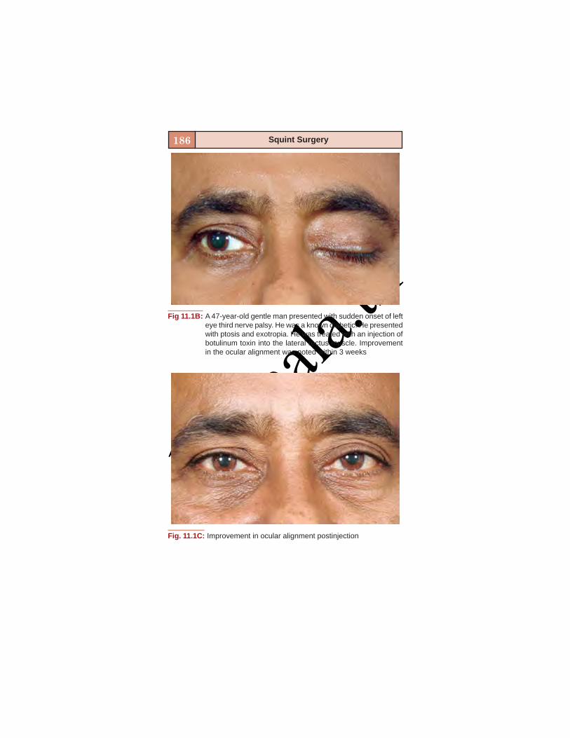
basm
ala.tk
186 Squint Surgery
Fig. 11.1C: Improvement in ocular alignment postinjection
Fig 11.1B: A 47-year-old gentle man presented with sudden onset of lefteye third nerve palsy. He was a known diabetic. He presentedwith ptosis and exotropia. He was treated with an injection ofbotulinum toxin into the lateral rectus muscle. Improvementin the ocular alignment was noted within 3 weeks

basm
ala.tk
187Botulinum Toxin in Squint
REFERENCES1. Klein AW. Botulinum toxins. Introduction. Semin Cutan
Med Surg 2001;20:69-70.2. Scott AB. Pharmacologic weakening of extraocular muscles.
Invest Ophthal Vis Sci 1973;12:924-7.3. Comella CL, Pullman SL. Botulinum toxins in neurologi-
cal disease. Muscle Nerve 2004;29:628-44.4. Sellin LC. The pharmacological mechanism of botulism.
Trends in Pharma Sci 1985;6:80-2.5. Siatowski RM. Third, fourth and sixth nerve palsies. Focal
points 1996;XIV:1-14.6. Merino P, Gomez de LIano P, Garcia C, et al. Bilateral supe-
rior oblique palsy and botulinum toxin. Arch Soc EspOphthalmol 2004;79:119-23.
7. Ozkan SB, Kir E, Dayanir V, Dundar SO. Botulinum toxin Ain the treatment of adherence syndrome. Ophthalmic SurgLasers Imaging 2003;34:391-5.
8. Murthy R, Kesarwani S. Botulinum toxin in the manage-ment of acquired motor fusion deficiency. Indian JOphthalmol 2009;57:463-4.
9. Owens PL, Strominger MB, Rubin PA, et al. Large angleexotropia corrected by intraoperative botulinum toxin A andmonocular recession resection surgery. J AAPOS 1998;2:144-6.
10. Dawson ELM, Murthy R, Lee J. Does Botulinum toxin havea role in the treatment of Oscillopsia? Neuro-Ophthalmol-ogy 2007;31(3):73-8.
11. Ruiz MF, Moreno M, Sanchez-Garrido CM, Rodriguez JM.Botulinum treatment of infantile esotropia with abductionnystagmus. J Pediatr Ophthalmol Strabismus 2000;37:196-205.
12. Murthy R, Dawson E, Khan S, Adams G, Lee J. Botulinumtoxin in the management of internuclear ophthalmople-gia. Journal of AAPOS 2007;11(5):456-9.
13. Dawson EL, Marshman WE, Lee JP. Role of botulinum toxinA in surgically corrected exotropia. J AAPOS 1999;3:269-71.
basm
ala.tk
188 Squint Surgery
14. Tejefor J, Rodriguez JM. Retreatment of children after sur-gery for acquired esotropia: Reoperation versus botulinuminjection. Br J Ophthalmol 1998;82:110-4.
15. Ruiz MF, Alvarez MT, Sanchez-Garrido CM, et al. Surgeryand botulinum toxin in congenital esotropia. Can JOphthalmol 2004;39:639-49.
16. Lawson JM, Kousoulides L, Lee JP. Long term results ofbotulinum toxin in consecutive and secondary exotropia:Outcome in patients initially treated with botulinum toxin.J AAPOS 1998;2:195-200.

basm
ala.tk
POSTOPERATIVECONSIDERATIONS
3

basm
ala.tk
basm
ala.tk
12
SQUINT POSTOPERATIVEFOLLOW-UP AND
TREATMENTMilind Killedar

basm
ala.tk
192 Squint Surgery
Squint surgery is one of the common surgeries amongstthe ophthalmic surgeries. The postoperative follow-up andtreatment is not very different from cataract except a fewchanges.
Although, there is no standard regime which is followedby everyone, the schedule given below can be convenientto follow and would avoid any missouts.
FIRST POSTOPERATIVE DAY
Usually the dressing is opened and then clean the eye. Adrop of paracaine will make child comfortably open theeyes and gain confidence. Although, in Western world it isa common practice to keep eyes open without a dressingand inform parents about bloody tears, it is not a commonpractice in India. But if it is bilateral surgery, I prefer tokeep one eye open (usually the eye with less no of musclesoperated). Keeping both eyes closed makes the childextremely unhappy. After removal of dressing and puttinga drop of paracaine wait for ½ to 1 hour and then see theocular alignment. If it is adjustable suture surgery take 3readings of ocular deviation at ½ hourly interval. Thenplan for readjustment under topical anesthesia or shortGA. The child is then given goggles for protection. Oneshould look for corneal abrasions, cells in A/C at this stage.
One Week Postoperative Follow-up
By one week, the lid edema subsides and patient cancomfortably open the eyes. At this time, I usually adviseconvergence exercises especially for exotropes. I usuallyallow all kinds of work , attending school at this follow-up.If the conjunctival sutures are lost and conjunctiva is notretracted much, it can be left alone.

basm
ala.tk
193Squint Postoperative Follow-up and Treatment
One Month Follow-up
At this time the congestion, subconjunctival hemorrhageis gone. The final alignment can be measured and decisionto operate other eye can be taken if necessary. Also testsfor binocular single vision and stereopsis should be done.The diplopia which is transient especially after purpose-fully overcorrected exotropes in adults usually goes by 8days and surely by one month. If it has not gone,prismotherapy or resurgery can be considered.
POSTOPERATIVE DRUG REGIME
This would consist of local and systemic medicines.
Locally
I usually prescribe steroid antibiotic combinations 4 timesa day and a steroid antibiotic ointment once at bed time.If there is a dellen noted, local lubricants can be added.
Systemic
In children I prescribe first generation cephalosporin25 mg/kg and nonsteroidal anti-inflammatory drug likeibuprofen or nimesulide in appropriate dosage. If the needarises, antiemetics can be given in injectable form on theoperative day.

basm
ala.tk

basm
ala.tk
13
COMPLICATIONS INSQUINT SURGERY
Prasad Walimbe
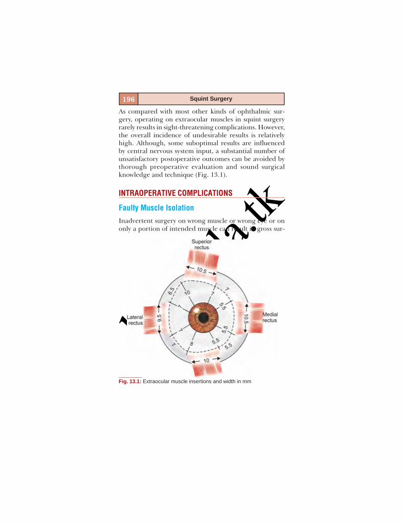
basm
ala.tk
196 Squint Surgery
As compared with most other kinds of ophthalmic sur-gery, operating on extraocular muscles in squint surgeryrarely results in sight-threatening complications. However,the overall incidence of undesirable results is relativelyhigh. Although, some suboptimal results are influencedby central nervous system input, a substantial number ofunsatisfactory postoperative outcomes can be avoided bythorough preoperative evaluation and sound surgicalknowledge and technique (Fig. 13.1).
INTRAOPERATIVE COMPLICATIONS
Faulty Muscle Isolation
Inadvertent surgery on wrong muscle or wrong eye or ononly a portion of intended muscle can result in gross sur-
Fig. 13.1: Extraocular muscle insertions and width in mm

basm
ala.tk
197Complications in Squint Surgery
gical undercorrection of squint or create a new and seri-ous disturbance in ocular motility.
Prevention
The surgeon must confirm the identity of patient, the de-tail surgical plan before surgery.
Hooks used to engage rectus muscle should bepositioned with great care. Sweeping even a few millimeterstoo posterior for lateral or superior rectus may lead toinclusion of IO or SO tendon respectively (Figs 13.2 and13.3). A very posterior sweep for medial rectus canoccasionally hit optic nerve. On the contrary superficialhooking may miss the muscle altogether, this can especiallyhappen in resurgery, when scar tissue is mistaken formuscle.
Fig. 13.2: Inferior oblique muscle

basm
ala.tk
198 Squint Surgery
Another possible complication of inferior obliqueisolation is damage to ciliary ganglion, causing post-operative mydriasis.
Hemorrhage
Hemorrhage during muscle surgery never leads to hemo-dynamically significant blood loss, but it often interfereswith visibility, thereby increasing the risk of other compli-cations and prolonging operating time.Conjunctiva: More bleeding may come from transectedconjunctival vessels, particularly if incision is too poste-rior or in the fornix.
Prevention: Instillation of topical phenylephrine be-fore surgery constricts conjunctival vessels. Light applica-tion of bipolar cautery or pressure on oozing site with cot-
Fig. 13.3: Superior oblique tendon

basm
ala.tk
199Complications in Squint Surgery
ton tipped swab soaked in 1:1000 adrenaline also aids inthis regard.
Muscle vessels: Anterior ciliary arteries and veins are cutduring most muscle operations during disinsertion, butthe bleeding is surprisingly not much. More extensive hem-orrhage occurs if a ciliary vessel is cut anterior or poste-rior to muscle. Cauterization should be performed only ifbleeding has not stopped before conjunctival closure.
Inferior oblique muscle is most likely to bleed after my-ectomy (Fig. 13.4) and the resulting ecchymosis resolvesin a few days.
Vortex veins: Vortex veins are close to vertical recti andobliques and hence they must be identified and avoidedduring muscle surgery.
Fig. 13.4: CT scan showing retrobulbar hemorrhage in left orbit after IOmyectomy. (Courtesy: Mark J Greenwald and Janice Lasky)

basm
ala.tk
200 Squint Surgery
Scleral Perforation
Penetration of globe by needle during muscle surgery hasbeen reported at a frequency ranging from 1 to 10%. Fineside cutting spatulated needles reduce the incidence ofthis complication. Fortunately scleral perforation usuallydoes not cause dangerous sequelae. However, the possibil-ity of vitreous hemorrhage, lens dislocation,endophthalmitis and RD makes avoidance of this compli-cation an important concern.
Prevention
Use of fine spatulated needles (Fig. 13.5).Use of hangback technique especially if sclera is verythin.Proper passage of needle so that it is visible throughoverlying scleral layer (Figs 13.6A to D).Detection of needle perforation is suspected if the
needle reaches a depth that precludes visualization of itstip or if there is loss of normal tissue resistance. There
Fig. 13.5: Scleral sections showing safety of spatulated needles.(Courtesy: Eugene Helveston)

basm
ala.tk
201Complications in Squint Surgery
may be pigment or vitreous from the needle track. Theperforation must be confirmed with indirect ophthalmos-copy with scleral indentation (Fig. 13.7). Frequently, theonly fundus abnormality is localized loss of choroidal pig-ment, that appears as a white lesion.Muscle disinsertion: Scleral penetration can occur whendisinserting muscle with scissors (especially in resurgerieswith tight muscle).Prevention: Disinsert muscle with small, controlled scissorbites under visualization.Treatment: Large, full thickness scleral breaks should beclosed with sutures. If retina is involved – cryopexy or lasershould be done with placement of scleral buckle. A partialthickness scleral wound does not require treatment.
Figs 13.6A to D: Overlying scleral layer. (Courtesy: Theodore Krupin,Kolker and Rosenberg)
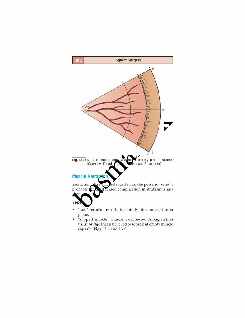
basm
ala.tk
202 Squint Surgery
Muscle Retraction
Retraction of a detached muscle into the posterior orbit isprobably the most feared complication in strabismus sur-gery.
Types
• ‘Lost’ muscle—muscle is entirely disconnected fromglobe.
• ‘Slipped’ muscle—muscle is connected through a thintissue bridge that is believed to represent empty musclecapsule (Figs 13.8 and 13.9).
Fig. 13.7: Needle tract lesion caused by deeply placed suture.(Courtesy: Theodore Krupin, Kolker and Rosenberg)
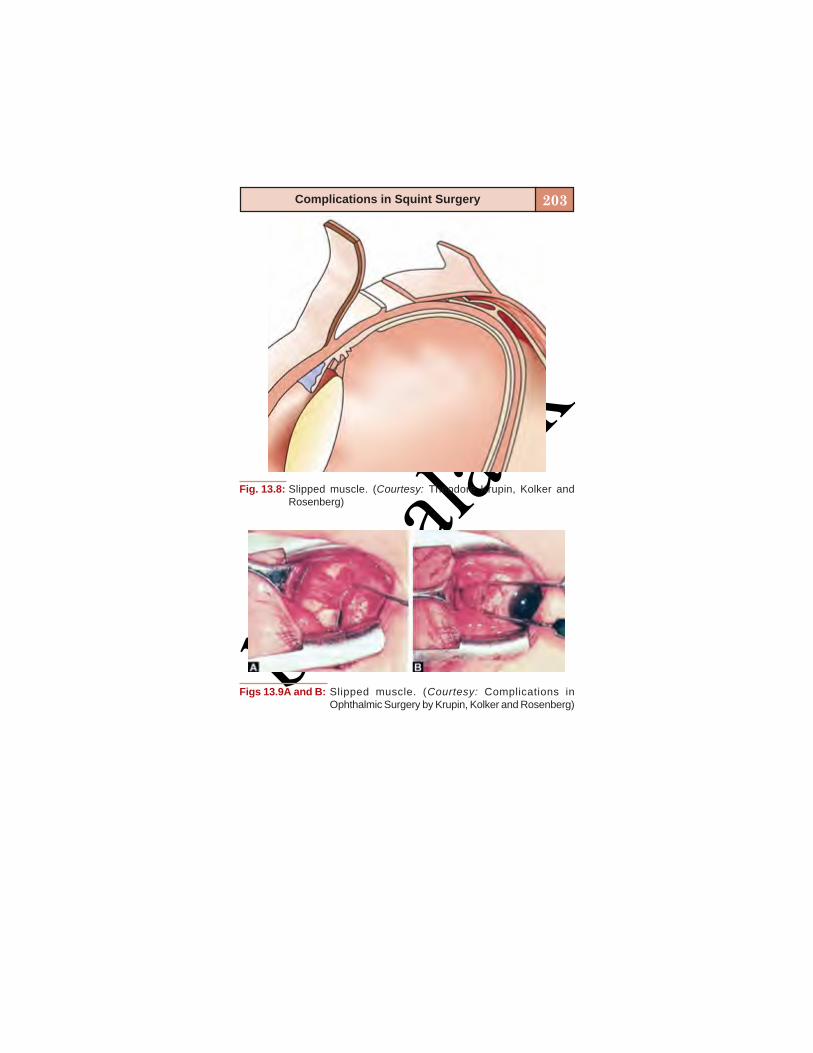
basm
ala.tk
203Complications in Squint Surgery
Fig. 13.8: Slipped muscle. (Courtesy: Theodore Krupin, Kolker andRosenberg)
Figs 13.9A and B: Slipped muscle. (Courtesy: Complications inOphthalmic Surgery by Krupin, Kolker and Rosenberg)

basm
ala.tk
204 Squint Surgery
• ‘Pulled-in two’ syndrome (PITS) – in some elderly ordebilitated patients, muscle fragmentation may resultwith muscle traction (Fig. 13.10).Prevention: Careful attention to suture preplacementbefore disinsertion particularly when making lockbites.In high-risk situations, anchor a temporary ‘securitystitch’ posteriorly in muscle belly. Pay particular atten-tion while operating on medial rectus.Identification and recovery of retracted muscle: Remain calmand approach intraoperatively retracted muscle
Fig. 13.10: “Pulled-in two” syndrome. (Courtesy: Theodore Krupin, Kolkerand Rosenberg)
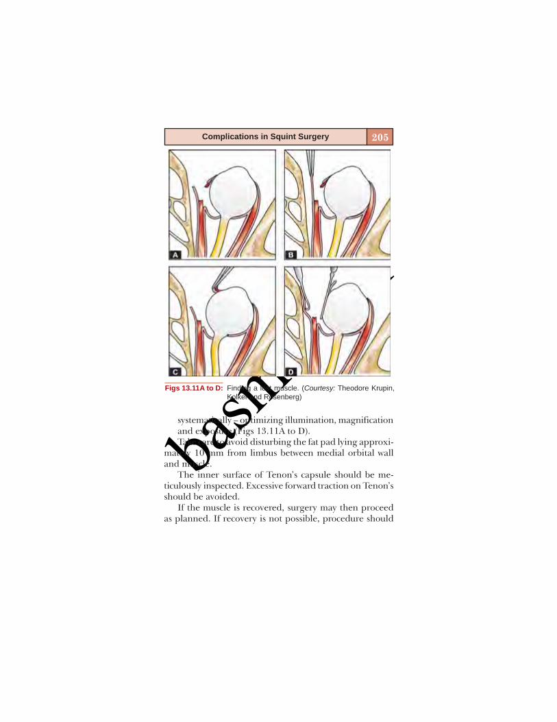
basm
ala.tk
205Complications in Squint Surgery
systematically – optimizing illumination, magnificationand exposure (Figs 13.11A to D).Take care to avoid disturbing the fat pad lying approxi-
mately 10 mm from limbus between medial orbital walland muscle.
The inner surface of Tenon’s capsule should be me-ticulously inspected. Excessive forward traction on Tenon’sshould be avoided.
If the muscle is recovered, surgery may then proceedas planned. If recovery is not possible, procedure should
Figs 13.11A to D: Finding a lost muscle. (Courtesy: Theodore Krupin,Kolker and Rosenberg)
basm
ala.tk
206 Squint Surgery
be aborted and secondary procedure like transposition ofadjacent recti should be done later.
POSTOPERATIVE COMPLICATIONS
Infection
Infective complications typically present 24 to 72 hours aftermuscle surgery, but are quite uncommon (overall incidenceof less than 1%). These include conjunctivitis, orbital cellulitisand endophthalmitis.Prevention:• Strict asepsis and sterility protocol• Topical application of a drop of 5% povidone – iodine
during preoperative preparation reduces the conjunc-tival bacterial load by 90%
• Prophylactic topical and oral antibiotics.
Immunologic Reactions
• Suture granuloma: Occasionally, an inflammatory,slightly tender lump over the suture may develop withina week or so. It is a foreign body reaction to suture orother foreign matter like cotton fibers (Fig. 13.12).Treatment: Corticosteroid drops are prescribed. If thisdoes not bring resolution, remove the suture and excisethe lump (10 days after surgery when the musclereattaches to sclera).
• Serious inflammatory complications like necrotizingscleritis and fulminant orbitopathy may occur in con-ditions like active Graves’ ophthalmopathy.Prevention: Do not operate until active ocular signs ofinflammation subside and squint is stable for manymonths.
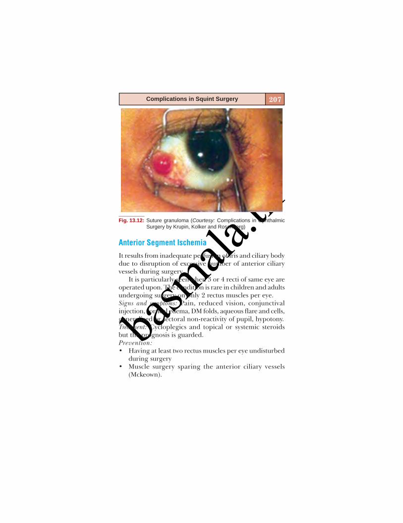
basm
ala.tk
207Complications in Squint Surgery
Anterior Segment Ischemia
It results from inadequate perfusion of iris and ciliary bodydue to disruption of excessive number of anterior ciliaryvessels during surgery.
It is particularly seen when 3 or 4 recti of same eye areoperated upon. The condition is rare in children and adultsundergoing surgery on only 2 rectus muscles per eye.Signs and symptoms: Pain, reduced vision, conjunctivalinjection, corneal edema, DM folds, aqueous flare and cells,generalized or sectoral non-reactivity of pupil, hypotony.Treatment: Cycloplegics and topical or systemic steroidsbut the prognosis is guarded.Prevention:• Having at least two rectus muscles per eye undisturbed
during surgery• Muscle surgery sparing the anterior ciliary vessels
(Mckeown).
Fig. 13.12: Suture granuloma (Courtesy: Complications in OphthalmicSurgery by Krupin, Kolker and Rosenberg)

basm
ala.tk
208 Squint Surgery
Poor Healing
Conjunctiva:
• Prolapse of Tenon’s capsulePrevention: Careful cleaning and good wound closure.Treatment: Wait a few days—Tenon’s may shrink insideconjunctiva—if not, it can be excised under local anes-thesia.
• Subconjunctival cysts: Entrapment of epithelial cellsbeneath the surface may cause subconjunctival cysts(Fig. 13.13).
• Adhesive or adherence syndrome: Intense postopera-tive fibroproliferative response that leads to severe re-striction of ocular motility.
Fig. 13.13: Subconjunctival cyst (Courtesy: Complications in OphthalmicSurgery by Krupin, Kolker and Rosenberg)

basm
ala.tk
209Complications in Squint Surgery
Corneal Problems
• Epithelial damage – common.• Dellen formation – if bunching of tissues near the limbus
disturbs the tear flow over ocular surface.• Induced astigmatism.
Change in Eyelid Position
A large unbalanced horizontal muscle surgery may causeslight globe retraction or proptosis with altered palpebralfissure.
Recession or resection of inferior rectus tends to dis-place the lid downward or upward due to its firm attach-ment through Lockwood’s ligament.
Disturbance of Binocular Vision
Postoperative diplopia
Persistence of diplopia beyond two weeks justifies a trialof optical correction like prism. Postoperative diplopia thatremains bothersome for two months or longer warrantsprescription of permanent prism glasses or further surgery.
Unsatisfactory Alignment
• Undercorrection: Persistence of significant degree oforiginal deviation.
• Overcorrection: Deviation in opposite direction.At least 10-20% of all operations fail to achieve their goalsin terms of ocular alignment, thus necessitating furthersurgery.

basm
ala.tk
210 Squint Surgery
Prevention: Thoughtful planning and skilled execution ofstrabismus surgery may reduce the frequency of postop-erative disappointments.
ANESTHETIC COMPLICATIONS
The possibility of anesthesia related complications espe-cially GA must be discussed with patient and/or relatives.
Cardiovascular and Respiratory Complications
• Mortality-risk is very less (< 1 in 1,00,000)• Oculocardiac reflex – mediated via trigeminal afferent
fibers from the orbit and vagal efferent fibers.Usually this causes mild transient bradycardia, but
rarely it may give rise to more serious arrhythmias likeprolonged asystole.
• Malignant hyperthermia: Rise of blood CO2 level, car-diac arrhythmia and elevation of body temperature.Treatment: Dantrolence sodium, 100% oxygen.
• Postanesthetic nausea and vomiting: Quite commonafter GA .Treatment: Antiementics like ondansetron, emeset.
REFERENCES1. Management of strabismus & amblyopia: John A pratt –
Johnson, Geraldine Tillson: PP 227-38.2. Complications in ophthalmic surgery: Theodore Krupin,
Allen Kolker and Lisa Rosenberg; chapter 10: M Greenwaldand J Hasky: PP 195-215.
3. Morris RJ: Incidence of globe perforation during strabismussurgery. Br J Ophthalmol 1990;74:490-3.
4. Plager DA, parks MM. Recognition and repair of ‘lost’ rectusmuscle. Ophthalmology 1990;97:131-7.
basm
ala.tk
211Complications in Squint Surgery
5. Kivlin JD, Wilson ME. Periocular infection after strabismussurgery. J Pediatric Ophthalmol strabismus 1995;32:42-9.
6. Kushner BJ. Subconjunctival cysts after strabismus surgery.Arch Ophthalmol 1992;110:1243-5.
7. McKeown CA. Preservation of anterior ciliary vessels duringmuscle surgery. Ophthalmology 1989;96:498-563.
8. Saunders RA. Ant. segment ischemia after strabismus surgery.Surv Ophthalmol 1994;38: 456-66.

basm
ala.tk

basm
ala.tk
RECENT ADVANCES
4

basm
ala.tk

basm
ala.tk
14
WHAT IS NEW INSTRABISMUS?
Stephen P Kraft
basm
ala.tk
216 Squint Surgery
INTRODUCTION
In recent years, there have been impressive advances inbasic sciences, clinical diagnosis, and treatment in all oph-thalmic specialties. The field of strabismus has seen its shareof major developments, many of which have changed prac-tice in the assessment and treatment of amblyopia and dis-orders of binocular vision.
This chapter will discuss many of these advances andhow they have shaped the course of the specialty as wecomplete the first decade of the 21st century. It is not pos-sible to cover all of the new information in this rapidlychanging field, but some of the more clinically relevantitems will be presented. These advances will be discussedunder four main headings:
Basic sciences and anatomyDiagnostic testingMedical therapySurgical treatment.
BASIC SCIENCES AND ANATOMY
Infantile Strabismus Syndrome
This common syndrome involves the combination of largeangle heterotropia (usually esotropia, rarely exotropia),dissociated vertical deviation (DVD), overaction of theinferior oblique muscles, and latent nystagmus.1 Anotherfeature recognized in recent years is asymmetry of thesmooth pursuit movement of each eye: slow-moving targetsgenerate a much smoother movement when the targetpasses nasalward before the eye, than do targets movingtemporalward. In addition, there are marked fusional ver-gence deficits as well as congenital deficits of binocularvision processing in the visual cortex.2
basm
ala.tk
217What is New in Strabismus?
In an effort to understand this combination of anoma-lies, research efforts have been directed to studies in normaland strabismic infants, and also in a primate model.3 Datafrom several laboratories revealed a number of anomaliesin the cortex that account for the components of theinfantile strabismus complex. The primate model ofinfantile esotropia has shown loss of horizontal connec-tions between ocular dominance columns in area V1 ofthe visual cortex, which results in loss of connectivitybetween the inputs from the two eyes. This decorrelationmanifests downstream in maldevelopment of the cerebralpursuit pathways, leading to the nasal-temporal pursuitaymmetry and the impaired disparity vergences. Theprimate model also shows clinical findings similar to theoblique and dissociated vertical anomalies seen in humans.3
An important finding from these studies is that the tim-ing and duration of the insult that upsets the binocularconnections is critical. The primate model predicts that,in the human, an insult must occur or exist within the first2 to 3 months of life to cause the collection of signs seen ininfantile strabismus. In addition, the insult must persistuntreated beyond the first 6 to 8 months of life. This sug-gests that realignment of the infant’s eyes should take placevery early in life, before 8 or 9 months of age. This is ear-lier than the longstanding, more typical practice, wherebyrealignment surgery is usually planned before age 18 to24 months, with the goal of restoring binocular fusion.4 Asa result, there are ongoing studies evaluating the benefitsof “very early” surgery, before age 6 to 9 months, with thegoal of not only regaining fusion but also reversing theanomalous eye movement and vergence disorders in thiscondition (see later discussion under Surgical Treatment).

basm
ala.tk
218 Squint Surgery
Advances in Orbit Anatomy
In the past 20 years, strabismus surgeons have re-exam-ined the structures and directional paths of the obliqueand rectus ocular muscles. Important findings include newunderstanding of the basic structure of the rectus musclefibers, and the presence of connective tissue sleeves (“pul-leys”) of the six ocular muscles.
Ocular Muscle Structure
The literature until the 1970s divided the ocular musclefibers into two types, based on the rapidity of response:slow twitch versus fast twitch. The former were character-ized by thin motor fibers showing slow sustained contrac-tion, while the latter were thick fibers that contractedquickly and fatigued quickly.5
In the past 20 years, the structural classification of aneye muscle underwent revisions as newer histologic,immunolocytologic, and recording techniques were devel-oped. These studies have led to the dividing of ocularmuscle fibers into six subtypes, based on location (globalversus orbital), innervation (singly- versus multiply-inner-vated), and color (red versus pale).6-8 One clinical impli-cation of this reclassification is the finding that botulinumtoxin injected into the ocular muscle binds preferentiallyto the singly-innervated orbital fiber subtype, rather thanto any of the other five subtypes.9 A second implication ofthis new paradigm is the fact that the orbital layers of theocular rectus muscles, which contain about half of the totalnumber of muscle fibers in the muscles, are the portionsthat insert via the ocular muscle pulleys (see next section).They are distinct in their mechanical action from the glo-bal layers, which contain the other half of the total num-ber of fibers and which insert onto the globe.8

basm
ala.tk
219What is New in Strabismus?
Along this theme, a third finding is that there are pali-sade endings in the global (inner) layer of the extraocularmuscles, and these are associated with non-twitch multiply-innervated muscle fibers (MIF). There has been a greatdeal of research in recent years into the possible role ofthese endings. One hypothesis states that they are affer-ent sensory organs providing information about eye posi-tion, while the other theory is that they are cholinergic(effector) motor organs.10 The “palisade ending-MIF motorunit” may be part of a sensory feedback system in eyemuscles, which should be considered in association withthe causes and treatment of strabismus. Finally, it has beennoted that singly-innervated motoneurons are more in-volved in driving eye movements, and that multiply-innervated motoneurons help to set the tonic tension ineye muscles.11
The ocular muscle subtypes have also been character-ized by their distribution of myosin heavy chain isoforms.These analyses have shown that the six ocular rotatorymuscles are immunocytologically different from the leva-tor palpebrae muscle, which is often included as one ofthe extraocular muscles, as well as from extraocular musclesin other species.7,8
Ocular Muscle Pulleys
A second major advance in eye muscle anatomy is the char-acterizing of the connective tissue sleeves, or “pulleys”, ofthe ocular rectus and oblique muscles. These pulleys arelocated within Tenon’s fascia, just posterior to the equatorof the globe. The structure of these pulleys includes softtissue, elastin, and richly innervated smooth muscle. Asnoted earlier, the fibers of the orbital layer terminate onthe sleeve while the global layer passes through the pulleyand inserts onto the globe.12 As a result of this dichotomy

basm
ala.tk
220 Squint Surgery
of anatomic insertions of the two portions of the muscle,the global layer mainly rotates the globe whereas the or-bital layer translates the pulley to control the direction ofglobe rotation. Pulleys have been described for the ob-lique muscles as well as the rectus muscles.13
This new anatomic paradigm has led to revised think-ing of the etiology of several common strabismus entities,as well as rethinking of the approach to surgical treatmentof these and other conditions. A good example is theetiology of the A and V syndromes, which have beenascribed primarily to oblique muscle dysfunction for manyyears. However, detailed radiologic studies, in concert withorbit dissections, have shown that some of the A and Vsyndromes are, in fact, caused by anomalous positions ofthe pulleys within the orbit. Thus, repositioning of thepulleys has been designed as a more physiologic andeffective approach in some cases, rather than surgery onthe oblique muscles. A second surgical application is a newvariation on the posterior fixation suture (“fadenoperation”),whereby suturing of the rectus muscle to the pulley maybe as effective as tacking down the rectus muscle to thesclera.12 Finally, radiologic analysis of transposition sur-gery for paralytic strabismus has shown that the benefit ofthis surgery in realigning the eye and improving ductionsis related to a marked lateral displacement of the pulleysof the transposed muscles.14
Computer Modeling of Strabismus
The exponential increase in computer capacity has alloweddevelopment of software that can model the orbital andocular motor “plant”. The updated versions of theseprograms incorporate recent advances in orbit andextraocular muscle anatomy, biochemistry, physiology, and
basm
ala.tk
221What is New in Strabismus?
biomechanics to simulate both simple and complex eyemovements.15,16 They also allow surgeons to predict theeffect of a small or large perturbation on the normal eyemovement plant, in terms of both clinical effects andpossible surgical implications.
Genetic Basis of Strabismus
In recent decades, there have been ongoing studies on theheritability of strabismus, including the common forms ofexotropia and esotropia as well as complex forms such asDuane syndrome, Brown syndrome, congenital fibrosis ofthe extraocular muscles (CFEOM), and congenital fourthnerve palsy.17 The pathogenesis of comitant strabismus,in the absence of structural abnormalities of the eye orbrain, is currently considered to be inherited as a complexgenetic trait, with contributions from both genes and theenvironment.18 Recent work has suggested that there aremultiple loci for comitant strabismus.19
Several congenital forms of incomitant strabismuspresent with nonprogressive restrictive strabismus. Theseinclude CFEOM and Duane syndrome, which until a fewyears ago were considered anatomically and physiologi-cally distinct entities. However, anatomic and neurophysi-ologic studies have challenged this thinking, and theseentities are now considered part of a spectrum of congeni-tal disorders of innervation. Several other disorders havenow been grouped with these two entities, including Möbiussyndrome, familial horizontal gaze palsies, and congenitalthird and fourth nerve palsies.18,20 These seemingly dis-parate clinical syndromes all arise out of mutations in genescritical to the development of ocular motor neurons andtheir connections.20 Thus in 2003, the term “congenitalcranial disinnervation disorders (CCDDs)” came into
basm
ala.tk
222 Squint Surgery
existence to apply to this group of strabismus entities.18
Several genetic loci have been identified for most of thesesyndromes.
DIAGNOSTIC TESTING
Assessment of Infant Vision
In the 1970s, initiatives arose to develop tools to assessvision in children who were too young to perform conven-tional vision tests including Snellen or Allen pictures. Thesestudies proceeded along two related but parallel paths, oneinvolving creation of manual tests for use in the clinicalsetting, and other utilizing visual technology and computergenerated images.
It is now possible to determine quantitative vision levelsin infants as young as a few months old using clinical testsbased on grating acuity cards, which include Teller acuitycards and Cardiff cards.21,22 These tests are widely used byorthoptists and ophthalmic assistants to diagnose visiondisorders in preverbal or nonverbal children, and to guidetreatment for amblyopia and refractive errors, among manyother ophthalmic conditions. However, there have beenconcerns raised about the specificity and sensitivity of thesetests in detecting amblyopia, when compared to optotypetests.22-25 Computerized tests, such as the pattern-onsetand the sweep visual evoked potential (VEP), allow quanti-tative vision assessments in the laboratory setting, bothfor diagnostic and research purposes.26-28 In addition, moreaccurate recognition tests for slightly older children arenow in common use, including LEA symbols and singleoptotype cards with interaction bars, which address theissue of the crowding phenomenon in amblyopia.
basm
ala.tk
223What is New in Strabismus?
Photoscreening
Large-scale vision screening of children has been an activearea of research in recent years. Instruments have beendeveloped that can detect various ocular disorders, andwhich are easy to use for both experienced and lay practi-tioners. At the same time, many devices provide accuratedata with high sensitivity and specificity. Among the mostuseful ones invented for these purposes are photoscreeners,which have the ability to detect misalignment of the eyes,refractive errors, and media opacities.
There have been several prototypes produced in re-cent years. The most widely-evaluated one is the MTIPhotoscreener (MTI, Cedar Falls, Iowa, USA), which canbe worked by a lay individual, while photos are analyzedby experienced observers. Successful programs using thisdevice have been undertaken in the United States.29 Otherphotoscreening instruments have been validated in otherstudies, each with its own advantages. In addition,autorefractors have been found to be valid as screeningtools for detecting significant (and amblyogenic) refrac-tive errors in large population-based studies.29
Plotting the Field of Binocular Single Vision
There are several means of recording data of eye musclebalance, with most schemes involving static measurementsof alignment in different fields of gaze. While such dataare helpful in following strabismus cases and planningsurgical dosages for surgery, strabismus specialists haverecognized that static data do not always reflect the dynamicsituation when the eyes are tracking targets. To addressthis gap, many investigators have developed alternativerecording methods, including the field of binocular single

basm
ala.tk
224 Squint Surgery
vision (BSV). This test documents the fusion field of thepatient as the eyes are moved into specified directions awayfrom the primary position.
The test is usually performed with a Goldmann or arcperimeter in order to capture the entire binocular field.The target used in a Goldmann apparatus is often a spot,but fusion targets have been incorporated into the testingdevices to enable detection of torsional as well as horizon-tal and vertical disparities.30 Within the last five years, analternative test using a cervical range of motion (CROM)method has also been introduced, and this procedure canbe used if a Goldmann perimeter is not available. In bothcases, a weighted scoring template is also available to pro-vide a BSV score for a plot.30,31
Weighted scoring methods of the field of BSV have sev-eral uses:30
They can allow the surgeon or orthoptist to follow apatient’s eye muscle problem serially over time.They provide a preoperative picture of the strabismusproblem and the areas of the field where fusion is lack-ing.The score can be used in prospective or retrospectivestudies to compare different surgical methods for aspecific entity.The scores can be used to compare the results of thesame surgery done by different surgeons.The weighted score can be used as a quantitative mea-sure of disability in medical-legal and insurance claims.
Eye Movement Recording
For several decades, investigators have used eye movementrecordings to document abnormalities of the various eyemovement systems, including saccades, smooth pursuit,

basm
ala.tk
225What is New in Strabismus?
vergences, and optokinetic anomalies. In recent years, thesedevices have become increasingly sophisticated and pre-cise in providing dynamic eye movement data to diagnoseand follow strabismus and neuro-ophthalmologic eyemovement disorders. Systems incorporating search coilsor infrared detection devices have allowed tracking of eyemovements in three dimensions.32,33 Eye movementrecordings have proven invaluable in assessment ofnystagmus, ocular motor nerve pareses, dissociated verticaldeviation, myasthenia, external ophthalmoplegia, andrestrictive strabismus.33-35
Studies using eye movement recordings have led toreclassifications for many disorders. One example is theabandoning of a long-held separation of “motor” versus“sensory” nystagmus to describe nystagmus waveformsresulting from, respectively, primarily efferent and prima-rily afferent ocular diseases. Modern eye movement re-cordings have shown that infantile nystagmus waveformsare complex, and cannot separate out those patients whohave a congenital eye disorder from those who do not haveone. This has led to a new terminology, with the olderterm “congenital nystagmus” being replaced by “infantilenystagmus syndrome (INS)”. Another new term is “fusionmaldevelopment nystagmus syndrome (FMNS)” to replacethe long-held term “latent/manifest latent nystagmus”.36
The quantitation of recordings has also led to new indicesof fixation quality to measure visual performance and pre-dict improvements after eye muscle surgery for nystagmus.
The “Four-Step Test”
There has been a steady increase in knowledge of the supra-nuclear pathways that control eye movements, includingthe pathways in the brainstem and cerebellum and their

basm
ala.tk
226 Squint Surgery
connections to the utricles and saccules of the inner ear.These pathways affect the eye muscles in several ways, oneof which is the control of ocular counterrolling and verticaleye movements.
In light of these gains in knowledge, a fourth step hasrecently been added to the traditional “3-step test” thathas been in use for many decades in order to diagnose a4th nerve paresis. This clinical test involves, firstly, the la-belling of the hypertropic eye in primary position. In thesecond step, one records the lateral gaze in which the ver-tical tropia worsens, and this step isolates the possibleparetic muscle to either the superior oblique of thehypertropic eye or the superior rectus of the fellow eye.The third step is the forced head tilt test (or Bielschowskyhead tilt test) to the right and left shoulders, and this stepisolates the offending muscle by determining on which tiltthe hypertropia worsens.37
However, one entity that can mimic some features of asuperior oblique paresis, especially an incomitanthypertropia or hypotropia, is a skew deviation, which is avertical strabismus caused by lesions in the brainstem teg-mentum or cerebellum, or due to peripheralvestibulopathy.38 In order to rule out a skew deviation, anew paradigm involves a fourth maneuver: measuring thevertical heterotropia and cyclotorsion in the sitting posi-tion, and again in the reclining position. Whereas a 4thnerve paresis will show no change in the vertical deviationand subjective excyclotorsion in the sitting and recliningpositions, a skew deviation will show marked reduction orelimination of the vertical deviation and torsion when thepatient reclines.38
basm
ala.tk
227What is New in Strabismus?
Radiologic Advances
Ultrasound Biomicroscopy
Ultrasound biomicroscopy (UBM), involving the use ofhigh frequency output (50 MHz), was developed in the1980s for diagnosis of anterior segment conditions suchas glaucoma and iris tumors. In the last 10 years it hasproven to be a valuable tool for detecting the insertions ofthe four ocular rectus muscles, both in patients undergo-ing primary strabismus surgery and in those undergoingreoperations.39,40 The probes used in this test can detectmuscle insertions as far posteriorly as 14 mm from thelimbus for the lateral rectus and 12 mm for the medialrectus and vertical rectus muscles. The UBM can alsodifferentiate a connective tissue “pseudotendon” from thetrue muscle insertion.39
Computerized Tomography (CT) andMagnetic Resonance Imaging (MRI)
Improvements in technology have made these two mo-dalities increasingly sensitive to subtle lesions and anoma-lies that can lead to strabismus problems. The field of CTscanning has undergone major changes in recent years withthe introduction of helical or spiral scanning to the con-ventional imaging programs. This modality is still thepreferred study for orbit bony problems, especially tovisualize fractures after trauma.41 Newer scanners can alsoproduce impressive 3-dimensional views of the orbits andcranial vault, and this technology has revolutionizedtreatment for craniofacial and orbit disorders, many ofwhich can affect the eye muscles.

basm
ala.tk
228 Squint Surgery
In recent years, MRI scanning has also undergonemajor advances, including a steady reduction in time in-tervals needed to complete a study. This factor has beenparticularly beneficial in the context of pediatric cases.Newer scanners give unprecedented details of the softtissues, allowing assessment of a wide range of eye muscledisorders. This technology has been used by researchersto model the extraocular muscles and to redefine theanatomy of the orbital soft tissues. One example is thecharacterization of the ocular muscle pulleys and their ef-fects on eye muscle alignment and dynamic eye movements(as described earlier).12
Dynamic MRI, or the imaging of the eyes in real timeduring guided eye movements, has proven instructive indiagnosing various eye muscle conditions. Following thetraumatic severing of an eye muscle, dynamic MRI can beused to assess the integrity of a residual portion of muscle.It is also useful to view the co-contraction of eye musclesin misinnervation syndromes such as Duane retraction syn-drome.42,43
Other Imaging Advances
Other modalities have been applied in assessing eye muscleproblems. Positron emission tomography (PET) detectsbiochemical changes in body tissues. It has been useful indetecting lesions that may be missed by MRI or CT scan,such as brainstem disorders that affect the eye muscles.44
Advances in optical coherence tomography (OCT) haveled to accurate depiction of the anterior segment of theeye, in addition to its value in imaging the posteriorsegment structures.45 It can yield detailed views of thecornea, chamber angle, and sclera, which may some dayallow accurate visualization of the extraocular muscleinsertions, now possible only with the ultrasound
basm
ala.tk
229What is New in Strabismus?
biomicroscope. Finally, a new high-resolution X-ray-basedimaging technique known as diffraction-enhanced imaging(DEI) gives very high-resolution images of the eye, and itmay in the future provide an alternative method ofdetecting eye muscle position.46
Telemedicine
Telemedicine is a method for transmitting images and dataover long distances for diagnostic and treatment purposes.It is being used in ophthalmology at an increasing rate inthe diagnosis of such disorders as retinopathy of prematu-rity and retinal diseases. It has also been shown to be use-ful in assessing strabismus cases, whereby static images ofa strabismus case can be sent to another city on the sameor a different continent for analysis.47
With advances in video technology, the analysis of stra-bismus cases can now be done via telehealth setups thatallow dynamic video recordings. The examination can bedone on a delayed telecast basis, or in real time as thepatient is examined by the ophthalmologist or orthoptistat the site of origin. The first study on the accuracy of real-time video to diagnose strabismus cases was carried out inCanada in 1999, with transmission over 1500 km betweenToronto, Ontario and Thunder Bay, Ontario. With thebandwidths available at that time, the study showed thathorizontal deviations were more accurately measured thanvertical deviations.48 It is expected that the markedimprovements in technology would allow much higheraccuracy in agreement between observers at the site of theexamination and the remote site.
basm
ala.tk
230 Squint Surgery
MEDICAL THERAPY
Botulinum Toxin
Botulinum toxin injections were introduced in 1978 as analternative to strabismus surgery. Injections of the toxinwere also found to be useful in controlling facial musclespasms in various forms of dystonia and in hemifacialspasm. Its range of uses increased rapidly over the ensu-ing years such that, currently, there are several dozen usesfor this drug both in the medical and cosmetic spheres.The main drawback to using this agent is its temporaryeffect. However, thirty years of experience with botulinumtoxin in strabismus has shown that it has a role to play invarious eye muscle disorders.49
Botulinum toxin has been used to treat various condi-tions in adults, especially sensory strabismus and acuteocular muscle palsies. Several years ago it was touted asimproving the recovery rate after acute sixth nerve palsiesif it was injected into the medial rectus within a few weeksof onset of the palsy. This expectation has not been sup-ported by recent studies on traumatic palsies.50 However,the injection can temporarily reduce a compensatory headposture and preserve binocularity until definitive treatmentis undertaken.51
Although some practitioners use botulinum toxin for awide variety of strabismus conditions, it has proven espe-cially effective in certain contexts.51,52 It has a high rate ofsuccess for small angle strabismus, usually less than 20prism diopters, and for postoperative undercorrections orovercorrections associated with diplopia.49,51 It is used asan adjunct for transposition surgery for sixth or third nervepalsies.51 It has also been effective in reducing symptomsof oscillopsia due to acquired nystagmus.53 Botulinuminjections can be helpful when surgery is risky, such as cases
basm
ala.tk
231What is New in Strabismus?
of pre-phthisical eyes or in eyes in which glaucoma orretinal surgery can increase the risk of anterior segmentischemia after surgery.
Botulinum toxin has proven effective in treatingcomitant strabismus in children. Some authors haveachieved success in treating infantile strabismus thatmatches surgical success rates, both in terms of motor andsensory outcomes.54,55 These authors injected muscles inboth eyes at the initial treatment. Experience in children,as in adults, has shown that the rates of satisfactory align-ment after injection of one eye muscle are higher for smallangles of strabismus, usually under 20 prism diopters.49
Injections of Local Anesthetic
Observations of eye muscle conditions resulting from un-intended intramuscular injections of local anesthetic forcataract and retinal surgery have led to the notion thatintentional injection of a rectus muscle could realign aneye. This has been borne out by pilot studies involving theinjection of bupivicaine, a long-acting local anesthetic, intothe lateral rectus muscles of patients who hadundercorrected strabismus after prior esotropia surgery.56
Further experience in larger studies, and in a variety ofconditions, will be required before it becomes an acceptedalternative to surgery.
Treatment of Amblyopia
Traditional Methods
There have been numerous prospective controlled ran-domized studies carried out in the last 10 years that haveconfirmed some long-held concepts and also yielded a

basm
ala.tk
232 Squint Surgery
great deal of new information on the treatment of am-blyopia. Many of these studies were carried out by a largegroup of pediatric eye specialists in North America knowncollectively as the Pediatric Eye Disease Investigator Group(PEDIG).
The following is a list of some of the important conclu-sions that have been learned thus far from these and otherrelated studies:57-60
For children age 3 to 7 years with unilateral amblyopiaand vision at least 6/30 (moderate amblyopia) and whohad no prior therapy for amblyopia, daily atropinepenalization leads to the same rate of improvement invision over a 6-month period as 6 hours per day ofpatching.For the same age group with the same range of start-ing visions, patching of 2 hours per day generates thesame rate of improvement as 6 hours per day over a4-month period.Also in this age group with moderate amblyopia, atro-pine drops given only on the weekend leads to the samerate of improvement as daily atropine administration.In the same age group but with vision poorer than 6/30(severe amblyopia), patching 6 hours per day yieldsthe same rate of improvement as full days of patchingover 4 months of follow-up.In treating anisometropic amblyopia, spectacle correc-tion alone can lead to an improvement in vision over a2-month period of follow-up. This improvement is alsopossible even if there is strabismus present with glasses.If the vision reaches a plateau with spectacles alone,adding patching for 2 hours per day can yield a furtherincrease in vision over a 2-month interval.Among patients between 7 and 12 years of age withmoderate or severe amblyopia, up to one-half will gain

basm
ala.tk
233What is New in Strabismus?
more than 2 lines’ improvement in vision using eitherpatching, or patching accompanied by penalizationover a 6-month period of follow-up.Among patients between 13 and 17 years of age withmoderate or severe amblyopia, patching or patchingplus penalization can yield an overall improvement invision of at least 2 lines in one quarter of individuals.The success rate is higher than this (at almost 50%)among the sub-group of patients who have not hadprior amblyopia therapy.Among patients whose vision improved at least 3 lineswith patching, abrupt cessation of patching will resultin a slipping of vision over the ensuing few months.Thus, after ceasing daily patching, the hours shouldbe tapered over a few months to be sure the vision doesnot slip.Correction of refractive error alone can lead to im-proved vision in a majority of patients with previouslyuntreated strabismic amblyopia.The diligent use of near work for the amblyopic eyeduring the treatment period does not improve the re-sults of therapy.The improvement in vision seen with atropine penal-ization is not contingent on inducing a switch in fixa-tion preference during the cycloplegia.Spectacles as the sole intervention for bilateral ame-tropic amblyopia can improve vision over a period ofup to 12 months.
Other studies in recent years have also clarifiedsome issues in the treatment of amblyopia, includingthe following information:61,62
– Once the maximum vision is reached with patchingtherapy, if no improvement is seen with continua-tion of treatment for three subsequent age-adjusted
basm
ala.tk
234 Squint Surgery
intervals (each up to 4 weeks maximum), then thetherapy should be tapered.
– Long-term follow-up of patients who had patchingtreatment for amblyopia in childhood showed thatthe vision improvement was maintained into adult-hood in at least two thirds of cases.
It should be evident from these many lines of evidencethat the chance of reversing amblyopia is no longer lim-ited only to children under the age of visual maturity (age8 or 9 years). Current practice requires the practitioner toconsider offering treatment to patients of any age if theyhave not previously had an adequate trial of acceptedtherapy. While no guarantee can be given as to efficacy oftherapy, it is not possible to predict which older child orteenager may respond. Thus, the mantra in the older agegroups is to “never say never”, but to discuss the option oftherapy with any patient who wishes to improve his or hervision.
Newer Alternative Approaches
There are oral medications that have shown efficacy inimproving vision in amblyopic patients, usually in conjunc-tion with patching. Dopaminergic analogs, usually L-Dopawith Carbidopa, have led to greater improvements in visionthan patching alone. Most of the improvements in visionwere evident within 6 weeks after initiation of treatment.In some cases the vision tended to regress slightly afterthe oral medication was discontinued, but it did not revertback to the original vision level.63,64 Another medication,citicholine (or CDP-choline), is available in Europe andhas been shown to stabilize the improved vision in con-junction with patching treatment for amblyopia.65

basm
ala.tk
235What is New in Strabismus?
In recent years, there has been a large and growinginterest in the plasticity of the human brain and produc-tion of agents or interventions that can prolong or actu-ally restore brain plasticity, especially in adults. Recent re-search has shown that the critical period in animals can berevived or prolonged with the use of drugs that alter bio-chemical signals in the visual pathway, such as Prozac,Valium, GABA-minergic compounds, and proteins such asNogo and Otx2.66 In addition, repetitive transcranial mag-netic stimulation (rTMS) has shown promise in improvingcontrast sensitivity in amblyopic eyes, indicating plasticityin the amblyopic visual cortex.67 These interventions mayhold promise for the future in improving the success ratesand rapidity of response in amblyopia, and also poten-tially to prolong the age range within which vision deficitscan be reversed.
SURGICAL TREATMENT
Refractive Surgery
There are two groups of patients for whom refractive sur-gery has become a fertile ground for research and treat-ment: adults with refractive-based strabismus, and childrenwith amblyopia who are not responsive to or who are notcandidates for traditional medical therapies.
Adult Refractive Strabismus
Refractive surgery can be a useful alternative in treatingsome forms of strabismus. There are small case series inadults that have documented the successful treatment ofaccommodative esotropia by eliminating the hyperopicrefractive error.68-73 Refractive surgery can also eliminate
basm
ala.tk
236 Squint Surgery
the accommodative component of a mixed accommoda-tive and non-accommodative form of esotropia.74 It hasbeen reported to eliminate exotropia in two patients withmyopic anisometropia when the refractive errors werecorrected in both eyes.71 Small vertical deviations haveresponded to correction of refractive errors with laser.73
On the other hand, refractive surgery can lead to un-expected or unwanted results. In one report, a minority ofpatients with longstanding accommodative esotropia hadno change in their esotropia despite successful correctionof the hyperopia. These patients could not be identifiedby sensorimotor testing prior to the laser surgery.70 Anotherarticle presented a series of myopic patients whoseexodeviations worsened significantly after refractive surgeryfor their myopic errors.75
Refractive Surgery in Children
In recent years it has become evident that some childrendo not benefit from traditional amblyopia therapy, whetherit be spectacles for bilateral significant ametropia or ani-sometropia, or patching or penalization for unilateralamblyopia. Thus, investigators in several countries haveturned to laser refractive surgery on a limited number ofchildren. Because of increasing experience with refractivesurgery in young children, the role of this treatment is beingbetter defined. In addition, the choice of procedures(LASIK, PRK, or LASEK) that are most effective and saf-est in specific disorders is also becoming clearer.
In current practice, refractive surgery is considered anoption for the treatment of bilateral high ametropia andsevere anisometropia when traditional therapy is nottolerated.76-78 This approach can be very helpful for childrenwith neurobehavioral disorders.79,80 Vision improvementshave been impressive in some of the patients studied, and
basm
ala.tk
237What is New in Strabismus?
postoperative refractive errors have been close to targetvalues in most of the patients.76
A new avenue of research has been in the area of clearlens extraction and refractive lens exchanges for some chil-dren with high refractive errors, as an alternative to lasersurgery. So far, most of the experience has been in chil-dren with neurobehavioral disorders, with these childrenshowing a high rate of success in terms of improved visualbehavior and overall demeanor.76,81 Finally, the use ofphakic intraocular lenses in severe anisometropia has beenreported by several surgeons, most of whom practise inEuropean centres. Thus far, these small series have shownsignificant vision recovery in the majority of patients.76
Timing of Surgery in Infantile Strabismus
Recent neuroanatomic and neurophysiologic findings thatare elucidating the pathology of the infantile strabismuscomplex were presented earlier. This new knowledge hasled to small pilot studies on “very early” surgery for infan-tile strabismus, specifically esotropia. Primary surgery hasbeen performed on infants less than 6 months old to try toreverse the vergence and binocular deficits associated withthis syndrome.82,83 Early evidence suggests that the windowfor restoring some of the defective processing is withinthe first few months of life.4 This work has spawned newprospective studies to try to determine definitively whether“very early” surgery has significantly better outcomes interms of binocular vision and motor vergences than tradi-tional “early” surgery that is performed in the 9 to 18 monthpostnatal time frame. The early evidence suggests thatsurgery for infantile strabismus can be performed up to10 months of age to accomplish these objectives.4
basm
ala.tk
238 Squint Surgery
Eye Muscle Surgery
In recent years, strabismus surgery has enjoyed significantprogress both in technical quality and variety of proce-dures available. There are very few strabismus conditionsthat are currently untreatable, considering the myriad toolsavailable to the surgeon. One paradigm that has shifted inrecent years is the design of the strabismus procedure,which is now individualized for each patient. It is no longeracceptable to simply plan correction for the primary posi-tion alignment. Planning for strabismus surgery shouldattempt to maximize the field of BSV (the field of fusion)so that the patient gains the widest possible range wherebythe eyes move in tandem. Ideally, single vision should begained within a range of 30° from the primary position.84
This may require shifting of the traditional planning ofsurgery in terms of the choice of muscles and the dosageof surgery.
It is not possible to describe all of the advances in stra-bismus surgery that have arisen in the last few years. In-stead, some of the more important recent approaches andtechniques will be discussed.
Surgery for Paralytic Strabismus
It has been known for many years that surgery for a com-plete third or sixth cranial nerve palsy is not successful inthe long-term with a traditional recess-and-resect proce-dure on the horizontal rectus muscles, even ifsupramaximal surgery is done. In these cases, the resectedmuscle does not function, and it cannot prevent the an-tagonist muscle from “unwinding” the tightening effect ofthe resection over time. In order to increase the chance ofsuccess, several innovative approaches have been devised.
basm
ala.tk
239What is New in Strabismus?
For a total sixth nerve palsy, transposition surgery hasbeen done on a wide scale for many years. The most com-mon approach has been a total vertical rectus muscle trans-position to the lateral rectus insertion, or some form ofmuscle tethering such as the Jensen procedure in whichhalves of the lateral rectus are tethered to the respectivelateral halves of the vertical rectus muscles. In both proce-dures, the medial rectus has to be weakened as part of theplan, either in the form of a recession, or by means of abotulinum injection.85 However, in the past 10 years avariation of the full muscle transposition has been usedsuccessfully in many cases. This option involvesretroequatorial myopexy sutures to redirect the verticalmuscles into a horizontal configuration above and belowthe lateral rectus. This variation, also known as the Fostermodification, has the advantage of not requiring concur-rent weakening of the medial rectus: a recession or botuli-num injection of the muscle is needed only for unexpectedundercorrections after the primary transposition.85,86
For complete third nerve palsy, a novel approachesinvolves completely detaching the antagonist lateral rectusand fixing it to the periosteum of the lateral orbital wall.87
The medial rectus is supramaximally resected at the sametime, but the fact the lateral rectus is off the globe reducesthe chance of recurrence of the exotropia with time. In thepast, an alternative approach was the large recess-and-re-sect procedure with nasal and medial transposition of thesuperior oblique tendon to just above the medial rectusinsertion. This is still performed in some centers, but seemsto have fallen out of favor among a majority of eye musclesurgeons.

basm
ala.tk
240 Squint Surgery
Surgery for Incomitant Strabismus
One of the current strategies in treating incomitant eyemuscle deviations is the balancing of rotations of yokemuscles to restore comitance of eye movements. The goalis to expand the field of binocular single vision (BSV) intothe field where diplopia or visual confusion is present. Theyoke muscle surgery is done on the fellow eye. This strat-egy is used in many scenarios:
For a paresis of a lateral rectus or mechanical restric-tion of abduction, one weakens the medial rectus ofthe contralateral eye.For a paresis of a medial rectus or an adduction restric-tion, one weakens the lateral rectus of the contralateraleye.For a downgaze paresis or a restriction to downgaze,one weakens the inferior rectus of the fellow eye.For many years the most common method of achiev-
ing this comitance was to perform a retroequatorial poste-rior fixation suture (“fadenoperation”) on the yoke muscleof a weak agonist.88,89 In recent years, two different surgi-cal options were introduced as alternatives to thefadenoperation. One approach involves a combination ofa recession and resection of the (same) yoke muscle. In themost current version of this procedure, the recession ismade 2 or 3 mm larger than the resection.90,91 The advan-tage of this option is the opportunity to use an adjustablesuture on the muscle in order to titrate the degree ofduction limitation to match that of the limited yoke muscle.
Another variation of the fadenoperation is derived fromthe anatomic studies of the rectus ocular muscle pulleysdescribed earlier. Rather than place intrascleral suturesretroequatorially on the muscle, the surgeon can fix therectus muscle belly directly to the pulley.92 This procedurehas been performed only on a limited number of subjects,
basm
ala.tk
241What is New in Strabismus?
so long-term experience is still lacking. However, it canalso be used in cases other than incomitant deviations, suchas convergence excess esotropia in children, whereby asurgeon wishes to create a medial rectus posterior fixationeffect.
Misplaced Ocular Muscle Pulleys: A and VSyndromes and High Myopia Strabismus Fixus
As noted earlier, characterization of the rectus ocularmuscle pulleys has opened new surgical paradigms fortreating strabismus entities. One example already discussedis the understanding of the anatomic causes of the A andV patterns, which are commonly seen in strabismus prac-tice. While most cases in the past were ascribed to obliquemuscle anomalies, it has been well documented that a sig-nificant minority of these patterns are, in fact, caused byanomalous rectus muscle pulley positions. For example,an A-pattern can be caused by superior oblique overactions,but it may also be due to superiorly malpositioned lateralrectus pulleys.12 The correct surgical option in the latterscenario would be a realigning of the lateral rectus pulleysby infraplacing the muscles, rather than utilizing a superioroblique weakening procedure. The opposite argumentapplies to a V-pattern exotropia.
One entity whose pathophysiology and surgical man-agement has been clearly redefined by the knowledge ofthe pulley system is the case of a very highly myopic globewith large esotropia and hypotropia. Scanning of theseorbits with MRI or CT shows a significant derangement ofthe rectus muscle pulleys, such that the superior rectus andinferior rectus are misplaced well medial to their normalpositions, while the lateral rectus is redirected inferiorlyand medially to where it is normally situated.93,94 A recess-and-resect of the horizontal rectus muscles worsens the
basm
ala.tk
242 Squint Surgery
abnormal physiology by driving the eye more inferiorlywhile not adequately dealing with the esotropia. Instead,a reanalysis of the orbit scans in these patients led to asolution involving closing the large superior-lateral gapthat exists between the lateral and superior rectus musclepulleys. A simple tethering of these two muscles with anonabsorbable suture near the equator restores the morenormal pulley positions and repositions the eye properlyin the primary position.94
Inferior Oblique Muscle Surgery
The past 20 years have witnessed the introduction of vari-ous innovative procedures on the inferior oblique muscle.One variation is the anterior transposition of the inferioroblique (ATIO) in which the muscle insertion is transposedto just lateral to the lateral pole of the inferior rectus in-sertion. This redirection of the muscle renders it an anti-elevator of the globe. The ATIO has become very popularas an alternative to superior rectus recession or superiorrectus posterior fixation suture for dissociated verticaldeviations (DVD).95,96 Although it is generally performedwhen the inferior oblique muscle is also overacting inaddition to the presence of DVD, it can be effective as aprocedure on its own when only DVD is present.95 Thissurgery is also used in treating superior oblique paresiswhen the inferior oblique is significantly overacting, andit is used as well in conjunction with superior rectus reces-sion to treat a chin-down head posture due to infantilenystagmus syndrome.97
More recently, other procedures on the inferior obliquehave been described. One variation with which surgeonshave had limited experience thus far is transposition ofthe inferior oblique to the nasal side of the inferior rectusinsertion. This option creates a downward vector to the
basm
ala.tk
243What is New in Strabismus?
inferior oblique, to aid in treating complex downgazeproblems.98
Superior Oblique Muscle Surgery
In parallel with developments in inferior oblique surgery,new options for dealing with the superior oblique tendonhave also been introduced. One popular innovation inrecent years is the weakening of the superior oblique onthe nasal side of the superior rectus by inserting an artifi-cial device between the tenotomized ends of the tendon.The two options for insertion are a retinal band (bandspacer) or a nonabsorbable suture (suture spacer).99,100
These advances have proven effective for treating Brownsyndrome and primary overactions of the superior obliquemuscle causing A-patterns.
Another effective surgery that has been performed formany years is transposition of the anterior fibers of thesuperior oblique tendon to the lateral rectus, to treatexcyclodiplopia. The two most popular variations of thisprocedure are the Harada-Ito procedure, in which the an-terior fibers of the tendon are displaced toward the lateralrectus without detaching them from the insertion, and theFells modification in which the anterior half of the tendonis detached from its insertion and moved to the upperborder of the lateral rectus. The surgery was also describedwith the addition of adjustable sutures.101 However,experience has shown that the surgery is highly predict-able in resolving excyclotropia such that adjustable suturesare not required in most cases.
Surgery for Duane Syndrome
One of the impressive advances in surgical treatment is theproliferation of approaches to dealing with this complexstrabismus syndrome. One of the fundamental changes has
basm
ala.tk
244 Squint Surgery
been the recognition that Duane syndrome is a spectrumof innervational anomalies, with the common feature ofglobe retraction on adduction due to co-innervation of thelateral and medial rectus on attempted adduction.102,103
Several lines of evidence have elucidated the underlyingmechanism of this spectrum of eye muscle disorders,including neuroanatomic studies showing hypoplasia ofthe sixth nerve and its nucleus, with miswiring of the thirdnerve to the lateral rectus.
Electromyographic and dynamic MRI studies (discussedearlier) have consistently demonstrated that the lateralrectus is anomalously innervated when the eye tries to ad-duct. In addition, there has been progress on the geneticfront, with new genetic loci found in association with somefamilial cases of Duane syndrome. Recent work, also dis-cussed in a previous section, has put this series of disor-ders under the banner of the congenital cranialdisinnervation disorders (CCDDs), which includes congeni-tal fibrosis of the extraocular muscles (CFEOM), Möbiussyndrome, congenital 4th nerve palsy, and congenital 3rdnerve palsy.18,20 Embryologic and epidemiological workhave also suggested that the in utero timing of developmentof the misinnervation in Duane syndrome occurs within anarrow time frame, between 6 and 8 weeks of gestation.104
As a result of these many lines of research and observa-tions that confirm the variability of the signs of this spec-trum of disorders, it is no longer acceptable to classifyDuane syndrome into 3 or 4 clinical types based solely onits clinical presentation. Instead, it is more effective to ana-lyze a given case according to five clinical features:102,103,105
Presence of a compensatory head postureThe type of heterotropia in primary position, if theeyes are not straightThe severity of retraction on adduction
basm
ala.tk
245What is New in Strabismus?
The presence of upshoots and/or downshoots onadduction of the eye, and whether they are due toinnervational or mechanical factors, or bothWhether the syndrome is unilateral or bilateral.In recent years, several innovative surgical approaches
have been developed to deal with the many clinical abnor-malities in the various forms of Duane syndrome, includ-ing: severely limited abduction in esotropic Duane syn-drome, upshoots and downshoots in exotropic Duane syn-drome, and severe retraction. Some of the newer proce-dures involve transposition of the vertical rectus muscles,with or without deactivation of the lateral rectus. Surgeryon the fellow eye in unilateral cases is sometimes neces-sary to optimize a result.102,103 Rarely, a small resection ofa lateral rectus can be used to treat some forms of Duanesyndrome with esotropia.106
Adjustable Suture Surgery
Since the 1960s, adjustable sutures have become common-place as an adjunct to eye muscle surgery in adults. Thetwo basic methods include a bow-tie knot or a suture handleslip knot.107 There have been many novel methods devel-oped over the years to optimize the ease and success ofthis useful addition to strabismus surgery. Some of theseadvances include methods allowing suture adjustmentmany days after the original surgery. Other approachesinvolve suturing the muscles such that if the alignment issatisfactory after surgery, then no manipulation of thesutures is needed, and they can be left in situ.
One of the developments in recent years has been theaddition of adjustable sutures for surgery in children. Thismethod is practiced by many strabismus surgeons world-wide.108 It usually requires leaving the sutures buried underthe conjunctiva at the end of surgery. Then the surgeon
basm
ala.tk
246 Squint Surgery
and anesthetist have to make time to assess the child’s eyealignment in the recovery room later on the day of surgerywhen he or she is wide awake. If the result is satisfactory,the sutures are left alone. If the result is not optimal, thenthe child is given a short-acting anesthetic, such as nitrousoxide, so that one attempt can be made to correct theresidual angle.
Ciliary Vessel-Sparing Surgery
A major advance in strabismus surgery was the develop-ment of effective means to perform rectus muscle repairswithout disturbing the ciliary artery supply to the muscle.109
This approach is used in situations where there is a risk ofanterior segment ischemia:
The patient has had multiple prior eye muscle repairson various muscles.There are serious systemic vascular issues.More than two rectus muscles have to be detached dur-ing the same surgery on one eye.The eye is at a risk of phthisis due to prior intraocularsurgeries.The vessel-sparing procedure is a very meticulous and
lengthy surgery requiring a long learning curve for thestrabismus surgeon. It is performed using an operatingmicroscope and specialized microsurgical instruments.When done by experienced surgeons, it can be a valuableadjunct to the strabismus surgery arsenal.
Nystagmus Surgery
Recent years have seen a great deal of interest in and newapproaches to the diagnosis and treatment of nystagmusin children and adults. Some of the newer terminologyand phenomenology has been discussed earlier. There are
basm
ala.tk
247What is New in Strabismus?
newly revised algorithms for determining the type andnature of a given case of nystagmus.110 By appropriatelycharacterizing the form of nystagmus, a reasonable surgi-cal approach can be devised.
The most common reason for surgery in a case of in-fantile nystagmus syndrome (formerly known as “congeni-tal motor nystagmus”) is a compensatory head posture dueto an eccentric null zone. There are now approaches todealing with all possible orientations of head postures,including combinations of face turns, chin-up or downpostures, and head tilts.110 Many cases require surgery onup to four muscles, often in both eyes. If there is an associ-ated strabismus, then the surgery on the extraocularmuscles can be adjusted to take this factor into account.
If there is no strabismus and no compensatory headposture, then surgery can be done to improve vision byreducing the intensity of the nystagmus. One approach isartificial divergence surgery involving large medial rectusrecessions, if convergence dampening can be demonstratedusing a trial of prisms.110,111 A newly described alternativemethod is bilateral tenotomy of the four rectus muscleswith reattachment at the original insertions.112 Thisoption takes advantage of new concepts about the pres-ence of proprioceptive afferents in the muscle tendons thatare involved in feedback to the ocular muscles. A less popu-lar but still widely practiced variation is supramaximalrecessions of all four horizontal rectus muscles. Finally, ifthere is strabismus present along with nystagmus, a newapproach is to perform two-muscle surgery to correct themisalignment and to add a tenotomy and reattachment ofthe remaining two rectus muscles.110 Further long-termstudies in multiple centers will be needed to determinethe efficacy and advantages of these newer approaches.

basm
ala.tk
248 Squint Surgery
Myths and Misconceptions aboutAdult Strabismus Surgery
A discussion of advances in strabismus surgery would notbe complete without addressing the functional benefits ofadult strabismus surgery. In recent years, there has been agrowing body of evidence substantiating the many advan-tages of correcting eye misalignments in patients beyondthe age of visual maturity. These studies counter a com-monly held belief that adult strabismus correction is purely“cosmetic”. In fact, strabismus is an abnormal motor andsensory state, and any realignment is actually “restorative”or “reconstructive” surgery.The benefits can be summarized under six categories:84
Elimination of symptoms: Eye muscle surgery in adultscan eliminate diplopia and asthenopia in a majority ofcases. Torticollis due to strabismus can also be reducedor eliminated in most cases.Regaining of binocular vision (fusion): Most adult pa-tients will show sensory fusion responses, and even ste-reopsis, when their angles of misalignment are reducedto under 10 prisms diopters.Restoring the static field of binocular vision: Realign-ing an eye restores the normal “panorama” of the bin-ocular visual field, which can be significantly contractedin esotropia and abnormally expanded in exotropia.Restoring the dynamic field of binocular single vision:In cases where the misalignment causes diplopia, sur-gery can expand the single vision field back to a usefulrange, thus reducing the level of disability and allow-ing the patient to more efficiently perform activities ofdaily living.Improving psychosocial functioning: Realigning an eyereverses many of the negative social and psychologic
basm
ala.tk
249What is New in Strabismus?
stresses that affect adults with strabismus. Numerousstudies have shown the positive gains, using indicatorsincluding employability, self-esteem, improved inter-personal relationships, and self-confidence.Quantitating cost-effectiveness: Cost-utility analyseshave confirmed that adult strabismus surgery is highlycost-effective. In addition, visual function studies haveshown negative subjective and quantitative effects onquality-of-life measures in adults with strabismus.
ACKNOWLEDGMENTS
The author thanks Drs Agnes Wong and David Smith forreviewing the manuscript and making helpful suggestions,and Frances Kraft for editorial assistance.
REFERENCES1. von Noorden GK, Campos EC Binocular Vision and Ocular
Motility. 6th edn. St Louis: Mosby; 2002; 320-9.2. Tychsen L: Infantile esotropia: Current neurophysiologic
concepts. In: Rosenbaum AL, Santiago AP, (Eds): ClinicalStrabismus Management. Philadelphia: Saunders 1999;117-38.
3. Tychsen L, Richards M, Wong AMF, et al. Decorrelation ofcerebral visual inputs as the sufficient cause of infantileesotropia. Am Orthoptic J 2008;58:60-9.
4. Wong AMF: Timing of surgery for infantile esotropia: sensoryand motor outcomes. Can J Ophthalmol 2008;43: 643-51.
5. von Noorden GK, Campos EC: op. cit., p 103-9.6. Lim KH, Poukens V, Demer JL: Fascicular specialization in
human and monkey rectus muscles: evidence for anatomicindependence of global and orbital layers. InvestOphthalmol Vis Sci 2007;48:3089-97.
7. Kjellgren D, Thornell LE, Andersen J, Pedrosa-Domellöf F.Myosin heavy chain isoforms in human extraocular muscles.Invest Ophthalmol Vis Sci 2003;44:1419-25.

basm
ala.tk
250 Squint Surgery
8. Rubenstein NA, Porter JD, Hoh JFY. The development oflongitudinal variation of myosin isoforms in the orbital fi-bers of extraocular muscles of rats. Invest Ophthalmol VisSci 2004;45:3067-72.
9. Spencer RF, McNeer KW: Botulinum toxin paralysis of adultmonkey extraocular muscle: Structural alterations in orbital,singly innervated muscle fibers. Arch Ophthalmol1987;105:1703-11.
10. Blumer R, Konacki KZ, Pomikal C, Wieczorek G, Lukas JR,Streicher J. Palisade endings: Cholinergic sensory organsor effector organs? Invest Ophthalmol Vis Sci 2009;50:1176-86.
11. Buttner Ennever JA, Eberhorn A, Horn AK. Motor and sen-sory innervation of extraocular muscles. Ann N Y Acad Sci2003;1004:40-9.
12. Demer JL. Pivotal role of orbital connective tissues in bin-ocular alignment and strabismus. The Friedenwald Lecture.Invest Ophthalmol Vis Sci 2004;45:729-38.
13. Demer JL. Oh SY, Clark RA, Poukens V. Evidence for a pulleyof the inferior oblique muscle. Invest Ophthalmol Vis Sci2003;44:3856-65.
14. Clark RA, Demer JL. Rectus extraocular muscle pulley dis-placement after surgical transposition and posterior fixa-tion for treatment of paralytic strabismus. Am J Ophthalmol2002;133:119-28.
15. Miller JM, Demer JL. Clinical applications of computermodels for strabismus. In: Rosenbaum AL, Santiago AP:op. cit., p 99-113.
16. Miller JM, Demer JL. Biomechanical analysis of strabismus.Binoc Vis Eye Muscle Surg Q 1992;7:233-48.
17. Paul TO, Hardage LK. The heritability of strabismus.Ophthalmic Genetics 1994;15:1-18.
18. Engle EC. Genetic basis of congenital strabismus. ArchOphthalmol 2007;125:189-95.
19. Shaaban S, Matsuo T, Fujiwara H, et al. Chromosomes4q28.3 and 7q31.2 as new susceptibility loci for comitantstrabismus. Invest Ophthalmol Vis Sci 2009;50:654-61.
basm
ala.tk
251What is New in Strabismus?
20. Engle EC, Leigh RJ. Genes, brainstem development, andeye movements. Neurology 2002;59:304-5.
21. Day SH, Sami DA. History, examination, and further inves-tigation. In: Taylor D, Hoyt CS, (Eds): Pediatric Ophthal-mology and Strabismus, 3rd edn. London:Elsevier Saunders2005, p 66-77.
22. Stiers P, Vanderkelen R, Vandenbussche E. Optotype andgrating visual acuity in preschool children. InvestOphthalmol Vis Sci 2003;44:4123-30.
23. Mash C, Dobson V: Long-term reliability and predictivevalidity of the Teller Acuity Card procedure. Vision Res1998;38:619-26.
24. Spierer A, Royzman Z, Chetrit A, Novikov I, Barkay A. Visionscreening of preverbal children with Teller acuity cards.Ophthalmology 1999;106:849-54.
25. Kushner BJ, Lucchese NJ, Morton GV. Grating visual acuitywith Teller cards compared with Snellen visual acuity inliterate patients. Arch Ophthalmol 1995;113:85-93.
26. Thompson D, Liasis A. Pediatric visual electrodiagnosis. In:Taylor D, Hoyt CS: op. cit., p 87-96.
27. McCulloch DL, Orbach H, Skarf B. Maturation of thepattern reversal VEP in human infants: a theoretical frame-work. Vision Res 1999;39:3673-80.
28. Norcia A, Tyler C, Hamer R, et al. Measurement of spatialcontrast sensitivity with the swept contrast VEP. Vision Res1989;29:627-37.
29. Donahue SP. Screening for pediatric ophthalmologicdisorders. In: Wilson ME, Saunders RA, Trivedi RH, (Eds):Pediatric Ophthalmology: Current Thought and A Practi-cal Guide. Berlin:Springer-Verlag 2009;61-72.
30. Sullivan TJ, Kraft SP, Burack C, O’Reilly C. A functionalscoring method for the field of binocular single vision. Oph-thalmology 1992;99:575-81.
31. Hatt SR, Leske DA, Holmes JM. Comparing methods ofquantifying diplopia. Ophthalmology 2007;114:2316-22.
32. Houben MMJ, Goumans J, van der Steen J. Recording three-dimensional eye movements: Scleral coils versus videooculography. Invest Ophthalmol Vis Sci 2006;47:179-87.

basm
ala.tk
252 Squint Surgery
33. Irving EL, Goltz, Steinbach MS, Kraft SP. Objective videoeye movement recording: A useful tool in the diagnosis ofdissociated vertical deviation. Binocul Vis & Strabismus Q1997;12:181-90.
34. Santiago AP, Rosenbaum AL: Tests of muscle function. In:Rosenbaum AL, Santiago AP: op. cit., p 40-9.
35. Guyton DL. Dissociated vertical deviation: Etiology, mecha-nism, and associated phenomena. Costenbader Lecture. Jof AAPOS 2000;4:131-44.
36. Dell’Osso LF, Hertle RW, Daroff RB. “Sensory and motornystagmus”: Erroneous and misleading terminology basedon misinterpretation of David Cogan’s observations. ArchOphthalmol 2007;125:1559-61.
37. von Noorden GK, Campos EC: op. cit., p 416-21, 434-3838. Parulekar MV, Dai S, Buncic JR, Wong AMF. Head posi-
tion-dependent changes in ocular torsion and vertical mis-alignment in skew deviation. Arch Ophthalmol 2008;126:899-905.
39. Dai S, Kraft SP, Smith DR, Buncic JR. Ultrasound biomi-croscopy in strabismus reoperations. J of AAPOS2006;10:202-5.
40. Solarte CE, Smith DR, Buncic JR, Najm-Tehrani N, KraftSP. Evaluation of vertical rectus muscles using ultrasoundbiomicroscopy (UBM). J of AAPOS 2008;12:128-31.
41. Davis PC, Newman NJ. Advances in neuroimaging of thevisual pathways. Am J Ophthalmol 1996;121:690-705.
42. Cadera W, Viirre E, Karlik S. Cine magnetic resonance im-aging of ocular motility. J Ped Ophthalmol Strabismus1992;29:120-2.
43. Demer JL, Miller JM. Orbital imaging in strabismus surgery.In: Rosenbaum AL, Santiago AP: op. cit., p 84-98.
44. Kiyosawa M, Ishii K, Inoue C, Kawasaki T, Senda M. Positronemission tomography in diagnosing brainstem vascularlesions that cause abnormal eye movements. Am JOphthalmol 1996;122:557-67.
45. Sarunic MV, Asrani S, Izatt JA. Imaging the ocular anteriorsegment with real-time, full-range Fourier-domain opticalcoherence tomography. Arch Ophthalmol 2008;126:537-42.

basm
ala.tk
253What is New in Strabismus?
46. Kelly ME, Coupal DJ, Beavis RC, et al. Diffraction-enhancedimaging of a porcine eye. Can J Ophthalmol 2007;42:731-3.
47. Helveston EM, Orge FH, Naranjo R, Hernandez L.Telemedicine: Strabismus e-consultation. J of AAPOS2001;5:291-6.
48. Cheung JC, Dick PT, Kraft SP, Yamada J, Macarthur C. Stra-bismus examination by telemedicine. Ophthalmology2000;107:1999-2005.
49. Coats DK, Olitsky SE. Strabismus Surgery and Its Compli-cations. New York:Springer; 2007, p. 160-3.
50. Holmes JM, Beck RW, Kip KE, Droste PJ, Leske DA. Botu-linum toxin treatment versus conservative management inacute traumatic sixth nerve palsy or paresis. J of AAPOS2000;4:145-9.
51. Rosenbaum AL. The current use of botulinum toxin therapyin strabismus. Arch Ophthalmol 1996;114:213-4.
52. Lee JP. Botulinum toxin in the management of ocular muscledisorders. Am Orthoptic J 1995;45:115-24.
53. Repka MX, Savino PJ, Reinecke RD. Treatment of acquirednystagmus with botulinum neurotoxin A. Arch Ophthalmol1994;112:1320-7.
54. McNeer KW, Tucker MG, Spencer RF. Botulinum toxin man-agement of essential infantile esotropia in children. ArchOphthalmol 1997;115:1411-18.
55. McNeer KW, Tucker MG, Guerry CH, Spencer RF. Incidenceof stereopsis after treatment of infantile esotropia with botu-linum toxin. J Pediatr Ophthalmol Strabismus 2003;40:288-92.
56. Scott AB, Miller JM, Shieh K. Bupivicaine injection of thelateral rectus muscle to treat esotropia. J of AAPOS2009;13:119-22.
57. Wallace DK. Amblyopia. In: Wilson EM, et al. op. cit., p 38-4558. Wallace DK, Chandler DL, Beck RW, et al. Treatment of
bilateral refractive amblyopia in children three to less thanten years of age. Am J Ophthalmol 2007;144:487-96.
59. Cotter SA, Edwards AR, Arnold RW, et al. Treatment of stra-bismic amblyopia with refractive correction. Am JOphthalmol 2007;143:1060-3.

basm
ala.tk
254 Squint Surgery
60. Bhola R, Keech RV, Kutschke P, Pfeifer W, Scott WE. Recur-rence of amblyopia after occlusion therapy. Ophthalmol-ogy 2006;113:2097-100.
61. Keech RV, Ottar W, Zhang L. The minimum occlusion trialfor the treatment of amblyopia. Ophthalmology 2002;109:2261-64.
62. Leiba H, Shimshoni M, Oliver M, Gottesman N, LevartovskyS. Long-term followup of occlusion therapy in amblyopia.Ophthalmology 2001;108:1552-5.
63. Leguire LE, Walson PD, Rogers GL, Bremer D, McGregorML. Levodopa/carbidopa treatment for amblyopia in olderchildren. J Pediatr Ophthalmol Strabismus 1995;32:143-50.
64. Procianoy E, Fuchs FD, Procianoy L, Procianoy F. The effectof increasing doses of levodopa on children with strabismicamblyopia. J of AAPOS 1999;3:337-40.
65. Fresina M, Dickmann A, Salerni A, De Gregorio F, CamposEC. Effect of oral CDP-choline on visual function in youngamblyopic patients. Graefes Arch Clin Exp Ophthalmol2008;246:143-50.
66. Stix G. Childhood recovered. Scient Amer 2009;300(2):27-8.67. Thompson B, Mansouri B, Koski L, Hess RF. Brain plastic-
ity in the adult: Modulation of function in amblyopia withrTMS. Curr Biol 2008;18(14):1067-71.
68. Sabetti L, Spadea L, D’Alessandri L, Balestrazzi E.Photorefractive keratectomy and laser in situ keratomileusisin refractive accommodative esotropia. J Cataract RefractSurg 2005;31:1899-903.
69. Farahi A, Hashemi H. The effect of hyperopic laser in situkeratomileusis on refractive accommodative esotropia. EurJ Ophthalmol 2005;15:688-94.
70. Stidham DB, Borissova O, Borissov V, Prager TC. Effect ofhyperopic laser in situ keratomileusis on ocular alignmentand stereopsis in patients with accommodative esotropia.Ophthalmology 2002;109:1148-53.
71. Nemet P, Levenger S, Nemet A. Refractive surgery forrefractive errors which cause strabismus: A report of 8 cases.Binocul Vis & Strabismus Q 2002;17(3):187-90.

basm
ala.tk
255What is New in Strabismus?
72. Nucci P, Serafino M, Hutchinson AK. Photorefractive kera-tectomy for the treatment of purely refractive accommoda-tive esotropia. J Cataract Refract Surg 2003;29:889-94.
73. Godts D, Trau R, Tassignon MJ. Effect of refractive surgeryon binocular vision and ocular alignment in patients withmanifest or intermittent strabismus. Br J Ophthalmol2006;90:1410-3.
74. Nucci P, Serafino M, Hutchinson AK. Photorefractive kera-tectomy followed by strabismus surgery for the treatment ofpartly accommodative esotropia. J of AAPOS 2004;8:555-9.
75. Snir M, Kremer I, Weinberger D, Sherf I, Axer-Siegel R.Decompensation of exodeviation after corneal refractive sur-gery for moderate to high myopia. Ophthalmic Surg LasersImaging 2003;34:363-70.
76. Paysse EA, Reddy AK, Weikert MP. Refractive surgery inchildren. In: Wilson ME, et al. op. cit., p 26-9.
77. Astle WF, Fawcett SL, Huang PT, Alewenah O, Ingram A.Long-term outcomes of photorefractive keratectomy andlaser-assisted subepithelial keratectomy in children. J Cata-ract Refract Surg 2008;34:411-6.
78. Daoud YJ, Hutchinson A, Walace DK, Song J, Kim T.Refractive surgery in children: treatment options, outcomes,and controversies. Am J Ophthalmol 2009;147:573-82.
79. Tychsen L, Hoekel J. Refractive surgery for high bilateralmyopia in children with neurobehavioral disorders. 2. La-ser-assisted subepithelial keratectomy (LASEK). J of AAPOS2006;10:364-70.
80. Tychsen L. Refractive surgery for special needs children.Arch Ophthalmol 2009;127:810-13.
81. Tychsen L, Packwood E, Hoekel J, et al. Refractive surgeryfor high bilateral myopia in children with neurobehavioraldisorders. 1. Clear lens extraction and refractive lensexchange. J of AAPOS 2006;10:357-63.
82. Birch EE, Fawcett S, Stager DR: Why does early surgicalalignment improve stereoacuity outcomes in infantileesotropia? J of AAPOS 2000:4:10-4.
83. Ing MR. Surgical alignment prior to six months of age forcongenital esotropia. Trans Am Ophthalmol Soc 1995;93:135-46.
basm
ala.tk
256 Squint Surgery
84. Kraft SP. The functional benefits of adult strabismus treat-ment. Am Orthoptic J 2008:58:2-9.
85. Coats DK, Olitsky SE. op. cit., p 131-9.86. Foster RS. Vertical muscle transposition with lateral fixa-
tion. J of AAPOS 1997:1:20-30.87. Coats DK, Olitsky SE. op. cit., p 51-5.88. Buckley EG, Meekins BB. Fadenoperation for the manage-
ment of complicated incomitant vertical strabismus. Am JOphthalmol 1988;105:304-10.
89. Buckley EG. General principles in the surgical treatment ofparalytic strabismus. In: Wilson ME, et al. op. cit., p 179-91.
90. Bock CJ Jr, Buckley EG, Freedman SF. Combined resectionand recession of a single rectus muscle for the treatment ofincomitant strabismus. J of AAPOS 1999;3:263-8.
91. Thacker NM, Velez FG, Rosenbaum AL. Combined adjust-able rectus muscle resection-recession for incomitant stra-bismus. J of AAPOS 2005;9:137-40.
92. Clark RA, Ariyasu R, Demer JL. Medial rectus pulley poste-rior fixation: A novel technique to augment recession. J ofAAPOS 2004;8:451-6.
93. Krzizok TH, Kaufmann H, Traupe H. Elucidation ofrestrictive motility in high myopia by magnetic resonanceimaging. Arch Ophthalmol 1997;115:1019-27.
94. Yamada M, Taniguchi S, Muroi T, Satofuka S, Nishina S.Rectus eye muscle paths after surgical correction of conver-gent strabismus fixus. Am J Ophthalmol 2002;134:630-32.
95. Quinn AG, Kraft SP, Day C, Taylor RS, Levin AV. Aprospective evaluation of anterior transposition of theinferior oblique muscle, with or without resection, in thetreatment of dissociated vertical deviation. J of AAPOS2000;4:348-353.
96. Bothun ED, Summers CG. Unilateral inferior obliqueanterior transposition for dissociated vertical deviation. Jof AAPOS 2004;8:259-63.
97. Coats DK, Olitsky SE. op. cit., p 113-5.98. Stager DR Jr, Beauchamp GR, Wright WW, Felius J. Anterior
and nasal transposition of the inferior oblique muscles. J ofAAPOS 2003;7:167-73.
basm
ala.tk
257What is New in Strabismus?
99. Stager DR Jr, Parks MM, Stager DR Sr, Pasheva M. Long-term results of silicone expander for moderate and severeBrown syndrome (Brown syndrome “plus”). J of AAPOS1999;3:328-32.
100. Suh DW, Oystreck DT, Hunter DG. Long-term results of anintraoperative adjustable superior oblique tendon suturespacer using nonabsorbable suture for Brown syndrome.Ophthalmology 2008;115:1800-4.
101. Metz HS, Lerner H. The adjustable Harada-Ito procedure.Arch Ophthalmol 1981;99:624-6.
102. Kraft SP: A surgical approach for Duane syndrome. J PediatrOphthalmol Strabismus 1988;25:119-30.
103. Teed RGW, Saunders RA. Diagnosis and surgical manage-ment of ocular motility syndromes. In: Wilson ME, et al.op. cit., p 194-200.
104. Miller MT, Stromland K. Ocular motility in thalidomideembryopathy. J Pediatr Ophthalmol Strabismus 1991;28:47-51.
105. Kraft SP. Surgery for Duane’s syndrome: An update. AmOrthoptic J 1993;43:18-26.
106. Morad Y, Kraft SP, Mims JL III. Unilateral recession andresection in Duane syndrome. J of AAPOS 2001;5:158-162.
107. Kraft SP, Jacobson ME: Adjustable suture techniques instrabismus surgery. In: Nelson LB, Lavrich J, (Eds). Oph-thalmology Clinics of North America: Strabismus Surgery.Philadelphia: WB Saunders;1992: p 93-104.
108. Dawson E, Bentley C, Lee J. Adjustable squint surgery inchildren. Strabismus 2001;9:221-4.
109. McKeown CA, Lambert HM, Shore JW. Preservation of theanterior ciliary vessels during extraocular muscle surgery.Ophthalmology 1989;96:498-506.
110. Hertle RW. Nystagmus in infancy and childhood. In: Wil-son ME, et al. op. cit., p 243-54.
111. Spielmann A. Clinical rationale for manifest congenital nys-tagmus surgery. Knapp Lecture. J of AAPOS 2000;4:67-74.
112. Hertle RW, Dell’Osso LF, FitzGibbon EJ, Thompson D, YangD, Mellow SD. Horizontal rectus tenotomy in patients withcongenital nystagmus. Ophthalmology 2003;110:2097-105.

basm
ala.tk
basm
ala.tk
INDEX
A
Actions of extraocularmuscles 25
Activeforce generation test 145thyroid orbitopathy 181
Adherence syndrome 208Adult refractive strabismus 235Advances in orbit anatomy 218Allen cards 40Alleviate diplopia 144Amyotrophic lateral
sclerosis 180Anesthesia 91
for squint surgery 3Anesthetic challenges in
squint surgery 10Aniseikonia 163Anisometropia 163Anterior
ciliary blood vesels 33segment ischemia 207
A-pattern strabismus 136Assessment of infant vision 222Atropine 8
B
Bagolini’sglasses 45, 48striated glasses 45
Barbie retractors 78, 79
Bilateralmyopia and esotropia 166symmetrical surgery 86
Binocular motor functions 49Blood supply 32Blow out fracture 144Botulinum toxin 180, 230BP handle 80Breakdown infusion 163Brown’s syndrome 49, 66, 136,
139, 144, 158Bruckner test 50Bull dog clamps 78
C
Capturing muscle 132Caruncle 21Castroviejo caliper 75Central fusion disruption 145Chavassee hook 79Choice of anesthesia 7Ciliary vessel-sparing
surgery 246Clostridium botulinum 178Combined recess-resect
procedure 146Compensatory head posture 48Complications in
squint surgery 195Computer modeling of
strabismus 220
basm
ala.tk
260 Squint Surgery
Computerized tomography 227Congenital fibrosis
syndrome 144Conjunctiva 20, 198, 208Conjunctival
closure 105incision 100
Consecutive strabismus 184Corneal light reflex tests 49Counseling for
squint surgery 63Cover tests 49, 50Cranial nerve palsies 181Curved locking fixation
forceps 80Cyclic esotropia 181Cycloplegic refraction 36, 60, 61
D
Dellen formation 209Desflurane 8Diplopia charting 144Direct trauma 163Disinsertion 163Dissimilar
image tests 49, 53target tests 49, 56
Dissociated verticaldeviation 127, 183
Disturbance ofbinocular vision 209
Double Maddox rod test 54, 55Duane’s syndrome 144Dysthyroid ophthalmopathy 144
E
E chart 41Eaton-Lambert syndrome 180
Epithelial damage 209Esotropia 65, 184
Exotropia 65, 185Explaining type of squint 64Exposure of muscle 101
with fornix incision 106Extraocular
movement charting 58
muscle insertions 26, 196Eye
movement recording 224muscle surgery 238
F
Fadenoperation 106suture
with recession 107without recession 107
Fat pad 21Faulty muscle isolation 196Field of binocular vision 59Fixation
forceps 76, 77pattern 37
Forced
duction test 9, 58, 145
generation test 59, 60Foster’s augmentation 147
of transposition 148Four-step test 225
Free tenotomies 106Frisby test 43, 44
Full tendon transposition 147
Fundus examination 36, 62Fusion with underlying
structures 21
basm
ala.tk
261Index
G
General anesthesia 8
H
Half bow tie method 170Halothane 8Harley’s classification 160Head posture 48Heavy eye syndrome 167Hemorrhage 198Hess’
charting 145screening 56
Heterotropia 162Hirschberg’s test 49, 50Horizontal
muscle surgery forstrabismus 99
rectus muscles 26strabismus 184
Hypertropia 65
I
Indications for surgery 131Infantile strabismus
syndrome 216Inferior
oblique 29muscle 30, 197, 292palsy 136weakening
procedures 129rectus 28, 127
Instruments forstrabismus surgery 73, 81
Intermuscular septa 115Intraocular pressure 10Isoflurane 8
J
Jameson muscle hooks 79
K
Ketamine 8
L
Lancaster red/green test 57Lang’s test 43, 44Laryngeal mask airway 9Lateral rectus 27
resection 146Lea’s symbols 41Light source 80Limbal
fusion of conjunctiva andanterior Tenon’s 22incision made with Wescottscissors 112
Local anesthesia 12
M
Macular ectopia/gliosis 163Maddox rod 54Major amblyoscope 46, 57Malignant hyperthermia 210Measurement of deviation 47Medial rectus 26
recession 146Modified
Krimsky’s test 50, 51muscle hooks 77
Moebius syndrome 144, 160Monocular elevation deficit 127Mosquito clamps 80Motor
neuropathy 180testing 48

basm
ala.tk
262 Squint Surgery
Muscle 20, 25anchored to sclera withlocking knots 117clamps 78, 79disinsertion 201hooks 74ischemia 163retraction 202secured to insertion 124slip 163surgery 88vessels 199
Myasthenia gravis 180Myectomy 133Myotomy 132
N
Nausea and vomiting 11Near stereoacuity 43Needle
holder 75, 76tract lesion 202
Nerve damage 163Neuromuscular junction
disorders 180Nondepolarizing muscle
relaxants 8Nonresolving paralytic
strabismus 144Nystagmus 183
surgery 246
O
Oblique muscle 29surgery 129pulleys 219structure 218
Oculocardiac reflex 10Ophthalmoplegia 184Opioids 8Optional instruments 77Optokinetic nystagmus 38Orbital fracture 165Overlying scleral layer 201
P
Palpebral fissure 20Paradoxical diplopia 183Paralytic strabismus 181Partial
paralysis 146tendon transposition 147third nerve palsy 149
Peribulbar anesthesia 12, 14Peripheral motor neuron
disease 180Plica semilunaris 21Position of lids 49Postanesthetic nausea and
vomiting 210Posterior
fixation suture 156tenectomy of superioroblique 137
Postoperativeadjustment of sutures 171, 174consecutive strabismus 181diplopia 209drug regime 193nausea and vomiting 9pain 11
Postscleral buckling 144Preferential looking tests 39, 40Pregnancy 180

basm
ala.tk
263Index
Previous multiple surgeries 181Principles of strabismus
surgery 83Prism bar cover test 52Propofol 8, 9Pulled-in two syndrome 204Pulse oximeter 81
Q
Quantification 130
R
Randot stereograms 43, 44Recession 103, 134Recognition acuity 40, 41Red filter test 54Redundant muscle tissue 123Refractive surgery 235
in children 236Relaxing incision 113Restrictive strabismus 144, 154Retrobulbar anesthesia 12, 13
S
Saccadic velocity 59Sclera 20, 32Sensory
strabismus 183tests 43
Sevoflurane 8Sheridan-Gardiner test 42Single muscle surgery 87Sliding noose method 172Slipped muscle 203Speculum 80Spiral of tilaux 29
Strabismology surgery 74Strabismus 221
in high myopia 144, 166Subconjunctival cysts 208Sub-Tenon’s anesthesia 12, 15Superior
obliquemuscle surgery 243procedures 136tendon 31, 150, 198tuck 141
rectus 28, 127Surgery after
blowout fracture 164scleral buckling
procedure 162Surgery for
Duane syndrome 243fourth nerve palsy 152incomitant strabismus 240paralytic strabismus 238
Surgery inBrown’s syndrome 158congenital fibrosis
syndrome 160Duane’s syndrome 154fourth nerve palsy 152Moebius syndrome 159sixth nerve palsy 146third nerve palsy 149, 151thyroid ophthalmopathy 161
Surgical points 26-29Suture
granuloma 206, 207tying forceps 76, 77
Suxamethonium 8Synoptophore 46

basm
ala.tk
264 Squint Surgery
T
Technique of injection 179Telemedicine 229Teller acuity cards 38Tenectomy 136Tenon’s capsule 20, 22Tenotomy 136Tests for
stereopsis 43suppression 43, 46
Thiopentone 8Thyroid ophthalmopathy 162Timing of surgery in infantile
strabismus 237Titmus stereo test 43, 44Topical anesthesia 12, 16Total
paralysis 146third nerve paralysis 149
Traditional methods 231Treatment of amblyopia 231
V
Various surgical strategies 149Vertical
and oblique musclesurgery 125
rectusmuscles 28
recession 127
strabismus 182Visual acuity 37
in nystagmus 42Visually evoked potential 39
Von Graefe hook slides 114
Vortex veins 199
W
Weakening procedures 106
Westcott’s scissors 75, 76
Worth 4 dot test 47


![Analytical and - docshare02.docshare.tipsdocshare02.docshare.tips/files/9186/91864173.pdf · chromatography [GC], high-performance liquid chromatography [HPLC]) were quite adequate](https://static.fdocuments.us/doc/165x107/5e2d840c63d7c445257855b6/analytical-and-chromatography-gc-high-performance-liquid-chromatography-hplc.jpg)