Basics of slit lamp microscopy - Bhatti Eyebhattieye.com/pdf/slbasics.pdfBasics of slit lamp...
59
Basics of slit lamp microscopy Dr. S S Bhatti www.bhattieye.com
-
Upload
doannguyet -
Category
Documents
-
view
243 -
download
7
Transcript of Basics of slit lamp microscopy - Bhatti Eyebhattieye.com/pdf/slbasics.pdfBasics of slit lamp...
Basics of slit lamp microscopy
Dr. S S Bhattiwww.bhattieye.com
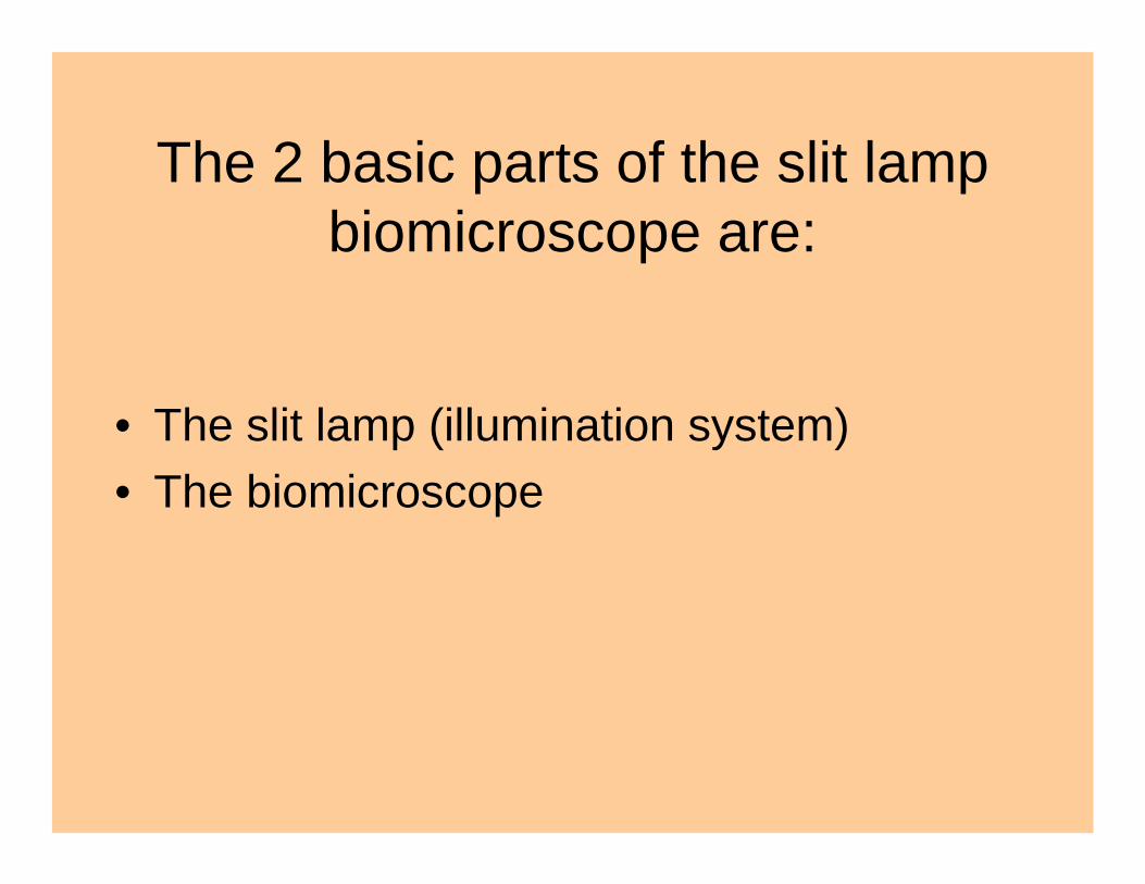
The 2 basic parts of the slit lamp biomicroscope are:
• The slit lamp (illumination system)• The biomicroscope

The illumination system can be
1. Of the Zeiss type2. Of the Haag Streit type

In the Zeiss type the illumination comes from below
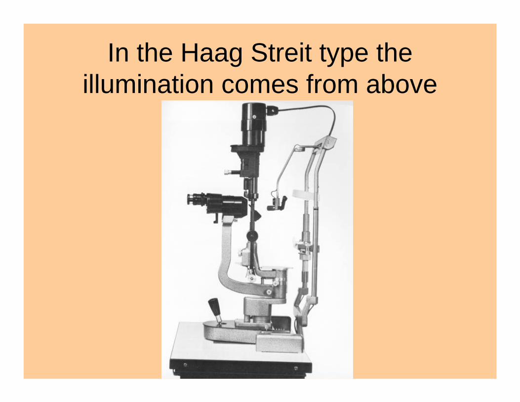
In the Haag Streit type the illumination comes from above
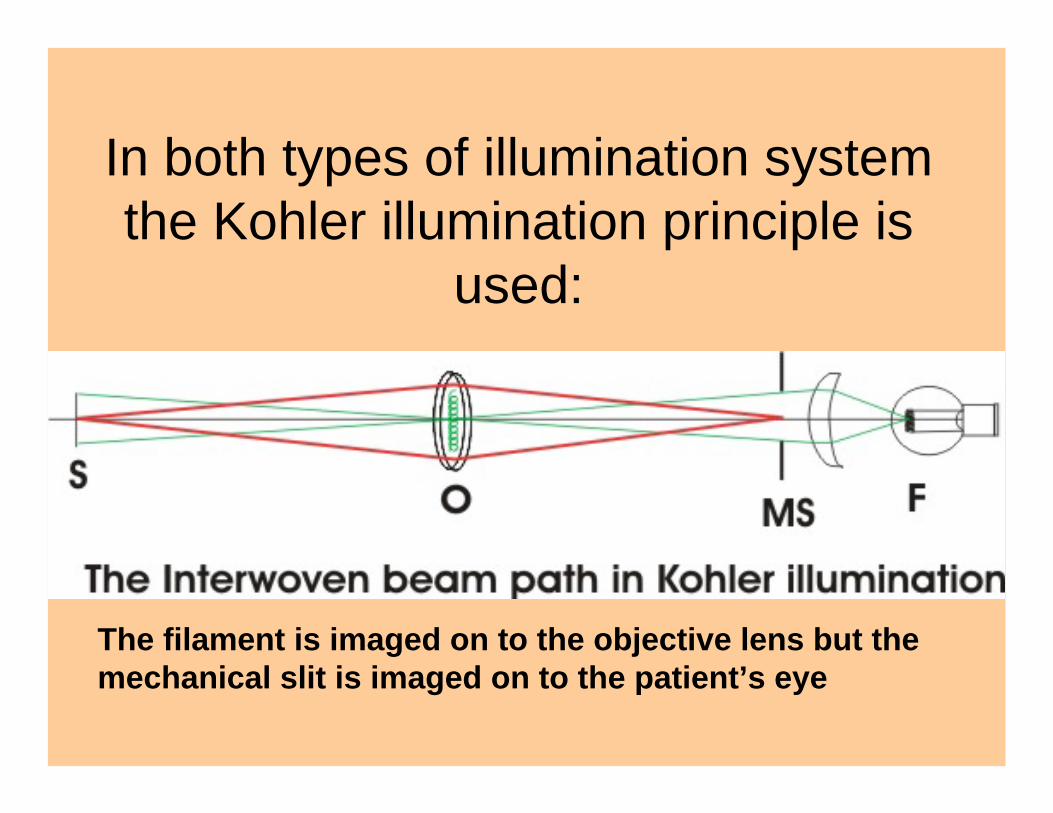
In both types of illumination system the Kohler illumination principle is
used:
The filament is imaged on to the objective lens but the mechanical slit is imaged on to the patient’s eye
The biomicroscope:based on the optics of a compound
microscope
• Two basic types:– The Grenough type– The Galilean changer type
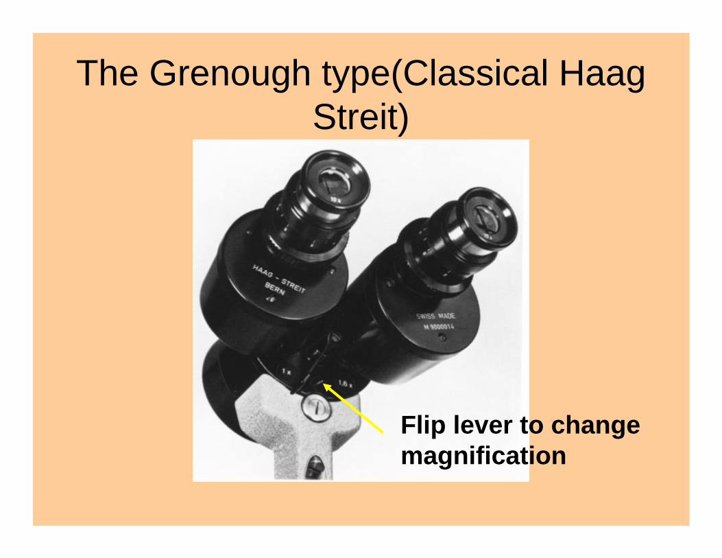
The Grenough type(Classical Haag Streit)
Flip lever to change magnification
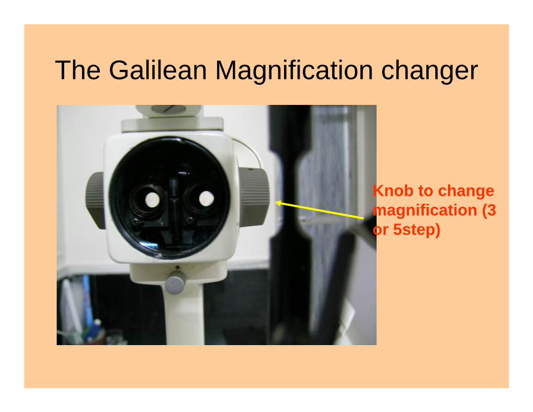
The Galilean Magnification changer
Knob to change magnification (3 or 5step)
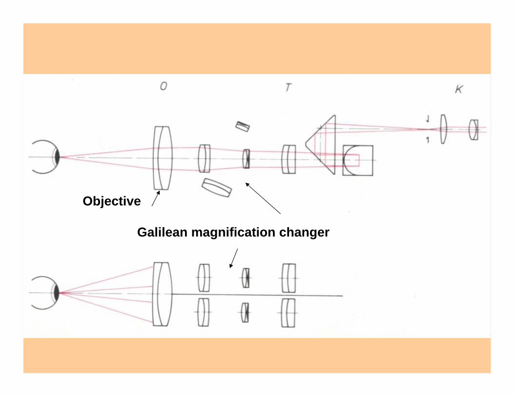
Galilean magnification changer
Objective
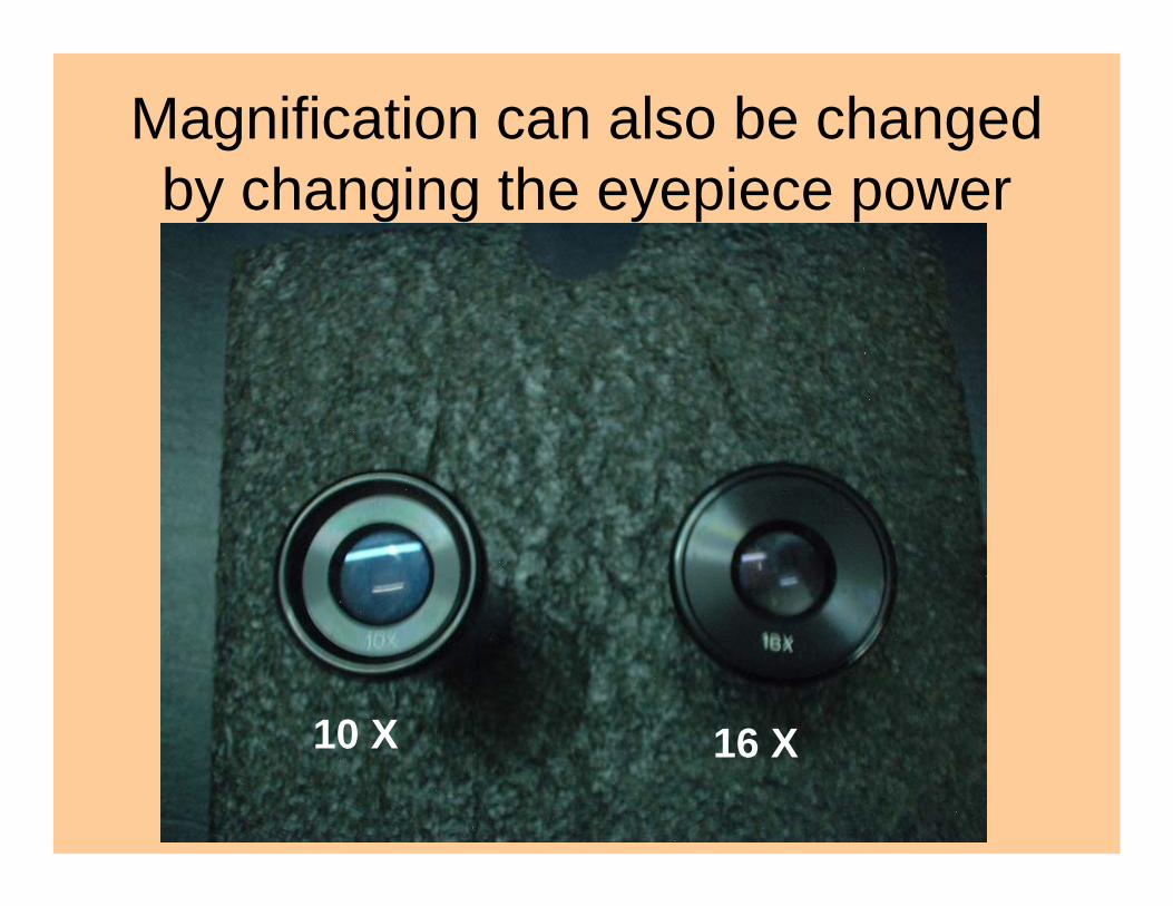
Magnification can also be changed by changing the eyepiece power
10 X 16 X
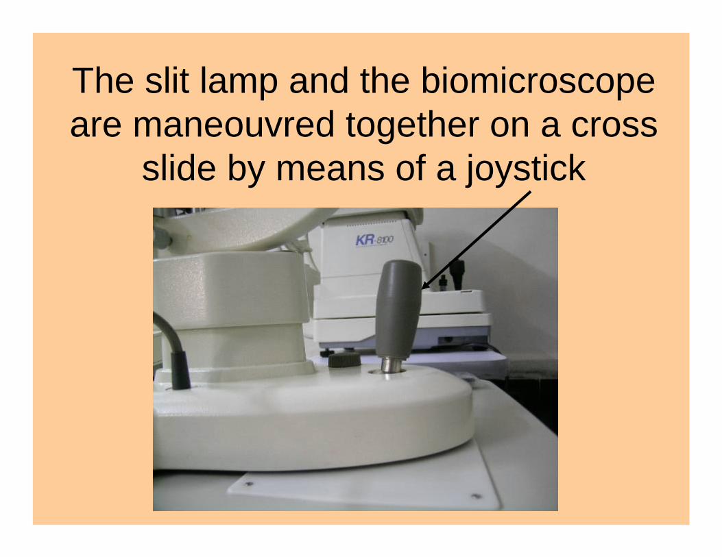
The slit lamp and the biomicroscopeare maneouvred together on a cross
slide by means of a joystick
The coupling between the slit lamp and the biomicroscope
• This is such as to make the system “parfocal”
• i.e the focus of the slit and the focus of the microscope are at the same point.
• This parfocality may occasionally need to be dissociated as for example in the technique of sclerotic scatter
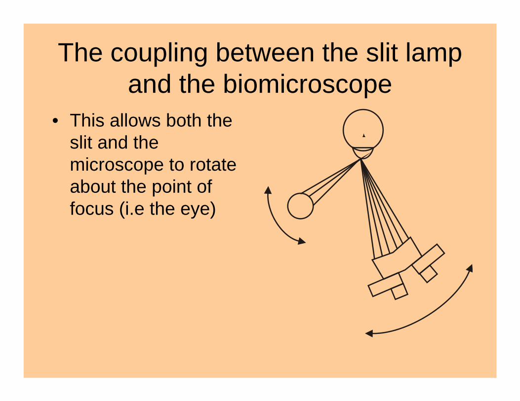
The coupling between the slit lamp and the biomicroscope
• This allows both the slit and the microscope to rotate about the point of focus (i.e the eye)
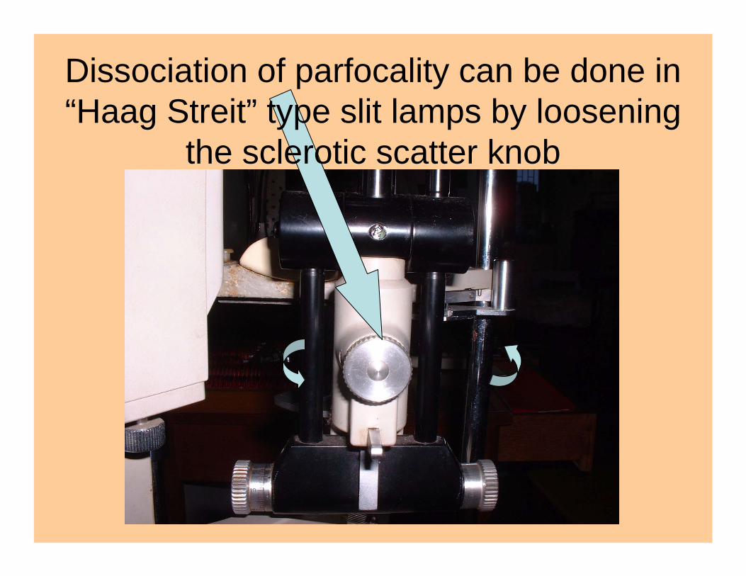
Dissociation of parfocality can be done in “Haag Streit” type slit lamps by loosening
the sclerotic scatter knob

This dissociation of parfocality is useful for indirect illumination,
sclerotic scatter and retroillumination
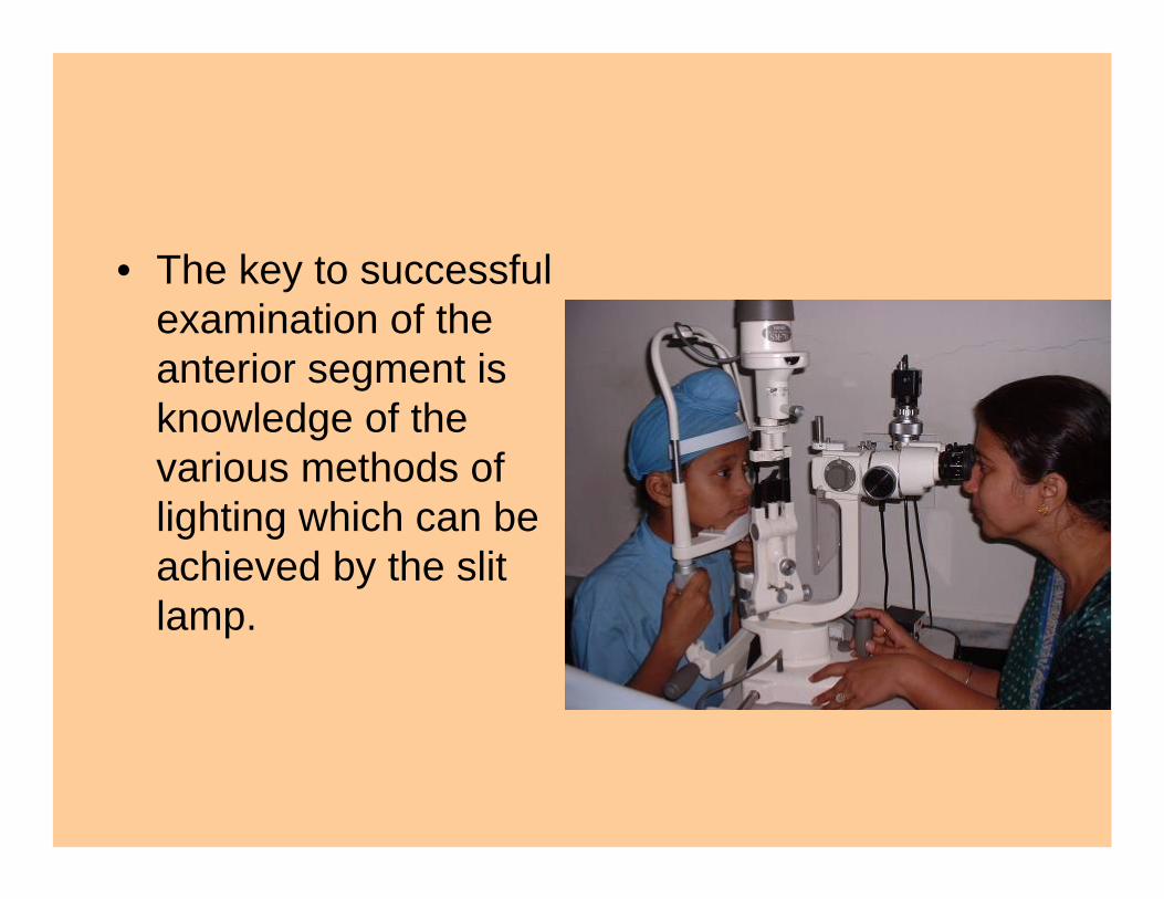
• The key to successful examination of the anterior segment is knowledge of the various methods of lighting which can be achieved by the slit lamp.
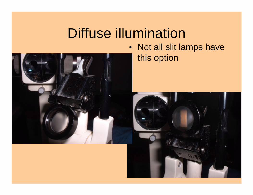
Diffuse illumination• Not all slit lamps have
this option
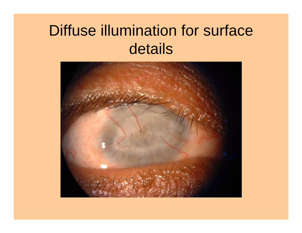
Diffuse illumination for surface details
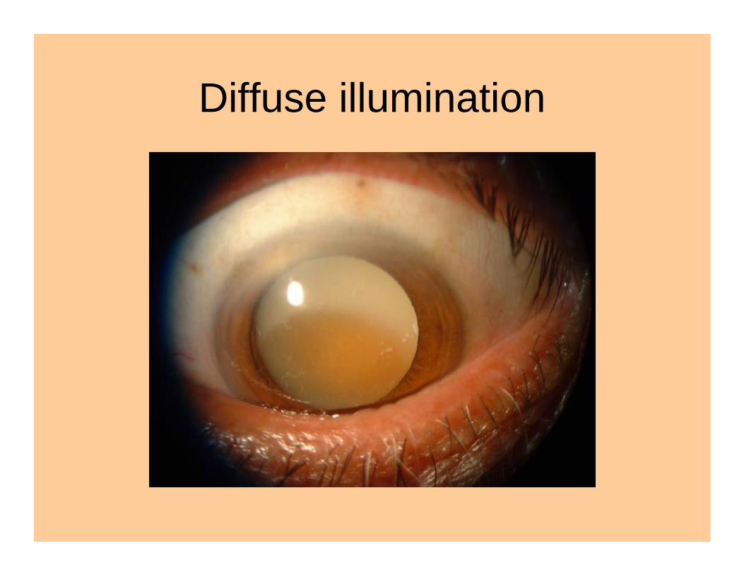
Diffuse illumination
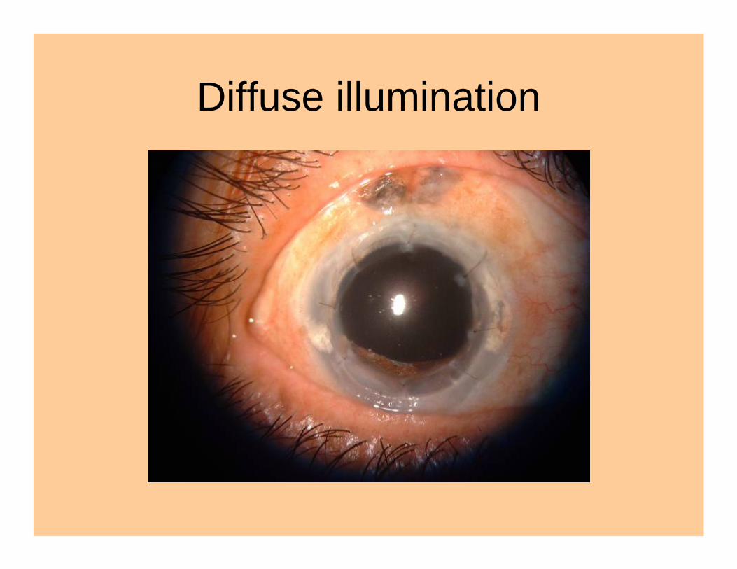
Diffuse illumination
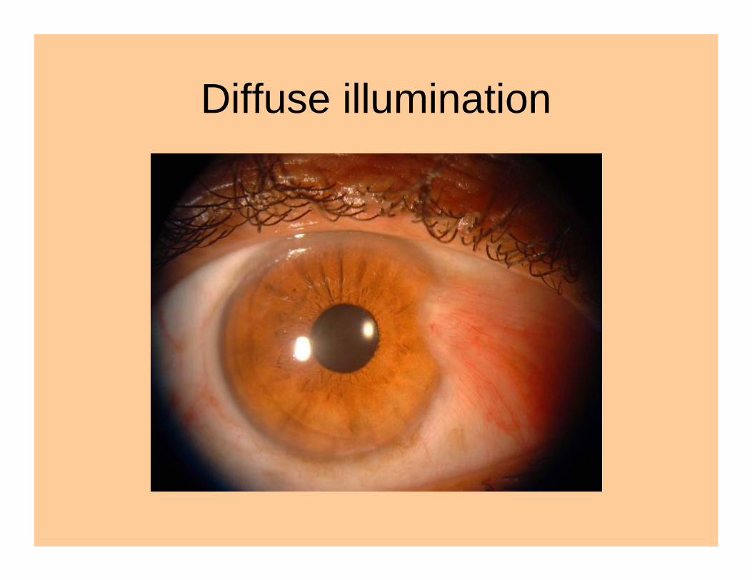
Diffuse illumination
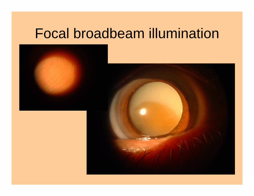
Focal broadbeam illumination
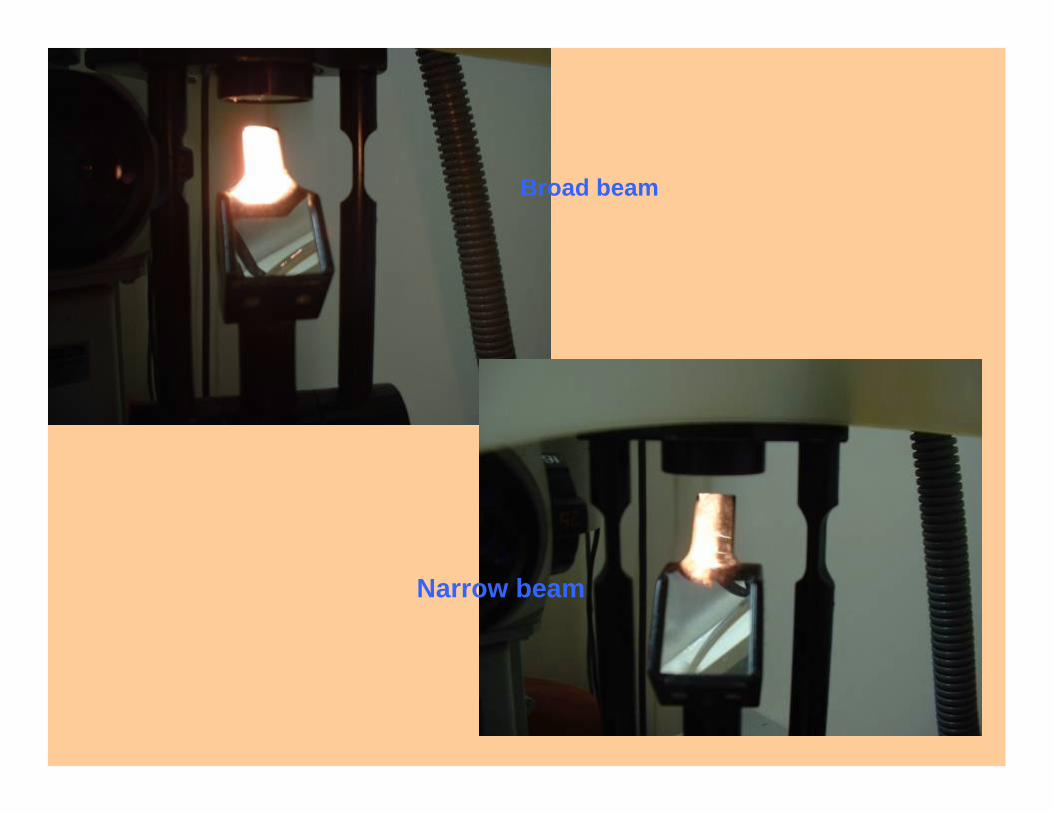
Broad beam
Narrow beam
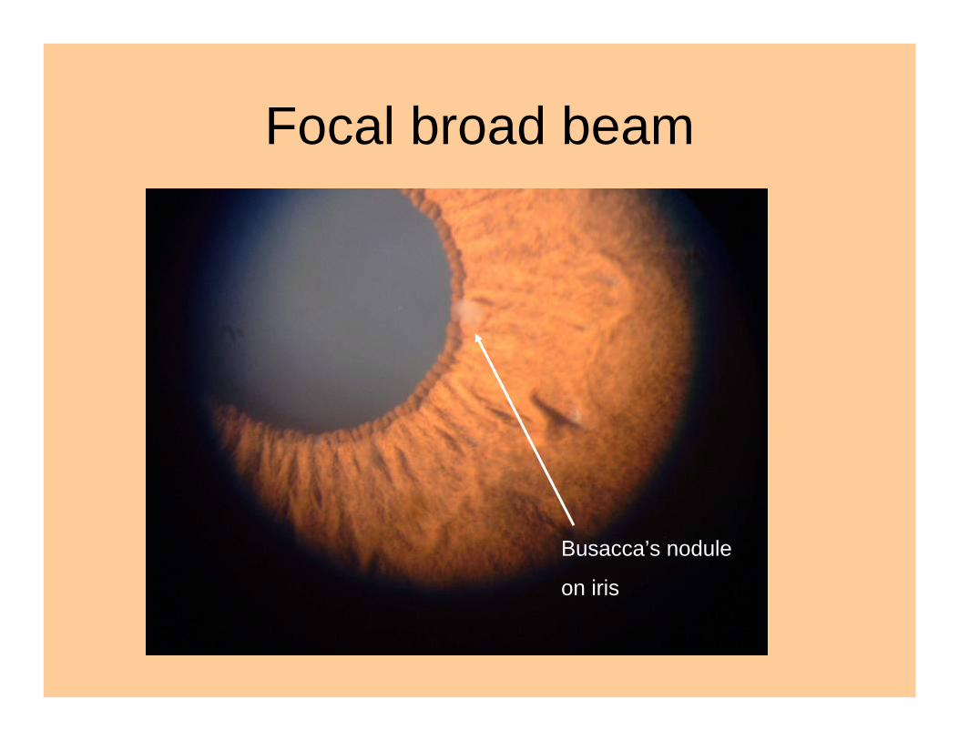
Focal broad beam
Busacca’s nodule
on iris
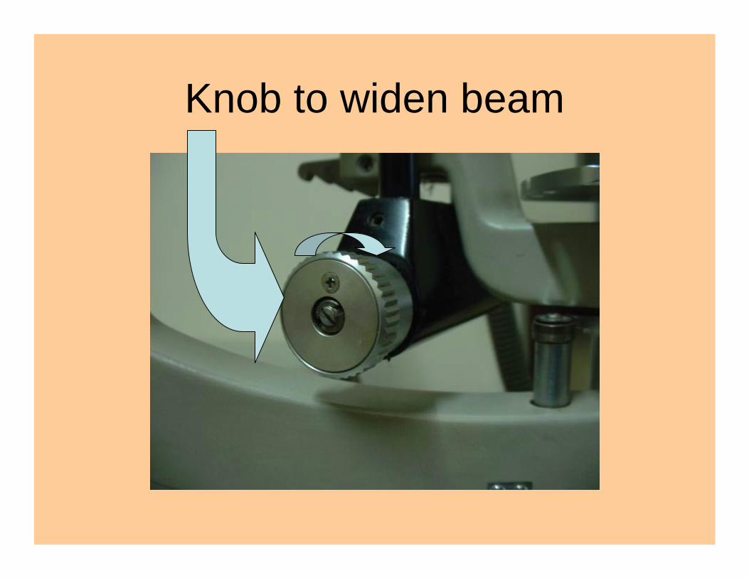
Knob to widen beam
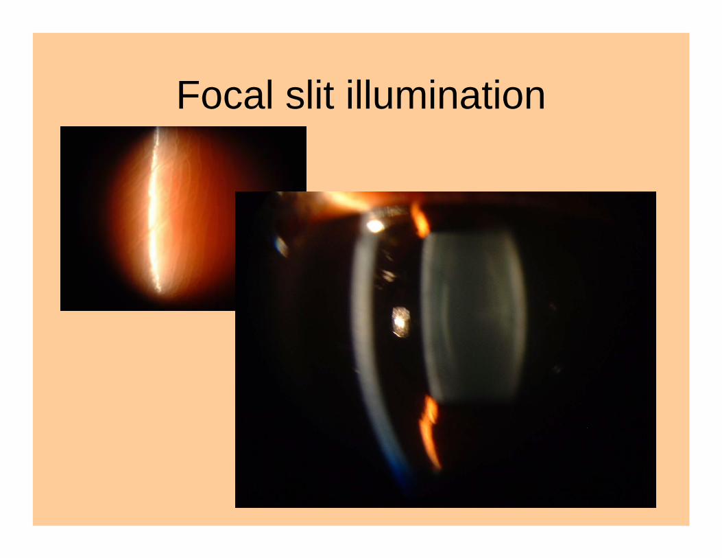
Focal slit illumination
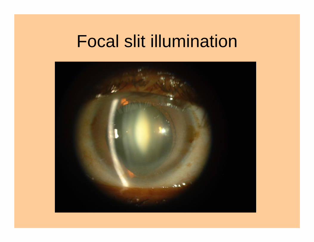
Focal slit illumination
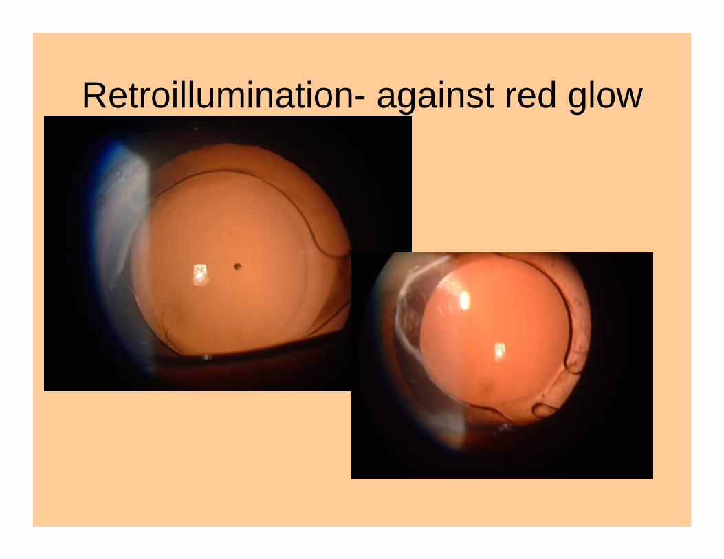
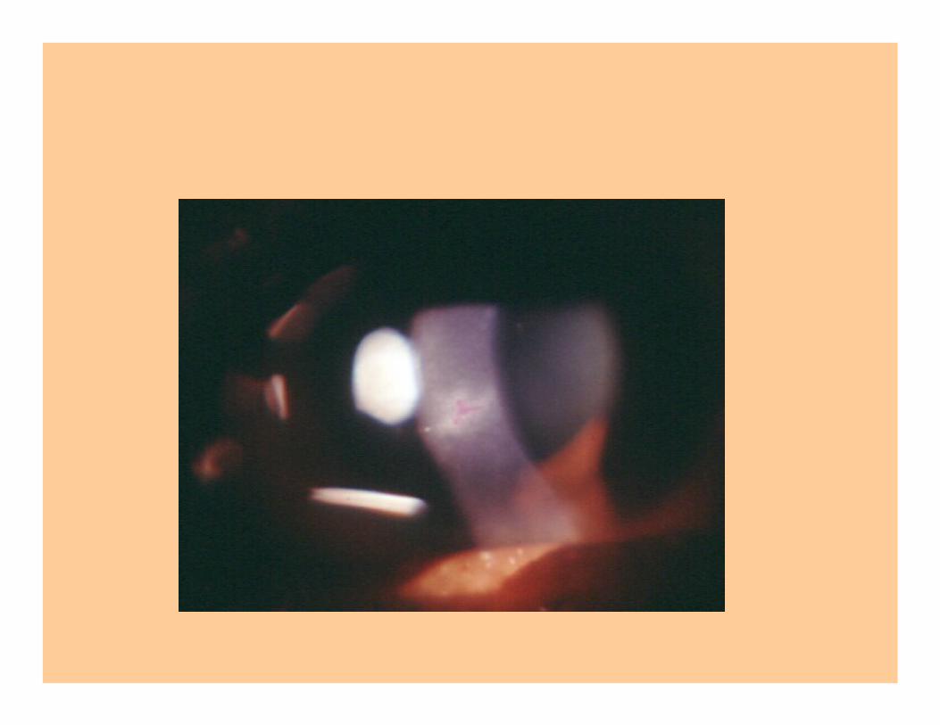
Retroillumination- against red glow
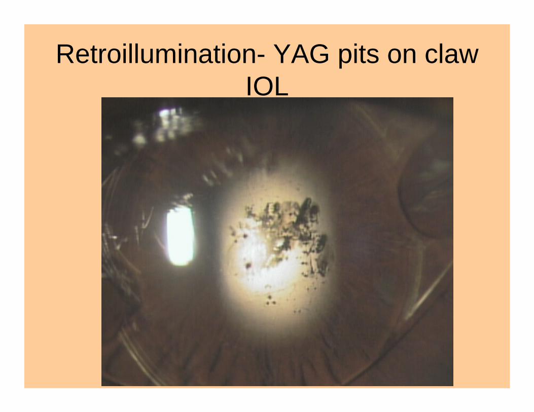
Retroillumination- YAG pits on claw IOL
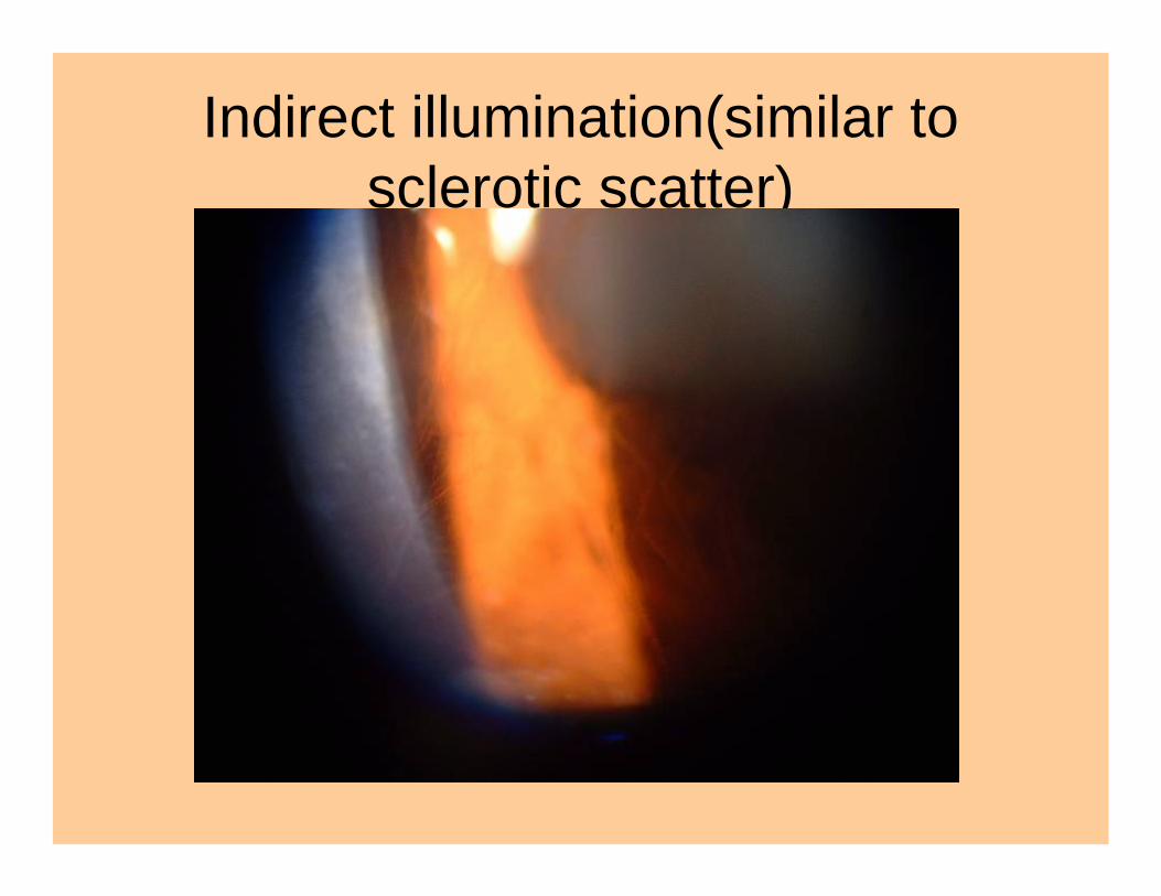
Indirect illumination(similar to sclerotic scatter)
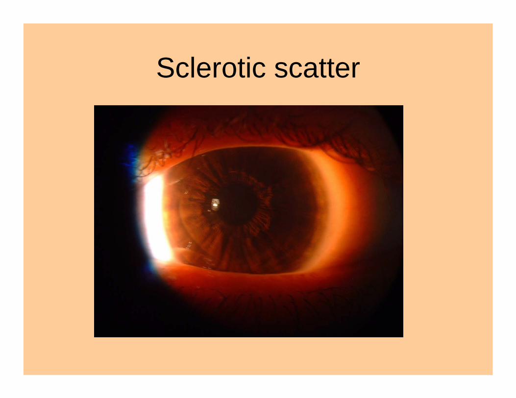
Sclerotic scatter
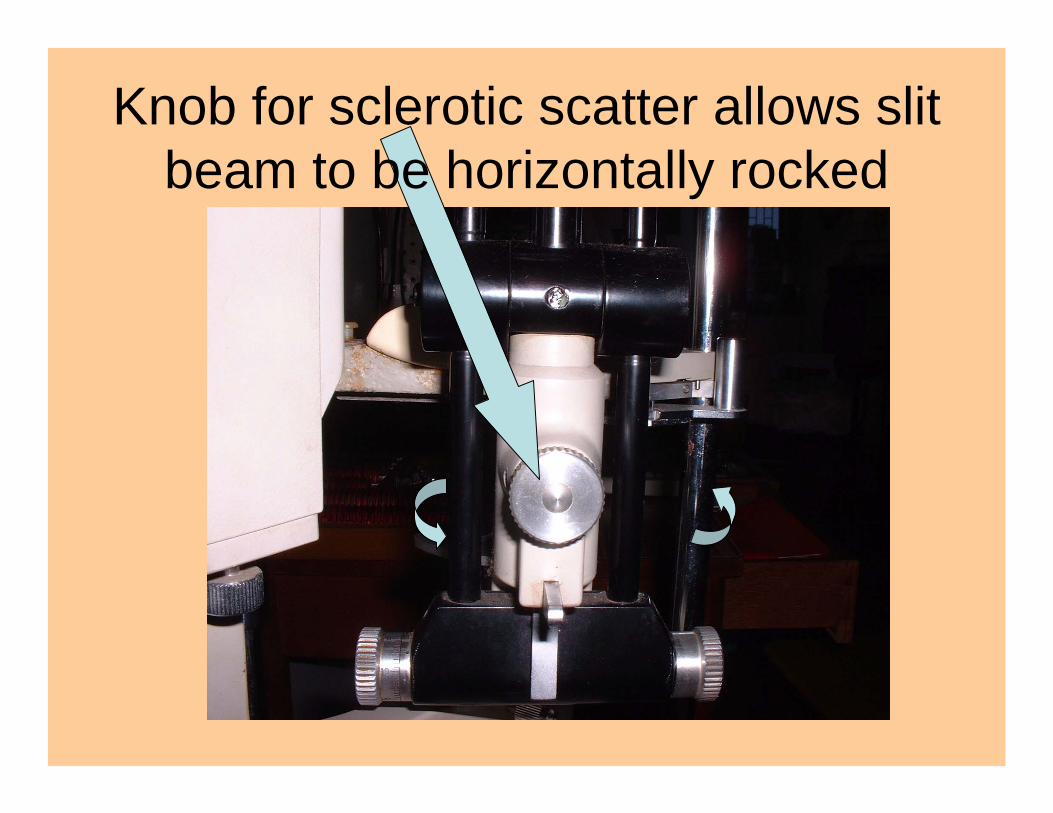
Knob for sclerotic scatter allows slit beam to be horizontally rocked

Parfocality of slit and viewing altered for sclerotic scatter
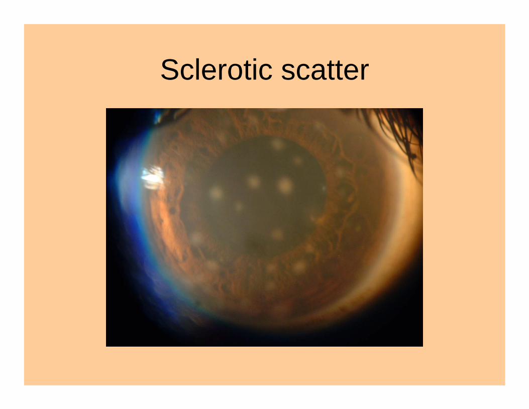
Sclerotic scatter
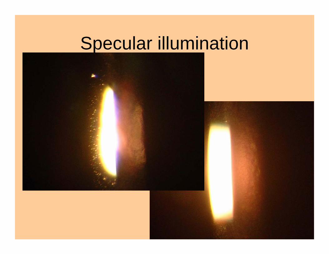
Specular illumination
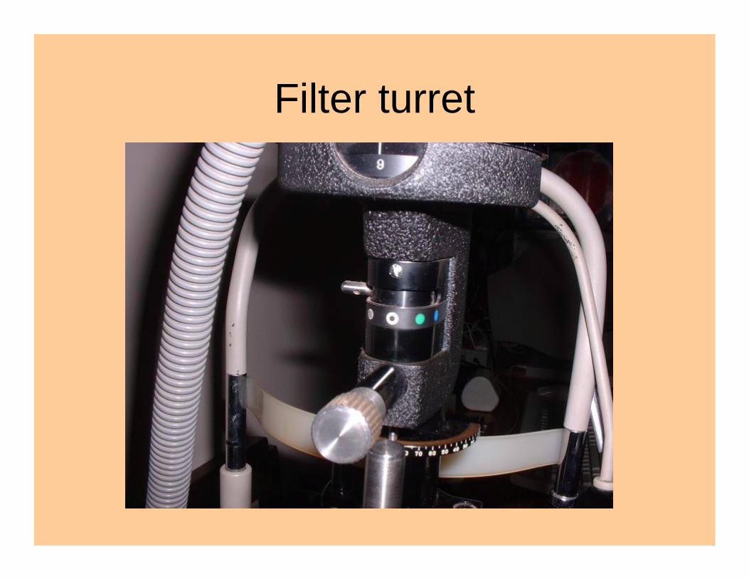
Filter turret
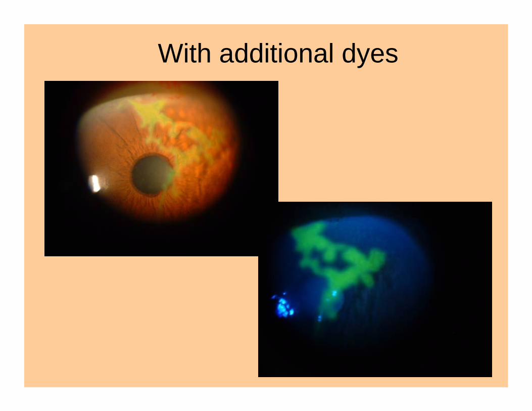
With additional dyes
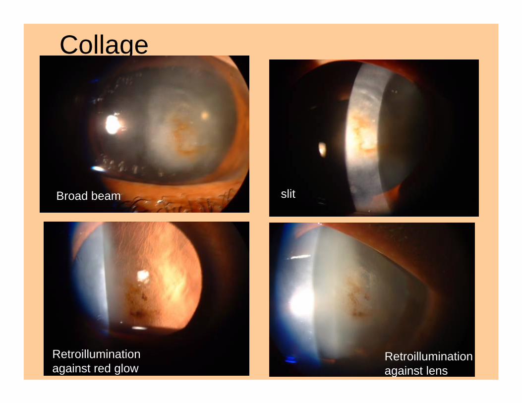
Collage
Broad beam slit
Retroilluminationagainst red glow
Retroilluminationagainst lens
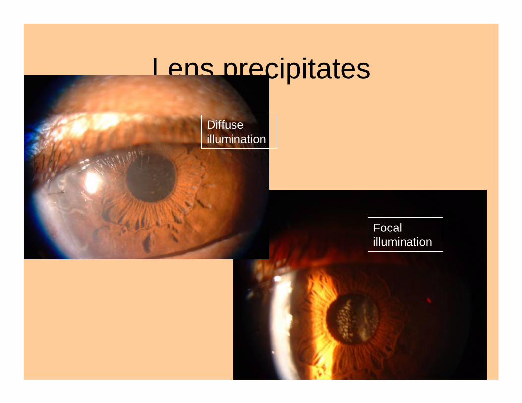
Lens precipitates
Diffuse illumination
Focal illumination
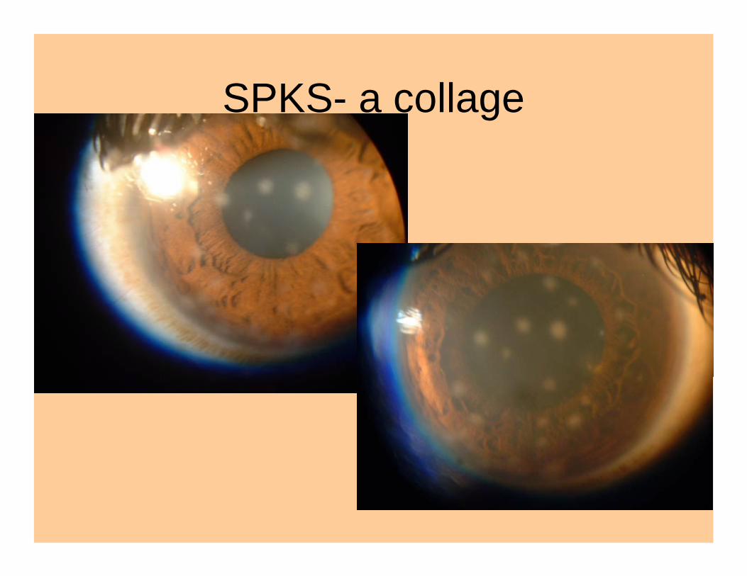
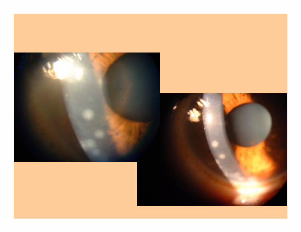
SPKS- a collage
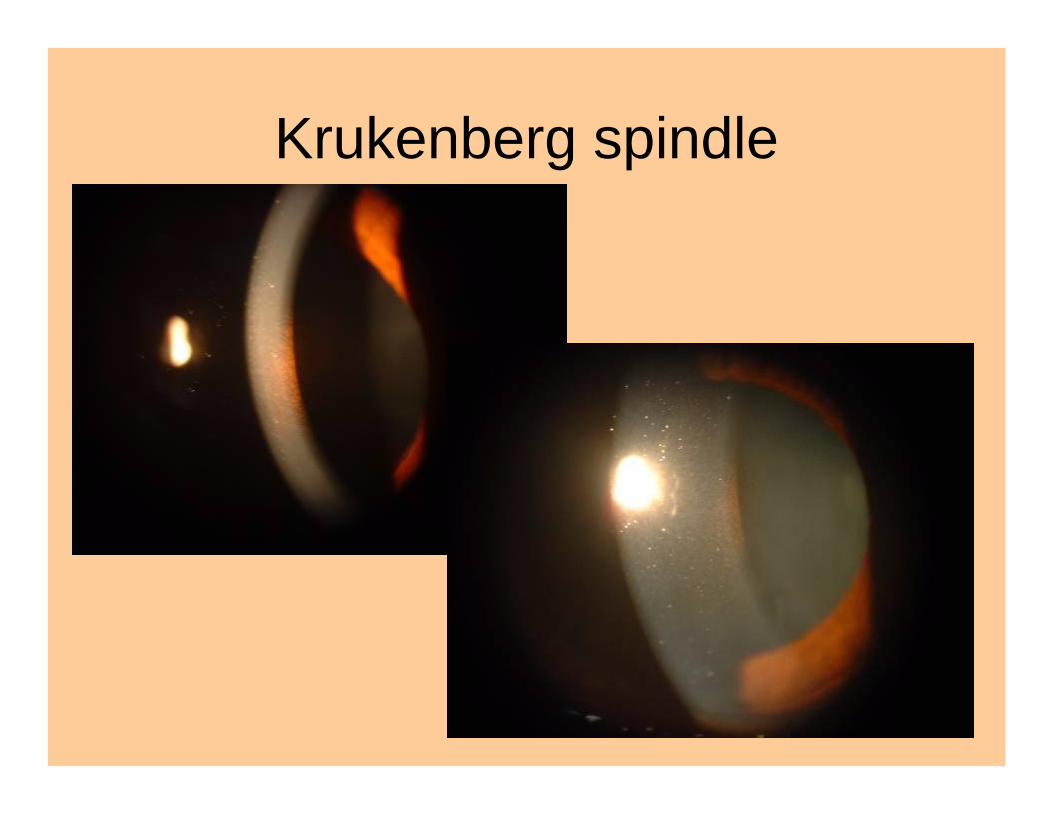
Krukenberg spindle
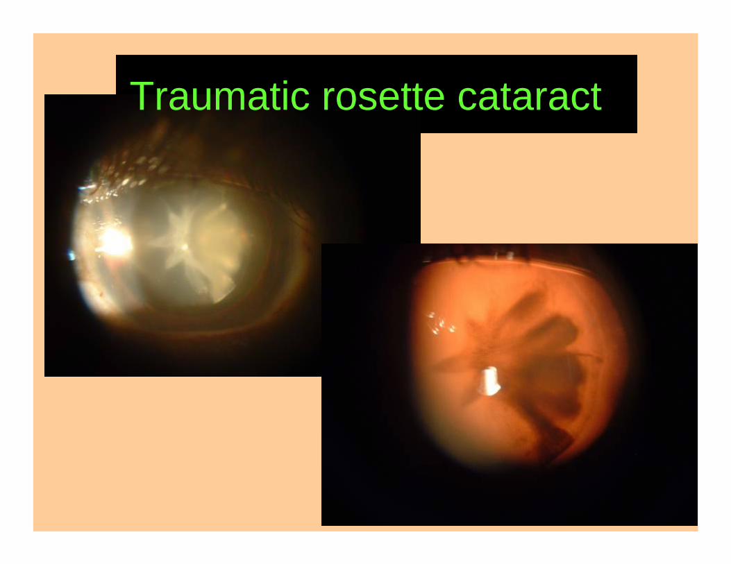
Traumatic rosette cataract
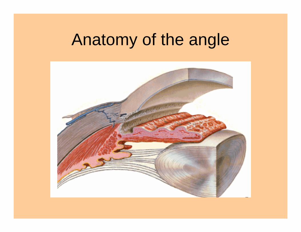
Anatomy of the angle
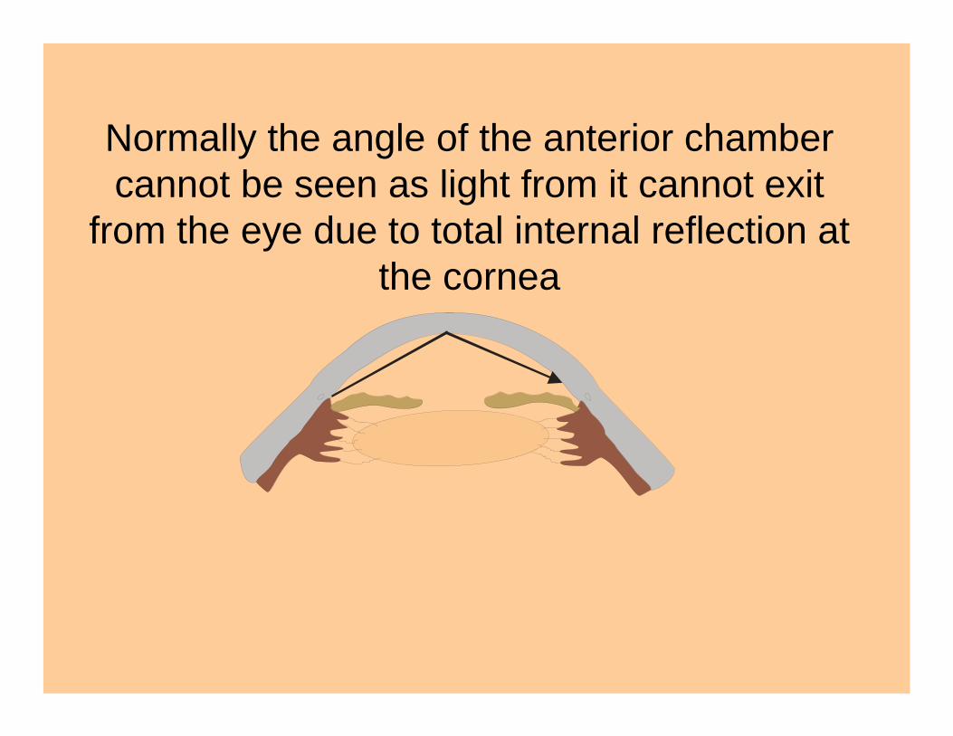
Normally the angle of the anterior chamber cannot be seen as light from it cannot exit
from the eye due to total internal reflection at the cornea
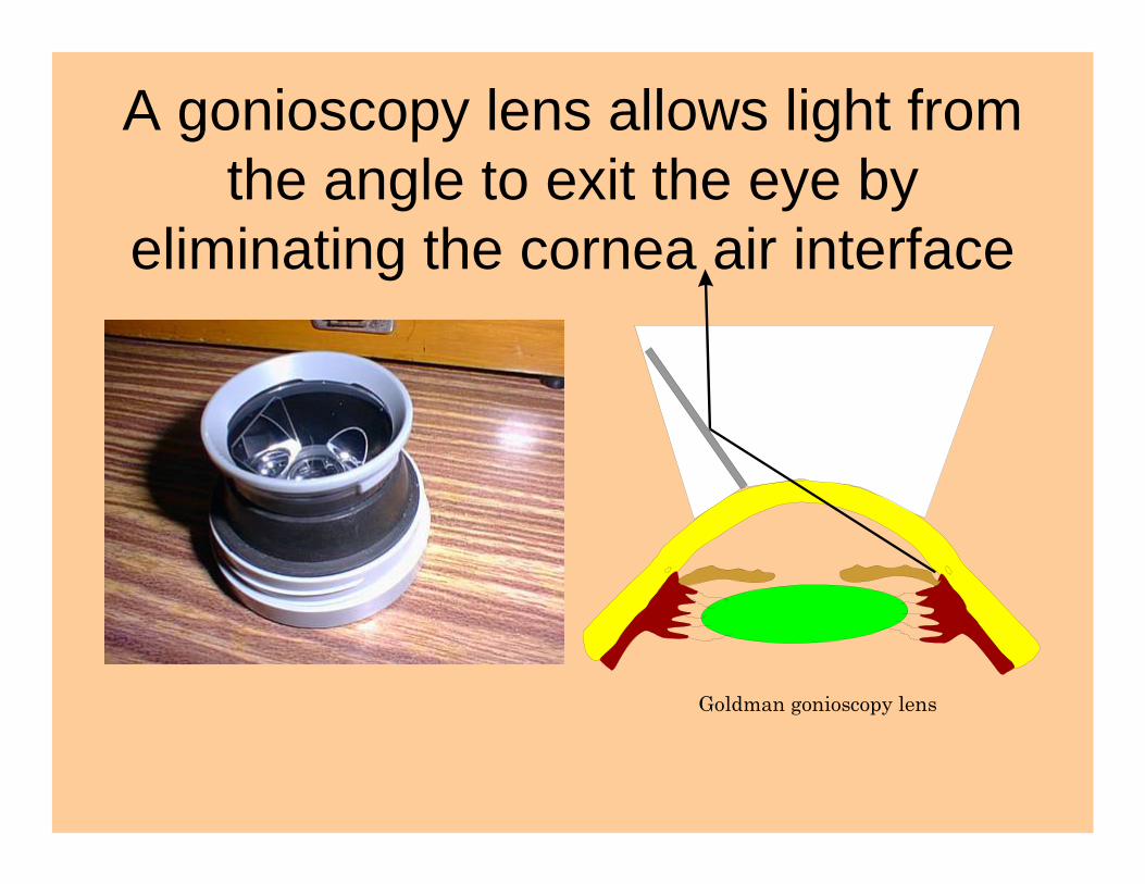
A gonioscopy lens allows light from the angle to exit the eye by
eliminating the cornea air interface
Goldman gonioscopy lens
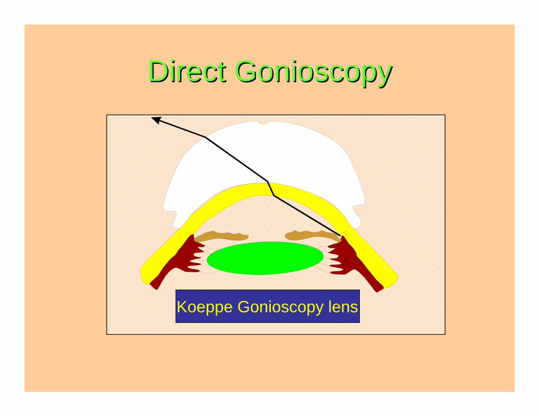
Direct Direct GonioscopyGonioscopy
Koeppe gonioscopy lensKoeppe Gonioscopy lens
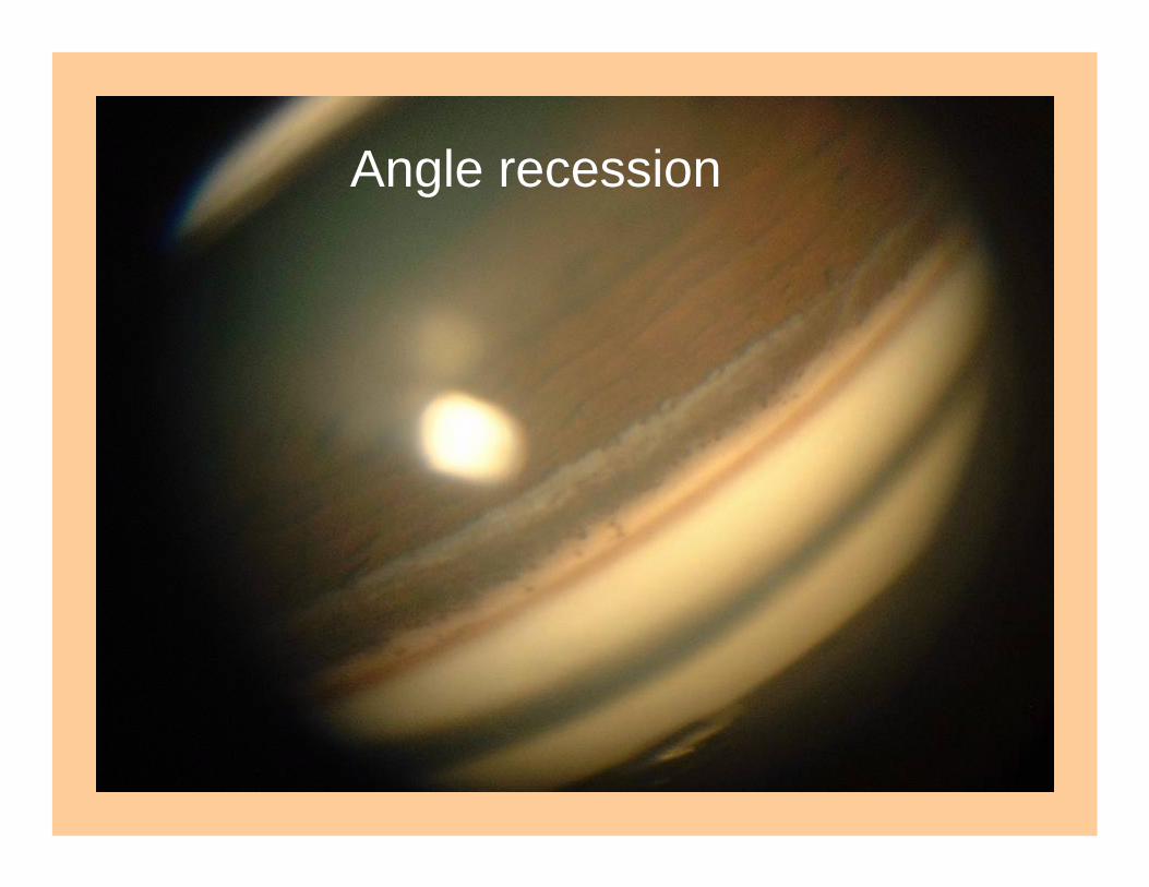
Angle recession
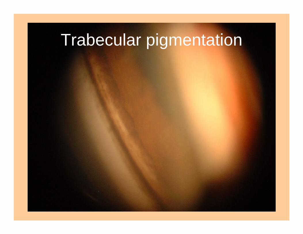
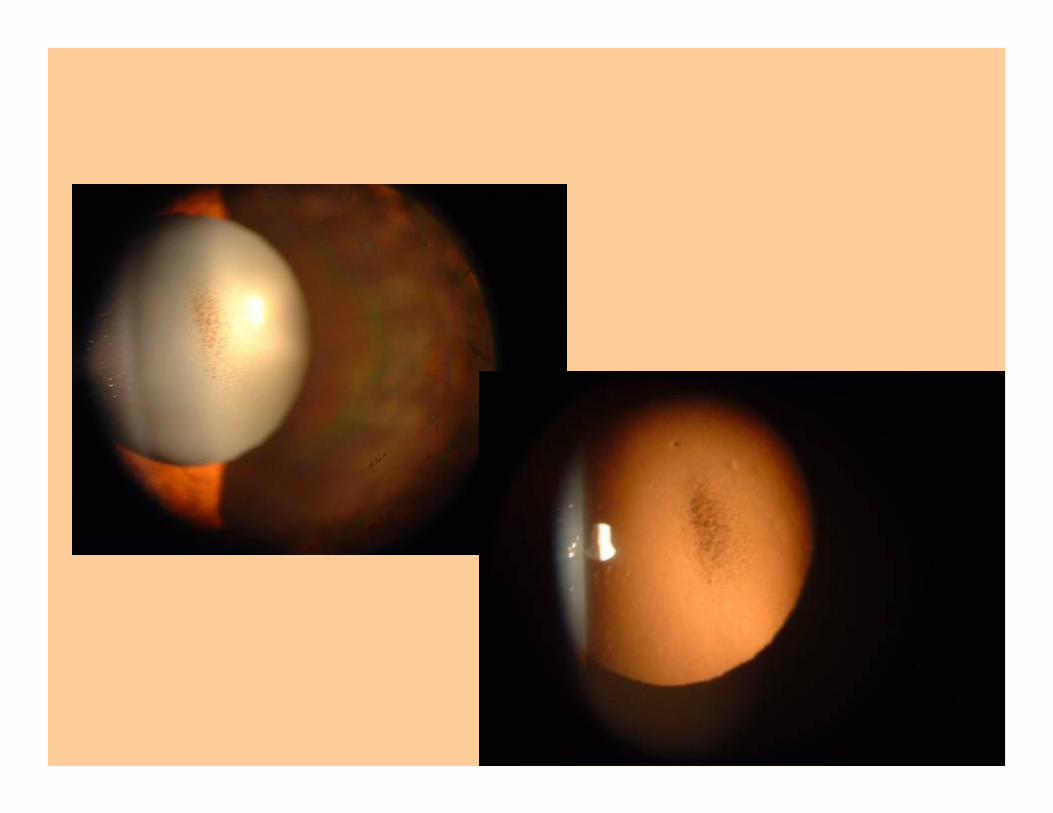
Trabecular pigmentation
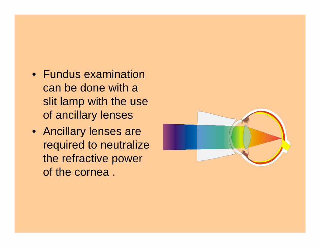
• Fundus examination can be done with a slit lamp with the use of ancillary lenses
• Ancillary lenses are required to neutralize the refractive power of the cornea .
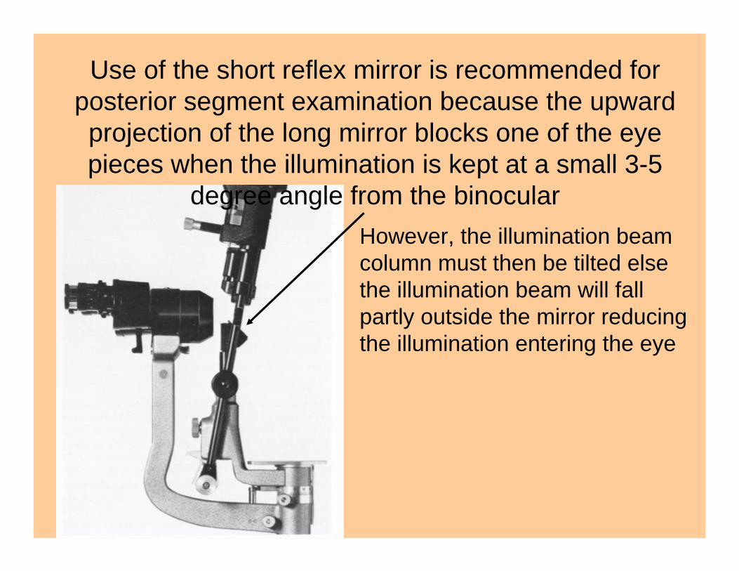
Use of the short reflex mirror is recommended for posterior segment examination because the upward projection of the long mirror blocks one of the eye pieces when the illumination is kept at a small 3-5
degree angle from the binocularHowever, the illumination beam column must then be tilted else the illumination beam will fall partly outside the mirror reducing the illumination entering the eye

Broad beam
Narrow beam
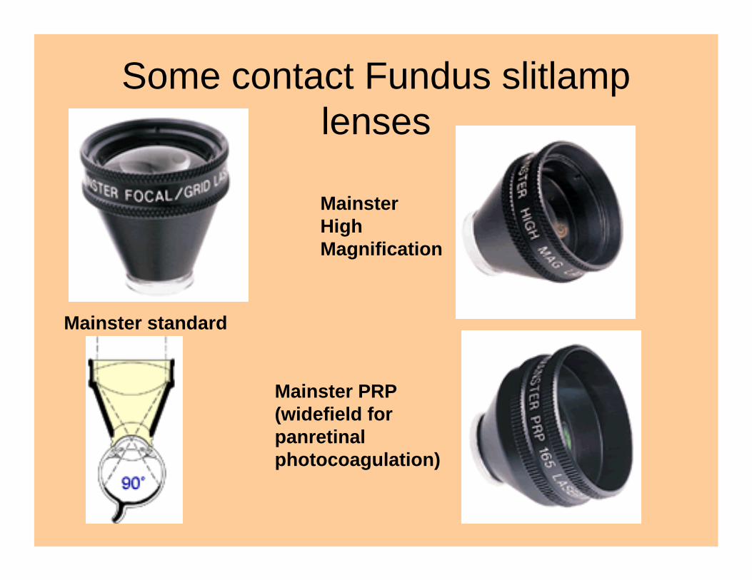
Some contact Fundus slitlamplenses
Mainster standard
MainsterHigh Magnification
Mainster PRP (widefield for panretinalphotocoagulation)
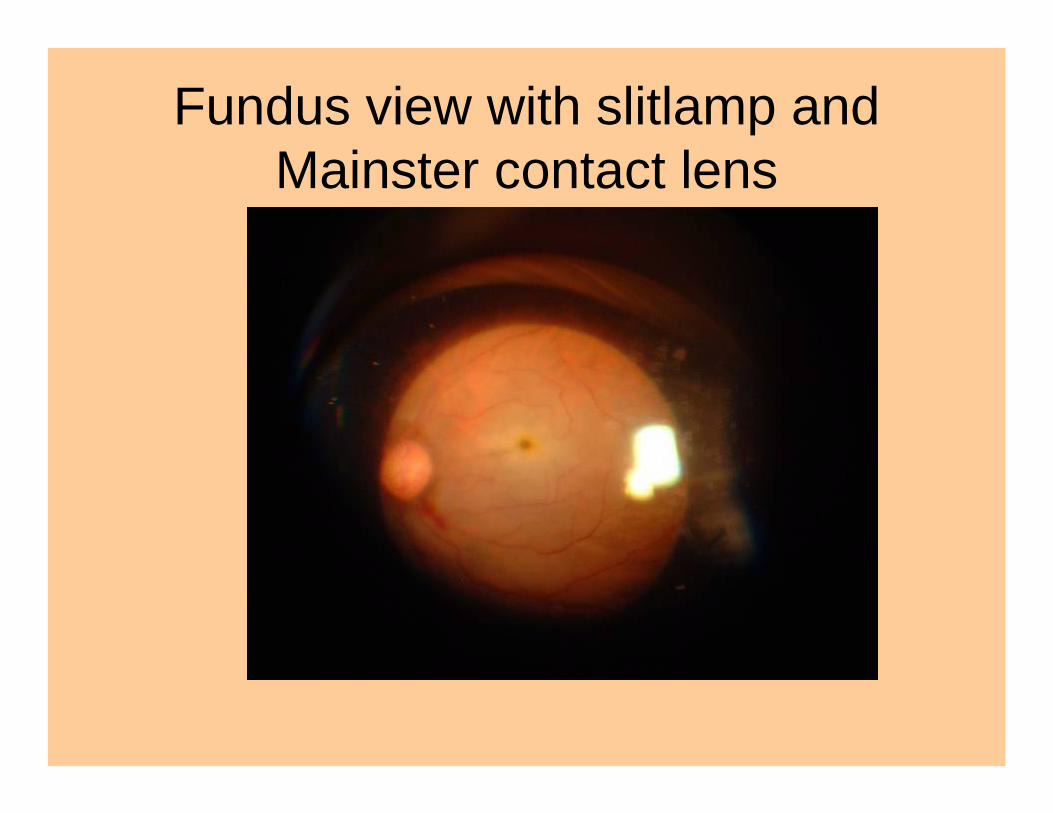
Fundus view with slitlamp and Mainster contact lens
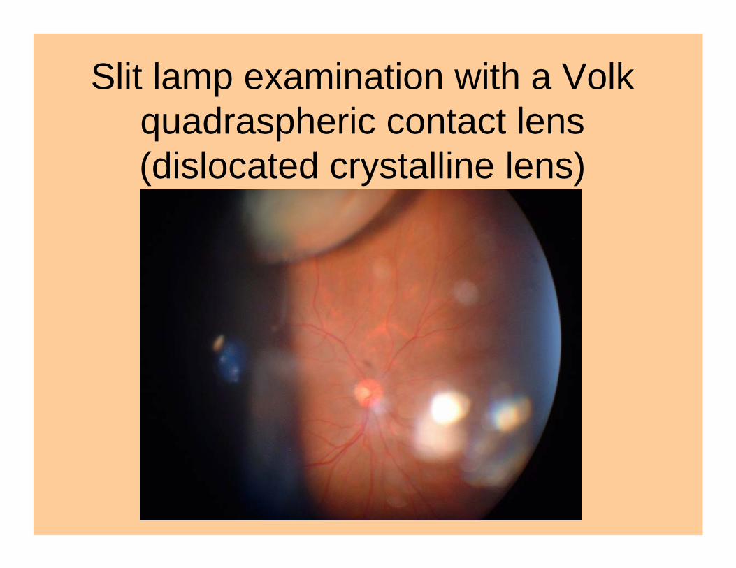
Slit lamp examination with a Volk quadraspheric contact lens (dislocated crystalline lens)