Basics of Sleep Medicine: From A to ZZZZZ’s - White · Basics of Sleep Medicine: From A to...
118
Basics of Sleep Medicine: From A to ZZZZZ’s Joyce K. Lee-Iannotti, MD Chief and Medical Director, Sleep Disorders Center BUMC IM Sleep Lecture October 18, 2016
Transcript of Basics of Sleep Medicine: From A to ZZZZZ’s - White · Basics of Sleep Medicine: From A to...

Basics of Sleep Medicine: From A to
ZZZZZ’sJoyce K. Lee-Iannotti, MD
Chief and Medical Director, Sleep Disorders Center
BUMC
IM Sleep LectureOctober 18, 2016

Objectives
►Normal Sleep►Physiology of sleep (just briefly)►Sleep stages►Introduction to the PSG►Sleep Disorders Insomnia, OSA, Narcolepsy

Basic Sleep Concepts►Drive for sleep exceeds the drive for food
and water, and freedom from pain►Sleep deprivation, total or chronic partial,
may have serious consequences death in experimental animals impaired perception and microsleeps in humans
►Sleep debt must eventually be repaid
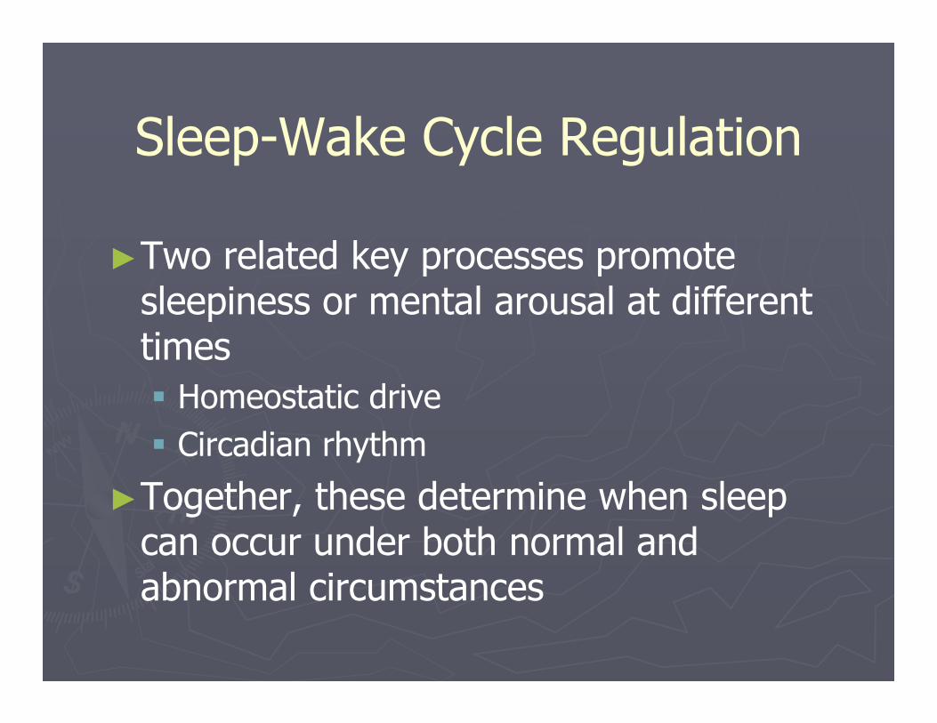
Sleep-Wake Cycle Regulation
►Two related key processes promote sleepiness or mental arousal at different times Homeostatic drive Circadian rhythm
►Together, these determine when sleep can occur under both normal and abnormal circumstances

Homeostatic drive
►Has a ratio of approximately 1/3 sleep and 2/3 waking
►Sleep deprivation, acute or chronic, increases the homeostatic sleep drive and therefore sleepiness
►Hypothetically, the homeostatic sleep drive could be satisfied by sleep at any hour

Circadian rhythm
►Entrained and synchronized ►Timing of sleepiness promoted by the
endogenous circadian clock►Facilitates the rhythmic cycle of sleep at the
same approximate nighttime hours (each day)
►Reinforced by the daily photoperiod, and possibly influenced by other light exposure

Sleep/Wake Cycle

Normal Sleep
4
3
2
1
REM
Awake
87654321Hours
REM NREM 1 NREM 2 NREM 3/4

Normal Sleep Architecture
Approximately 90 minute cycle including NREM and REM
Slow wave dominates first third of night REM sleep dominates last third of night
(early morning hours) REM sleep: 20-25% total sleep time Can see REM-rebound with sleep deprivation,
abrupt withdrawal of REM suppressants


Introduction to the PSG

Types of sleep studies Diagnostic – overnight study In-lab (OSA, PLMD/RLS, RBD, parasomnias, sz) Home sleep study (just for OSA)
CPAP titration - Once a patient is identified as having sleep apnea another study is performed in which the technician adjusts the CPAP level during the test/mask fitting
Split Night - Combines a diagnostic study and a CPAP titration study into one night. The patient is diagnosed during the first half of the night (AHI >40); CPAP applied the second half if required by protocol
MSLT - Multiple Sleep Latency Test MWT – Maintenance of Wakefulness Test

Indications for PSG
►Excessive daytime sleepiness (EDS)►Unexplained behavioral events in sleep►Insomnia or unexplained awakenings►Sleep-related breathing disturbances►Effect of treatment for sleep disorders

PSG Parameters
► EEG► EOG (electro-
oculogram)►Chin EMG► Leg EMG► ECG
►Airflow► Effort ►Oxygen ►Body position

EEG► Minimum of 3 EEG derivations required to sample from frontal, central
and occipital regions► Recommended derivations
F4-M1 C4-M1 O2-M1 F3, C3, O1 and M2 placed for backup
► Alternative derivations Fz-Cz Cz-Oz C4-M1 Fpz, C3, O1 and M2 placed for backup
► Additional derivations required for evaluation of seizures International 10-20 electrode placement
► Paper speed: 10 mm/sec (30 sec epochs)


EOG EOG records voltage changes caused by EM Recommended derivations:
E1-M2 (E1 placed 1 cm below LOC) E2-M2 (E2 placed 1 cm above ROC)
Alternative derivations: E1-FPz (E1 placed 1 cm below/lateral to LOC) E2-FPz (E2 placed 1 cm above/lateral to ROC)
Wake: random, high amplitude
Stage 1: slow rolling, conjugate, regular
REM: conjugate, irregular, sharply peaked EM

EMG Recorded as the potential between two surface
electrodes placed several centimeters apart Typically, the chin (submental) muscle is used
because it exhibits large differences during sleep, aiding in the identification of stages
Wake - high activity
Sleep - lower activity
REM sleep - paralysis of skeletal muscles

EMG Placement
Chin Electrode placement (2 required) Midline 1 cm above
inferior edge of mandible (optional)
2 cm below inferior edge of mandible to right of midline
2 cm below inferior edge of mandible to left of midline

REM vs. NREM Sleep►Non-REM Physical restoration Driven by homeostatic drive Quiet brain, active body
►REM Mental restoration/memory Driven by circadian rhythm Active brain, quiet body

REM vs. NREM Sleep
Physiologic Variable NREM REM
Heart rate Regular Irregular
Respiratory rate Regular Irregular
Blood pressure Regular Variable
Skeletal muscle tone Preserved Absent
Brain 02 consumption Reduced Increased
Ventilatory response Normal Reduced
Temperature Normal Poikilothermic

Overview of sleep stages
Combined to become N32007 AASM scoring guidelines
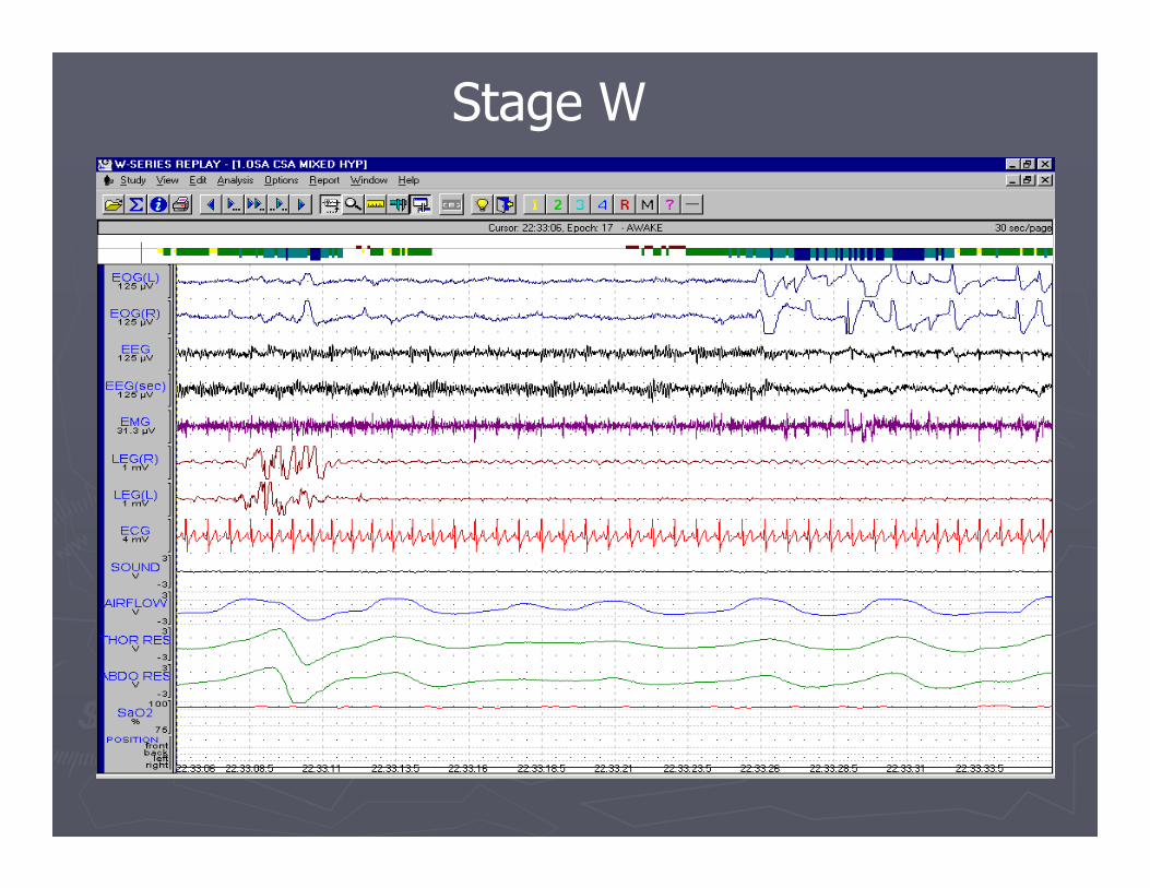
Stage W

Stage 1

Stage 2

Stage 3
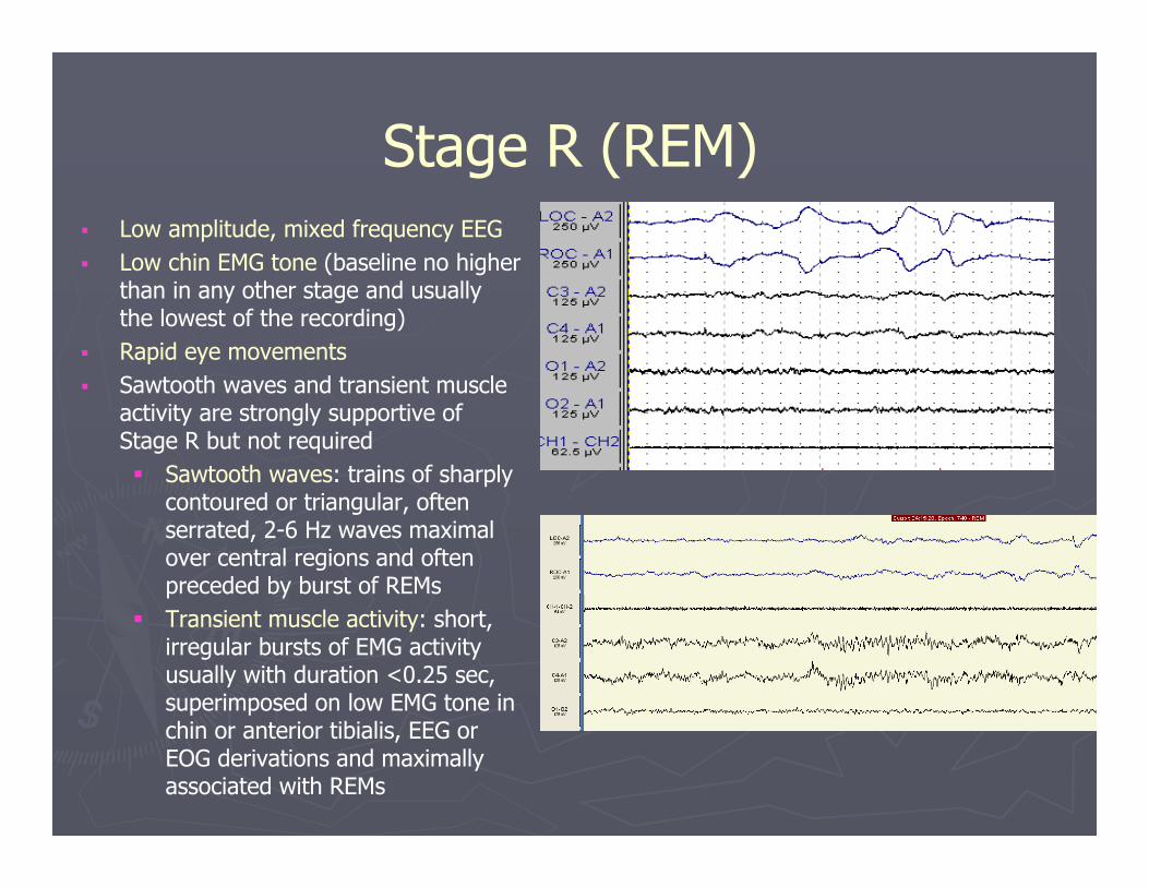
Stage R (REM) Low amplitude, mixed frequency EEG Low chin EMG tone (baseline no higher
than in any other stage and usually the lowest of the recording)
Rapid eye movements Sawtooth waves and transient muscle
activity are strongly supportive of Stage R but not required Sawtooth waves: trains of sharply
contoured or triangular, often serrated, 2-6 Hz waves maximal over central regions and often preceded by burst of REMs
Transient muscle activity: short, irregular bursts of EMG activity usually with duration <0.25 sec, superimposed on low EMG tone in chin or anterior tibialis, EEG or EOG derivations and maximally associated with REMs
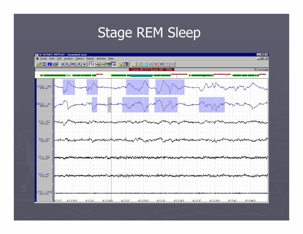
Stage REM Sleep

Respiratory Variables
Respiratory effort (thoracic and abdominal belts) Airflow (thermistor, thermocouple, nasal
pressure, ETCO2) SpO2 (pulse oximetry) Snoring microphone Optional signals
• ETCO2• tcCO2
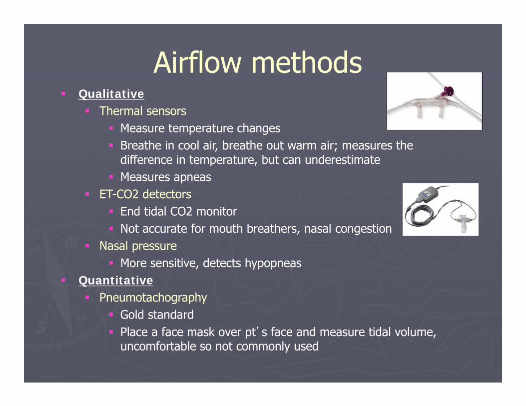
Airflow methods Qualitative
Thermal sensors Measure temperature changes Breathe in cool air, breathe out warm air; measures the
difference in temperature, but can underestimate Measures apneas
ET-CO2 detectors End tidal CO2 monitor Not accurate for mouth breathers, nasal congestion
Nasal pressure More sensitive, detects hypopneas
Quantitative Pneumotachography
Gold standard Place a face mask over pt’s face and measure tidal volume,
uncomfortable so not commonly used

Effort methods
Qualitative Piezo-electric belts (crystals embedded in belt that
sense movement) Intercostal EMG
Semi-quantitative Respiratory inductive plethysmography (RIP): can
give tidal volume, but not very accurately Gold standard: Esophageal pressure (balloon
inserted into lower esophagus)

Other Variables Typically Recorded
ECG Leg movement: EMG Video Body position

Respiratory Events
Apneas – absence of airflow Drop in peak thermal sensor excursion by >90% of
baseline Duration of events lasts at least 10 seconds At least 90% of event’s duration meets the amplitude
reduction criteria for apnea Hypopneas – reduced airflow Respiratory Event Related Arousals (RERA)
• Respiratory event does not meet the criteria for event types above
• Causes a disruption of the sleep architecture

Types of Apnea
Obstructive: Associated with continued or increased inspiratory
effort, but absent airflow
Central: Absent inspiratory effort and airflow
Mixed: Absent inspiratory effort initially, followed by resumption
of effort in the second portion of the event

Example - Obstructive Apnea

Example - Central Apnea
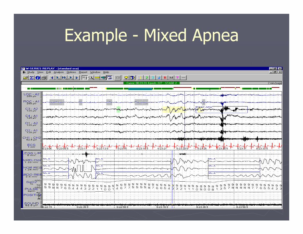
Example - Mixed Apnea
Hypopnea
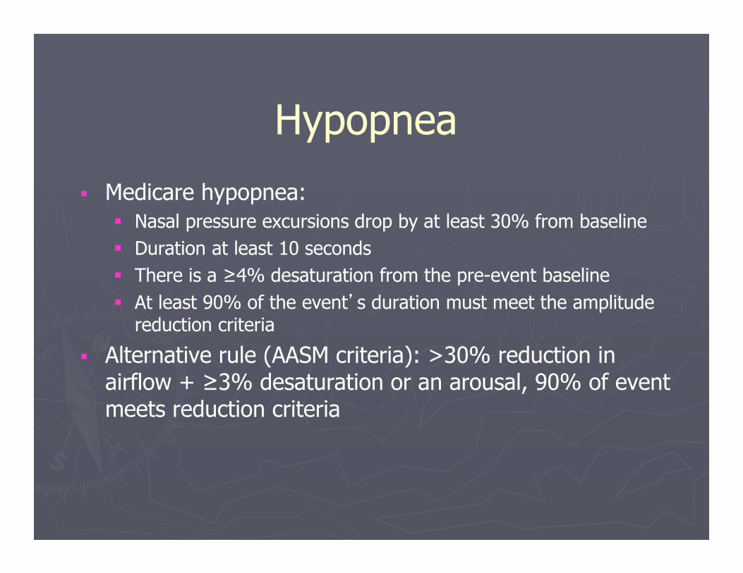
Medicare hypopnea: Nasal pressure excursions drop by at least 30% from baseline Duration at least 10 seconds There is a ≥4% desaturation from the pre-event baseline At least 90% of the event’s duration must meet the amplitude
reduction criteria
Alternative rule (AASM criteria): >30% reduction in airflow + ≥3% desaturation or an arousal, 90% of event meets reduction criteria

Example - Hypopnea

Example - PLMS

Sleep disorders: An overview

ICSD-2 (2006)► Insomnia► Sleep Related
Breathing Disorders►Hypersomnias of
Central Origin►Circadian Rhythm
Sleep Disorders
► Parasomnias► Sleep Related
Movement Disorders► Isolated symptoms
and normal variants►Other
70 distinct sleep disorders categorized

ICSD-3 (2014)

Case 1 69 yo F, travel agent presents with insomnia x
15+ years PMH: hypothyroidism, OA, MVP, irritable bowel
syndrome, migraines headaches Rx: levothyroxine, sumatriptan Currently rx’d temazepam 30 mg qhs for insomnia
but c/o morning grogginess Other tried rx:
► lorazepam 1-2 mg, diazepam 2 mg – initially worked, lost effectiveness
►zolpidem 10-20 mg – nocturnal eating, sleep walking►Trazodone, imipramine, paroxetine, seroquel – “like a
zombie”

Case 1 Sleep routine
►BT: 22:30 (admits to reading but in lounge chair next to bed)
►SL: 45-60 min►Awakenings: 1-3 x with variable SL after each (10-60
min), admits to rumination (stressors: finances, parents)►WT: 7 AM►Estimated TST: 5-6 hours (desires 7 hours)
No symptoms to suggest OSA, RLS/PLMD, parasomnias, REM behavior disorder
No psychiatric co-morbidities but family label her a “worry-wart”
No drug or excessive caffeine/ETOH use, non-smoker

Case 1 Exam
►BMI 23.5►BP 126/78, pulse 72, RR 13, O2 sat 97% RA►Friedman tongue position 1 (Mallampati 1), no nasal
obstruction►Rest of exam nl (cardio/lungs/neuro/affect/etc)
Questionnaires►Epworth sleepiness score: 6►Beck Depression Inventory Score 5 (mild) ►Pittsburgh Sleep Quality Index 9 (moderate insomnia)
Lab work: TSH, CBC, Vit D, B12, Fe all wnl PSG 1 year ago, showed no OSA
►Sleep latency 66 min, TST 246 min, SE 73%, no N3 sleep, 15% REM

Differential Diagnosis?

Case 1 Differential Diagnosis
►OSA? Negative PSG, no symptoms/signs►Insomnia due to poor sleep hygiene? Overall good (no
excessive late night caffeine/tob, reads out of bed, no clockwatching
►Insomnia due to medication effect? Levothyroxine and sumatriptan not known to cause insomnia
►Insomnia due to a co-morbid medical condition? TSH, lab work wnl
►Insomnia due to a co-morbid psychiatric condition? Perhaps but no clinical diagnosis of anxiety/depression, overall questionnaire values wnl
FINAL DIAGNOSIS……

Case 1 CHRONIC INSOMNIA
►Sleep onset and sleep maintenance►Treatment:
Both behavioral + pharmacological treatments are reasonable Behavioral:
► Sleep restriction in bed► Delaying bedtime until sleepy► Stimulus control (getting out of bed when unable to sleep)► Regular BT/WT (even on weekends)
Pharmacological: ► Benzodiazepines can be used for <3months (with co-morbid
anxiety) but recommended as short-term therapy; >6 months develop tolerance and dependence
► Other anxiolytics with SE of sedation: TCA’s► GBP (concomitant tx for migraines/OA pain), “Vitamin G”► Other sedative-hypnotics (next slide)

Drug Duration Onset of action Hypnotic dose Half life
Zaleplon (Sonata) Short 15-30 min 10-20 mg 1 hr
Zolpidem (Ambien) Short 30 min 5-10 mg 2.5 hrs
Ramelteon (Rozerem) Short 30-45 min 8mg 1-2.6 hours
Triazolam (Halcion) Short 15-30 min 0.125-0.25 mg 2.9 hrs
Suvorexant(Belsomnra) Intermediate 30-60 min 10-20 mg 12 hours
Eszopiclone (Lunesta) Intermediate 30 min 1-3 mg 6 hours
Oxazepam (Serax) Intermediate 45-60 min 15-30 mg 8.0 hrs
Estazolam Intermediate 15-60 min 1-2 mg 10-24 hrs
Lorazepam (Ativan) Intermediate 30-60 min 1-2 mg 14 hrs
Temazepam (Restoril) Intermediate 45-60 min 15-30 mg 11 hrs
Clonazepam (Klonopin) Long 30-60 min 0.5 mg-1 mg 23 hrs
Diazepam (Valium) Long 15-30 min 5-10 mg 43 hrs*
Flurazepam (Dalmane) Long 30-60 min 15-30 mg 74 hrs*
Prescription Sedative-Hypnotics

Insomnia► 2012 Sleep In America Poll by NSF – 58% of
American Adults experience insomnia a few nights a week or more
► Insomnia definition: sleep latency >30 min + dysfunction
► ICSD-3 recognizes 3 types: Short-term – “adjustment” or “transient”, <3 mos Chronic – at least 3x/week for >3 mos Other – catch-all group
► 3 patterns Sleep Onset Insomnia Sleep Maintenance Insomnia Terminal Insomnia (Early Morning Awakening)
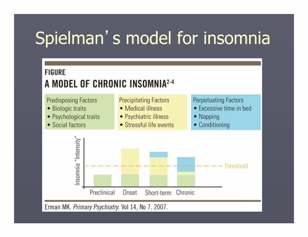
Spielman’s model for insomnia
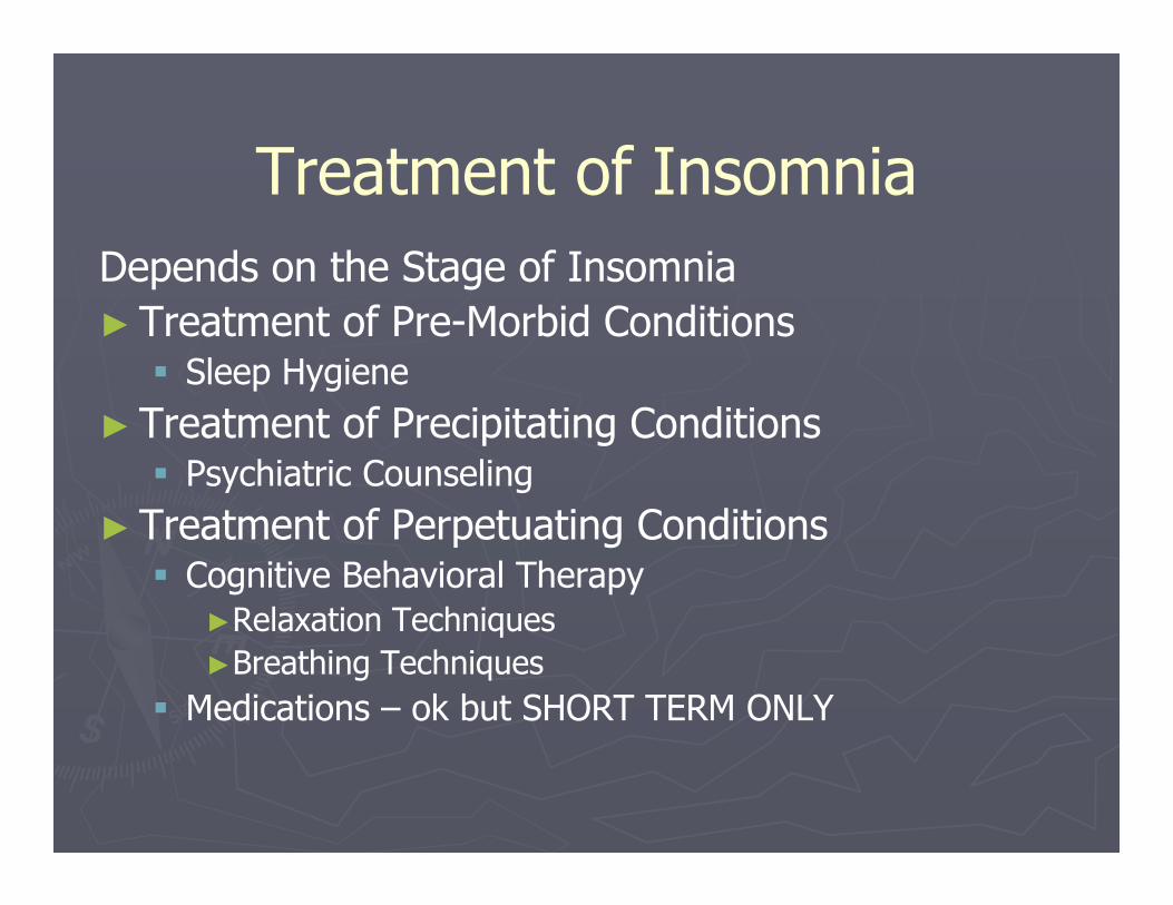
Treatment of InsomniaDepends on the Stage of Insomnia►Treatment of Pre-Morbid Conditions Sleep Hygiene
►Treatment of Precipitating Conditions Psychiatric Counseling
►Treatment of Perpetuating Conditions Cognitive Behavioral Therapy
►Relaxation Techniques►Breathing Techniques
Medications – ok but SHORT TERM ONLY

Good sleep hygiene tips

Insomnia inducing Rx


ICSD-3 (2014)

Case 2
73 yo RH German man PMH: HTN, HPL, CAD, CHF (EF 30%), V-
fib s/p ICD, paroxysmal AF Recent left MCA stroke secondary to AF-
related cardiac emboli with residual right HP and expressive aphasia

Case 2
Noted to have abnormal overnight oximetry while in stroke rehab
Evidence of periodic desaturations in saw-tooth pattern with lowest O2 saturation of 82%, a pattern suggestive of sleep apnea.

Split study

Case 2
Pt used CPAP 8 cm H2O No stroke recurrence
• Patient symptomatically improved, able to cooperate with rehabilitation
• NIHSS 125
Downloaded data showed good compliance (88%) and efficacy (AHI 453)
Sleep Related Breathing Disorders
►Obstructive Sleep Apnea Most common cause of EDS and sleep
disruption
►Central Sleep Apnea►Hypoventilation Syndromes

What is OSA?“… characterized by repetitive episodes of upper airway obstruction that occur during sleep, usually associated with a reduction in blood oxygen saturation…” with associated features of daytime sleepiness and snoring.

OSA Definitions• Obstructive Apnea – cessation of airflow for
10 s with continued respiratory effort.• Central Apnea – cessation of airflow for 10 s
without respiratory effort.• Obstructive Hypopnea – “some” reduction in
airflow for at least 10 s.- 30-50% reduction in airflow- associated with either an arousal or
desaturation (3-4%)

What is OSA Syndrome?
• Apnea – Hypopnea Index (AHI or RDI) ≥5 events/hour in conjunction with symptoms
• What is a relevant AHI?• Consensus Statement 1999: “RDI of 5 (or
greater) accompanied by symptoms...”Loube et al, Chest 1999
• Medicare 2014: AHI ≥ 5 with symptoms, or HTN, CAD or CVA

Prevalence of OSA
• Wisconsin Sleep Cohort Study• Population based study: 602 working
subjects, aged 30-60 years studied with PSG• Definition OSAS: AHI ≥5 and
hypersomnolenceF M
OSA 9% 24%OSAS 2% 4%
Young et al, NEJM 1993

Pathophysiology of OSA
• Narrowing or collapse of the upper airway • Decreased tidal volume hypercapnia and hypoxia• Increased respiratory effort• Arousal opens airway• Ensuing hyperpnea with hypocapnia and adequate oxygenation

Demographics of OSAS
• In younger, but not middle aged groups, OSAS has been reported to be more prevalent in AA’s compared to Caucasians
• Despite lower BMI, Asians have a predisposition of OSA thought to be due to cranio-facial features
• Prevalence of OSA increases with age

Risk Factors for OSA• Sleep Heart Health Study: male, age, BMI, neck girth,
snoring, and witnessed apnea predict AHI >15Young et al. Arch IM.2002
• Craniofacial abnormalities - nasal obstruction, enlarged uvula/tongue/tonsils, long soft palate, retrognathia, micrognathia, brachycephaly (flat posterior head)
• Family History (increases risk of OSAS 2-4 fold)• Co-morbid illness
• cardiopulmonary disease (CHF, OHV)• metabolic disorders (hypothyroidism, acromegaly)• neurologic disorders (CVA, neuromuscular disorders e.g.
MD)• Down’s syndrome (macroglossia)
• Environmental Factors - tobacco use, ETOH, sedatives

Symptoms/Signs of OSA
► Snoring► Witnessed apneas► Daytime sleepiness► Sleep fragmentation► Night sweats► Nocturia► Dry mouth/sore throat► Leg kicking while sleeping► Morning headaches► Mood changes► Decreased libido► Memory problems
► Obesity► Associated diseases
Hypertension Cardiac disease Stroke Glucose intolerance Hypothyroidism Acromegaly

STOP-BANG Questionnaire
Chung F et al. Anesthesiology. 2008 May;108(5):812-21

Clinical Examination• Vital signs (hypertensive, arrhythmias)• Obese (BMI >30)
• 40% of those with BMI >40 have OSAS and 50% of those with BMI >50 have OSAS
Kripke et al. Sleep 1997.• Neck circumference
• ≥40 cm associated with sensitivity of 61% and specificity of 93% for OSAS
• Men >17 inches, women >16 inches• Oral airway
• Retrognathia (narrows the upper airway behind the base of the tongue)• Dental malocclusion and overlapping teeth (indicated small oral cavity)• Macroglossia• Edema and erythema of the uvula• Elongated soft palate• Narrow high arched palate• Tonsillar hypertrophy• Lateral airway narrowing
• Nasal airway• Nasal valve collapse with sniff test• Nare size and asymmetry• Septal deviation• Enlarged inferior turbinates
Nasion
Gnathion
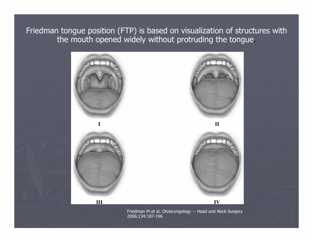
Friedman tongue position (FTP) is based on visualization of structures with the mouth opened widely without protruding the tongue.
Friedman M et al. Otolaryngology -- Head and Neck Surgery 2006;134:187-196

OSA – Example of a PSG
SpO2
Airflow
Thoracic
Abd

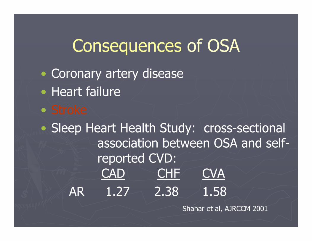
Consequences of OSA• Coronary artery disease• Heart failure• Stroke• Sleep Heart Health Study: cross-sectional
association between OSA and self-reported CVD:CAD CHF CVA
AR 1.27 2.38 1.58Shahar et al, AJRCCM 2001
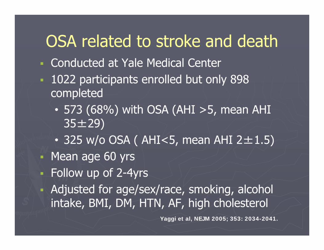
OSA related to stroke and death Conducted at Yale Medical Center 1022 participants enrolled but only 898
completed• 573 (68%) with OSA (AHI >5, mean AHI
35±29)• 325 w/o OSA ( AHI<5, mean AHI 2±1.5)
Mean age 60 yrs Follow up of 2-4yrs Adjusted for age/sex/race, smoking, alcohol
intake, BMI, DM, HTN, AF, high cholesterolYaggi et al, NEJM 2005; 353: 2034-2041.
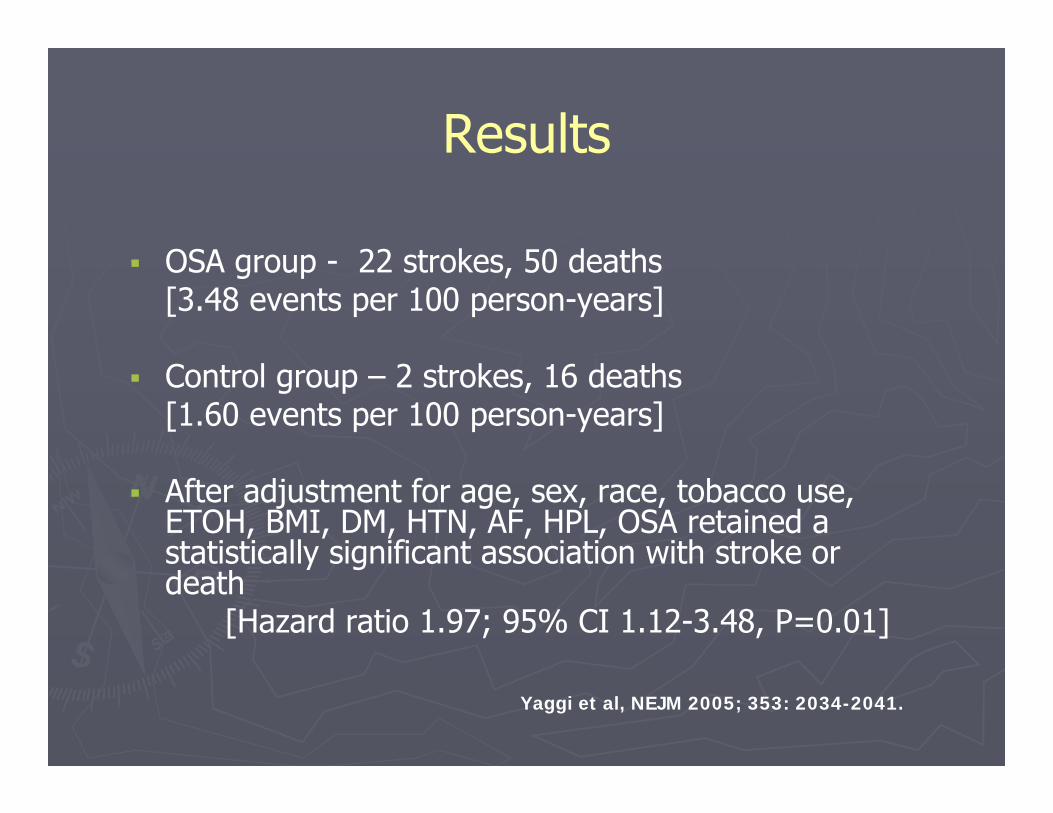
Results
OSA group - 22 strokes, 50 deaths[3.48 events per 100 person-years]
Control group – 2 strokes, 16 deaths[1.60 events per 100 person-years]
After adjustment for age, sex, race, tobacco use, ETOH, BMI, DM, HTN, AF, HPL, OSA retained a statistically significant association with stroke or death
[Hazard ratio 1.97; 95% CI 1.12-3.48, P=0.01]
Yaggi et al, NEJM 2005; 353: 2034-2041.

Trend analysis showed a step-wise increase in the risk of stroke/death as a function of increased severity of OSA (p=0.005)
The risk of stroke/death in pts in the most severe quartile of OSA was 3 x that in the controls
Yaggi et al, NEJM 2005; 353: 2034-2041.

Other Consequences of OSA
• Pulmonary HTN• Cor Pulmonale• Cardiac Arrhythmias (atrial fibrillation)• GERD• Increased frequency of seizures in
epileptics• Increased headache syndromes
(migraines)
Consequences of OSA
• Psychiatric/mood - depression, anxiety, irritability
• Social and sexual dysfunction• Neurocognitive impairment – general
intellectual ability, learning and memory, attention, information processing efficiency, visual and psychomotor performance

Consequences of OSA• Increased traffic accidents - case-controlled
study found those with AHI > 10 had OR of 6.3 for MVA
Teran-Santos et al, NEJM 1999
• Increased utilization of Health Care Services• Increased mortality - relative risk 2.7-3.3 • All of these adverse outcomes can be improved
by treatment
Treatment of OSA: Conservative Measures
• Weight loss • 10% weight loss leads to 26-50% decrease
in AHI• pharyngeal function improves as weight
decreases• extensive weight loss (i.e. following gastric
bypass surgery) may resolve OSA• almost always should be combined with
other therapies

Treatment of OSA:Conservative Measures
• Lateral positioning• Elevating the head of the bed• Avoiding upper airway irritants - tobacco• Minimizing sedating agents - alcohol,
sedatives
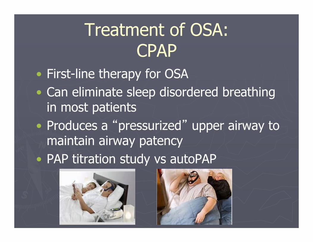
Treatment of OSA: CPAP
• First-line therapy for OSA• Can eliminate sleep disordered breathing
in most patients • Produces a “pressurized” upper airway to
maintain airway patency• PAP titration study vs autoPAP
Treatment of OSA:CPAP
• Benefits • decreases sleep-disordered breathing and EDS• improves oxygenation, exercise function • improves neuropsychiatric measures• decreases MVAs and hospitalizations• appears to decrease mortality
• Problems• acceptance suboptimal
• compliance poor at times but can overcome

Alternative treatment for OSA:Oral Appliances
• Relatively new therapy for OSA• Two categories:
Mandibular Advancing DevicesTongue Retaining Devices
• Work by enlarging the pharyngeal cross-sectional area
• Consider in patients with mild/moderate OSA• RCT suggest about equal efficacy to CPAP
with better tolerance

Alternative treatment of OSA
• Provent nasal strips
• Positional therapy
• Hypoglossal nerve stimulator

Treatment of OSA:Surgery
• Numerous approaches have been tried
• Surgical data limited• Procedures in general use:
• Nasal surgery• Tonsillectomy +/-
adenoidectomy• UPPP• Genioglossus advancement• Maxillomandibular Advancement
(MMA)• Tracheotomy

Treatment of OSA:Pharmacotherapy
• Little successes at this point in time
• “Some” efficacy may be present in thefollowing situations:Condition MedicationOHV MedroxyprogesteroneREM OSA SSRIs, TCAs
CHF Theophylline

ICSD-3 (2014)

Case 3
►17 yo M presents with EDS x 2 years►C/o decline in academic performance due to
falling asleep in classes ►Dx’d with ADD by PCP, rx’d Adderall 40 mg palipitations, HA
►Since age 12, he’s had multiple episodes of knees bucking and facial twitching with laughter
►When he woke up, he felt paralyzed for 30 seconds, couldn’t speak
Case 3
►He reported seeing little minions running around his room right before falling asleep
►He would finish chores without recollection of doing them
►He would nap throughout the day (10-20 min each) and noted vivid dreams with all naps
►No recent head injury, no drug/substance abuse

Case 3
►Bedtime routine:►BT: 23:00►SL: minutes►Awakenings: 2-5 times, unclear reasons►WT: 6:30, snoozes alarm multiple times
►Exam: normal but you crack a joke and he slumps over for 15 secs (no LOC, no DTR’s)
►MRI negative for hypothalamic lesions

Case 3►PSG: SL 5 min, normal AHI, fragmented
sleep►MSLT
►Napped during 5 nap trials►Mean sleep latency: 4.5 minutes►+ SOREMP in 3 (REM <15 min)►Reported vivid dreams in 3 naps
►Negative urine drug screen prior to MSLT (off stimulants x 2 weeks)
►CSF hypocretin-1 assay 90 pg/ML►+ HLP DQB1*0602
►+90% of narcolepsy 1 pts, 25% general population

Differential Diagnosis?

Case 3
►Narcolepsy type 1 ►EDS >3 months►Cataplexy►Sleep paralysis►Hypnagogic hallucinations►Automatic behavior►Vivid dreams shortly after sleep onset
►+ MSLT with negative drug screen, low hypocretin-1 assay levels, + HLA haplotype

Case 3
►Pt was started on Provigil 200 mg qAM, with extra 200 mg at noon prn with drop in is ESS from 18 to 9
►Started on Effexor for cataplexy (down from 5 episodes a week to 0-1, could attend comedy shows now)
►Grades improved, feeling better
NarcolepsyA central nervous system disorder that is an important cause of persistent sleepiness.
The second most common cause of disabling daytime sleepiness after sleep apnea.*Should be on differential for syncope!

Epidemiology/Prevalance• Affects 1 in 200 people in Western Europe and North America
• Prevalence men = women
• Typically begins in the teens and early 20’s, but can occur as early as age 5 or age 40
• Symptoms may worsen over the first few years and then persist for life
• Half of patients report that symptoms interfere with job, marriage or social life

Gelineau (1862) applied the term "narcolepsy" to a clinical syndrome of daytime sleepiness with…
- Hypnagogic hallucinations (vivid, often frightening hallucinations that occur just as the patient is falling asleep)
- Sleep paralysis (complete inability to move for 1-2 minutes after awakening)
- Cataplexy (sudden episodes of bilateral muscle weakness leading to partial or complete collapse; often triggered by strong emotions, last 1-2 minutes with preserved LOC)
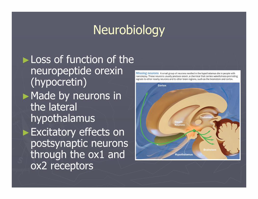
Neurobiology
►Loss of function of the neuropeptide orexin (hypocretin)
►Made by neurons in the lateral hypothalamus
►Excitatory effects on postsynaptic neurons through the ox1 and ox2 receptors

Genetic factors
►Usually sporadic, but genetic factors play important role
►Most narcoleptics (50-90 percent) have HLA DR2 and DQ1
►Environmental factors appear to be even more important: only about 25 percent of affected monozygotic twins are concordant for narcolepsy
►On rare occasions, narcolepsy runs in families.

Narcolepsy +/- Cataplexy
► ICSD-2 diagnostic criteria Complaint of EDS almost daily for >3 months +/- cataplexy
►Sudden transient weakness of muscles during periods of stress, great emotion (buckling of knees, facial weakness, drop attacks)
Diagnosis confirmed by noctural PSG followed by MSLT►MSL is ≤ 8 minutes►2 or more SOREM
Alternatively, CSF hypocretin-1 levels ≤110 can be used to confirm diagnosis

ICSD-3: Narcolepsy, type 1 and 2►Narcolepsy type 1 (with hypocretin
deficiency) Both criteria must be met:
►Daily periods of irrepressible need to sleep or daytime lapses into sleep, occurring for at least 3 months
►Presence of one or both: Cataplexy and a MSL of up to 8 min or 2+ SOREMP (15
min) on MSLT (1 SOREMP on preceding PSG can count as one)
CSF hypocretin-1 concentration is either up to 110 picograms/ml measured by immunoreactivity or <1/3 of mean values obtained in normal subjects with the standardized assay

ICSD-3: Narcolepsy, type 1 and 2►Narcolepsy type 2 (without hypocretin
deficiency) All 5 of the following must be met:
►Daily pds of irrepressible need to sleep or EDS >3 mos
►MSL ≤8 min or 2 or more SOREMP (15 min) on MSLT (1 SOREMP on preceding PSG can count)
►Cataplexy is absent►CSF hypocretin-1 concentration is >110
picograms/ml measured by immunoreactivity or >1/3 of mean values obtained in normal subjects with the standardized assay
►No other causes (OSA,DSPS, rx/substance)

MSLT
► Full night PSG is performed prior► A patient is given four or five opportunities to nap
every two hours► On average, healthy subjects fall asleep in about 10-15
minutes► People with narcolepsy often fall asleep in less
than five minutes► The naps of narcoleptics often include REM
sleep► Occurrence of sleep onset REM periods
(SOREMs) in two or more naps is an essential feature in establishing the diagnosis of narcolepsy
Drug Effects…
►REM sleep-suppressing medications (TCAs, SSRIs) or withdrawal from these drugs also can produce SOREMs ("rebound" phenomenon)
►Stimulants obscure results►These drugs should be discontinued at
least three weeks before the MSLT if possible
DIFFERENTIAL DIAGNOSIS
►With Cataplexy: Hypothalamic lesions Prader-Willi syndrome Niemann-Pick disease type C Norrie disease
►Without Cataplexy:- OSA- PLMD- Idiopathic hypersomnia
TREATMENT
►Mainstays of therapy are -Stimulants for the treatment of sleepiness-REM sleep-suppressing medications for the treatment of cataplexy
►Napping and sleep hygiene►Psychosocial support

Medication
► Amphetamines (methylphenidate, dextroamphetamine) Oldest, used since 1930’s
► Modafinil (200-400 mg qAM) SE: HA, n/v, dry mouth, anorexia, diarrhea Lack of sympathomimetic effects makes it ideal for
older pts with HTN, CAD► Gamma hydroxybutyrate (Xyrem)
2002 –approved by FDA for treatment of cataplexy Metabolite of GABA, mechanism unknown Can also improve EDS SE: 14% UT, somnambulism, n/v Potential for abuse, overdose can be fatal


BUMC-P Sleep • Clinic – Neuroscience clinic in Rehab
building• Sleep lab – West tower, 1st floor• We see everything – insomnia, OSA, CSA,
narcolepsy, RBD, nocturnal epilepsy, etc• Office number: 602-351-2200 • Cyrus Guevarra (sleep lab manager)• Crystal McDonald (field representative) • Email me: joyce.lee-


Summary►Sleep Medicine is a relatively new field►Normal Sleep is dictated by homeostatic
pressure and circadian rhythms►PSG is the gold standard for diagnosis of
most sleep disorders►Insomnia is the most common sleep
disorder, but OSA is the most common cause of EDS
►Narcolepsy is not common, but can be debilitating

Sleep is cool, dude~We welcome
rotators all the time

QUESTIONS??