Basic Neuroscience Lecture 1
47
Basic Neuroscience: Neuroanatomy Dr. Bodnar: Lecture 1 Gross Brain Neuroanatomy
-
Upload
monster40lbs -
Category
Documents
-
view
14 -
download
0
description
Introductory PDF for Neuroanatomy
Transcript of Basic Neuroscience Lecture 1

Basic Neuroscience: Neuroanatomy
Dr. Bodnar: Lecture 1
Gross Brain Neuroanatomy

Human Brain• Weighs ~1400 grams; 2% of body weight• Consumes 20% of oxygen• Requires 17% of cardiac output• Blood flow: ~750 ml/min• Oxygen consumption: 46 ml/min

Planes of Section• Dorsal or Posterior: towards the back• Superior: towards the vertex of skull• Ventral or Anterior: towards the stomach• Inferior: towards the palate

Axes of Central Nervous System
• Vertical axis: parallel to spinal cordUse dorsal (posterior) for vertical axisUse ventral (anterior) for vertical axis
• Horizontal axis: parallel to eye socketsUse superior for horizontal axisUse inferior for horizontal axis
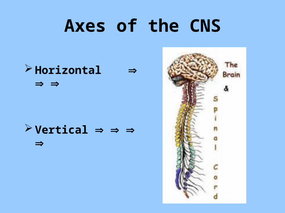
Axes of the CNS
Horizontal
Vertical

Other Directions
• Rostral: towards the head (vertical)• Rostral: towards the nose (horizontal)• Caudal: towards the tail (vertical)• Caudal: towards the occiput (horizontal)• Medial: towards the midline or middle• Lateral: towards the outside

Horizontal Coronal Sagittal
CNS Axes and Cuts

Horizontal Cuts• HorizontalParallel to dorsal-ventralPerpendicular to rostral-caudal and medial-
lateral

Coronal Cuts• CoronalParallel to rostral-caudalPerpendicular to medial-lateral and
dorsal-ventral

Sagittal Cuts
SagittalParallel to medial-lateralPerpendicular to rostral-caudal and
dorsal-ventral

Oblique CutsObliqueAny other angular cut; observed in virtually
all CAT, PET or MRI scans
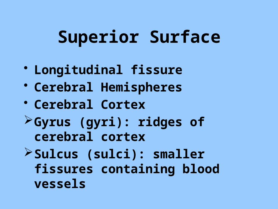
Superior Surface
• Longitudinal fissure• Cerebral Hemispheres• Cerebral CortexGyrus (gyri): ridges of cerebral cortexSulcus (sulci): smaller fissures containing
blood vessels
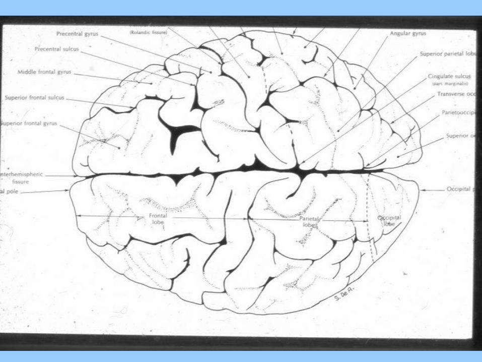

Central Sulcus or Rolandic Fissure
• Rostral to Central Sulcus: Immediate: Pre-Central Gyrus (Primary Motor
Cortex) General: Frontal Lobe• Caudal to Central Sulcus: Immediate: Post-Central Gyrus (Primary
Somatosensory Cortex) General: Parietal Lobe• No sulcus on superior surface demarcating
occipital lobe


Lateral Surface
• Central SulcusSeparates frontal and parietal lobe on lateral
surface• Lateral sulcus or Sylvian fissureSeparates temporal lobe from frontal and
parietal lobes on lateral surface• No sulcus demarcating occipital lobe on
lateral surface


Frontal Lobe and Lateral Surface
• Pre-Central Gyrus Pre-Central Sulcus
• Superior Frontal Gyrus Superior Frontal Sulcus
• Middle Frontal Gyrus Inferior Frontal Sulcus
• Inferior Frontal Gyrus
• Inferior Frontal GyrusPars opercularisPars triangularisPars orbitalis (Broca’s area)
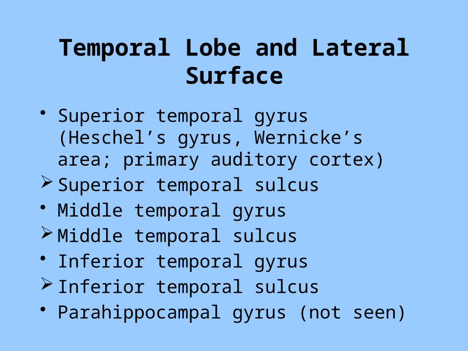
Temporal Lobe and Lateral Surface
• Superior temporal gyrus (Heschel’s gyrus, Wernicke’s area; primary auditory cortex)
Superior temporal sulcus• Middle temporal gyrus Middle temporal sulcus• Inferior temporal gyrus Inferior temporal sulcus• Parahippocampal gyrus (not seen)
Parietal and Occipital Lobes and Lateral Surface
• Parietal Lobe• Post-central gyrus• Superior Parietal Lobule (Temporo-parietal
auditory Association area)• Inferior Parietal Lobule (Temporo-occipital
visual Association area)• Occipital Pole (Primary Visual Cortex)


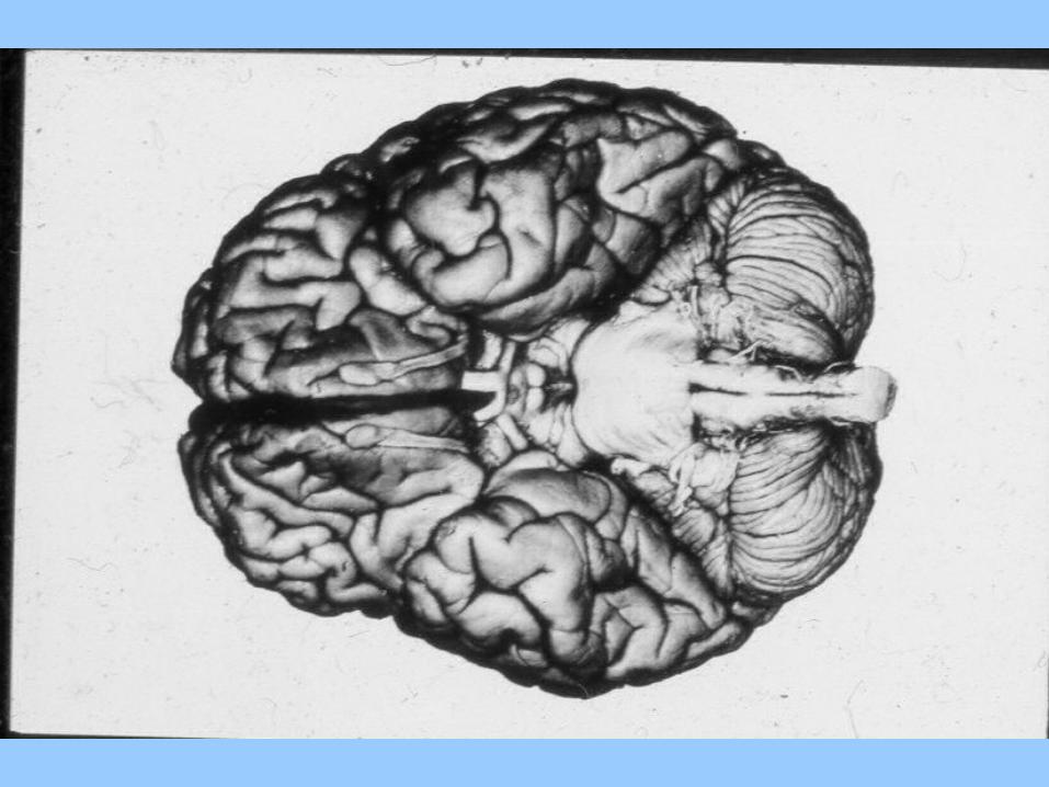
Inferior Surface
• Encephalic Levels and Development• Cranial Nerves• Hypothalamo-Hypophysial Axis• Frontal Lobe: Orbital gyri and Longitudinal
Fissure • Temporal Lobe: Parahippocampal Gyrus and
Uncus• Cerebral and Cerebellar Peduncles• Pyramids and Olives

Encephalic Development
• Prosencephalon Telencephalon: Cortex, Basal Ganglia and Limbic
System Diencephalon: Thalamus and Hypothalamus• Mesencephalon: Midbrain• Rhombencephalon Metencephalon: Pons Mylencephalon: Medulla and Cerebellum

Cranial Nerves 1-6
Number Name Level Type Path
1 Olfactory Telenceph. Sensory (Smell)
Enters Medial
2 Optic Dienceph. Sensory (Vision)
Enters Medial
3 Oculomotor Mesenceph. Motor (Eye Movements)
Exits Medial
4 Trochlear Mesenceph. Motor (Eye Movements)
Exits Medial
5 Trigeminal Metenceph. Mixed Lateral Path
6 Abducens Metenceph-Mylenceph.
Motor (Eye Movements)
Exits Medial
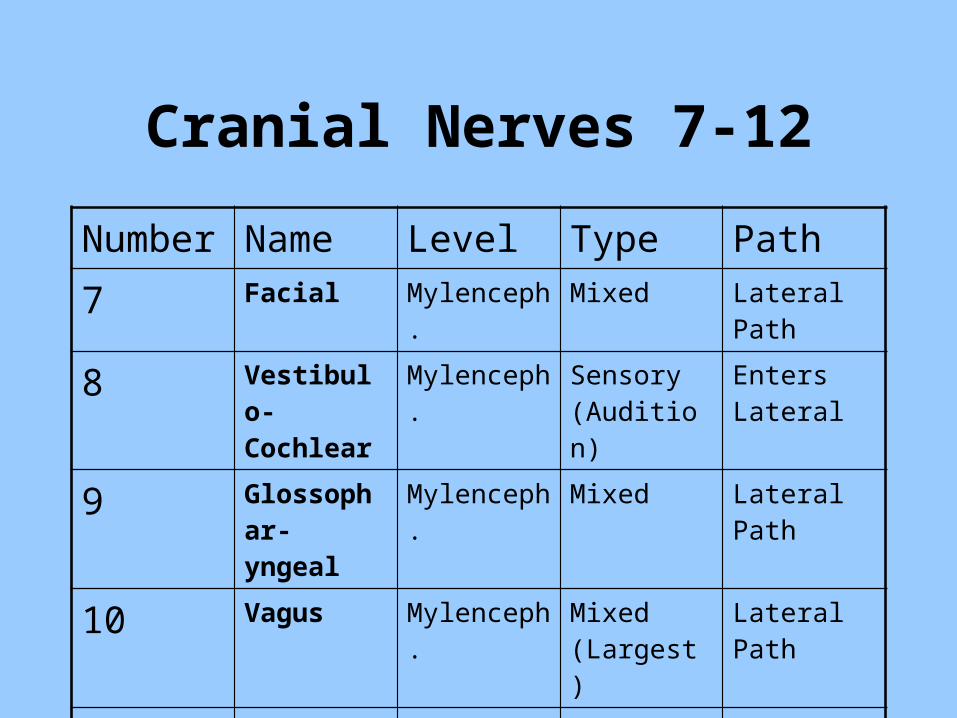
Cranial Nerves 7-12
Number Name Level Type Path
7 Facial Mylenceph. Mixed Lateral Path
8 Vestibulo-Cochlear
Mylenceph. Sensory (Audition)
Enters Lateral
9 Glossophar-yngeal
Mylenceph. Mixed Lateral Path
10 Vagus Mylenceph. Mixed (Largest)
Lateral Path
11 Spinal Accessory
Mylenceph. Motor (Smallest)
Exits Lateral
12 Hypoglossal Mylenceph. Motor Exits Medial
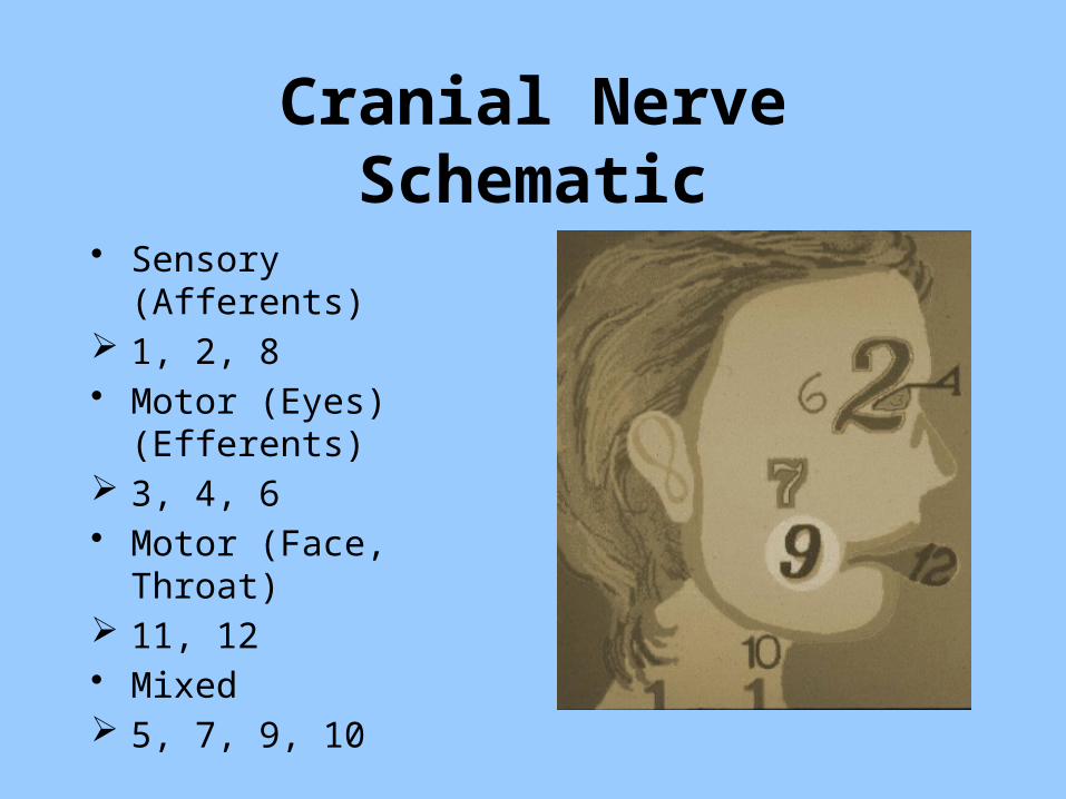
Cranial Nerve Schematic
• Sensory (Afferents) 1, 2, 8• Motor (Eyes) (Efferents) 3, 4, 6• Motor (Face, Throat) 11, 12• Mixed 5, 7, 9, 10


Cerebellar Peduncles
• Superior Cerebellar Peduncle or Brachium Conjunctivum: connections to thalamus and basal ganglia
• Middle Cerebellar Peduncle or Brachium Pontis: connections to ventral midbrain, pons and medulla
• Inferior Cerebellar Peduncle or Restiform Body: afferent connections from spinal cord

Cerebral Peduncle and Pyramids
• Cortico-spinal tractPre-Central gyrus (telencephalon)Internal capsule and Crus cerebri
(diencephalon)Cerebral peduncle (mesencephalon)Cortico-spinal tract (metencephalon)Pyramids and Pyramidal decussation
(mylencephalon)

Hypothalmo-Hypophysial Portal System
• Paraventricular & Supraoptic Hypothalamus Neurohypophysial System: Magnocellular-
Median eminence, zona interna to infindibular stalk to posterior lobe- Vasopressin, Oxytocin release into blood.
Adenohypophysial System: Parvocellular- Median eminence, zona externa (hypothalamic releasing factors: e.g., LHRH) to portal system to anterior and intermediate lobe (pituitary releasing factors: e.g., LH) into blood acting on target (e.g., ovary- progesterone).
Medial Surface
• Mid-sagittal cut• Central sulcus• Septum pellucidum• Pineal gland• Calcarine sulcus: Macula and Primary
Visual Cortex• Three Means of Interhemispheric
Communication


Means of Interhemispheric Communication
• Corpus callosumFrontal, parietal, temporal & occipital lobes• Anterior commissureMedial temporal lobe structures• Posterior commissureMedial parietal and mesencephalic
subcortical structures
Somatosensory Systems
• Dorsal Column System, Anterior Spinothalamic and Lateral Spinothalamic pathways from Medulla to Thalamus
• Somatosensory: Dorsal or Posterior Path• Motor: Ventral or Anterior Path

Circumventricular Structures
• Lateral VentriclesDorsal: Corpus Callosum and Cingulate
Gyrus (limbic system)Ventral: Fornix (hippocampal-septal and
mammillary body interconnections)Medial: Septum PellucidumLateral: Caudate Nucleus of the Basal
Ganglia


Superior View without Cortex
• Thalamus, Medial Geniculate Body and Pineal Gland
• Corpus quadragemini Superior Colliculus or Optic Tectum Inferior Colliculus or Auditory Tectum• Cerebellar Peduncles• Gracile and Cuneate Tubercles and Fasciculi• Obex

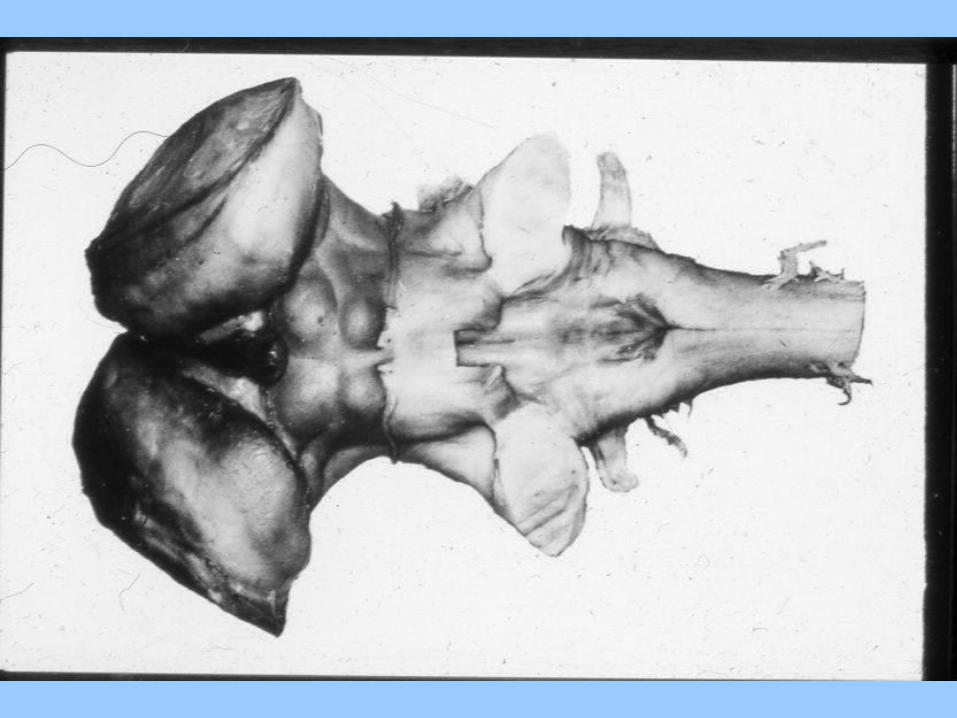
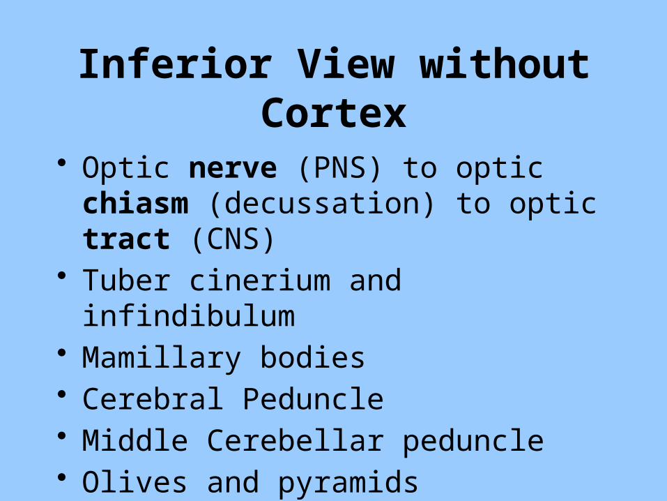
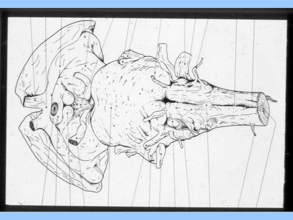
Inferior View without Cortex
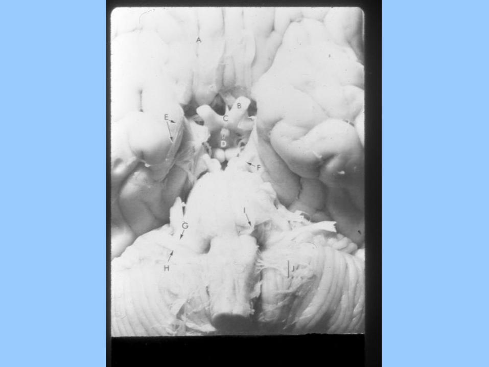
• Optic nerve (PNS) to optic chiasm (decussation) to optic tract (CNS)
• Tuber cinerium and infindibulum• Mamillary bodies• Cerebral Peduncle• Middle Cerebellar peduncle• Olives and pyramids


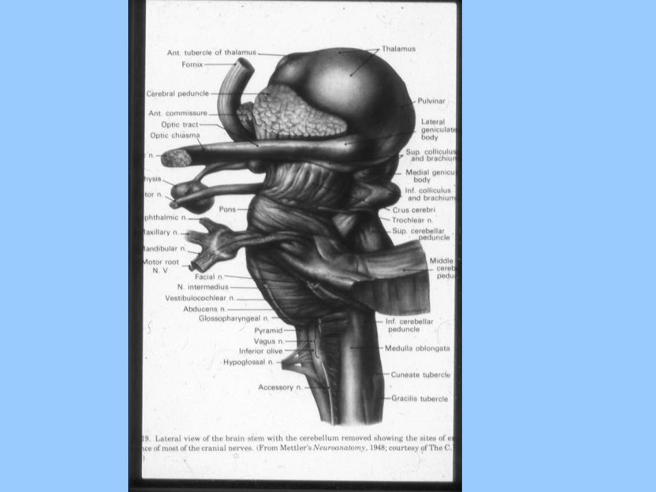
Lateral Surface without Cortex or Cerebellum
• Thalamus• Fornix• Cerebral Peduncle• Optic Tract• Hypophysis• Oculomotor nerve• Trigeminal nerve
• Crus Cerebri• Superior and Inferior Colliculi• Superior, Middle and Inferior Cerebellar
Peduncles• Gracile and Cuneate nuclei• Olives and Pyramids