Basic Canine NeuroAnatomy and MRI Imaging Planes Sagittal...
5
Basic Canine NeuroAnatomy and MRI Imaging Planes Sagittal - slice (blue line) thru the length of the body dividing it into left and right sides. Transverse (axial or cross-section) – slice (blue line) cuts across the body Sagittal of the spine Transverse spine
Transcript of Basic Canine NeuroAnatomy and MRI Imaging Planes Sagittal...

Basic Canine NeuroAnatomy and MRI
Imaging Planes
Sagittal - slice (blue line) thru the length of
the body dividing it into left and
right sides.
Transverse (axial or cross-section) – slice (blue line) cuts across the body
Sagittal of the spine
Transverse spine

Canine Spine
The cervical spine is located in the neck.
The thoracic spine is located in the chest. Ribs attach to the thoracic vertebrae.
The lumbar spine is the lower back area.
The sacral spine is in the tail.
Cervical
Spine
Thoracic
Spine
Lumbar
Spine Sacral
Spine

MRI Basics
Magnetic Resonance Imaging (MRI) does not use radiation,
and there are no known side effects. A very strong magnet is
used to align the hydrogen protons in the body. By applying a
radio frequency (RF)pulse, the protons can be flipped into a
different plane. As they realign back to the original plane, they
give off a signal that is read by the MR computer, and
reconstructed into an image. Depending on the timing of the
RF pulses different types of signals are generated that create
different types of images. The most common are T1 and T2.
T1 and T2
T1 images typically provide better imaging of anatomy and bone. CSF is dark on a T1 image.
T2 images typically provide better imaging of fluid and pathologies (although bone is visible). CSF is
white on a T2 image.
On Left: On Right:
T2 image T1 image
White CSF Dark CSF
T2 shows CSF better than a T1, therefore it shows a syrinx better. The T2 will also show
hydrocephalus and if the 4th ventricle is compressed (the 4th ventricle should be a little triangle in
front of the cerebellum and the brainstem).
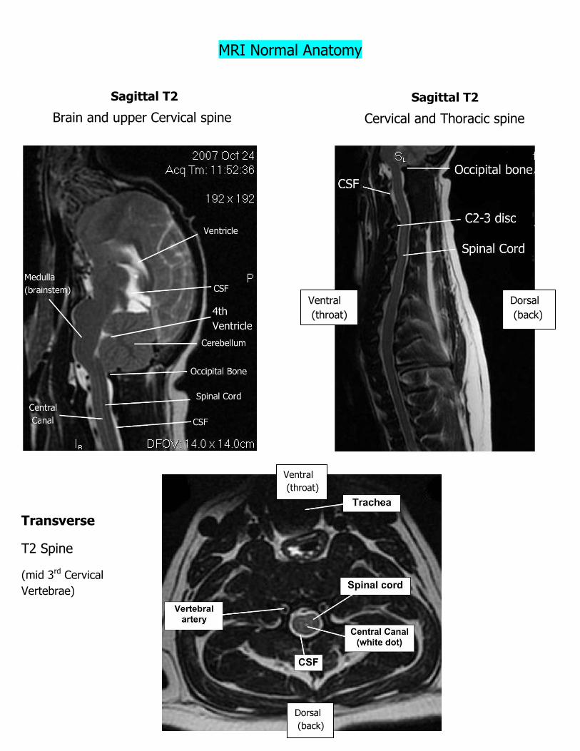
MRI Normal Anatomy
Sagittal T2
Brain and upper Cervical spine
Dorsal
(back) 4th
Ventricle
Sagittal T2
Cervical and Thoracic spine
Ventral
(throat)
Transverse
T2 Spine
(mid 3rd Cervical
Vertebrae)
Ventral
(throat)
Dorsal
(back)

Glossary of Anatomical Directions
Term Equivalent Term Meaning Short Form
Dorsal Posterior Toward the back
Ventral Anterior Toward the front (belly, abdomen, throat)
Cranial Cephalad Toward the head
Caudal Toward the tail
Rostral Toward the nose
Lateral Toward the side
Medial Toward the midline
Transverse Axial Cross section Tran
Sagittal Sag
Cervical spine Vertebrae in neck C spine
Thoracic spine Vertebrae in chest/ mid back
T spine
Lumbar spine Vertebrae in lower back L spine
Cervical + Thoracic spine CT spine
Thoracic + Lumbar spine TL spine
cranial
caudal
dorsal
ventral