Bacteria §Cells are prokaryotic and amongst the smallest known cells ( length 0.5-20 µm ). §No...
32
-
Upload
mervin-knight -
Category
Documents
-
view
217 -
download
1
Transcript of Bacteria §Cells are prokaryotic and amongst the smallest known cells ( length 0.5-20 µm ). §No...


Bacteria
Cells are prokaryotic and amongst the smallest known cells ( length 0.5-20 µm ).
No membrane bound nucleus.Have ribosomes,but no other organelles.DNA present as a long circular molecule.( see handout for bacterial structure)

Habitat
Bacteria are found everywhere.AirWaterSoilOn plants and animals.In plants and animals.

Types of Bacteria
There are three major groups of bacteria based on their shapes; coccus, bacillus, and spirillum.
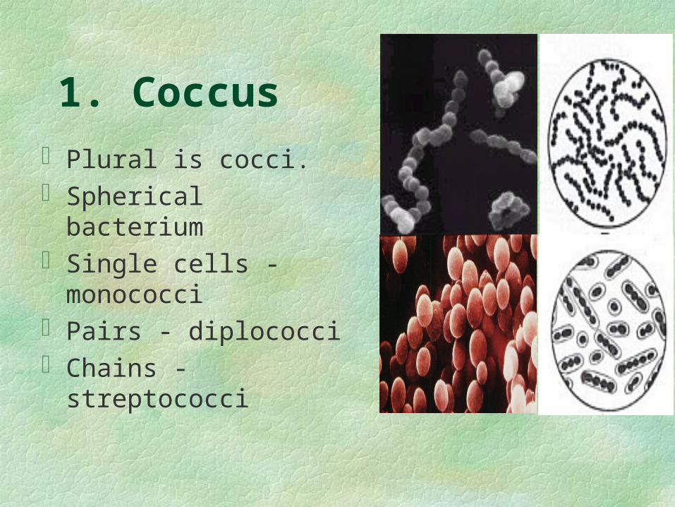
1. Coccus
Plural is cocci.Spherical bacteriumSingle cells - monococciPairs - diplococciChains - streptococci

2.BacillusPlural is bacilli.A rod - shaped bacterium.Exists as single cells, in
pairs (diplobacilli), and in chains (streptobacilli).

3. SpirillumPlural is spirilli.A spiral - shaped
bacterium.Exist only as single
cells.

Bacterial Structure
Cell Wall - outermost structure of the cell.
Made up of a substance called peptidoglycan ( a long chain of sugars linked to amino acids ).
Penicillin destroys bacteria by interfering with the peptidoglycan molecules.

Bacterial Structure Continued
Cell Membrane - Found beneath the cell wall.
May be folded inward.

Bacterial Structure Continued
Capsule: This is a layer of slime secreted over the cell wall of the bacterium.
The capsule provides protection for the bacterium.
Bacteria that have capsules are said to be encapsulated.
Most pathogenic bacteria are encapsulated.

Bacterial Structure Continued
Flagella - Some bacteria in the bacilli and spirilli categories can move by way of flagella ( whip - like structures that propel the bacteria).
Note: Cocci do not have flagella.
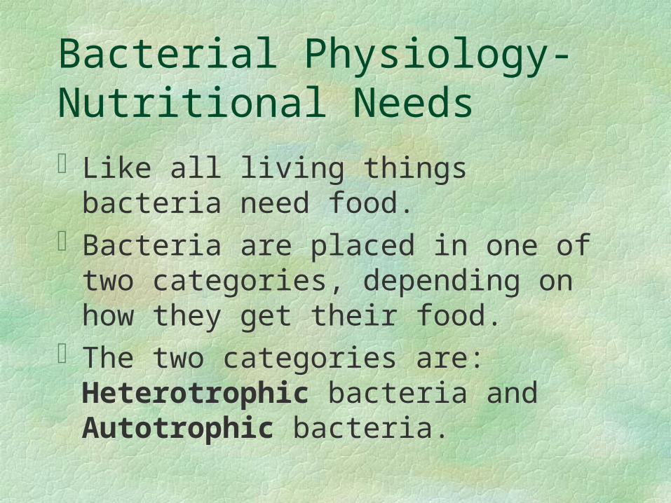
Bacterial Physiology-Nutritional NeedsLike all living things bacteria need food.Bacteria are placed in one of two
categories, depending on how they get their food.
The two categories are: Heterotrophic bacteria and Autotrophic bacteria.

Heterotrophic Bacteria
Heterotrophs must get their food from a source of pre-formed organic matter:
(A) Saprobes- feed on the remains of dead plants and animals.
(B) Parasites - live on or in the organism and cause disease. For example, Mycobacterium tuberculosis.

Autotrophic Bacteria
Autotrophs can make their own food:
Photosynthetic-use a special type of chlorophyll called bacteriochlorophyll.
O2 is not released in bacterial photosynthesis
Chemosynthetic -obtain energy by breaking down inorganic material such as iron or sulfur.
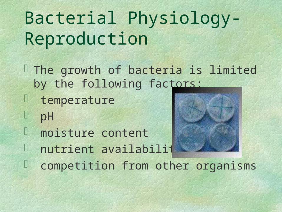
Bacterial Physiology-Reproduction
The growth of bacteria is limited by the following factors:
temperature pH moisture content nutrient availability competition from other organisms

Bacterial Physiology-Reproduction continued
Bacteria reproduce asexually by binary fission. Using this process bacteria reproduce about every 20 minutes.
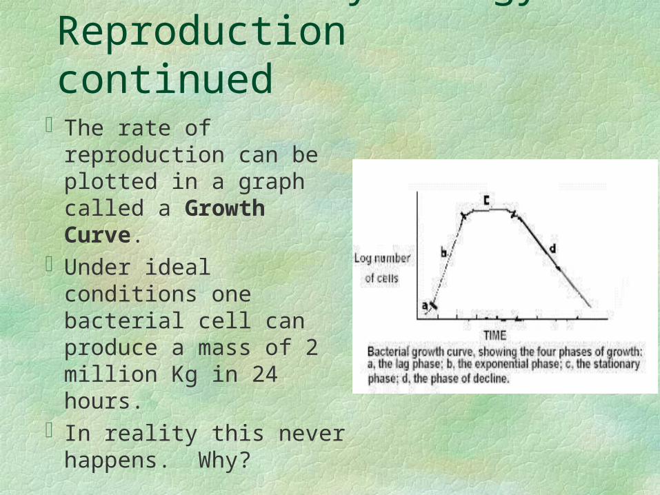
Bacterial Physiology-Reproduction continuedThe rate of
reproduction can be plotted in a graph called a Growth Curve.
Under ideal conditions one bacterial cell can produce a mass of 2 million Kg in 24 hours.
In reality this never happens. Why?

GRAM STAINING

REAGENTS USED IN GRAM STAIN
1. CRYSTAL VIOLET Primary stain Violet colored, stains all micro-org2. GRAM IODINE Mordant Forms Crystal violet iodine complexes3. DECOLORIZER Acetone + Methanol Removes Crystal violet iodine complex
from thin peptidoglycan layers Dissolves outer layer of Gram negative
org

REAGENTS USED IN GRAM STAIN
4. GRAM SAFRANINE Counter stain Red colored Stains thin walled
Gram neg org Pus cells cytoplasm &
lobes of nuclei also stain red

The Gram Stain ProcedureStep 1 - Prepare a Smear
Watch what happens to the “Bacteria” at each step
“Bacteria”
Suspend some of the material to be stained in a drop of water on a microscope slide, spread the drop to about the size of a nickel.
Allow to air dry. Heat fix by gently warming

The Gram Stain ProcedureStep 2 - Apply the Primary Stain
Flood the Smear with Crystal Violet
Allow to stand for 1 min
Rinse with water to remove excess stain

The Gram Stain ProcedureStep 3 - Apply the Mordant
Flood the Smear with Iodine solution
Allow to stand 2 min

The Gram Stain ProcedureStep 4 - Rinse
Rinse with water to remove excess Iodine

The Gram Stain ProcedureStep 5 - Decolorize
Drip Decolorizer (80% Methanol +20% Acetone) across the slide about 5 sec
The effluent should appear pale or clear
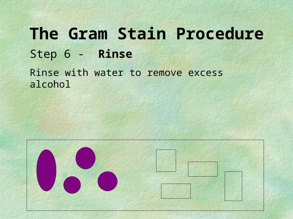
The Gram Stain ProcedureStep 6 - Rinse
Rinse with water to remove excess alcohol

The Gram Stain ProcedureStep 7 - Counterstain
Flood the slide with Safranin solution
Let stand for 2 minutes
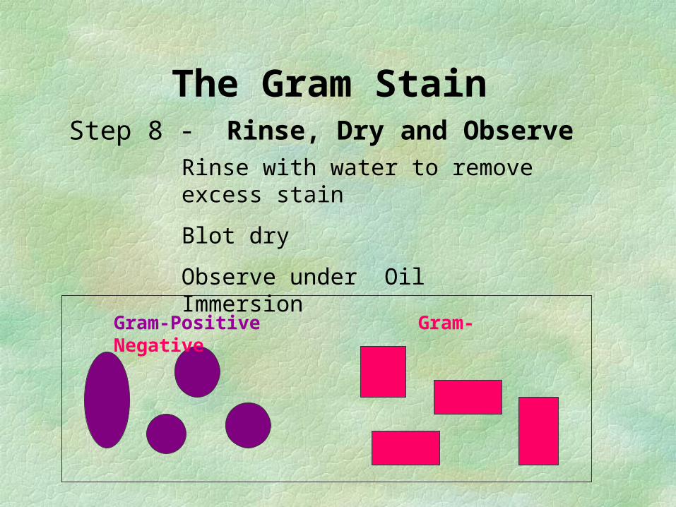
The Gram Stain
Step 8 - Rinse, Dry and Observe
Gram-Positive Gram-Negative
Rinse with water to remove excess stain
Blot dry
Observe under Oil Immersion

CELL WALL OF GRAM POS & NEG

CELL WALL OF GRAM POS & NEG

GRAM + & GRAM – BACTERIACell Wall Structures Gram Positive
organismsGram Negative organisms
Inner cytoplasmic membrane
Present Present
Peptidoglycan layer Thick Thin
Teichoic Acid Present Absent
Outer membrane layer Absent Present
Lipid A, LPS , Lipo-protien components
Absent Present
Peri-plasmic space Absent Present
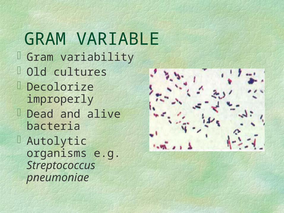
GRAM VARIABLEGram variability Old culturesDecolorize
improperlyDead and alive
bacteriaAutolytic organisms
e.g. Streptococcus pneumoniae