Infections of the Central Nervous System MLAB 2434 – Microbiology Keri Brophy-Martinez.
Upload
benedict-alexanderCategory
view
229download
2
Bacteremia
MLAB 2434 –Microbiology Keri Brophy-Martinez

Definitions
PseudobacteremiaFalse bacteremiaContamination of a blood culture
during or after collection

Definitions
Bacteremia – presence of bacteria in blood stream Some conditions have a period of
bacteremia as part of the disease process (ex. Meningitis, endocarditis)
Usually occurs due to a disruption of skin or mucosal barriers to bacterial invasion

Classifications of Bacteremia
Classified by Site of Origin Classified by Causative Agent Classified by Place of Acquisition Classified by Duration

Classification by Site of Origin
Primary Bacteremia Blood stream or endovascular bacterial
invasion with no preceding or simultaneous site of infection with the same microorganism
Secondary Bacteremia Isolation of a microorganism from
blood as well as other site(s) Fever of Unknown Origin (FUO)
Source unknown

Classification by Causative Agent
Gram positive bacteremia Gram negative bacteremia Anaerobic bacteremia Polymicrobial bacteremia

Classification by Place of Acquisition
Community-acquired
Health-care acquired/NosocomialDefined as occurring 72 hours
post admission

Classification by Duration Transient
Comes and goes Usually occurs after a procedural
manipulation (ex. Dental procedures) Intermittent
Can occur from abscesses at some body site that is “seeding” the blood
Continuous Bacteremia Organisms from an intravascular
source that are consistently present in bloodstream

Sepsis & Septicemia
Presence of active bacteria Results from continuous bacteremia Clinical signs and symptoms of bacterial
invasion and toxin production Apply the SIRS criteria
Systemic response to bacterial infection

Septic shock Results from body’s reactions to
bacterial bi-products• Endotoxins: lipopolysaccharide• Exotoxins
Disrupts many body functions• Hemodynamic changes, decreased
tissue perfusion and compromised organ & tissue function
Mortality 40% to 50%
Bacteremia Complications

Bacteremia/Septicemia Risk Factors Immunocompromised patients
Due to decrease in circulating neutrophils Increased use of invasive procedures &
indwelling devices Disrupts normal flora
Age of patient Young: defect in humoral immunity Old: Decreased immune competency
Administration of drug therapy Broad spectrum antibiotics decrease
normal flora Increase in antimicrobial resistance

Sources of Bacteremia
Pericarditis and Peritonitis Pneumonias Pressure sores Prosthetic medical devices Total hip replacement Skeletal system Skin and soft tissue Urinary Tract Infections

Clinical Signs and Symptoms Abrupt onset of chills, fever, or
hypothermia and hypotension Prostration (exhaustion/weakness) and
diaphoresis (perspiration) Tachypnea (rapid breathing) is an
early sign of bacteremia Delirium, stupor, agitation Nausea, vomiting

Clinical Signs and Symptoms (cont’d) Laboratory Values in Bacteremia
Thrombocytopenia Leukocytosis or leukopenia Acidosis Abnormal liver functions Coagulopathy DIC Elevations in CRP, haptoglobin,
fibrinogen, ESR, procalcitonin
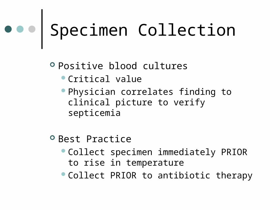
Specimen Collection
Positive blood culturesCritical valuePhysician correlates finding to clinical
picture to verify septicemia
Best PracticeCollect specimen immediately PRIOR
to rise in temperatureCollect PRIOR to antibiotic therapy

Specimen Collection
Aseptic collection procedure is critical Cleansing agents
• Tincture of iodine (1-2%)• Leave on skin for 30 seconds
• Povidine-iodine (10%)• Leave on skin 1.5 to 2 minutes
• Chlorhexidine/ChloraPrep• Leave on skin for 30 seconds• 2% chlorhexidine gluconate + 70% isopropyl alcohol
Cleansing Technique• In concentric fashion, from inside to out• After cleaning, wait 1.5-2 minutes
Acceptable Contamination Rate• 1-3%

Collection sites
PreferredPeripheral venousArterial sites
Less commonCentral venous cathetersArterial lines

Blood Collection Devices
Traditional set Aerobic bottle
• Selects for aerobic & facultative anaerobes
Anaerobic bottle• Selects for obligate anaerobes
ARD bottle (Antibiotic Removal Device) Used when patient is on antibiotics
prior to blood collection
SPS= Sodium polyanetholsulfonate

Blood Collection Devices
Anticoagulants SPS= Sodium polyanetholsulfonate
• Function/Purpose• Anticoagulant• Neutralizes human serum• Prevents phagocytosis• Inactivates certain antimicrobial agents
SAS(sodium amylosulfate)• Similar to SPS, but less effective in neutralizing serum
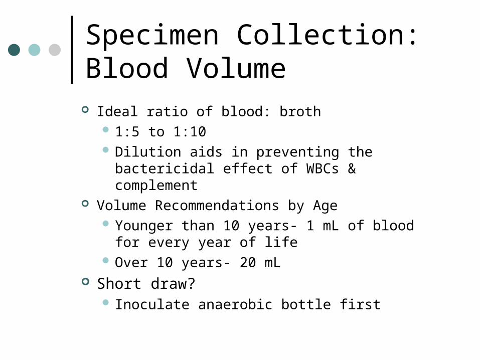
Specimen Collection:Blood Volume
Ideal ratio of blood: broth 1:5 to 1:10 Dilution aids in preventing the
bactericidal effect of WBCs & complement Volume Recommendations by Age
Younger than 10 years- 1 mL of blood for every year of life
Over 10 years- 20 mL Short draw?
Inoculate anaerobic bottle first

Specimen Collection:Frequency of Collection Depends if bacteremia is transient,
intermediate or continuous General guidelines
Usually x2 from different body sites, when patient is spiking a fever
Endocarditis• 3 sets from 3 different sites within 1-2 hours of
clinical presentation Fever of Unknown Origin (FUO)
• Initially 2 sets; 24-36 hours later, obtain 2 more

Specimen Collection:Frequency of Collection If a catheter-related bloodstream
infection is suspected: One set drawn peripherally One set drawn via catheter

Blood Culture Methods
Conventional Broth Systems Aerobic broth contains soybean casein digest
broth, tryptic or trypticase soy broth, Brucella agar or Columbia broth base
Anaerobic broth is usually the same as aerobic with addition of 0.5% cysteine in an aerobic environment
Must be subcultured and gram stained manually, at 12, 24 and 48 hours
Method not recommended due to risk of needlestick and contamination; not cost effective

Blood Culture Methods (cont’d)
Biphasic Broth-Slide System Agar “paddles” attached to top of
bottle; includes CA, MAC, malt extract agars
Incubate at 35 OC for 7 days Allows for blind subcultures Closed system

Blood Culture Methods (cont’d) Lysis-Centrifugation Blood Culture Systems
(Isolator) Used in the recovery of Fungus and AFB The Isolator is a special tube that contains
saponin, a chemical that lyses cells and other anticoagulants
Approximately 7.5-10 ml of blood is placed in the tube, then centrifuged to concentrate microorganisms; sediment is subcultured to fungal and/or mycobacterial media

Blood Culture Methods (cont’d)
Automatic Blood Culture Systems BacTec 9000 Series
• Fluorescent light is used to detect changes in CO2 levels

Bactec 9000 Series

Automatic Blood Culture Systems (con’t)
ESP( Extra Sensing Power) Now VersaTREK Measures
consumption/production of gases; such as CO2 H2, N2 and O2 in the headspace of each bottle
Detects a change in pressure

Automatic Blood Culture Systems (con’t)
• BacT-Alert• Carbon dioxide
production results in a pH change
• pH change results in color change detected by system as “positive”

Blood Culture Workup
Incubation times Routine aerobic/anaerobic
• 5-7 days Endocarditis
• 2 weeks Brucellosis/Fungemia/HACEK
• 21-28 days Reporting results
Initial report is sent out at 24 hours Final report is sent out at 5-7 days for
all no growth specimens

Blood Culture Workup
Positive Cultures Gram stain the bottle to determine the
morphology of the organism present Call the results of the gram stain to the
physician or nurse, including how many sets etc., so that antibiotic therapy can be initiated
Subculture to appropriate media Identify organism and perform
sensitivity testing

Blood Cultures: Pathogens
Staphylococcus aureus Streptococcus pneumoniae Haemophilus influenza Pseudomonas species Neisseria species Coagulase negative Staphylococcus species
(immunocompromised) Group B Streptococcus (infants) Alpha hemolytic Streptococcus viridans group Gram negative rods Yeasts and molds Anaerobes

Blood Cultures: Contaminants Coagulase negative
Staphylococcus Propionibacterium acnes Alpha hemolytic Streptococcus
viridans group Bacillus species Diphtheroids Growth of multiple organism

Treatment & Prevention
Treatment Empirical treatment, initially, with broad
spectrum antibiotic Antisepsis therapy; physiological support,
anticoagulation agents, glucocorticoids Adjunctive measures; draining fluids,
removing catheters Prevention
Vaccines; S. pneumo, influenza, varicella

References
Broyles, M. (2013, June). A Closer Look at Sepsis. ADVANCE for Medical Laboratory Professionals, 25(5), 12-13.
http://www.achats-publics.fr/Fournisseurs/BIOMERIEUX.htm http://www.bd.com/ds/productCenter/212536.asp
http://www.bd.com/ds/productCenter/445718.asp http://www.temple.edu/medicine/microbiology_lab.htm Kiser, K. M., Payne, W. C., & Taff, T. A. (2011). Clinical
Laboratory Microbiology: A Practical Approach . Upper Saddle River, NJ: Pearson Education.
Mahon, C. R., Lehman, D. C., & Manuselis, G. (2011). Textbook of Diagnostic Microbiology (4th ed.). Maryland Heights, MO: Saunders.