Award Number: DAMD17-03-1-0759 TITLE: Strategies … · Bisphosphonates sequester calcium and...
45
AD_________________ Award Number: DAMD17-03-1-0759 TITLE: Strategies for Treatment of Bone Metastases from Breast Cancer PRINCIPAL INVESTIGATOR: Rajagopalan Sridhar, Ph.D. CONTRACTING ORGANIZATION: Howard University Washington, DC 20059 REPORT DATE: October 2005 TYPE OF REPORT: Annual PREPARED FOR: U.S. Army Medical Research and Materiel Command Fort Detrick, Maryland 21702-5012 DISTRIBUTION STATEMENT: Approved for Public Release; Distribution Unlimited The views, opinions and/or findings contained in this report are those of the author(s) and should not be construed as an official Department of the Army position, policy or decision unless so designated by other documentation.
-
Upload
vuongtuyen -
Category
Documents
-
view
216 -
download
0
Transcript of Award Number: DAMD17-03-1-0759 TITLE: Strategies … · Bisphosphonates sequester calcium and...

AD_________________ Award Number: DAMD17-03-1-0759 TITLE: Strategies for Treatment of Bone Metastases from Breast Cancer PRINCIPAL INVESTIGATOR: Rajagopalan Sridhar, Ph.D. CONTRACTING ORGANIZATION: Howard University Washington, DC 20059 REPORT DATE: October 2005 TYPE OF REPORT: Annual PREPARED FOR: U.S. Army Medical Research and Materiel Command Fort Detrick, Maryland 21702-5012 DISTRIBUTION STATEMENT: Approved for Public Release; Distribution Unlimited The views, opinions and/or findings contained in this report are those of the author(s) and should not be construed as an official Department of the Army position, policy or decision unless so designated by other documentation.

REPORT DOCUMENTATION PAGE Form Approved
OMB No. 0704-0188 Public reporting burden for this collection of information is estimated to average 1 hour per response, including the time for reviewing instructions, searching existing data sources, gathering and maintaining the data needed, and completing and reviewing this collection of information. Send comments regarding this burden estimate or any other aspect of this collection of information, including suggestions for reducing this burden to Department of Defense, Washington Headquarters Services, Directorate for Information Operations and Reports (0704-0188), 1215 Jefferson Davis Highway, Suite 1204, Arlington, VA 22202-4302. Respondents should be aware that notwithstanding any other provision of law, no person shall be subject to any penalty for failing to comply with a collection of information if it does not display a currently valid OMB control number. PLEASE DO NOT RETURN YOUR FORM TO THE ABOVE ADDRESS. 1. REPORT DATE (DD-MM-YYYY)01-10-2005
2. REPORT TYPEAnnual
3. DATES COVERED (From - To)8 Sep 2004 – 7 Sep 2005
4. TITLE AND SUBTITLE
5a. CONTRACT NUMBER
Strategies for Treatment of Bone Metastases from Breast Cancer 5b. GRANT NUMBER DAMD17-03-1-0759
5c. PROGRAM ELEMENT NUMBER
6. AUTHOR(S)
5d. PROJECT NUMBER
Rajagopalan Sridhar, Ph.D. 5e. TASK NUMBER
E-Mail: [email protected]
5f. WORK UNIT NUMBER
7. PERFORMING ORGANIZATION NAME(S) AND ADDRESS(ES)
8. PERFORMING ORGANIZATION REPORT NUMBER
Howard University Washington, DC 20059
9. SPONSORING / MONITORING AGENCY NAME(S) AND ADDRESS(ES) 10. SPONSOR/MONITOR’S ACRONYM(S)U.S. Army Medical Research and Materiel Command
Fort Detrick, Maryland 21702-5012 11. SPONSOR/MONITOR’S REPORT NUMBER(S) 12. DISTRIBUTION / AVAILABILITY STATEMENT Approved for Public Release; Distribution Unlimited
13. SUPPLEMENTARY NOTES- Original contains colored plates: ALL DTIC reproductions will be in black and white.
14. ABSTRACT The major goal of this project is to optimize the strategies for using radioactive strontium for palliation of bone pain due to breast cancer metastases. One approach is to determine ways of selectively increasing strontium uptake at the metastatic site. Bisphosphonates are useful in the treatment of osteoporosis and bone metastases. Bisphosphonates sequester calcium and strontium. Strontium was non toxic to human breast cancer cells. Bisphosphonates are charged molecules that do not easily cross hydrophobic cell membranes. The nature of the counter ion to the anionic phosphonate was found to have an effect on the cellular uptake of bisphosphonate such as etidronate. The cytotoxicty of zoledronic acid towards MCF-7 cells greater than pamidronate and etidronate. The presence of strontium chloride in the culture medium inhibited calcium uptake by MDA-MB 231 cells. Etidronic acid treatment perturbed cell cycle progression of MCF-7 cells, causing a G2/M block and decreased the proportion of cells in S-phase. This suggests that a bisphosphonate such as etidronate may be useful in combination with ionizing radiation for treatment of bone metastases.
15. SUBJECT TERMS No subject terms provided
16. SECURITY CLASSIFICATION OF:
17. LIMITATION OF ABSTRACT
18. NUMBER OF PAGES
19a. NAME OF RESPONSIBLE PERSONUSAMRMC
a. REPORT U
b. ABSTRACTU
c. THIS PAGEU
UU
45
19b. TELEPHONE NUMBER (include area code)
Standard Form 298 (Rev. 8-98)Prescribed by ANSI Std. Z39.18

Table of Contents
Introduction…………………………………………………………….……………. 4 Body………………………………………………………………………………… 4-11
Key Research Accomplishments………………………………………………...11 Reportable Outcomes……………………………………………………… ...11-12 Conclusions………………………………………………………………………… 12 References……………………………………………………………………… 13-16 List of Appended Abstracts and Paper …………………………… ………….17 Appendex 1- 3 Publications and Abstracts

4
INTRODUCTION
In this project we have concentrated mainly on establishing monolayer cultures of human fetal osteoblasts, breast cancer cells and murine osteoclasts. We have also established the conditions of low calcium levels for tissue culture experiments. Experiments are in progress for estimating calcium and strontium in cells. Tasks 2, 4 and 5 have not been initiated, while progress has been made in tasks 1, 3, 6 and 7. One manuscript and four abstracts have been published during this report period. BODY BACKGROUND More than 50% of patients with primary breast cancer will eventually develop bone metastases and 75% of patients with breast carcinoma were found to be with bone metastases at autopsy (1,2). In spite of skeletal metastases, breast cancer patients can survive for long periods (5 year survival is about 20%), but these persons will have much pain and suffering due to cancer mediated bone destruction (2). In addition to bone pain, these individuals may develop bone fractures, spinal cord compression (which can cause paralysis) and hypercalcemia of malignancy (HCM) (2-4). Breast neoplasms have a strong tendency to metastasize to the bone. For this to occur, both the tumor cell and host cells at the site of metastasis interact significantly and selectively (5-7). Once the cancer cell reaches the bone surface, the proteolytic enzymes of the neoplastic cell can facilitate the break down of the stroma and pave the way for metastasis to become entrenched. Another possibility is for the cancer cell to recruit and promote the proliferation of osteoblasts (bone forming cells) and new bone formation. Soluble growth factors from the cancer cell enable new bone to be formed around the neoplastic cell. Alternatively the growth factors produced by the migrating tumor cell could stimulate the proliferation of osteoclasts which are involved in bone destruction. The breast carcinoma cells can also destroy the bone. Destruction of the bone by the metastatic process can lead to hypercalcemia and associated problems. Certain breast tumors produce parathyroid hormone which promotes bone resorption. In particular, parathyroid hormone stimulates osteoclasts which degrade the bone. It appears that parathyroid hormone stimulates the growth of osteoclasts, and enhances parathyroid hormone production by the bone compared to normal tissues and tumor (7,8). Other factors known to increase osteoclast mediated bone resorption are interleukin 1 (9), interleukin 8 (10), interleukin 6 (11) and cathepsin K (12). Preosteoblasts are differentiating intermitotic cells derived from vegetative intermitotic mesenchymal stem cell. The preosteoblasts differentiate further to yield mature osteoblasts that are involved in bone matrix synthesis. Preosteoclasts are also of the differentiating intermitotic types that are derived from hematopoietic stem cell. Preosteoclasts differentiate further into mature multinucleated osteoclasts that are involved in bone resorption. Cytokines such as TGF-β, macrophage-colony stimulating factor (M-CSF), IL-6, TNF-α, and osteoprotogerin (OPG)/osteoprotogerin ligand (OPG-L) from a variety of sources including mature osteoblasts are involved in promoting proliferation and differentiation of preosteoclasts to mature osteoclasts (13). Estrogen deficiency is associated with enhanced bone osteoclast formation and bone loss. Correction of estrogen deficiency prevents bone loss. Estrogen action is mediated through cytokine production. Bisphosphonates also protect against bone loss. There is evidence to suggest that both estrogen and bisphosphonates can have direct effects on osteoblasts and osteoclasts (13-15). There

5
are examples of bisphosphonates covalently linked to estrogens and anti-estrogens (16, 17). The anti-estrogen, tamoxifen and selective estrogen response modifiers protect against osteoclast mediated bone resorption (18, 19). The effects of conjugates of bisphosphonates with estrogens and anti-estrogens on osteoblasts and osteoclasts are not known. Investigation of such conjugates may help identify compounds that protect against bone lysis caused by breast cancer metastases. The radioisotope Strontium-89 (Sr-89) has a physical half life of 50.5 days. It is a pure beta emitter (undergoes beta minus decay with a beta emission of 1.463 MeV). The maximum range of the beta emission is about 8 mm. Strontium is a calcium mimic and a bone seeking mineral. When in the bone the radiation from this isotope is mainly to the cortical and trabecular bone, with less to bone marrow and minimal dose to adjacent soft tissues. Strontium-89 is usually given at a dose of 4 mCi (or 40-60µCi/kg body weight [1.5MBq/kg] is used as a single injection. About 65% of the patients gain relief from pain. The success of Sr-89 treatment depends on the ability of the radioisotope to infiltrate into the osteoblasts and osteoclasts, Once the radioisxzotope is taken up by the cells, the beta emission can damage the cell. If the isotope is retained long enough then the accumulated damage can be lethal to bone cells. Strontium-89 has been used for palliative treatment of bone metastases from breast and other cancers (20-24). Strontium-89 is sold under the trade name metastron. We have used metastron for treating bone pain in about 41 patients (25). There is considerable variation in treatment response with respect to pain relief. There may be several reasons for this. For this treatment to be effective, radioactive strontium has to be taken up in sufficient amounts by the intended target (osteoblasts and or osteoclasts). Sr-89 does not exert an appreciable lethal effect towards tumor cells. Since strontium is a calcium analog, the uptake of strontium may depend on a variety of factors including systemic calcium levels. If the systemic calcium levels are high, cellular uptake of strontium may be poor because calcium may competitively inhibit the uptake of strontium. Bisphosphonates are useful in the treatment of bony metastases, especially those traceable to an osteoclast etiology (26-34, 35-37). Bisphosphonates and estrogen metabolites appear to have a direct effect on bone cells (38, 39). Bisphosphonates also appear to have antineoplastic effect by inducing apoptosis of tumor cells in vivo and in vitro (40). Osteoprotegerin (OPG) is a potent inhibitor of osteoclast formation and activity. OPG is a decoy receptor which neutralizes OPG-ligand (OPG-L) which is the ultimate effector of osteoclastogenesis (41). Recently there has been some concern about adverse effects of bisphosphonates such as the development of osteonecrosis. These effects are usually seen with prolonged use of bisphosphonates. The severity of the adverse effects appears to be related to the potency of bisphosphonates and instances of osteonecrosis appear to be more common with newer generation bisphosphonate such as zoledronic acid compared to earlier generation bisphosphonates (44, 45).. HYPOTHESIS/RATIONALE/PURPOSE The purpose is to improve the use of Sr-89 and bisphosphonates for treating bone metastases from breast cancer. The rationale is as follows: Nearly 50% of breast cancer patients develop bone metastases due to osteotropic nature of the primary breast cancer cells. Breast cancer cells, osteoblasts and osteoclasts have some growth factors in common. Thus the breast cancer cells promote the growth of either or both types of cells. The osteoblsts surround the cancer cell which has adhered to the bone matrix and starts depositing new bone which ultimtely leads to bony
7

6
metastasis. In contrast, the osteoclasts utilize the growth factors differently. They destroy the bone by resorption which results in hypercalcemia. Sr-89 is usually satisfactory for the treatment of lesions caused by osteoblasts. Osteoblastic activity attracts breast cancer cells and utilizes calcium for making new bone. Strontium is an analog of calcium. Sr-89 is easily taken up instead of calcium by the active osteoblasts. Once inside the osteoblast, the beta emission from the radionuclide inactivates the osteoblast. This can lead to some relief from pain, If the lesion is caused by osteoclast, which does not consume calcium or strontium ions avidly, Sr-89 is not quite so effective against osteoclasts. Bisphosphonates are pyrophosphate analogs which have a rather high affinity for bone. Although bisphosphonates such as pamidronate are useful in the treatment of bony metastases, it is important to know if the high levels of hypercalcemia have to be decreased in order to achieve effective treatment with pamidronate or Sr-89. Since bisphosphonates and Sr-89 complement each other, addition of a bisphosphonate can enhance the efficacy of Sr-89 and vice versa. Such logic can be extended to arrive at other combinations based on our knowledge of the mechanisms involved.
Hypothesis 1: Calcium concentration in the system affects strontium uptake by the cells. For improved use of Sr-89, strategies for decreasing cellular calcium pools are necessary. Gallium nitrate or a bisphosphonate can be used for lowering the calcium levels in cells.
Hypothesis 2: Antiestrogens and estrogens can modify the effects of bisphosphonates on osteoblasts and osteoclasts. Bisphosphonates linked covalently to anti estrogen and estrogen moieties will have greater affinity for bone cells and may be more active.
Hypothesis 3: Hormones and hormone antagonists can be combined for modulating the effects bisphosphonates on bone cells. Specific Aims:
1. Treat osteoblast and osteoclast-like cells in culture with graded concentrations of strontium in the presence of different amounts of calcium and measure the kinetics of uptake and retention of strontium by cells. Atomic absorption spectroscopy will be used for estimating strontium concentration.
2. Test the influence of the bisphosphonates pamidronate and its more potent analog zolendronate on the uptake of strontium by the osteoblast cell lines in culture. It is not known if bisphosphonates alter the bioavailability of strontium ions.
3. Synthesize bisphosphonates conjugated to estrogen and anti-estrogen moieties and test their effects on the viability of breast cancer cell lines and human fetal osteoblasts in culture. Cell viability will be assessed on the basis of apoptosis assays and clonogenicity measurements where feasible.
4. Determine if gallium nitrate, calcium channel blockers, nifedipine and verapamil and the antiestrogens tamoxifen and raloxifene alter strontium uptake by human fetal osteoblasts. grown as monolayers and as multicell spheroids. Both estrogen receptor positive and receptor negative human fetal osteoblasts and breast cancer cells are being utilized in our experiments. Estrogen receptor positive human fetal osteoblast (hFOB/ER9) and estrogen receptor negative human fetal osteoblasts (hFOB1.19) are to be studied. Estrogen responsive MCF-7 and estrogen independent MDA-MB231 are some of the breast cancer cells in our laboratory.

7
STATEMENT OF WORK
Task 1. Establish osteoclast and osteoblast cultures (months 1 through 6). Standardize assays for strontium and calcium using atomic absorption spectrophotometry (months 1 through 3). Determine the baseline values of strontium and calcium in cultures grown in defined media. Evaluate the effect of calcium levels in the medium on strontium uptake by hFOB1.19 and hFOB ER/9. Compare the results obtained using serum-free and serum supplemented culture media. Test the effects of gallium nitrate and calcium channel blockers verapamil and nifedipine on strontium uptake and retention (months 1 through 15).
Task 2. Synthesize bisphosphonates conjugated to estrogenic and antiestrogenic
moieties (months 1 through 18). Task 3. Test the effects of bisphosphonates (including pamidronate, zolendronate and
those synthesized in task 2) on strontium uptake by osteoblasts (months 6 through 30).
Task 4. Determine the influence of gallium nitrate on the uptake of strontium by the
afore-mentioned cell lines (months 2 through 12). Task 5. Measure the uptake and retention of strontium by multicellular spheroids of
osteoblasts in the presence and absence of agents which affect calcium homeostasis (months 6 through 30).
Task 6. Evaluate the differences, if any, in the uptake kinetics of strontium and
calcium in the different cell lines. Determine any correlations that may exist among the cellular levels of calcium and strontium on the cytotoxicity of bisphosphonate. Utilize isobologram analysis to reveal any synergistic or antagonistic interactions between bisphosphonates and strontium and/or calcium (throughout the 36 month project period).
Task 7. Assays for strontium, calcium and clonogenicity assays for cell viability and
apoptosis will be carried out throughout the 36 month project.
Materials and Methods: Cell lines: Estrogen responsive MCF-7 and estrogen independent MCF-7MDR clone 10.3 and MDA-MB231-luc human breast cancer cell lines were maintained as monolayer cultures growing in RPMI-1640 medium supplemented with 10% fetal bovine serum, glutamine, pyruvate, insulin,

8
penicillin and streptomycin. MCF-7 cells were also grown as multicellular spheroids. We have also grown spheroids with matrigel. Culturing hFOB 1.19 and hFOB/ER cell lines. These osteoblast cells (42, 43) were obtained from Dr. Thomas Spelsberg of Mayo Clinic, Rochester. The following is their description of the cells along with the recommended procedure for the growth and maintenance of these cells. The hFOB 1.19 cells were cultured as monolayers at 34o C in DMEM-F12 medium supplemented with 10% fetal bovine serum and 300µg/ml geneticin, and induced to differentiate more fully either by culturing past confluence or by culturing at 39o C, as described by Harris SA et al, Bone Miner Res 10:178-186, 1995 (42). The hFOB/ER9 cell lines were derived from the hFOB 1.19 cells were also cultured at 34o C in DMEM: F12 (1:1), but supplemented with 10% charcoal-stripped FBS and either geneticin at 300µg/ml or hygromycin B at 100µg /ml. Spheroid culture: We have acquired high aspect ratio vessels for culturing hFOB spheroids. These spheroids are more difficult to grow compared to multicell spheroids of MCF-7 cells. MCF-7 cells form spheroids in stationary cultures with unstirred medium and in spinner flasks. We have utilized matrigel to facilitate spheroid formation from MCF-7 cells in stationary cultures. The breast cancer line MCF-7 cells were obtained from ATCC and grown in RPMI 1640 medium. Pure cell cultures were incubated at 37 0C for 6 days. On the third day, media was removed and fresh media added to maintain good cell growth. At the end of 6 days, media was removed and the adherent cells recovered by adding Trypsin/EDTA. The trypsinized cell culture was collected in fresh media and 2 x 10 6 cells/ml used for treating the cells. Cells were seeded on plates and incubated overnight. The following nine treatments were imposed on the cell cultures and incubated for 6 and 20 hours. Control; 0, 3.5 mM and 7.0 mM Strontium Chloride applied separately and in combination with 10 and 20 mM etidronic acid. At the end of 6 and 20 hours incubation, cells were harvested by scraping and transferred into 15 ml centrifuge tubes. One set was used for cell cycle analysis and the second set was reserved for DNA extraction. MTT Assay for Cell Viability:
The cells were treated with graded concentrations of etidronate for different treatment periods of incubation (1, 3, 6 and 12 hrs). Mitochondial dehydrogenase levels, which are correlated to cell viability, were determined by the enzyme mediated cleaving of the tetrazolium salt ring of 3-[4, 5-dimethylthiazol-2-yl]-2, 5-diphenyl tetrazolium bromide to yield purple formazan crystals. These crystals were dissolved in isopropanol, and the absorption at 560 nm was determined spectrophotometrically. Appropriate control experiments were run to determine spectrophotometric background and absorption due to reagent blanks. Clonogenicity Assay: Following treatment, cells were trypsinized at 37°C for 5 minutes, and pipetted up and down 5 times to break up cell clumps and obtain a single cell suspension. The same volume of single cell suspension was plated on 100-mm tissue culture dishes with fresh medium and kept at 37°C, 5%

9
CO2 incubator for 10~14 days. Colonies consisting of more than 50 cells were counted. Survival was calculated as percentage rate of the number of colonies formed at a given treatment condition to the number of colonies produced by related untreated control cells. Hyperthermia treatment: For 96-well plate: Cells were seeded in sterile 96-well plates at a density of 1 x10⁴ cells/well and incubated overnight. For 24-well plate: cells were seeded at densities of 1x10³, 1x10⁴, 1x10⁵ and 5x10⁵ /well. Hyperthermia was applied at 43°C by sealing the plates with parafilm and enclosing in a Ziploc bag and then immersing the bag into a temperature controlled water bath maintained at 43o(± 0.1°C). The continuous heating period ranged from 10 to 120 minutes. Controls were sealed in ziplock bags and immersed in a 37°C water bath. After heating, plates were ready for optical imaging, and for MTT and clonogenic assays. Apopain Assay: Apopain/Caspase 3 is derived from the proenzyme CPP32 at the onset of apoptosis and plays a pivotal role in programmed cell death. This assay indicates apopain activity associated with apoptosis. The FluorAce apopain assay kit was used (Bio-Rad, Catalog number 170-3130). Flow Cytometry Analysis for Cell Cycle and Apoptosis: The effect of etidronate treatment on cell cycle and apoptosis was analyzed using flow cytometry assay. The cells were trypsinzed and washed twice with PBS after treatment. The suspended cells were fixed with 80% ethanol for 30 min on ice, and then centrifuged for 5 min at 1500rpm. The fixed cells were washed again with PBS and the supernatant was removed. The cells were stained at 4o C in the dark with 1 ml of propidium iodide (PI) solution and/or fluorescein-conjugated
annexin V (apoptotic marker), and then stored at 4oC until analysis. The cell cycle distribution was analyzed by FACS caliber flow cytometry (Becton Dickson, San Jose, CA). Ten thousand cells were analyzed per sample. PI solution contained 100 Units/ml or 50 µg/ml RNase A and 50 µg/ml PI in PBS. Similarly the effect of different concentrations of strontium chloride and etidronate on cell cycle was analyzed using flow cytometry. Cytotoxicity of Etidronic Acid Towards Human Breast Cancer Cells. Human breast cancer frequently metastasizes to the skeleton to cause osteolysis and subsequent pain, pathological fracture, and hypercalcemia. The bone continuously releases growth factors stored in bone matrix by bone resorption and provides a favorable environment for metastatic breast cancer cells to proliferate. Reducing the amount of bone destruction and increasing the rate of bone repair can reduce pain and lower the risk of fractures by keeping bones strong. Inhibition of bone resorption is a useful adjuvant therapy in patients with breast cancer. Bisphosphonates are potent inhibitors of osteoclastic resorption and are used in the treatment of osteoporosis, hypercalcemia, and bone metastases. Alendronate, etidronate and risedronate increase bone density and prevent spine and hip fractures (46). Etidronic acid has been shown to inhibit bone resorption and increase bone mineral density.These drugs bind permanently to the surfaces of the bones and slow down the osteoclasts (bone-eroding cells). This allows the osteoblasts (bone-building cells) to work more effectively. Bisphosphonates may induce human osteoblast differentiation via inhibition of the

10
mevalonate pathway or other mechanisms. These drugs may also induce cell death by apoptosis through the inhibition of protein farmesylation and geranylgeranylation. Cell proliferation of normal and cancer tissues requires post-translational isoprenylation by farmesyl-transferase and geranylgeranyl-transferase. Strontium, an isotope that imitates the in-vivo behavior of calcium, is known to stimulate bone formation and has been used in the treatment of osteoporosis. It is readily taken up by bone and is concentrated at the osteoblastic skeletal metastatic sites. The combined effects of etidronate and strontium on breast cancer cells has not been studied.. The purpose of the present investigation is to determine the combined effects of etidronate and strontium on apoptosis and the mutational status of the p53 gene in MCF-7. The tumor suppressor gene p53 plays an essential role in cell proliferation and apoptosis. Missense mutations of p53 occur in 50 % of all cancers. Due to its relevance to cancer therapy, most studies have focused on the cellular consequences of p53 activation in relation to cytotoxic drugs. Anticancer drugs are known to induce apoptosis by triggering biochemical events involved in apoptotic pathways and in cell cycle regulators (47, 48). Results from this study will provide some insight into the management of bone metastases in breast cancer patients. Etidronic acid (1-hydroxyethane-1,1,-diphosphonic acid; 1-hydroxyethylidenediphosphonic acid) (Eti) affects calcium metabolism and slows down abnormal bone resorption. The anti osteolytic activity of this compound has led to research on the use of bisphosphonates for the treatment of Paget’s disease, osteoporosis and cancer metastases to the bone. Strontium, which imitates the in vivo behavior of calcium, stimulates bone formation and has been used in the treatment of post menopausal osteoporosis. There have been several reports on anti tumor effects of bisphosphonates. The cytotoxicity of etidronic acid (a first generation bisphosphonate) towards MCF-7 human breast cancer cells and its multidrug resistant derivative MCF-7 clone 10.3 cells, was studied in the presence and absence of strontium chloride (Sr). Clonogenicity assays revealed that a 24 hour exposure to etidronic acid (10 mM) was more toxic to MCF-7 compared to MCF-7 clone 10.3 cells, while the addition of strontium chloride had no effect. Flow cytometry studies revealed that etidronic acid caused a decrease in the s-phase population with concomitant increase in G2/M phase population . Again strontium was without any effect. Within the cell cycle, late s-phase cells are the most radioresistant while G2/M cells are the most radiosensitive. Therefore the decrease in s-phase population with corresponding increase in G2/M would position the cells in a relatively more radiosensitive setting. Such a shift in cell cycle distribution may be useful if etidronic acid were combined with radioactive strontium ( 89 Sr, metastron), which is a beta emitter used in the treatment of bone metastases from breast cancer. Inhibition of cellular uptake of calcium by strontium. Our assumption that calcium levels would influence cellular uptake of strontium were not borne out in hormone independent MDA MB-231 cells. In fact the presence of strontium in the medium inhibited calcium uptake by cells. Preliminary indications are that strontium in combination with the calcium channel blocker verapamil strongly inhibited calcium uptake.

11
The normal steady state calcium levels in MDA-MB231 cells was around 230 nM based on digitized fluorescence microscopy of single cells using Fura-2, AM ester as a fluorescent probe. Even a 10 minute exposure to strontium chloride (1 mM) decreased cellular calcium levels to 140 nM. More experiments are needed to understand the uptake kinetics of strontium and calcium in cells. Comparison of cell viability assays, We observed that cells deemed to be viable on the basis of trypan blue dye exclusion as well as the MTT assay proved to be dead on the basis of clonogenicity assays. Since clonogenicity is the gold standard for evaluating cancer chemotherapeutic agents, we compared different assays for cell viability. KEY RESEARCH ACCOMPLISHMENTS
• Monolayer cultures of the cell lines needed for our research have been established, but only
MCF-7 human breast cancer cells have been grown consistently as multicell spheroids. Spheroid formation was facilitated in the presence of matrigel.
• Assays for cell viability have been standardized and utilized for evaluating the cytotoxicity
of etidronate and analogs towards the different cell lines in culture. • Presence of strontium in the culture medium decreases calcium uptake by hormone
independent breast cancer cells. • Toxicity of etidronate towards osteoclast and breast cancer cells was demonstrated. The cytotoxicity of etidronic acid was enhanced by hyperthermia. • Flow cytometry assays for cell cycle analysis and estimation of apoptosis have been
standardized and applied to study the effect of etidronate towards breast cancer cells.
• Strontium chloride (up to 7 mM) was non toxic to breast cancer cells. Strontium decreased the uptake of calcium by MDA-MB-231 breast cancer cells.
REPORTABLE OUTCOMES PUBLICATIONS
1. Sridhar, R. RNA processing in health and disease: a brief review. J Clin Ligand Assay 28 (#2): 61-67, 2005
ABSTRACTS
1. Sridhar R, Kassa A and Ashayeri E. Modulation of bisphosphonate toxicity towards human breast cancer cells in culture. Era of Hope, Department of Defense Breast Cancer Research Program Meeting, Abstract P43-17, Philadelphia, PA, 2005. (Abstract)

12
2. Zhou Y, Pang X, Zhang R, Gu X, P Kc, Sridhar R. Loperamide sensitizes multidrug resistant
MCF-7 clone 10.3 human breast cancer cells to doxorubicin, Proc. Amer. Assoc. Cancer Res. 46: 538, 2005.
3. Zhang R, Brown S, Guerrier K, Kassa A,Zhou Y, Gu X, Ashayeri E and Sridhar R. Cytotoxicity
of Lipoxygenase Inhibitors towards prostate cancer cells in culture, Proc. Amer. Assoc. Cancer Res. 46: 5863, 2005.
4. Zhang R, Zhou Y, Kassa A, Gu X, Ashayeri E and Sridhar R. In Vitro Cytotoxicity of
Lipoxygenase Inhibitors towards PC3 and DU-145 Human Prostate Cancer cells, RCMI International Symposium on Health Disparities: Scientific Program and Abstracts, pp 73-74, Baltimore, MD, December 8-11, 2004.
CONCLUSIONS In addition to their effect on calcium, bisphosphonates are cytotoxic towards breast cancer cells in culture. The bisphosphonate etidronate is toxic to breast cancer cells and perturbs the cell cycle as judged by flow cytometry. Combining etidronate with calcium or strontium chloride causes precipitation of insoluble material. Calcium and strontium salts of etidronic acid are sparingly soluble. Strontium chloride up to 7 mM was not toxic to MCF 7 breast cancer cells in culture. However, strontium chloride caused the precipitation of etidronic acid. Presence of strontium in the culture medium decreases calcium load in breast cancer cells. Bisphosphonates decrease the proportion of cancer cells in the DNA synthetic phase (s-phase). This may be due to the replacement of critical pyrophosphate links by non hydrolysable phosphonate linkage to bases in nucleotides. We have obtained 31P NMR spectrum of etidronic acid. This will be useful for monitoring its presence in cell cultures.

13
REFERENCES 1. Malawer, M.M., Delaney, T.F. Treatment of metastatic cancer to bone. In: DeVita, V.T.,
Hellman, S., Rosenberg, S.A. Eds., Cancer: Principles and Practice of Oncology. Philadelphia: J.B. Lippincott Co., 4th Edition, 1993, pp 2225-2245.
2. Lipton, A. Bisphosphonates and Breast Carcinoma.. Skeletal Complications of Malignancy
Cancer 80: Suppl. 1668-1673, 1997. 3. Coleman, R.E., Rubens, R.D. The clinical course of bone metastases from breast cancer.
Br. J. cancer 55: 61-66, 1987. 4. Galasko, C.S.B., Burn, J.L. Hypercalcemia in patients with advanced mammary cancer. Br.
Med. J. 3: 573-577, 1971. 5. Liotta, L.A.Kohn, E. Cancer invasion and metastases. JAMA 263: 1123-1126, 1990. 6. Zetter, B.R. The cellular basis of site-specific tumor metastases. N. Engl. J. Med. 322:605-
612, 1990. 7. Mundy, G.R. Mechanisms of Bone Metastases. Cancer 80: Suppl.1546-1563, 1997. 8. Yu, X., Scholler, J. Foged, N.T. Interaction between effects of parathyroid hormone and
bisphosphonate on regulation of osteoclast activity by the osteoblast-like cell line UMR-106.Bone 19:339-345, 1996.
9. Jimi, E., Nakamura, I., Duong, L.T., Ikebe, T., Takahashi, N., Rodan, G.A., Suda, T.
Interleukin 1 induces multinucleation and bone-resorbing activity of osteoclasts in the absence of osteoblasts/stromal cells. Exp. Cell Res. 247: 84-93, 1999.
10. Rothe, L., Collin-Osbody, P., Chen, Y., Sunyer, T., Chaudhary, L., Tsay, A., Goldring, S.,
Avioli, L, Osbody, P. Human osteoclasts and osteoclast-like cells synthesize and release high basal and inflammatory stimulated levels of the potent chemokine interleukin 8. Endocrinology 139: 4353-4363, 1998.
11. Schiller, C., Gruber, R., Redlich, K., Ho, G.M., Katzgraber, F., Willheim, M., Pietschmann,
P., Peterlick, M. 17 Beta-estradiol antagonizes effects of 1 alpha, 25-dihydroxyvitamin D3 on interleukin production and osteoclast-like cell formation in mouse bone marrow primary cultures. Endocrinology 138: 4567-4571, 1997.
12. Littlewood-Evans, A.J., Bilbe, G., Bowler, W.B., Farley, D., Wlodarski, B., Kokubo, T.,
Inaoka, T., Sloane, J., Evans, D.B., Gallagher, J.A.. The osteoclast associated protease cathepsin K is expressed in human breast carcinoma, Cancer Res, 57: 5386-5390
13. Rickard, D.J., Subramaniam, M., Spelsberg, T.C. Molecular and cellular mechanisms of
estrogen action on the skeleton. J. Cell. Biochem. Suppl. 32/33:123-132, 1999. 14. Ramalho, A.C., Jullienne, A., Couttet, Ph. , Graulet, A.M., Morieux, C., deVernejoul,

14
M.C., Cohen-Sohal, M.E. Effect of estradiol on cytokine production in immortalized human marrow stromal cell lines. Cytokine 16: 126-130, 2001.
15. Heino, T.J., Hentunen, T.A., Vaananen, K. Osteocytes inhibit osteoclastic bone resorption
through transforming growth factor-β: Enhancement by estrogen. J. Cell. Biochem. 85: 185-197, 2002.
16. Page, P.C.B., McKenzie, M.J. and Gallagher, J.A. Novel synthesis of bis(phosphonic acid)-
steroid conjugates. J. Org. Chem. 66: 3704-3708, 2001.
17. Bauss, F., Esswein, A., Reiff, K., Sponer, G., Muller-Beckmann, B. Effect of 17beta-estradiol-bisphosphonate conjugates, potential bone-seeking estrogen prodrugs, on 17beta-estradiol serum kinetics and bone mass in rats. Calcif. Tissue Int. 59: 168-173, 1996.
18. Williams, J.P., McKenna, M.A., Thames, A.M. 3rd, McDonald, J.M. tamoxifen inhibits phorbol ester stimulated osteoclastic bone resorption: an effect mediated by calmoulin. Biochem. Cell. Biol. 78: 715-723, 2000.
19. deVernejoul, M.C., Cohen-Sohal, M., Benichou, O. Physiology of bone loss and
pharmacologic approach of selective estrogen receptor modulators. Joint Bone Spine 67 Suppl. 1:7s-13s, 2000.
20. Patel, B.R., Flowers, W.M. Jr. Systemic radionuclide therapy with strontium chloride Sr-89
for painful skeletal metastases in prostat and breast cancer. South. Med. J. 90: 506-508, 1997.
21. Pizzocaro, C., Panarotto, M.B., De Agostini, A., Pagliani, R., Bestagno, M. Experience with
89-strontium treatment of opainful osseous metastases from breast cancer. Tumori 83: 558-559, 1997.
22. McEwan, A.J. Unsealed source therapy of painful bone metastases: an update, Semin. Nucl.
Med. 27: 165-182, 1997. 23. DeKlerk, J.M., Zonnenberg, B.A., Huiskes, A.W., Han, S.H., Blijham, G.H. Van Rijk,
P.P.Palliative treatment of bone metastases with bone-seeking radionuclides. 142: 2618-2622, 1998.
24. Porter, A.T., Ben-Josef, E., Davis, L. Systemic administration of new therapeutic isotopes,
including phosphorus, strontium, samarium and rhenium. Curr. Opin. Oncol. 6: 607-610, 1994.
25. Ashayeri, E., Adedamola, O., Sridhar, R., and Shankar, R.A. 2002 Strontium 89 in the
treatment of pain due to diffuse osseous metastases: A university hospital experience. J Natl Med Assoc. 94: 706-711, 2002.
26. Van der Pluijm, G., Vloedgraven, H., van Beek, E., van der Wee-Pals, L., Lowick, C.,
Papapoulos, S. Bisphosphonates inhibit the adhesion of breast cancer cells to bone matrices in vitro, J. Clin. Invest. 98: 698-705, 1996.

15
27. Diel, I.J., Solomayer, E.F., Costa, S.D., Gollan, C., Goerner, R., Wallweiner, D., Kaufmann,
M., Bastert, G. Reduction in new metastases in breast cancer with adjuvant clodronate treatment, N. Engl. J. Med. 339: 357-363, 1998.
28. Lipton, A., Aredia: the once-monthly infusion for the treatment of bone metastases.. Curr.
Opin. Oncol. , 10 Suppl. 10:S1-5, 1998. 29. Coleman, R.E., Houston, S., Purohit, OP., Rubens, R.D., Kandra, A., Ford, J. A randomised
phase II study of oral pamidronate for the treatment of bone metastases from breast cancer. Eur. J. Cancer 34: 820-824, 1998.
30. Hortobagyi, G.N., Theriault, R.L., Porter, L., Blayney, D., Lipton, A. Sinoff, C., Wheeler,
H., Simeone, J.F., Seaman, J., Knight, R D. N. Engl. J. Med. 335: 1785-1791, 1996. 31. Body, J.J. Clinical research update: zoledronate. Cancer 80(8Suppl): 1699-1701, 1997. 32. Lipton, A. Zoledronate in the treatment of osteolytic bone metastases. Br. J. Clin. Pract.
Suppl. 87: 21-22, 1996. 33. Kristensen, B., Ejlertsen, B., Mouridsen, H.T. and Loft, H. Survival in breast cancer
patients after the first episode of hypercalcemia. J. Intern. Med. 244: 189-198, 1998. 34. Coleman, R.E. How can we improve the treatment of bone metastases further? Curr. Opin.
Oncol. 10 Suppl. 10: S7-13, 1998 35. Diel, I.J., Solomayer, E.F. and Bastert, G. Bisphosphonates and prevention of metastases:
first evidences from preclinical and clinical studies. Cancer 88(12 Suppl): 3080-3088, 2000. 36. Lipton, A. Bisphosphonates and breast carcinoma: oresent and future. Cancer 88 (12 Suppl ):3033-3037, 2000. 37. Body, J.J. Current and future directions in medical therapy: hypercalcemia. Cancer 88(12
Suppl):3054-3058, 2000. 38. Reinholtz, G.G., Getz, B., Pederson., L., Sanders, E.S., Subramaniam, M., Ingle, J.N. and
Spelsberg, T.C. Bisphosphonates directly regulate cell proliferation, differentiation and gene expression in human osteoblasts. Cancer Res. 60: 6001-6007, 2000.
39. Robinson, J.A., Waters, K.M., Turner, R.T. and Spelsberg, T.C. Direct action of naturally
occurring estrogen metabolites on human osteoblastic cells. J. Bone Miner. Res. 15: 499-506, 2000.
40. Mundy, G.R. Bisphosphonates as anticancer drugs. Expert Opin. Investig. Drugs 8: 2009-
2015, 1999. 41. Hofbauer, L.C., Gori, F., Riggs, B.L., Lacey, D.L., Dunstan, C.R., Spelsberg, T.C. and
Khosla, S. Stimulation of osteoprotegerin production by glucocorticoids in human

16
osteoblastic lineage cells: potential paracrine mechanisms of glucocorticoid-induced osteoporosis. Endocrinology 140: 140: 4382-4389, 1999.
42. Harris, S.A., Enger, R.J., Riggs, B.L. and Spelsberg, T.C. Development and
characterization of a conditionally immortalized human fetal osteoblast cell line. J. Bone Miner. Res. 10: 178-186, 1995.
43. Harris, S.A., Tau, K.R., Enger, R.J., Toft, D.O., Riggs, B.L. and Spelsberg, T.C. Estrogen
response in the hFOB 1.19 human fetal osteoblastic cell line stably transfected with the human estrogen receptor gene. J. Cell. Biochem. 59: 193-201, 1995.
44. Robinson, N.A., Yeo, J.F. Bisphosphonates-a word of caution. Ann. Acad. Med. Singapore,
33 (4 Suppl): 48-49, 2004. 45. Migliorati, C.A., Schubert, M.M., Peterson, D.E., Seneda, L.M. Bisphosphonate associated
osteonecrosis of mandibular and maxillary bone: an emerging oral complication of supportive cancer therapy. Cancer 104: 83-93, 2005.

17
List of appended abstracts and paper Abstracts: 1. Sridhar R, Kassa A and Ashayeri E. Modulation of bisphosphonate toxicity
towards human breast cancer cells in culture. Era of Hope, Department of Defense Breast Cancer Research Program Meeting, Abstract P43-17, Philadelphia, PA, 2005.
4. Zhou Y, Pang X, Zhang R, Gu X, P Kc, Sridhar R. Loperamide sensitizes
multidrug resistant MCF-7 clone 10.3 human breast cancer cells to doxorubicin, Proc. Amer. Assoc Cancer Res. 46: 538, 2005.
5. Zhang R, Brown S, Guerrier K, Kassa A,Zhou Y, Gu X, Ashayeri E and Sridhar, R. Cytotoxicity of Lipoxygenase Inhibitors towards prostate cancer cells in culture, Proc.Amer. Assoc. Cancer Res. 46: 5863, 2005. 7. Zhou, Y., Sridhar, R., Gu, X., Pang, X., Balachandran, R., Magarian, R.A., and Day, B.W. Evaluation of a series of 1,1-dichloro-2,3-di-and tri-arylcyclopropanes as MDR reversing agents. Proc. Amer. Assoc. Cancer Res. 45: Abstract 2139, 2004 8. Zhang R, Zhou Y, Kassa A, Gu X, Ashayeri E and Sridhar R. In Vitro Cytotoxicity of Lipoxygenase Inhibitors towards PC3 and DU-145 Human Prostate Cancer cells, RCMI International Symposium on Health Disparities: Scientific Program and Abstracts, pp 73-74, Baltimore, MD, December 8-11, 2004. Paper
1. Sridhar, R. RNA processing in health and disease: a brief review. J Clin Ligand Assay 28 (#2): 61-67, 2005.

Appendix 1: Cytotoxicity and cell cycle effects of bisphosphonates: Bisphosphonic acids are relatively strong acids, which are charged polar compounds. Because of their charged nature, these compounds do not easily cross hydrophobic cell membranes. The acidic groups in bisphosphonic acids containing one or more basic nitrogen atoms are more potent pharmacological agents compared to those without a basic nitrogen atom in their structure. It is possible that the basic nitrogen atom ties up the acidic phosphonic acid by an intramolecular or intermolecular interaction. Where possible an intramolecular unimolecular reaction will be favoured over intermolecular interaction that will obey bimolecular kinetics. The interaction of the bisphosphonic acid group with the basic nitrogen atom will result in a species that is likely to cross the cell membrane more easily than bisphosphonic acid derivatives lacking a basic nitrogen atom. Formation of bisphosphonate salts with divalent ions such as calcium or zinc may also increase cellular bioavailability of bisphosphonates. Experiments utilizing chelators such as EDTA or EGTA are sometimes used to rule out calcium mediated effects on cytotoxic effects of bisphosphonates. These experiments ignore the possibility that insoluble or slightly soluble calcium bisphosphonates can be involved in the mechanism of bisphosphonate action. Bisphosphonates, upon entering the cell may become trapped as insoluble calcium salt. On the other hand if the calcium bisphosphonate is formed outside the cell it will not be bioavailable at the cellular level. This may explain the high residence time of bisphosphonates which may find their way to the site of bone formation or bone metastases. Zoledronic acid is a highly potent bisphosphonate, which is in clinical use for the treatment of osteoporosis and bone metastases. It is significant that the imidazole moiety in its structure provides two basic nitrogen atoms for neutralizing the bisphosphonic acid moiety. This internal neutralization of the net acidity may partially account for its high potency. In principle, neutralizing the bisphosphonic acid moiety either through the use of divalent cations or organic amines should improve the cross membrane transport of bisphosphonates into cells. Liposome encapsulated bisphosphonates are more cytotoxic than the corresponding unencapsulated free drugs since the liposomal formulation permits improved delivery of bisphosphonates to cells. Some polyamines marginally increased the cytotoxicity of etidronic acid when tested in the millimolar range. The results varied from experiment to experiment and even a slight protection was seen when imidazole was combined with etidronate. Imidazole interacts with etidronic acid to neutralize the acidity. When calcium chloride solution was added to the neutralized solution, a milky solution was formed, presumably due to the insolubility of calcium etidronate. Photomicrograph of the precipitate is shown. The formation of this insoluble product may account for the erratic kinetics of cell killing in the presence of bisphosphonates. Increasing the time of exposure had less than expected effect. Since bisphosphonates are not particularly unstable, we suggest

that their precipitation in the extracellular space renders them unavailable to most cells, except macrophages and other phagocytosing cells. Cytotoxicity of etidronic acid towards MCF-7 cells
Structure of etidronic acid (Didronel)
0
20
40
60
80
100
120
0 1000 2000 3000 4000 5000
Etidronic Acid (micro M)
Rela
tive
Surv
ival
(%)

Cytotoxicity of pamidronic acid towards MCF-7 cells
Structure of pamidronate (Aredia)
0
20
40
60
80
100
120
0 50 100 150 200 250 300
Pamidronic Acid (micro M)
Rela
tive
Surv
ival
(%)

Cytotoxicity of zoledronic acid towards MCF-7 cells
Structure of zoledronic acid (zometa)
0
20
40
60
80
100
120
0 50 100 150 200 250 300
Zoledronic Acid (micro M)
Rel
ativ
e Su
rviv
al(%
)

Photomicrograhs: Light microscopy revealed damage caused to cultures exposed to etidronic acid. (Photomicrograph shown for control and treated cells are shown on page 26 and 27 respectively) Addition of calcium chloride to etidronic acid caused precipitation of calcium etidronate, which could be observed under the microscope. (Photomicrograph shown) Bisphosphonates etidronate, pamidronate and zoledronate are cytotoxic to MCF-7 human breast cancer cells in culture, zoledronate being the most potent, followed by pamidronate and etidronate, which is the least potent. Prolonged use of the more potent bisphosphonate such as zoledronate (zometa) can cause the adverse effect of osteonecrosis in some patients. Etidronic acid is less potent but safer than zoledronic acid. The dose response curves for cell survival for the three bisphosphonates indicated increased cytotoxicity with increasing dose. Surprisingly, increasing the duration of exposure to the bisphosphonates did not cause a proportional increase in cell killing. The cellular bioavailability of the polar bisphosphonates depends on the structure of the bisphosphonates. The highly charged bisphosphonate such as etidronic acid is not likely to be easily transported across cell membranes. Transport across cell membrane will be facilitated by masking the highly acidic bisphosphonate groups by cationic species.

Photomicrograph of MCF control cells

Photomicrograph of etidronate treated cells

Photomicrograph of calcium etidronate precipitate
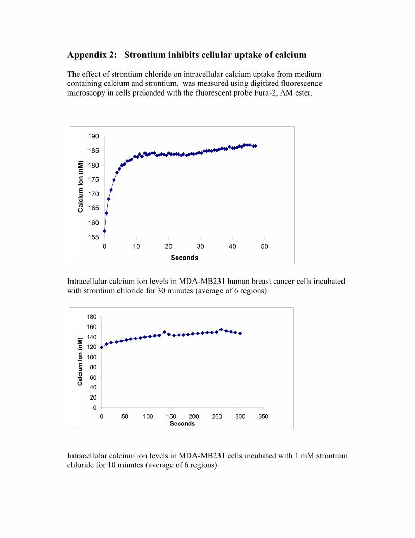
Appendix 2: Strontium inhibits cellular uptake of calcium The effect of strontium chloride on intracellular calcium uptake from medium containing calcium and strontium, was measured using digitized fluorescence microscopy in cells preloaded with the fluorescent probe Fura-2, AM ester.
155
160
165
170
175
180
185
190
0 10 20 30 40 50
Seconds
Cal
cium
Ion
(nM
)
Intracellular calcium ion levels in MDA-MB231 human breast cancer cells incubated with strontium chloride for 30 minutes (average of 6 regions)
0
20
406080
100
120140
160
180
0 50 100 150 200 250 300 350Seconds
Cal
cium
Ion
(nM
)
Intracellular calcium ion levels in MDA-MB231 cells incubated with 1 mM strontium chloride for 10 minutes (average of 6 regions)

116118120122124126128130132134136
0 10 20 30 40Seconds
Cal
cium
Ion
(nM
)
Intracellular calcium in MDA-MB231 cells incubated with sstrontium chloride (1 mM) for 15 minutes (average of 2 regions)
212
214
216
218
220
222
224
226
228
230
0 5 10 15 20 25Seconds
Cal
cium
Ion
Intracellular calcium in MDA-MB231 control cells (average of 5 regions)

148
150
152
154
156
158
160
0 5 10 15 20Seconds
Cal
cium
Ion
(nM
)
Intracellular calcium of MDA-MB231 cells incubated with strontium chloride (0.1 mM) for 10 minutes.
146148150152154156158160162164166168
0 5 10 15 20 25 30Seconds
Cal
cium
Ion
(nM
)
Intracellular calcium ion concentration in MDA-MB 231 cells incubated with strontium chloride (0.1 mM) for 15 minutes.

212
214
216
218
220
222
224
226
228
0 5 10 15 20 25 30 35
Seconds
Cal
cium
ion
( nM
)
Intracellular calcium ion concentration in control MDA-MB231 cells incubated in medium without strontium chloride

Appendix 3: Comparison of cell viability assays. Materials and methods Cell line MDA-MB-231-luc human breast cancer cell line (obtained from Xenogen, Alameda, CA) was used for this study. This cell line has been transfected with luciferase gene and expresses high level of luciferase. Cells were routinely maintained as monolayers in DMEM medium supplemented with 10% heat inactivated fetal bovine serum (FBS), penicillin (50 units/ml), and streptomycin (50 µg/ml) (Invitrogen), and kept at 37°C in humidified atmosphere containing 5% CO2 in air. Hyperthermia treatment For 96-well plate: Cells were seeded in sterile 96-well plates at a density of 1 x10⁴ cells/well and incubated overnight. For 24-well plate: cells were seeded at densities of 1x10³, 1x10⁴, 1x10⁵ and 5x10⁵ /well. Hyperthermia was applied at 43°C by sealing the plates with parafilm and enclosing in a Ziploc bag and then immersing the bag into a temperature controlled water bath maintained at 43o(± 0.1°C). The continuous heating period ranged from 10 to 120 minutes. Controls were sealed in ziplock bags and immersed in a 37°C water bath. After heating, plates were ready for optical imaging, and for MTT and clonogenic assays. MTT Assay The MTT colorimetric assay was performed to detect tumor cell viability based upon the reduction of the tetrazolium dye MTT [3-(4,5-dimethylthiazol-2-yl)-2,5-diphenyltetrazolium bromide] (Sigma Chemical Co., St. Louis, MO) by viable cells. This assay detects any reduction of metabolic viability, with or without relationship to apoptosis or necrosis. Immediately after exposure of the cells to 43°C for different times from 10 minutes up to two hours, the old medium was removed, MTT solution (100µl; 0.5mg/ml) in RPMI medium (phenol red free) was added. After incubation for 3 hours at 37°C, cellular mitochondrial dehydrogenase activity reduced the yellow MTT dye to a purple formazan, which was then solubilized with DMSO, the absorbance was determined at a wavelength of 560nm using multiwell scanning spectrophotometer. For delayed MTT assay, the Cells were incubated at 37oC for three to five days after the hyperthermia treatment, the cells were returned to 37oC incubator for three to five days followed by addition of MTT solution (100 µl; 0.5mg/ml) in RPMI medium (phenol red free). The assay was then completed as described above.

Clonogenic assay Following hyperthermia treatment, cells were trypsinized at 37°C for 5 minutes, and pipetted up and down 5 times to break up cell clumps and obtain a single cell suspension. The same volume of single cell suspension was plated on 100-mm tissue culture dishes with fresh medium and kept at 37°C, 5% CO2 incubator for 10~14 days. Colonies consisting of more than 50 cells were counted. Survival curves were generated by plotting percentage rate of the number of colonies formed at a given heating condition to the number of colonies produced by related unheated control cells versus the heating time at the given temperature. We are trying to adjust the MTT assay protocol with a view to obtaining results that reflect the clonogenic potential of cells.
0
20
40
60
80
100
0 20 40 60 80 100 120
Heating time(minutes)
Rel
ativ
e su
rviv
al(%
)
Results of MTT assay performed immediately after heat treatment at 430C.

0102030405060708090
100110
0 20 40 60 80 100 120
Heating time(min)
rela
tive
surv
ival
(%)
Results of MTT assay performed five days after heat treatment
0102030405060708090
100110
0 20 40 60 80 100 120
Heating time(minutes)
Rel
ativ
e su
rviv
al(%
)
Cells were subjected to 43o C hyperthermia for different durations and cell survival was determined using clonogenicity assay.

00.20.40.60.8
11.2
1000 10000 100000 500000
Number of cellsAbs
orba
nce
at 5
60nm
Control
150min heat
The effect of initial cell numbers on MTT assay carried out immediately after hyperthermis treatment.
00.20.40.60.8
11.2
1000 10000 100000 500000
Number of cellsAbs
orba
nce
at 5
60nm
Control150min heat
The effect of initial cell numbers on the results of MTT assay carried out 5 days after 2 hours of hyperthermis treatment at 43o C.