Avi Jacob, Ph.D. Head of Light Microscopy Part1:...
22
J. Paul Robinson, Ph.D. SVM Professor of Cytomics & Prof. Biomedical Engineering Director, Purdue University Cytometry Laboratories Most of the slides in this lecture were taken from Paul Robinson Microscopy BIU Equipment Center course for M.Sc. Students Avi Jacob, Ph.D. Head of Light Microscopy Part1: History
Transcript of Avi Jacob, Ph.D. Head of Light Microscopy Part1:...
J. Paul Robinson, Ph.D.
SVM Professor of Cytomics & Prof. Biomedical Engineering
Director, Purdue University Cytometry Laboratories
Most of the slides in this lecture were taken from
Paul Robinson
Microscopy BIU Equipment Center course for M.Sc. Students
Avi Jacob, Ph.D.
Head of Light Microscopy
Part1: History

Introduction
• Early Microscope History
• Fundamental Discoveries
• Key Individuals in the 17, 18 and 19th
centuries
• Modern Microscopy – 20th century
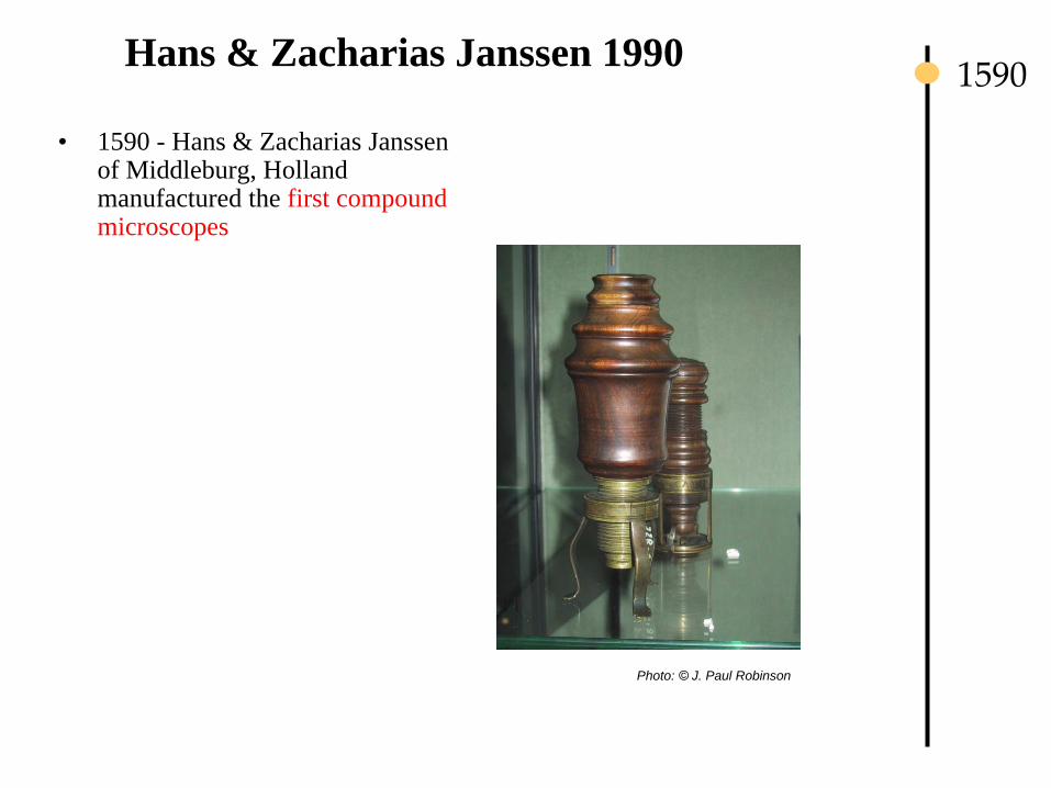
Hans & Zacharias Janssen 1990
• 1590 - Hans & Zacharias Janssen of Middleburg, Holland manufactured the first compound microscopes
1590
Photo: © J. Paul Robinson

Galileo Galilei (1564-1642)
• 1610 - he began publicly supporting the heliocentric view, which placed the Sun at the centre of the universe
• Galileo has been variously called – the "father of modern observational astronomy – the "father of modern physics – the "father of science
• The name "telescope" was coined for Galileo's instrument by a Greek mathematician, Giovanni Demisiani, at a banquet held in 1611 by Prince Federico Cesi to make Galileo a member of his Accademia dei Lincei
1610
• Telescope was derived from the Greek tele = 'far' and skopein = 'to look or see'.
In 1610, he used a telescope at close range to magnify the parts of insects.
• Denounced to the Roman Inquisition early in 1615
• 1624 he had perfected a compound microscope
• The Linceans played a role again in naming the "microscope" a year later when
fellow academy member Giovanni Faber coined the word for Galileo's invention
from the Greek words μικρόν (micron) meaning "small," and σκοπεῖν (skopein)
meaning "to look at."
• Published “Dialogue Concerning the Two Chief World Systems” in 1632, and
was tried by the Inquisition, found "vehemently suspect of heresy," forced to
recant, and spent the rest of his life under house arrest (to 1642)

Robert Hooke (1635-1703)-
© J.Paul Robinson
The Royal Society of London founded in 1616 during the reign of King James I
•1665 - Robert Hooke (1635-1703)- book Micrographia, published in 1665, devised the compound microscope most famous microscopical observation was his study of thin slices of cork. Named the term “Cell”
1665
Photo: © J. Paul Robinson
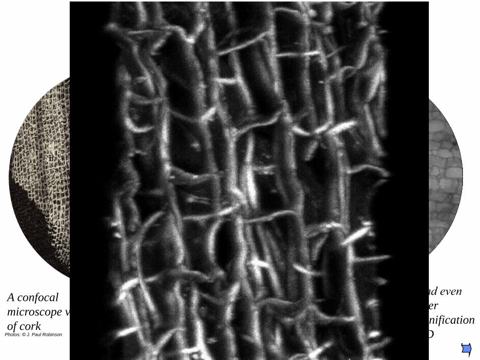
What did Hooke see when he looked at cork?
Hooke, 1665 The Purdue version of the
Hooke cork (2002)
A confocal
microscope view
of cork
…And even
higher
Magnification
in 3D Photos: © J. Paul Robinson

Antioni van Leeuwenhoek (1632-1723)
• 1673 - Antioni van Leeuwenhoek (1632-1723) Delft, Holland, worked as a
draper (a fabric merchant); he is also known to have worked as a surveyor, a
wine assayer, and as a minor city official.
• Leeuwenhoek is incorrectly called "the inventor of the microscope"
• Created a “simple” microscope that could magnify to about 275x, and
published drawings of microorganisms in 1683
• Could reach magnifications of over 200x with simple ground lenses -
however compound microscopes were mostly of poor quality and could only
magnify up to 20-30 times. Hooke claimed they were too difficult to use - his
eyesight was poor.
• Discovered bacteria, free-living and parasitic microscopic
protists, sperm cells, blood cells, microscopic nematodes
• In 1673, Leeuwenhoek began writing letters to the Royal
Society of London - published in Philosophical Transactions
of the Royal Society
• In 1680 he was elected a full member of the Royal Society,
joining Robert Hooke, Henry Oldenburg, Robert Boyle,
Christopher Wren
1673
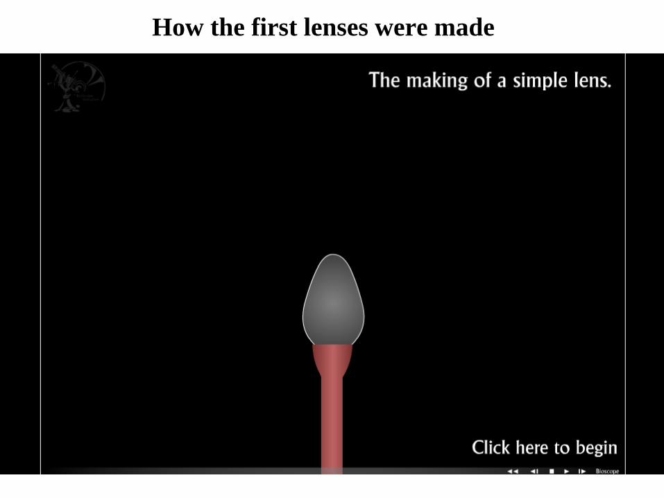
How the first lenses were made

Joseph Lister
• In 1830, by Joseph Jackson Lister (father of Lord Joseph Lister)
solved the problem of Spherical Aberration - caused by light
passing through different parts of the same lens. He solved it
mathematically and published this in the Philosophical
Transactions in 1830
© J.Paul Robinson
Joseph Lister
1830
Photos: © J. Paul Robinson
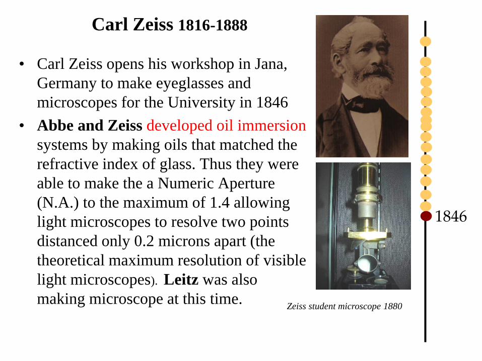
Carl Zeiss 1816-1888
• Carl Zeiss opens his workshop in Jana,
Germany to make eyeglasses and
microscopes for the University in 1846
• Abbe and Zeiss developed oil immersion
systems by making oils that matched the
refractive index of glass. Thus they were
able to make the a Numeric Aperture
(N.A.) to the maximum of 1.4 allowing
light microscopes to resolve two points
distanced only 0.2 microns apart (the
theoretical maximum resolution of visible
light microscopes). Leitz was also
making microscope at this time.
1846
Zeiss student microscope 1880

Pasteur - 1860
Louis Pasteur – his microscope was made in Paris by Nachet in
about 1860 and was made of brass
1860
Photo: © J. Paul Robinson
Photos: taken in London Science Museum by J. Paul Robinson

Abbe & Zeiss
• Ernst Abbe together with Carl Zeiss published a paper in 1877
defining the physical laws that determined resolving distance of
an objective. Known as Abbe’s Law
“minimum resolving distance (d) is related to the wavelength of light (lambda)
divided by the Numeric Aperture, which is proportional to the angle of the light
cone (theta) formed by a point on the object, to the objective”. “The impetus for the emergence into the industrial age was given by
Ernst Abbe (appointed Associate Professor in 1870), who, while still in
his early 30s, developed his theory of microscope image formation, which
took into consideration the familiar phenomenon of diffraction, and thus
made the leap in microscope construction from trial and error to
methodical design. He was given this commission by a university
mechanic, Carl Zeiss, who had been steadily perfecting the construction
of optical equipment in his private workshops. Otto Schott, who received
his doctorate at Jena in 1875, was the third to enter into this alliance by
founding, at Abbe’s urging, a "Laboratory for Glass Technology" in 1884,
to produce the highly pure special lenses for Zeiss’s microscopes and
optical equipment. Humboldt’s pupil Matthias Jakob Schleiden, Professor
of Botany and famous for his cell theory, encouraged -- and later
benefited from -- this process, which was to prove exemplary in German
economic history.”
http://www.uni-jena.de/History-lang-en.html
Abbe
Ernst Abbe joins Zeiss (Jena), develops Abbe sine condition optics, improving optics significantly
in 1873
1877
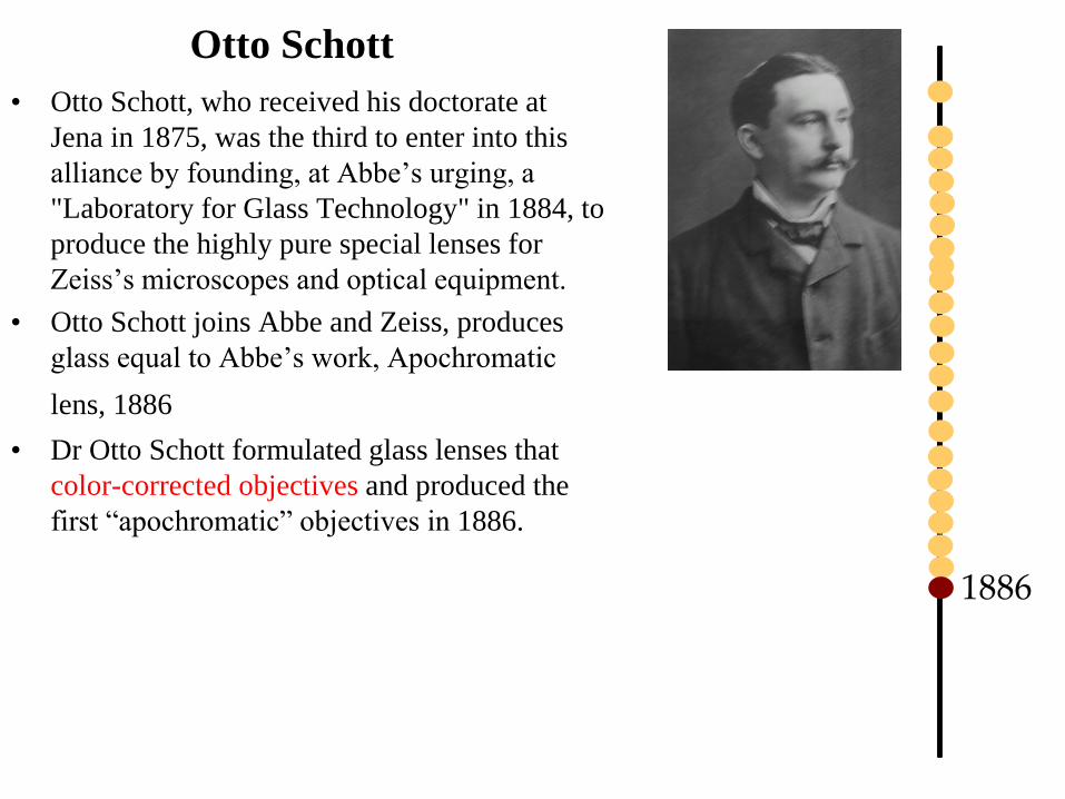
Otto Schott
• Otto Schott, who received his doctorate at
Jena in 1875, was the third to enter into this
alliance by founding, at Abbe’s urging, a
"Laboratory for Glass Technology" in 1884, to
produce the highly pure special lenses for
Zeiss’s microscopes and optical equipment.
• Otto Schott joins Abbe and Zeiss, produces
glass equal to Abbe’s work, Apochromatic
lens, 1886 • Dr Otto Schott formulated glass lenses that
color-corrected objectives and produced the
first “apochromatic” objectives in 1886.
1886

August Karl Johann Valentin Köhler (1866-1948)
• Early 20th Century Professor Köhler developed the
method of illumination still called “Köhler
Illumination”
• In 1900, he was invited to join the Zeiss Optical Works
company in Jena, Germany, by Siegfried Czapski based
on his earlier work on improving microscope
illumination. He stayed with Zeiss as a physicist for 45
years and became instrumental to the development of
modern light microscope design.
• Köhler recognized that using shorter wavelength light
(UV) could improve resolution
• The driving force for Köhler’s even illumunation
invention was the use of gas lamps and similar uneven
light sources that created serious problems in trying to
gain even and constant illumination
Image source: http://en.wikipedia.org/wiki/File:August_Koehler.jpg
1900

1900
Köhler
• Köhler illumination creates an evenly
illuminated field of view while illuminating
the specimen with a very wide cone of light
• Two conjugate image planes are formed
– one contains an image of the specimen and the
other the filament from the light
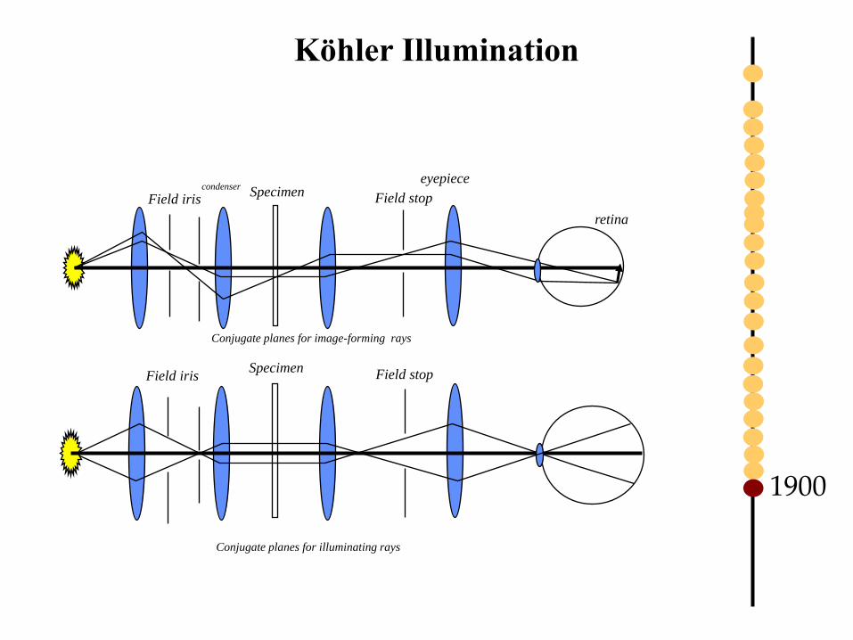
Köhler Illumination
Specimen Field stop Field iris
Conjugate planes for illuminating rays
Specimen Field stop Field iris
Conjugate planes for image-forming rays
condenser eyepiece
retina
1900
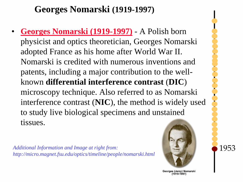
Georges Nomarski (1919-1997)
• Georges Nomarski (1919-1997) - A Polish born
physicist and optics theoretician, Georges Nomarski
adopted France as his home after World War II.
Nomarski is credited with numerous inventions and
patents, including a major contribution to the well-
known differential interference contrast (DIC)
microscopy technique. Also referred to as Nomarski
interference contrast (NIC), the method is widely used
to study live biological specimens and unstained
tissues.
Additional Information and Image at right from:
http://micro.magnet.fsu.edu/optics/timeline/people/nomarski.html 1953
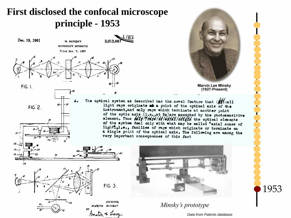
First disclosed the confocal microscope
principle - 1953
1953
Minsky’s prototype
Data from Patents database

Please memorize this
• Janssen brothers: first compound microscopes
• Galileo: perfected compound microscope
• Robert Hooke: coined the term “Cell”
• Antioni van Leeuwenhoek: discovered bacteria with simple ground
lenses
• Joseph Lister: first to fix abberations
• Carl Zeiss and Ernst Abbe: developed oil immersion systems
• Ernst Abbe: Abbe’s Law for minimum resolving distance
• Otto Schott: developed good glass
• Karl Kochler: method of illumination called “Kochler Illumination
• Georges Nomarski: major contribution to differential interference
contrast
• Marvin Minskey: confocal microscope
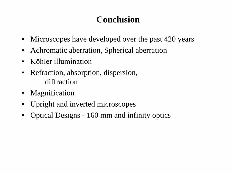
Conclusion
• Microscopes have developed over the past 420 years
• Achromatic aberration, Spherical aberration
• Köhler illumination
• Refraction, absorption, dispersion,
diffraction
• Magnification
• Upright and inverted microscopes
• Optical Designs - 160 mm and infinity optics

Summary Lecture 1
• Historical context of discovery of microscopes
• The major players in microscopy
• Variety of imaging tools developed to focus on specific
problems
• New inventions for high resolution imaging
• Linking automated microscopy to image processing