Avascular necrosis of the hip in a 41-year-old male: a ... · Avascular necrosis 138 J Can Chiropr...
5
J Can Chiropr Assoc 2004; 48(2) 137 0008-3194/2004/137–141/$2.00/©JCCA 2004 Avascular necrosis of the hip in a 41-year-old male: a case study Rahim Karim, BSc, DC, FCCRS(C), DACRB* Kambiz D Goel, DC, FCCRS(C) ** This case deals with the avascular necrosis or osteonecrosis of the femoral head in a 41-year-old male presenting to a chiropractor’s office. In addition to the clinical picture, diagnostic imaging should be performed to confirm the presence and extent of hip avascular necrosis. Referral to an orthopedic specialist is key and treatment is mainly surgical. (JCCA 2004; 48(2):137–141) key words: avascular necrosis, total hip arthroplasty, rehabilitation, osteonecrosis, chiropractic, hip pain. Ce cas traite de la nécrose avasculaire ou de l’ostéonécrose de la tête fémorale chez un homme de 41 ans se présentant dans un bureau de chiropraticien. En plus de dresser un portrait clinique, il est nécessaire d’effectuer une imagerie diagnostique afin de confirmer la présence et l’ampleur de la nécrose avasculaire de la hanche. Il est primordial d’aiguiller le patient chez un orthopédiste et le traitement est principalement chirurgical. (JACC 2004; 48(2):137–141) mots clés : nécrose avasculaire, arthroplastie totale de hanche, réadaptation, ostéonécrose, chiropratique, coxalgie. Case report A 41-year-old male presented to a chiropractor’s office for ongoing right sided low back, hip and knee pain for the past six months following jumping off a two meter high roof and landing on his feet. He had immigrated to Canada from abroad two months prior to consulting us. The patient had an antalgic limp and walked with the help of a cane. He complained of intermittent pain radiat- ing into his right groin and anteriomedial thigh region. He stated that his symptoms were aggravated by walking and stair climbing. His pain was relieved by sitting and resting. The patient did not report numbness or paresthe- sias in his lower extremities. There was no bowel and bladder dysfunction. The patient did not complain of any night sweats, fever or chills. The patient had previously sought medical advice and was prescribed pain killers and antiinflammatories. At that time, he had radiographs of his lumbar spine done which he stated were normal. He then saw a chiropractor for approximately one month with no improvement. Sub- sequently he had a lumbar spine MRI done and was re- ferred to a neurosurgeon for consult. The MRI showed a small disc bulge in the T12/L1 region. The neurosur- geon’s report stated that he was unable to correlate the patient symptoms with the MRI findings and recom- mended EMG studies. He then moved to Canada and pre- sented to our office for continuing management. Past history revealed that the patient received a flu vaccine 14 months prior to injury. He subsequently developed an allergic reaction and was diagnosed with ** Canadian Memorial Chiropractic College, 1900 Bayview Avenue, Toronto, Ontario, Canada M4G 3E6. Tel: 416-738-5035; E-mail [email protected] ** Private practice, Toronto, Ontario. © JCCA 2004.
Transcript of Avascular necrosis of the hip in a 41-year-old male: a ... · Avascular necrosis 138 J Can Chiropr...
J Can Chiropr Assoc 2004; 48(2) 137
0008-3194/2004/137–141/$2.00/©JCCA 2004
Avascular necrosis of the hipin a 41-year-old male:a case studyRahim Karim, BSc, DC, FCCRS(C), DACRB*Kambiz D Goel, DC, FCCRS(C) **
This case deals with the avascular necrosis or osteonecrosis of the femoral head in a 41-year-old male presenting to a chiropractor’s office. In addition to the clinical picture, diagnostic imaging should be performed to confirm the presence and extent of hip avascular necrosis. Referral to an orthopedic specialist is key and treatment is mainly surgical.(JCCA 2004; 48(2):137–141)
key words: avascular necrosis, total hip arthroplasty, rehabilitation, osteonecrosis, chiropractic, hip pain.
Ce cas traite de la nécrose avasculaire ou de l’ostéonécrose de la tête fémorale chez un homme de 41 ans se présentant dans un bureau de chiropraticien. En plus de dresser un portrait clinique, il est nécessaire d’effectuer une imagerie diagnostique afin de confirmer la présence et l’ampleur de la nécrose avasculaire de la hanche. Il est primordial d’aiguiller le patient chez un orthopédiste et le traitement est principalement chirurgical.(JACC 2004; 48(2):137–141)
mots clés : nécrose avasculaire, arthroplastie totale de hanche, réadaptation, ostéonécrose, chiropratique, coxalgie.
Case reportA 41-year-old male presented to a chiropractor’s officefor ongoing right sided low back, hip and knee pain forthe past six months following jumping off a two meterhigh roof and landing on his feet. He had immigrated toCanada from abroad two months prior to consulting us.The patient had an antalgic limp and walked with thehelp of a cane. He complained of intermittent pain radiat-ing into his right groin and anteriomedial thigh region.He stated that his symptoms were aggravated by walkingand stair climbing. His pain was relieved by sitting andresting. The patient did not report numbness or paresthe-sias in his lower extremities. There was no bowel andbladder dysfunction. The patient did not complain of anynight sweats, fever or chills.
The patient had previously sought medical advice andwas prescribed pain killers and antiinflammatories. Atthat time, he had radiographs of his lumbar spine donewhich he stated were normal. He then saw a chiropractorfor approximately one month with no improvement. Sub-sequently he had a lumbar spine MRI done and was re-ferred to a neurosurgeon for consult. The MRI showed asmall disc bulge in the T12/L1 region. The neurosur-geon’s report stated that he was unable to correlate thepatient symptoms with the MRI findings and recom-mended EMG studies. He then moved to Canada and pre-sented to our office for continuing management.
Past history revealed that the patient received a fluvaccine 14 months prior to injury. He subsequentlydeveloped an allergic reaction and was diagnosed with
** Canadian Memorial Chiropractic College, 1900 Bayview Avenue, Toronto, Ontario, Canada M4G 3E6.Tel: 416-738-5035; E-mail [email protected]
** Private practice, Toronto, Ontario.© JCCA 2004.
Avascular necrosis
138 J Can Chiropr Assoc 2004; 48(2)
leukocytoclastic vasculitis skin eruptions. This was treat-ed with four months of oral corticosteroid therapy withdoses up to 50 mg per day. The skin lesions resolved withtreatment. However he developed corticosteroid-inducedglucose intolerance subsequent to treatment. Past historyalso revealed a nasal fracture six years ago which re-quired two surgical interventions. The patient stated hedoes not drink or smoke currently and has not in the past.The patient stated that he does not have any night pain. Inaddition, he reported that he has had no noticeable weightchange in the past year. He stated that he has not workedsince his injury.
On physical examination, range of motion of the righthip was severely limited and painful in all ranges, withmost pain being felt in abduction and internal rotation.Palpation of the right hip region revealed extreme tender-ness. Muscle palpation revealed tenderness in the rightthigh and pelvic musculature. Muscle atrophy was alsonoted in the right thigh musculature. Lumbar spine rangeof motion was full with end range pain in right lateralflexion and right rotation. Valsalva was unremarkable.Straight leg raise produced right hip pain. Posterior jointprovocation tests were painful for L4, L5. SI testing waspainful for the right sacroiliac joint. Muscle palpation re-vealed tenderness in the lumbar paraspinal and right glu-teal musculature. Range of motion of the right knee wasfull and pain free and no effusion was noted. Muscle pal-pation revealed tenderness in the right TFL and quad-riceps musculature. Lower limb neurological testingrevealed normal reflexes and sensory testing bilaterally.Global muscle weakness was noted in the right lowerlimb when compared to the left.
ImpressionThe patient was suspected as having avascular necrosis ofthe right hip with differential diagnoses of hip osteoar-thritis or healed fracture. He was referred to a medical ra-diology facility for radiographs of the lumbar spine, righthip and pelvis. The radiology report stated that there wasmarked irregularity to the right femoral head with sclero-sis, subchondral lucency and mild collapse. This reportled to the diagnosis of avascular necrosis of the right hip.The patient was also diagnosed clinically as having lum-bosacral and sacroiliac strain/strain. Based on the radiol-ogy report, the patient was immediately referred to anorthopedic surgeon for consult and advice. The patient
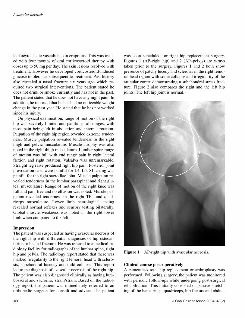
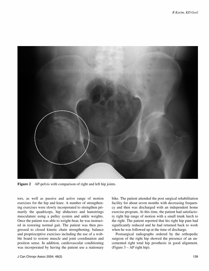
was soon scheduled for right hip replacement surgery.Figures 1 (AP–right hip) and 2 (AP–pelvis) are x-raystaken prior to the surgery. Figures 1 and 2 both showpresence of patchy luceny and sclerosis in the right femo-ral head region with some collapse and irregularity of thearticular cortex demonstrating a subchondral stress frac-ture. Figure 2 also compares the right and the left hipjoints. The left hip joint is normal.
Clinical course post-operativelyA cementless total hip replacement or arthroplasty wasperformed. Following surgery, the patient was monitoredwith periodic follow-ups while undergoing post-surgicalrehabilitation. This initially consisted of passive stretch-ing of the hamstrings, quadriceps, hip flexors and abduc-
Figure 1 AP-right hip with avascular necrosis.
R Karim, KD Goel
J Can Chiropr Assoc 2004; 48(2) 139
tors, as well as passive and active range of motionexercises for the hip and knee. A number of strengthen-ing exercises were slowly incorporated to strengthen pri-marily the quadriceps, hip abductors and hamstringsmusculature using a pulley system and ankle weights.Once the patient was able to weight-bear, he was instruct-ed in restoring normal gait. The patient was then pro-gressed to closed kinetic chain strengthening, balanceand proprioceptive exercises including the use of a wob-ble board to restore muscle and joint coordination andposition sense. In addition, cardiovascular conditioningwas incorporated by having the patient use a stationary
bike. The patient attended the post surgical rehabilitationfacility for about seven months with decreasing frequen-cy and then was discharged with an independent homeexercise program. At this time, the patient had satisfacto-ry right hip range of motion with a small trunk lurch tothe right. The patient reported that his right hip pain hadsignificantly reduced and he had returned back to workwhen he was followed up at the time of discharge.
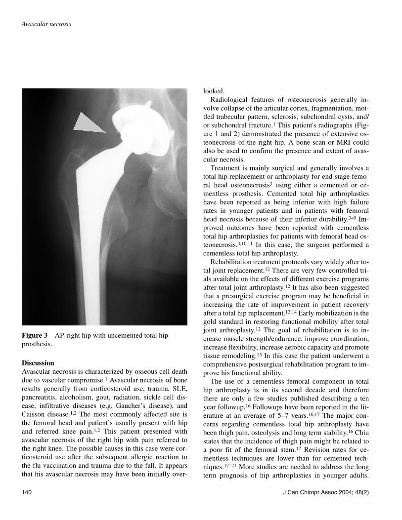
Postsurgical radiographs ordered by the orthopedicsurgeon of the right hip showed the presence of an un-cemented right total hip prosthesis in good alignment.(Figure 3 – AP right hip).
Figure 2 AP-pelvis with comparison of right and left hip joints.
Avascular necrosis
140 J Can Chiropr Assoc 2004; 48(2)
DiscussionAvascular necrosis is characterized by osseous cell deathdue to vascular compromise.1 Avascular necrosis of boneresults generally from corticosteroid use, trauma, SLE,pancreatitis, alcoholism, gout, radiation, sickle cell dis-ease, infiltrative diseases (e.g. Gaucher’s disease), andCaisson disease.1,2 The most commonly affected site isthe femoral head and patient’s usually present with hipand referred knee pain.1,2 This patient presented withavascular necrosis of the right hip with pain referred tothe right knee. The possible causes in this case were cor-ticosteroid use after the subsequent allergic reaction tothe flu vaccination and trauma due to the fall. It appearsthat his avascular necrosis may have been initially over-
looked.Radiological features of osteonecrosis generally in-
volve collapse of the articular cortex, fragmentation, mot-tled trabecular pattern, sclerosis, subchondral cysts, and/or subchondral fracture.1 This patient's radiographs (Fig-ure 1 and 2) demonstrated the presence of extensive os-teonecrosis of the right hip. A bone-scan or MRI couldalso be used to confirm the presence and extent of avas-cular necrosis.
Treatment is mainly surgical and generally involves atotal hip replacement or arthroplasty for end-stage femo-ral head osteonecrosis3 using either a cemented or ce-mentless prosthesis. Cemented total hip arthroplastieshave been reported as being inferior with high failurerates in younger patients and in patients with femoralhead necrosis because of their inferior durability.3–9 Im-proved outcomes have been reported with cementlesstotal hip arthroplasties for patients with femoral head os-teonecrosis.3,10,11 In this case, the surgeon performed acementless total hip arthroplasty.
Rehabilitation treatment protocols vary widely after to-tal joint replacement.12 There are very few controlled tri-als available on the effects of different exercise programsafter total joint arthroplasty.12 It has also been suggestedthat a presurgical exercise program may be beneficial inincreasing the rate of improvement in patient recoveryafter a total hip replacement.13,14 Early mobilization is thegold standard in restoring functional mobility after totaljoint arthroplasty.12 The goal of rehabilitation is to in-crease muscle strength/endurance, improve coordination,increase flexibility, increase aerobic capacity and promotetissue remodeling.15 In this case the patient underwent acomprehensive postsurgical rehabilitation program to im-prove his functional ability.
The use of a cementless femoral component in totalhip arthroplasty is in its second decade and thereforethere are only a few studies published describing a tenyear followup.16 Followups have been reported in the lit-erature at an average of 5–7 years.16,17 The major con-cerns regarding cementless total hip arthroplasty havebeen thigh pain, osteolysis and long term stability.16 Chiustates that the incidence of thigh pain might be related toa poor fit of the femoral stem.17 Revision rates for ce-mentless techniques are lower than for cemented tech-niques.17–21 More studies are needed to address the longterm prognosis of hip arthroplasties in younger adults.
Figure 3 AP-right hip with uncemented total hip prosthesis.
R Karim, KD Goel
J Can Chiropr Assoc 2004; 48(2) 141
Considering the age of this patient, periodic follow-upswith an orthopedic specialist are essential in helpingmonitor the integrity of the surgical procedure.
ConclusionWhenever a patient presents with hip pain secondary totrauma and/or corticosteroid use, the clinician must in-clude avascular necrosis as a differential. The diagnosisis confirmed by imaging procedures. The chiropractorshould refer the patient to a physician or specialist ifthere is radiological evidence of avascular or osteonecro-sis of the hip.
References1 Yochum T, Rowe L. Essentials of skeletal radiology. 2nd
ed. Baltimore: Williams & Wilkins 1996; 1260–1263.2 Tierney Jr. LM, McPhee SJ, Papadakis MA. Current
Medical diagnosis and treatment. 36th ed. Stamford: Appleton & Lange 1997; 798–799.
3 Fye MA, Huo MH, Zatorski LE, Keggi KJ. Total hip arthroplasty performed without cement in patients with femoral head osteonecrosis who are less than 50 years old. J Arthroplasty 1998; 13:876–881.
4 Callaghan JJ. Results of primary total hip arthroplasty in young patients. J Bone Joint Surg Am 1993; 75:1728.
5 Chandler HP, Reineck FT, Klixson RL, McCarthy JC. Total hip replacement in patients younger than 30 years old. J Bone Joint Surg Am 1981; 63:1426.
6 Devlin VJ, Einhorn TA, Gordon SL, et al. Total hip arthroplasty after renal transplantation: Long-term follow-up stuffy and assessment of metabolic bone status. J Arthroplasty 1988; 3:205.
7 Dorr LD, Takel GL, Conaty JP. Total hip arthroplasty in patients less than 45 years old. J Bone Joint Surg Am 1983; 65:474.
8 Saito S, Saito M, Nishina T, et al. Long-term results of total hip arthroplasty for osteonecrosis of the femoral head: A comparison with osteoarthritis. Clin Orthop 1989; 244:198.
9 Salvati EA, Cornell CN. Long-term follow-up of total hip replacement in patients with avascular necrosis. AAOS Instr Course Lec 1988; 37:67.
10 Piston RW, Engh CA, Carvalho PI, Suthers K. Osteonecrosis of the femoral head treated with total hip arthroplasty without cement. J Bone Joint Surg Am 1994; 76:202.
11 Phillips FM, Pottenger LA, Finn HA, Vandermolen J. Cementless total hip arthroplasty in patients with steroid-induced avascular necrosis of the hip. Clin Orhop 1994; 303:147.
12 Roos EW. Effectiveness and practice variation of rehabilitation after joint replacement. Curr Opin Rheumatol 2003; 15:160–162.
13 Gilbry HJ, Ackland RR, Wang AW, Morton AR, Trouchet T, Tapper J. Exercise improves early functional recovery after total hip arthroplasty. Clin Orthop 2003; 408:193–200.
14 Wang AW, Gilbey HJ, Ackland TR. Perioperative exercise programs improve early return of ambulatory function after total hip arthroplasty. Am J Phys Med Rehabil 2002; 81:801–806.
15 Liebensen C. Rehabilitation of the spine. Baltimore: Williams & Wilkins 1996; 31–34.
16 Hellman EJ, Capello WN, Feinberg JR. Omnifit Cementless total hip arthroplasty. Clin Orthop 1999; 364:164–174.
17 Chiu KH, Shen WY, Ko CK, Chan KM. Osteonecrosis of the femoral head treated with cementless total hip arthroplasty. J Arthroplasty 1997; 12:683–688.
18 Brinker MR, Rosenbery AG, Kull L, Galante JO. Primary total hip arthroplasty using noncemented porous-coated femoral components in patients with osteonecrosis of the femoral head. J Arthroplastry 1994; 9:457.
19 Kim YH, Oh JH, Oh SH. Cementless total hip arthroplasty in patients with osteonecrosis of the femoral head. Clin Orthop 1995; 320:73.
20 Piston RW, Engh CA, Carvalho PL, Suthers K. Osteonecrosis of the femoral head treated with total hip arthroplasty without cement. J Bone Joint Surg 1994; 76A:22.
21 Phillips FM, Pottenger LA, Finn HA, Vandermolen J. Cementless total hip arthroplasty in patients with steroid-induced avascular necrosis of the hip: a 62-month follow-up study. Clin Orthop 1994; 303:147.