Autopsy
24
AUTOPSY PREPARED BY : CHRISTIAN RAVEINA
-
Upload
rv252 -
Category
Health & Medicine
-
view
230 -
download
0
Transcript of Autopsy

AUTOPSY PREPARED BY : CHRISTIAN RAVEINA
INTRODUCTION •An autopsy is also known as postmortem examination, necropsy or obduction • It is a medical procedure that consists of a thorough examination of a dead body to determine the cause & manner of death & to evaluate any disease or injury that may be present • It is usually performed by a specialized medical doctor called a pathologist
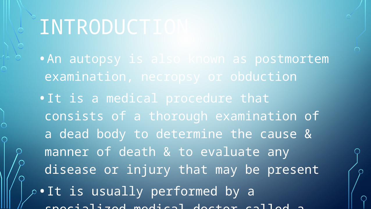
•The term “autopsy” derived from the Greek word “to see for oneself” •“ Necropsy” is form the Greek word “ seeing a dead body” •Necropsy is the term for a post-mortem examination on animal
CLASSIFICATION
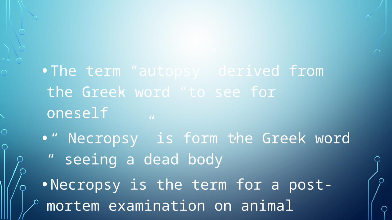
•Three main types :1.Forensic 2.Clinical/academic 3.Coroner’s

1.FORENSIC •This is done for medical legal purposes •No family permission is required to complete this type of autopsy •This is carried out when the cause of death may be a criminal matter such as accident or burns

2. CLINICAL/ ACADEMIC •This is usually performed in hospitals for research & study purposes•For a clinical autopsy to take place, cause of death must be established & a death certificate completed •To complete this type of autopsy, permission from the deceased’s legal next of kin is required

3.CORONER’S •This type of autopsy involves cases where no medical cause of death is readily available •Cause, manner & mechanism of death are in question •Eventually, the prospectors will identify whether the cases deserve comprehensive forensic autopsy or a routine postmortem

FORENSIC AUTOPSY
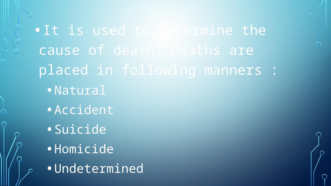
•It is used to determine the cause of death. Deaths are placed in following manners : •Natural •Accident •Suicide •Homicide •Undetermined
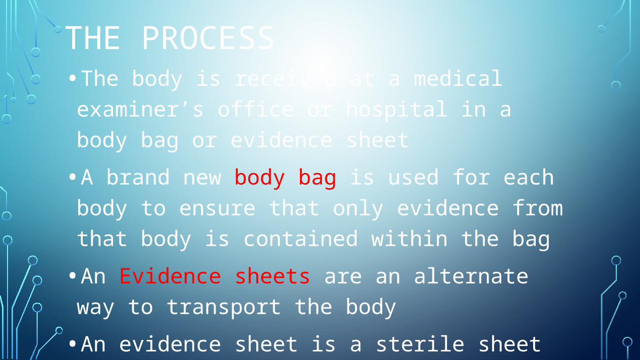
THE PROCESS •The body is received at a medical examiner’s office or hospital in a body bag or evidence sheet •A brand new body bag is used for each body to ensure that only evidence from that body is contained within the bag •An Evidence sheets are an alternate way to transport the body •An evidence sheet is a sterile sheet in which the body is covered

PHYSICAL EXAMINATION INVOLVES •2 parts :•External examination •Internal examination
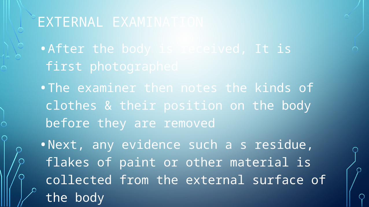
EXTERNAL EXAMINATION •After the body is received, It is first photographed•The examiner then notes the kinds of clothes & their position on the body before they are removed •Next, any evidence such a s residue, flakes of paint or other material is collected from the external surface of the body •Ultraviolet light may also be used to search body surfaces for any evidence not easily visible to the naked eye

•Samples of hair, nails are taken & the body may also be radio graphically imaged •Once the external evidence is collected, the body is removed from the bag, undressed & any wound present are examined •The body is cleaned, weighed & measured in preparation for the internal examination •The scale used to weigh the body is often designed to accommodate the cart that body is transported on, its weight is then deducted from the total weight shown to give the weight of the body
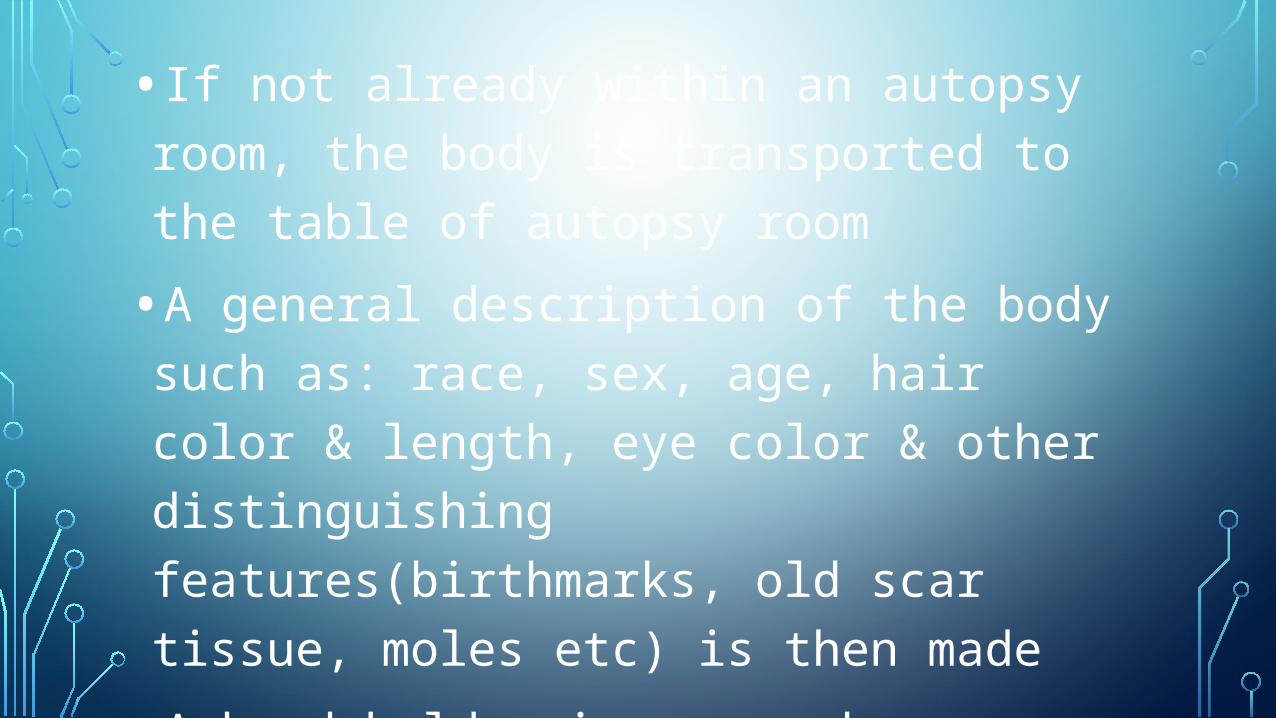
•If not already within an autopsy room, the body is transported to the table of autopsy room •A general description of the body such as: race, sex, age, hair color & length, eye color & other distinguishing features(birthmarks, old scar tissue, moles etc) is then made •A hand held voice recorder or a standard examination form is normally used to record this information

INTERNAL EXAMINATION • If not already in place, a plastic or rubber brick
called a “body block” is placed under the back of the body, causing the arms & neck to fall backward while stretching & pushing the chest upward to make it easier to cut open • This gives the prospector/a pathologist or
assistance, maximum exposure to the trunk • After this, the internal examination begins • The internal examination consists of inspecting the
internal organs of the body for evidence of trauma or other indications of cause of death

•A large & deep “Y” shape incision can be made from behind each ear & running down the sides of neck, meeting at the breast bone •This is the approach most often used in forensic autopsies so as to allow maximum exposure of neck structures for later detailed examination

•A “T” shape incision made from the tips of both shoulder, in a horizontal line across the region of the collar bones to meet at sternum in the middle •This initial cut is used more often to produce a more aesthetic finish to the body when it is re-constituted as stitching marks will not be as apparent as with a “Y”shape •A single vertical cut is made from the middle of the neck


RECONSTITUTION OF THE BODY •After the examination, the body has an open & empty chest cavity with chest flaps open on both sides, the top of the skull is missing & the skull flaps are pulled over the face & neck • It is unusual to examine the face, arms, hands or legs internally. •All the organs & tissue must be returned to the body unless permission is given by family to retain any tissue for further investigation

•Normally, the internal body cavity is lined with cotton wood or an appropriate material, the organs are then placed into a plastic bag to prevent leakage & returned to the body cavity •The chest flaps are then closed & sewn back together & the skull cap is sewed back in place •Then the body may be wrapped in a sheet

Thank you…