AUTOIMMUNE DISEASES AUTOIMMUNE DISEASES Martina Vachová Department of Immunology and Allergology...
53
AUTOIMMUNE DISEASES Martina Vachová Department of Immunology and Allergology Faculty of Medicine and Faculty Hospital in Pilsen
-
Upload
kelly-owens -
Category
Documents
-
view
217 -
download
0
Transcript of AUTOIMMUNE DISEASES AUTOIMMUNE DISEASES Martina Vachová Department of Immunology and Allergology...

AUTOIMMUNE DISEASES
Martina Vachová
Department of Immunology and Allergology Faculty of Medicine and Faculty Hospital in Pilsen
AUTOIMMUNE DISEASES
chronic and usually irreversible incidence: 5%-7% of population, higher frequencies in women, increases with age
Autoimmune diseases
Result from a failure of self-tolerance Immunological tolerance is specific unresponsiveness
to an antigen All individuals are tolerant of their own (self) antigens
Autoimmunity
is defined as an immune response against self antigens
The principal factors in the development of autoimmunity are the inheritance of susceptibility genes and environmental triggers, such as infections
Most autoimmune diseases are polygenic and are asssociated with multiple gene loci, the most important of which are the MHC genes
Infections may activate self-reactive lymphocytes, thereby triggering the development of autoimmune diseases
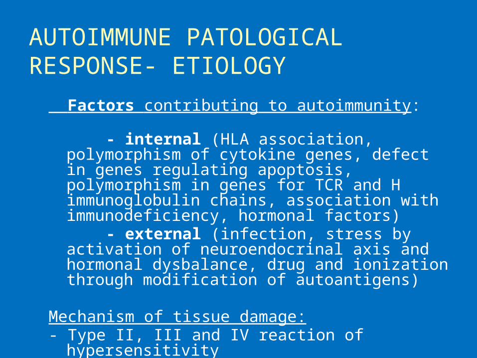
AUTOIMMUNE PATOLOGICAL RESPONSE- ETIOLOGY
Factors contributing to autoimmunity: - internal (HLA association, polymorphism of cytokine
genes, defect in genes regulating apoptosis, polymorphism in genes for TCR and H immunoglobulin chains, association with immunodeficiency, hormonal factors)
- external (infection, stress by activation of neuroendocrinal axis and hormonal dysbalance, drug and ionization through modification of autoantigens)
Mechanism of tissue damage:- Type II, III and IV reaction of hypersensitivity

Type II hypersensitivity reaction
IgM and IgG Ab promote the phagocytosis of cells which they bind, induce inflammation by complement – and Fc receptor- mediated leukocyte recruitment , and may interfere with the functions of cells by binding to essential molecules and receptors.
Graves‘ disease, Pernicious anemia, Myasthenia gravis, Acute rheumatic fever, Goodpasture‘s syndrome, Pemphigus vulgaris, Autoimmune hemolytic anemia or thrombocytopenic purpura

Type III hypersensitivity reaction
Ab may bind to circulating antigens to form immune complexes, which deposit in vessels and cause tissue injury
Injury is mainly due to leukocyte recruitment and inflammation
Systemic lupus erythematosus, Polyarteritis nodosa, Poststreptococcal glomerulonephritis
Type IV hypersensitivity reaction
T cell- mediated diseases are caused by Th1-mediated delayed-type hypersensitivity reactions or Th17- mediated inflammatory reactions, or by killing of host cells by CD8+ CTLs (cytotoxic lymphocytes).
Diabetes mellitus (insulin-dependent), Rheumatoid arthritis, Multiple sclerosis, Inflammatory bowel disease

Autoimmune diseases
Systemic Organ-specific
Organ-localised
CLINICAL CATEGORIES systemic - affect many organs and tissues - organ non-specific autoantibodies
organ specific - affect one organ - organ specific autoantibodies or autoreactive T
lymphocytes
organ localised - affect predominantly one organ accompanied by
affection of other organs (inflammatory bowel diseases, coeliac disease, AI hepatitis, pulmonary fibrosis)
- organ non-specific autoantibodies
EXAMPLES OF SYSTEMIC AUTOIMMUNE DISEASES
examples autoantibodies

SYSTEMIC AUTOIMMUNE DISEASES
Systemic lupus erythematosus Rheumathoid arthritis Sjögren‘s syndrome Dermatopolymyositis Systemic sclerosis Mixed connective tissue disease Vasculitis

SYSTEMIC LUPUS ERYTHEMATOSUS
chronic, inflammatory, multiorgan disorder
autoantibodies react with nuclear material and attack cell function, immune complexes with dsDNA deposit in the tissues
general symptoms: include malaise, fever, weight loss
multiple tissues are involved including the skin, mucosa, kidney, joints, brain and cardiovascular system
characteristic features: butterfly rash, renal involvement, CNS manifestation, pulmonary fibrosis

DIAGNOSTIC TESTS
an elevated ESR (erythrocyte sedimentation rate), low CRP, trombocytopenia, leucopenia, hemolytic anemia, decreased levels of complement compounds (C4, C3), elevated serum Ig levels, immune complexes in serum

AUTOANTIBODIES
Autoantibodies: ANA, dsDNA (double-stranded), ENA (SS-A/Ro, SS-B/La, Sm), against histones, phospholipids

RHEUMATOID ARTHRITIS
chronic, inflammatory disease with systemic involvement characterized by an inflammatory joint lesion in the synovial
membrane, destruction of the cartilage and bone, results in the joint deformation
clinical features: arthritis, fever, fatigue, weakness, weight loss systemic features: vasculitis, pericarditis, uveitis, nodules under skin,
intersticial pulmonary fibrosis diagnostic tests: elevated C- reactive protein and ESR, elevated serum gammaglobulin levels - autoantibodies against IgG = rheumatoid factor (RF), a-CCP (cyclic citrulline peptid), ANA - X-rays of hands and legs- show a periarticular porosis, marginal erosion
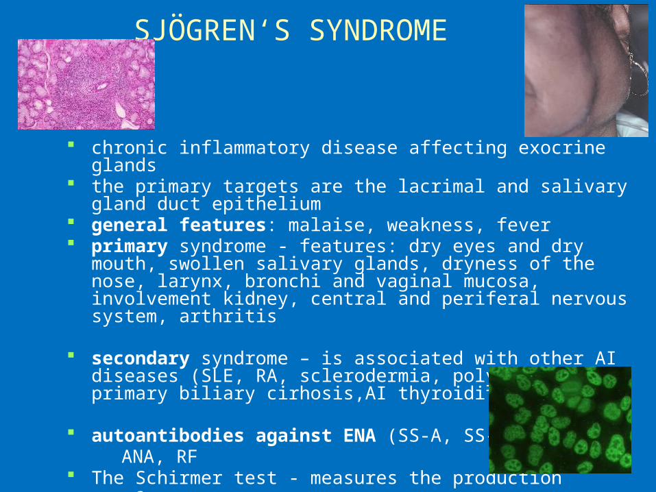
SJÖGREN‘S SYNDROME
chronic inflammatory disease affecting exocrine glands the primary targets are the lacrimal and salivary gland duct
epithelium general features: malaise, weakness, fever primary syndrome - features: dry eyes and dry mouth, swollen
salivary glands, dryness of the nose, larynx, bronchi and vaginal mucosa, involvement kidney, central and periferal nervous system, arthritis
secondary syndrome – is associated with other AI diseases (SLE, RA, sclerodermia, polymyositis, primary biliary cirhosis,AI thyroiditis)
autoantibodies against ENA (SS-A, SS-B), ANA, RF The Schirmer test - measures the production of tears

Heliotrope rash is a violaceous eruption on the upper eyelids, often with swelling
• a connective-tissue disease characterized by inflammation
of the muscles and the skin.
Gottron's sign is an erythematous, scaly eruption occurring in symmetric fashion over the MCP and interphalangeal joints
Dermatopolymyositis

Dermatopolymyositis
elevated creatine phosphokinase (CPK)
muscle biopsy (a mixed B- and T-cell perivascular inflammatory infiltrate, perifascicular muscle fiber atrophy)
EMG (electromyogram) autoantibodies - ENA (Jo-1)

Systemic sclerosis
sclerosis in the skin or other organs Diffuse scleroderma (progressive systemic
sclerosis) is the most severe form, involves skin, will generally cause internal organ
damage (specifically the lungs and gastrointestinal tract)
The limited form is much milder The limited form is often referred to as CREST
syndrome (CREST is an acronym for the five main features: Calcinosis, Raynaud's syndrome, Esophageal dysmotility, Sclerodactyly, Telangiectasia
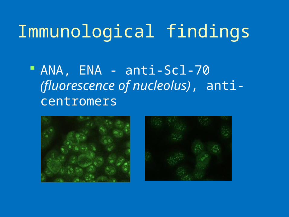
Immunological findings
ANA, ENA - anti-Scl-70 (fluorescence of nucleolus), anti-centromers
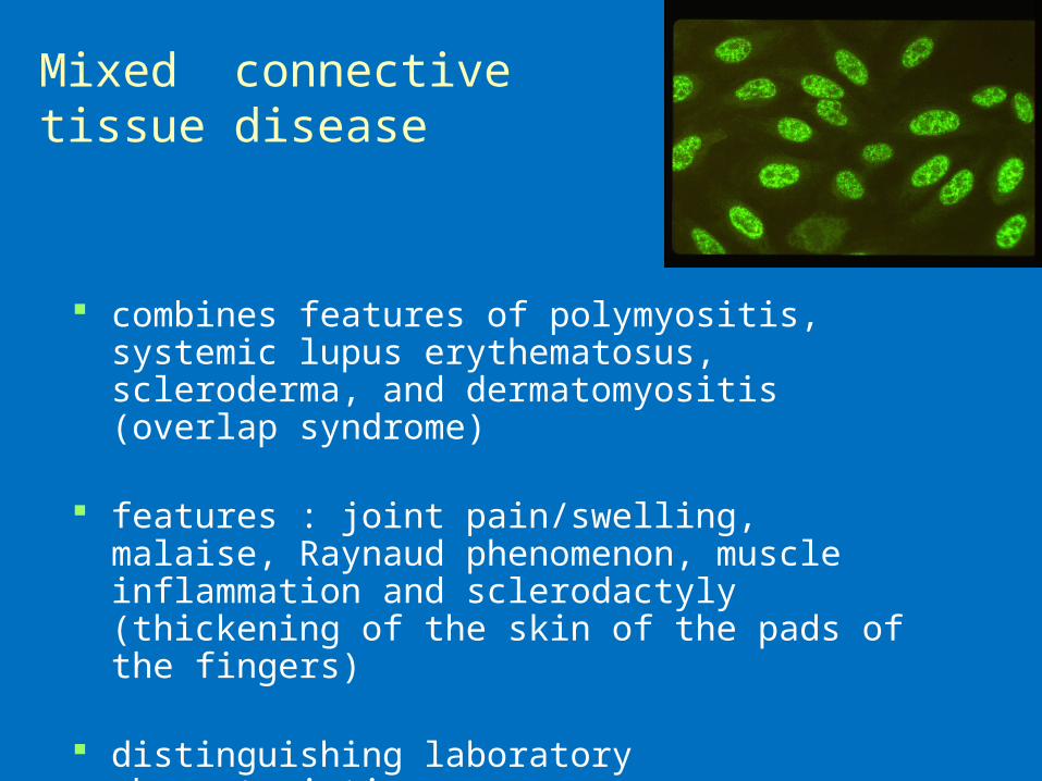
Mixed connective tissue disease
combines features of polymyositis, systemic lupus erythematosus, scleroderma, and dermatomyositis (overlap syndrome)
features : joint pain/swelling, malaise, Raynaud phenomenon, muscle inflammation and sclerodactyly (thickening of the skin of the pads of the fingers)
distinguishing laboratory characteristics: a positive, speckled anti-nuclear antibody (ANA) and
anti-U1-RNP antibody (ENA)

Vasculitis
characterized by inflammatory destruction of vessels leading to thrombosis and
aneurysms
affect mostly lung, kidneys, skin
diagnostic tests: elevated ESR, CRP, leucocytosis, biopsy of affected organ (necrosis, granulomas), angiography

Vasculitis
p- ANCA (myeloperoxidase) positivity (Polyarteritis nodosa, Churg- Strauss, Microscopic polyarteritis nodosa)
c- ANCA (serin proteinase) positivity (Wegener granulomatosis, Churg- Strauss syndrome)

Classification
Large vessel vasculitis (Takayasu arteritis, Giant cell (temporal) arteritis)
Medium vessel vasculitis (Polyarteritis nodosa, Wegener's granulomatosis, Kawasaki disease)
Small vessel vasculitis (Churg-Strauss arteritis, Microscopic polyarteritis, Henoch-Schönlein purpura)
Symptoms: fatigue, weakness, fever, arthralgias, abdominal pain, hypertension, renal insufficiency, and neurologic dysfunction
EXAMPLES OF ORGAN LOCALISED AUTOIMMUNE DISEASES
diseases autoantibodies
ORGAN LOCALIZED AUTOIMMUNE DISEASES
Ulcerative colitis Crohn‘s disease Autoimmune hepatitis Primary biliary cirhosis Pulmonary fibrosis

Ulcerative colitis
chronic inflammation of the large intestine mucosa and submucosa
features: diarrhea, bloody and mucus stools extraintestinal features (arthritis, uveitis) autoantibodies against pANCA, a- large intestine

Crohn‘s disease
the granulomatous inflammation of whole intestinal wall with ulceration and scarring that can result in abscess and fistula formation
the inflammation in Crohn's disease the most commonly affects the terminal ileum, presents with diarrhea and is accompanied by extraintestinal features - iridocyclitis, uveitis, artritis, spondylitis
antibodies against Saccharomyces cerevisiae
(ASCA), a- pancreas
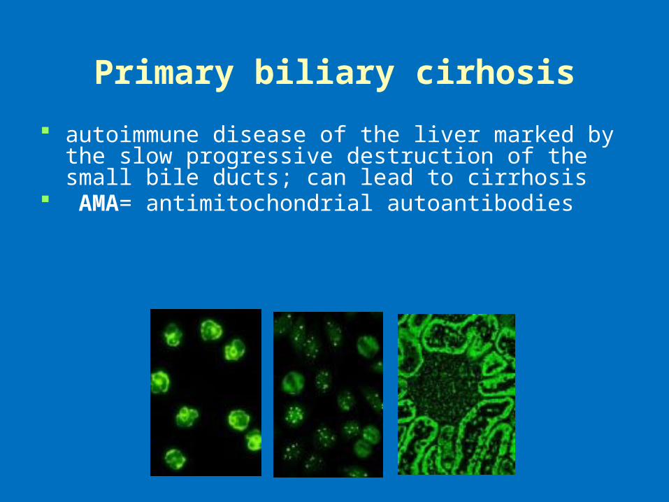
Primary biliary cirhosis
autoimmune disease of the liver marked by the slow progressive destruction of the small bile ducts; can lead to cirrhosis
AMA= antimitochondrial autoantibodies
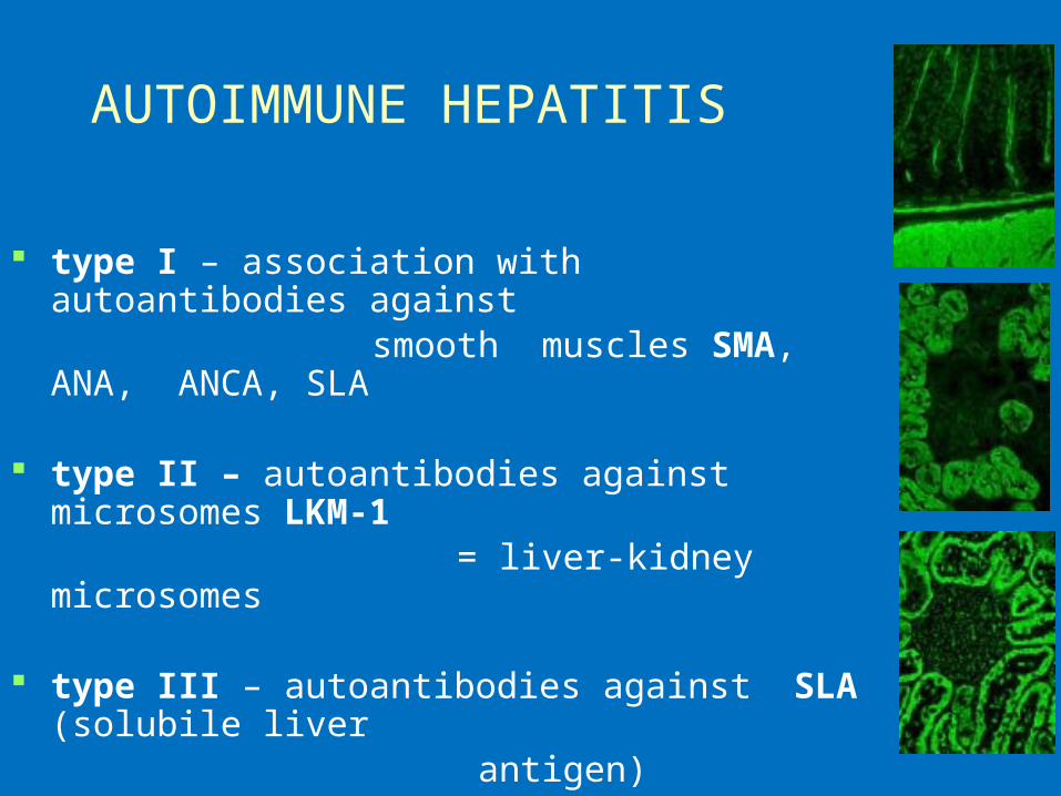
AUTOIMMUNE HEPATITIS
type I – association with autoantibodies against smooth muscles SMA, ANA, ANCA, SLA
type II – autoantibodies against microsomes LKM-1
= liver-kidney microsomes
type III – autoantibodies against SLA (solubile liver
antigen)

ORGAN SPECIFIC AUTOIMMUNE DISEASES
Autoimmune endocrinopathy Autoimmune neurological diseases Autoimmune cytopenia

AUTOIMMUNE ENDOCRINOPATHY
Hashimoto‘s thyroiditis Graves-Basedow disease Diabetes mellitus I. type Addison‘s disease Autoimmune polyglandular syndrome Pernicious anemia

Hashimoto‘s thyroiditis
thyroid disease result to hypothyroidism on the base of lymphocytes and plasma cells infiltrate
autoantibodies against thyroidal peroxidase (a-
TPO) and/or against thyroglobulin (a-TG)
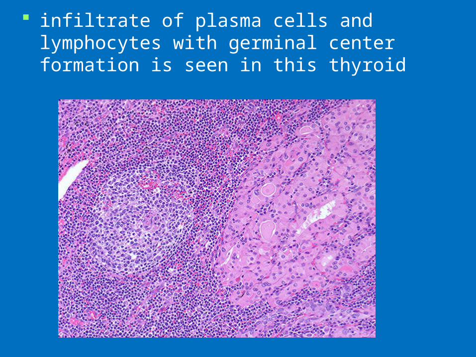
infiltrate of plasma cells and lymphocytes with germinal center formation is seen in this thyroid

Grave‘s disease
thyrotoxicosis from overproduction of thyroid hormone (patient exhibit fatigue, nervousness, increased sweating, palpitations, weight loss,
exophtalmus)
autoantibodies against thyrotropin receptor, autoantibodies cause thyroid cells proliferation

Diabetes mellitus (insulin- dependent)
characterized by an inability to process sugars in the diet, due to a decrease in or total absence of insulin production
results from immunologic destruction of the insuline- producing β-cells of the islets of Langerhans in the pancreas
autoantibodies against GAD- glutamic acid decarboxylase = primary antigen), autoantibodies anti- islet cell, anti- insulin
islets are infiltrated with B and T cells

Polyglandular autoimmune syndrome
combination of several different AI endocrinopathies
autoantibodies appear in according with the
connected disorders

Pernicious anemia
the deficiency of the intrinsic factor results in inadequate and abnormal formation of erythrocytes and failure to absorb vitamin B12
clinical feature- atrophic gastritis, macrocytic anemia autoantibodies against parietal cells of gastric
mucose, against intrinsic factor (transportation of B12 vitamin)

AUTOIMMUNE NEUROPATHY
Guillain-Barré syndrome (acute idiopathic polyneuritis)
Myasthenia gravis
Multiple sclerosis
Guillain-Barré syndrome
inflammation demyelinates peripheral nerves that causes progressive muscle weakness and paralysis
the cause is the loss of myelin occurs often 1-3 weeks after infection
(Campylobacter jej.)
features: progressive weakness and paresthesia of the lower and later upper extremitas and respiratory muscles, weakness can leads to paralysis and respiratory failure
immunologic findings: autoantibodies against ganglioside membrane
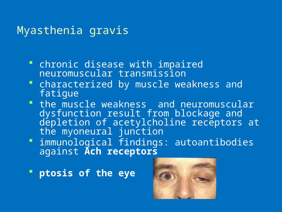
Myasthenia gravis
chronic disease with impaired neuromuscular transmission
characterized by muscle weakness and fatigue the muscle weakness and neuromuscular dysfunction
result from blockage and depletion of acetylcholine receptors at the myoneural junction
immunological findings: autoantibodies against Ach receptors
ptosis of the eye
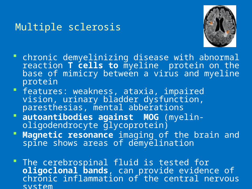
Multiple sclerosis
chronic demyelinizing disease with abnormal reaction T cells to myeline protein on the base of mimicry between a virus and myeline protein
features: weakness, ataxia, impaired vision, urinary bladder dysfunction, paresthesias, mental abberations
autoantibodies against MOG (myelin-oligodendrocyte glycoprotein)
Magnetic resonance imaging of the brain and spine shows areas of demyelination
The cerebrospinal fluid is tested for oligoclonal bands, can provide evidence of chronic inflammation of the central nervous system

AUTOIMMUNE CYTOPENIA
AI hemolytic disease- autoantibodies against membrane erythrocyte antigens
AI trombocytopenia - autoantibodies against
trombocyte antigens (GPIIb/IIIa)
AI neutropenia - autoantibodies against membrane neutrofil antigens

IMMUNOSUPPRESSION
non-specific treatmentexamples of drugsindicationrisks

Immunosuppressants
Drugs that inhibit or prevent activity of the immune system
They are used in immunosuppressive therapy to: Prevent the rejection of transplanted organs and
tissues (bone marrow, heart, kidney, liver) Treat autoimmune diseases or diseases that are
most likely of autoimmune origin (rheumatoid arthritis, multiple sclerosis, myasthenia gravis, systemic lupus erythematosus, Crohn's disease, pemphigus, ulcerative colitis).
Treat some other non-autoimmune inflammatory diseases (allergic asthma, atopic eczema).
Glucocorticoids
suppress the cell-mediated immunity- act by inhibiting genes that code for various cytokines (e.g.IL-2)
decrease cytokine production reduces the T cell proliferation.
suppress the humoral immunity, causing B cells to express smaller amounts of IL-2 and IL-2 receptors- this diminishes both B cell clone expansion and antibody synthesis.

Glucocorticoids leads to diminished eicosanoid production,
suppression of the cyclooxygenase expression Glucocorticoids also stimulate the lipocortin-1
escaping to the extracellular space, where it binds to the leucocyte membrane receptors and inhibits : epithelial adhesion, migration, chemotaxis, phagocytosis, respiratory burst, and the release of various inflammatory mediators from neutrophils, macrophages, and mastocytes.
side-effects: hypertension, dyslipidemia, hyperglycemia, peptic ulcers, osteoporosis, disturbed growth in children

Drugs affecting the proliferation of both T cells and B cells
Cyclophosphamide -very efficient in the therapy of systemic lupus erythematosus, autoimmune hemolytic anemias
high doses cause pancytopenia and hemorrhagic cystitis
Methotrexate is a folic acid antagonist, acts during DNA and RNA synthesis, and thus it is cytotoxic during the S-phase of the cell cycle; used in the treatment of autoimmune diseases (RA, Crohn's disease) and in transplantations.
Drugs affecting the proliferation of both T cells and B cells
Azathioprine is a purine synthesis inhibitor, inhibiting the proliferation of cells, especially leucocytes; SLE, RA, sclerosis multiplex, transplantation
Mycophenolate mofetil – affects the enzyme that controls the purine synthesis
Used in transplantation of solid organ

Drugs blocking the activation of lymphocytes Tacrolimus - prevents the cell from transitioning from
the G0 into G1 phase of the cell cycle Used to prevent rejection reactions, atopic eczema
Cyclosporin A- inhibits calcineurin, which is responsible for activating the transcription of interleukin-2; inhibits cytokines production and interleukin release
Used to prevent rejection reactions
Side effects: nephrotoxicity, neurotoxicity, hypertension, dyslipidemia, hyperglycemia
Monoclonal antibodies
Monoclonal antibodies are directed towards exactly defined antigens
Daclizumab - acts by binding the IL-2a receptor's α chain, preventing the IL-2 induced clonal expansion of activated lymphocytes and shortening their survival
used in the prophylaxis of the acute organ rejection after the bilateral kidney transplantation

Thank you for your attention!