Author's personal copy - University of California, Berkeley Publications/Vazin... · copy is...
9
This article appeared in a journal published by Elsevier. The attached copy is furnished to the author for internal non-commercial research and education use, including for instruction at the authors institution and sharing with colleagues. Other uses, including reproduction and distribution, or selling or licensing copies, or posting to personal, institutional or third party websites are prohibited. In most cases authors are permitted to post their version of the article (e.g. in Word or Tex form) to their personal website or institutional repository. Authors requiring further information regarding Elsevier’s archiving and manuscript policies are encouraged to visit: http://www.elsevier.com/authorsrights
Transcript of Author's personal copy - University of California, Berkeley Publications/Vazin... · copy is...

This article appeared in a journal published by Elsevier. The attachedcopy is furnished to the author for internal non-commercial researchand education use, including for instruction at the authors institution
and sharing with colleagues.
Other uses, including reproduction and distribution, or selling orlicensing copies, or posting to personal, institutional or third party
websites are prohibited.
In most cases authors are permitted to post their version of thearticle (e.g. in Word or Tex form) to their personal website orinstitutional repository. Authors requiring further information
regarding Elsevier’s archiving and manuscript policies areencouraged to visit:
http://www.elsevier.com/authorsrights
Author's personal copy
The effect of multivalent Sonic hedgehog on differentiation of humanembryonic stem cells into dopaminergic and GABAergic neurons
Tandis Vazin a,1, Randolph S. Ashton a,1, Anthony Conwaya, Nikhil A. Rode b, Susan M. Lee c,Verenice Bravo d, Kevin E. Healy b, Ravi S. Kane e,**, David V. Schaffer a,*aChemical and Biomolecular Engineering, and The Helen Wills Neuroscience Institute, University of California Berkeley, Berkeley, CA 94720, USAbDepartment of Bioengineering, Department of Materials Science and Engineering, University of California Berkeley, Berkeley, CA 94720, USAcDepartment of Molecular and Cell Biology, University of California Berkeley, Berkeley, CA 94720, USAdDepartment of Integrative Biology, University of California Berkeley, Berkeley, CA 94720, USAeDepartment of Chemical and Biological Engineering, Center for Biotechnology and Interdisciplinary Studies, Rensselaer Polytechnic Institute, Troy,NY 12180, USA
a r t i c l e i n f o
Article history:Received 27 August 2013Accepted 5 October 2013Available online 27 October 2013
Keywords:MultivalencySonic hedgehogBioconjugatesDopaminergicGABAergicHuman pluripotent stem cells
a b s t r a c t
Stem cell differentiation is regulated by complex repertoires of signaling ligands which often usemultivalent interactions, where multiple ligands tethered to one entity interact with multiple cellularreceptors to yield oligomeric complexes. One such ligand is Sonic hedgehog (Shh), whose post-translational lipid modifications and assembly into multimers enhance its biological potency, potentiallythrough receptor clustering. Investigations of Shh typically utilize recombinant, monomeric protein, andthus the impact of multivalency on ligand potency is unexplored. Among its many activities, Shh isrequired for ventralization of the midbrain and forebrain and is therefore critical for the development ofmidbrain dopaminergic (mDA) and forebrain gamma-aminobutyric acid (GABA) inhibitory neurons. Wehave designed multivalent biomaterials presenting Shh in defined spatial arrangements and investigatedthe role of Shh valency in ventral specification of human embryonic stem cells (hESCs) into thesetherapeutically relevant cell types. Multivalent Shh conjugates with optimal valencies, compared to themonomeric Shh, increased the percentages of neurons belonging to mDA or forebrain GABAergic fatesfrom 33% to 60% or 52% to 86%, respectively. Thus, multivalent Shh bioconjugates can enhance neuronallineage commitment of pluripotent stem cells and thereby facilitate efficient derivation of neurons thatcould be used to treat Parkinson’s and epilepsy patients.
� 2013 Elsevier Ltd. All rights reserved.
1. Introduction
Biochemical cues within the stem cell niche that instruct cellfate decisions are often incorporated into larger structures, forexample via self-assembly or by immobilization to the extracellularmatrix. Understanding the importance of this nanoscale spatialorganization in controlling cell behavior both advances our basicknowledge of stem cell and developmental biology and enablesapplications in tissue engineering and regenerative medicine.Multivalent interactions e where multiple ligands on one entity
bind multiple receptors on another e are one such class of nano-scale organization that naturally occur in many biological processesranging from growth factor and morphogen signaling to theattachment of a virus to a cell surface [1]. Multivalent ligands areoften collectively more potent than corresponding monovalentinteractions due to an enhanced ability to occupy and/or clustertheir receptors [2e4]. Engineered biomaterials are increasinglybeing employed to design systems that emulate important bio-logical features of natural niches [5,6], and synthetic biomimeticmultivalent ligands may be more potent than monovalent ligandsin regulating cell fate decisions, such as stem cell differentiation.
Shh, a potent morphogen that specifies cell fate choices in tis-sues throughout the developing embryo is one factor that has beensuggested to function as a multimeric form [7e9]. Within theneural tube e the germinal origin of the central nervous system(CNS) e a gradient of Shh initially emanating from ventralmesenchyme tissue and subsequently the floor plate cells within
* Corresponding author. Fax: þ1 510 642 5198.** Corresponding author. Fax: þ1 518 276 4030.
E-mail addresses: [email protected] (R.S. Kane), [email protected](D.V. Schaffer).
1 These authors contributed equally to this work.
Contents lists available at ScienceDirect
Biomaterials
journal homepage: www.elsevier .com/locate/biomater ia ls
0142-9612/$ e see front matter � 2013 Elsevier Ltd. All rights reserved.http://dx.doi.org/10.1016/j.biomaterials.2013.10.025
Biomaterials 35 (2014) 941e948

Author's personal copy
the tube patterns differentiation of ventral progenitor domains in aconcentration dependent manner [10]. The resulting ventral pro-genitors give rise to many neuronal cell types along the neuraxisincluding midbrain dopaminergic (mDA) [11] and GABA producinginhibitory neurons [12], which undergo degeneration in Parkin-son’s disease (PD) [13] or are impaired in epileptic disorders [14e16], respectively. Efforts to develop cell replacement therapies forthese intractable neurodegenerative diseases routinely manipulateShh signaling to ventralize neurally differentiating human plurip-otent stem cells (hPSCs) and thereby generate mDA [17] orGABAergic [15] progenitors.
Small molecule agonists of Shh signaling have been used toinduce the dopaminergic and GABAergic differentiation of hPSCs.Recombinant Shh, however, is 1e2 orders of magnitude morepotent on a molar basis in patterning neural cell fate [18,19], andwhen used in combination with saturating levels of Shh agonistpurmorphamine, recombinant Shh can further increase the efficacyof lineage-specific neuronal differentiation protocols [17]. There-fore, it appears that activation of Shh signaling with the proteinligand, which binds to the cell membrane receptor Patched, offerspotential advantages compared to small molecule agonists, whichregulate the downstream effector Smoothened (SMO).
Naturally produced Shh is covalently modified by cholesteroland palmitate [20,21]. These lipid moieties were initially believedto tether the protein to the cellular plasma membrane, yet in theneural tube Shh secreted from the notochord and floor plate act inlong range to organize the pattern of ventral neurogenesis [7,22].Shh’s long-range signaling effects observed during organismaldevelopment have recently been attributed to its nanoscale clus-tering and multimerization within the secreting cell’s membraneprior to release as a diffusible and multivalent molecule [7e9,23,24]. Furthermore, recent observations that Shh can beassembled into a soluble multimeric protein complex with a hy-drophobic core of lipids help explain this transport [25]. Theresulting multimeric form of Shh is also reported in vitro to be evenmore potent than monovalent recombinant Shh [7,24]; however,Shh multimerization and secretion rely on complex mammalianposttranslational modifications and secretory mechanisms that arenot fully understood [26] and that render the production of natural,multivalent Shh for regenerative medicine applications problem-atic. We previously demonstrated that a bio-inspired, multivalent,conjugate form of Shh was more active in a murine fibroblastbioassay [27], raising the possibility that multivalent Shh bio-conjugates may potentially serve as valuable materials to moreeffectively direct the differentiation of hPSCs into therapeuticallyvaluable cell types compared to recombinant Shh or small moleculeagonists of Shh signaling.
In this work we generated multivalent Shh with defined spatialdistribution by conjugating recombinant Shh to linear Hyaluronicacid (HyA) polymers at various stoichiometric ratios and investi-gated the putative role of multivalency in Shh signaling duringneuronal differentiation of hPSCs. We evaluated whether bio-mimetic Shh conjugates can be used as a bioactive material toenhance ventralization of neural progenitors and lineage commit-ment to therapeutically relevant neuronal phenotypes includingmDA and GABAergic neurons in direct comparison with equimolaramounts of monomeric recombinant Shh, and ten-fold higherlevels of Shh pathway small molecule agonist (SAG).
2. Materials and methods
2.1. Recombinant protein production, purification, and bioconjugation
A bacterial expression vector encoding Shh with a C-terminal hexahistidine tagand cysteine (pBADeShh) was transformed into chemically competent BL21Escherichia coli. In addition, valine and isoleucine residues were introduced to theShh N-terminus to increase potency by mimicking the hydrophobic palmitic acid
modification of endogenous Shh. Protein expression was induced by the addition of0.1% (w/v) L-arabinose in TB media for 5 h at 30 �C. Cells were lysed, and Shh waspurified via immobilized metaleion affinity chromatography (IMAC) on a BiologicDuoFlow System. The purified protein was dialyzed into pH 6.5 PBS containing 10%glycerol and EDTA. SDS-PAGE revealed a single band of the predicted size. Shh wasconjugated to 800 kDa HyA through a two-step reaction using carbodiimidechemistry at the HyA carboxylate group and a maleimide reaction at the protein C-terminal cysteine. In the first step, EMCH, Sulfo-NHS, and EDC were added to asolution of HyA in MES buffer and allowed to react at 4 �C for 4 h, followed bydialysis. Recombinant Shh was reduced with a 200-fold molar excess of TCEP at 4 �Cfor 5 min. The Shh was added to HyAeEMCH at the desired molar ratios andallowed to react at 4 �C overnight. The Shh-conjugated HyAwas dialyzed to removeunreacted Shh. Shh concentrations were measured using a BCA assay. Also, thedegrees of substitution and valency of bioconjugates were analyzed by size-exclusion chromatography with multiangle laser light scattering paired withrefractive index detection and ultraviolet spectroscopy (SEC-MALS-RI-UV) asdescribed [28].
2.2. Dopaminergic and GABAergic differentiation of human embryonic stem cells
The H1 (WiCell) hESC line was cultured on Matrigel-coated cell culture plates(BD) in X-Vivo medium (Lonza) supplemented with 80 ng/ml FGF2 (PeproTech) and0.5 ng/ml TGF-b1 (R&D Systems) or in mTeSR1 maintenance medium (Stem CellTechnologies). HESCs were removed from the tissue culture plates using a sterilecell scraper and partially dissociated by gentle pipetting. The cell clusters wereresuspended in hESC culture medium lacking the mTeSR1 supplement, or lackingFGF2 and TGF-b1 for X-Vivo culture, and transferred to ultra low-attachment plates(Corning Incorporated) for embryoid body (EB) formation. HESCs were aggregatedfor 5 days and then seeded on Matrigel-coated plates, and the culture medium wasthen supplemented with N2 and B27 (Invitrogen) and treated with 100 ng/ml FGF-8 and 200 ng/ml Shh (R&D-Shh), SAG, m-Shh, or HyAeShh (1:5, 1:10, 1:20, 1:40).After 9 days, cells were mechanically passaged onto poly-L-ornithine (SigmaAldrich) and laminin (Invitrogen, 20 mg/ml) coated plates and cultured with FGF8and Shh for an additional 5 days. Thereafter, FGF8 and Shh were withdrawn, andcells were matured for 16 days with BDNF (10 ng/ml) and GDNF (10 ng/ml,Peprotech).
To further increase neural progenitor yield, neural rosettes were isolated. EBswere seeded onto Matrigel coated plates for 14 days under high density conditions(100�103/cm2) with FGF8 and Shh conjugates or controls. At day 14, structures witha rosette-like morphology were mechanically isolated and plated on poly-L-orni-thine and laminin coated plates or 8-well chamber slides at a lower density(20� 103/cm2) in the presence of Shh for an additional week. Next, BDNF (10 ng/ml)and GDNF (10 ng/ml) were added to achieve neuronal maturation for an additional2e3 weeks. As a control, corresponding amounts of HyA polymer without Shhconjugation were added. For GABAergic differentiation, cells were differentiatedsimilarly, except FGF8 was excluded during neural patterning, and GDNF wasexcluded during neuronal maturation.
2.3. Gene expression analysis by RT-PCR
Using random primers and MultiScribe Reverse Transcriptase (Applied Bio-systems) in a 20 ml reaction, complementary DNA was synthesized from 1 mg totalRNA isolated from undifferentiated hESCs, EBs after 5 days of culture in suspension,and at days 9, 14, and 30 of neural differentiation following EB formation. The PCRanalysis was carried out with Taq DNA polymerase (New England Biolabs). Equalamounts of RNA were tested in PCR reactions under the same conditions to verifythe absence of genomic DNA amplification. The housekeeping gene glyceraldehyde-3-phosphate dehydrogenase (GAPDH) was amplified as an internal control in geneexpression analysis. Primer sequences (see Table. S1 in Supplementary data) wereobtained from the PrimerBank website (http://pga.mgh.harvard.edu/primerbank/)and synthesized by Life Technologies.
2.4. Dopamine analysis
Media conditioned for 48 h by DA neuron cultures (differentiated for 30 days)were collected. Alternatively, dopamine release was induced by first conditioningcultured cells in Hanks’ balanced salt solution (HBSS) for 15 min and then replacingit with HBSS containing 56 mM KCl for 15 min at 37 �C. 20 ml of dopamine stabili-zation buffer, consisting of 2.4 mM EGTA and 2.3 mM gluthatione in 10 ml of 0.1 M
NaOH, were added. A HPLC kit (Chromsystems) was used to extract monoamines,whose levels were then determined by HPLC coupled to an electrochemical detectorusing MD-TM mobile phase.
2.5. Immunocytochemistry
Cultures were fixed with 4% paraformaldehyde for 10e15 min. The primaryantibodies used were: mouse anti-Oct4 (1:100, Santa Cruz Biotechnology), mouseanti-SSEA-4 (1:500, Millipore), rabbit anti-Pax6 (1:200, Covance), mouse anti-Otx2(1:50, R&D Systems), mouse anti-Msx1/2 (1:100, Developmental Studies HybridomaBank), mouse anti-bIII-Tubulin (Tuj-1) (1:250, Sigma), mouse anti-Microtubule-Associated Protein 2 (MAP2) (1:500, BD Biosciences), rabbit anti-TH (1:1000, Pel-
T. Vazin et al. / Biomaterials 35 (2014) 941e948942

Author's personal copy
Freez), and rabbit anti-GABA (1:2000, Sigma Aldrich). Cultures were incubated withsecondary antibodies conjugated Cy3 or Cy5 (1:500, Jackson ImmunoResearchLaboratories), or Alexa 594-conjugated anti-rabbit and Alexa 488-conjugated anti-mouse antibodies (1:1000, Invitrogen), in PBS containing 1% BSA for 2 h. Cultureswere counter-stained with DAPI (Molecular probes) and imaged using a Zeiss AxioObserver A1 inverted microscope or a Zeiss LSM 710 confocal microscope.
2.6. Cell quantification and statistics
The numbers of neurons were quantifiedmanually. The numbers of cells stainedwith DAPI were counted automatically with ImageJ software. For cell quantifica-tions, images of 9 different fields per well from three wells were acquired using a20� objective. For neuronal cell counts, fields where Tuj-1 and MAP2 positiveneurons could clearly be seen and distinguished from each other were selectedwithout consideration of number of neurons present in the field or neurotransmitterexpression in neurons. Neuronal cell morphology was carefully examined to confirmexpression of markers Tuj-1, MAP2, TH, and GABA. Differences in percentages of cellswere tested by analysis of variance followed by pair-wise comparisons of groupmeans using the TukeyeKramer method for multiple comparisons generated by theGraphPad InStat software (GraphPad Software Inc.). Levels of significance are indi-cated by asterisks on the line graphs and figure legends. Differences were considered
significant at p < .05. Data represent the mean values (�SD) of cell counts fromtriplicate samples from three independent experiments.
3. Results
3.1. Multivalent Sonic hedgehog bioconjugate synthesis
To investigate whether valency plays a role in Shh bioactivityduring neuronal patterning of hESC differentiation, we synthesizedmultivalent bioactive Shh conjugates by grafting recombinantlyproduced, cysteine-modified, N-terminal Shh to a biological poly-mer, high molecular weight HyA, using a hydrazideemaleimidehetereobifunctional cross-linker (EMCH) [28] (Fig. 1A). To enableinvestigation of whether ligand valency impacts neuronal fate re-striction, we generated ShheHyA conjugates at various stoichio-metric ratios ranging from 4 to 27 Shh molecules per HyA polymerchain. Conjugate valencies were determined by a protein assay as
Fig. 1. (A) Representation of the chemical synthesis of hyaluronic acid (HyA)eSonic hedgehog (Shh) bioconjugates using a hydrazide-maleimide hetereobifunctional cross-linker(EMCH) to anchor recombinantly produced cysteine-modified Shh to the carboxylic acid groups of the linear HyA polymers. (B) Bioconjugate valency characterization of HyAeShhproducts by SEC-MALS. (C) Schematic representation of the paradigm for differentiating hESCs into midbrain dopaminergic (mDA) neurons with the aid of the midbrain instructivefactors Shh and FGF8. Cultures stained with antibodies against pluripotent stem cell markers Oct3/4 and SSEA4 at day 1, the early neural progenitor marker Pax6 and midbrainprogenitor markers Otx2 and Msx1 at day 14, and the neuronal and DA marker Tuj-1 and TH at day 35 confirmed the DA differentiation of hESCs. Scale bar ¼ 100 mm(D) Complementary DNA from undifferentiated cultures, embryoid bodies (EBs), and cells at various stages of differentiation as illustrated in the differentiation scheme in panel(A) were analyzed by RT-PCR for a number of midbrain specific transcription factors including Engrailed 1 (En1), Lmx1a, Lmx1b, Pitx3, and Nurr1, and TH and the Dopaminetransporter (DAT) expressed in mature DA neurons. The levels of Shh expression were assessed in all cultures. Amplification was performed for 35 cycles, and GAPDH was amplifiedsimultaneously as an internal control under the same conditions.
T. Vazin et al. / Biomaterials 35 (2014) 941e948 943
Author's personal copy
well as refractive index detection, ultraviolet spectroscopy, and sizeexclusion chromatography coupled to multi-angle light scattering(SEC-MALS) [28] (Fig. 1B).
3.2. Effect of multivalent Shh on dopaminergic differentiation
To establish a timeline for mDA neuronal differentiation, hESCswere first cultured in suspension for 5 days to form embryoidbodies (EBs). Midbrain DA differentiation was then achieved bydifferentiating the resulting EBs in adherent conditions for 9 days inthe presence of the established mDA specifying factors Shh(monomeric) and FGF8 [11]. After 9 days of differentiation inadherent cultures (Fig. 1C, DA Induction I), expression of the neuralprogenitor marker Pax6 [29] and midbrain progenitor markersOtx2 [30] andMsx1 [31] was detected in themajority of colonies. Atthis time, cultures were passaged and exposed to Shh and FGF8 foran additional 5 days (DA Induction II), followed by exposure to glialcell line-derived neurotrophic factor (GDNF) and brain-derivedneurotrophic factor (BDNF) to promote DA maturation [32] for an
additional 16 days (Fig. 1C, DA Maturation). RT-PCR analysis of thepluripotency marker Oct3/4; the midbrain specific transcriptionfactors Engrailed 1 (EN1), Lmx1a, Lmx1b, Pitx3, and Nurr1; and themature DA neuronal dopamine transporter (DAT) proteinconfirmed the developmental progression of hESCs to a maturemDA phenotype [33] (Fig. 1D).
As the specification of midbrain neural progenitors is predom-inantly mediated by Shh [11]which has been suggested to functionas a soluble multimeric complex during development [7e10,23,24],we next investigated whether this factor’s multivalency impactsmidbrain differentiation of hESCs in vitro. EBs were again formedfor 5 days and transferred to adherent conditions for neural in-duction. On the first day of adherent culture differentiation, cellswere exposed to the mDA-inducing factor FGF8 in combinationwith a Shh signaling activator e either the small molecule SMOagonist SAG, a commonly used commercial form of Shh (R&D), ourrecombinantly producedmonomeric Shh (termedm-Shh), or HyAeShh bioconjugates with polymer:Shh ratios of 1:4, 1:11, 1:17, or1:27. Cells were cultured for 14 days (Fig. 1C, DA Induction I and II)
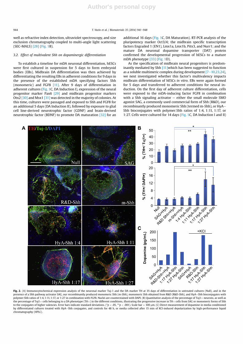
Fig. 2. (A) Immunocytochemical expression analysis of the neuronal marker Tuj-1 and the DA marker TH at 35 days of differentiation in untreated cultures (Null), and in thepresence of a Shh pathway activator SAG, our recombinantly produced monomeric Shh (m-Shh), monomeric Shh obtained from R&D (R&D-Shh), and HyAeShh bioconjugates withpolymer:Shh ratios of 1:4, 1:11, 1:17, or 1:27 in combination with FGF8. Nuclei are counterstained with DAPI. (B) Quantitative analysis of the percentage of Tuj1þ neurons, as well asthe percentage of Tuj1þ cells belonging to a DA phenotype (THþ) in the different conditions, illustrating the progressive increase in THþ cells from SAG or monomeric forms of Shhto the conjugates of higher valencies. Error bars indicate standard deviations. (*p < .05, **p < .001). Scale bar ¼ 100 mm. (C) Direct measurement of dopamine in media conditionedby differentiated cultures treated with HyAeShh conjugates, and controls for 48 h, or media collected after 15 min of KCl-induced depolarization by high-performance liquidchromatography (HPLC).
T. Vazin et al. / Biomaterials 35 (2014) 941e948944

Author's personal copy
with a passage on day 9. FGF8 and the Shh signal activators werethen withdrawn, and GDNF and BDNF were added to promote DAneuronal survival and maturation. After 30 total days of neuraldifferentiation, immunocytochemical analysis of the DA markertyrosine hydroxylase (TH) and neuronal marker Tuj-1 revealed thatShh with a valency of 1:17 increased the overall number ofTH þ neurons approximately 3-fold and the fraction of neuronsexpressing THmore than 2-fold (Fig. 2A,B) compared tomonomericShh. Results with a higher valency, 1:27, were similar (Fig. 2A,B).Since the ability to synthesize and release the neurotransmitterdopamine is the characteristic feature of dopaminergic neurons, weused high-performance liquid chromatography (HPLC) to comparethe ability of the cells generated under conditions with the Shhpathway agonist SAG, monomeric R&D-Shh, or the higher valencyShh conjugates (1:17 and 1:27) to release dopamine. Mediaconditioned for 48 h by differentiated cultures treated with HyAeShh conjugates or controls showed higher levels of dopamine inconditions with Shh bioconjugates with polymer:Shh ratio of 1:17(Fig. 2C). Analogously, media collected after 15 min of KCl-induceddepolarization also exhibited increased levels of dopamine fromcultures patterned with higher valencies of Shh (1:17, 1:27)compared to SAG or monomeric Shh (R&D-Shh) (Fig. 2C). Theseresults establish that morphogen valency impacts neuronal speci-fication and maturation of mDA neurons.
To further increase the fraction of cells that belong to a mDA fatefollowing EB formation, we increased the cell density and initialdifferentiation time in the presence of FGF8 and various forms ofShh (Fig. 1C, DA Induction I), isolated rosette-like structurescomprised mainly of neural progenitor cells, and further differen-tiated them in adherent conditions at lower densities to enhanceneuronal differentiation (Fig. 1C, DA Induction II). Also, to assessreproducibility within this study, we used newly synthesized bio-conjugates at valencies close to those that previously exhibitedmaximal potencies (w1:17 in Fig. 2B). After the 14 day incubation inFGF8 and Shh, DA induction of neural progenitors was significantlyhigher in conditions with high HyA:Shh ratio conjugates (1:12 and1:16) as indicated by the expression of mDA progenitor markersLmx1a and Msx1 (Fig. 3A) and the DA marker TH (Fig. 3B).
Cultures under the influence of HyA:Shh conjugateswith ratios of1:16 induced expression of Lmx1a and Msx1 in 61% of colonies ascompared to 36% in cultures treatedwith themonomeric formof Shh(R&D-Shh) (Fig. 3A). Colonies containing 30 or more TH-expressingneurons comprised about 71% of colonies in cultures influenced byShh bioconjugates with polymer:Shh ratios of 1:16, versus 34% ofcolonies in cultures treated with the monomeric Shh (Fig. 3B).
These neural progenitors were then matured into neurons(Fig. 1C, DA Maturation). Consistent with the increase in mDAneural progenitor marker expression in colonies after the initial
Fig. 3. (A) Quantitative analysis of the co-expression of midbrain progenitor transcription factors Msx1 and Lmx1a or TH in colonies treated with FGF8 and SAG, monomeric formsof Shh (R&D-Shh and m-Shh), or the Shh multimers with valencies of 1:4, 1:10, 1:12, or 1:16 after 14 days of neural induction. The image insert in the top bar graph shows anexample of an immunostained colony expressing Msx1 and Lmx1a in conditions treated with FGF8 and HyA polymers carrying 16 Shh ligands. *p < .05, **p < .001, **p < .001.(B) Representative images of colonies expressing TH and MAP2 in the different conditions indicate an increase in the generation of TH þ neurons in cultures exposed to multivalentShh in relation to monomeric form of Shh or SAG. Scale bar ¼ 100 mm.
T. Vazin et al. / Biomaterials 35 (2014) 941e948 945

Author's personal copy
neural induction stage (Fig. 3A), immunocytochemical analysis af-ter a total of 35 days of differentiation post-EB formation estab-lished that DA differentiation was enhanced with the multimericforms of Shh (Fig. 4A,B).
Specifically, the fraction of TH þ neurons in the cultureincreased from 17% for monomeric Shh (R&D-Shh) to 36% in cul-tures treated with Shh with a valency of 16 (Fig. 4A,B), and thefraction of neurons that were committed to a mDA phenotypeincreased from 33% to 60% (Fig. 4B).
3.3. Effect of multivalent Shh on GABAergic differentiation
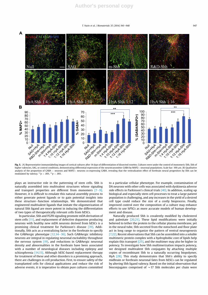
During development, Shh also acts as a cell fate specifying factorin the forebrain, giving rise to ventral forebrain neural progenitorsthat subsequently develop into GABAergic interneurons [12,34,35],which may offer promise for the treatment of epilepsy and seizure[14e16]. It has been shown that exposure to Shh can increase theGABAergic differentiation of hPSCs [36]. To demonstrate broaderapplication for multivalent Shh in directed stem cell differentiation,we investigated whether Shh valency impacts differentiation ofhESCs to forebrain GABAergic neurons. Similar to the optimized DAdifferentiation paradigm, forebrain differentiation was carried out
via 5 day EB-formation followed by 14 days of differentiation in thepresence of the small molecule agonist SAG, multivalent Shh con-jugates, or monomeric forms of Shh. At this stage, rosettes weremanually isolated and further exposed to Shh conjugates or con-trols for 7 days. Isolated neural progenitor cells werematured for anadditional 14 days in the presence of BDNF, previously shown toplay a crucial role in the development and functional maturation offorebrain neurons [37,38]. Immunocytochemical staining of theGABA neurotransmitter and MAP2, a marker for mature neurons,demonstrated that multivalent conjugates substantially increasedthe commitment of neural progenitors to a GABAergic lineagecompared to monomeric Shh from 30% to 56% of all cells, and from52% to 86% of neurons (Fig. 5), resulting in a relatively pureGABAergic neuronal population.
4. Discussion
While the signaling events that govern stem cell fate decisionsare intricate, chemistry and materials synthesis can enable theengineering of biomimetic systems and structure that progressivelyemulate and restore the complexity of the stem cell niche signals. Inthis study, we addressed the question of whether the valency of Shh
Fig. 4. (A) Midbrain neuronal lineage specification analysis of isolated neural rosettes after neuronal maturation by TH and MAP2 expression. Cultures were differentiated in theabsence of any instructive cues, continued exposure to SAG, or conjugates bearing various Shh valencies for 7 days and further matured in the presence of neurotrophic factors. Scalebar ¼ 100 mm. (B) Quantification results of percentages of cell expressing TH and MAP2 are presented in bar graphs, revealing that the percentages of TH expressing neurons and thefraction of the mature neuronal population restricted to a midbrain DA phenotype can be nearly doubled by increasing the valency of Shh during the course of neuronaldifferentiation*p < .05, **p < .001, **p < .001.
T. Vazin et al. / Biomaterials 35 (2014) 941e948946
Author's personal copy
plays an instructive role in the patterning of stem cells. Shh isnaturally assembled into multivalent structures whose signalingand transport properties are different from monomers [7e9].However, it is difficult to emulate this natural assembly process toeither generate potent ligands or to gain potential insights intothese structureefunction relationships. We demonstrated thatengineered multivalent ligands that imitate the oligomerization ofnatural Shh ligand are more potent in inducing the differentiationof two types of therapeutically relevant cells from hESCs.
In particular, Shh and FGF8 signaling promotemDA derivation ofstem cells [11], and replacement of defective dopamine producingneurons with healthy new mDA neurons derived from hESCs is apromising clinical treatment for Parkinson’s disease [39]. Addi-tionally, Shh acts as a ventralizing factor in the forebrain to specifythe GABAergic phenotype [12,34e36]. Such GABAergic inhibitoryneurons are integral in regulating neuronal excitability throughoutthe nervous system [19], and reductions in GABAergic neuronaldensity and abnormalities in the forebrain have been associatedwith a number of neurological diseases including epilepsy andschizophrenia [14,15]. Although hESC-based replacement therapyfor treatment of these and other disorders is a promising approach,there are challenges in cell production. First, to ensure safety of thetransplanted cells for clinical applications and reduce the risk ofadverse events, it is imperative to obtain pure cultures committed
to a particular cellular phenotype. For example, contamination ofDA neurons with other cells was associatedwith dyskinesia adverseside effects in Parkinson’s clinical trials [40]. In addition, scaling upbiological and especially stem cell processes to treat a large patientpopulation is challenging, and any increases in the yield of a desiredcell type could reduce the size of a costly bioprocess. Finally,improved control over the composition of a culture may enhanceefforts to use hPSCs as more accurate models of human develop-ment and disease.
Naturally produced Shh is covalently modified by cholesteroland palmitate [20,21]. These lipid modifications were initiallybelieved to tether the protein to the cellular plasma membrane, yetin the neural tube, Shh secreted from the notochord and floor plateact in long range to organize the pattern of ventral neurogenesis[7,22]. Recent observations that Shh can be assembled into a solublemultimeric protein complex with a hydrophobic core of lipids helpexplain this transport [25], and the multimer may also be higher inpotency. To investigate how Shh multimerization impacts potency,we designed multivalent Shh conjugates by attaching multiplecopies of recombinant Shh to a naturally occurring biopolymer,HyA [28]. This study demonstrates that Shh’s ability to specifymidbrain or forebrain neuronal fates from hESCs can be regulatedby altering Shh ligand valency. Based on the initial observation thatbioconjugates comprised of w17 Shh molecules per chain were
Fig. 5. (A) Representative immunolabeling images of cortical cultures after 14 days of differentiation of dissected rosettes. Cultures were under the control of monomeric Shh, Shh ofhigher valencies, SAG, or control conditions, demonstrating differential expression of the neurotransmitter GABA by MAP2þ neuronal populations. Scale bar: 100 mm. (B) Qualitativeanalysis of the proportion of GABA þ neurons and MAP2þ neurons co-expressing GABA, revealing that the ventralization effect of forebrain neural progenitors by Shh can bemodulated by valency. **p < .001, **p < .001.
T. Vazin et al. / Biomaterials 35 (2014) 941e948 947
Author's personal copy
superior for mDA differentiation (Fig. 2), we optimized our protocolto increase the yield of mDA and GABAergic neurons by isolatingneural rosettes and increasing the exposure time to these Shhconjugates. As a result, lineage commitment of hESCs to mDA andforebrain GABAergic neurons increased approximately 2-fold rela-tive to either monomeric Shh or the small molecule agonist. Futurework may explore how the binding of these flexible polymereligand complexes to the receptor may compare to the binding ofnatural Shh oligomers, what role receptor clustering may play insignaling, and whether cells can vary the valency to tune thebioactivity of the ligand.
5. Conclusions
We demonstrate that engineered multivalent ligands that mimicthe oligomerization of natural Shh ligand are more potent inenhancing fate specification of hESCs into mDA and GABAergicneurons, for strategies to treat Parkinson’s disease and epilepsy andseizure, respectively. Patterning factors with increased potencycould improve the purity of a desired cell type and thereby poten-tially reduce deleterious outcomes. These results can both improvethe ability to generate therapeutically relevant cell types, as well aslend future insights into the mechanisms of cell patterning.
Acknowledgments
We thank Dr. Agnieszka Ciesielska (Bankiewicz Lab, UCSF) forhelp with HPLC analysis of dopamine. This work was supported byCIRM Award TG2-01164 and CIRM grant RT2-02022.
Appendix A. Supplementary data
Supplementary data related to this article can be found at http://dx.doi.org/10.1016/j.biomaterials.2013.10.025.
References
[1] Mammen M, Choi SK, Whitesides GM. Polyvalent interactions in biologicalsystems: implications for design and use of multivalent ligands and inhibitors.Angew Chem Int Ed 1998;37:2755e94.
[2] Bray D, Levin MD, Morton-Firth CJ. Receptor clustering as a cellular mecha-nism to control sensitivity. Nature 1998;393:85e8.
[3] Heldin CH. Dimerization of cell surface receptors in signal transduction. Cell.1995;80:213e23.
[4] Wess J. G-protein-coupled receptors: molecular mechanisms involved in re-ceptor activation and selectivity of G-protein recognition. Faseb J 1997;11:346e54.
[5] Metallo CM, Mohr JC, Detzel CJ, de Pablo JJ, Van Wie BJ, Palecek SP. Engi-neering the stem cell microenvironment. Biotechnol Prog 2007;23:18e23.
[6] Vazin T, Schaffer DV. Engineering strategies to emulate the stem cell niche.Trends Biotechnol 28:117e24.
[7] Zeng X, Goetz JA, Suber LM, Scott Jr WJ, Schreiner CM, Robbins DJ. A freelydiffusible form of Sonic hedgehog mediates long-range signalling. Nature2001;411:716e20.
[8] Goetz JA, Singh S, Suber LM, Kull FJ, Robbins DJ. A highly conserved amino-terminal region of sonic hedgehog is required for the formation of its freelydiffusible multimeric form. J Biol Chem 2006;281:4087e93.
[9] Vyas N, Goswami D, Manonmani A, Sharma P, Ranganath HA,VijayRaghavan K, et al. Nanoscale organization of hedgehog is essential forlong-range signaling. Cell 2008;133:1214e27.
[10] Briscoe J, Chen Y, Jessell TM, Struhl G. A hedgehog-insensitive form of patchedprovides evidence for direct long-range morphogen activity of sonic hedge-hog in the neural tube. Mol Cell 2001;7:1279e91.
[11] Yan Y, Yang D, Zarnowska ED, Du Z, Werbel B, Valliere C, et al. Directed dif-ferentiation of dopaminergic neuronal subtypes from human embryonic stemcells. Stem Cells 2005;23:781e90.
[12] Wilson SW, Rubenstein JL. Induction and dorsoventral patterning of thetelencephalon. Neuron 2000;28:641e51.
[13] Damier P, Hirsch EC, Agid Y, Graybiel AM. The substantia nigra of the humanbrain e II. Patterns of loss of dopamine-containing neurons in Parkinson’sdisease. Brain 1999;122:1437e48.
[14] Alvarez Dolado M, Broccoli V. GABAergic neuronal precursor grafting: impli-cations in brain regeneration and plasticity. Neural Plast 2011;2011. 384216.
[15] Treiman DM. GABAergic mechanisms in epilepsy. Epilepsia 2001;42(Suppl. 3):8e12.
[16] Hunt RF, Girskis KM, Rubenstein JL, Alvarez-Buylla A, Baraban SC. GABAprogenitors grafted into the adult epileptic brain control seizures andabnormal behavior. Nat Neurosci 2013;16:692e7.
[17] Kriks S, Shim JW, Piao J, Ganat YM, Wakeman DR, Xie Z, et al. Dopamineneurons derived from human ES cells efficiently engraft in animal models ofParkinson’s disease. Nature 2011;480:547e51.
[18] Ma L, Hu B, Liu Y, Vermilyea SC, Liu H, Gao L, et al. Human embryonic stemcell-derived GABA neurons correct locomotion deficits in quinolinic acid-lesioned mice. Cell Stem Cell. 2012;10:455e64.
[19] Li K, Xu E. The role and the mechanism of gamma-aminobutyric acid duringcentral nervous system development. Neurosci Bull 2008;24:195e200.
[20] Mann RK, Beachy PA. Novel lipid modifications of secreted protein signals.Annu Rev Biochem 2004;73:891e923.
[21] Buglino JA, Resh MD. Hhat is a palmitoylacyltransferase with specificity for N-palmitoylation of Sonic hedgehog. J Biol Chem 2008;283:22076e88.
[22] Chamberlain CE, Jeong J, Guo C, Allen BL, McMahon AP. Notochord-derivedShh concentrates in close association with the apically positioned basal bodyin neural target cells and forms a dynamic gradient during neural patterning.Development 2008;135:1097e106.
[23] Ohlig S, Farshi P, Pickhinke U, van den Boom J, Hoing S, Jakuschev S, et al.Sonic hedgehog shedding results in functional activation of the solubilizedprotein. Dev Cell 2011;20:764e74.
[24] Dierker T, Dreier R, Petersen A, Bordych C, Grobe K. Heparan sulfate-modulated, metalloprotease-mediated sonic hedgehog release from produc-ing cells. J Biol Chem 2009;284:8013e22.
[25] Chen MH, Li YJ, Kawakami T, Xu SM, Chuang PT. Palmitoylation is required forthe production of a soluble multimeric hedgehog protein complex and long-range signaling in vertebrates. Genes Dev 2004;18:641e59.
[26] Etheridge LA, Crawford TQ, Zhang S, Roelink H. Evidence for a role of verte-brate Disp1 in long-range Shh signaling. Development 2010;137:133e40.
[27] Wall ST, Saha K, Ashton RS, Kam KR, Schaffer DV, Healy KE. Multivalency ofSonic hedgehog conjugated to linear polymer chains modulates protein po-tency. Bioconjug Chem 2008;19:806e12.
[28] Pollock JF, Ashton RS, Rode NA, Schaffer DV, Healy KE. Molecular character-ization of multivalent bioconjugates by size-exclusion chromatography withmultiangle laser light scattering. Bioconjug Chem 2012;23:1794e801.
[29] Gotz M, Stoykova A, Gruss P. Pax6 controls radial glia differentiation in thecerebral cortex. Neuron 1998;21:1031e44.
[30] Puelles E, Acampora D, Lacroix E, Signore M, Annino A, Tuorto F, et al. Otxdose-dependent integrated control of antero-posterior and dorso-ventralpatterning of midbrain. Nat Neurosci 2003;6:453e60.
[31] Andersson E, Tryggvason U, Deng Q, Friling S, Alekseenko Z, Robert B, et al.Identification of intrinsic determinants of midbrain dopamine neurons. Cell2006;124:393e405.
[32] Lin LF, Doherty DH, Lile JD, Bektesh S, Collins F. GDNF: a glial cell line-derivedneurotrophic factor for midbrain dopaminergic neurons. Science 1993;260:1130e2.
[33] Simon HH, Bhatt L, Gherbassi D, Sgado P, Alberi L. Midbrain dopaminergicneurons e determination of their developmental fate by transcription factors.Ann NY Acad Sci 2003;991:36e47.
[34] Watanabe K, Kamiya D, Nishiyama A, Katayama T, Nozaki S, Kawasaki H, et al.Directed differentiation of telencephalic precursors from embryonic stemcells. Nat Neurosci 2005;8:288e96.
[35] Gaspard N, Bouschet T, Hourez R, Dimidschstein J, Naeije G, van den Ameele J,et al. An intrinsic mechanism of corticogenesis from embryonic stem cells.Nature 2008;455:351e7.
[36] Shi Y, Kirwan P, Smith J, Robinson HP, Livesey FJ. Human cerebral cortexdevelopment from pluripotent stem cells to functional excitatory synapses.Nat Neurosci 15:477e86, S1.
[37] Rutherford LC, DeWan A, Lauer HM, Turrigiano GG. Brain-derived neuro-trophic factor mediates the activity-dependent regulation of inhibition inneocortical cultures. J Neurosci 1997;17:4527e35.
[38] Yamada MK, Nakanishi K, Ohba S, Nakamura T, Ikegaya Y, Nishiyama N, et al.Brain-derived neurotrophic factor promotes the maturation of GABAergicmechanisms in cultured hippocampal neurons. J Neurosci 2002;22:7580e5.
[39] Emborg ME, Liu Y, Xi J, Zhang X, Yin Y, Lu J, et al. Induced pluripotent stemcell-derived neural cells survive and mature in the nonhuman primate brain.Cell Rep 2013;3:646e50.
[40] Politis M, Wu K, Loane C, Quinn NP, Brooks DJ, Rehncrona S, et al. Serotonergicneurons mediate dyskinesia side effects in Parkinson’s patients with neuraltransplants. Sci Transl Med 2010;2:38ra46.
T. Vazin et al. / Biomaterials 35 (2014) 941e948948