
Author Manuscript NIH Public Access *,1, *,2,7, Joshua E ... Manuscript NIH Public Access *,1,...
28
Bone morphogenetic proteins, eye patterning, and retinocollicular map formation in the mouse Daniel T. Plas *,1 , Onkar Dhande *,2,7 , Joshua E. Lopez 1 , Deepa Murali 5 , Christina Thaller 2,3 , Mark Henkemeyer 6 , Yasuhide Furuta 5 , Paul Overbeek 1,2,4 , and Michael C. Crair 1,2,7 1Department of Neuroscience, Baylor College of Medicine, One Baylor Plaza S-603, Houston, Texas, 77030 2Program in Developmental Biology, Baylor College of Medicine, One Baylor Plaza S-603, Houston, Texas, 77030 3Department of Biochemistry, Baylor College of Medicine, One Baylor Plaza S-603, Houston, Texas, 77030 4Department of Cell Biology, Baylor College of Medicine, One Baylor Plaza S-603, Houston, Texas, 77030 5Department of Biochemistry and Molecular Biology, University of Texas, MD Anderson Cancer Center, Houston, Texas, 77030 6Department of Developmental Biology, University of Texas Southwestern Medical Center, Dallas, Texas 75390 7Department of Neurobiology, Yale University School of Medicine, 333 Cedar St., New Haven, CT 06510 Abstract Patterning events during early eye formation determine retinal cell fate and can dictate the behavior of retinal ganglion cell (RGC) axons as they navigate toward central brain targets. The temporally and spatially regulated expression of bone morphogenetic proteins (BMPs) and their receptors in the retina are thought to play a key role in this process, initiating gene expression cascades that distinguish different regions of the retina, particularly along the dorsoventral axis. Here, we examine the role of BMP and a potential downstream effector, EphB, in retinotopic map formation in the lateral geniculate nucleus (LGN) and superior colliculus (SC). RGC axon behaviors during retinotopic map formation in wild type mice are compared with those in several strains of mice with engineered defects of BMP and EphB signaling. Normal RGC axon sorting produces axon order in the optic tract that reflects the dorsoventral position of the parent RGCs in the eye. A dramatic consequence of disrupting BMP signaling is a missorting of RGC axons as they exit the optic chiasm. This sorting is not dependent on EphB. When BMP signaling in the developing eye is genetically modified, RGC order in the optic tract and targeting in the LGN and SC are correspondingly disrupted. These experiments show that BMP signaling regulates dorsoventral RGC cell fate, RGC axon behavior in the ascending optic tract and retinotopic map formation in the LGN and SC through mechanisms that are in part distinct from EphB signaling in the LGN and SC. Corresponding Author: Michael C. Crair, [email protected]. * These authors contributed equally Senior Editor: Dr. Moses Chao Section Editor: Dr. David Fitzpatrick NIH Public Access Author Manuscript J Neurosci. Author manuscript; available in PMC 2009 April 13. Published in final edited form as: J Neurosci. 2008 July 9; 28(28): 7057–7067. doi:10.1523/JNEUROSCI.3598-06.2008. NIH-PA Author Manuscript NIH-PA Author Manuscript NIH-PA Author Manuscript
Transcript of Author Manuscript NIH Public Access *,1, *,2,7, Joshua E ... Manuscript NIH Public Access *,1,...

Bone morphogenetic proteins, eye patterning, and retinocollicularmap formation in the mouse
Daniel T. Plas*,1, Onkar Dhande*,2,7, Joshua E. Lopez1, Deepa Murali5, ChristinaThaller2,3, Mark Henkemeyer6, Yasuhide Furuta5, Paul Overbeek1,2,4, and Michael C.Crair1,2,7
1Department of Neuroscience, Baylor College of Medicine, One Baylor Plaza S-603, Houston, Texas, 77030
2Program in Developmental Biology, Baylor College of Medicine, One Baylor Plaza S-603, Houston, Texas,77030
3Department of Biochemistry, Baylor College of Medicine, One Baylor Plaza S-603, Houston, Texas, 77030
4Department of Cell Biology, Baylor College of Medicine, One Baylor Plaza S-603, Houston, Texas, 77030
5Department of Biochemistry and Molecular Biology, University of Texas, MD Anderson Cancer Center,Houston, Texas, 77030
6Department of Developmental Biology, University of Texas Southwestern Medical Center, Dallas, Texas75390
7Department of Neurobiology, Yale University School of Medicine, 333 Cedar St., New Haven, CT 06510
AbstractPatterning events during early eye formation determine retinal cell fate and can dictate the behaviorof retinal ganglion cell (RGC) axons as they navigate toward central brain targets. The temporallyand spatially regulated expression of bone morphogenetic proteins (BMPs) and their receptors in theretina are thought to play a key role in this process, initiating gene expression cascades that distinguishdifferent regions of the retina, particularly along the dorsoventral axis. Here, we examine the role ofBMP and a potential downstream effector, EphB, in retinotopic map formation in the lateralgeniculate nucleus (LGN) and superior colliculus (SC). RGC axon behaviors during retinotopic mapformation in wild type mice are compared with those in several strains of mice with engineereddefects of BMP and EphB signaling. Normal RGC axon sorting produces axon order in the optictract that reflects the dorsoventral position of the parent RGCs in the eye. A dramatic consequenceof disrupting BMP signaling is a missorting of RGC axons as they exit the optic chiasm. This sortingis not dependent on EphB. When BMP signaling in the developing eye is genetically modified, RGCorder in the optic tract and targeting in the LGN and SC are correspondingly disrupted. Theseexperiments show that BMP signaling regulates dorsoventral RGC cell fate, RGC axon behavior inthe ascending optic tract and retinotopic map formation in the LGN and SC through mechanisms thatare in part distinct from EphB signaling in the LGN and SC.
Corresponding Author: Michael C. Crair, [email protected].*These authors contributed equallySenior Editor: Dr. Moses ChaoSection Editor: Dr. David Fitzpatrick
NIH Public AccessAuthor ManuscriptJ Neurosci. Author manuscript; available in PMC 2009 April 13.
Published in final edited form as:J Neurosci. 2008 July 9; 28(28): 7057–7067. doi:10.1523/JNEUROSCI.3598-06.2008.
NIH
-PA Author Manuscript
NIH
-PA Author Manuscript
NIH
-PA Author Manuscript

KeywordsBone Morphogenetic Proteins; retinotopic map; superior colliculus; lateral geniculate nucleus; visualdevelopment; EphB; EphrinB
IntroductionEarly patterning events during eye formation determine subsequent retinal ganglion cell (RGC)axon behavior, which in turn affects the formation of a map of the retina in the optic tectum(superior colliculus (SC)). In this map, RGC axons lying along the nasotemporal axis of theretina project along the rostrocaudal axis of the SC, while axons originating on the dorsoventralaxis of the retina project along the mediolateral axis of the SC. Sperry recognized 50 years agothat it was unlikely there were individual molecular labels for each RGC axon, and insteadproposed a simple model with two sets of complementary chemical gradients in the retina andoptic tectum (Sperry, 1963). Although oversimplified, this ‘chemoaffinity hypothesis’ hascontributed substantially toward our understanding of the mechanisms underlying the mappingof the nasotemporal retinal axis, where complementary gradients of EphA receptor expressedon RGC axons and ephrinA ligands in the tectum play a crucial role in the development of thisretinotopic axis (for reviews, see (McLaughlin and O'Leary, 2005; Luo and Flanagan, 2007)).
The mechanisms for patterning the dorsoventral retinal axis onto the mediolateral axis of theSC are less well understood (Hindges et al., 2002; Mann et al., 2002; Schmitt et al., 2006; Luoand Flanagan, 2007; Buhusi et al., 2008), and may be quite distinct from those of thenasotemporal retinal axis (Chandrasekaran et al., 2005; Cang et al., 2008). At least two distinctmolecular mechanisms appear to contribute to correct dorsoventral mapping. One of theseoperates within the colliculus itself and biases the direction in which side-branches orcollaterals on RGC axons form along the mediolateral axis of the colliculus. This behaviordepends, in part, on the expression patterns of EphB and Ryk receptors in the retina and EphrinBand Wnt ligands in the tectum (Hindges et al., 2002; Schmitt et al., 2006; Buhusi et al.,2008). The other mechanism is evident within the ascending optic tract, and results in sortingof axons within the tract according to their origin along the dorsoventral retinal axis. Althoughthe molecular mechanism of this sorting has not been determined, it occurs as the axons exitthe chiasm and is well established by the time the axons reach their central brain target (Simonand O'Leary, 1991; Chan and Guillery, 1994; Plas et al., 2005). The discovery that BoneMorphogenetic Protein (BMP) 4 is strongly expressed only in dorsal retina soon afterretinogenesis begins, and that ectopic expression of BMP2, BMP4 or Tbx5, a transcriptionfactor regulated by BMP signaling, causes mis-expression of dorsoventral markers and mis-targeting of RGC axons along the dorsoventral axis of the retina in the chick provides animportant clue about the mechanisms responsible for retinocollicular topography (Furuta andHogan, 1998; Koshiba-Takeuchi et al., 2000; Sakuta et al., 2006).
BMP signaling has a well-known role in dorsoventral midline patterning during embryogenesis(Dale et al., 1997; Graff, 1997), and appears to perform a similar role in specifying the dorsalretina during development. In this report, we explore the role of BMP signaling in thedevelopment of topographic maps in the SC and LGN using several relevant mouse models inwhich BMP signaling in the eye is disrupted. We also examine the potential role of EphB/ephrinB signaling in pretartget RGC axon behavior. Our results demonstrate that BMPsignaling is responsible for the establishment of dorsoventral retinal cell fate, and disruptingthis morphogen gradient in the retina correspondingly disrupts pretarget sorting of RGC axons,and the establishment of retinal topography in the LGN and SC.
Plas et al. Page 2
J Neurosci. Author manuscript; available in PMC 2009 April 13.
NIH
-PA Author Manuscript
NIH
-PA Author Manuscript
NIH
-PA Author Manuscript
Methods and MaterialsAnimals
Genotypes of mice used in this study include C57/BL6 wild type (WT) mice, two transgeniclines expressing either human BMP2 (hBMP2) or Xenopus Noggin in the lens of the eye(“BMP-transgenic or BMP-tg.” and “Noggin-transgenic or Noggin-tg.” respectively) on a C57/BL6 background, mice with retina specific deletion of BMP receptor BMPR1a which werealso heterozygote for BMP receptor BMPR1b (“BMP-receptor mutant”), and mice with singleor double mutations in EphB2 and EphB3 on a CD1 background. The day of birth, which wasnoted by checking pregnant females every 12 hours, was designated as post-natal day 0 (P0).
The BMP-transgenic (BMP-tg) mice, generated in the Overbeek lab at Baylor College ofMedicine, were homozygous for the BMP2 transgene, and back-crossed eight generations ontothe C57/BL6 background and genotyped as described (Hung et al., 2002). The BMP transgenewas a lens-specific αA-crystallin promoter, which becomes active no later than E12.5(Overbeek et al., 1985), driving expression of hBMP2 cDNA resulting in lens-specificexpression (OVE1202A mice in (Hung et al., 2002)). The retina of BMP-transgenics, studiedas homozygotes, appears grossly normal at P8, though their eyes are somewhat smaller thannormal (Supplementary Fig. 1B). Transcription of the BMP2 gene in the transgenic retina wasverified by in situ hybridization. In WT eyes at P1, no expression of BMP2 could be detected,as expected, but there was strong expression throughout the lens of the BMP-transgenicembryos at P1 (Supplementary Fig.1D).
Noggin-transgenic (Noggin-tg) mice, also generated in the Overbeek lab at Baylor College ofMedicine, were made using the same lens-specific αA-crystallin promoter to drive theexpression of Xenopus Noggin cDNA, and back-crossed eight generations onto the C57/BL6background and genotyped as described (Zhao et al., 2002). Noggin-transgenic mice were usedas heterozygotes, as homozygotes had hypertrophic eyes. Heterozygote Noggin-tg mice alsohad grossly normal retinal lamination at P8, though their eyes are somewhat smaller than WTlittermates (Supplementary Fig. 1C).
The BMP-receptor mutant mice, generated by the Furuta lab at MD Anderson Cancer Center,were made using the Six3Cre transgene to drive Cre recombinase in order to specifically disruptthe expression of BMP receptor 1a (BMPR1a) in the retina. The BMP-receptor mutant micewere made on a null background for BMP receptor 1b (BMPR1b). Mice that were null for bothBMPR1a and BMPR1b were anopthalmic. Mice that had one copy of BMPR1b but were nullfor BMPR1a had grossly normal eye morphology and were used for this study (Murali et al.,2005).
EphB2 and EphB3 mutant mice were generated as previously described (Henkemeyer et al.,1996; Orioli et al., 1996). The mice were maintained on a CD1 background, and were examinedas either EphB2+/-;EphB3-/- (heterozygous for EphB2, homozygous for EphB3) or EphB2-/-;EphB3-/- (homozygous for both EphB2 and EphB3).
Retinal labelingPups were anesthetized with an IP injection (0.7 ml/kg) of a combination anesthetic (Ketamine-4.28mg/ml, Xylazine- 0.82mg/ml, Acepromazine-0.07mg/ml). After surgically opening theeyelid, the eye was protruded and a small injection (2-6nL) of dye (‘DiI’ (Molecular Probes),10% in dimethylformamide) was made beneath the sclera. The injection was through a glasspipette attached to a nanoinjector (Nanoinject II, Drummond Scientific). Animals were allowedto recover from the anesthesia and were put back with their mother, then sacrificed after 48hours, or 24 hours for neonatal mice. Upon sacrifice, the injected eye was fixed in 10% bufferedformalin for later examination to localize the injection site relative to the four major eye muscles
Plas et al. Page 3
J Neurosci. Author manuscript; available in PMC 2009 April 13.
NIH
-PA Author Manuscript
NIH
-PA Author Manuscript
NIH
-PA Author Manuscript
(the superior rectus (SR), medial rectus (MR), inferior rectus (IR), and lateral rectus (LR)).Animals with dye injections that spread beyond a focal spot in the retina were eliminated fromfurther analysis. The injection position along the perimeter of the retina was reliably localizedto a given one-third of each muscle or inter-muscle space, yielding 24 possible injection sites.The inter-muscle spaces and the spans of the insertion of the four muscles are not equal; theywere measured in three P14 animals and the means are indicated schematically in Fig. 2B. Forthe purposes of clarity, we will refer to injections localized to the superior rectus (quadrants1-3 in Fig 2B) as ‘dorsal’, medial rectus injections as ‘nasal’, lateral rectus as ‘temporal’ andinferior rectus as ‘ventral’.
Distribution of label in whole mount preparationsAt the time of sacrifice, the brain was removed and the cerebral cortex was dissected away.Digital images of dye label were acquired under epifluorescent illumination using a CCDcamera and associated software (Epix, Inc., Houston, TX). The superior colliculus was firstimaged using a 2.5X objective. The focus was then shifted to the anterior edge of the superiorcolliculus and again imaged at 2.5X in order to assess the distribution of axons as they passedinto the SC from the brachium (bSC, Fig. 2A).
After the superior colliculus and brachium were digitally imaged, the brain was bisected mid-sagittally and the half contralateral to the injected eye was oriented with the lateral side up sothat the uppermost part of the optic tract could be visualized. This is the region just before thetract reaches the ventral lateral geniculate nucleus, and is referred to here as the delta of theoptic tract (dOT, Fig. 2A). Fluorescent digital images of the dOT were obtained using a 5Xobjective.
We quantified the axon distribution in the dOT and brachium of the SC (bSC) using softwarewritten in IDL (Research Systems Inc) and Matlab. First, a path was defined crossing the tractfrom the medial to the lateral edge. The fluorescence of the pixels in this path was weaklysmoothed with a Gaussian filter and recorded as a fluorescence profile (FP), which gives thefiltered fluorescence as a function of mediolateral position in the tract. The FP was backgroundsubtracted and normalized by the total area under the FP curve. The position of labeled retinalaxons in the tract was quantified by calculating the center of mass of fluorescent label alongthe defined mediolateral path. A center of mass value of 0 % indicates that all axons lie on themedial edge of the tract, and a value of 100% indicates that all axons lie on the lateral edge ofthe tract.
All results are reported as means +/- SEM. Error bars in figures represent SEM. Means arecompared with a Students t-test, and corrected for multiple comparisons where appropriate.Results are considered significant at the p=0.05 level.
In situ hybridizationTissue preparation, automated in situ hybridization (ISH) and digital imaging were performedas previously described (Carson et al., 2002; Visel et al., 2004; Yaylaoglu et al., 2005) and asdescribed online at http://www.genepaint.org/RNA.htm. Briefly, coronal serial sections ofheads of P1 WT, BMP-tg and Noggin-tg mice were cut with a cryostat. After PFA fixation andacetylation the slides were assembled into flow-through hybridization chambers and placed ina Tecan (Mannedorf, Switzerland) Genesis 200 liquid-handling robot. Templates for synthesisof digoxygenin (DIG)-labeled riboprobes for EphB2, efnB2, and hBMP2 have been describedpreviously (Chapman et al., 1996; Birgbauer et al., 2000; Barbieri et al., 2002). Hybridizedanti sense probes were detected by catalysed reporter deposition (CARD) using biotinylatedtyramide followed by colorimetric detection of biotin with avidin coupled to alkalinephosphatase (Carson et al., 2005; Yaylaoglu et al., 2005).
Plas et al. Page 4
J Neurosci. Author manuscript; available in PMC 2009 April 13.
NIH
-PA Author Manuscript
NIH
-PA Author Manuscript
NIH
-PA Author Manuscript

ResultsRetinotopic projections from ventral retina are disrupted in BMP-transgenic mice
Retinal ganglion cell (RGC) axons of the mouse project primarily to the contralateral brain,where they form retinotopic maps in the ventral and dorsal divisions of the lateral geniculatenucleus (LGN) and in the superior colliculus (SC, Fig. 1A). To study the retinotopicprojections, we made small, focal injections of DiI into the retina and identified the locationof the injections around the perimeter of the retina relative to the four principal extraocularmuscles, as described previously (Fig. 1B) (Plas et al., 2005). This ‘muscle coordinate system’permitted us to confidently examine RGC axon origin regardless of age or size of the eye.
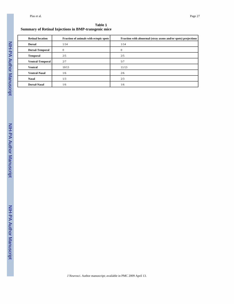
Injections of DiI were made in postnatal day 6 (P6) BMP-tg mice and compared to WT miceinjected at the same age. Labeled projections in the SC were analyzed on P8, an age whenretinotopic projections are anatomically mature (Simon and O'Leary, 1992a). In general,injections into dorsal retina of BMP-tg mice produced normal projection patterns withoutectopic spots (1 of 14 dorsal injections had ectopic spots, Fig. 1E and Table 1). On the otherhand, ventral injections in the BMP-tg mice usually produced a normal target zone withmultiple ectopic projections (11 of 14 cases, Fig. 1F and Table 1). Ectopic spots, which arenever seen in WT mice (Fig. 1C and 1D), are caused by nearby RGC axons projecting toinappropriate targets in the SC. At retinal locations intermediate between dorsal and ventral,such as nasal or temporal locations, there was a graded tendency to form ectopic projections,suggesting that BMP also has weak effects on targeting of RGC axons along the nasotemporalaxis (summarized schematically in Fig. 1G and quantified in Table 1).
Mediolateral entry of ventral axons into SC is abnormal in BMP-transgenic miceIn WT mice, axons that terminate in a given target zone enter the superior colliculus roughlyaligned with the mediolateral position of the target zone (Plas et al., 2005). For example, axonsfrom ventral retina enter the SC on the medial side of the brachium and will terminate in medialSC (Fig. 2A). In general, the degree of alignment of the incoming axons with the target zoneis greater when the target zone is on the lateral or medial edge of the SC, that is, when the axonsoriginate in extreme dorsal or ventral retina.
The pattern of ventrally labeled axons is distinctly different for BMP-tg mice (Fig.2B) where‘off-target’ axons are observed at the point of entry into SC. We quantified this targeting defectby examining the distribution of fluorescence across the width of the brachium of the SC, withexample florescence profiles shown in Fig. 2C. The center of mass of the axon distributionfrom ventral RGC axons (Fig. 2C, red) for a population of WT and BMP-transgenic mice inthe brachium of the SC quantitatively confirms that WT axons are typically arrayed medially(Fig. 2D; center of mass at 28+/-8%, n=5), whereas axons from BMP-tg mice show nopositional bias (Fig. 2D; center of mass at 47+/-7%, n=10; p< 0.001).
In contrast, injections into dorsal retina resulted in similar axon distributions in WT and BMP-tg mice. For example, axons from dorsal injections were distributed in the lateral brachium inboth BMP-tg and WT mice (Fig. 2C, blue). Summary quantification using the center of massof the fluorescence label in the brachium again confirms this qualitative point, with nodifference in the distribution of dorsal axons in BMP-tg and WT mice (Fig. 3E; BMP-transgeniccenter of mass = 63+/-5%, n-11; WT center of mass = 63+/-7%, n=6; P = 0.954). Thus, dorsalaxons in BMP-tg mice project normally to the SC, but the trajectory of ventral axons is severelydisturbed. This suggests that graded BMP signaling acts as a dorsalizing factor in thedeveloping retina, and confirms that the dorsoventral identity of axons is partially reflected inthe position of axons before they enter the SC at the brachium.
Plas et al. Page 5
J Neurosci. Author manuscript; available in PMC 2009 April 13.
NIH
-PA Author Manuscript
NIH
-PA Author Manuscript
NIH
-PA Author Manuscript

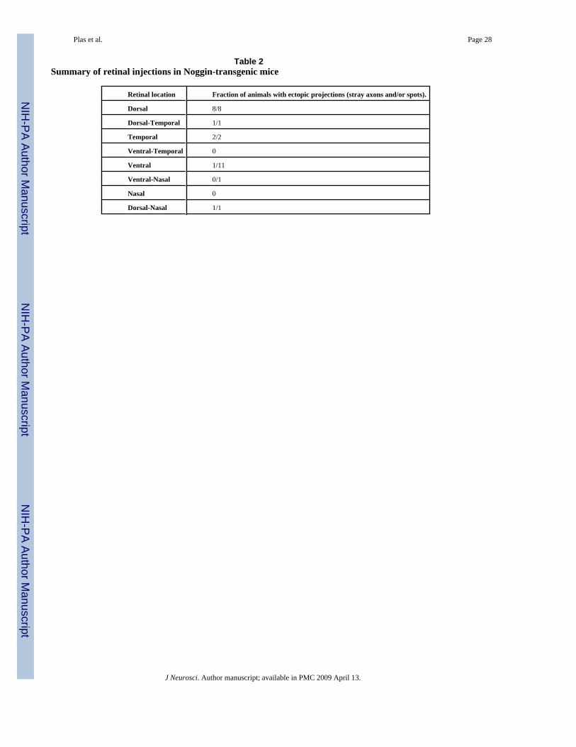
Projection and sorting defects in Noggin-transgenic miceNoggin is a BMP antagonist that inhibits BMP signaling by blocking the binding of the BMPligand to both types of BMP receptors (De Robertis and Kuroda, 2004). In Noggin-tg mice,which secrete Noggin from the lens, we hypothesize that the suppression of BMP signalingdue to high levels of Noggin in the eye will interfere with the specification of dorsal retinalcell fate by endogenous BMP. Indeed, topographic projections from the retina to the colliculusare disrupted in Noggin-tg mice, with targeting of dorsal RGC axons most severely disturbed(Fig. 3A), and only the projections from the most ventral part of retina remain largelyundisturbed (Fig. 3B). Specifically, all injections into the dorsal retina resulted in mistargetedaxons in the SC (8 of 8 cases had ectopic spots, Table 2), while injections into ventral retinararely resulted in ectopic spots (1 of 11 cases, Table 2, summarized schematically in Fig. 3C).The dorsal injections in Noggin-tg mice also showed evidence of naso-temporal targeting errors(Figure 3A). Moreover, the few cases in which the injections were localized to nasal or temporalretina often, though not always, showed misprojections (Table 2), suggesting that BMPsignaling also impacts nasotemporal targeting.
The mediolateral distribution of axons entering the SC at the brachium is also altered in theNoggin-tg mice, with axons from the dorsal retina most severely disturbed (Fig. 3D). Summaryquantification using the center of mass of the fluorescence label in the brachium shows thatdorsal RGC axons from Noggin-tg mice are pushed into the mid-brachium (Noggin-tg centersof mass at 48.3 +/- 4.2%, n=7; WT center of mass at 63.3 +/- 4.7%, n=6; p < 0.0001). RGCaxons from the ventral retina in Noggin-tg mice are also somewhat disturbed (Noggin-tgcenters of mass at 41.3 +/- 7.0 %, n=8; WT center of mass at 28.1 +/- 7.9 %, n=6; p < 0.01),though not as severely as dorsal axons. Thus, the sorting of dorsal axons in Noggin-tg mice asthey enter the SC, as well as their targeting in the SC, is completely compromised. The sortingof ventral RGC axons in Noggin-tg is partially disturbed, and the targeting of ventral axons inthe SC is nearly normal. This is consistent with a model in which the high dorsal expressionof BMP in the developing eye helps specify dorsal retinal ganglion cell fate, and interferingwith this signaling with transgenic expression of Noggin, a BMP antagonist, has its greatesteffect on dorsal axons. Similarly, overwhelming the endogenous dorsoventral gradient of BMPin BMP-tg mice interferes especially with ventral axons, has weaker effects on nasal andtemporal axons, but leaves the projection from dorsal axons intact. Furthermore, dorsoventralaxon sorting errors in the bSC are typically associated with mistargeting in the SC itself.
Mediolateral axon sorting in the Optic Tract is disrupted in BMP-tg and Noggin-tg miceThe SC is the final target of retinal ganglion cell axons. By the time the axons have reachedthis point they have already traversed the ventral and dorsal divisions of the lateral geniculatenucleus (LGN) of the thalamus, where they also form retinotopic maps. We investigated in P8mice whether the disruption of mediolateral axon order in BMP- and Noggin-tg mice is evidentin the optic tract, at a point before the axons have not even reached their first topographic target.We measured axon sorting in the optic tract just prior to its entry into the ventral LGN, whichwe refer to as the delta of the optic tract (dOT; Fig. 1A). In WT mice, axons from ventral anddorsal retina are clearly sorted in the medial and lateral parts of the optic tract (dOT),respectively (Fig. 4A and 4B). Axons at the dOT from dorsal retina in BMP-tg are also quitelateralized (Fig. 4F), like in WT mice. In contrast, axons from ventral retina at the dOT inBMP-tg mice are broadly distributed without any apparent sorting (Fig. 4E). In Noggin-tgmice, sorting of axons from the dorsal retina is completely disturbed (Fig. 4C), and the sortingof axons from the ventral retina is relatively intact (Fig. 4C). Quantification of this sorting wasagain performed by calculating the center of mass of the fluorescent distribution of label at thedOT across its mediolateral width for dorsal and ventral retinal injections (Fig. 4G). Thisquantification reveals that ventral RGC axons in BMP-tg mice and dorsal RGC axons inNoggin-tg mice are completely missorted in the dOT (WT ventral injection center of mass =
Plas et al. Page 6
J Neurosci. Author manuscript; available in PMC 2009 April 13.
NIH
-PA Author Manuscript
NIH
-PA Author Manuscript
NIH
-PA Author Manuscript

24+/-1%, n=2; WT dorsal center of mass = 69+/-5%, n=3; Noggin-tg ventral injection centerof mass = 38+/-2%, n=6; Noggin-tg dorsal center of mass = 53+/-2%, n=7; BMP-tg ventralinjection center of mass = 55+/-2%, n=10; BMP-tg dorsal center of mass = 69+/-3%, n=11, p< 0.01 for difference between WT and BMP-tg ventral injections and WT and Noggin-tg dorsalinjections. All other comparisons with WT are not statistically significant).
Aberrant sorting of axons in the OT is evident prior to target formation in the SCBy P1, nearly all RGC axons have entered the colliculus, though no target zone is yetestablished. At this early age, RGC axons from all parts of the retina extend nearly the entirelength of the colliculus, and axon branching is still quite minimal (Hindges et al., 2002). Weexamined the sorting of axons in BMP-transgenic mice at P1 to determine if RGC axon-targetinteractions were somehow responsible for the disruption in sorting in the optic tract at laterages. Instead, we found that even at P1 axons from ventral retina in BMP-tg mice (Fig. 5B)are not at all confined to the medial optic tract, as they are in WT mice (Fig. 5A). Quantificationof the distribution of axons in the optic tract at P1 confirms this qualitative impression (Fig.5C; center of mass for BMP-tg is 56 +/- 4%, n=5; center of mass for WT is 31 +/- 1%, n=11,p << 0.001). Given this data, it is difficult to argue that axon-target interactions are responsiblefor the severe disruption in sorting seen in the ascending optic tract at these early ages.
Off-target axons mature into ectopic spots in the SCDuring normal development, a small number of retinal axons enter the SC at inappropriatemediolateral positions (white arrows in Fig. 6A), but by maturity, most of these missortedaxons are eliminated (Simon and O'Leary, 1992a,b). Target zones begin to emerge at P3 at amediolateral position in SC that corresponds to the mediolateral location of the peak densityof incoming axons from the brachium (red arrows in Fig. 6A). In the BMP-transgenic mice atP3 (Fig. 6B and 6C), axons labeled by ventral retinal injections are much more widespread inthe brachium, and numerous axons enter the SC at incorrect mediolateral locations. In the SC,these missorted axons form dense branching networks visible even at off-target locations (Fig.6C).
Although it is often difficult to visualize the individual axons which give rise to ectopic spotsat P8, because the axons are not superficial, we see clear evidence that ectopic spots result fromaxons which have entered the anterior edge of the SC at an inappropriate mediolateral location(Fig. 6D and 6E). In these cases, just as in the wild type mice, the axons entry point and finalarborization are aligned, albeit at a mediolaterally inappropriate position.
Topography of LGN map is also disturbed in BMP-transgenic miceRetinal axons form topographic termination patterns in the ventral lateral geniculate nucleus(vLGN) and the dorsal lateral geniculate nucleus (dLGN) as well as the superior colliculus.We wondered whether the retinotopic maps in LGN were also disrupted by the transgenicoverexpression of BMP2. Indeed, this was typically the case (Fig. 7). The LGN is much morecompact in the rodent than the SC, so it is more difficult to quantitatively characterize targetingerrors in the LGN. Nonetheless, it was clear that the normally predictable patterns of label inthe LGN of WT mice resulting from focal injection into ventral retina were profoundlyperturbed in the LGN of BMP-tg mice. For example, targeted injections into ventral retina inBMP-tg mice, which typically lead to ectopic spots in the superior colliculus (Fig. 7A and 7B),cause corresponding ectopic spots in the same mice in dorsal and ventral LGN (Fig. 7C and7D). This suggests that common mechanisms are responsible for retinotopic map developmentin the lateral geniculate nucleus and superior colliculus.
Plas et al. Page 7
J Neurosci. Author manuscript; available in PMC 2009 April 13.
NIH
-PA Author Manuscript
NIH
-PA Author Manuscript
NIH
-PA Author Manuscript

Pretarget sorting in BMP-receptor mutants is similar to that in Noggin-tg miceWe examined the projections from dorsal retina in mice lacking one or both copies of the BMPreceptors BMPR1a and BMPR1b. BMPR1a is expressed ubiquitously, whereas BMPR1b hasa ventral-high to dorsal-low gradient of expression in the developing retina (Furuta and Hogan,1998). Loss of both of these receptors entirely prevents the development of the eye, while lossof both copies of either receptor alone or one copy of both receptors has no detectable effecton retinotectal topography ((Murali et al., 2005) and Fig. 8A, ‘control mice’). Mice homozygotefor BMPR1a and heterozygote for BMPR1b (‘BMP-receptor mutant mice’) have eyes withnormal structure but a strong disruption of topography from dorsal retina (Fig. 8B), quitereminiscent of the pattern of ectopic spots in Noggin-tg mice. We examined whether thistargeting defect is accompanied by a failure of pretarget sorting in the optic tract. In controlmice, sorting in the optic tract for dorsal retinal injections was normal (Fig. 8C and 8E), whilein BMP-receptor mutant mice, which have targeting defects in the colliculus for dorsal retinalaxons, sorting in the optic tract was also disturbed (Fig. 8D, 8E; center of mass for BMP-receptor mutant is 39 +/- 2%, n=3; center of mass for control mice is 69 +/- 1%, n=16, p <<0.001).
Role of EphB/ephrinB signaling in pretarget sortingRetinal axon behavior that differs depending on the dorsal or ventral retinal origin of the axonpresumably is the result of differential patterns of gene expression in the retina. For example,EphB receptors and their ligands, which are thought to mediate aspects of axon branching alongthe mediolateral axis of the SC, have expression patterns that vary with position along thedorsoventral axis during embryonic development (Henkemeyer, 1996; Braisted et al., 1997;Birgbauer et al., 2000; Hindges et al., 2002; Mann et al., 2002; McLaughlin et al., 2003). Weinvestigated whether the early spatial patterns of BMP expression are responsible forsubsequent patterns of gene expression of members of the EphB/ephrinB receptor-ligandsystem consistent with their role in mediolateral targeting in the SC. Using ISH, we visualizedthe expression pattern of EphB2 and ephrinB2 in the retina of P1 WT, BMP-tg and Noggin-tgmice (Fig. 9). In WT mice, ephrinB2 has a high-dorsal to low-ventral pattern of expression andEphB2 has a high-ventral to low-dorsal pattern of expression, consistent with what waspreviously observed (Henkemeyer et al., 1996; Birgbauer et al., 2000; Hindges et al., 2002;Murali et al., 2005). In BMP-tg mice, the expression of ephrinB2 appears unaltered, but theexpression of EphB2 is reduced and the high-ventral to low-dorsal gradient is absent (Fig. 9B,E). In Noggin-tg mice, the expression of ephrinB2 is suppressed, and the graded expression ofEphB2 is lost, though overall expression levels appear intact (Fig. 9C, F). This is consistentwith results from BMP-receptor mutants, where ephrinB2 expression in the retina was alsofound to be completely suppressed and the graded expression of EphB2 was absent (Murali etal., 2005). These results show that BMP signaling regulates the dorsoventral expression patternof axon guidance factors (EphB2/ephrinB2) that are known to play a role in RGC axonbranching and collicular target formation in the SC. Since EphB/ephrinB signaling has beenspecifically implicated in mapping of the dorsoventral axis of the retina onto the colliculus, weexamined RGC axon behavior in EphB2/B3 mutant mice to determine if EphB/ephrinBsignaling also plays a role in pretarget sorting. Consistent with previous reports (Hindges etal., 2002), we frequently observed ectopic target zones in SC projections from EphB2/B3 nullventral RGC axons (EphB2+/-;B3-/-: 1 out of 6 and EphB2-/-;B3-/-: 6 out of 12 mice).However, pretarget sorting in the brachium of the SC was not impaired in EphB2/B3 mutants(center of mass for control mice = 40 +/- 1%, n=11; center of mass for EphB2+/-;B3-/- = 35+/- 3%, n=6; center of mass for EphB2-/-;B3-/- mice = 32 +/- 1%, n=12; sorting is slightlybetter (smaller center of mass) in EphB2-/-;B3-/- mice than in control mice, p<0.01), implyingthat a mechanism apart from EphB/ephrinB signaling is principally responsible for this axonguidance behavior in the ascending optic tract (Fig. 9G-J).
Plas et al. Page 8
J Neurosci. Author manuscript; available in PMC 2009 April 13.
NIH
-PA Author Manuscript
NIH
-PA Author Manuscript
NIH
-PA Author Manuscript

DiscussionWe used several different lines of knockout and transgenic mice to establish and clarify therole of BMP signaling in retinotopic map formation and pre-target sorting in the developingretinofugal pathway of the rodent. In BMP-tg and Noggin-tg mice, SC target formation andpretarget sorting of ventral and dorsal axons, respectively, were dramatically disturbed. InBMP-receptor mutant mice, pretarget sorting of dorsal but not ventral axons was disrupted.We also found that dorsoventral targeting errors in the SC and LGN of these transgenics arealways associated with sorting errors in the ascending optic tract, suggesting a mechanisticlink. Finally, we showed that RGC axon pretarget sorting was undisturbed in EphB2/B3 mutantmice. Together, this data demonstrates that BMP signaling patterns the dorsoventral axis ofthe retina and influences RGC axon guidance behavior in the ascending optic tract through amechanism that is, at least in part, independent of EphB/ephrinB signaling.
Dorsoventral topography depends on BMP signalingWe demonstrated that transgenic induction of high levels of BMP throughout the developingeye leads ventral axons to mis-target in the LGN and SC. The targeting of dorsal axons, incontrast, is not disturbed in BMP-tg mice. Given the endogenous high-dorsal to low-ventralexpression of BMP in vertebrates, we conclude that the establishment of dorsoventraltopography depends on BMP signaling, with high levels of BMP leading to dorsal cell fate,and low levels to ventral cell fate. This model is reinforced by results from the Noggin-tg mice,which have an opposite phenotype to that of the BMP-tg, with relatively normal targeting ofventral RGC axons and grossly abnormal targeting and sorting of dorsal RGC axons. This isan expected phenotype, given that Noggin binds to endogenous BMP and prevents it fromactivating the BMP receptor. Finally, eye-specific BMP-receptor mutant mice have a similartargeting phenotype as the Noggin-tg mice, reinforcing the hypothesis that activation of theBMP receptor is necessary to establish dorsal cell fate in the developing retina.
The high-ventral to low-dorsal expression pattern of EphB2 receptor in the retina is thought toplay an important role in the development of retinotopy in the SC and LGN (Hindges et al.,2002; Mann et al., 2002). In BMP-tg mice, ephrinB2 expression in the retina is intact, but thegraded expression of EphB2 is disturbed. In Noggin-tg, ephrinB2 expression is dramaticallyreduced, and the high-ventral to low-dorsal expression of EphB2 is also disturbed. Thissuggests that BMP signaling is necessary for ephrinB2 expression in the retina, and that highlevels of BMP suppresses EphB2 expression, but BMP signaling is not necessary for EphB2expression. Moreover, the graded expression pattern of EphB2 is dependent on the gradedexpression of BMP in the eye. Finally, the effect of Noggin on EphB2/ephrinB2 expressionobserved in Noggin-tg mice is very similar to that found in BMP-receptor mutant mice (Muraliet al., 2005), confirming the role of BMP in regulating the expression of EphB2/ephrinB2 inthe retina. These results are also consistent with published reports in the chick (Koshiba-Takeuchi et al., 2000; Sakuta et al., 2001; Sakuta et al., 2006) arguing for a similar role acrossspecies for an endogenous dorsoventral gradient of BMP in the developing eye establishingdorsoventral retinal cell fate.
Pretarget sorting in bSC and dOT rely on BMP signalingAxons originating along the dorsoventral axis of the retina show evidence of presorting in theoptic tract of the rodent soon after they exit the optic chiasm (Simon and O'Leary, 1991; Chanand Guillery, 1994; Plas et al., 2005). Molecular/chemical guidance cues downstream of BMPsignaling in the retina are responsible for this pre-target sorting, since axons at the brachiumof the SC (bSC) and in the ascending ‘delta’ of the optic tract (dOT) are disorganized in theBMP-tg, Noggin-tg and BMP-receptor mutants (Figures 2-4, 8).
Plas et al. Page 9
J Neurosci. Author manuscript; available in PMC 2009 April 13.
NIH
-PA Author Manuscript
NIH
-PA Author Manuscript
NIH
-PA Author Manuscript

LGN targeting depends on BMP signalingRetinotopic precision in the mouse LGN is difficult to assay because of the small size of theLGN relative to the SC and its convoluted anatomy. Nonetheless, we frequently observedmistargeting (ectopic spots) in the LGN that was associated with sorting errors in the ascendingoptic tract and the formation of ectopic spots in the SC (Figures 7, 8). This correspondencesuggests that retinotopy in the LGN is regulated by similar mechanisms as in the SC, both ofwhich are downstream of BMP signaling in the retina.
Effects of BMP signaling on nasotemporal targetingDorsal axons are targeted and sorted normally in BMP-tg mice, whereas ventral axons areprofoundly disturbed. In Noggin-tg mice, dorsal axons are profoundly disturbed, but evenventral axons are partially disrupted. RGC axons originating along the nasotemporal axis ofthe mouse retina show no evidence of sorting in the ascending optic tract (Plas et al., 2005).Thus, targeting errors along the rostral-caudal axis of the SC in the BMP-tg, Noggin-tg andBMP-receptor mutant mice are not likely due to sorting errors in the optic tract. Disturbing theexpression of BMP or transcription factors downstream of BMP can cause changes in theexpression of ephrinA2 and EphA5 ((Sakuta et al., 2001; Mui et al., 2002; Liu et al., 2003;Sakuta et al., 2006), however see, (Barbieri et al., 2002)). This suggests that the partial sortingphenotype of ventral axons in Noggin-tg mice and the anterior-posterior targeting errors in theBMP-tg, Noggin-tg and BMP-receptor mutant mice are due to the effects of BMP signaling inthe regulation of EphA/ephrinA expression in the retina.
Pretarget sorting defects in BMP-tg mice are evident before target formationThe retinal ganglion cell axon sorting defects observed in the BMP-tg, Noggin-tg and BMP-receptor mutant mice are clearly evident in anatomical structures en route to their targets.Moreover, these sorting defects are present in the optic tract at a developmental stage (P0-P1)when no clear target zone has established itself in the SC or LGN. Sorting in the ascendingoptic tract is therefore not a byproduct of targeting in the LGN or SC, and may serve a necessaryfunction in the formation of topographically accurate projections by retinal ganglion cell axonsto their central targets.
In the neonatal (P2-3) SC of BMP-tg mice, there is a high density of retinal ganglion cell axonsat off-target locations relative to WT controls, even before focal target zones or ectopic spotshave formed in the colliculus (Figures 5 and 6). One week later, when retinocollicular maprefinement is complete (Simon and O'Leary, 1992a), ectopic spots in BMP-tg SC receive someof their axonal innervation directly from missorted axons that orginate from the wrongmediolateral position in the brachium of the SC (Figure 6). This suggests that the sorting ofaxons before they reach their target may produce a threshold density of axons necessary foractivity-dependent factors to refine retinal projections into a mature topographic map(Chandrasekaran et al., 2005). Errors in direction-specific branching that occur subsequent tomissorting likely also contribute to the formation of ectopic target zones in the colliculus(Hindges et al., 2002; McLaughlin et al., 2003).
EphB2/B3 signaling is not responsible for pretarget sortingWhat are the mechanisms responsible for pretarget sorting of RGC axons based upon theirdorsoventral origin? One possibility is that axon-axon interactions between RGCs based upontheir expression of EphBs and ephrinBs in a counter-gradient may lead to sorting via contactmediated repulsion (Marston et al., 2003; Zimmer et al., 2003). Alternatively, RGC axon-bornEphB receptors may interact with ephrinB ligands expressed in specific populations ofchiasmatic cells, in a manner analogous to the mechanism of ipsilateral-contralateral sortingthat has recently been described at the chiasm (Nakagawa et al., 2000; Williams et al., 2003).
Plas et al. Page 10
J Neurosci. Author manuscript; available in PMC 2009 April 13.
NIH
-PA Author Manuscript
NIH
-PA Author Manuscript
NIH
-PA Author Manuscript

Hypothesizing a role for EphB receptors on RGC axons is parsimonious, as they havepreviously been shown to play a role in target-specific branch formation in the superiorcolliculus (Hindges et al., 2002; McLaughlin et al., 2003). However, we did not find thatpretarget sorting defects of RGC axons in EphB2/B3 mutant mice were worse than controls.If anything, we found that sorting in the mutants was marginally better than controls, whichcould be a byproduct of EphB/ephrinB signaling in the control of axon fasciculation (Orioli etal., 1996; Chen et al., 2004), or related to the albino background of the EphB2/B3 mutant mice.Interestingly, the sorting of ventral axons in the albino (CD1) background of the EphB2/B3mice was clearly worse than wildtype (C57/Bl6) mice, suggesting that defects in thedevelopment of the retinofugal pathway known to exist in albino mice (Guillery et al., 1995;Rice et al., 1995; Marcus et al., 1996) may also affect sorting in the ascending optic track. Themuch stronger mapping (targeting) phenotypes in BMP-tg, Noggin-tg and BMP-receptor micein comparison to EphB2/EphB3 receptor mutants also suggests that additional guidancemechanisms beyond EphB/ephrinB signaling in the target are crucial for mapping thedorsoventral axis of the retina.
Our experiments have ruled out EphB2/B3 receptor signaling as the mechanism by which BMPsignaling regulates axon sorting in the ascending optic tract. Thus, the mechanisms regulatingdorsoventral sorting in the optic tract of the mouse are at least in part distinct from thosecontrolling target specific branching in the superior colliculus, as has been previouslydemonstrated in Xenopus (Chien et al., 1995). There is ample evidence for other signalingpathways playing a role in dorsoventral RGC axon guidance and targeting. Wnt-Ryk signalingis thought to mediate aspects of retinotectal mapping of dorsoventral RGC axons in the chick,but little is known of their function in the mouse (Schmitt et al., 2006). BMP signaling mightalso regulate heparan sulfate proteoglycan function, which is required for sorting of dorsalretinal axons in the optic tract of zebrafish and retinal axon guidance at the chiasm in mice(Inatani et al., 2003; Lee et al., 2004; Pratt et al., 2006). Cell adhesion molecules (L1) haverecently been implicated in the regulation of mediolateral topography in the colliculus (Buhusiet al., 2008), and are also differentially expressed in the ascending optic tract (L1 and NCAM;(Chung et al., 2004)), and are therefore potential candidates to mediate RGC pretartget sorting.
Our data shows that BMPs play an important role in the establishment of retinal ganglion cellfate, with strong effects along the dorsoventral axis, and more circumscribed effects along thenasotemporal axis. Disrupting BMP signaling early in eye development causes profound errorsin retinal ganglion cell axon sorting in the ascending optic tract through a mechanismindependent of EphB2/B3 receptors. Targeting errors caused by interfering with retinal BMPsignaling are also quite severe, suggesting that mechanisms regulating sorting in the ascendingoptic tract and branching in the central target both contribute to the retinotopic mapping ofdorsoventral RGC axons.
Supplementary MaterialRefer to Web version on PubMed Central for supplementary material.
AcknowledgementsThis work was supported by NIH grants R01 MH62639, R01 EY015788 and P30 EY000785 to MCC and R01EY017434 to MH.
ReferencesBarbieri AM, Broccoli V, Bovolenta P, Alfano G, Marchitiello A, Mocchetti C, Crippa L, Bulfone A,
Marigo V, Ballabio A, Banfi S. Vax2 inactivation in mouse determines alteration of the eye dorsal-
Plas et al. Page 11
J Neurosci. Author manuscript; available in PMC 2009 April 13.
NIH
-PA Author Manuscript
NIH
-PA Author Manuscript
NIH
-PA Author Manuscript

ventral axis, misrouting of the optic fibres and eye coloboma. Development 2002;129:805–813.[PubMed: 11830579]
Birgbauer E, Cowan CA, Sretavan DW, Henkemeyer M. Kinase independent function of EphB receptorsin retinal axon pathfinding to the optic disc from dorsal but not ventral retina. Development2000;127:1231–1241. [PubMed: 10683176]
Braisted JE, McLaughlin T, Wang HU, Friedman GC, Anderson DJ, O'Leary DD. Graded and lamina-specific distributions of ligands of EphB receptor tyrosine kinases in the developing retinotectalsystem. Dev Biol 1997;191:14–28. [PubMed: 9356168]
Buhusi M, Schlatter MC, Demyanenko GP, Thresher R, Maness PF. L1 interaction with ankyrin regulatesmediolateral topography in the retinocollicular projection. J Neurosci 2008;28:177–188. [PubMed:18171935]
Cang J, Niell CM, Liu X, Pfeiffenberger C, Feldheim DA, Stryker MP. Selective Disruption of OneCartesian Axis of Cortical Maps and Receptive Fields by Deficiency in Ephrin-As and StructuredActivity. Neuron 2008;57:511–523. [PubMed: 18304481]
Carson JP, Thaller C, Eichele G. A transcriptome atlas of the mouse brain at cellular resolution. CurrOpin Neurobiol 2002;12:562–565. [PubMed: 12367636]
Carson JP, Ju T, Lu HC, Thaller C, Xu M, Pallas SL, Crair MC, Warren J, Chiu W, Eichele G. A digitalatlas to characterize the mouse brain transcriptome. PLoS Comput Biol 2005;1:e41. [PubMed:16184189]
Chan SO, Guillery RW. Changes in fiber order in the optic nerve and tract of rat embryos. J Comp Neurol1994;344:20–32. [PubMed: 8063954]
Chandrasekaran AR, Plas DT, Gonzalez E, Crair MC. Evidence for an instructive role of retinal activityin retinotopic map refinement in the superior colliculus of the mouse. J Neurosci 2005;25:6929–6938.[PubMed: 16033903]
Chapman DL, Garvey N, Hancock S, Alexiou M, Agulnik SI, Gibson-Brown JJ, Cebra-Thomas J, BollagRJ, Silver LM, Papaioannou VE. Expression of the T-box family genes, Tbx1-Tbx5, during earlymouse development. Dev Dyn 1996;206:379–390. [PubMed: 8853987]
Chen ZY, Sun C, Reuhl K, Bergemann A, Henkemeyer M, Zhou R. Abnormal hippocampal axonbundling in EphB receptor mutant mice. J Neurosci 2004;24:2366–2374. [PubMed: 15014111]
Chien CB, Cornel EM, Holt CE. Absence of topography in precociously innervated tecta. Development1995;121:2621–2631. [PubMed: 7671824]
Chung KY, Leung KM, Lin CC, Tam KC, Hao YL, Taylor JS, Chan SO. Regionally specific expressionof L1 and sialylated NCAM in the retinofugal pathway of mouse embryos. J Comp Neurol2004;471:482–498. [PubMed: 15022265]
Dale JK, Vesque C, Lints TJ, Sampath TK, Furley A, Dodd J, Placzek M. Cooperation of BMP7 andSHH in the induction of forebrain ventral midline cells by prechordal mesoderm. Cell 1997;90:257–269. [PubMed: 9244300]
De Robertis EM, Kuroda H. Dorsal-ventral patterning and neural induction in Xenopus embryos. AnnuRev Cell Dev Biol 2004;20:285–308. [PubMed: 15473842]
Furuta Y, Hogan BL. BMP4 is essential for lens induction in the mouse embryo. Genes Dev1998;12:3764–3775. [PubMed: 9851982]
Graff JM. Embryonic patterning: to BMP or not to BMP, that is the question. Cell 1997;89:171–174.[PubMed: 9108472]
Guillery RW, Mason CA, Taylor JS. Developmental determinants at the mammalian optic chiasm. JNeurosci 1995;15:4727–4737. [PubMed: 7623106]
Henkemeyer M, Orioli D, Henderson JT, Saxton TM, Roder J, Pawson T, Klein R. Nuk controlspathfinding of commissural axons in the mammalian central nervous system. Cell 1996;86:35–46.[PubMed: 8689685]
Hindges R, McLaughlin T, Genoud N, Henkemeyer M, O'Leary DD. EphB forward signaling controlsdirectional branch extension and arborization required for dorsal-ventral retinotopic mapping.Neuron 2002;35:475–487. [PubMed: 12165470]
Hung FC, Zhao S, Chen Q, Overbeek PA. Retinal ablation and altered lens differentiation induced byocular overexpression of BMP7. Vision Res 2002;42:427–438. [PubMed: 11853758]
Plas et al. Page 12
J Neurosci. Author manuscript; available in PMC 2009 April 13.
NIH
-PA Author Manuscript
NIH
-PA Author Manuscript
NIH
-PA Author Manuscript

Koshiba-Takeuchi K, Takeuchi JK, Matsumoto K, Momose T, Uno K, Hoepker V, Ogura K, TakahashiN, Nakamura H, Yasuda K, Ogura T. Tbx5 and the retinotectum projection. Science 2000;287:134–137. [PubMed: 10615048]
Inatani M, Irie F, Plump AS, Tessier-Lavigne M, Yamaguchi Y. Mammalian brain morphogenesis andmidline axon guidance require heparan sulfate. Science 2003;302:1044–1046. [PubMed: 14605369]
Lee JS, von der Hardt S, Rusch MA, Stringer SE, Stickney HL, Talbot WS, Geisler R, Nusslein-VolhardC, Selleck SB, Chien CB, Roehl H. Axon Sorting in the Optic Tract Requires HSPG Synthesis byext2 (dackel) and extl3 (boxer). Neuron 2004;44:947–960. [PubMed: 15603738]
Liu J, Wilson S, Reh T. BMP receptor 1b is required for axon guidance and cell survival in the developingretina. Dev Biol 2003;256:34–48. [PubMed: 12654290]
Luo L, Flanagan JG. Development of continuous and discrete neural maps. Neuron 2007;56:284–300.[PubMed: 17964246]
Mann F, Ray S, Harris W, Holt C. Topographic mapping in dorsoventral axis of the Xenopus retinotectalsystem depends on signaling through ephrin-B ligands. Neuron 2002;35:461–473. [PubMed:12165469]
Marcus RC, Wang LC, Mason CA. Retinal axon divergence in the optic chiasm: midline cells areunaffected by the albino mutation. Development 1996;122:859–868. [PubMed: 8631264]
Marston DJ, Dickinson S, Nobes CD. Rac-dependent trans-endocytosis of ephrinBs regulates Eph-ephrincontact repulsion. Nat Cell Biol 2003;5:879–888. [PubMed: 12973357]
Massague J. TGF-beta signal transduction. Annu Rev Biochem 1998;67:753–791. [PubMed: 9759503]McLaughlin T, O'Leary DD. Molecular gradients and development of retinotopic maps. Annu Rev
Neurosci 2005;28:327–355. [PubMed: 16022599]McLaughlin T, Hindges R, Yates PA, O'Leary DD. Bifunctional action of ephrin-B1 as a repellent and
attractant to control bidirectional branch extension in dorsal-ventral retinotopic mapping.Development 2003;130:2407–2418. [PubMed: 12702655]
Mui SH, Hindges R, O'Leary DD, Lemke G, Bertuzzi S. The homeodomain protein Vax2 patterns thedorsoventral and nasotemporal axes of the eye. Development 2002;129:797–804. [PubMed:11830578]
Murali D, Yoshikawa S, Corrigan RR, Plas DJ, Crair MC, Oliver G, Lyons KM, Mishina Y, Furuta Y.Distinct developmental programs require different levels of Bmp signaling during mouse retinaldevelopment. Development 2005;132:913–923. [PubMed: 15673568]
Nakagawa S, Brennan C, Johnson KG, Shewan D, Harris WA, Holt CE. Ephrin-B regulates the Ipsilateralrouting of retinal axons at the optic chiasm. Neuron 2000;25:599–610. [PubMed: 10774728]
Orioli D, Henkemeyer M, Lemke G, Klein R, Pawson T. Sek4 and Nuk receptors cooperate in guidanceof commissural axons and in palate formation. Embo J 1996;15:6035–6049. [PubMed: 8947026]
Overbeek PA, Chepelinsky AB, Khillan JS, Piatigorsky J, Westphal H. Lens-specific expression anddevelopmental regulation of the bacterial chloramphenicol acetyltransferase gene driven by themurine alpha A-crystallin promoter in transgenic mice. Proc Nat Acad Sci U S A 1985;82:7815–7819.
Plas DT, Lopez JE, Crair MC. Pretarget sorting of retinocollicular axons in the mouse. J Comp Neurol2005;491:305–319. [PubMed: 16175549]
Pratt T, Conway CD, Tian NM, Price DJ, Mason JO. Heparan sulphation patterns generated by specificheparan sulfotransferase enzymes direct distinct aspects of retinal axon guidance at the optic chiasm.J Neurosci 2006;26:6911–6923. [PubMed: 16807321]
Rice DS, Williams RW, Goldowitz D. Genetic control of retinal projections in inbred strains of albinomice. J Comp Neurol 1995;354:459–469. [PubMed: 7608332]
Sakuta H, Takahashi H, Shintani T, Etani K, Aoshima A, Noda M. Role of bone morphogenic protein 2in retinal patterning and retinotectal projection. J Neurosci 2006;26:10868–10878. [PubMed:17050724]
Sakuta H, Suzuki R, Takahashi H, Kato A, Shintani T, Iemura S, Yamamoto TS, Ueno N, Noda M.Ventroptin: a BMP-4 antagonist expressed in a double-gradient pattern in the retina. Science2001;293:111–115. [PubMed: 11441185]
Schmitt AM, Shi J, Wolf AM, Lu CC, King LA, Zou Y. Wnt-Ryk signalling mediates medial-lateralretinotectal topographic mapping. Nature 2006;439:31–37. [PubMed: 16280981]
Plas et al. Page 13
J Neurosci. Author manuscript; available in PMC 2009 April 13.
NIH
-PA Author Manuscript
NIH
-PA Author Manuscript
NIH
-PA Author Manuscript

Simon DK, O'Leary DD. Relationship of retinotopic ordering of axons in the optic pathway to theformation of visual maps in central targets. J Comp Neurol 1991;307:393–404. [PubMed: 1856329]
Simon DK, O'Leary DD. Development of topographic order in the mammalian retinocollicular projection.J Neurosci 1992a;12:1212–1232. [PubMed: 1313491]
Simon DK, O'Leary DD. Influence of position along the medial-lateral axis of the superior colliculus onthe topographic targeting and survival of retinal axons. Brain Res Dev Brain Res 1992b;69:167–172.
Sperry RW. Chemoaffinity in the Orderly Growth of Nerve Fiber Patterns and Connections. Proc NatlAcad Sci U S A 1963;50:703–710. [PubMed: 14077501]
Visel A, Thaller C, Eichele G. GenePaint.org: an atlas of gene expression patterns in the mouse embryo.Nucleic Acids Res 2004;32(Database):D552–556. [PubMed: 14681479]
Williams SE, Mann F, Erskine L, Sakurai T, Wei S, Rossi DJ, Gale NW, Holt CE, Mason CA,Henkemeyer M. Ephrin-B2 and EphB1 mediate retinal axon divergence at the optic chiasm. Neuron2003;39:919–935. [PubMed: 12971893]
Yaylaoglu MB, Titmus A, Visel A, Alvarez-Bolado G, Thaller C, Eichele G. Comprehensive expressionatlas of fibroblast growth factors and their receptors generated by a novel robotic in situ hybridizationplatform. Dev Dyn 2005;234:371–386. [PubMed: 16123981]
Zhao S, Chen Q, Hung FC, Overbeek PA. BMP signaling is required for development of the ciliary body.Development 2002;129:4435–4442. [PubMed: 12223402]
Zimmer M, Palmer A, Kohler J, Klein R. EphB-ephrinB bi-directional endocytosis terminates adhesionallowing contact mediated repulsion. Nat Cell Biol 2003;5:869–878. [PubMed: 12973358]
Plas et al. Page 14
J Neurosci. Author manuscript; available in PMC 2009 April 13.
NIH
-PA Author Manuscript
NIH
-PA Author Manuscript
NIH
-PA Author Manuscript

Figure 1.Projections from ventral, but not dorsal retina are inappropriate in BMP transgenic mice at P8.A. Schematic of mouse visual system, showing the major retinal projections, including thedorsal and ventral LGN and the SC. The accessory optic system is not shown. The ‘delta ofthe Optic Tract’ is the area of the ascending optic tract before it enters the ventral LGN. The‘brachium of the Superior Colliculus’ is the area of the optic tract between the LGN and SC,just before the ganglion cell axons enter the SC. B. The retina is assigned coordinates basedon the insertion of the four major occulomotor muscles, namely the superior, inferior, lateral,and medial recti (SR, IR, LR and MR), see Plas et al., 2005. C. A focal injection into dorsalretina of a WT mouse labels a target zone (TZ) in lateral SC. D. A focal injection into ventralretina of a WT mouse labels a TZ in medial SC. E. In BMP transgenic mice, the projectionfrom dorsal retina is similar to that in WT mice and topographically correct. F. Projectionsfrom ventral retina in BMP transgenic mice show ‘ectopic spots’ (arrows) lateral to the expectedTZ. G. Summary cartoon depicting graphically the frequency of inappropriate projections tothe SC as a function of the position of the retinal dye injection in the BMP transgenic mouse.Dorsal retinal injections lead to normal collicular projections to lateral (L) colliculus (blue).Ventral retinal injections lead to a medial target zone (M) in colliculus and many ectopic spots(red). Temporal (green) and nasal (purple) retinal injections are largely normal, with somecases of inappropriate projections to the colliculus.
Plas et al. Page 15
J Neurosci. Author manuscript; available in PMC 2009 April 13.
NIH
-PA Author Manuscript
NIH
-PA Author Manuscript
NIH
-PA Author Manuscript

Figure 2.Ventral retinal axons in BMP transgenics are misplaced in the brachium of the SuperiorColliculus (bSC) at P8. A. Axons originating in ventral retina of WT mice enter the SC fromthe medial edge of the bSC in WT mice. B. In BMP-transgenic mice, ventral axons lose theirconfinement and spread laterally in the bSC. C. The difference in the distribution of RGC axonsfrom ventral (red) retina in the bSC is shown as normalized fluorescence across the width ofthe bSC (red bar in Fig. 2A and 2B) for representative examples of WT (dashed line) and BMP-transgenic mice (solid line). The axon distributions in the bSC resulting from dorsal (blue)injections are similar in the two genotypes. D. These results are summarized as the meanposition of the center of mass of the fluorescent distribution of labeled RGC axons from ventral
Plas et al. Page 16
J Neurosci. Author manuscript; available in PMC 2009 April 13.
NIH
-PA Author Manuscript
NIH
-PA Author Manuscript
NIH
-PA Author Manuscript

(red) or dorsal (blue) injections across the width of the bSC, with the number of animalsindicated above each bar. The mean centers of mass for the ventral retinal injections retina aresignificantly different between the two genotypes (*; p<0.001).
Plas et al. Page 17
J Neurosci. Author manuscript; available in PMC 2009 April 13.
NIH
-PA Author Manuscript
NIH
-PA Author Manuscript
NIH
-PA Author Manuscript

Figure 3.Dorsal retinal axons in Noggin transgenic mice are misplaced in the bSC at P8. A. Exampleof a dorsal retinal injection in a Noggin transgenic leading to multiple ectopic spots (arrows)at inappropriate locations in the SC. B. Ventral retinal injections nearly always terminatenormally in the SC. C. Summary cartoon showing that dorsal injections lead to misprojectionsin Noggin transgenics, but ventral retinal injections are relatively normal. D. Summaryquantification showing that the distribution of RGC axons in the bSC in Noggin transgenicsis completely disturbed for dorsal injections (blue), and partially disturbed for ventral injections(red) so that dorsal (*; p<0.0001). and ventral (*; p<0.01) axons in Noggin transgenics are lessconfined to the lateral and medial sides of the bSC, respectively, than in WT mice.
Plas et al. Page 18
J Neurosci. Author manuscript; available in PMC 2009 April 13.
NIH
-PA Author Manuscript
NIH
-PA Author Manuscript
NIH
-PA Author Manuscript

Figure 4.Axons in the ascending ‘delta’ of the Optic tract (dOT) are missorted at P8 in BMP and Noggintransgenics. A, B. Axons originating from ventral or dorsal retina in WT mice travel along themedial (M), or lateral (L) edges of the optic tract. C, D. In the Noggin-transgenic mice, axonsfrom both ventral and dorsal retina lose their confinement in the dOT, though the disturbanceof the dorsal axons is more severe E, F. In BMP transgenic mice, the ventrally originatingaxons lose their restriction to medial dOT, while those from dorsal retina remain on the lateralside. G. Summary quantification of the position of the center of mass of fluorescent label acrossthe medial-lateral width of the dOT (red bar in Fig. 4B) in WT, BMP transgenic and Noggintransgenic mice for dorsal (blue) and ventral (red) retinal injections. In BMP-transgenic mice,only injections from ventral retinal show an inappropriate distribution in the dOT, where as inNoggin transgenics, dorsal RGC axons are the most inappropriately distributed (*; p < 0.01).
Plas et al. Page 19
J Neurosci. Author manuscript; available in PMC 2009 April 13.
NIH
-PA Author Manuscript
NIH
-PA Author Manuscript
NIH
-PA Author Manuscript

Figure 5.Axon sorting even at P1 is disturbed in BMP-transgenic mice. A and B. Axon label from ventralretina is segregated to medial (M) optic tract by P1 in WT mice but already extends into lateral(L) optic tract in BMP transgenics at P1. C. The distribution of ventral label in the optic tractsof the two genotypes is quite different (BMP-transgenic is red; WT is blue; Dashed lines areexample fluorescent profiles, solid lines are average fluorescent profiles). Each curvecorresponds to the distribution of fluorescent label across the dOT (red bar in Fig. 5A and 5B)in one animal. Note that the fluorescent profiles for ventral retinal axons in BMP-transgenicmice (red) are not sorted to the medial optic tract, as they are in WT mice (blue). Summaryquantification (inset) for the center of mass of fluorescent label in the dOT in P1 WT (blue;n=11) and BMP transgenic (red; n=5; *, p << 0.001) mice.
Plas et al. Page 20
J Neurosci. Author manuscript; available in PMC 2009 April 13.
NIH
-PA Author Manuscript
NIH
-PA Author Manuscript
NIH
-PA Author Manuscript

Figure 6.Mistargeted axons persist in BMP-transgenics. A. In WT mice at P3, nearly all retinal axonshave invaded the SC, but the target zone has not been elaborated. Axons from ventral retina inWT mice enter the anterior SC at its medial edge (lower red arrow), and are clearly confinedto medial SC (upper red arrow). Very few axons are in off target regions (white arrows). B. InBMP-transgenic mice at P3, axons from ventral retina are diffusely spread across themediolateral extent of the SC, with many off target RGC axons forming branches atinappropriate locations in the SC. C. High magnification (10X) image of white boxed regionin (B) shows an ‘off-target’ area in which a dense network of branching axons is evident. Thisdense network of off-target axons doesn't exist in WT mice. D. At P8 in BMP-transgenics,some ventral RGC axons that entered the SC lateral (small white arrows) to their normaltrajectory in bSC maintain a misprojection to ‘ectopic spots’ (large red arrow) lateral to theirnormal target zone in medial SC. E. High magnification (10X) image of white boxed regionin (D) shows the misprojecting ventral axons in BMP transgenic mice that enter the SC fromlateral locations often form ectopic spots (red arrow) in the SC. Lateral (L) and Caudal (C)designation in B applies to all panels. Scale bars: 500 μm in A, B, D; 100 μm in C, E.
Plas et al. Page 21
J Neurosci. Author manuscript; available in PMC 2009 April 13.
NIH
-PA Author Manuscript
NIH
-PA Author Manuscript
NIH
-PA Author Manuscript

Figure 7.The LGN in BMP-transgenic mice contains axons that are mistargeted in a manner similar tothat seen in the SC. A, B. In P8 BMP-transgenic mice, ventral axons often form ‘ectopicspots’ (ES) lateral to the normal target zone. C, D. The dorsal and lateral subdivisions of theLGN each contain a retinotopic representation of the retina that is formed by the retinal axonsas they pass through these structures on their way to the SC making topographic appropriatesynaptic connections via axon collaterals. In the BMP-transgenic mice, the disturbance oftopography in the projection from ventral retina seen in the SC (A, B) is frequentlyaccompanied by similar disruptions of retinotopy in the LGN (C, D). Scale bars: 500 μm in A,B; 250 μm in C, D.
Plas et al. Page 22
J Neurosci. Author manuscript; available in PMC 2009 April 13.
NIH
-PA Author Manuscript
NIH
-PA Author Manuscript
NIH
-PA Author Manuscript

Figure 8.Axon sorting disturbed in BMP-receptor mutants is similar to that in Noggin transgenics at P8.A. Dorsal retinal axons form a single focal TZ in lateral SC in control mice. B. In a retinalacking BMPR1a and one copy of BMPR1b (BMP-receptor mutants), dorsal retinal axons formectopic projections in addition to the normal TZ. C. Dorsal axons at the dOT in control miceare segregated to the lateral (L) optic tract. D. In the BMP-receptor mutant mice, dorsal axonsfail to segregate to lateral optic tract. E. Distribution of dorsal RGC axons in the dOT of BMP-receptor mutants is shown as normalized fluorescence across the width of the dOT. Individualexamples are shown as solid red line and the average is represented as a dashed red line.Summary quantification (inset) shows the mean center of mass of or fluorescently labeled
Plas et al. Page 23
J Neurosci. Author manuscript; available in PMC 2009 April 13.
NIH
-PA Author Manuscript
NIH
-PA Author Manuscript
NIH
-PA Author Manuscript

axons in the dOT for dorsal injections in WT mice and BMP-receptor mutant mice. The centerof mass of label at the dOT in BMP-receptor mutant mice is significantly more medial (M)than that for WT mice (p<0.001). Scale bars: 500 μm in A, B; 250 μm in C, D.
Plas et al. Page 24
J Neurosci. Author manuscript; available in PMC 2009 April 13.
NIH
-PA Author Manuscript
NIH
-PA Author Manuscript
NIH
-PA Author Manuscript

Figure 9.In situ hybridization reveals altered expression patterns of EphB2 and ephrinB2 in the retinaof BMP-tg and Noggin-tg mice, but ventral axons are not missorted in EphB2/B3 mutant mice.A. In WT mice, ephrinB2 is expressed in a high-dorsal to low-ventral gradient. B. The ephrinB2expression pattern is not altered in BMP-tg mice. C. EphrinB2 expression is dramaticallysuppressed in Noggin-tg mice. D. In WT mice, EphB2 shows a high-ventral to low-dorsalexpression pattern. E. EphB2 expression is suppressed in BMP-tg, and no obvious gradient ispresent. F. EphB2 expression lacks a high-ventral to low-dorsal pattern in Noggin-tg mice,though average expression levels are normal. G-I. At P8, relative to controls (G), ventral RGCaxons in EphB2+/-;B3-/- (H) and EphB2-/-;B3-/- mice (I) are not missorted in the brachium
Plas et al. Page 25
J Neurosci. Author manuscript; available in PMC 2009 April 13.
NIH
-PA Author Manuscript
NIH
-PA Author Manuscript
NIH
-PA Author Manuscript

of the SC (red bar in G and H). Summary quantification of center of mass for ventral injectionsin the bSC of WT and EphB2/B3 mutants. The sorting of EphB2/B3 double mutants is slightlybetter (with a smaller Center of Mass) than controls. D, dorsal; L, lateral; C, cadual; Scale bar:100μm in G-I.
Plas et al. Page 26
J Neurosci. Author manuscript; available in PMC 2009 April 13.
NIH
-PA Author Manuscript
NIH
-PA Author Manuscript
NIH
-PA Author Manuscript
NIH
-PA Author Manuscript
NIH
-PA Author Manuscript
NIH
-PA Author Manuscript
Plas et al. Page 27
Table 1Summary of Retinal Injections in BMP-transgenic mice
Retinal location Fraction of animals with ectopic spots Fraction with abnormal (stray axons and/or spots) projections
Dorsal 1/14 1/14
Dorsal-Temporal 0 0
Temporal 2/5 2/5
Ventral-Temporal 2/7 5/7
Ventral 10/13 11/13
Ventral-Nasal 1/6 2/6
Nasal 1/3 2/3
Dorsal-Nasal 1/6 1/6
J Neurosci. Author manuscript; available in PMC 2009 April 13.
NIH
-PA Author Manuscript
NIH
-PA Author Manuscript
NIH
-PA Author Manuscript
Plas et al. Page 28
Table 2Summary of retinal injections in Noggin-transgenic mice
Retinal location Fraction of animals with ectopic projections (stray axons and/or spots).
Dorsal 8/8
Dorsal-Temporal 1/1
Temporal 2/2
Ventral-Temporal 0
Ventral 1/11
Ventral-Nasal 0/1
Nasal 0
Dorsal-Nasal 1/1
J Neurosci. Author manuscript; available in PMC 2009 April 13.


![Company introducing[PDF, 2,7 MB]](https://static.fdocuments.us/doc/165x107/58a2cfcc1a28abbe5a8bc6d4/company-introducingpdf-27-mb.jpg)