Author: Ida Lucy Iacobucci, 2015 License: Unless … · MEASUREMENTS IN OCULAR MOTILITY 1. Corneal...
24
Author: Ida Lucy Iacobucci, 2015 License: Unless otherwise noted, this material is made available under the terms of the Creative Commons Attribution-NonCommercial-Share Alike 4.0 License: http://creativecommons.org/licenses/by-nc-sa/4.0/ The University of Michigan Open.Michigan initiative has reviewed this material in accordance with U.S. Copyright Law and has tried to maximize your ability to use, share, and adapt it. Copyright holders of content included in this material should contact [email protected] with any questions, corrections, or clarification regarding the use of content. For more information about how to attribute these materials, please visit: http://open.umich.edu/education/about/terms-of-use.
Transcript of Author: Ida Lucy Iacobucci, 2015 License: Unless … · MEASUREMENTS IN OCULAR MOTILITY 1. Corneal...

Author: Ida Lucy Iacobucci, 2015
License: Unless otherwise noted, this material is made available under the terms of the Creative Commons Attribution-NonCommercial-Share Alike 4.0 License: http://creativecommons.org/licenses/by-nc-sa/4.0/
The University of Michigan Open.Michigan initiative has reviewed this material in accordance with U.S. Copyright Law and has tried to maximize your ability to use, share, and adapt it. Copyright holders of content included in this material should contact [email protected] with any questions, corrections, or clarification regarding the use of content.
For more information about how to attribute these materials, please visit:
http://open.umich.edu/education/about/terms-of-use.

Chapter 1
BASIC TESTS TO EVALUATE OCULAR MOTILITY I. VISION ASSESSMENT IN CHILDREN UNDER FIVE YEARS
OF AGE
1. Visual Acuity Timeline: - Newborn = 20/600, child will respond to bright lights by
squeezing lids. - 1 month = 20/400 child will follow a large object of light. - 3-5 months = 20/200 child will reliably follow a target in all gazes. - 12 months = 20/40 (estimate) hand-eye coordination developing
will grab objects. - 2-3 years = 20/30 child can identify/match various pictures. - 4+years = 20/20 child can reliably identify letters.
2. Obtaining Reliable VA
- Always measure monocularly with adhesive eye patch. - Exception: Nystagmus, low vision - use opaque occluder. - Never use hand to occlude the eye. - Don’t give up too soon!
3. Preverbal Children a) Light - Used for children < 2 months. - Shine light into each eye and watch response. - Blinking, shielding, squeezing eyes, crying – all normal responses. b) OKN Drum/Tape-Evaluate Smooth & Pursuit Nystagmus
Movements - Rotate drum vertically and watch for lid movement
upward/downward. - Monocular/binocular – horizontal and vertical movements. - Used in children under 6-8 months or developmentally delayed. c) Mirror/Doll’s Head - Hold mirror up to baby so they can see their reflection and move
it side to side - Normal response = baby will follow image in the mirror - Move baby’s head from side to side, they will keep focus on your
face if they can see you

d) Induced Tropia Test - 12-20 prism used base up, base down or base in. - Prism placed over one eye while covering the fellow eye as
child fixates on near target. - Cover removed inducing diplopia. - Watch eye under prism to maintain fixation of target, repeat
process with fellow eye. - Looking for symmetrical response between both eyes.
An abnormal response would be: - Eye under prism does not pick up fixation. - Eye under prism initially picks up fixation but quickly reverts
to fellow eye. - The response determines the notation in the chart i.e. FFM,
FF(M), FFnoM.
e) Teller Acuity Cards - Child offered a choice between patterned stimuli (black and
white stripes) vs. homogenous background. - Based on principle that child will choose a more interesting
object to view if the pattern is above VA threshold. - Cards become progressively more difficult. - Used best in children under 6 months or developmentally
delayed. - May underestimate VA, especially with strabismic amblyopia. - Cannot be compared to recognition tests like Snellen.
f) Verbal Children - Allen pictures - HOTV - Tumbling E - Landolt Rings - Sheridan Gardner - Leah Figures - Snellen Letters
g) Linear vs. Isolated Pictures, E’s-HOTV or Letters - Linear acuity is always more accurate than isolated due to
secondary crowding phenomenon. - Crowding bar letters, pictures, and HOTV are considered
equally reliable in linear acuity. - Using isolated letters/pictures is not reliable in detecting
amblyopia. - If child is hesitant with linear acuity, the examiner can point
to each symbol or use crowding bars.

II. OCULAR MOTILITY 1. Ductions: movement ability measured independently from that of the
other eye 2. Versions: parallel movement of both eyes in any direction. 3. Vergence: movement of both eyes in opposite directions to obtain or
maintain single binocular vision. 4. Forced Duction: eye passively rotated in the direction to be tested by
force in the office. - Restriction: shows positive forced duction. - Paralysis: shows negative forced duction.
5. Forced Generation: performed in the office, conjunctiva grasped over
the muscle to be tested. Patient is asked to slowly move eye in the direction to be tested. Normal force generated 60-80g. - Restriction: normal force generated by muscle. - Paralysis: decreased force generated.
6. Agonist: muscle that produces a specific movement when contracted. 7. Antagonist: muscle with opposite action of the agonist. 8. Synergist: muscle that assists the primary muscle helper for making a
particular eye movement.
EYE MOVEMENT SYSTEMS
1. Saccadic system – all fast eye movements OKN (optokinetic nystagmus) are also the function of saccades.
2. Smooth pursuit system – eyes follow a slow moving object. 3. Vergence system – eyes move toward or away from either to
eliminate diplopia. 4. Vestibular (non-optic) - includes the labyrinthine or otolithic. 5. Position maintenance – maintains eyes on target.

PRIMARY, SECONDARY ACTIONS AND SYNERGISTS OF THE OCULAR MOTOR MUSCLES1
Muscle Primary Action Secondary Action Synergists
LR ABduction None SO & I0 MR ADduction None SR & LR
SR Elevation
IO MR & IR SO
ADduction INcycloduction
IR Depression
SO MR & SR IO
ADduction EXcycloduction
SO INcycloduction
SR IR LR & IO
Depression ABduction
IO EXcycloduction
IR SR LR & SO
Elevation Abduction
PRIMARY, SECONDARY ACTIONS AND ANTAGONISTS OF
OCULAR MOTOR MUSCLES1 Muscle Action Antagonists
Primary Secondary LR ABduction MR SR & IR MR ADduction LR SO & IO SR
Elevation IR SO ADduction SO, LR, IO INcycloduction IO & IR
IR Depression SR IO ADduction SO, LR, IO EXcycloduction SO, SR
SO INcycloduction IO IR Depression SR & IO ABduction SR, MR, IR
IO EXcycloduction SO SR Elevation IR & SO Abduction SR, MR, IR
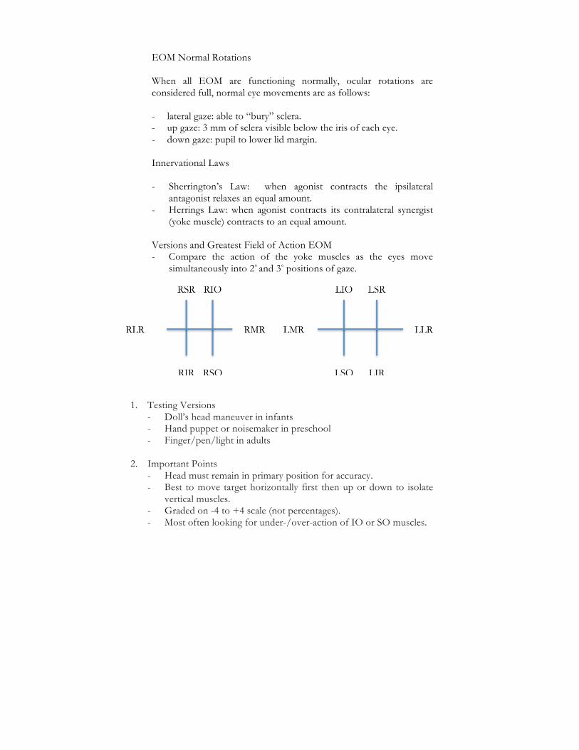
EOM Normal Rotations When all EOM are functioning normally, ocular rotations are considered full, normal eye movements are as follows: - lateral gaze: able to “bury” sclera. - up gaze: 3 mm of sclera visible below the iris of each eye. - down gaze: pupil to lower lid margin.
Innervational Laws - Sherrington’s Law: when agonist contracts the ipsilateral
antagonist relaxes an equal amount. - Herrings Law: when agonist contracts its contralateral synergist
(yoke muscle) contracts to an equal amount.
Versions and Greatest Field of Action EOM - Compare the action of the yoke muscles as the eyes move
simultaneously into 2° and 3° positions of gaze.
1. Testing Versions
- Doll’s head maneuver in infants - Hand puppet or noisemaker in preschool - Finger/pen/light in adults
2. Important Points
- Head must remain in primary position for accuracy. - Best to move target horizontally first then up or down to isolate
vertical muscles. - Graded on -4 to +4 scale (not percentages). - Most often looking for under-/over-action of IO or SO muscles.

III. MEASUREMENTS IN OCULAR MOTILITY
1. Corneal Light Reflex Test Used in patients with: - Poor visual acuity/fixation - Limited eye movements - Poor cooperation - Test is only accurate at near
2. Hirschberg (H) Test - Estimation of misalignment based on light reflex. - 1 mm of decentration corresponds to 7 degrees. - Penlight held at 1/3 m, and patient asked to fixate on light or target
next to it. - Important for examiner to be directly in front of patient for
accurate measurement.
3. Krimsky (K) Test - Same principle as (H), but quantifies deviation with prisms - Goal is completely symmetric reflexes - Krimsky: prism is placed over the deviating eye. - Modified Krimsky: prism is placed over the fixating eye.
4. Single Cover Test
a) Unilateral tropia
If one eye is constantly deviated and only picks up fixation when the preferred eye is covered and returns to the tropic position when the preferred eye is uncovered, a monocular tropia is present.
b) Alternate tropia
If either eye moves to take up fixation when the other eye is covered and maintains fixation when the cover is removed, an alternating tropia is present.
c) No tropia – phoria
- If neither eye makes a movement to take up fixation when the other eye is covered because each eye is fixating, no tropia is present.
- If the eye under cover makes a fusional movement to regain binocular fixation when the cover is removed, the patient has a phoric deviation.

d) Poor vision If one eye makes wavering or wandering type of movement when the other eye is covered, poor visual acuity should be suspected and the patient may have eccentric (non-foveal) fixation.
5. Alternate prism cover test
a) Comitant (primary and secondary deviation)
Having diagnosed the presence of a heterotropia or heterophoria with the cover-uncover test, the alternate cover test (shifting cover from one eye to the other eye) with prisms is used to quantitate the size of the deviation.
The retino-motor value for each retinal element is determined by the distance of the retinal element from the fovea. Prisms properly placed in front of an eye bring the retinal image closer to the fovea until the correct size prism places the image onto the fovea and the movement is eliminated.
Good clinical technique is important. The examiner must be sure the patient is attending. The prisms are placed: - Base-out for esodeviation - Base-in for an exodeviation - Base-up OD for a left hyper or right hypodeviation - Base-down OD for a right hyper or left hypodeviation In other words, the base is placed in the direction the eye moves to take up fixation as it is uncovered. The prisms may be placed in front of either eye or split between the two eyes providing the prisms are placed in the proper direction for each eye. Hold the occluder over each eye long enough so that the patient has sufficient time to take up fixation as you rapidly shift the occluder from one eye to the other. One eye is always covered to maintain constant dissociation. Go to reversal (beyond the end point and movement is now towards the apex of the prism) with prism measurements and subtract 2 prism diopters for the reversal to obtain the full size deviation.
b) Incomitant FOD/FOS (primary and secondary deviation)
When the movement of one eye is reversed, the other eye’s movement may not be neutralized. In the presence of a paretic muscle, the angle of deviation is dependent upon which eye fixates. According to Herring’s Law of Equal Intervention, the primary deviation is less than the secondary deviation. The primary deviation is with the normal eye fixing (paretic eye deviated) and the secondary deviation is with the paretic eye fixing (the normal eye deviated). The prisms may be placed in front of either eye. During the alternate cover and prism test, the examiner observes and measures only the deviation of the non-fixing eye. To measure the deviation of the
left eye with fixing right eye, watch only the left eye’s movement until reversals are noted. Then repeat the alternate cover test, observing and measuring the movement of the right eye with OS fixing. If the deviation of the left eye is larger, the fixing right eye has the paretic muscle.
6. Phoria-Tropia-Simultaneous Cover Test
There are cases in which a very small deviation is present ·with cover-uncover (tropic deviation) which increase with alternate cover (maximal deviation). The maximal deviation minus the tropia is the phoria. In order to measure the size of the tropia from the phoric part of the maximal deviation, the Simultaneous Prism and Cover Test method is used. This is done by placing a prism in front of the deviated eye and simultaneously placing the occluder before the fixing eye. Uncover both eyes and repeat with a larger prism until reversal of deviated eye is observed. The Simultaneous Prism and Cover Test (SΔCT) is used primarily for micro tropia, monofixational phorias, and phoria-tropias. Basic theory is that peripheral fusion is in effect (phoria) with a facultative central scotoma (tropia).

7. Four Base-Out Prism Test Purpose This test is to verify bifoveal fixation or detect a small central scotoma of one eye secondary to a microstrabismus. Principle Sudden displacement of an image from the fovea onto parafoveal retina will elicit a refixation movement if the image has been displaced onto normally functioning retina. By Herring's Law of Equal Innervation, both eyes move simultaneously in the same direction (version movement). If a prism is used to suddenly displace an image from the fovea of one eye of a patient with normal bifoveal fixation, both eyes will move (version) and then the fellow eye subsequently performs a fusional movement (refixation) in the opposite direction to correct for image displacement from the version. However, if the central foveal and parafoveal area of the eye behind the prism is scotomatous (non-functioning), the displacement will not elicit any version or refixation movement. If the central or parafoveal retina of the fellow eye is scotomatous, the version will occur but, the displacement on the fellow eye will not elicit refixation movement. Procedures
1. Patient has both eyes open (binocular). 2. Patient fixates on an object (attends) during the test. 3. A 4Δ
base-out prism is quickly placed over the right eye.
4. The examiner observes the movements of the left eye -i.e. normally out and then back in.
5. The test is repeated by' placing the prism over the left eye and observing the right eye.
Responses 1. Scotoma OD (Microtropia)
a. Prism over OD and no response OU. b. Prism over OS and OD moves out (version) but not in (re-
fixation).

2. Scotoma OS (Microtropia) a. Prism over OD and. OS moves out but not in. b. Prism over OS and no response OU.
3. Negative (Bifoveal) a. Prism over OD and OS goes out and in. b. Prism over OS and OD goes out and in.
IV. SENSORY AND MOTOR TESTS
1. Amplitudes of fusion
a. Absolute, Relative, Recovery Relative fusional amplitudes are the amount of convergence and divergence with accommodation fixed. Blurring is the end point and means accommodation is no longer controlled. For the asthenopic patient, this is more significant than the absolute amplitudes. Absolute amplitudes are the amount of divergence and convergence without controlling accommodation and diplopia is the break point. Recovery point is the point at which the patient regains single binocular vision as the amount of prism is reduced; if lower than 5Δ from the break point, this indicates fatigue in fusional stability.
b. Clinical Technique
Clinical testing can be done with a rotary prism, loose prism, or a prism bar and should be done slowly having the patient fix a 20/30 isolated letter both at 6 meters and 1/3 meter. Check base-in for divergence and then base-out for convergence.
c. Norms
Norms for amplitudes of fusion are:
At Distance At Near
6-7Δ……………Divergence (Base-In)………………12-15Δ
25-35Δ…………Convergence (Base-Out)……………25-35Δ
2-3Δ……………Vertical (Base-Up and Base-Down…..2-3Δ
2. Near point of convergence
NPC demonstrates the amount of convergence ability a person can elicit when following an object to his nose.
3. PCB (punctum convergence basalis)
10 mm from the bridge of the nose to the anterior part of the eye: 15 mm from the anterior part of the cornea to the center of rotation of the eye.
4. Near point of accommodation
Near point of accommodation (NPA). Clinically, it is the nearest point of clear vision. This measurement should be done separately for each eye and then repeated binocularly. The nearest point at which a small print is readable denotes the NPA. The prince rule or a similar device is used to measure the near point of accommodation in centimeters.
5. Red glass test
The Red Glass Test is used to diagnose the cortical relationship between the two eyes and the red is a means of differentiating the image of one eye from the image of the other eye. When testing, the red filter is placed before the patient’s preferred eye, the patient fixes a white light at 20 feet (later repeated at 13 inches), and the patient is asked how many he sees. The patient may report 1 or 2 images; in order to interpret the response, the examiner must observe whether the patient is phoric or tropic during the testing. The normal sensory for a phoria is fusion and correct diplopia for a tropia. A tropia without diplopia is a sensory adaptation of suppression or ARC (“abnormal fusion”). Incorrect diplopia with tropia denotes ARC. Clinically, the examiner must observe whether the patient is phoric or tropic when the patient reports his response. If the patient reports one image, the response may mean normal fusion, abnormal binocularity (ARC), or suppression. If the color is both red and white mixed (light red), both eyes are being used. To help the patient distinguish mixed color from red only, cover the unfiltered eye and ask if the color remains the same or becomes darker.

a. Fusion. If the light red (mixed red and white) was claimed, a quick cover-uncover test is performed to see if the patient is phoric. If the cover-uncover test verifies parallelism, the response means normal fusion. If no shift was seen on the cover-uncover test but the patient does not distinguish a color change (darker) when the unfiltered eye is covered, another technique can be used to verify fusion. Move a -1.00 cylinder with the axis at 90° up and down in front of the red filter. The patient should report that the red light appears to move above and below the white if the patient is fusing.
b. ARC. If a light red is claimed and the cover-uncover test reveals a tropia, the patient is reporting abnormal fusion (harmonious ARC).
c. Suppression. If the red does not change when the unfiltered eye
is covered and uncovered, the white image was not perceived but suppressed; and the cover-uncover test should reveal a deviation of the unfiltered eye. Suppression may be monocular in which case the patient cannot hold fixation with the non-preferred eye when both eyes are uncovered. Patients with alternate suppression will be able to hold fixation with either eye and switch fixation from the red image to the white image but both images are not seen simultaneously.
If a patient reports two images without alternating eye movements, he is reporting diplopia, which is the normal response to a tropia. In an exotropic patient, the light image falls on the temporal retina of the deviated eye and is perceived in the opposite field producing crossed diplopia. In an esotropic patient, the light image falls on the nasal retina and is seen in the same field producing uncrossed diplopia.
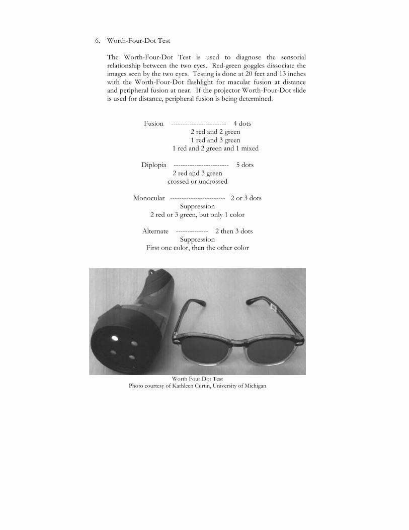
6. Worth-Four-Dot Test The Worth-Four-Dot Test is used to diagnose the sensorial relationship between the two eyes. Red-green goggles dissociate the images seen by the two eyes. Testing is done at 20 feet and 13 inches with the Worth-Four-Dot flashlight for macular fusion at distance and peripheral fusion at near. If the projector Worth-Four-Dot slide is used for distance, peripheral fusion is being determined.
Fusion ------------------------ 4 dots 2 red and 2 green 1 red and 3 green
1 red and 2 green and 1 mixed
Diplopia ------------------------ 5 dots 2 red and 3 green
crossed or uncrossed
Monocular ------------------------ 2 or 3 dots Suppression
2 red or 3 green, but only 1 color
Alternate -------------- 2 then 3 dots Suppression
First one color, then the other color
Worth Four Dot Test
Photo courtesy of Kathleen Curtin, University of Michigan

7. Double Maddox Rod Test
This test is a subjective test to determine the presence and quality of a cyclodeviation. Two Maddox Rods are placed at 90°. The red Maddox is placed before the eye that is suspected of having a cyclodeviation and the white Maddox before the other eye. If no vertical is present, a 6Δ vertical prism is placed before one eye to separate the images. The patient fixes a small bright light and is asked if both horizontal lines are parallel. If both lines are parallel, no cyclodeviation is present. If one line is not perfectly horizontal, the Maddox rod is rotated until both lines are parallel. If the Maddox rod had to be rotated outward to achieve parallelism, an excyclodeviation is present. If the Maddox rod has to be rotated inward to achieve parallelism, an incyclodeviation is present. If torsion is measured to be 5 degrees or greater it becomes a significant problem in strabismus management.
Maddox Rod Test
Photo courtesy of Marc Stephens, University of Michigan
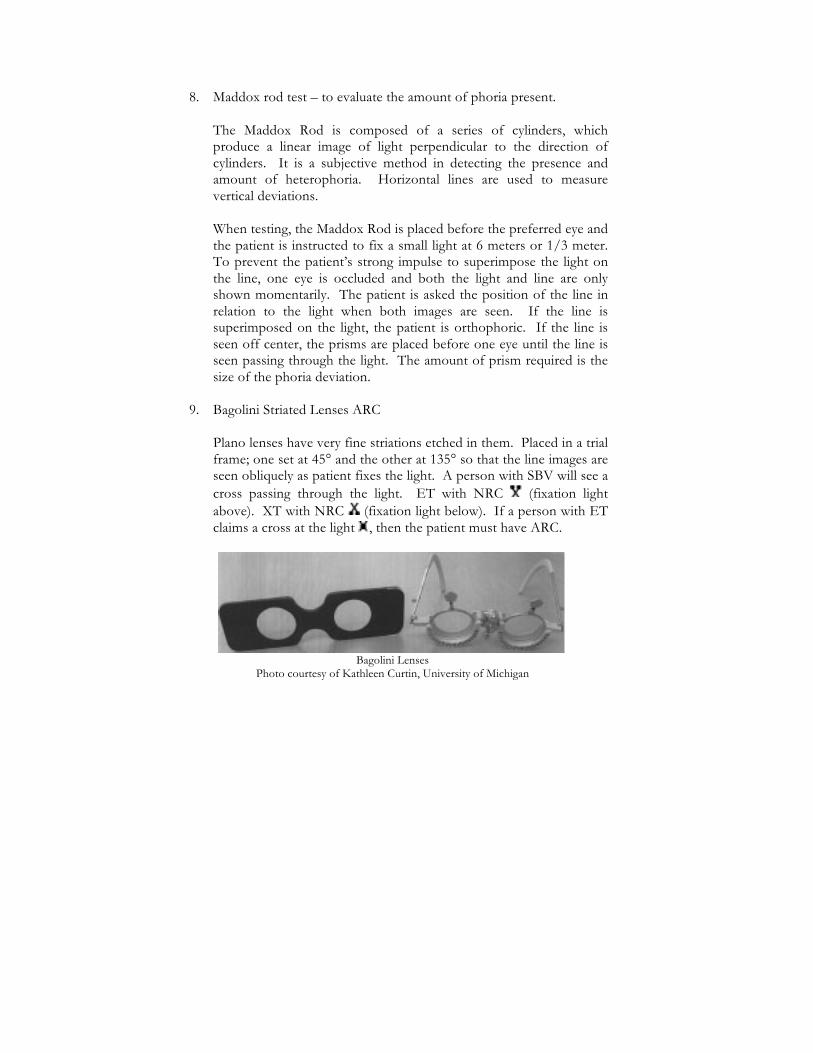
8. Maddox rod test – to evaluate the amount of phoria present.
The Maddox Rod is composed of a series of cylinders, which produce a linear image of light perpendicular to the direction of cylinders. It is a subjective method in detecting the presence and amount of heterophoria. Horizontal lines are used to measure vertical deviations.
When testing, the Maddox Rod is placed before the preferred eye and the patient is instructed to fix a small light at 6 meters or 1/3 meter. To prevent the patient’s strong impulse to superimpose the light on the line, one eye is occluded and both the light and line are only shown momentarily. The patient is asked the position of the line in relation to the light when both images are seen. If the line is superimposed on the light, the patient is orthophoric. If the line is seen off center, the prisms are placed before one eye until the line is seen passing through the light. The amount of prism required is the size of the phoria deviation.
9. Bagolini Striated Lenses ARC
Plano lenses have very fine striations etched in them. Placed in a trial frame; one set at 45° and the other at 135° so that the line images are seen obliquely as patient fixes the light. A person with SBV will see a cross passing through the light. ET with NRC (fixation light above). XT with NRC (fixation light below). If a person with ET claims a cross at the light , then the patient must have ARC.
Bagolini Lenses
Photo courtesy of Kathleen Curtin, University of Michigan

After Image Lamp
Photo courtesy of Kathleen Curtin, University of Michigan
10. After image test ARC
The deviated eye is occluded and the patient is instructed to fix on the dot in the center, while the horizontal filament is shown to the dominant eye for 20 seconds. Light is extinguished, rotated 90°, the opposite is occluded and process repeated. Present the vertical light to the non-dominant eye. Patient is instructed to close their eyes and observe the white lines against the eyelid (positive after image). Patient looks at the blank wall and blinks their eyes to see black lines (negative after image). Each image should have a central gap corresponding to the dot, which indicates the visual direction of the fovea. It is the position of this gap or space in the image that is important. This is a fovea-to-fovea test; it cannot be performed if the patient has eccentric fixation.

11. Synoptophore (Major Amblyopscope, Troposcope)
This is an instrument that is used in the diagnosis of strabismus and anomalies of binocular vision using visual targets that are presented to each eye through a haploscopic arrangement. Description of the Synoptophore It is an instrument that consists of: - A base with controls. - 2 tubes with a light source that can be moved horizontally or
vertically. - An illuminated slide carrier and slides with visual targets of
various sizes and shapes. - A reflecting mirror with an eyepiece lens +6.50D. so that the
distance for the patient’s eye is 15cm. This equals the focal length of the lens so that the eye is focused for distance fixation and the patient does not have to exert any accommodation if he is emmetropic or is wearing his glasses.
Synoptophore, Clement Clark International Ltd.
Photo courtesy of Marc Stephens, University of Michigan Disadvantage-proximal convergence is induced. - There is a mm scale to adjust the IPD. - A movable chin rest.

The scales measure: - The horizontal deviation in degrees and prism diopters. - The vertical deviation in prism diopters. - The cyclodeviation is measured in degrees. - Fusional amplitude ranges in degrees and prism diopters – in for
convergence and out for divergence.
Slides -‐ The slides are of various size to test foveal, para foveal, and
peripheral function. -‐ Both slides are placed in the slide holders with the dots facing in
the same direction. -‐ The visual targets test three grades of fusion:
Simultaneous perception slides -(First Degree) -‐ The slides have a red border. -‐ The targets are dissimilar. -‐ This is to test the first grade of fusion that is sensory fusion. -‐ Foveal size targets are the fish and tank. -‐ Parafoveal - Flowerpot in window. -‐ Peripheral size - parrot in cage and truck in garage.
Fusion - (Second Degree) -‐ The slides have a green border. -‐ They test the second grade of fusion that is motor fusion. -‐ They are similar visual targets with a different detail in each slide. -‐ Fusion slides are - apple with cherries and a strawberry, a frog on
a lily pad with dragonfly and a lily, a bear with blue pants and a red shirt, and a car with a driver and a passenger.
Stereopsis - (Third Degree) -‐ The slides have a yellow border. -‐ This is the third grade of fusion. -‐ They are a circle with lines, a horse and jump with a red and blue
flag, a swing with a red and blue flower and fish tank with crab and a lobster.

Special slides To measure: -‐ Torsion – A circle with a cross slide and the slide with an X
surrounded by a border. -‐ Aniseikonia slides. -‐ After image – the slides have a blue border with a horizontal and
vertical lines with a red gap in the middle of the lines. -‐ Angle kappa - the slide has letters and numbers – EDCBA012345
– spaced at 1 degree intervals.
Diagnostic uses
It is used to measure: -‐ The objective and subjective angles of deviation and the angle
anomaly. -‐ The angle kappa using a special slide on which there are letters and
numbers at 1 degree intervals. -‐ The primary deviation in the deviating eye and fixing with the fixating
eye. -‐ The secondary deviation of the fixating eye and fixing with the
deviating eye. -‐ The deviation in the cardinal directions of gaze.
Estimation of binocular vision: -‐ The state of retinal correspondence – NRC, ARC, angle of anomaly
to determine the type of ARC – harmonious or unharmonious. -‐ To determine the presence of ARC or NRC there must be a 8 diopter
or 4 degree difference between the subjective and objective angle. -‐ The presence, type and area of suppression – foveal, parafoveal,
macular and peripheral. -‐ Simultaneous perception. -‐ The presence, degree and range of sensory fusion and motor fusion –
fusional divergence and convergence amplitudes – note the break and recovery points.
-‐ The presence of stereopsis.
Clinical application – to determine if surgery is recommended in the presence of cyclotorsion: -‐ Using the fusion slides, the torsion is corrected and the presence or
absence of fusion is determined. -‐ If fusion is demonstrated then EOM surgery is indicated to correct
the torsion and restore fusion.

-‐ If it is absent, this indicates that the patient may not fuse after surgery.
Therapeutic uses in the treatment of: -‐ Suppression -‐ ARC -‐ Eccentric fixation with the Haidinger brushes. -‐ Accommodative esotropia – dissociation training. -‐ Improvement of fusional amplitudes.
Method
Diagnostic uses -‐ The chin rest and the IPD are adjusted.
Measurement of the objective angle of deviation -‐ The simultaneous perception (SP) slides – the lion and the cage are
placed into the slide holders.
To detect the type of deviation a cover test is performed. -‐ The lion is placed in front of the fixating eye – the right eye - and the
cage in front of the left eye and the light in front of the left eye is turned off.
-‐ The right arm is set at zero and the left one in the vicinity of zero – on the base outside for esodeviations and base in for exodeviations.
To measure the deviation an alternate cover test is performed. -‐ With the patient fixating the lion with the right eye, the light in front
of the left eye is turned on as the light in front of the right eye is turned off.
-‐ The patient is asked to look directly in the center of the cage and watch the left eye as it moves to take up fixation.
-‐ The left arm is moved o in toward a convergent position - if the eye is seen to
move out = ET. o out for a divergent position = XT. o If the eye moves downward the tube has to be raised =
HT o If it moves upwards the tube is lowered = hypotropia.
-‐ The alternating flashing is continued until there is no movement when either eye picks up fixation.
-‐ The reading on the left arm on the horizontal scale and the vertical scale represents the objective angle of deviation.

-‐ For example if the left arm is at base out 15 pd and has to be raised by 2 pd then the angle is recorded as ET 15, LHT 2.
Measurement of the subjective angle of deviation -‐ If the lion is in the cage, then superimposition is present and the
objective angle is also the subjective angle. -‐ If this is not the case, the patient is asked to move the left arm until
the lion is in the cage. This is the patient’s subjective angle. -‐ In some cases the lion may not be placed in the cage and is seen to
suddenly cross to the other side of the cage – crossed diplopia in ET, or uncrossed diplopia with XT. The crossing point is the subjective angle.
-‐ Suppression may also be present.
Measurement of the angle of anomaly -‐ The difference between the objective and subjective angle is the angle
of anomaly. -‐ If the objective angle is ET 20 base out and the subjective angle is 6
base out, the angle of anomaly is 14 pd.
Measurement of the angle of deviation at near - -3.00 spheres are inserted into the lens holders in front of the eye piece
lenses. - The patient has to exert 3.00D of accommodation in order to get a
clear image of the slides. -‐ Each eye exerts 3pd of convergence for each diopter of
accommodation. C = 3pd/A=1D. -‐ Or 9pd of convergence on one eye. +3.00 lens = 3D x 3 A = 9pd of
C for one eye or 18 pd for both eyes – 9+9 = 18 pd -‐ IPD = 60mm. (for a smaller IPD the convergence requirement is
less, a bigger one – more). -‐ To record the angle of deviation, either 18 pd is added in an ET or
subtracted for an XT from the readings. -‐ If a recording of 20 base out is recorded as ET 2, 20pd – 18pd = 2
pd. -‐ 20 pd base as XT 38 pd. 20pd + 18 pd = 38 pd.
Measurement of subjective cyclodeviations -‐ SP slides are used – the lion in front of the right eye and the cage in
front of the left eye. -‐ The patient is asked to look at each one in turn and is asked if the
cage appears level.
-‐ If it is tilted – torsion is present. -‐ If the left side of the cage is lower than the right – intorsion is
present and extorsion if it is lower on the left side. -‐ The tilt is corrected and read in degrees on the torsion scale that is
located on the slide holder. -‐ The images are tilted in the opposite direction to the tilt of the eye.
Determining angle kappa -‐ A special slide that consists of a row of number and letters
(EDCBA012345) at 1 degree intervals. -‐ The patient is asked to look at the zero. -‐ Positive angle kappa - The corneal light reflex is on the nasal side of the
pupil, appears XT. -‐ Negative angle kappa - The corneal light reflex is on the temporal side
of the pupil, appears ET. -‐ The patient is asked to look in turn at either the letters or numbers
until the reflex is centered. -‐ The degree of deviation corresponding to the letter or number is
recorded. -‐ For example – if the corneal reflex in the right eye is centered on the
C, the patient has a 3 degree negative angle kappa in the right eye. Determining fusional amplitudes - The fusion slides (green border) are placed into the slide holders. - These are similar targets with control marks for each eye –
mouse/bear with red shirt and blue pants.) - If the patient fuses the targets and sees them as one with both
control marks, the arms are locked at the objective angle, divided equally between the two arms.
- Using the horizontal controls both arms are first diverged and the point where fusion breaks is recorded.
-‐ The arms are then moved back to a less divergent position and the point at which fusion is recovered is recorded.
-‐ The arms continue to be converged until fusion breaks and is recovered.
Stereopsis - The stereo slides (yellow border) are placed in the slide holders. - The image should be seen as a single image with both the controls. - The target is seen in depth – the swing appears towards or away from
you.

Recording results Distance:
Objective and subjective angle – ET 26, RHT 2. First and second grade fusion – present at angle. Convergence – to BO 40/recovery at BO 30. Stereopsis – present with foveal, para foveal or peripheral slides.
Near with -3.00D spheres:
Objective and subjective angles – ET 46, RHT 2. Convergence – to BO 58/recovery at BO 46. Divergence – no divergence past angle, suppression OD. Stereopsis – absent.
12. Lancaster Red Green Test
The Lancaster Red-Green is a foveal projection test used for detecting incomitance of a deviation in a patient with normal sensory relationship. It is not reliable in patients with ARC and deep suppression.
The tangent screen is composed of lines which form squares 7 cm apart. The patient is positioned 1 or 2 meters from the tangent screen and the eyes should be at the level with the zero center mark on the screen. At 2 meters, each square subtends an angle of 3.5o or 7Δ for 1 meter. The patient is given red and green goggles (usually red in front of OD, green OS) and a torch flashlight is held by the patient and the other held by the examiner. One torch flashlight projects red which can only be seen through the red lens and the other torch flashlight projects green which can be seen only through the green lens. The color held by the examiner determines the fixing eye. For example, the examiner projects a green light on the zero mark and the patient is asked to superimpose his red light on the examiner’s green light. This is repeated in the 6 diagnostic positions of gaze. The distance of light (projected by the patient) from the diagnostic positions (projected by the examiner) determines the amount of deviation. The flashlights are traded and the test is repeated. The results are plotted on a chart that is an exact duplicate of the screen. Once plotted one can easily observe 1) the diagnostic position at which the separation of light is greatest, 2) primary and secondary deviation, 3) identify the paretic muscles.