AUSA Director, Family Forum II Installation Services 26
13
The p14 Fusion-associated Small Transmembrane (FAST) Protein Effects Membrane Fusion from a Subset of Membrane Microdomains * □ S Received for publication, March 20, 2006, and in revised form, August 24, 2006 Published, JBC Papers in Press, August 26, 2006, DOI 10.1074/jbc.M602566200 Jennifer A. Corcoran ‡ , Jayme Salsman ‡1 , Roberto de Antueno ‡ , Ahmed Touhami § , Manfred H. Jericho § , Eileen K. Clancy ‡2 , and Roy Duncan ‡3 From the Departments of ‡ Microbiology and Immunology and § Physics, Dalhousie University, Halifax, Nova Scotia B3H 1X5, Canada The reovirus fusion-associated small transmembrane (FAST) proteins are a unique family of viral membrane fusion proteins. These nonstructural viral proteins induce efficient cell-cell rather than virus-cell membrane fusion. We analyzed the lipid environment in which the reptilian reovirus p14 FAST protein resides to determine the influence of the cell membrane on the fusion activity of the FAST proteins. Topographical mapping of the surface of fusogenic p14-containing liposomes by atomic force microscopy under aqueous conditions revealed that p14 resides almost exclusively in thickened membrane microdo- mains. In transfected cells, p14 was found in both Lubrol WX- and Triton X-100-resistant membrane complexes. Cholesterol depletion of donor cell membranes led to preferential disrup- tion of p14 association with Lubrol WX (but not Triton X-100)- resistant membranes and decreased cell-cell fusion activity, both of which were reversed upon subsequent cholesterol reple- tion. Furthermore, co-patching analysis by fluorescence micros- copy indicated that p14 did not co-localize with classical lipid- anchored raft markers. These data suggest that the p14 FAST protein associates with heterogeneous membrane microdo- mains, a distinct subset of which is defined by cholesterol-de- pendent Lubrol WX resistance and which may be more relevant to the membrane fusion process. Biological membrane fusion is dependent on protein cata- lysts to mediate the lipid rearrangements required for mem- brane merger (1, 2). The fusion-associated small transmem- brane (FAST) proteins are one such family of membrane fusion catalysts (3). The FAST proteins are a unique group of small (95–140 amino acids) integral membrane proteins encoded by the fusogenic reoviruses, an unusual group of non-enveloped viruses that induce syncytium formation (3– 6). Three distinct members of the FAST protein family have been described in recent years: the homologous p10 proteins of avian reovirus and Nelson Bay reovirus and the unrelated p14 and p15 pro- teins of reptilian reovirus and baboon reovirus, respectively (3–5). Unlike the well characterized fusion proteins of envel- oped viruses (7), the FAST proteins are nonstructural viral pro- teins and are therefore not involved in viral entry into cells. Following their expression inside virus-infected or -transfected cells, the FAST proteins traffic through the endoplasmic retic- ulum-Golgi pathway to assume a bitopic N exoplasmic /C cytoplasmic topology in the plasma membrane, where they mediate fusion of virus-infected cells to neighboring uninfected cells (8 –10). Therefore, the FAST proteins function more as “cellular” rather than viral fusion proteins, mediating cell-cell rather than virus- cell membrane fusion. Furthermore, a recent study using the purified p14 FAST protein of reptilian reovirus reconstituted into artificial lipid bilayers indicated that the FAST proteins are both necessary and sufficient to mediate membrane fusion (11). In addition to their unique role in the viral replication cycle, the FAST proteins are also structurally distinct from enveloped viral fusion proteins. In particular, at only 22– 44 residues in size, the FAST protein ectodomains are considerably less com- plex than the ectodomains of enveloped viral fusion proteins. Current models propose that extensive structural remodeling of the large metastable enveloped viral fusion protein ectodo- mains provides the energy required to drive the fusion reaction (1, 12–15). Because the biophysical features of the FAST pro- teins are incompatible with a fusion mechanism dependent on dramatic conformational changes in structurally complex ectodomains, alternative models of protein-mediated mem- brane fusion must be considered. In the case of the 125-residue p14 FAST protein, structural motifs relevant to the membrane fusion activity include the transmembrane domain, a fusion peptide loop and myristate moiety in the 36-residue N-terminal ectodomain, and a membrane-proximal polybasic region in the 68-residue endodomain (4, 9). We currently favor a model of FAST protein-mediated membrane fusion whereby mem- brane-interactive structural motifs function from either side of the membrane to disrupt the boundary water layer asso- ciated with lipid head groups and/or alter lipid packing, * This work was supported in part by grants from the Canadian Institutes of Health Research (to R. D.) and by grants from the Natural Sciences and Engineering Research Council (to M. H. J.). The costs of publication of this article were defrayed in part by the payment of page charges. This article must therefore be hereby marked “advertisement” in accordance with 18 U.S.C. Section 1734 solely to indicate this fact. □ S The on-line version of this article (available at http://www.jbc.org) contains detailed experimental results and methods for Fig. 1C. 1 Supported by scholarships from the Natural Sciences and Engineering Research Council, the Cancer Research Training Program of Cancer Care Nova Scotia, and the Nova Scotia Health Research Foundation. 2 Supported by a scholarship from the Cancer Research Training Program of Cancer Care Nova Scotia. 3 Recipient of a Regional Partnerships Program Investigator Award from the Canadian Institutes of Health Research. To whom correspondence should be addressed: Dept. of Microbiology and Immunology, Tupper Medical Bldg., Rm. 7S, Dalhousie University, Halifax, Nova Scotia B3H 1X5, Canada. Tel.: 902-494-6770; Fax: 902-494-5125; E-mail: [email protected]. THE JOURNAL OF BIOLOGICAL CHEMISTRY VOL. 281, NO. 42, pp. 31778 –31789, October 20, 2006 © 2006 by The American Society for Biochemistry and Molecular Biology, Inc. Printed in the U.S.A. 31778 JOURNAL OF BIOLOGICAL CHEMISTRY VOLUME 281 • NUMBER 42 • OCTOBER 20, 2006 by guest on April 4, 2019 http://www.jbc.org/ Downloaded from
Transcript of AUSA Director, Family Forum II Installation Services 26

The p14 Fusion-associated Small Transmembrane (FAST)Protein Effects Membrane Fusion from a Subset ofMembrane Microdomains*□S
Received for publication, March 20, 2006, and in revised form, August 24, 2006 Published, JBC Papers in Press, August 26, 2006, DOI 10.1074/jbc.M602566200
Jennifer A. Corcoran‡, Jayme Salsman‡1, Roberto de Antueno‡, Ahmed Touhami§, Manfred H. Jericho§,Eileen K. Clancy‡2, and Roy Duncan‡3
From the Departments of ‡Microbiology and Immunology and §Physics, Dalhousie University, Halifax,Nova Scotia B3H 1X5, Canada
The reovirus fusion-associated small transmembrane (FAST)proteins are a unique family of viral membrane fusion proteins.These nonstructural viral proteins induce efficient cell-cellrather than virus-cell membrane fusion. We analyzed the lipidenvironment in which the reptilian reovirus p14 FAST proteinresides to determine the influence of the cell membrane on thefusion activity of the FAST proteins. Topographical mapping ofthe surface of fusogenic p14-containing liposomes by atomicforce microscopy under aqueous conditions revealed that p14resides almost exclusively in thickened membrane microdo-mains. In transfected cells, p14 was found in both Lubrol WX-and Triton X-100-resistant membrane complexes. Cholesteroldepletion of donor cell membranes led to preferential disrup-tion of p14 association with LubrolWX (but not Triton X-100)-resistant membranes and decreased cell-cell fusion activity,both of whichwere reversed upon subsequent cholesterol reple-tion. Furthermore, co-patching analysis by fluorescencemicros-copy indicated that p14 did not co-localize with classical lipid-anchored raft markers. These data suggest that the p14 FASTprotein associates with heterogeneous membrane microdo-mains, a distinct subset of which is defined by cholesterol-de-pendent LubrolWX resistance and whichmay bemore relevantto the membrane fusion process.
Biological membrane fusion is dependent on protein cata-lysts to mediate the lipid rearrangements required for mem-brane merger (1, 2). The fusion-associated small transmem-brane (FAST) proteins are one such family of membrane fusion
catalysts (3). The FAST proteins are a unique group of small(95–140 amino acids) integral membrane proteins encoded bythe fusogenic reoviruses, an unusual group of non-envelopedviruses that induce syncytium formation (3–6). Three distinctmembers of the FAST protein family have been described inrecent years: the homologous p10 proteins of avian reovirusand Nelson Bay reovirus and the unrelated p14 and p15 pro-teins of reptilian reovirus and baboon reovirus, respectively(3–5). Unlike the well characterized fusion proteins of envel-oped viruses (7), the FAST proteins are nonstructural viral pro-teins and are therefore not involved in viral entry into cells.Following their expression inside virus-infected or -transfectedcells, the FAST proteins traffic through the endoplasmic retic-ulum-Golgi pathway to assume a bitopicNexoplasmic/Ccytoplasmictopology in the plasma membrane, where they mediate fusionof virus-infected cells to neighboring uninfected cells (8–10).Therefore, the FASTproteins functionmore as “cellular” ratherthan viral fusion proteins, mediating cell-cell rather than virus-cell membrane fusion. Furthermore, a recent study using thepurified p14 FAST protein of reptilian reovirus reconstitutedinto artificial lipid bilayers indicated that the FAST proteins areboth necessary and sufficient tomediatemembrane fusion (11).In addition to their unique role in the viral replication cycle,
the FAST proteins are also structurally distinct from envelopedviral fusion proteins. In particular, at only �22–44 residues insize, the FAST protein ectodomains are considerably less com-plex than the ectodomains of enveloped viral fusion proteins.Current models propose that extensive structural remodelingof the large metastable enveloped viral fusion protein ectodo-mains provides the energy required to drive the fusion reaction(1, 12–15). Because the biophysical features of the FAST pro-teins are incompatible with a fusion mechanism dependenton dramatic conformational changes in structurally complexectodomains, alternative models of protein-mediated mem-brane fusionmust be considered. In the case of the 125-residuep14 FAST protein, structural motifs relevant to the membranefusion activity include the transmembrane domain, a fusionpeptide loop andmyristatemoiety in the 36-residueN-terminalectodomain, and amembrane-proximal polybasic region in the68-residue endodomain (4, 9). We currently favor a model ofFAST protein-mediated membrane fusion whereby mem-brane-interactive structural motifs function from either sideof the membrane to disrupt the boundary water layer asso-ciated with lipid head groups and/or alter lipid packing,
* This work was supported in part by grants from the Canadian Institutes ofHealth Research (to R. D.) and by grants from the Natural Sciences andEngineering Research Council (to M. H. J.). The costs of publication of thisarticle were defrayed in part by the payment of page charges. This articlemust therefore be hereby marked “advertisement” in accordance with 18U.S.C. Section 1734 solely to indicate this fact.
□S The on-line version of this article (available at http://www.jbc.org) containsdetailed experimental results and methods for Fig. 1C.
1 Supported by scholarships from the Natural Sciences and EngineeringResearch Council, the Cancer Research Training Program of Cancer CareNova Scotia, and the Nova Scotia Health Research Foundation.
2 Supported by a scholarship from the Cancer Research Training Program ofCancer Care Nova Scotia.
3 Recipient of a Regional Partnerships Program Investigator Award from theCanadian Institutes of Health Research. To whom correspondence shouldbe addressed: Dept. of Microbiology and Immunology, Tupper MedicalBldg., Rm. 7S, Dalhousie University, Halifax, Nova Scotia B3H 1X5, Canada.Tel.: 902-494-6770; Fax: 902-494-5125; E-mail: [email protected].
THE JOURNAL OF BIOLOGICAL CHEMISTRY VOL. 281, NO. 42, pp. 31778 –31789, October 20, 2006© 2006 by The American Society for Biochemistry and Molecular Biology, Inc. Printed in the U.S.A.
31778 JOURNAL OF BIOLOGICAL CHEMISTRY VOLUME 281 • NUMBER 42 • OCTOBER 20, 2006
by guest on April 4, 2019
http://ww
w.jbc.org/
Dow
nloaded from

thereby lowering the energy barriers that impede membranefusion (8–11, 16).The realignment of lipids during the membrane fusion proc-
ess is believed to proceed through a series of sequential stepsinvolving merger of the outer leaflets of two closely apposedmembranes to form a stalk; formation of a hemifusion dia-phragm; and then merger of the inner leaflets, leading to poreformation andpore expansion (2, 13). The lipid rearrangementsrequired to achieve these steps are complex and are believed torequire the formation of different non-bilayer structuresinvolving negative membrane curvature, acyl chain tilt, andaltered lipid packing (13, 17). The liquid-disordered fluid stateof biological membranes makes it easy to envision how lipidmolecules could be rearranged to form such non-bilayer struc-tures. In recent years, however, the concept of a biologicalmembrane as a fluid mosaic containing randomly dispersedmembrane proteins moving through a fluid lipid bilayer hasbeen supplanted by the realization that biological membraneslikely contain regions highly biased in both their lipid and pro-tein composition (18). These lipid-based membrane microdo-mains, so-called “rafts,” are currently viewed as small dynamicclusters of protein and lipid enriched in cholesterol and/orsphingomyelin (19). The tight lipid packing promoted by cho-lesterol and the unsaturated acyl chains present in such mem-brane microdomains promotes the formation of a liquid-or-dered state in model membranes, which may render these lipidmicrodomains more resistant to detergent solubilization (18,20). However, the complex interactions between detergentsand the lipids and proteins present in membranes make itunclear as to whether the biochemical and biophysical proper-ties of isolated detergent-resistant membranes (DRMs)4 accu-rately reflect the properties of membrane microdomains pres-ent in native membranes (21–23).Regardless of the exact relationship between DRMs and
actual membrane microdomains, considerable detergent-dependent and -independent analyses support the concept ofmembrane microdomains with distinct lipid/protein composi-tions that may serve as organizational centers for numerousproteins (19, 24, 25). In the case of enveloped viral fusion pro-teins (26), membrane microdomains generally serve as plat-forms either to concentrate viral glycoproteins in the cell mem-brane to facilitate viral assembly or to localize viral receptorsduring the entry process (27–30). If membrane fusion proteinsdo function from lipidmicrodomains, then the potential liquid-ordered state of these microdomains may need to be rational-ized with the formation of non-bilayer structures that are con-sidered to be essential for the fusion process. It is possible thatthe fusion proteins migrate out of the microdomains into themore fluid adjacent lipid bilayer during the fusion process, assuggested for influenza virus hemagglutinin, where disruptionof membrane microdomains by cholesterol depletion has no
adverse effects on the membrane fusion reaction (31). How-ever, theoretical and experimental evidence suggests that somemembrane fusion reactions may be mediated within orderedlipidmicrodomains (32–34).Mechanisticmodels ofmembranefusion may therefore need to adapt to the involvement of liq-uid-ordered lipid bilayers as a component of the fusion process.We are particularly interested in understanding how the
FAST proteins induce cell-cell fusion. Because the membraneenvironment fromwhich the FAST proteinsmediate the fusionreaction is the cell plasma membrane and in view of the poten-tially significant impact of the biophysical properties of plasmamembrane lipid microdomains on models of the membranefusion reaction, we determined whether this recently discov-ered novel family of membrane fusion proteins associates withmembrane microdomains. Based on both detergent and non-detergent approaches, our results indicate that the p14 FASTprotein associates with distinct membrane microdomains, asubset of which may contain p14 molecules more directlyinvolved in the membrane fusion process.
EXPERIMENTAL PROCEDURES
Cells and Materials—The rabbit anti-p14 polyclonal anti-serum and the maintenance of QM5 (quail fibroblast) cells wasas described previously (4). N-BP-2 cells are Chinese hamsterovary cell derivatives deficient in cholesterol synthesis becauseof a deletion in the site-2 protease that activates the sterol reg-ulatory element-binding proteins (35). These cells wereobtained fromN. Ridgway (DalhousieUniversity) andwere cul-tured in Dulbecco’s modified Eagle’s medium supplementedwith 5% fetal bovine serum and cholesterol as described previ-ously (36). Anti-placental alkaline phosphatase (PLAP) andanti-human transferrin receptor monoclonal antibodies werepurchased from Dako and Zymed Laboratories Inc., respec-tively. Horseradish peroxidase-conjugated goat anti-mouseand goat anti-rabbit antibodies were obtained from Kirkegaard& Perry Laboratories, Inc. Lubrol WX was purchased fromSERVA; all other detergents and cholesterol-depleting reagents(methyl-�-cyclodextrin (M�CD)) were from Sigma. Cell-Tracker dyes were fromMolecular Probes.Expression Plasmids—The authentic p14 and non-myristoy-
lated p14-G2A expression plasmids were as described previ-ously (4). Plasmid vectors expressing PLAPor the human trans-ferrin receptor were generously provided by D. Brown (StateUniversity of New York) and C. Parish (Cornell University),respectively. A eukaryotic expression plasmid encoding thevesicular stomatitis virus G protein (VSV-G; Indiana serotype)was provided by P. Lee (Dalhousie University).Transfection, Cell Staining, and Syncytial Indexing—QM5
quail cell fibroblasts were transfected with p14 expression plas-mids using Lipofectamine 2000 (Invitrogen) according to themanufacturer’s instructions. At various times post-transfec-tion, cell monolayers were fixed and stained with Wright-Gi-emsa, and the syncytial index was determined by quantifyingthe numbers of syncytial nuclei present in randommicroscopicfields as described previously (4). Results are reported as themeans � S.E. of three separate experiments.Cholesterol Depletion andRepletion—Membrane cholesterol
was depleted fromp14-transfectedQM5 cells prior to the onset
4 The abbreviations used are: DRM(s), detergent-resistant membrane(s);PLAP, placental alkaline phosphatase; M�CD, methyl-�-cyclodextrin;VSV-G, vesicular stomatitis virus G protein; cLPDS, complete lipoprotein-deficient serum; HBSS, Hanks’ buffered saline solution; PBS, phosphate-buffered saline; AFM, atomic force microscopy; LRMs, Lubrol WX-resistantmembranes; TRM(s), Triton X-100-resistant membrane(s); GM1, ganglio-side GM1.
FAST Proteins and Membrane Microdomains
OCTOBER 20, 2006 • VOLUME 281 • NUMBER 42 JOURNAL OF BIOLOGICAL CHEMISTRY 31779
by guest on April 4, 2019
http://ww
w.jbc.org/
Dow
nloaded from
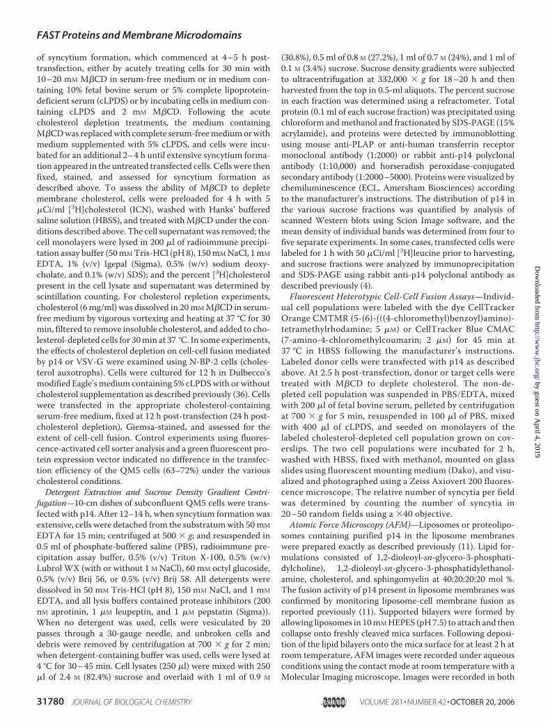
of syncytium formation, which commenced at 4–5 h post-transfection, either by acutely treating cells for 30 min with10–20 mM M�CD in serum-free medium or in medium con-taining 10% fetal bovine serum or 5% complete lipoprotein-deficient serum (cLPDS) or by incubating cells in medium con-taining cLPDS and 2 mM M�CD. Following the acutecholesterol depletion treatments, the medium containingM�CDwas replacedwith complete serum-freemediumorwithmedium supplemented with 5% cLPDS, and cells were incu-bated for an additional 2–4 h until extensive syncytium forma-tion appeared in the untreated transfected cells. Cells were thenfixed, stained, and assessed for syncytium formation asdescribed above. To assess the ability of M�CD to depletemembrane cholesterol, cells were preloaded for 4 h with 5�Ci/ml [3H]cholesterol (ICN), washed with Hanks’ bufferedsaline solution (HBSS), and treated withM�CD under the con-ditions described above. The cell supernatant was removed; thecell monolayers were lysed in 200 �l of radioimmune precipi-tation assay buffer (50mMTris-HCl (pH8), 150mMNaCl, 1mMEDTA, 1% (v/v) Igepal (Sigma), 0.5% (w/v) sodium deoxy-cholate, and 0.1% (w/v) SDS); and the percent [3H]cholesterolpresent in the cell lysate and supernatant was determined byscintillation counting. For cholesterol repletion experiments,cholesterol (6mg/ml) was dissolved in 20mMM�CD in serum-free medium by vigorous vortexing and heating at 37 °C for 30min, filtered to remove insoluble cholesterol, and added to cho-lesterol-depleted cells for 30min at 37 °C. In some experiments,the effects of cholesterol depletion on cell-cell fusion mediatedby p14 or VSV-G were examined using N-BP-2 cells (choles-terol auxotrophs). Cells were cultured for 12 h in Dulbecco’smodified Eagle’smediumcontaining 5% cLPDSwith orwithoutcholesterol supplementation as described previously (36). Cellswere transfected in the appropriate cholesterol-containingserum-free medium, fixed at 12 h post-transfection (24 h post-cholesterol depletion), Giemsa-stained, and assessed for theextent of cell-cell fusion. Control experiments using fluores-cence-activated cell sorter analysis and a green fluorescent pro-tein expression vector indicated no difference in the transfec-tion efficiency of the QM5 cells (63–72%) under the variouscholesterol conditions.Detergent Extraction and Sucrose Density Gradient Centri-
fugation—10-cm dishes of subconfluent QM5 cells were trans-fected with p14. After 12–14 h, when syncytium formation wasextensive, cells were detached from the substratumwith 50mMEDTA for 15 min; centrifuged at 500 � g; and resuspended in0.5 ml of phosphate-buffered saline (PBS), radioimmune pre-cipitation assay buffer, 0.5% (v/v) Triton X-100, 0.5% (w/v)Lubrol WX (with or without 1 M NaCl), 60 mM octyl glucoside,0.5% (v/v) Brij 56, or 0.5% (v/v) Brij 58. All detergents weredissolved in 50 mM Tris-HCl (pH 8), 150 mM NaCl, and 1 mMEDTA, and all lysis buffers contained protease inhibitors (200nM aprotinin, 1 �M leupeptin, and 1 �M pepstatin (Sigma)).When no detergent was used, cells were vesiculated by 20passes through a 30-gauge needle, and unbroken cells anddebris were removed by centrifugation at 700 � g for 2 min;when detergent-containing buffer was used, cells were lysed at4 °C for 30–45 min. Cell lysates (250 �l) were mixed with 250�l of 2.4 M (82.4%) sucrose and overlaid with 1 ml of 0.9 M
(30.8%), 0.5 ml of 0.8 M (27.2%), 1 ml of 0.7 M (24%), and 1 ml of0.1 M (3.4%) sucrose. Sucrose density gradients were subjectedto ultracentrifugation at 332,000 � g for 18–20 h and thenharvested from the top in 0.5-ml aliquots. The percent sucrosein each fraction was determined using a refractometer. Totalprotein (0.1 ml of each sucrose fraction) was precipitated usingchloroform andmethanol and fractionated by SDS-PAGE (15%acrylamide), and proteins were detected by immunoblottingusing mouse anti-PLAP or anti-human transferrin receptormonoclonal antibody (1:2000) or rabbit anti-p14 polyclonalantibody (1:10,000) and horseradish peroxidase-conjugatedsecondary antibody (1:2000–5000). Proteins were visualized bychemiluminescence (ECL, Amersham Biosciences) accordingto the manufacturer’s instructions. The distribution of p14 inthe various sucrose fractions was quantified by analysis ofscanned Western blots using Scion Image software, and themean density of individual bands was determined from four tofive separate experiments. In some cases, transfected cells werelabeled for 1 h with 50 �Ci/ml [3H]leucine prior to harvesting,and sucrose fractions were analyzed by immunoprecipitationand SDS-PAGE using rabbit anti-p14 polyclonal antibody asdescribed previously (4).Fluorescent Heterotypic Cell-Cell Fusion Assays—Individ-
ual cell populations were labeled with the dye CellTrackerOrange CMTMR (5-(6)-(((4-chloromethyl)benzoyl)amino)-tetramethylrhodamine; 5 �M) or CellTracker Blue CMAC(7-amino-4-chloromethylcoumarin; 2 �M) for 45 min at37 °C in HBSS following the manufacturer’s instructions.Labeled donor cells were transfected with p14 as describedabove. At 2.5 h post-transfection, donor or target cells weretreated with M�CD to deplete cholesterol. The non-de-pleted cell population was suspended in PBS/EDTA, mixedwith 200 �l of fetal bovine serum, pelleted by centrifugationat 700 � g for 5 min, resuspended in 100 �l of PBS, mixedwith 400 �l of cLPDS, and seeded on monolayers of thelabeled cholesterol-depleted cell population grown on cov-erslips. The two cell populations were incubated for 2 h,washed with HBSS, fixed with methanol, mounted on glassslides using fluorescent mounting medium (Dako), and visu-alized and photographed using a Zeiss Axiovert 200 fluores-cence microscope. The relative number of syncytia per fieldwas determined by counting the number of syncytia in20–50 random fields using a �40 objective.Atomic Force Microscopy (AFM)—Liposomes or proteolipo-
somes containing purified p14 in the liposome membraneswere prepared exactly as described previously (11). Lipid for-mulations consisted of 1,2-dioleoyl-sn-glycero-3-phosphati-dylcholine), 1,2-dioleoyl-sn-glycero-3-phosphatidylethanol-amine, cholesterol, and sphingomyelin at 40:20:20:20 mol %.The fusion activity of p14 present in liposome membranes wasconfirmed by monitoring liposome-cell membrane fusion asreported previously (11). Supported bilayers were formed byallowing liposomes in 10mMHEPES (pH7.5) to attach and thencollapse onto freshly cleaved mica surfaces. Following deposi-tion of the lipid bilayers onto the mica surface for at least 2 h atroom temperature, AFM images were recorded under aqueousconditions using the contact mode at room temperature with aMolecular Imaging microscope. Images were recorded in both
FAST Proteins and Membrane Microdomains
31780 JOURNAL OF BIOLOGICAL CHEMISTRY VOLUME 281 • NUMBER 42 • OCTOBER 20, 2006
by guest on April 4, 2019
http://ww
w.jbc.org/
Dow
nloaded from

height and deflection modes. V-shaped cantilevers with oxide-sharpened Si3N4 tips (Veeco) were used with spring constantsof 0.01 newton-meter as specified by the manufacturer. Highresolution images were recorded with optimized feedbackparameters at scan frequencies of 4 Hz. Very low forces wereapplied during the scan, and the threshold for imaging was esti-mated at �400 piconewtons. Images were obtained from atleast three different samples prepared on different days, andseveral macroscopically separate areas were scanned on eachsample. Several images were analyzed from each experiment,and�100AFMcross-section profiles were used to estimate theheight of the observed membrane microdomains and p14 pro-tein spikes (height of the microdomains � 0.8 � 0.2 nm (n �30) and height of the spikes � 1.5 � 0.5 nm (n � 70)).Surface Fluorescence Microscopy and Co-localization—
QM5 cells grown on coverslips were transfected or cotrans-fected and stained for surface immunofluorescence. Forcotransfection experiments, the non-fusogenic p14 construct(p14-G2A) was used to prevent syncytium formation and toallow expression of PLAP. At 24 h post-transfection, cells werepreblocked with whole goat IgG (1:1000) in HBSS for 30 min at4 °C, treated with primary antibody (rabbit anti-p14 polyclonalantibody (1:200) and/or mouse anti-PLAP monoclonal anti-body (1:50) in the blocking buffer) for 60 min at 4 °C, washedwith HBSS, and then treated with secondary antibody (AlexaFluor 488- or Alexa Fluor 555-conjugated goat anti-mouseand/or goat anti-rabbit IgG (1:200; Molecular Probes) in theblocking buffer). After secondary antibody addition and wash-ing, cells were fixed with 3.7% formaldehyde in PBS andmounted on glass slides using fluorescent mounting medium.GM1 was stained using Alexa Fluor 555-conjugated choleratoxin B (0.1 �g/ml) during the primary antibody incubation.Afterwashing as described above, anti-cholera toxinB antibody(1:200) was added during the secondary antibody incubation tocause individual GM1molecules on the cell surface to coalesce(37). Stained cells were visualized and photographed using aZeiss LSM 510 META scanning argon laser confocal micro-scope and a �100 objective.Thin Layer Chromatography—Untransfected or p14-trans-
fected QM5 cells were labeled with 10 �Ci of [14C]acetate/dish(107 cells) for 16–18 h. After labeling, cells were left untreatedor treated with M�CD for cholesterol depletion/repletion asdescribed above and extracted with Lubrol WX or TritonX-100 prior to isolation of DRMs by sucrose density gradientultracentrifugation as described above. Total lipid from eachfraction or fraction pool was isolated using 2:1 (v/v) chloro-form/methanol (38), and aliquots of the lipid extract containingequal amounts of radioactivity (20,000 dpm) were applied toSilica Gel G plates (Macherey-Nagel). Cholesterol and phos-pholipids were fractionated by thin layer chromatographyusing a 24:30:6:30 (v/v/v/v) chloroform/ethanol/water/triethyl-aminemobile phase as described previously (39). Phospholipid/cholesterol standardswere run in parallel and detected by treat-ment with sulfuric acid and charring at 180 °C. The percentageof radioactivity recovered in each lipid spot was quantifiedusing aModel GS-525molecular imager system andMolecularAnalyst 1.5 software (Bio-Rad).
RESULTS
p14-induced Membrane Fusion Is Sensitive to Donor Mem-brane Cholesterol Levels—In view of the importance of the lipidenvironment tomembrane fusion and the absence of any infor-mation on the effect of this environment on the function of therecently discovered FAST proteins, we examined the influenceof cholesterol on syncytium formation induced by the p14FAST protein. QM5 fibroblasts were transfected with a p14expression plasmid, and membrane cholesterol was depletedwith M�CD prior to the onset of cell-cell fusion. Quantitativeanalysis indicated that M�CD extracted 60–90% of the mem-brane cholesterol under various extraction conditions (Fig. 1D)and that p14-induced syncytium formation was inhibited by80–90% under all conditions (Fig. 1, A and B). Furthermore,restoration of membrane cholesterol by treatment of cells withcholesterol-loaded M�CD substantially restored the rate ofsyncytium formation (Fig. 1C). Accounting for the 60-min lagbetween the time course of syncytium formation in treatedversus untreated cells (i.e. the time course of syncytium for-mation in treated cells was delayed by the cholesterol deple-tion and repletion steps), cholesterol repletion essentiallyrestored p14-induced syncytium formation to the levelsobserved in untreated cells (data not shown).To control for possible indirect effects of cholesterol deple-
tion on p14-mediated cell-cell fusion, we examined the effectsof acute cholesterol depletion by M�CD on the cell-cell fusionactivity of theVSV-G fusion protein. AlthoughVSV-Gdoes notappear to associatewith lipidmicrodomains based ondetergentfractionation analysis (29), the effects of cholesterol depletionby M�CD on VSV-G cell-cell fusion activity have never beenreported. To serve as a suitable control for p14 inhibition bycholesterol depletion, rather than using low pH treatment totrigger VSV-G fusion activity, we exploited the fact that tran-sient overexpression of VSV-G in cells that partially acidify themedium results in gradual cell-cell fusion. This phenotype par-allels the progressive fusion activity of the FAST proteins,which are untriggered fusogens. As shown in Fig. 1C, cell-cellfusion mediated by VSV-G was completely unaffected by acutecholesterol depletion conditions that inhibited p14 fusionactivity by �80%.We also examined p14 fusion activity in N-BP-2 cells, a cho-
lesterol auxotrophic cell line (35). Under conditions in whichN-BP-2 cells are deficient in membrane cholesterol (24 h ofcholesterol depletion), p14-induced cell-cell fusion was inhib-ited by �50% (Fig. 1C). However, the same experimental con-ditions resulted in �70% inhibition of VSV-G cell-cell fusionboth when VSV-G fusion occurred under the neutral pH con-ditions described above (data not shown) and when fusion wastriggered by low pH treatment (Fig. 1C). The different effects ofthe two cholesterol depletion protocols (i.e.M�CD or auxotro-phic cells) on VSV-G fusion activity most likely reflect differ-ences in the cellular response to acute versus extended choles-terol depletion.Because cholesterol depletion can exert its effect on mem-
brane fusion proteins from either the donor or target mem-brane (30, 34, 40), we used a fluorescent heterotypic cell-cellfusion assay and acute cholesterol depletion with M�CD to
FAST Proteins and Membrane Microdomains
OCTOBER 20, 2006 • VOLUME 281 • NUMBER 42 JOURNAL OF BIOLOGICAL CHEMISTRY 31781
by guest on April 4, 2019
http://ww
w.jbc.org/
Dow
nloaded from

examine the influence of donor versus target membrane cho-lesterol levels on p14-mediated cell-cell fusion. p14-transfectedcells (donor membranes) and untransfected cells (target mem-branes) were fluorescently labeled with different CellTrackerdyes, depleted of cholesterol using 10 or 20 mM M�CD, andthen co-cultured and monitored for cell-cell fusion. Quantify-ing the formation of heterotypic syncytia (i.e. syncytia contain-ing both donor (Fig. 1E, red) and target (blue) nuclei) revealedthat depletion of donor membrane cholesterol resulted in adose-dependent decrease in p14-induced syncytium formation(�90% inhibition), whereas depletion of target membrane cho-lesterol had little, if any, significant effect on syncytium forma-tion (Fig. 1F). The cell-cell fusion activity of p14 thereforeappears to be sensitive to the cholesterol content of the mem-brane in which p14 resides.p14AssociateswithDetergent-resistantMembranes in aMyr-
istoylation-independent Manner—The donor membrane cho-lesterol dependence of p14-induced syncytium formation sug-gested that p14 might localize to cholesterol-rich membranemicrodomains. In accord with this possibility, a significant pro-portion of p14 remained detergent-insoluble when cells wereextracted with the non-ionic detergents Triton X-100, LubrolWX, Brij 56, and Brij 58 (Fig. 2A). Qualitative assessments indi-cated that p14 exhibited differential resistance to solubilizationby the different detergents, with the greatest proportion ofinsoluble p14 observed following Lubrol WX extraction. Theinclusion of high salt in the lysis buffer did not disrupt theLubrol WX-insoluble nature of p14, whereas treatment of cellswith octyl glucoside, which tends to preserve cytoskeleton-as-sociated membrane proteins but solubilizes membranemicrodomains (41, 42), rendered p14 completely soluble (Fig.2A), suggesting that p14 association with DRMs is not due toresidual cytoskeletal interactions.Sucrose density gradient centrifugation confirmed the asso-
ciation of p14 with DRMs as evidenced by the migration of aproportion of the Triton X-100- and Lubrol WX-insoluble p14to the lowdensity 10–20% sucrose fractions (Fig. 2B). The pres-ence of p14 in DRMswas not due to inadequate detergent/lipidor detergent/protein ratios required to efficiently solubilizemembranes, as indicated by the complete solubilization byLubrol WX of the transferrin receptor, an archetypal mem-brane protein that does not associate with DRMs (Fig. 2B). Incontrast to PLAP, a glycophosphatidylinositol-linkedmicrodo-main-associated protein which localized exclusively to the lowdensity DRM sucrose fractions, p14 exhibited a broad andsomewhat variable (e.g. compare Figs. 2B and 3) density distri-bution following extraction with either Triton X-100 or LubrolWX, a profile similar to that of numerous other microdomain-associated transmembrane proteins (43–46). Quantitativeanalysis of p14 distribution in the sucrose density fractionsbased on pixel analysis of the fluorograms from four to fiveindividual experiments indicated that, on average, approxi-mately half of the total p14 present in detergent cell lysates wasassociated with DRMs, with the remainder distributedthroughout the intermediate and high density sucrose fractions(Fig. 2C). The fact that Lubrol WX solubilized more p14 com-pared with Triton X-100 (i.e. 38 and 20% of p14 were found inthe soluble high density sucrose fractions following extraction
FIGURE 1. p14-induced syncytium formation is influenced by mem-brane cholesterol in the donor membrane. A and B, p14-transfectedQM5 cells were left untreated or were treated, respectively, with 2 mM
M�CD in medium containing 5% cLPDS just prior to and during syncytiumformation. Cells were Giemsa-stained and observed under a light micro-scope for the appearance of multinucleated syncytia. Arrows in A indicatenumerous syncytial foci. The arrow in B indicates a single small syncytium.Scale bars � 100 �m. C, QM5 cells transfected with the p14 or VSV-Gexpression plasmid were treated for 30 min with 10 mM M�CD (CD) justprior to the onset of syncytium formation. A sample of p14-transfected,M�CD-treated QM5 cells was subsequently supplemented with exoge-nous cholesterol for 30 min at 37 °C using M�CD-cholesterol complexes(CDC). All cells were maintained in serum-free medium at 37 °C for anadditional 2– 4 h. Similar studies were performed in p14- or VSV-G-trans-fected N-BP-2 cells (NBP), except that cholesterol depletion was inducedby culturing these cholesterol auxotrophs under cholesterol-limiting con-ditions for 24 h as described under “Experimental Procedures.” At the com-pletion of each experiment, cells were Giemsa-stained, and the average numberof nuclei present in syncytia was determined by microscopic examination of fiverandom fields and used to determine the percent fusion relative to untreatedcells. Results are expressed as the means � S.E. of three separate experiments,except for the VSV-G results in N-BP-2 cells, which are the means � S.D. of a singleexperiment. Results from the replicate experiments are provided in the sup-plemental material. D, p14-transfected QM5 cells were prelabeled with[3H]cholesterol and then left untreated (UT ) or treated with 2 or 20 mM M�CD(2-CD and 20-CD, respectively) in serum-free medium (white bars), 5% cLPDS(black bars), or 10% fetal bovine serum (gray bars). The percent [3H]cholesterolextracted from the cells by M�CD was quantified by scintillation counting. E,untransfected QM5 cells prelabeled with CellTracker Blue CMAC (blue) wereco-cultured with p14-transfected cells prelabeled with CellTracker OrangeCMTMR (red). Fluorescence microscopy revealed the presence of syncytialfoci containing both red and blue cell nuclei, indicating that p14-transfecteddonor cells fuse to untransfected target cells. Scale bar � 30 �m. Arrows indi-cate the edges of a single syncytium. F, the same experiment as described forE was conducted, except that p14-transfected donor cells or untransfectedtarget cells were treated with 0, 10, or 20 mM M�CD prior to co-culturing thetwo cell populations. The number of syncytial nuclei per field was determinedby counting random microscopic fields and used to calculate the percentfusion relative to the untreated sample. Results are the means � S.E. of threeexperiments.
FAST Proteins and Membrane Microdomains
31782 JOURNAL OF BIOLOGICAL CHEMISTRY VOLUME 281 • NUMBER 42 • OCTOBER 20, 2006
by guest on April 4, 2019
http://ww
w.jbc.org/
Dow
nloaded from

with LubrolWX and Triton X-100, respectively) argues againstthe concern that Lubrol WX is a less efficient detergent thanTriton X-100 and therefore not useful for identifying microdo-main-associated proteins (47). Both the detergent resistanceand cholesterol sensitivity of p14 were suggestive of p14 asso-ciation with membrane microdomains.Because myristate can serve as a targeting signal for mem-
brane microdomains (41), the demonstration that p14 asso-ciates with DRMs provided a possible explanation for theessential role of N-terminal myristoylation in the fusionactivity of p14 (4). However, analysis of a previously charac-terized non-myristoylated construct of p14, p14-G2A (9),indicated that this was not the case; aside from slight varia-tions between experiments in the relative distribution of p14in the low and intermediate density sucrose fractions, the
p14-G2A construct displayed the same general distributionin sucrose gradients as authentic p14 (Fig. 3). The p14 pro-tein therefore associates with DRMs (and by inference,membrane microdomains in cells) in a myristoylation-inde-pendent manner.Preferential Association of p14 with Lipid Microdomains—
Although detergent insolubility serves as the most commonoperational definition of a microdomain-associated protein,the uncertain nature of the relationship between DRMs andfunctional membrane microdomains complicates this simpleinterpretation (19, 21–23). Detergent-independent evidencefor p14 association with lipid microdomains was thereforeobtained byAFMusing supported lipid bilayers containing p14.Following deposition of lipid vesicles onto mica surfaces, theliposomes flatten and merge to form double bilayer discs thatsubsequently rupture to form a continuous supported phos-pholipid bilayer (48). Such supported bilayers provide a plat-form for the analysis of membrane proteins under near-physi-ological conditions and have been used recently in conjunctionwith AFM to study lipid microdomains and microdomain-as-sociated proteins (49–51). AFM is based on scanning the sur-face of mica-supported lipid bilayers using a sharpened Si3N4probe attached to a flexible cantilever. Vertical deflections ofthe probe are detected and used to map the surface contour ofthe membrane at very high resolution (�1 nm) (52). We dem-onstrated recently that purified p14 reconstituted into 400 nmliposomes composed of a model mixture of membrane lipids (1,2-dioleoyl-sn-glycero-3-phosphatidylcholine/1,2-dioleoyl-sn-glycero-3-phosphatidylethanolamine/cholesterol/sphingomyelinat 40:20:20:20mol%)mediates efficient liposome-cell fusion, indi-cating that p14 in these liposomes resides in a fusion-competentmembrane environment (11).We therefore used these fusogenicliposomes to generate supported lipid bilayers, and the surfacetopography of the bilayers was examined by high resolutionAFM under aqueous conditions.Membranemicrodomains spontaneously formed in protein-
free lipid bilayers andwere readily observed by AFM as discretepatches of membrane extending �0.8 � 0.2 nm above the lipidbilayer (Fig. 4, A–C). The difference in height of the membranemicrodomains and surrounding lipid bilayer approximated thedifference in length between 1,2-dioleoyl-sn-glycero-3-phos-
FIGURE 2. p14 associates with both Lubrol WX- and Triton X-100-resistantmembranes. A, radiolabeled p14-transfected QM5 cells were treated withPBS, radioimmune precipitation assay (RIPA) buffer, 0.5% Triton X-100 (TX),0.5% Lubrol WX (LB), 0.5% Lubrol WX in 1 M NaCl (LB/salt), 60 mM octyl gluco-side (OG), 0.5% Brij 56, or 0.5% Brij 58 on ice for 45 min. Cell extracts werefractionated into soluble (S) and insoluble membrane (M) fractions by ultra-centrifugation, and the presence of p14 in each fraction was detected byimmunoprecipitation, SDS-PAGE, and fluorography. B, p14-, PLAP-, or humantransferrin receptor (TfR)-transfected QM5 cells were lysed with either 0.5%Triton X-100 (left panels) or 0.5% Lubrol WX (right panels) and subjected tosucrose density gradient ultracentrifugation. Sucrose fractions were analyzedfor the presence of protein by SDS-PAGE and immunoblotting with anti-p14,anti-PLAP, or anti-human transferrin receptor antibody. The percent sucrosepresent in each fraction of the gradient is indicated below the correspondingpanel. C, the percentage of total p14 protein present in the pooled raft, inter-mediate (inter), and soluble fractions from sucrose gradients similar to thosein B was quantified using Scion Image software, and results are presented asthe means � S.E. of four to five separate experiments.
FIGURE 3. p14 association with DRMs is myristoylation-independent.QM5 cells transfected with authentic p14 (b and d) or non-myristoylated p14-G2A (a and c) were lysed with either Triton X-100 (a and b) or Lubrol WX (c andd) and subjected to sucrose density gradient ultracentrifugation. Sucrosefractions were analyzed for the presence of p14 by SDS-PAGE and immuno-blotting with anti-p14 polyclonal antibody. The percent sucrose in each frac-tion of the gradient is indicated below the corresponding panel. inter, inter-mediate density sucrose fraction.
FAST Proteins and Membrane Microdomains
OCTOBER 20, 2006 • VOLUME 281 • NUMBER 42 JOURNAL OF BIOLOGICAL CHEMISTRY 31783
by guest on April 4, 2019
http://ww
w.jbc.org/
Dow
nloaded from

phatidylcholine and sphingomyelin molecules, suggesting thatthese microdomains are enriched in sphingomyelin and likelyexist in a liquid-ordered phase. These results were in goodagreement with previous AFM reports on the spontaneous for-mation and thickness of cholesterol- and/or sphingomyelin-rich lipid microdomains in supported lipid bilayers (49, 50, 53).The lipid microdomains were heterogeneous in size, rangingfrom �50 to 500 nm in diameter, a size distribution that prob-ably reflects lateral mobility and merger of smaller microdo-mains during the transition from liposomes to a supportedbilayer (50). Microdomains with similar two-dimensional pro-portions were observed in supported lipid bilayers that con-tained p14, although their surface topography was markedlyaltered by the presence of numerous protrusions extendingabove the height of the membranemicrodomains (Fig. 4,D–F).Such protrusions were never observed in the membranes ofprotein-free liposomes, suggesting that they represent the p14protein.When viewed from above, the protrusions were visiblein the shaded height projection as white spots (Fig. 4D). Incross-sections, the protrusions projected �1.5 � 0.5 nm abovethe surface of the microdomains, which themselves extended�0.8 nm above the lipid bilayer (Fig. 4E).When viewed in threedimensions (Fig. 4F), the protrusions appeared as distinctspikes, similar in appearance although smaller in height (i.e. 1.5versus 10 nm) than the microdomain-associated spikes of themuch larger PLAP protein previously observed by AFM (49).Most notable was the fact that the majority of the p14 proteinspikes were found associated with the thickened membranemicrodomain platforms. This is evident from the preferentialassociation of the white spots with the obvious microdomainplatforms in the lower magnification two-dimensional shadedheight projection (Fig. 4D) and by the clustering of the protein
spikes in the higher magnification three-dimensional projec-tion (Fig. 4F).The high resolution of AFM revealed further complexity in
the surface topography of the p14-containing lipid microdo-mains. In addition to the increased variations in height con-ferred by the prominent 1.5-nm tall protein spikes, a secondseries of what appeared to be shorter protrusions was observedin the microdomains containing p14. The height of thesesmaller projections averaged�0.7 nm above the lipidmicrodo-mains (Fig. 4E). In the three-dimensional image, these smallerprotrusions imparted a more irregular surface to the p14-con-taining lipidmicrodomains (Fig. 4F).We suggest that these twopopulations of protrusionsmost likely represent two topologiesof p14 in liposome membranes (both the correct N-exoplas-mic/C-cytoplasmic and the inverse C-exoplasmic/N-cytoplas-mic topology), as indicated previously by flow cytometry ofp14-containing liposomes using anti-p14 N and C termini-spe-cific antisera (11). Support for this conjecture derives from the�2-fold difference in the size of the p14 N-terminal ectodo-main and C-terminal endodomain (i.e. 36 versus 68 residues),which closely mirrors the 2-fold difference in the averageheights of the projections (i.e. 0.7 versus 1.5 nm). The AFMresults provide compelling detergent-independent evidencethat p14 preferentially associates with lipid microdomainsunder aqueous conditions.Distinct Populations of p14-containing Detergent-resistant
Membranes—Further analysis of the detergent solubility of p14extracted from transfected cells unveiled two additional nota-ble features of p14 association with DRMs. The first related tothe approximately equivalent percentages of p14 associatedwith either LubrolWX-resistant membranes (LRMs) or TritonX-100-resistant membranes (TRMs) (Fig. 2, B and C). This
FIGURE 4. p14 associates with membrane microdomains under aqueous conditions. AFM images were obtained from supported lipid bilayers preparedfrom liposomes either lacking (A–C ) or containing (D–F ) purified p14. The images in A and D were taken from above, and height projections were color-matched (lowest to highest corresponds to black to white). The black background represents the lipid bilayer; the gray regions correspond to raised lipidmicrodomains; and the white spots in D correspond to protrusions projecting above the height of the lipid microdomains in the p14-containing bilayers. Thehorizontal white lines in A and D indicate the AFM tracing used to obtain the corresponding virtual cross-section profiles of each AFM image presented in B andE. The horizontal lines in B and E denote the height of the lipid microdomains above the lipid bilayers. Numbers indicate height differences (in nanometers) ofthe indicated topographical features. The surface contours of the bilayers are presented in three-dimensions in C and F, showing the raised lipid microdomainscontaining the protruding p14 protein spikes. Scale bars � 300-nm horizontal distance (C and F ).
FAST Proteins and Membrane Microdomains
31784 JOURNAL OF BIOLOGICAL CHEMISTRY VOLUME 281 • NUMBER 42 • OCTOBER 20, 2006
by guest on April 4, 2019
http://ww
w.jbc.org/
Dow
nloaded from

observation appeared to be at odds with previous studies sug-gesting that LRMs and TRMs contain distinct subpopulationsof membranemicrodomains that differ both in their lipid com-position and in the proteins that preferentially associate withthem (43–45, 54). However, other proteins that associate withboth types of DRMs have been identified, suggesting that cer-tain proteinsmay not be as restricted in their choice of compat-ible lipid environments (43, 46, 55). To determine whether thislatter case applies to p14, thin layer chromatography was usedto ascertain whether the DRMs isolated with the differentdetergents were compositionally distinct. As shown in Fig. 5A,LRMs isolated from quail cells contained slightly more choles-terol and sphingomyelin (27 versus 20% and 11 versus 9%,respectively) and less phosphatidylcholine (35 versus 45%,respectively) compared with TRMs, percentages that were rea-sonably consistent in two independent experiments. Further-more, the relative lipid compositions of the DRMs obtainedfrom untransfected cells extracted by the two detergents weresimilar to those observed in identical fractions obtained fromp14-transfected cells extracted in the same manner, particu-larly in regard to their cholesterol and sphingomyelin content(Fig. 5B). Differences were noted in some of the more minorlipid components (e.g. glucosylceramide). These results suggestthat the effect, if any, of p14 recruitment of a specific subset ofmembrane lipids into DRMs is moderate and indicate that p14can associate with biochemically distinct DRM environments.This latter point does not imply that the different detergentsextract intact membrane microdomains, merely that p14 canassociate with distinct lipid environments.
The second notable feature of p14 segregation betweendetergent-soluble and -insoluble fractions became apparentfollowing analysis of the effects of cholesterol depletion/reple-tion on p14 resistance to detergent extraction. In four to fiveseparate experiments, cholesterol depletion led to an average50% decrease in the proportion of p14 found in the LRMs and aconcomitant increase in the percentage of soluble p14 in thehigh density sucrose fractions. Subsequent restoration ofmem-brane cholesterol redistributed p14 back into the Lubrol WX-resistant low density sucrose fractions (Fig. 6, B and C). Suchwas not the case with TRMs; cholesterol depletion/repletionhad little, if any, effect on redistributing p14 between the solu-ble and TRM fractions (Fig. 6, A and C). The same situationapplied to PLAP (i.e. cholesterol-dependent association withLRMs, but not TRMs) and has been previously noted for theassociation of PLAP and other membrane proteins with TRMs(56–58). We noted that the differential effect of cholesteroldepletion/repletion on p14 association with LRMs (but notTRMs) correlated both with the increased cholesterol contentof LRMs (Fig. 5) and with the inhibitory effects of cholesteroldepletion on p14-induced cell-cell fusion (Fig. 1C). The choles-terol depletion/repletion studies indicated that p14 associateswith biochemically distinct subsets of membrane microdo-mains, one or more of which are partially resistant to LubrolWX extraction and which may contain a subpopulation of p14actively involved in the cell-cell fusion process.Further detergent-independent evidence that p14 associ-
ates with heterogeneous membrane microdomains wasobtained by fluorescence microscopy. Patching of surface-
FIGURE 5. Lipid profiles of LRMs and TRMs. A, untransfected QM5 cells were radiolabeled with [14C]acetate. After sucrose density gradient ultracentrifugationof Lubrol WX (L)- or Triton X-100 (T)-extracted membranes, the low density DRM fractions (fractions 2– 4) were pooled, and total lipids were fractionated by thinlayer chromatography and detected by fluorography. Each lipid was identified by comparison with known standards that were run in parallel. The amount ofradioactivity corresponding to each lipid was quantified and is displayed in the corresponding table. Values are reported for two independent experiments,along with the average (boldface). NLs, neutral lipids; chol, cholesterol; Glu-cer: glucosylceramide; Gal-cer, galactosylceramide; ?, unidentified phospholipid; PE,phosphatidylethanolamine; PI, phosphatidylinositol; PS/Lac-cer, phosphatidylserine/lactosylceramide; PC, phosphatidylcholine; Sph, sphingomyelin. B,untransfected (�) or p14-transfected (�) QM5 cells were radiolabeled with [14C]acetate, lysed with either Lubrol WX (Lub) or Triton X-100 (TX), and fractionatedby sucrose density gradient ultracentrifugation. The lipid contents of the DRM fractions were analyzed by thin layer chromatography as described for A. Theamount of radioactivity corresponding to each lipid was quantified and is displayed in the corresponding table. Values are reported for a single experiment.
FAST Proteins and Membrane Microdomains
OCTOBER 20, 2006 • VOLUME 281 • NUMBER 42 JOURNAL OF BIOLOGICAL CHEMISTRY 31785
by guest on April 4, 2019
http://ww
w.jbc.org/
Dow
nloaded from

expressed p14 with anti-p14 polyclonal antiserum prior tofixation did not result in obvious co-localization with theglycophosphatidylinositol-linked protein PLAP, similarlyclustered using anti-PLAP antibody, or with the sphingolipidGM1, clustered using subunit B of cholera toxin and anti-cholera toxin antibody (Fig. 7). Aside from a very low level of
co-localization, one might expect, on a purely random basis,the majority of p14 to appear in clusters that do not overlapwith patches containing either PLAP or GM1, suggestingthat p14 associates with subsets of membrane microdomainsthat may be distinct from those containing certain classicalraft markers. Interestingly, although PLAP and GM1 havebeen reported to co-patch in T cells (37), they did not co-localize following patching in QM5 cells (Fig. 7, g–i). A sim-ilar lack of co-patching of microdomain-associated proteinshas been noted in other systems (59, 60), results consistentwith the emerging view that membrane microdomains aremost likely heterogeneous, small, and dynamic structures.Consequently, proteins that transiently associate with thesame microdomain would not necessarily co-patch follow-ing antibody-mediated clustering. Alternative approachessuch as fluorescence resonance energy transfer mightaddress whether p14 transiently associates with othermicrodomain-associated proteins.
DISCUSSION
Biochemical, microscopic, and morphological analyses sup-port the concept that p14 preferentially associates with mem-branemicrodomainswith features (i.e. sensitivity to cholesteroldepletion and detergent insolubility) that are consistent withcholesterol- and/or sphingolipid-enriched microdomains. TheAFM results also indicated that p14 can associate, at least inmodel membranes, with lipid microdomains that likely exist ina liquid-ordered phase. The association of p14 with membrane
FIGURE 6. p14-containing LRMs are sensitive to cholesterol depletion. Aand B, p14- or PLAP-transfected QM5 cells were lysed with either Triton X-100or Lubrol WX, respectively. After sucrose density gradient ultracentrifugation,fractions of each gradient were analyzed for the presence of protein by SDS-PAGE and immunoblotting with anti-p14 polyclonal or anti-PLAP monoclonalantibody. In each experiment, prior to detergent extraction, cells were eitherleft untreated ((�)CD) or were treated with 20 mM M�CD ((�)CD) or withM�CD followed immediately by cholesterol repletion using M�CD-choles-terol complexes ((�)CDC). The percent sucrose in each fraction of the gradi-ent is indicated below the corresponding panel. inter, intermediate densitysucrose fraction. C, the percentage of total p14 protein in pooled fractionsfrom sucrose gradients similar to those in A and B was quantified using ScionImage software. Fractions 2– 4, low density DRM fractions; fractions 5– 6, inter-mediate density fractions; fractions 7– 8, soluble high density fractions.Results are displayed graphically and represent the means � S.E. of fourto five separate experiments. MBCD, M�CD; MBCD�C, M�CD-cholesterolcomplexes.
FIGURE 7. p14 does not co-localize with classical raft markers. In a– c,cells were cotransfected with PLAP and the non-fusogenic p14-G2A con-struct so as to prevent syncytium formation and to allow sufficient time forPLAP expression. Unfixed cells were surface-stained using anti-p14 poly-clonal and anti-PLAP monoclonal antibodies and fluorescently taggedsecondary antibodies prior to fixation. c is the merged image of a and b. Ind–f, cells were transfected with authentic p14 and stained using anti-p14polyclonal antibody and fluorescently tagged secondary antibodies, andthe endogenous ganglioside GM1 was patched and detected using fluo-rescently conjugated cholera toxin B and anti-cholera toxin antibody. f isthe merged image of d and e. In g–i, cells were transfected with PLAP, andPLAP and GM1 were patched and detected as described above. i is themerged image of g and h. Scale bars � 10 �m.
FAST Proteins and Membrane Microdomains
31786 JOURNAL OF BIOLOGICAL CHEMISTRY VOLUME 281 • NUMBER 42 • OCTOBER 20, 2006
by guest on April 4, 2019
http://ww
w.jbc.org/
Dow
nloaded from

microdomains has important implications for models of p14-induced membrane fusion.As an aside, we have also shown, for the first time, that the
cell-cell fusion activity of VSV-G is unaffected by acute choles-terol depletion, an observation consistent with the designationofVSV-Gas a “non-raft”membrane protein. Cholesterol deple-tion using the auxotrophic N-BP-2 cells resulted, however, insubstantial inhibition of VSV-G fusion activity. This contrastswith previous results indicating that VSV-G can induce cell-cellfusion in other cholesterol-deprived cell types (61). The dis-crepancies in the cholesterol dependence of VSV-G fusionactivity presumably reflect different pleiotropic effects arisingfrom the various approaches used to depletemembrane choles-terol (i.e. acute chemical depletion versus extended cholesteroldeprivation in different cell types). These results suggest that, atleast for VSV-G, acute chemical depletion may be a preferredapproach for depleting membrane cholesterol.Our data also suggest that p14 is an example of a microdo-
main-associated protein that can be extracted into biochemi-cally distinct DRM environments, one of which is operationallydefined by cholesterol-dependent resistance to extraction withLubrolWX; displacement of p14 from these LubrolWX-resist-ant membranes coincided with the inhibition of p14-inducedcell fusion. On the basis of these observations, we propose thatp14 localizes to membrane microdomains that exhibit hetero-geneity in their biophysical properties and that a subset of thesemicrodomains may be more relevant to the process of p14-induced syncytium formation.In addition to the standard detergent extraction and choles-
terol depletion approachesmost commonly employed to definemicrodomain-associated proteins, we exploited our recentlydeveloped fusogenic p14-containing liposome system and thehigh resolution of AFM to reveal the preferential association offunctional p14 with lipid microdomains under aqueous condi-tions (Fig. 4). Supported planar lipid bilayers have beenemployed under aqueous conditions in conjunction with AFMto provide important insights into the spontaneous formationof lipid microdomains and the association of purified proteinswith thesemicrodomains (49–51, 53, 62, 63). Suchwas the casein this study, in which we observed the spontaneous formationof microdomains with a projection above the lipid bilayer thatclosely corresponded with previous estimates on the thicknessof cholesterol- and/or sphingomyelin-enriched lipid microdo-mains. The clear presence of protein spikes projecting abovethese lipid microdomains in p14-containing bilayers indicatedthat p14 associates with what are presumably liquid-orderedmicrodomains under aqueous conditions.Although detergent-based analysis indicated that approxi-
mately half of the p14 present in transfected cells was not iso-lated in association with DRMs (Fig. 2C), the AFM results indi-cated that, when p14 resides in a lipid bilayer that containsliquid-ordered lipid microdomains, it has a high affinity forassociation with such microdomains. The under-representa-tion of p14 association with lipid microdomains followingdetergent extractionmay reflect either partial detergent extrac-tion from lipid microdomains or trafficking of p14 through theendoplasmic reticulum-Golgi pathway (4), resulting in thepresence of p14 in cell membranes generally considered to be
deficient in microdomains (i.e. the endoplasmic reticulum). Inany event, the AFM results clearly demonstrated that associa-tion of p14 with lipid microdomains does not depend on deter-gent-induced protein-lipid interactions.Further detergent-based analysis provided insights into
the heterogeneity of p14 association with cell membranemicrodomains. Cell fractionation studies revealed that a sig-nificant proportion of p14 resides in detergent-resistantmembrane environments, a typical feature of microdomain-associated proteins. Recent evidence suggests that DRMsmaynot capture intact microdomains, but may be composed of thelipid and/or protein components extracted from a heterogene-ous population of distinct membrane microdomains that sharecertain detergent solubility properties (43, 54, 60, 64). Our bio-chemical and morphological studies support the concept ofheterogeneous membrane microdomains that can be differen-tially extracted by select detergents into DRMs containingoverlapping subsets of microdomains and that p14 associateswith different subsets of these membrane microdomains. First,chromatographic results indicated that Triton X-100- andLubrol WX-resistant membranes extracted from quail fibro-blasts were composed of different lipid compositions, withLRMs relatively more enriched in cholesterol and containingless phosphatidylcholine compared with TRMs (Fig. 5). Similarresults have been reported for other cell types, although deter-gent- and cell type-dependent influences on the nature of theisolated DRMs contribute to differences in the exact lipid for-mulations of the DRMs (43, 46). Regardless of the actual lipidcomposition of differentDRMs, the fact that p14was isolated inassociation with two distinct DRM populations indicates thatp14 can associatewith different lipid environments. This obser-vation was consistent with our previous demonstration thatpurified p14 is functional in liposomes composed of differentlipid formulations (11).Second, although PLAP and p14 displayed the same deter-
gent solubility properties and sensitivity to cholesterol deple-tion (Fig. 6), antibody-based co-patching and immunostainingof both proteins in intact plasma membranes revealed that p14did not co-cluster with classical lipid-anchored raft markers(Fig. 7). These results are in accord with the view that plasmamembranes contain distinct membrane microdomains and/orthat these microdomains are highly dynamic structures. Fur-thermore, although different microdomains may share similardetergent solubility properties, these results further cautionagainst directly equating the biochemical and biophysical prop-erties of DRMs with the actual features of membrane microdo-mains present in intact cell membranes. As noted by others(21–23, 60), detergent extraction is likely to promote partialdisruption and/or exchange between different membranemicrodomains, making the presence of proteins in the sameDRM fraction a poor prognosticator of their co-localization innative membranes.The segregation of certain proteins into DRMs obtained
using different detergents suggests that some microdomainscan be subclassified based on their detergent solubility proper-ties. Such is the case for proteins such as themicrovillar proteinprominin-1, the ATP cassette-binding transporter ABCA1, thesmall GTPase CDC42, and mature components of the �-secre-
FAST Proteins and Membrane Microdomains
OCTOBER 20, 2006 • VOLUME 281 • NUMBER 42 JOURNAL OF BIOLOGICAL CHEMISTRY 31787
by guest on April 4, 2019
http://ww
w.jbc.org/
Dow
nloaded from

tase complex, all of which selectively associate in specific celltypes with LRMs, but not TRMs (44–46). Proteins present inothermicrodomains escape subclassification based ondifferen-tial detergent solubility because, aswith p14, they are isolated inassociation with both LRMs and TRMs (45, 46, 55). However,cholesterol depletion helped to identify a subset of microdo-main-associated p14 operationally defined by its cholesterol-dependent resistance to Lubrol WX extraction, providing athird line of evidence that p14 associates with different subsetsof membrane microdomains. Cholesterol depletion resulted ina 50% displacement of p14 from LRMs (Fig. 6) and a coincident80% inhibition of p14-induced syncytium formation, effectsthat were largely reversed by repletion of membrane choles-terol (Fig. 1). In contrast, cholesterol depletion had no signifi-cant effect on the association of p14 or PLAP with TRMs. Thedifferential effects of cholesterol depletion on p14 associationwith DRMs also correlated with the increased cholesterol con-tent of the LRMs versus TRMs (Fig. 5). We suggest that LRMsmay capture a subpopulation of cholesterol-dependentmicrodomains not represented in the Triton X-100-resistantmembrane fraction. Moreover, the effects of cholesterol deple-tion on p14-induced syncytium formation further suggestedthat the collection of compositionally distinct microdomainsrecovered following Lubrol WX extraction might contain asubpopulation of membrane microdomain-associated p14more likely to be involved in the cell-cell fusion process.Insights into the nature of the FAST proteins and the possi-
ble roles of microdomain populations present in LRMs providea potential explanation for the role of Lubrol WX-resistant,p14-containing microdomains in the mechanism of p14-in-duced cell fusion. Microdomain-associated proteins extractedinto LRMs have been implicated in regulating protein traffick-ing to, or the formation of, plasma membrane protrusions. Forexample, prominin-1 (CD133) is a pentaspan membrane pro-tein that selectively localizes to microvilli or plasmamembraneprotrusions and that associates exclusively with LRMs, leadingto the proposal that some Lubrol WX-resistant microdomainsregulate protein localization to microvilli (45). Similarly,ABCA1 and CDC42 are both involved in filopodial formationand both associate with LRMs, suggesting that Lubrol WX-resistant microdomains may be involved in coordinating theformation of membrane protrusions (46, 65, 66). Based on theabove considerations, it is plausible that p14 may use a subsetof membrane microdomains, defined by their cholesterol-de-pendent resistance to Lubrol WX extraction, to localize a per-centage of p14 to regions of the plasma membrane involved inclose cell-cell contact. In addition to promoting cell-cell con-tact, such protrusions are likely to exhibit curvature stress andaltered lipid packing. The altered biophysical properties ofmembranes in constrained protruding regions of the plasmamembrane (33) may well contribute to lowered energy barriersfor membrane fusion that can be circumvented by the FASTproteins, without the need for extensive energy input derivedfrom structural rearrangements of complexmetastable ectodo-mains. We are currently exploring these and other possibleimplications of p14 association with distinct subsets of mem-brane microdomains in the mechanism of p14-induced cellfusion.
Acknowledgments—We thank Jingyun Shou for expert technicalassistance and Neale Ridgway for assistance with the N-BP-2 cellstudies.
REFERENCES1. Jahn, R., Lang, T., and Sudhof, T. C. (2003) Cell 112, 519–5332. Blumenthal, R., Clague, M. J., Durell, S. R., and Epand, R. M. (2003)Chem.
Rev. 103, 53–693. Shmulevitz, M., and Duncan, R. (2000) EMBO J. 19, 902–9124. Corcoran, J. A., and Duncan, R. (2004) J. Virol. 78, 4342–43515. Dawe, S., and Duncan, R. (2002) J. Virol. 76, 2131–21406. Duncan, R., Corcoran, J., Shou, J., and Stoltz, D. (2004) Virology 319,
131–1407. Earp, L. J., Delos, S. E., Park, H. E., and White, J. M. (2005) Curr. Top.
Microbiol. Immunol. 285, 25–668. Shmulevitz, M., Epand, R. F., Epand, R. M., and Duncan, R. (2004) J. Virol.
78, 2808–28189. Corcoran, J. A., Syvitski, R., Top, D., Epand, R. M., Epand, R. F., Jakeman,
D., and Duncan, R. (2004) J. Biol. Chem. 279, 51386–5139410. Dawe, S., Corcoran, J. A., Clancy, E. K., Salsman, J., and Duncan, R. (2005)
J. Virol. 79, 6216–622611. Top, D., de Antueno, R., Salsman, J., Corcoran, J. A., Mader, J., Hoskin, D.,
Touhami, A., Jericho, M. H., and Duncan, R. (2005) EMBO J. 24,2980–2988
12. Kozlov,M.M., and Chernomordik, L. V. (1998) Biophys. J. 75, 1384–139613. Cohen, F. S., and Melikyan, G. B. (2004) J. Membr. Biol. 199, 1–1414. Bentz, J. (2000) Biophys. J. 78, 886–90015. Markosyan, R. M., Bates, P., Cohen, F. S., and Melikyan, G. B. (2004)
Biophys. J. 87, 3291–329816. Shmulevitz,M., Salsman, J., andDuncan, R. (2003) J. Virol. 77, 9769–977917. Chernomordik, L. (1996) Chem. Phys. Lipids 81, 203–21318. Simons, K., and Ikonen, E. (1997) Nature 387, 569–57219. Edidin, M. (2003) Annu. Rev. Biophys. Biomol. Struct. 32, 257–28320. Brown,D.A., andLondon, E. (1998)Annu. Rev. Cell Dev. Biol.14, 111–13621. Heerklotz, H. (2002) Biophys. J. 83, 2693–270122. Lichtenberg, D., Goni, F. M., and Heerklotz, H. (2005) Trends Biochem.
Sci. 30, 430–43623. Munro, S. (2003) Cell 115, 377–38824. Kenworthy, A. K., Nichols, B. J., Remmert, C. L., Hendrix, G. M., Kumar,
M., Zimmerberg, J., and Lippincott-Schwartz, J. (2004) J. Cell Biol. 165,735–746
25. Simons, K., and Toomre, D. (2000) Nat. Rev. Mol. Cell Biol. 1, 31–3926. Chazal, N., and Gerlier, D. (2003)Microbiol. Mol. Biol. Rev. 67, 226–23727. Bavari, S., Bosio, C. M., Wiegand, E., Ruthel, G., Will, A. B., Geisbert,
T.W., Hevey,M., Schmaljohn, C., Schmaljohn, A., and Aman,M. J. (2002)J. Exp. Med. 195, 593–602
28. Nguyen, D. H., and Hildreth, J. E. (2000) J. Virol. 74, 3264–327229. Scheiffele, P., Rietveld, A., Wilk, T., and Simons, K. (1999) J. Biol. Chem.
274, 2038–204430. Kozak, S. L., Heard, J. M., and Kabat, D. (2002) J. Virol. 76, 1802–181531. Takeda, M., Leser, G. P., Russell, C. J., and Lamb, R. A. (2003) Proc. Natl.
Acad. Sci. U. S. A. 100, 14610–1461732. Fratti, R. A., Jun, Y., Merz, A. J., Margolis, N., and Wickner, W. (2004)
J. Cell Biol. 167, 1087–109833. Huttner, W. B., and Zimmerberg, J. (2001) Curr. Opin. Cell Biol. 13,
478–48434. Liao, Z., Graham, D. R., and Hildreth, J. E. (2003) AIDS Res. Hum. Retro-
viruses 19, 675–68735. Pai, J. T., Guryev,O., Brown,M. S., andGoldstein, J. L. (1998) J. Biol. Chem.
273, 26138–2614836. Ridgway, N. D., and Lagace, T. A. (2003) Biochem. J. 372, 811–81937. Harder, T., Scheiffele, P., Verkade, P., and Simons, K. (1998) J. Cell Biol.
141, 929–94238. Folch, J., Lees, M., and Sloane Stanley, G. H. (1957) J. Biol. Chem. 226,
497–50939. de Antueno, R. J., Elliot,M., andHorrobin, D. F. (1994) Lipids 29, 327–331
FAST Proteins and Membrane Microdomains
31788 JOURNAL OF BIOLOGICAL CHEMISTRY VOLUME 281 • NUMBER 42 • OCTOBER 20, 2006
by guest on April 4, 2019
http://ww
w.jbc.org/
Dow
nloaded from

40. Kielian, M., Chatterjee, P. K., Gibbons, D. L., and Lu, Y. E. (2000) Subcell.Biochem. 34, 409–455
41. Melkonian, K. A., Ostermeyer, A. G., Chen, J. Z., Roth, M. G., and Brown,D. A. (1999) J. Biol. Chem. 274, 3910–3917
42. Kunimoto, M., Shibata, K., and Miura, T. (1989) J. Biochem. (Tokyo) 105,190–195
43. Schuck, S., Honsho,M., Ekroos, K., Shevchenko, A., and Simons, K. (2003)Proc. Natl. Acad. Sci. U. S. A. 100, 5795–5800
44. Vetrivel, K. S., Cheng, H., Lin, W., Sakurai, T., Li, T., Nukina, N., Wong,P. C., Xu, H., and Thinakaran, G. (2004) J. Biol. Chem. 279, 44945–44954
45. Roper, K., Corbeil, D., andHuttner,W. B. (2000)Nat. Cell Biol. 2, 582–59246. Drobnik,W., Borsukova, H., Bottcher, A., Pfeiffer, A., Liebisch, G., Schutz,
G. J., Schindler, H., and Schmitz, G. (2002) Traffic 3, 268–27847. Shogomori, H., and Brown, D. A. (2003) Biol. Chem. 384, 1259–126348. Reviakine, I., and Brisson, A. (2000) Langmuir 16, 1806–181549. Saslowsky, D. E., Lawrence, J., Ren, X., Brown, D. A., Henderson, R. M.,
and Edwardson, J. M. (2002) J. Biol. Chem. 277, 26966–2697050. Lawrence, J. C., Saslowsky, D. E., Edwardson, J. M., and Henderson, R. M.
(2003) Biophys. J. 84, 1827–183251. Nicolini, C., Baranski, J., Schlummer, S., Palomo, J., Lumbierres-Burgues,
M., Kahms,M., Kuhlmann, J., Sanchez, S., Gratton, E.,Waldmann, H., andWinter, R. (2006) J. Am. Chem. Soc. 128, 192–201
52. Henderson, R. M., Edwardson, J. M., Geisse, N. A., and Saslowsky, D. E.(2004) News Physiol. Sci. 19, 39–43
53. van Duyl, B. Y., Ganchev, D., Chupin, V., de Kruijff, B., and Killian, J. A.(2003) FEBS Lett. 547, 101–106
54. Brugger, B., Graham, C., Leibrecht, I., Mombelli, E., Jen, A., Wieland, F.,and Morris, R. (2004) J. Biol. Chem. 279, 7530–7536
55. Slimane, T. A., Trugnan, G., van IJzendoorn, S. C. D., and Hoekstra, D.(2003)Mol. Biol. Cell 14, 611–624
56. Hansen, G. H., Immerdal, L., Thorsen, E., Niels-Christiansen, L. L., Nys-trom, B. T., Demant, E. J., and Danielsen, E. M. (2001) J. Biol. Chem. 276,32338–32344
57. Hao, M., Mukherjee, S., and Maxfield, F. R. (2001) Proc. Natl. Acad. Sci.U. S. A. 98, 13072–13077
58. Schroeder, R. J., Ahmed, S. N., Zhu, Y., London, E., and Brown, D. A.(1998) J. Biol. Chem. 273, 1150–1157
59. Vyas, K. A., Patel, H. V., Vyas, A. A., and Schnaar, R. L. (2001) Biol. Chem.382, 241–250
60. Wilson, B. S., Steinberg, S. L., Liederman, K., Pfeiffer, J. R., Surviladze, Z.,Zhang, J., Samelson, L. E., Yang, L.H., Kotula, P.G., andOliver, J.M. (2004)Mol. Biol. Cell 15, 2580–2592
61. Cleverley, D. Z., Geller, H. M., and Lenard, J. (1997) Exp. Cell Res. 233,288–296
62. Rinia, H. A., Snel, M. M., van der Eerden, J. P., and de Kruijff, B. (2001)FEBS Lett. 501, 92–96
63. Saslowsky, D. E., Lawrence, J. C., Henderson, R. M., and Edwardson, J. M.(2003) J. Membr. Biol. 194, 153–164
64. Vetrivel, K. S., Cheng, H., Kim, S. H., Chen, Y., Barnes, N. Y., Parent, A. T.,Sisodia, S. S., and Thinakaran, G. (2005) J. Biol. Chem. 280, 25892–25900
65. Tsukamoto, K., Hirano, K., Tsujii, K., Ikegami, C., Zhongyan, Z., Nishida,Y., Ohama, T., Matsuura, F., Yamashita, S., and Matsuzawa, Y. (2001)Biochem. Biophys. Res. Commun. 287, 757–765
66. Bared, S.M., Buechler, C., Boettcher, A., Dayoub, R., Sigruener, A., Grandl,M., Rudolph, C., Dada, A., and Schmitz, G. (2004) Mol. Biol. Cell 15,5399–5407
FAST Proteins and Membrane Microdomains
OCTOBER 20, 2006 • VOLUME 281 • NUMBER 42 JOURNAL OF BIOLOGICAL CHEMISTRY 31789
by guest on April 4, 2019
http://ww
w.jbc.org/
Dow
nloaded from

H. Jericho, Eileen K. Clancy and Roy DuncanJennifer A. Corcoran, Jayme Salsman, Roberto de Antueno, Ahmed Touhami, Manfred
Membrane Fusion from a Subset of Membrane MicrodomainsThe p14 Fusion-associated Small Transmembrane (FAST) Protein Effects
doi: 10.1074/jbc.M602566200 originally published online August 26, 20062006, 281:31778-31789.J. Biol. Chem.
10.1074/jbc.M602566200Access the most updated version of this article at doi:
Alerts:
When a correction for this article is posted•
When this article is cited•
to choose from all of JBC's e-mail alertsClick here
http://www.jbc.org/content/281/42/31778.full.html#ref-list-1
This article cites 66 references, 30 of which can be accessed free at
by guest on April 4, 2019
http://ww
w.jbc.org/
Dow
nloaded from