Auditory selective attention reveals preparatory activity...
9
ORIGINAL RESEARCH ARTICLE published: 07 January 2013 doi: 10.3389/fnins.2012.00190 Auditory selective attention reveals preparatory activity in different cortical regions for selection based on source location and source pitch Adrian K. C. Lee 1,2,3 *, Siddharth Rajaram 3 , Jing Xia 3 , Hari Bharadwaj 1,3,4 , Eric Larson 2 , Matti S. Hämäläinen 1 and Barbara G. Shinn-Cunningham 3,4 1 Athinoula A. Martinos Center for Biomedical Imaging, Massachusetts General Hospital, Charlestown, MA, USA 2 Department of Speech and Hearing Sciences, Institute for Learning and Brain Sciences, University of Washington, Seattle, WA, USA 3 Center for Computational Neuroscience and NeuralTechnology, Boston University, Boston, MA, USA 4 Department of Biomedical Engineering, Boston University, Boston, MA, USA Edited by: Josef P. Rauschecker, Georgetown University School of Medicine, USA Reviewed by: Teemu Rinne, University of Helsinki, Finland Anna Seydell-Greenwald, Georgetown University, USA *Correspondence: Adrian K. C. Lee, University of Washington, Portage Bay Building Box 357988, 1715 NE Columbia Road, Seattle, WA 98195, USA. e-mail: [email protected] In order to extract information in a rich environment, we focus on different features that allow us to direct attention to whatever source is of interest.The cortical network deployed during spatial attention, especially in vision, is well characterized. For example, visuospatial attention engages a frontoparietal network including the frontal eye fields (FEFs), which modulate activity in visual sensory areas to enhance the representation of an attended visual object. However, relatively little is known about the neural circuitry controlling atten- tion directed to non-spatial features, or to auditory objects or features (either spatial or non-spatial). Here, using combined magnetoencephalography (MEG) and anatomical infor- mation obtained from MRI, we contrasted cortical activity when observers attended to different auditory features given the same acoustic mixture of two simultaneous spoken digits. Leveraging the fine temporal resolution of MEG, we establish that activity in left FEF is enhanced both prior to and throughout the auditory stimulus when listeners direct audi- tory attention to target location compared to when they focus on target pitch. In contrast, activity in the left posterior superior temporal sulcus (STS), a region previously associated with auditory pitch categorization, is greater when listeners direct attention to target pitch rather than target location.This differential enhancement is only significant after observers are instructed which cue to attend, but before the acoustic stimuli begin. We therefore argue that left FEF participates more strongly in directing auditory spatial attention, while the left STS aids auditory object selection based on the non-spatial acoustic feature of pitch. Keywords: frontal eye fields, superior temporal sulcus, magnetoencephalography, auditory attention, auditory spatial processing, pitch processing INTRODUCTION The ability to selectively attend to one of multiple simultaneous sensory stimuli is very flexible, allowing attention to be directed to various spatial or non-spatial features of a source. However, we know little about how much selective attention processes are conserved across sensory modalities, or how different areas of the brain are differentially engaged depending on the feature being attended. In vision, where sensory acuity changes with eccentricity from the fovea, spatial attention, and eye gaze circuitry are intimately intertwined (Corbetta et al., 2008). The frontal eye fields (FEFs), located in premotor cortex, both control eye gaze and participate in the cortical network that directs spatial attention even with- out eye movement (Bruce et al., 1985; Wardak et al., 2006). In audition, where gaze direction does not alter the acoustic infor- mation reaching the ears, the relationship between eye gaze and selective attention is less clear (Gherri et al., 2008). Nonetheless, neuroimaging studies show that auditory spatial attention tasks evoke FEF activity (Mayer et al., 2006; Wu et al., 2007; Salmi et al., 2009), including when attention is directed outside the visual field of view (Tark and Curtis, 2009). Moreover, saccade preparation and gaze direction can affect performance in audiospatial tasks (Pavani et al., 2005). Past visual studies suggest that a frontoparietal network par- ticipates in top-down control of attention to both spatial and non-spatial features (Giesbrecht et al., 2003; Slagter et al., 2007). However, non-spatial attention engages additional areas beyond those involved in spatial attention. For instance, prior to visual stimulus onset, attention to color increases activity in a color- responsive region in occipital cortex, suggesting modulatory con- trol in anticipation of upcoming stimuli (Slagter et al., 2007). In a study involving shifting of attention between auditory and visual stimuli, Shomstein and Yantis (2006) observed distributed acti- vation including bilateral parietal lobule [and superior temporal sulcus (STS)], consistent with the idea that audition and vision share a common supramodal attention network. Here, we contrast www.frontiersin.org January 2013 |Volume 6 | Article 190 | 1
Transcript of Auditory selective attention reveals preparatory activity...

ORIGINAL RESEARCH ARTICLEpublished: 07 January 2013
doi: 10.3389/fnins.2012.00190
Auditory selective attention reveals preparatory activity indifferent cortical regions for selection based on sourcelocation and source pitchAdrian K. C. Lee1,2,3*, Siddharth Rajaram3, Jing Xia3, Hari Bharadwaj 1,3,4, Eric Larson2, Matti S. Hämäläinen1
and Barbara G. Shinn-Cunningham3,4
1 Athinoula A. Martinos Center for Biomedical Imaging, Massachusetts General Hospital, Charlestown, MA, USA2 Department of Speech and Hearing Sciences, Institute for Learning and Brain Sciences, University of Washington, Seattle, WA, USA3 Center for Computational Neuroscience and Neural Technology, Boston University, Boston, MA, USA4 Department of Biomedical Engineering, Boston University, Boston, MA, USA
Edited by:Josef P. Rauschecker, GeorgetownUniversity School of Medicine, USA
Reviewed by:Teemu Rinne, University of Helsinki,FinlandAnna Seydell-Greenwald,Georgetown University, USA
*Correspondence:Adrian K. C. Lee, University ofWashington, Portage Bay BuildingBox 357988, 1715 NE Columbia Road,Seattle, WA 98195, USA.e-mail: [email protected]
In order to extract information in a rich environment, we focus on different features thatallow us to direct attention to whatever source is of interest.The cortical network deployedduring spatial attention, especially in vision, is well characterized. For example, visuospatialattention engages a frontoparietal network including the frontal eye fields (FEFs), whichmodulate activity in visual sensory areas to enhance the representation of an attendedvisual object. However, relatively little is known about the neural circuitry controlling atten-tion directed to non-spatial features, or to auditory objects or features (either spatial ornon-spatial). Here, using combined magnetoencephalography (MEG) and anatomical infor-mation obtained from MRI, we contrasted cortical activity when observers attended todifferent auditory features given the same acoustic mixture of two simultaneous spokendigits. Leveraging the fine temporal resolution of MEG, we establish that activity in left FEFis enhanced both prior to and throughout the auditory stimulus when listeners direct audi-tory attention to target location compared to when they focus on target pitch. In contrast,activity in the left posterior superior temporal sulcus (STS), a region previously associatedwith auditory pitch categorization, is greater when listeners direct attention to target pitchrather than target location.This differential enhancement is only significant after observersare instructed which cue to attend, but before the acoustic stimuli begin. We thereforeargue that left FEF participates more strongly in directing auditory spatial attention, whilethe left STS aids auditory object selection based on the non-spatial acoustic feature ofpitch.
Keywords: frontal eye fields, superior temporal sulcus, magnetoencephalography, auditory attention, auditoryspatial processing, pitch processing
INTRODUCTIONThe ability to selectively attend to one of multiple simultaneoussensory stimuli is very flexible, allowing attention to be directedto various spatial or non-spatial features of a source. However,we know little about how much selective attention processes areconserved across sensory modalities, or how different areas of thebrain are differentially engaged depending on the feature beingattended.
In vision, where sensory acuity changes with eccentricity fromthe fovea, spatial attention, and eye gaze circuitry are intimatelyintertwined (Corbetta et al., 2008). The frontal eye fields (FEFs),located in premotor cortex, both control eye gaze and participatein the cortical network that directs spatial attention even with-out eye movement (Bruce et al., 1985; Wardak et al., 2006). Inaudition, where gaze direction does not alter the acoustic infor-mation reaching the ears, the relationship between eye gaze andselective attention is less clear (Gherri et al., 2008). Nonetheless,neuroimaging studies show that auditory spatial attention tasks
evoke FEF activity (Mayer et al., 2006; Wu et al., 2007; Salmi et al.,2009), including when attention is directed outside the visual fieldof view (Tark and Curtis, 2009). Moreover, saccade preparationand gaze direction can affect performance in audiospatial tasks(Pavani et al., 2005).
Past visual studies suggest that a frontoparietal network par-ticipates in top-down control of attention to both spatial andnon-spatial features (Giesbrecht et al., 2003; Slagter et al., 2007).However, non-spatial attention engages additional areas beyondthose involved in spatial attention. For instance, prior to visualstimulus onset, attention to color increases activity in a color-responsive region in occipital cortex, suggesting modulatory con-trol in anticipation of upcoming stimuli (Slagter et al., 2007). In astudy involving shifting of attention between auditory and visualstimuli, Shomstein and Yantis (2006) observed distributed acti-vation including bilateral parietal lobule [and superior temporalsulcus (STS)], consistent with the idea that audition and visionshare a common supramodal attention network. Here, we contrast
www.frontiersin.org January 2013 | Volume 6 | Article 190 | 1
Lee et al. Attending to space and pitch
whole-brain activity during spatial and non-spatial auditory atten-tion tasks, asking whether different cortical areas participate whenlisteners attend to location versus pitch. Given the involvement ofthe FEFs in visual spatial attention, gaze control, and orientation,we hypothesized that when preparing to attend to an upcomingsound, FEFs would be engaged more strongly when attention wasdirected to spatial location than to pitch.
Multiple previous studies have contrasted neural activity fortasks in which subjects either judged spatial location or judgedpitch in order to understand how these different features areencoded. When listeners judged sequences where either locationor pitch changed from token to token, differences in cortical activ-ity were found at the level of planum temporale and Heschl’s gyrus(Warren and Griffiths, 2003). Left and right premotors areas showdifferent levels of fMRI response when localizing auditory stim-uli compared to when recognizing stimuli (Maeder et al., 2001).Another study showed that bilateral premotor areas (likely includ-ing FEFs) are more strongly activated when attending to stimulibased on space versus pitch differences, whereas bilateral superiortemporal areas are active in both tasks (Degerman et al., 2006).While all of these studies point to areas that are preferentiallyinvolved in processing location over pitch (or vice versa), in eachof these studies, the stimuli changed along with the task demands.Thus, activity differences could be the result of bottom-up stim-ulus differences as well as task demands; results of these studiescannot disentangle which activity differences are due purely to dif-ferences in type and distribution of input stimuli and which aredue to differences in attentional focus.
A smaller set of studies have utilized identical auditory stim-uli while manipulating what aspect of the stimulus is judged orwhich competing sound a listener must attend to perform a task.One PET study found that right premotor areas (possibly includ-ing right FEF) are involved when auditory attention is based oneither the location or the pitch of tone sequences (Zatorre et al.,1999). Ahveninen et al. (2006) found evidence for “what” and“where” pathways in the STS, as shown by adaptation to repeatedpresentation of stimuli, with sharper spatial tuning in posteriorSTS and sharper phoneme tuning in anterior STS. Although thesestudies implicate premotor areas and likely FEFs (along side STSand many other regions) in auditory selective attention, they donot directly differentiate between the activity evoked when a lis-tener prepares to attend to a particular auditory stimulus versusthe activity evoked during auditory object selection (when theattended stimulus is being presented).
One previous fMRI study contrasting activity for attention tolocation and pitch found differences even on catch trials whereno acoustic targets were presented, suggesting that preparatorycontrol signals modulate neural activity in anticipation of upcom-ing stimuli (Hill and Miller, 2010). Here, both the inferior frontalgyrus (linked to language processing) and posterior STS showedgreater activity when attention was directed to pitch versus loca-tion. Similarly, it has been shown that preparing to attend to asound expected to originate from a given direction biases audi-tory cortex contralateral to that direction (Voisin et al., 2006).These studies indicate that preparing to attend in the audi-tory modality engages a distributed cortical network. However,it remains unclear the extent to which the FEFs are involved
in such preparatory activity (and how this differs depending onwhether the subject prepares to attend based on spatial location orsome non-spatial feature, such as pitch), despite their previouslyobserved involvement in auditory attention.
Moreover, although these kinds of fMRI and PET designs maytease apart neural activity linked purely to attentional control(i.e., before the to-be-attended stimulus onset) versus changes inresponsiveness to particular sensory inputs (i.e., during the stim-ulus), the long time scale of the BOLD response obscures rapidcortical dynamics. Moreover, while source localization in anatom-ically constrained magnetoencephalography (MEG) studies is notas precise as fMRI, MEG also allows us to measure neuronal cur-rents directly instead of indirectly via the fMRI BOLD signal. Oneearly event-related potential study, for example, observed scalppotentials spanning 80–700 ms following auditory stimuli con-taining to-be-attended spatial or frequency characteristics (Woodset al., 1994), although the neural generators of such potentialsremain unclear.
Here, we asked subjects to report a spoken target digit froma mixture of two simultaneous digits while we measured MEGsignals. Anatomical information from MRI scans constrained esti-mates of the sources of neural activity. By trading some of thespatial precision of fMRI (for comparison, see Sharon et al., 2007)for the millisecond time resolution of MEG, this approach allowedus to determine the cortical regions engaged during auditory atten-tion direction (in the pre-stimulus preparatory period) as wellas object selection (in the stimulus period) with fine temporalprecision.
MATERIALS AND METHODSSUBJECTSSeventeen normal-hearing subjects participated in the experi-ment (18–35 years of age, two females. This unequal distributionoccurred by chance; however, we do not expect the generalizabilityof our findings to be affected by the inclusion of only two femalesas, to our knowledge, there is no evidence for sex differences inattention networks). Each gave informed consent approved byMassachusetts General Hospital and Boston University. All partic-ipants had clinically normal-hearing (bilateral thresholds within20 dB of normal-hearing thresholds). Since the MEG room is mag-netically but not acoustically shielded, continuous, diffuse whitenoise (inverted at one ear to generate interaural differences thatcause the noise to“fill the head,”rather than coming from a distinctlocation) was presented at 60 dB SPL throughout the experimentto mask any environmental sounds during MEG acquisition. Thetoken-to-noise ratio was 20 dB, ensuring that all speech stimuliwere heard easily and were intelligible.
STIMULIVisual stimuli (left, right, up, and down cue arrows; response circle;and fixation dot) were presented using PsychToolbox (Brainard,1997) and a Digital Light Processing InFocus 350 projector (TexasInstruments) onto a back-projection screen placed 1 m in frontof participants. Auditory tokens consisted of the spoken digits1–4 (average duration of ∼400 ms) from the TIDIGIT data-base (Leonard et al., 1984). Digits were sampled at 24.4 kHzand windowed by 10-ms-long squared-cosine rise/fall ramps. The
Frontiers in Neuroscience | Auditory Cognitive Neuroscience January 2013 | Volume 6 | Article 190 | 2
Lee et al. Attending to space and pitch
pitch of each token was monotonized using Praat (Boersma andWeenink, 2012); the high- and low-pitch stimuli were generatedat 100 Hz± 3 semitones (=119 and 84 Hz), respectively. Tokenswere processed by non-individualized head-related transfer func-tions to simulate sources 30˚ to the left or right of midline (withHRTFs sourced from Shinn-Cunningham et al., 2005). Soundstimuli were presented using Tucker-Davis Technologies hard-ware (RP2.1 and HB7) and Tubal Insertion Earphones (NicoletBiomedical Instruments, WI, USA; model TIP-300 300 Ω).
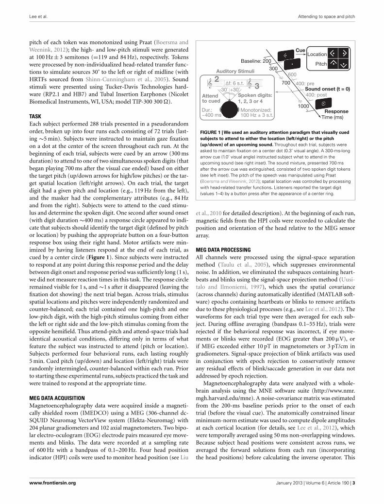
TASKEach subject performed 288 trials presented in a pseudorandomorder, broken up into four runs each consisting of 72 trials (last-ing ∼5 min). Subjects were instructed to maintain gaze fixationon a dot at the center of the screen throughout each run. At thebeginning of each trial, subjects were cued by an arrow (300 msduration) to attend to one of two simultaneous spoken digits (thatbegan playing 700 ms after the visual cue ended) based on eitherthe target pitch (up/down arrows for high/low pitches) or the tar-get spatial location (left/right arrows). On each trial, the targetdigit had a given pitch and location (e.g., 119 Hz from the left),and the masker had the complementary attributes (e.g., 84 Hzand from the right). Subjects were to attend to the cued stimu-lus and determine the spoken digit. One second after sound onset(with digit duration ∼400 ms) a response circle appeared to indi-cate that subjects should identify the target digit (defined by pitchor location) by pushing the appropriate button on a four-buttonresponse box using their right hand. Motor artifacts were min-imized by having listeners respond at the end of each trial, ascued by a center circle (Figure 1). Since subjects were instructedto respond at any point during this response period and the delaybetween digit onset and response period was sufficiently long (1 s),we did not measure reaction times in this task. The response circleremained visible for 1 s, and ∼1 s after it disappeared (leaving thefixation dot showing) the next trial began. Across trials, stimulusspatial locations and pitches were independently randomized andcounter-balanced; each trial contained one high-pitch and onelow-pitch digit, with the high-pitch stimulus coming from eitherthe left or right side and the low-pitch stimulus coming from theopposite hemifield. Thus attend-pitch and attend-space trials hadidentical acoustical conditions, differing only in terms of whatfeature the subject was instructed to attend (pitch or location).Subjects performed four behavioral runs, each lasting roughly5 min. Cued pitch (up/down) and location (left/right) trials wererandomly intermingled, counter-balanced within each run. Priorto starting these experimental runs, subjects practiced the task andwere trained to respond at the appropriate time.
MEG DATA ACQUISITIONMagnetoencephalography data were acquired inside a magneti-cally shielded room (IMEDCO) using a MEG (306-channel dc-SQUID Neuromag VectorView system (Elekta-Neuromag) with204 planar gradiometers and 102 axial magnetometers. Two bipo-lar electro-oculogram (EOG) electrode pairs measured eye move-ments and blinks. The data were recorded at a sampling rateof 600 Hz with a bandpass of 0.1–200 Hz. Four head positionindicator (HPI) coils were used to monitor head position (see Liu
FIGURE 1 | We used an auditory attention paradigm that visually cuedsubjects to attend to either the location (left/right) or the pitch(up/down) of an upcoming sound. Throughout each trial, subjects wereasked to maintain fixation on a center dot (0.3˚ visual angle). A 300-ms-longarrow cue (1.0˚ visual angle) instructed subject what to attend in theupcoming sound (see right inset). The sound mixture, presented 700 msafter the arrow cue was extinguished, consisted of two spoken digit tokens(see left inset). The pitch of the speech was manipulated using Praat(Boersma and Weenink, 2012); spatial location was controlled by processingwith head-related transfer functions. Listeners reported the target digit(values 1–4) by a button press after the appearance of a center ring.
et al., 2010 for detailed description). At the beginning of each run,magnetic fields from the HPI coils were recorded to calculate theposition and orientation of the head relative to the MEG sensorarray.
MEG DATA PROCESSINGAll channels were processed using the signal-space separationmethod (Taulu et al., 2005), which suppresses environmentalnoise. In addition, we eliminated the subspaces containing heart-beats and blinks using the signal-space projection method (Uusi-talo and Ilmoniemi, 1997), which uses the spatial covariance(across channels) during automatically identified (MATLAB soft-ware) epochs containing heartbeats or blinks to remove artifactsdue to these physiological processes (e.g., see Lee et al., 2012). Thewaveforms for each trial type were then averaged for each sub-ject. During offline averaging (bandpass 0.1–55 Hz), trials wererejected if the behavioral response was incorrect, if eye move-ments or blinks were recorded (EOG greater than 200 µV), orif MEG exceeded either 10 pT in magnetometers or 3 pT/cm ingradiometers. Signal-space projection of blink artifacts was usedin conjunction with epoch rejection to conservatively removeany residual effects of blink/saccade generation in our data notaddressed by epoch rejection.
Magnetoencephalography data were analyzed with a whole-brain analysis using the MNE software suite (http://www.nmr.mgh.harvard.edu/mne). A noise-covariance matrix was estimatedfrom the 200-ms baseline periods prior to the onset of eachtrial (before the visual cue). The anatomically constrained linearminimum-norm estimate was used to compute dipole amplitudesat each cortical location (for details, see Lee et al., 2012), whichwere temporally averaged using 50 ms non-overlapping windows.Because subject head positions were consistent across runs, weaveraged the forward solutions from each run (incorporatingthe head positions) before calculating the inverse operator. This
www.frontiersin.org January 2013 | Volume 6 | Article 190 | 3

Lee et al. Attending to space and pitch
allowed us to use a single inverse per subject. For pre- and post-sound stimulus analysis, activity was measured during 400-msepochs prior to and after sound onset (preparatory and stimu-lus periods) following a baseline correction calculated from the200-ms period prior to the visual cue (up/down/left/right) thatsignaled the start of each trial. For across-subject comparisons,source localized data were morphed to a template brain, optimallyaligning individual sulcal-gyral patterns (Fischl et al., 1999).
STATISTICAL ANALYSISFor displaying group-level activity on the cortical surface, we firstspatially smoothed individual subject data across neighboring ver-tices. Specifically, for 25 successive iterations of the spreadingoperator, the new value at each vertex was the sum of the previousvalues of the vertex and its immediate neighbors (adjacent verticesin the parcellation of the cortical surface), divided by the numberof non-zero values included. This smoothing helps to compensatefor expected anatomical and functional subject differences. Thesmoothed estimates were used to compute a location versus pitchcontrast at each vertex on the cortical surface. The resulting esti-mates were submitted to a repeated-measures ANOVA, treatingtime as an experimental factor over eight consecutive 50-ms timeframes making up both the preparatory and stimulus epochs. Aconservative Greenhouse–Geisser non-sphericity correction wasused within each 400-ms epoch to mitigate the effect of possiblecorrelations of the residuals over the time bins. To compensatefor multiple-comparisons, we took the conservative approach ofdisplaying p-values on the cortical maps after a stringent Bonfer-roni correction taking into account the number of source verticesacross the brain (∼10,000/hemisphere). Although a given brainarea may be activated strongly in both the attend-location andattend-pitch conditions, this within-subjects analysis only mea-sures the differences in activation elicited during the two tasks.This analysis ignores the overall activation levels in the conditionsto reduce the potential for biases in our measurements to affectresults. Given this, we only discuss the differential activity acrossthe two tasks. To examine large regions of activity, we only examineclusters containing at least 100 vertices.
SACCADE PARADIGM AND EYE MOVEMENT PROCESSINGWe used 192 trials of a memory-guided go/no-go saccade task toobtain an FEF functional localizer. Each saccadic trial lasted 3.3 s(counter-balanced for left/right movement). A left or right arrow(300 ms in duration) began each trial, after which a 1-s-long ringwas presented, shifted to the side by 10˚ in the direction of the pre-ceding arrow cue. Subjects were asked to maintain center fixation(white dot subtending 0.3˚) unless otherwise instructed. The colorof the arrow on a given trial cued the subjects either to move theeyes (“go”; green) or to keep eyes fixed (“no-go”; red). On a sepa-rate block, we asked subjects to track a white dot (subtended visualangle, 0.3˚) with their gaze as it moved to one of six locations (± 3˚,6˚, or 9˚ from center, each presented once in random order withina run, with four runs making up the block). These tracking datawere used to obtain an individualized linear transform relatingEOG to eye gaze eccentricity in degrees. Saccades were identifiedas horizontal eye movements with velocities exceeding 100˚/s. Theonset of a saccade was defined as the point at which the velocity
of the eye first exceeded 30˚/s. Trials with saccade latencies lessthan 100 ms were considered anticipatory and were not includedin subsequent analysis.
ADDITIONAL SACCADE MONITORINGIn order to rule out eye saccade explanations of results in ourspatial auditory attention task, one subject was invited back toperform a full auditory attention session in the MEG environmentwhile eye gaze was monitored using high-resolution binocular eye-tracking. Real-time (1000 Hz sampling) binocular gaze positionwas determined using an Eyelink 1000 MEG compatible eye-tracking system (SR Research, Ltd., ON, Canada) calibrated usinga 9-point fixation paradigm at the beginning of each block anddrift corrected at the beginning of each trial. This system has aspatial resolution of 0.02˚ (RMS) and accuracy of (average biasup to) 0.25˚. We used eye-tracking data from this system bothto determine if there were significant differences between the eyepositions in attend-space and attend-pitch conditions, and to testwhether there was a correlation between eye position and FEF acti-vation on a single-trial level. Since it is possible that single-trialMEG activations may be too noisy to provide a meaningful com-parison between conditions, we also compared trial-averaged FEFactivations and EOG levels across subjects. To remove effects dueto bias (e.g., variation in overall EOG or FEF amplitudes acrosssubjects), we compared EOG to FEF activations using the nor-malized differences of each in the space and pitch conditions as(space− pitch)/(space+ pitch). Additionally, to deal with the pos-sibility that left and right trials could have opposing eye-directionmovements that would cancel out in averaging, we comparedLFEF activation to the mean of the (1) magnitude of the left-trial-averaged EOG and (2) magnitude of the right-trial-averagedEOG.
FEF-ROI FUNCTIONAL LOCALIZERA priori, we focused only on the FEFs, located in and around theprecentral sulcus and gyrus (Simó et al., 2005). For each sub-ject, we used a functional localizer to obtain a region of interest(ROI) anatomically constrained to the bilateral superior and infe-rior precentral sulci and the precentral gyri, as defined by anautomated surface-based parcellation (Fischl et al., 2004). Withinthese regions in the averaged group data, we functionally con-strained the FEF-ROI to vertices showing activity (i.e., differencesin dipole strengths) in the “go” versus “no-go” saccade contrastwith a threshold of p < 0.05 following a conservative Greenhouse–Geisser non-sphericity correction. For this analysis, activity wasestimated every 50 ms between 0 and 300 ms after the onset ofthe peripheral ring. This contrast between the “go” and “no-go”trials isolates saccade-generating signals associated with the FEFs.This provided cross-subject spatial localization data for the FEFsto compare to our findings from the whole-brain analysis.
RESULTSLEFT-DOMINANT DIFFERENTIAL ENGAGEMENT IN AUDITORY SPATIALAND PITCH ATTENTIONTo quantify the cortical involvement in top-down attention, weanalyzed the differential cortical source estimates between locationand pitch trials in the 400-ms-long preparatory (from 600 ms after
Frontiers in Neuroscience | Auditory Cognitive Neuroscience January 2013 | Volume 6 | Article 190 | 4

Lee et al. Attending to space and pitch
the onset of the visual cue directing attention up to sound onset)and stimulus epochs (from sound onset to 400 ms later; note thatthe stimulus epoch analysis window encapsulates the duration ofeach token; see Figure 1). We also located our primary a priori ROI,FEFs, based on a combination of anatomical landmarks (limitinganalysis to the precentral sulcus and gyrus; see Materials and Meth-ods) and significant functional activity from our memory-guided,go/no-go saccade task, which revealed a larger area of activationfor in the left hemisphere than the right (Figure 2A, green labels;left/right ROI center of mass x =−47.5, y =−1.8, z = 39.6, andx = 34.8, y=−9.0, z= 52.8, respectively; activation traces shownin Figure 2B). This may reflect the hemispheric asymmetry relatedto the functional localizer task used, consistent with recent find-ings that the oculomotor system is more asymmetric in humansthan in monkeys (Kagan et al., 2010).
A region in the left, but not right, dorsal precentral sulcus/gyruswas more active when subjects attended to location than when they
attended to pitch (Figure 2A), both in the preparatory and stim-ulus epochs. Importantly, this region of enhanced activity duringspatial attention (MNI centroid coordinates: x =−36.3, y =−4.8,z = 42.4) overlapped with the left FEF-ROI. We also found thatthe left, but not right, posterior STS (MNI centroid coordinates:x =−49.7, −45.1, 7.1) was more active when subjects attendedto pitch than when they attended to location, but only in thepreparatory epoch.
Electro-oculogram results revealed no systematic differences ineye movements for the location and pitch trials. For the subject inwhom eye movements were recorded, there was no evidence forany consistent directional bias in either eye position or gaze veloc-ity that depended on trial type, either when treated as a function ofperistimulus time or when using the ergodic average (p > 0.2 forall paired t -test comparisons; left, right, and pitch trial types). Inthe attend-pitch trials, we found no correlation between eye posi-tion and the activation in the LFEF region that was significantly
4.5
3.5
2.5
1.5
1
2
3
4
4.5
3.5
2.5
1.5
1
2
3
4
A
B C
FIGURE 2 | Left FEF and left STG are more active prior to sound onsetwhen subjects attend to space and pitch, respectively, based oncontrasts of the cortical signal in the two conditions. (A) Statistical map(group average) displayed on the inflated cortical surface of the left and righthemispheres, illustrating a vertex-by-vertex comparison (yellow: greater
activity in location trials; blue: greater activity in pitch trials; minimumcluster-size threshold 100 vertices). Functionally localized FEF regions arehighlighted in green. B/C: normalized evoked cortical current time courses forthe significant left FEF (B) and left STG regions (C) for both location and pitchconditions, shown with standard error bars (µ±SEM) across subjects.
www.frontiersin.org January 2013 | Volume 6 | Article 190 | 5

Lee et al. Attending to space and pitch
more active during attend-space than attend-pitch trials (Kendallp= 0.308 for both vertical and horizontal position, N = 116).In addition, following a multiple-comparisons threshold correc-tion (α= 0.05/4= 0.0125), the correlation between horizontal eyeposition and FEF activation on pitch trials was also not statis-tically significant (Kendall p= 0.413, N = 68); importantly, evenif this difference was found to be statistically significant, it can-not explain why LFEF activation was greater in space trials thanin pitch trials. Moreover, when examining LFEF across all sub-jects, the normalized trial-averaged activation differences in LFEFdid not correlate with either the horizontal or vertical (Kendallp > 0.73 for both, N = 17) EOG. These data all suggest that thedifferential FEF engagement was not simply due to eye movementsin“attend left”and“attend right” trials. We also compared bilateral“attend left” and “attend right” activity, and found no significantdifference in activity in either left or right FEF with direction ofspatial attention (data not shown).
BEHAVIORAL RESULTS AND RELATIONSHIP TO NEURAL ACTIVITYOverall, subjects performed the task accurately in both locationand pitch trials, but were slightly better on location trials (per-cent correct 86.42± 3.27%, mean± SEM) than on pitch trials(78.32± 3.97%; P = 0.0002, paired t -test). All subjects performedbetter than chance (25% correct), although performance variedsubstantially across subjects, from 56.3 to 99.3% when attendinglocation and from 46.5 to 99.3% when attending pitch. Any tri-als in which a valid response was not recorded during the fixedresponse period were designated misses. In general, misses repre-sented a small fraction of trials: 1.3–8.9% for all but three subjects,who had miss rates of 13.9–25.0%. For the three subjects withhigher miss rates, most misses arose because responses were madebefore the response period (i.e., the subjects performed the taskand responded, but did so too quickly). If responses up to 0.4 sbefore the onset of the response circle (i.e., at least 200 ms fol-lowing sound offset) were included, the miss rates for all three ofthese subjects would decrease to 1.4–5.2%. To ensure that motorresponses did not interfere with the results presented here, theseanticipatory responses were not included in our MEG analysis.
Percent correct performance was correlated in attend-locationand attend-pitch trials across subjects (Kendall tau= 0.857,P < 0.000003, N = 17), showing that some of the individual differ-ences were unrelated to what feature was attended, coming insteadfrom individual differences in the ability to focus auditory selectiveattention and perform the task. Similarly, the degree of modulationin left FEF activity for attend-location versus attend-pitch trialswas negatively correlated with the degree of attended-feature-specific modulation of activity in left STS (Kendall tau=−0.467,P < 0.011, N = 17), consistent with there being a common atten-tional control signal that affects activity in these areas in a mannerspecific to the feature to which attention is directed. However, com-paring average performance on the two tasks to the average of themodulation in left FEF and the negative of the modulation in leftSTS, there was no significant relationship (Kendall tau=−0.081,P = 0.680, N = 17). Comparing the level of neural activation (rel-ative to baseline) in the space condition with the activation inleft FEF, and in the pitch condition with left STS, correlationswere insignificant but trending (Kendall tau= 0.32 and, P = 0.082
and 0.069, respectively, N = 17). Although left FEF activation inthe pitch condition was correlated with performance on the pitchtask (Kendall tau= 0.41, P = 0.026, N = 17), after Bonferroni cor-rection for six comparisons (adjusted α= 0.0083), this differencewas not statistically significant. Activity in the left STS was notsignificantly correlated with performance on the space condition(Kendall tau= 0.31, P = 0.097, N = 17).
DISCUSSIONOur results demonstrate that regions of the cortex are engaged indirecting attention to acoustic features even before the soundsbegin; moreover, different regions are engaged more stronglydepending on what feature is directly selective auditory atten-tion: left FEF when attending location and left posterior STSwhen attending pitch. Previous fMRI studies demonstrate thatactivity in a left dominated frontoparietal network is enhancedduring attentionally demanding trials compared to fixation tri-als, whether subjects attend to a spatial or a non-spatial feature,and in both visual and auditory tasks. During a visual attentiontask, anatomical regions proximal to bilateral FEFs were moreactive during spatial attention, while the left ventral occipital cor-tex was more active when subjects attended to color (Giesbrechtet al., 2003; Slagter et al., 2007). During an auditory task, leftFEF showed enhanced activity for both location and pitch, evenon catch trials where there was no acoustic stimulus (Hill andMiller, 2010), consistent with the anticipatory activity we foundin our task. However, in this earlier study, right FEF was moreactive in location trials, and the inferior frontal gyrus (a regionlinked to language processing) was more active in pitch trials.While it is difficult to directly compare results of studies usingdifferent sensory stimuli and different neuroimaging techniques,especially since the relationship between neural activity measuredusing MEG and BOLD responses measured using fMRI is not wellestablished, our results add to evidence that FEFs are involvedin control of covert spatial attention across different modalities(while other areas may be similarly engaged when attending tonon-spatial features). In contrast to previous studies, our resultssuggest that there is an asymmetry in auditory processing wherebyleft FEF is more strongly involved in attending to auditory stimulibased on spatial location compared to pitch. We also show thatthis attention-specific control begins in preparation for upcom-ing stimuli containing a to-be-attended feature. It is worth notingthat the activity observed in preparing to attend to stimuli based onspatial condition may be due to the deployment of both auditoryand visual attention to the spatial location of interest, as wouldlikely be the case if auditory and visual spatial attention share acommon supramodal network. Teasing this apart could be inter-esting in future studies that make use of either auditory-only cuesor visual cues that come on well before auditory attention mustbe directed. However, our observations here are unlikely to be duesolely to the deployment of visual attention.
It has been shown previously that preparing to attend to a soundlikely to originate from a given direction biases cortical activity inauditory cortex contralateral to that direction (Voisin et al., 2006),indicating that prior to sound onset, listeners “prime” cortical rep-resentations to favor upcoming sounds from the direction to beattended. Given this, our results are consistent with our listeners
Frontiers in Neuroscience | Auditory Cognitive Neuroscience January 2013 | Volume 6 | Article 190 | 6

Lee et al. Attending to space and pitch
engaging auditory attention to perform our tasks, although wecannot rule out the possibility that listeners co-deploy both audi-tory and visual spatial attention networks in anticipation of anupcoming sound. Additionally, although some evoked responsesare visible in the traces of left FEF and STS (likely due to leakagefrom primary sensory cortices), the observed differences reportedhere must be due to the task condition (attend-space versus attend-pitch) since the acoustic stimuli used in the two conditions areidentical.
The left lateralization of FEF activity initially seems at odds withpast reports of “hemispheric dominance.”It is well established thatthe right hemisphere processes information in both visual fields,whereas the left hemisphere exclusively encodes the right visualfield (Mesulam, 1981). This raises the question of why left, butnot right, FEF is more active in our location trials, regardless ofthe direction of the target, and why there are no significant differ-ences in FEF activity for “attend left” versus “attend right” trials.It is unlikely that this is due to preparatory motor activation (i.e.,preparing to press a button with the right hand), since such activ-ity would be the same for both the space task and the pitch taskthat were contrasted, yet differences in activation were observedbefore a sound stimulus was presented (and thus before an appro-priate response could be prepared). We believe this left FEF biasmay reflect its participation in a dorsal, top-down attention net-work (Corbetta et al., 2008), with right FEF involved in top-downattention, exogenous attention, and shifting of attention. Previousauditory fMRI studies that find bilateral FEF activation duringauditory spatial attention tasks used paradigms that differ fromours: most either required subjects to explicitly shift their auditoryspatial attention (Salmi et al., 2007, 2009), or exogenously cued theauditory location to attend (Wu et al., 2007). Moreover, because ofscanner noise, listeners in these studies may have deployed someform of non-spatial attention to focus on the desired acousticstimuli instead of or in addition to engaging spatial attention.The one study that required top-down deployment of auditoryspatial attention (Hill and Miller, 2010) yielded poor behavioralperformance (especially early in the experiment), suggesting thatthe subjects were not always successful in deploying attention, andsometimes reoriented attention while trying to perform the task. Incontrast, our study presented a brief stimulus (one syllable long),yet subjects performed the task reasonably well, showing that theysuccessfully deployed top-down spatial attention in the prepara-tory period. Thus, we suggest that the left FEF is differentially moreinvolved in top-down auditory spatial attention, consistent withthe supramodal attentional network previously proposed (Cor-betta et al.,2008). Note that although left FEF shows greater activityduring spatial rather than pitch-based attention trials, left FEF alsomay well play a role in non-spatial attention as well, as evidencedby a significant (before a multiple-comparisons correction) cor-relation between activity in left FEF and behavioral performanceon attend-pitch trials. The pre-auditory-stimulus left FEF activityobserved here is also similar to the anticipatory activity in FEFsreported in past visual studies that is linked to top-down controlof spatial attention (Awh et al., 2006).
We found that left posterior STS, which was not chosen a priorias an ROI, was recruited in attend-pitch trials during the prepara-tory period, a result that mirrors previous findings of other cortical
regions showing attention biases for non-spatial features: ventraloccipital cortex for attention to color (Giesbrecht et al., 2003, 2006;Slagter et al., 2007), inferior frontal regions for attention to spec-tral features in a language-related task (Hill and Miller, 2010),and preparatory activity in auditory cortex contralateral to theexpected location of an upcoming sound (Voisin et al., 2006). Sev-eral studies have associated the left STS with the identification orcategorization of sounds based on non-spatial attributes (Möttö-nen et al., 2006; Liebenthal et al., 2010), especially for people withabsolute pitch (Schulze et al., 2009). Although this area is likelyinvolved in performing categorization in both the spatial locationand pitch tasks, our results suggest that the cortical region asso-ciated with categorization of pitch information becomes moreactive and helps listeners prepare to select a target stimulus basedon pitch. These results support the existence of different path-ways for processing “what” and “where” sound attributes (e.g.,Rauschecker and Tian, 2000; Ahveninen et al., 2006); however,since the posterior location of the left STS activation observed hereis more consistent with the previously reported “where” pathway,additional experiments will be necessary to provide the spatialresolution required to definitively tease apart the contributionsof these areas. Moreover, we were relatively conservative in ourdata analysis (e.g., Bonferroni correction) to decrease the likeli-hood of false positives; however, this increases the chance thatadditional areas are significantly involved during tasks like thoseused here; experiments that relax constraints or that employ otherstatistical approaches might expose such other activity (e.g., Singhet al., 2003; Maris and Oostenveld, 2007). For example, the use ofmasking noise could obscure differences in activity that the twotasks might have evoked in auditory cortex for presentations inquiet, especially given the conservative analyses we adopted. It isalso possible that there is an underlying activity difference in leftSTS during the stimulus (“post” period; as seen in Figure 2C) aswell; future studies with better SNR or less strict thresholding maywell observe significant left-biased activity differences during thestimuli when attending based on pitch.
Finally,our results show that left FEF is involved both before andafter the onset of sound while activity in left posterior STS is signif-icantly enhanced only prior to the onset of sound. These changeswere correlated across subjects, as if the degree of attentional mod-ulation in both attend-location and attend-pitch trials depends onsome common signal, regardless of the feature attended. How-ever, these activity differences are not significantly correlated withbehavioral performance, even though listener ability varies widelyacross subjects. In our neural analysis, we contrasted two con-ditions, each of which engages selective auditory attention; thus,any differences in the strength with which listeners engage corticalregions that are common to both attend-location and attend-pitchtrials is invisible in our analysis; we only see the indirect effectsof such common control in the strength of modulation of thefeature-specific areas left FEF and left STS. Combined with theobservation that across subjects, performance is strongly corre-lated in the attend-location and attend-pitch trials, our resultssuggest that overall selective attention performance depends onthe degree of engagement of neural areas that are employed bothwhen attending to location and when attending to pitch, and/oron individual differences in the fidelity of sensory encoding of the
www.frontiersin.org January 2013 | Volume 6 | Article 190 | 7

Lee et al. Attending to space and pitch
basic acoustic information needed to compute auditory featureslike location and pitch (Ruggles et al., 2011). Here, we also foundinsignificant but trending correlations between activity in attend-space and attend-pitch trials. However, the estimates of neuralactivity normalized to baseline used here are influenced by thesignal-to-noise ratio and the number of valid trials for each subject,which could contribute to the lack of observed significant corre-lations. Future experiments thus could be undertaken to explorethe degree to which the overall activity of a general “attention net-work” helps to predict individual ability on this kind of selectiveauditory attention task. Additionally, although we did not havea sufficient number of incorrect trials to perform a meaningfulanalysis here, future studies could also look at activations in errortrials to also examine the auditory selective attention network.
Taken in the context of previous psychoacoustical and neu-roimaging work, our findings support the conclusions that(1) left FEF is involved in both directing and sustainingauditory spatial attention and (2) the left STS aids objectselection based on its pitch feature prior to the onset ofsound.
ACKNOWLEDGMENTSWe thank Nick Kurkjy for his assistance with data collection.This work was supported by National Institutes of Health (NIH)grants to Adrian K. C. Lee (K99/R00 DC010196) and Matti S.Hämäläinen (P41 RR014075), an NSSEFF fellowship to BarbaraG. Shinn-Cunningham, and training grant T32DC000018 (EricLarson).
REFERENCESAhveninen, J., Jaaskelainen, I., Raij,
T., Bonmassar, G., Devore, S.,Hamalainen, M., et al. (2006).Task-modulated “what” and “where”pathways in human auditory cor-tex. Proc. Natl. Acad. Sci. U.S.A. 103,14608–14613.
Awh, E., Armstrong, K. M., and Moore,T. (2006). Visual and oculomotorselection: links, causes and impli-cations for spatial attention. TrendsCogn. Sci. (Regul. Ed.) 10, 124–130.
Boersma, P., and Weenink, D. (2012).Praat: Doing Phonetics by Computer[Computer Program], Version 5.3.10.Available at: http://www.praat.org/
Brainard, D. H. (1997). The psy-chophysics toolbox. Spat. Vis. 10,433–436.
Bruce, C. J., Goldberg, M. E., Bushnell,M. C., and Stanton, G. B. (1985).Primate frontal eye fields. II. Physio-logical and anatomical correlates ofelectrically evoked eye movements.J. Neurophysiol. 54, 714–734.
Corbetta, M., Patel, G., and Shulman,G. L. (2008). The reorienting systemof the human brain: from environ-ment to theory of mind. Neuron 58,306–324.
Degerman, A., Rinne, T., Salmi, J., Salo-nen, O., and Alho, K. (2006). Selec-tive attention to sound location orpitch studied with fMRI. Brain Res.1077, 123–134.
Fischl, B., Salat, D. H., van der Kouwe,A. J. W., Makris, N., Ségonne, F.,Quinn, B. T., et al. (2004). Sequence-independent segmentation of mag-netic resonance images. Neuroimage23(Suppl. 1), S69–S84.
Fischl, B., Sereno, M., and Dale,A. (1999). Cortical surface-basedanalysis. II: inflation, flattening, anda surface-based coordinate system.NeuroImage 9, 195–207.
Gherri, E., Driver, J., and Eimer, M.(2008). Eye movement preparationcauses spatially-specific modulation
of auditory processing: new evidencefrom event-related brain potentials.Brain Res. 1224, 88–101.
Giesbrecht, B., Weissman, D. H.,Woldorff, M. G., and Mangun, G. R.(2006). Pre-target activity in visualcortex predicts behavioral perfor-mance on spatial and feature atten-tion tasks. Brain Res. 1080, 63–72.
Giesbrecht, B., Woldorff, M. G., Song,A. W., and Mangun, G. R. (2003).Neural mechanisms of top-downcontrol during spatial and featureattention. Neuroimage 19, 496–512.
Hill, K., and Miller, L. (2010). Audi-tory attentional control and selec-tion during cocktail party listening.Cereb. Cortex 20, 583–590.
Kagan, I., Iyer, A., Lindner, A., andAndersen, R. A. (2010). Space repre-sentation for eye movements is morecontralateral in monkeys than inhumans. Proc. Natl. Acad. Sci. U.S.A.107, 7933–7938.
Lee, A. K., Larson, E. D., Mad-dox, R. K. (2012). Mapping cor-tical dynamics using simultane-ous MEG/EEG and anatomically-constrained minimum-norm esti-mates: an auditory attention exam-ple. J. Vis. Exp. 68, e4262.
Leonard, J. B., Foster, K. R., and Athey,T. W. (1984). A database for speaker-independent digit recognition. Proc.IEEE Int. Conf. Acoust. Speech. Sig-nal. Process. 9, 328–331.
Liebenthal, E., Desai, R., Ellingson, M.M., Ramachandran, B., Desai, A.,and Binder, J. R. (2010). Specializa-tion along the left superior temporalsulcus for auditory categorization.Cereb. Cortex 20, 2958–2970.
Liu, H., Tanaka, N., Stufflebeam, S.,Ahlfors, S., and Hämäläinen, M.(2010). Functional mapping withsimultaneous MEG and EEG. J.Vis. Exp. Available at: http://www.jove.com/video/1668/functional-mapping-with-simultaneous-meg-and-eeg.
Maeder, P. P., Meuli, R. A., Adriani, M.,Bellmann, A., Fornari, E., Thiran, J.P., et al. (2001). Distinct pathwaysinvolved in sound recognition andlocalization: a human fMRI study.Neuroimage 14, 802–816.
Maris, E., and Oostenveld, R. (2007).Nonparametric statistical testing of,EEG- and MEG-data. J. Neurosci.Methods 164, 177–90.
Mayer, A. R., Harrington, D., Adair, J. C.,and Lee, R. (2006). The neural net-works underlying endogenous audi-tory covert orienting and reorient-ing. Neuroimage 30, 938–949.
Mesulam, M. M. (1981). A corticalnetwork for directed attention andunilateral neglect. Ann. Neurol. 10,309–325.
Möttönen, R., Calvert, G. A., Jääskeläi-nen, I. P., Matthews, P. M., Thesen,T., Tuomainen, J., et al. (2006). Per-ceiving identical sounds as speechor non-speech modulates activ-ity in the left posterior superiortemporal sulcus. Neuroimage 30,563–569.
Pavani, F., Làdavas, E., and Driver, J.(2005). Gaze direction modulatesauditory spatial deficits in strokepatients with neglect. Cortex 41,181–188.
Rauschecker, J., and Tian, B. (2000).Mechanisms and streams for pro-cessing of “what” and “where” inauditory cortex. Proc. Natl. Acad. Sci.U.S.A. 97, 11800–11806.
Ruggles, D., Bharadwaj, H., andShinn-Cunningham, B. G. (2011).Normal hearing is not enoughto guarantee robust encoding ofsuprathreshold features impor-tant in everyday communication.Proc. Natl. Acad. Sci. U.S.A. 108,15516–15521.
Salmi, J., Rinne, T., Degerman, A., Salo-nen, O., and Alho, K. (2007). Ori-enting and maintenance of spatialattention in audition and vision:multimodal and modality-specific
brain activations. Brain Struct.Funct. 212, 181–194.
Salmi, J., Rinne, T., Koistinen, S., Salo-nen, O., and Alho, K. (2009). Brainnetworks of bottom-up triggeredand top-down controlled shifting ofauditory attention. Brain Res. 1286,155–164.
Schulze, K., Gaab, N., and Schlaug, G.(2009). Perceiving pitch absolutely:comparing absolute and relativepitch possessors in a pitch mem-ory task. BMC Neurosci. 10:106.doi:10.1186/1471-2202-10-106
Sharon, D., Hämäläinen, M. S., Tootell,R. B. H., Halgren, E., and Bel-liveau, J. W. (2007). The advantageof combining, MEG and EEG: com-parison to fMRI in focally stimu-lated visual cortex. Neuroimage 36,1225–1235.
Shinn-Cunningham, B., Kopco, N., andMartin, T. (2005). Localizing nearbysound sources in a classroom: binau-ral room impulse response. J. Acous.Soc. Am. 117, 3100–3115.
Shomstein, S., and Yantis, S. (2006).Parietal cortex mediates voluntarycontrol of spatial and nonspatialauditory attention. J. Neurosci. 26,435–439.
Simó, L. S., Krisky, C. M., and Sweeney, J.A. (2005). Functional neuroanatomyof anticipatory behavior: dissoci-ation between sensory-driven andmemory-driven systems. Cereb. Cor-tex 15, 1982–1991.
Singh, K. D., Barnes, G. R., and Hille-brand, A. (2003). Group imagingof task-related changes in corticalsynchronisation using nonparamet-ric permutation testing. Neuroimage19, 1589–1601.
Slagter, H. A., Giesbrecht, B., Kok,A., Weissman, D. H., Kenemans, J.L., Woldorff, M. G., et al. (2007).fMRI evidence for both general-ized and specialized components ofattentional control. Brain Res. 1177,90–102.
Frontiers in Neuroscience | Auditory Cognitive Neuroscience January 2013 | Volume 6 | Article 190 | 8

Lee et al. Attending to space and pitch
Tark, K.-J., and Curtis, C. E. (2009). Per-sistent neural activity in the humanfrontal cortex when maintainingspace that is off the map. Nat. Neu-rosci. 12, 1463–1468.
Taulu, S., Simola, J., and Kajola, M.(2005). Applications of the sig-nal space separation method. IEEETrans. Signal Process. 53, 3359.
Uusitalo, M. A., and Ilmoniemi, R.J. (1997). Signal-space projectionmethod for separating, MEG or EEGinto components. Med. Biol. Eng.Comput. 35, 135–140.
Voisin, J., Bidet-Caulet, A., Bertrand,O., and Fonlupt, P. (2006). Lis-tening in silence activates auditoryareas: a functional magnetic reso-nance imaging study. J. Neurosci. 26,273–278.
Wardak, C., Ibos, G., Duhamel, J.-R.,and Olivier, E. (2006). Contributionof the monkey frontal eye field tocovert visual attention. J. Neurosci.26, 4228–4235.
Warren, J., and Griffiths, T. (2003).Distinct mechanisms for process-ing spatial sequences and pitchsequences in the human auditorybrain. J. Neurosci. 23, 5799–5804.
Woods, D. L., Alho, K., and Algazi, A.(1994). Stages of auditory featureconjunction: an event-related brainpotential study. J. Exp. Psychol. Hum.Percept. Perform. 20, 81–94.
Wu, C. T., Weissman, D. H., Roberts,K. C., and Woldorff, M. G. (2007).The neural circuitry underlying theexecutive control of auditory spatialattention. Brain Res. 1134, 187–198.
Zatorre, R. J., Mondor, T. A., and Evans,A. C. (1999). Auditory attention tospace and frequency activates simi-lar cerebral systems. Neuroimage 10,544–554.
Conflict of Interest Statement: Theauthors declare that the research wasconducted in the absence of any com-mercial or financial relationships thatcould be construed as a potential con-flict of interest.
Received: 02 November 2012; paperpending published: 14 November 2012;accepted: 16 December 2012; publishedonline: 07 January 2013.Citation: Lee AKC, Rajaram S, Xia J,Bharadwaj H, Larson E, Hämäläinen
MS and Shinn-Cunningham BG (2013)Auditory selective attention revealspreparatory activity in different corticalregions for selection based on source loca-tion and source pitch. Front. Neurosci.6:190. doi: 10.3389/fnins.2012.00190This article was submitted to Frontiersin Auditory Cognitive Neuroscience, aspecialty of Frontiers in Neuroscience.Copyright © 2013 Lee, Rajaram, Xia,Bharadwaj, Larson, Hämäläinen andShinn-Cunningham. This is an open-access article distributed under the termsof the Creative Commons AttributionLicense, which permits use, distributionand reproduction in other forums, pro-vided the original authors and sourceare credited and subject to any copy-right notices concerning any third-partygraphics etc.
www.frontiersin.org January 2013 | Volume 6 | Article 190 | 9