Atrial fibrillation ablation using very short duration 50 W … · Statistical analysis was done...
8
Atrial fibrillation ablation using very short duration 50 W ablations and contact force sensing catheters Roger A. Winkle 1,2 & Ryan Moskovitz 3 & R. Hardwin Mead 1 & Gregory Engel 1 & Melissa H. Kong 1 & William Fleming 1 & Jonathan Salcedo 1 & Rob A. Patrawala 1 & John H. Tranter 3 & Isaac Shai 3 Received: 20 November 2017 /Accepted: 26 January 2018 /Published online: 19 February 2018 # The Author(s) 2018. This article is an open access publication Abstract Purpose The optimal radiofrequency (RF) power and lesion duration using contact force (CF) sensing catheters for atrial fibrillation (AF) ablation are unknown. We evaluate 50 W RF power for very short durations using CF sensing catheters during AF ablation. Methods We evaluated 51 patients with paroxysmal (n = 20) or persistent (n = 31) AF undergoing initial RF ablation. Results A total of 3961 50 W RF lesions were given (average 77.6 ± 19.1/patient) for an average duration of only 11.2 ± 3.7 s. As CF increased from < 10 to > 40 g, the RF application duration decreased from 13.7 ± 4.4 to 8.6 ± 2.5 s (p < 0.0005). Impedance drops occurred in all ablations, and for patients in sinus rhythm, there was loss of pacing capture during RF delivery suggesting lesion creation. Only 3% of the ablation lesions were at < 5 g and 1% at > 40 g of force. As CF increased, the force time integral (FTI) increased from 47 ± 24 to 376 ± 102 gs (p < 0.0005) and the lesion index (LSI) increased from 4.10 ± 0.51 to 7.63 ± 0.50 (p < 0.0005). Both procedure time (101 ± 19.7 min) and total RF energy time (895 ± 258 s) were very short. For paroxysmal AF, the single procedure freedom from AF was 86% at 1 and 2 years. For persistent AF, it was 83% at 1 year and 72% at 2 years. There were no complications. Conclusions Short duration 50 W ablations using CF sensing catheters are safe and result in excellent long-term freedom from AF for both paroxysmal and persistent AF with short procedure times and small amounts of total RF energy delivery. Keywords Atrial fibrillation . AF ablation . Contact force catheters 1 Introduction Radiofrequency (RF) ablation is widely utilized to treat atrial fibrillation (AF). The predominant goal for catheter ablation of AF is durable pulmonary vein (PV) isolation. Virtually, all veins appear isolated at the end of a procedure, but late recurrences can occur and repeat studies often show PV reconnection. Lesion formation for PV isolation and other ablations depend upon RF current delivered, duration of RF energy delivery, and good ablation catheter tissue contact. Increasing the power (and, therefore, current) to 50 W for longer duration lesions results in increased ablation efficacy but is associated with increased complications [1]. Shorter duration 50 W lesions also provide better long-term outcomes but without an increase in complica- tions [2]. The availability of catheters which measure tissue con- tact force (CF) provides a tool to potentially improve PV isola- tion and long-term outcomes. Prior studies using CF have used average power of 25–28 W for durations of 40 to 75 s at each site for a total RF energy delivery time of 37.6 to 46.5 min [3–5]. Our study evaluates the feasibility of doing AF ablation using very short duration ablations at a power of 50 W with CF sens- ing catheter technology. 2 Methods 2.1 Patient population The subjects were consecutive symptomatic patients with par- oxysmal or persistent AF undergoing initial AF ablation done * Roger A. Winkle [email protected] 1 Silicon Valley Cardiology, Palo Alto Medical Foundation, Sutter Health, E. Palo Alto and Sequoia Hospital, Redwood City, CA, USA 2 Silicon Valley Cardiology, 1950 University Avenue, Suite 160, E, Palo Alto, CA 94303, USA 3 St. Jude Medical, Inc, St. Paul, MN, USA Journal of Interventional Cardiac Electrophysiology (2018) 52:1–8 https://doi.org/10.1007/s10840-018-0322-6
Transcript of Atrial fibrillation ablation using very short duration 50 W … · Statistical analysis was done...
Atrial fibrillation ablation using very short duration 50 W ablationsand contact force sensing catheters
Roger A. Winkle1,2 & Ryan Moskovitz3 & R. Hardwin Mead1& Gregory Engel1 & Melissa H. Kong1
& William Fleming1&
Jonathan Salcedo1& Rob A. Patrawala1 & John H. Tranter3 & Isaac Shai3
Received: 20 November 2017 /Accepted: 26 January 2018 /Published online: 19 February 2018# The Author(s) 2018. This article is an open access publication
AbstractPurpose The optimal radiofrequency (RF) power and lesion duration using contact force (CF) sensing catheters for atrialfibrillation (AF) ablation are unknown. We evaluate 50 W RF power for very short durations using CF sensing catheters duringAF ablation.Methods We evaluated 51 patients with paroxysmal (n = 20) or persistent (n = 31) AF undergoing initial RF ablation.Results A total of 3961 50WRF lesions were given (average 77.6 ± 19.1/patient) for an average duration of only 11.2 ± 3.7 s. As CFincreased from < 10 to > 40 g, the RF application duration decreased from 13.7 ± 4.4 to 8.6 ± 2.5 s (p < 0.0005). Impedance dropsoccurred in all ablations, and for patients in sinus rhythm, there was loss of pacing capture during RF delivery suggesting lesioncreation. Only 3% of the ablation lesions were at < 5 g and 1% at > 40 g of force. As CF increased, the force time integral (FTI)increased from 47 ± 24 to 376 ± 102 gs (p < 0.0005) and the lesion index (LSI) increased from 4.10 ± 0.51 to 7.63 ± 0.50 (p < 0.0005).Both procedure time (101 ± 19.7 min) and total RF energy time (895 ± 258 s) were very short. For paroxysmal AF, the singleprocedure freedom from AF was 86% at 1 and 2 years. For persistent AF, it was 83% at 1 year and 72% at 2 years. There wereno complications.Conclusions Short duration 50Wablations using CF sensing catheters are safe and result in excellent long-term freedom fromAFfor both paroxysmal and persistent AF with short procedure times and small amounts of total RF energy delivery.
Keywords Atrial fibrillation . AF ablation . Contact force catheters
1 Introduction
Radiofrequency (RF) ablation is widely utilized to treat atrialfibrillation (AF). The predominant goal for catheter ablation ofAF is durable pulmonary vein (PV) isolation. Virtually, all veinsappear isolated at the end of a procedure, but late recurrences canoccur and repeat studies often show PV reconnection. Lesionformation for PV isolation and other ablations depend upon RFcurrent delivered, duration of RF energy delivery, and goodablation catheter tissue contact. Increasing the power (and,
therefore, current) to 50 W for longer duration lesions resultsin increased ablation efficacy but is associated with increasedcomplications [1]. Shorter duration 50 W lesions also providebetter long-term outcomes but without an increase in complica-tions [2]. The availability of catheters which measure tissue con-tact force (CF) provides a tool to potentially improve PV isola-tion and long-term outcomes. Prior studies using CF have usedaverage power of 25–28 W for durations of 40 to 75 s at eachsite for a total RF energy delivery time of 37.6 to 46.5min [3–5].Our study evaluates the feasibility of doing AF ablation usingvery short duration ablations at a power of 50 W with CF sens-ing catheter technology.
2 Methods
2.1 Patient population
The subjects were consecutive symptomatic patients with par-oxysmal or persistent AF undergoing initial AF ablation done
* Roger A. [email protected]
1 Silicon Valley Cardiology, Palo Alto Medical Foundation, SutterHealth, E. Palo Alto and Sequoia Hospital, Redwood City, CA, USA
2 Silicon Valley Cardiology, 1950 University Avenue, Suite 160, E,Palo Alto, CA 94303, USA
3 St. Jude Medical, Inc, St. Paul, MN, USA
Journal of Interventional Cardiac Electrophysiology (2018) 52:1–8https://doi.org/10.1007/s10840-018-0322-6
by point by point RF ablation with the St. Jude (St. Paul, MN)TactiCath® CF sensing catheter at Sequoia Hospital,Redwood City, California. All patients signed written in-formed consent. The study was approved by the WesternInstitutional Review Board. The AF type was categorized asparoxysmal: lasting < 1 week or persistent: lasting > 1 weekand < 1 year or requiring pharmacological or electrical cardio-version in < 1 week. Patients with longstanding persistent AFlasting >1 year were excluded.
2.2 Ablation protocol
Our ablation protocol [6] and our periproceduralanticoagulation protocols [7] have been previously described.Antiarrhythmic drugs were stopped at least five half-lives andamiodarone at least 3 months before ablation. The St. JudeEnSite™ Velocity™ system was used in all cases for 3Dmapping. All patients underwent circumferential PV isolationand other ablations as clinically indicated. All ablations wereat 50 W, including the posterior wall, using the St. JudeTactiCath™ open irrigated-tip CF sensing catheter and theSt. Jude Ampere™ RF generator with a target CF of 10–40 g. CF readings of 5–10 g were accepted, provided visualinspection of the CF waveform showed a stable pattern with-out respiratory variation and was constantly above 0 g. If wedid not get adequate CF, the transseptal sheath was ex-changed for a St. Jude Agilis™ steerable sheath. We used a2s ramp time, a catheter irrigation rate of 30 ml/min, and a50 °C temperature cutoff in the LA and 42° for the RA isth-mus. Lower temperature cutoff was used in the RA isthmus toavoid steam pops if the tip was buried in a trabeculation. Forpatients in sinus rhythm, pacing was undertaken from thedistal bipole of the ablation catheter at 10 mA and 2 msduration during RF energy delivery at a rate approximately20 bpm faster than sinus. We terminated RF energy deliveryseveral seconds after there was loss of capture, confirmed bysudden return to the sinus rate and loss of atrial capture by thepacing spikes. For patents in AF, we used a target LSI of 5.5–6 at all locations. After the veins were encircled, patients inAF were cardioverted. We evaluated entrance and exit blockusing a St. Jude Spiral™ circular mapping catheterdocumenting lack of vein potentials and failure of pacinginside the vein to propagate to the atrium. The esophaguswas marked with a thermistor catheter. Our RF lesions wereso short that any temperature rise was seen after RF wasterminated. If there was a small temperature rise, we did notresume ablation until the esophageal temperature fell back tobaseline. After vein isolation, other additional clinically indi-cated ablations were performed. Isoproterenol was given tolook for non-PV triggers and arrhythmia induction performedwith bursts of rapid atrial pacing before and during isoproter-enol. Non-PV triggers or induced atrial flutters or tachycar-dias were mapped and ablated.
2.3 Data collection and analysis
For each patient, we recorded preablation age, gender, durationof AF, AF type, prior antiarrhythmic drug therapy, CHADS2 andCHA2DS2-VASC scores, cardioversions, body mass index(BMI), LA size, prior strokes/transient ischemic attacks (TIAs),and the presence of hypertension, diabetes, coronary artery dis-ease (CAD), cardiomyopathy, and obstructive sleep apnea. Foreach RF energy delivery, we recorded the duration of RF time inseconds, the CF in grams sampled at 50 Hz and averaged overthe duration of each RF application, the force time integral (FTI)in gram-seconds (gs), the LSI, and the percent impedance dropduring RF energy delivery. FTI is the average CF of each lesionin grams multiplied by the duration of the lesion in seconds, andthe LSI was empirically derived in animals to reflect lesion size.The LSI metric is calculated and displayed in real time. LSI isderived using a complex proprietary mathematical formula thattakes into account a 6-s moving average of CF and current aswell as time. Procedure timewas defined as time from groin stickto sheath removal. A successful ablation procedure was definedas no AF, flutter, or tachycardia lasting more than 30 s off ofantiarrhythmic drugs after a 3-month blanking period.
2.4 Follow-up
No patients received antiarrhythmic drugs during the blankingperiod. Patients whowent into persistent AFwere cardioverted atthe end of the blanking period. Patients sent daily transtelephonicECG strips for 1–3 months after ablation and were seen at3 months when a 7- to 14-day continuous ECG patch monitorwas done. Initial failures were encouraged to undergo a repeatablation after the blanking period; however, only the initial abla-tion was used for outcome analysis. Patients were seen orcontacted frequently from 3 to 12 months and seen at 1 yearwhen they underwent another 7- to 14-day continuous ECGpatch monitor. Thereafter, patients were seen directly orcontacted by phone at least annually and arrhythmia recordsobtained from hospitals and referring physicians. ECG recorderswere reissued for arrhythmia symptoms. Pacemaker AF datawere utilized when available.
2.5 Statistical analysis
Statistical analysis was done using XLSTAT 2014. Continuousdata were described as mean ± standard deviation and countsand percent if categorical. Analysis of variance was done for theFTI, LSI duration of lesions, and average impedance drop dur-ing RF energy delivery by ranges of CF. Kaplan-Meier curveswere generated for AF-free survival after the initial ablation forpatients with paroxysmal and persistent AF and for patientsgrouped by the percent of the total number of ablation lesionsdone with CF < 10 g. All statistical tests were two sided, andp < 0.05 was considered statistically significant.
2 J Interv Card Electrophysiol (2018) 52:1–8
3 Results
3.1 Patient population
The patient population is summarized in Table 1. There were31 males and 20 females with an average age of 68.4 ±9.3 years. Twenty-one patients had paroxysmal AF and 30patients had persistent AF. Thirteen patients were in AF atthe time of ablation. The average duration of AF was 6.1 ±7.0 years, and patients had failed an average of 0.92 ± 0.91antiarrhythmic drugs.
3.2 RF energy delivery
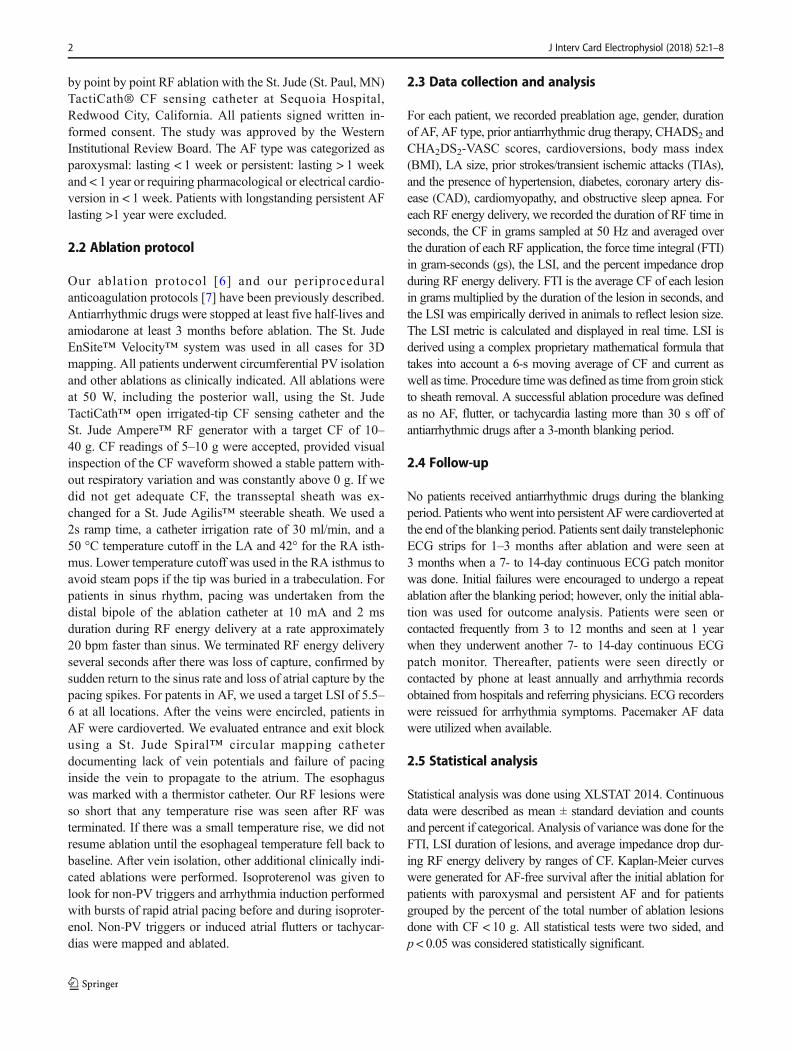
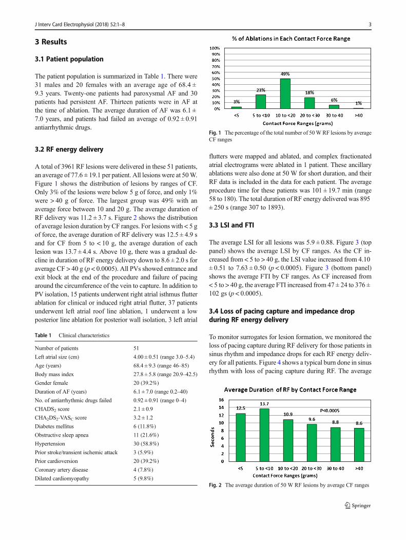
A total of 3961 RF lesions were delivered in these 51 patients,an average of 77.6 ± 19.1 per patient. All lesions were at 50W.Figure 1 shows the distribution of lesions by ranges of CF.Only 3% of the lesions were below 5 g of force, and only 1%were > 40 g of force. The largest group was 49% with anaverage force between 10 and 20 g. The average duration ofRF delivery was 11.2 ± 3.7 s. Figure 2 shows the distributionof average lesion duration byCF ranges. For lesions with < 5 gof force, the average duration of RF delivery was 12.5 ± 4.9 sand for CF from 5 to < 10 g, the average duration of eachlesion was 13.7 ± 4.4 s. Above 10 g, there was a gradual de-cline in duration of RF energy delivery down to 8.6 ± 2.0 s foraverage CF > 40 g (p < 0.0005). All PVs showed entrance andexit block at the end of the procedure and failure of pacingaround the circumference of the vein to capture. In addition toPV isolation, 15 patients underwent right atrial isthmus flutterablation for clinical or induced right atrial flutter, 37 patientsunderwent left atrial roof line ablation, 1 underwent a lowposterior line ablation for posterior wall isolation, 3 left atrial
flutters were mapped and ablated, and complex fractionatedatrial electrograms were ablated in 1 patient. These ancillaryablations were also done at 50 W for short duration, and theirRF data is included in the data for each patient. The averageprocedure time for these patients was 101 ± 19.7 min (range58 to 180). The total duration of RF energy delivered was 895± 250 s (range 307 to 1893).
3.3 LSI and FTI
The average LSI for all lesions was 5.9 ± 0.88. Figure 3 (toppanel) shows the average LSI by CF ranges. As the CF in-creased from < 5 to > 40 g, the LSI value increased from 4.10± 0.51 to 7.63 ± 0.50 (p < 0.0005). Figure 3 (bottom panel)shows the average FTI by CF ranges. As CF increased from< 5 to > 40 g, the average FTI increased from 47 ± 24 to 376 ±102 gs (p < 0.0005).
3.4 Loss of pacing capture and impedance dropduring RF energy delivery
To monitor surrogates for lesion formation, we monitored theloss of pacing capture during RF delivery for those patients insinus rhythm and impedance drops for each RF energy deliv-ery for all patients. Figure 4 shows a typical burn done in sinusrhythm with loss of pacing capture during RF. The average
Table 1 Clinical characteristics
Number of patients 51
Left atrial size (cm) 4.00 ± 0.51 (range 3.0–5.4)
Age (years) 68.4 ± 9.3 (range 46–85)
Body mass index 27.8 ± 5.8 (range 20.9–42.5)
Gender female 20 (39.2%)
Duration of AF (years) 6.1 ± 7.0 (range 0.2–40)
No. of antiarrhythmic drugs failed 0.92 ± 0.91 (range 0–4)
CHADS2 score 2.1 ± 0.9
CHA2DS2-VASC score 3.2 ± 1.2
Diabetes mellitus 6 (11.8%)
Obstructive sleep apnea 11 (21.6%)
Hypertension 30 (58.8%)
Prior stroke/transient ischemic attack 3 (5.9%)
Prior cardioversion 20 (39.2%)
Coronary artery disease 4 (7.8%)
Dilated cardiomyopathy 5 (9.8%)
Fig. 1 The percentage of the total number of 50WRF lesions by averageCF ranges
Fig. 2 The average duration of 50 W RF lesions by average CF ranges
J Interv Card Electrophysiol (2018) 52:1–8 3

impedance drop for all lesions was 13.5 ± 5.5%. As CF in-creased, the average impedance drop per lesion decreasedfrom 14.5 ± 5.2% for the 5 to < 10 g force range to 9.6 +5.2% for CF > 40 g (p < 0.0005) (Fig. 5). We did not havean audible or tactile steam pop in any of the 3961 RF appli-cations. No char was noted on any catheters. For all lesionsdelivered in sinus rhythm, we paced during RF energy deliv-ery. Except for an occasional lesion on the anterior portion of
the right upper PV, there was always loss of capture prior totermination of RF energy delivery. For lesions on the anteriorright upper vein, where there was no loss of capture, westopped RF at an LSI of 6.0. Rechecking these areas afterterminating RF showed no electrograms and no capture afterrepositioning the catheter. We felt that the proximal pole of thecatheter may have been touching tissue and causing anodalstimulation or there may have been adjacent right atrial tissuecapture to explain continued atrial capture when the LSIreached 6.0. Prior to terminating the procedure, we pacedalong all lesion sets to further document failure to capture.All veins were isolated in all patients, and there were no acutereconnections noted.
3.5 Long-term outcomes
All patients considered free of AF underwent 3 and12 month 7- to 14-day continuous monitors. The averagefollow-up was 1.74 ± 0.61 years. Figure 6 shows the
Fig. 3 The average lesion index (LSI) (top panel) and the average forcetime integral (FTI) (bottom panel) by CF ranges
Fig. 4 Loss of pacing captureduring RF. This figure shows asingle lesion created near the topof the left upper pulmonary vein.While pacing the distal bipole ofthe ablation catheter, RF is turnedon at 50 W. After 10 s of RF(when the LSI was 4.9), there isloss of capture. The RF wascontinued for a total RF time of13.4 s. The average CFwas 11.0 g
Fig. 5 The average percent impedance drop per lesion by CF ranges
4 J Interv Card Electrophysiol (2018) 52:1–8

Kaplan-Meier single procedure AF-free rates by AF type(paroxysmal vs. persistent). For paroxysmal AF, the singleprocedure freedom from AF was 86% at 1 and 2 years.For persistent AF, the single procedure AF-free rate was83% at 1 year and 72% at 2 years. The difference in long-term outcome between paroxysmal and persistent AF wasnot significant (p = 0.331).
We also examined the single procedure AF-free rate for theentire cohort by the percent of lesions in each patient withaverage CF < 10 g. We compared patients who had < 20%of their RF lesions at < 10 g to those with 20–30% of thelesions at < 10 g and those with > 30% of the lesions at <10 g. Figure 7 shows the Kaplan-Meier curves for these threegroups. There was no difference in single procedure AF-freerates, suggesting that even the lesions created with < 10 g offorce using 50 W, the majority of which were between 5 and10 g (averaging 8.0 g of force), were making durable lesions.This may have been due to our requiring an optimal CF wave-form for CF between 5 and 10 g.
Nine patients returned for a repeat ablation after theirinitial ablation failed. All had at least one active vein.There was more than one site of vein reconnection of24.2% and a single site of reconnection in 27.3% of theveins. Eight of the nine patients had atrial arrhythmiaseliminated by the redo ablation.
3.6 Complications
There were no instances of stroke or TIA, pericardialtamponade, atrial-esophageal fistulae, PV stenosis, or death.
4 Discussion
The main finding of this study is that using CF sensing cath-eters and 50Wablations averaging only 11.2 s/burn, we safelyisolated the PVs in all patients with much shorter proceduretimes and total RF energy delivery (< 15 min/patient) thanreported in prior studies using lower power and longer dura-tion RF applications [3–5]. We had an excellent single proce-dure 1 and 2-year freedom from AF.
In the present study, we did point by point ablation to ex-amine the characteristics of each lesion regarding RF duration,loss of pacing capture during RF delivery, impedance drop,and measurement of LSI and FTI. The average duration of alllesions was 11.2 ± 3.7 s. The RF generator we utilized takes2 s to get up to full 50 W power when RF energy is applied tothe catheter. Several animal studies support the use of 50 Wablation for 5–10 s. In an in vitro and in vivo sheep model,Bhaskaran et al. [8] compared 50 and 60 W ablations for 5 swith the conventional 40 Wablations for 30 s, all delivered ata CF of 10 g. They delivered energy after the RF generator hadreached full power. Their 5-s ablation would be comparable toa 7-s ablation in our study. They demonstrated that 50 and60 W ablations for 5 s achieved transmural lesions and weresafer than the 40W 30-s ablations. Steam pops occurred in 8%of the lesions using 40W for 30 s and in none of the 5-s 50 and60 Wablations. When they evaluated 80 W for 5 s, there wasan 11% occurrence of steam pops, suggesting an upper limiton safe power for short duration lesions. Another study by
Fig. 6 Kaplan-Meier curves showing the single procedure freedom fromAF by AF type (red = paroxysmal AF and green = persistent AF)
Fig. 7 Kaplan-Meier curves showing the single procedure freedom fromAF by the percentage of 50WRF lesions with<10 g of CF in each patient(green = < 20% of the lesions at < 10 g, blue = 20–30% of the lesions <10 g, and red = > 30% of the lesions < 10 g)
J Interv Card Electrophysiol (2018) 52:1–8 5
Goyal et al. [9] in fresh killed porcine ventricles showed thatfor 20 g of CF, the time needed to create a 4-mm deep lesiondecreased from just over 20 s for 20W to 6–7 s for 50W. Thatstudy suggested that these high power and short durationsmight help to reduce collateral injury.
Several previous clinical studies evaluated 50 W ablationsusing non-CF catheters. Kanj et al. [1] compared 35 vs. 50 Wablations. They randomized 180 patients (85% with paroxys-mal AF) to ablation using either an 8-mm non-irrigated cath-eter or an open irrigated tip catheter (OITC) at either 35 or at50 W. For the OITC, they showed a 6-month freedom fromAF of 82% at 50 W and only 66% at 35 W with shorterfluoroscopy and left atrial times with 50 W. They did notemore steam pops, pericardial effusions, and gastrointestinalcomplaints at 50W, probably because they ablated at each sitefor prolonged periods of time and did not shorten the RFdelivery time for the 50 W lesions. Bunch and Day [10] re-ported on the use of 50 W and a “painting” technique wherethey moved the catheter back and forth across a small areauntil it was devoid of electrograms and reported no esophagealinjuries and an 85% freedom from AF after one or two abla-tions with a mean follow-up of 338 days. In a previous study,we reported a technique like that of Bunch and Day, termed“perpetual motion,” using open irrigated tip catheters at 50 W[2] for short durations at each site. Compared with lower pow-er ablations for longer durations, the short 50 Wablations hadbetter long-term freedom from AF and shorter procedural, leftatrial and fluoroscopy times. There was no increase in com-plications with the use of 50 W for short durations. The pres-ent study extrapolates those observations about short duration50 W ablations to the use of CF sensing catheters.
The appropriate range of CF and appropriate method forreal-time monitoring of CF ablations is still being determined.In the TOCATTA study [4], using 15 to 40 Wof power, therewas a higher 1-year success rate when the CF was maintainedabove 20 g. That study also suggested the FTI should be >500 gs and possibly > 1000 gs for best outcomes. TheEFFICAS I study [3], using a median of 25 W of power forup to 60 s at each site, showed fewer PV reconnections whenthe FTI was > 400 gs. The FTI has been commonly used as areal-time surrogate for durable lesion formation. There are atleast four determinants of lesion formation at each site: CF, RFenergy duration, RF current, and catheter stability. The onlycomponents the FTI considers are the CF and the RF lesionduration. The FTI does not consider either current or powerlevel or catheter stability. Das et al. [11] recently reported onthe use of the “ablation index” which is similar to the FTI butalso includes power in a weighted formula. They found theablation index to be superior to FTI for acutely monitoring PVisolation. In our study, we followed the LSI measurementwhich also utilizes CF, RF duration, and RF current (whichreflects delivered power) in determining a real-time number toguide ablations. We also visually incorporated the CF
waveform as a measure of catheter stability, especially forablations done at < 10 g of force. We monitored both imped-ance drop at all sites and loss of capture for the patients insinus rhythm and always saw loss of capture and a fall inimpedance, indicating good lesion formation by the time theLSI reached a value of 5.5–6.0. This would suggest that whenusing 50 W ablations for patients in AF, where loss of pacingcapture cannot be monitored, one should deliver energy at allsites long enough to achieve an LSI in the 5.5–6.0 range. Forpatients in sinus rhythm, when ablating the anterior portion ofthe right upper PV while pacing, one should stop the ablationwhen the LSI reaches 6.0, even if there is still atrial capture, asthe capture may anodal form the proximal pacing electrode ofthe ablation catheter. Since force quality is not incorporatedinto the LSI measurement, for CFs between 5 and 10 g, oneshould carefully monitor CF quality by continuous visual in-spection of the CF waveform. If this waveform frequentlydrops to 0 g or is labile, one should abort that lesion and startanew with higher or more stable CF. When using 50 W abla-tions, one cannot use the prior target FTI numbers reported inthe literature, as these were all derived using RF energy wellbelow 50W. In our study using short duration 50Wablations,we did not achieve an average FTI of 400 gs, even for lesionswith a CF > 40 g. There was an eightfold range with widestandard deviations (from 47 ± 24 to 376 ± 102 gs) for averageFTIs as CF went from < 5 to > 40 g and less than a twofoldrange with smaller standard deviations (from 4.10 ± 0.51 to7.63 ± 0.50) for average LSI over the same CF ranges. Thissuggests that the LSI is a better number to monitor than theFTI, as it takes RF current into consideration and is less var-iable from lesion to lesion over a range of CFs.
Our study suggests that, when using CF sensing cathe-ters at 50 W, if one wants to use drag lesions, perpetualmotion or painting techniques to keep the RF generator onwhile moving the ablation catheter to a new site, oneshould remain at each spot for approximately 8–9 s forhigher CF sites and for a few seconds longer at lower CFsites, before moving on to the next spot.
Studies have indicated that a fall in impedance during RFenergy delivery [12] or loss of pace capture [13] indicateslesion formation. Studies evaluating CF suggest that there isa greater fall in impedance with more CF [14]. For unclearreasons, we found a smaller decrease in impedance as CFincreased. This may be due to the fact that we used endpointsof lesion formation (loss of pace capture and LSI) to guide thetermination of RF rather than measuring impedance drop at afixed time after the onset of RF delivery and that our ablationtimes were very short.
In the TOCCASTAR study [5] for paroxysmal AF ablation,there was no difference in the 1-year freedom from AF whenthe CF sensing catheter was compared to the non-CF sensingcatheter. However, when the data were analyzed comparingpatients who received ≥ 90% of the ablation lesions with ≥
6 J Interv Card Electrophysiol (2018) 52:1–8
10 g of force to those who had < 90% of their lesions at ≥ 10 g,the group with a higher percentage of high CF lesionsachieved a 75.9% 1-year freedom from AF compared to58.1% for the group where CFwas less optimal. In our presentstudy, having a higher percentage of ablation sites at less than10 g of force did not significantly impact the outcome. Inaddition, for paroxysmal AF, our 1-year 86% freedom fromAFwas considerably higher than the overall 67.8% seen in theTOCCASTAR trial for paroxysmal AF. This could be due toour requirement that for CF between 5 and 10 g, the CF wave-formmust be ideal and consistent with good contact or it couldbe that 50 W ablations overcome the limitations of slightlylower CF.
Radiofrequency energy delivery to tissue is a complex in-teraction and is well summarized in a recent review [12].There is a resistive component adjacent to the catheter elec-trode which results in local heating and dissipation of radio-frequency energy as heat. This resistive heating depends uponcurrent delivered to the tissue and the resistance seen by theRF generator. Resistive heating probably occurs relatively ear-ly in the RF application. Greater resistive heating can beachieved by the use of higher RF power or lower resistance,achieved by the use of larger tip catheters or extra skin elec-trodes. There is also a secondary passive heating of deepertissue which increases with longer duration RF applications.Tissue needs to be heated to 50 °C or higher for several sec-onds to achieve irreversible coagulation necrosis which resultsin an electrically silent scar. The use of catheter tip cooling andgreater energy both result in larger and deeper scar formation.Open irrigated tip catheters, such as the one used in the presentstudy, results in tip temperature increases of only a few de-grees Celsius which are associated with 20–30° of tissue tem-perature increases [15]. Thus, with the open-irrigated tip cath-eters, one can heat and ablate adjacent tissue at 50Wof powerwithout having catheter tip coagulation in the blood pool. Theshorter, higher energy RF applications we used in the presentstudy may also result in fewer complications by achievingrapid local resistive tissue ablation and avoiding deeper col-lateral passive heating seen with longer and lower power RFapplications. Although the present study is too small to exam-ine the rate of infrequent complications, over the past 12 years,we have performed more than 4500 AF ablations using 50 Wfor short durations, including the posterior wall, with only asingle non-fatal atrioesophageal fistula, a single patient withPV stenosis requiring intervention, a pericardial tamponaderate of 0.34%, and a 48-h stroke rate of 0.145%.
5 Limitations
This was a single center study. There were a relatively smallnumber of patients evaluated. However, there were almost4000 ablation applications evaluated and almost five million
individual data points examined with 50Hz sampling. Despitethe small number of patients, the excellent long-term singleprocedure outcomes support the animal data referenced previ-ously and provides “proof of concept” for using this techniquein future trials of AF ablation. Although we saw no complica-tions, the number of patients is too small to evaluate for infre-quent serious complications that can occur with AF ablation.
6 Conclusions
Monitoring the quality of the CF waveform, documenting lossof capture during pacing during RF delivery for patients insinus rhythm, and following a parameter that takes currentor power into account provide a safe and rational method formonitoring short duration RF ablations at 50 W using CFmeasuring catheters. The use of short duration 50 W RF le-sions permits shorter procedures with minimal total RF energydelivery and excellent outcomes.
Acknowledgements Patricia Barberini, R.N., Cynthia Lebsack, Pharm.D, and Glenda Rhodes assisted with data and manuscript management.
Compliance with ethical standards
The study was approved by the Western Institutional Review Board.
Conflict of interest Dr. Winkle: none; Mr. Moskovitz, Mr. Tranter, andMr. Shai: employed by St. Jude Medical, Inc.; Dr. Mead: consultantMedtronic; Dr. Engel: consultant Medtronic; Dr. Kong: advisory boardMedtronic, consultant Peerbridge, EBR Systems, Medtronic; Mr.Fleming: none; Dr. Patrawala: consultant for St. Jude Medical, Inc.; Dr.Salcedo: none.
Open Access This article is distributed under the terms of the CreativeCommons At t r ibut ion 4 .0 In te rna t ional License (h t tp : / /creativecommons.org/licenses/by/4.0/), which permits unrestricted use,distribution, and reproduction in any medium, provided you give appro-priate credit to the original author(s) and the source, provide a link to theCreative Commons license, and indicate if changes were made.
References
1. Kanj MH, Wazni O, Fahmy T, Thal S, Patel D, Elay C, et al.Pulmonary vein antral isolation using an open irrigation ablationcatheter for the treatment of atrial fibrillation. J Am Coll Cardiol.2007;49(15):1634–41. https://doi.org/10.1016/j.jacc.2006.12.041.
2. Winkle RA, Mead RH, Engel G, Patrawala RA. Atrial fibrilla-tion ablation: perpetual motion of open irrigated tip catheters at50 watts is safe and improves outcomes. Pacing ClinElecrophysiol. 2011;34(5):531–9. https://doi.org/10.1111/j.1540-8159.2010.02990.x.
3. Nuezil P, Reddy VY, Kautzner J, Petru J,Wichterle D, Shah D, et al.Electrical reconnection after pulmonary vein isolation is contingenton contact force during initial treatment results from the EFFICAS I
J Interv Card Electrophysiol (2018) 52:1–8 7
study. Cire Arrhythm Electophysiol. 2013;6(2):327–33. https://doi.org/10.1161/CIRCEP.113.000374.
4. Reddy VY, Shah D, Kautzner J, Schmidt B, Saoudi N, Herrera C,et al. The relationship between contact force and clinical outcomeduring radiofrequency catheter ablation of atrial fibrillation in theTOCCATA study. Heart Rhythm. 2012;9(11):1789–95. https://doi.org/10.1016/j.hrthm.2012.07.016.
5. Reddy VY, Dukkipati SR, Neuzil P, Natale A, Albenque JP,Kautzner J, et al. Randomized, controlled trial of the safety andeffectiveness of a contact force-sensing irrigated catheter for abla-tion of paroxysmal atrial fibrillation: results of the TactiCathContact Force Ablation Catheter Study for Atrial Fibrillation(TOCCASTAR) study. Circulation. 2015;132:907–15.
6. Winkle RA, Mead RH, Engel G, Patrawala RA. Long term resultsof atrial fibrillation ablation: the importance of all initial ablationfailures undergoing a repeat ablation. Am Heart J. 2011;162(1):193–200. https://doi.org/10.1016/j.ahj.2011.04.013.
7. Winkle RA, Mead RH, Engel G, Kong MH, Patrawala PA. Peri-procedural interrupted oral anticoagulation for atrial fibrillation ab-lation: comparison of aspirin, warfarin, dabigatran, andrivaroxaban. Europace. 2014;16(10):1443–9. https://doi.org/10.1093/europace/euu196.
8. Bhaskaran A, Chik W, Pouliopoulos J, Nalliah C, Quin P, Barry T,et al. Five seconds of 50–60W radiofrequency atrial ablations weretransmural and safe: an in vitro mechanistic assessment and force-controlled in vivo validation. Europace. 2016;19:874–80.
9. Goyal V, Ali-Ahmed F, Patel M, Haines ED, WongWS. Low flow,high power, short ablation duration-is this the key to avoid collateraldamage? Heart Rhythm. 2017;14(May Supplement):S464.
10. Bunch TJ, Day JD. Novel ablative approach for atrial fibrillation todecrease risk of esophageal injury. Heart Rhythm. 2008;5(4):624–7. https://doi.org/10.1016/j.hrthm.2007.11.007.
11. Das M, Loveday JJ, Wynn GJ, Gome S, Saeed Y, Bonnett LJ, et al.Ablation index, a novel marker of ablation lesion quality: predictionof pulmonary vein reconnection at repeat electrophysiological studyand regional differences in targeted areas. Europace. 2017;17:775–83.
12. Kumar S, Barbhaiya CR, Balindger S, John RM, Epstein LM,Koplan BA, et al. Better lesion creation and assessment duringcatheter ablation. J Atr Fibrillation. 2015;8:1189.
13. Kosmidou I, Houde-Walter H, Foley L, Michaud G. Loss of pacecapture after radiofrequency application predicts the formation ofuniform transmural lesions. Europace. 2013;15:6010606.
14. Reichlin T, Knecht S, Lane C, Kuhne M, Nof E, Chopra N, et al.Initial impedance decrease as an indicator of good catheter contact:insights from radiofrequency ablation with force sensing catheters.Heart Rhythm. 2014;11:194–201.
15. Bruce GK, Bunch TJ, Milton MA, Sarabanda A, Johnson SB,Packer DL. Discrepancies between catheter tip and tissue tempera-ture in cooled-tip ablation. Relevance to guiding left atrial ablation.Circulation. 2005;112(7):954–60. https://doi.org/10.1161/CIRCULATIONAHA.104.492439.
8 J Interv Card Electrophysiol (2018) 52:1–8