ATLAS OF CAPSULE Juan Manuel Herrerías Miguel Mascarenhas ... · Saraiva. It has been my good...
38
1 AUTHORS: ATLAS OF CAPSULE ENDOSCOPY Published by Juan Manuel Herrerías Miguel Mascarenhas-Saraiva
Transcript of ATLAS OF CAPSULE Juan Manuel Herrerías Miguel Mascarenhas ... · Saraiva. It has been my good...
1
AUTHORS:
ATLAS OF CAPSULE ENDOSCOPY
ATLASOF CAPSULE ENDOSCOPY
Published by
Juan Manuel HerreríasMiguel Mascarenhas-Saraiva
ATLASOF CAPSULE ENDOSCOPY
Published by
Juan Manuel HerreríasMiguel Mascarenhas-Saraiva
FOREWORD
It is a great privilege and a real pleasure for me to have been invited to write the foreword for the 2nd edition of Atlas of Capsule endoscopy published by my prestigious friends Juan Manuel Herrerías and Miguel Mascarenhas-Saraiva. It has been my good fortune to have known these two distinguished physicians for many years and to have seen them grow as respected clinicians and educators in the fi eld of gastroenterology.
Until a few years ago, the small bowel was an organ which was very diffi cult to explore with the available endoscopic, radiological and nuclear medicine techniques. In routine practice only the last few centimeters of the ileum were accessible to retrograde visualization by ileocolonoscopy. Explorations from the proximal side by push, sonde or intraoperative enteroscopy were invasive procedures that do not always allow us to visualize the lesions in the small bowel. Sonde enteroscopy had been abandoned in the 90’s because it was a tedious exploration (long duration of the procedure) and it had several technical limitations. Push enteroscopy is limited by the depth of insertion of the scope and it is poorly tolerated. Intraoperative enteroscopy is the most effective of these techniques, but is the most invasive with an important percentage of adverse side effects.
I witnessed Dr. Paul Swain fi rst presenting the use of the wireless capsule endoscope in May 2000 at Digestive Disease Week in San Diego, during the Plenary Session of the American Society for Gastrointestinal Endoscopy, there was a tremendously enthusiastic response. Comparisons were made to the miniaturized spaceship used to examine the body’s inner spaces in the science fi ction movie “Fantastic Voyage”.
Capsule endoscopy was launched at the beginning of this millennium and since then has had a very important impact on managing obscure gastrointestinal bleeding and many other small bowel diseases. The initial capsule endoscope was developed by Given Imaging (Yoqneam, Israel) and approved in Europe by the European Agency and in the United States by the Food and Drug Administration in 2001.
With Wireless capsule endoscopy (CE) we can provide a simple, safe, non invasive, reliable, procedure, well accepted and tolerated by the patient, which has revoluzioned the study of the small bowel. This technique evaluates endoscopically, with high resolution images, what has been called “the last frontier” of endoscopy, the small bowel, avoiding any sedation, surgery or radiation exposure.
Currently CE is recommended as a third stage examination, after negative gastroscopy and colonoscopy in patients with obscure gastrointestinal bleeding. Also many studies have established, with a growing body of evidence, that this technique is cost-effective in other clinical situations, such as detection of small bowel lesions in Crohn’s disease in patients in which other methods fail to prove the diagnosis, non steroidal anti-infl ammatory drug enteropaties, celiac disease, small bowel polyposis syndromes and small bowel tumors. Other possible indications are HIV patients with gastrointestinal symptoms, malabsortive syndromes other than celiac disease, Henoch-Schonlein purpura, patients with small bowel transplants and with intestinal graft versus host disease, particularly in monitoring the response to immunosuppressive therapy.
The acquired knowledge of the wide range of lesions that can be found in the small bowel, encouraged the implementation of some diagnostic and therapeutic techniques, such as double balloon enteroscopy, MRI-enteroclysis and CT-enteroclysis.
The main contraindication of performing the CE is the suspicion or knowledge of an obstruction in the GI tract.
The device retention is the main complication of the procedure and is defi ned when CE remains in the digestive tract for a minimum of 2 weeks. The frequency of this problem varies, depending mostly on the clinical indication for CE, and ranges from 0% in healthy subjects, to 1.5 % in patients with obscure GI bleeding, to 5% in patients with suspected Crohn’s disease and to 21% in patients with intestinal obstruction.
At present CE has some technical limitations, it can not be used to obtain biopsy specimens or for endoscopic treatment and it can not be controlled remotely. CE has also some clinical limitations which are the problem in sizing and locating small bowel lesions, a possible false-negative CE result, due to the fact that the global miss rate is about 11%, ranging from 0.5% for ulcerative lesions to 18.9% for neoplastic disease and the fact that some times we can get fi ndings of uncertain relevance in healthy subjects. Other drawback is that in almost 20% of procedures the capsule does not reach the cecum while it is active.
This technique is available in over 5000 gastrointestinal centers throughout the world.
Since its arrival, more than 650,000 capsules have been swallowed worldwide and more than 1000 peer-reviewed publications have appeared in medical literature. The most important GI societies have published guidelines about its use.
In latter years, breakthrough developments in CE technology have enabled the direct visualization of the upper and lower segments of the gut using specifi cally designed capsules.
This updated second edition of the atlas is much improved compared to the fi rst edition; many new chapters, authors and technological advances have been added.
The editors have chosen the authors of each chapter very well, from eight different countries, with a mixture of established leaders and rising younger colleagues who represent the next generation staking its claim to this rapidly evolving fi eld of the gastrointestinal endoscopy.
The images are well chosen most of them of high quality and superbly produced, raising the exceptionally high quality of the fi rst edition.
The atlas is divided into six parts. The fi rst part consists of 10 chapters and covers general aspects of the technique. The second part deals with its usefulness for the study of the esophagus, the third shows the possibilities that the capsule gastroscopy presently offers as well as the fi ndings that we can see in the stomach when we are performing an exploration with the capsule. The fourth consists of 16 chapters and deals with the multiple applications that this technique has in the small intestine, including motility studies, and with the alternative techniques for enteroscopy. The fi fth part deals with capsule colonoscopy and the possibility of performing pan-endoscopy with the colon capsule, the sixth part discusses the utility of capsule endoscopy in pediatric patients, in patients with abdominal pain and fi nally the future developments of capsule endoscopy
I believe that this atlas has much to offer to individuals at all levels of involvement in the fi eld of gastroenterology, from students to even the most seasoned clinicians. And fi nally, I want to congratulate not only the publishers but also the authors for their excellent contributions to this atlas.
Miguel Muñoz-Navas MD, PhDMiguel Muñoz-Navas MD, PhDDirector Gastroenterology DepartmentDirector Gastroenterology DepartmentUniversity of Navarra ClinicUniversity of Navarra ClinicSchool of Medicine. University of Navarra. Pamplona. Spain.School of Medicine. University of Navarra. Pamplona. Spain.
1. Technology of capsule endoscopy
1.1 The development of capsule endoscopy Eitan Scapa
1.2 Description of the different capsule endoscopes Rolando Pinho, Miguel Mascarenhas-Saraiva
1.3 Patient’s preparation for capsule endoscopy Josefa Mª García-Montes, Federico Argüelles-Arias, Belén Maldonado-Pérez, Francisco Pellicer-Bautista, Juan Manuel Herrerías
1.4 The procedure of capsule endoscopy Miguel Mascarenhas-Saraiva
1.5 Role of the assistant in capsule endoscopy Eduardo Oliveira, Miguel Mascarenhas-Saraiva
1.6 Reading capsule endoscopy Rolando Pinho, Miguel Mascarenhas-Saraiva
1.7 Standard terminology for capsule endoscopy Michel Delvaux, Gérard Gay
1.8 Indications for capsule endoscopy Miguel Mascarenhas-Saraiva
1.9 Contra-indications, complications and special situations Emanuele Rondonotti, Roberto de Franchis
1.10 Patency and Agile capsules Ángel Caunedo-Álvarez, Javier Romero-Vázquez, Mileidis San Juan Acosta, Juan Manuel Herrerías
2. Capsule endoscopy of the esophagus
2.1 Fields of application Alba Belda-Cuesta, María Luisa Morales-Barroso, Federico Argüelles-Arias, Ángel Caunedo-Álvarez, Juan Manuel Herrerías
2.2 Esophageal capsule endoscopy: normal and pathologic esophagus Andrés Sánchez-Yagüe, Andrés M. Sánchez-Cantos, Francisco Pellicer-Bautista, Juan Manuel Herrerías
2.3 Esophageal fi ndings with non-esophageal capsules Vanesa Méndez-Rufi án, Belén Maldonado-Pérez, Juan Manuel Herrerías
2.4 String capsule endoscopy Francisco C. Ramírez
3. Gastric fi ndings on capsule endoscopy
3.1 Capsule gastroscopy: current possibilities Javier Romero-Vázquez, Vanesa Méndez-Rufi án, Ángel Caunedo-Álvarez, Francisco Pellicer-Bautista, Juan Manuel Herrerías
3.2 Gastric fi ndings during capsule endoscopy procedures Javier Romero-Vázquez, Mileidis San Juan-Acosta, Ángel Caunedo-Álvarez, Juan Manuel Herrerías
TABLE OF CONTENTS
4. Capsule enteroscopy
4.1 Fields of applications Quetzalihuitl Arroyo-Martínez, Manuel Rodríguez-Téllez, Belén Maldonado-Pérez, Juan Manuel Herrerías
4.2 Obscure gastrointestinal bleeding Mª Teresa Galiano-de Sánchez
4.3 Infl ammatory bowel disease of the small bowel Marta Salgado, Miguel Mascarenhas-Saraiva
4.4 Malabsorption syndromes and capsule endoscopy Javier Romero-Vázquez, Patricia Cordero-Ruiz, Ángel Caunedo-Álvarez, Juan Manuel Herrerías
4.5 Neoplastic lesions of the small bowel Miguel Mascarenhas-Saraiva
4.6 Intestinal polyposis José Soares
4.7 Iatrogenic lesions of the small bowel Begoña González-Suárez, Josep Llach-Vila
4.8 Vascular alterations of small bowel Miguel Mascarenhas-Saraiva
4.9 Portal enteropathy Pedro Narra Figueiredo
4.10 Infectious diseases of the small bowel Martin Keuchel, Nageshwar Reddy, Fritz Hagenmüller
4.11 Graft-vs-Host disease Samuel Adler
4.12 Other fi ndings in small bowel Miguel Mascarenhas-Saraiva
4.13 Motility studies using capsule endoscopy Carolina Malagelada, Fernando Azpiroz-Vidaur
4.14 Enteroscopy with non-SB capsule Manuel de Sola-Romero, Pedro Hergueta-Delgado, Juan Manuel Herrerías
4.15.1 Complementary procedures to capsule enteroscopy: new ways of enteroscopy Enrique Pérez-Cuadrado Martínez
4.15.2 Non-endoscopic imaging studies: Cross sectional imaging of the small bowel Miguel Ramalho, Luís Guimarães, Vasco Herédia, Fernando Ramalho, Richard C. Semelka
5. Capsule colonoscopy
5.1 Fields of applications Iñaki Fernández-Urién, Erika Borobio, Ana Borda, Inmaculada Elizalde, Francisco Javier Jiménez-Pérez
5.2 Colon capsule endoscopy: normal and pathologic colon Cristina Carretero, Carlos Prieto, Miguel Muñoz-Navas
5.3 Colonic fi ndings with non-colon capsules Mileidis E. San Juan-Acosta, Francisco Pellicer-Bautista, Juan Manuel Herrerías
5.4 The concept of pan-endoscopy and its possibilities Alba Belda-Cuesta, Javier Romero-Vázquez, Juan Manuel Herrerías

Muñoz-Navas, Miguel - MDGastroenterology Service. University of Navarra. Pamplona. Spain.Narra Figueiredo, Pedro - MD, PhDMedical School, University of Coimbra. University Hospitals of Coimbra. Coimbra. Portugal.
Oliveira, Eduardo - MDEndoscopy nurse. ManopH, Laboratório de Endoscoppia e Motilidade Digestiva. Instituto CUF, Porto, Portugal.
Pellicer-Bautista, Francisco - MDGastroenterology Service. Hospital Universitario Virgen Macarena. Sevilla. Spain.
Pérez-Cuadrado Martínez, Enrique - MDHead of Gastroenterology. Hospital General Universitario Morales Meseguer. Profesor Asociado de la UMU Universidad de Murcia, Murcia.
Pinho, Rolando - MDGastroenterology Department. Centro Hospitalar de Vila Nova de Gaia. ManopH, Instituto CUF, Porto, Portugal.
Prieto, Carlos - MDGastroenterology Service. University of Navarra. Pamplona. Spain.
Ramalho, Fernando - MD, PhDDepartment of Gastroenterology and Hepatology, School of Medicine of Lisbon, Lisbon. Portugal.
Ramalho, Miguel - MDDepartment of Radiology, Hospital Garcia de Orta, Almada. Portugal.
Ramírez, Francisco C. - MD, FACG, AGAFChief of Gastroenterology Service. Carl T Hayden VA Medical Center, Phoenix, Arizona. Professor of Clinical Medicine. University of Arizona College of Medicine, Phoenix, Arizona, USA.
Reddy, Nageshwar - MD, PhDAsian Institute of Gastroenterology, Somajiguda, Hyderabad. India.
Rodríguez-Téllez, Manuel - MDGastroenterology Service. Hospital Universitario Virgen Macarena. Sevilla. Spain.
Romero-Vázquez, Javier - MDGastroenterology Service. Hospital Universitario Virgen Macarena. Sevilla. Spain.
Rondonotti, Emanuele - MD, PhDGastroenterology Service. Ospedale Valduce, Como, Italy.
Salgado, Marta - MDServiço de Gastrenterologia. Hospital de Santo António, Centro Hospitalar do Porto. Porto, Portugal.
Sánchez-Cantos, Andrés M. - MDGastroenterology Service. Hospital Costa del Sol, Marbella, Málaga.
Sánchez-Yagüe, Andrés - MD, PhDGastroenterology Service. Hospital Costa del Sol, Marbella, Málaga.
San Juan Acosta, Mileidis - MDGastroenterology Service. Hospital Universitario Virgen Macarena. Sevilla. Spain.
Scapa, Eitan - MD, PhDGastroenterology Department. Assaf Harofef Medical Center. P.O. Beer Yaacov. Zerifi n, Israel.
Semelka, Richard C. - MDDepartment of Radiology, University of North Carolina at Chapel Hill, Chapel Hill, North Carolina. USA.
Soares, José M. - MDHospital Pedro Hispano. Matosinhos, Porto, Portugal.
Valverde-Fernández, Justo - MDPaediatric Service. Hospital Universitario Virgen Macarena. Sevilla. Spain.
ABBREVIATIONS
AGA American Gastroenterological AssociationAIDS Acquired immunodefi ciency syndromeAPC Argon plasma coagulationAPS Active pixel sensorASIC Application-specifi c integrated circuitAVM Arteriovenous malformationBAE Balloon-assisted enteroscopyBE Balloon enteroscopyBE Barret´s EsophagusCCD Charged couple deviceCCE Colon capsule endoscopy CD Crohn´s diseaseCDAI Crohn´s disease activity indexCE Capsule endoscopyCECDAI Capsule endoscopy Crohn´s disease activity indexCEST Capsule endoscopy structured terminologyCMOS Complementary metal oxide semiconductorCMUSE Cryptogenic multifocal ulcerous stenosing enteritisCMV CytomegalovirusCPs PacemakersCRC Colorectal cancerCT Computed tomographyCTE Computed tomography enterographyCVID Common variable immunodefi ciencyDBE Double Balloon Enteroscopy DICOM Digital imaging and communications in medicineDR Data recorderDVD Digital video diskECE Esophageal capsule endoscopyEE Eosinophilic enteropathyEIO Intraoperative enteroscopyER Emergency RoomESGE European Society of Gastrointestinal EndoscopyESPGHAN European Society for Pediatric Gastroenterology and NutritionFAP Familial adenomatous polyposisFICE Fujinon intelligent color-enhacementGERD Gastro esophageal refl ux diseaseGIFD Gluten free dietGIST Gastrointestinal stromal tumoursGIVEN GastroIntestinal Video EndoscopyGVHD Graft-versus-host diseaseHHT Hereditary hemorrhagic telangiectasiaIBDU Infl ammatory bowel disease unclassifi edIBS Irritable bowel syndromeICCE International Conference of Capsule EndoscopyICDs Implanted cardiac defi brillatorsIDF Israeli Defense ForcesIL Intestinal lymphangiectasiaLED Light-emitting diodeMAC Mycobacterium avium complexMAI Mycobacterium avium intracellulareMDCT Multidetector CTMRE Magnetic Resonance EnterographyMRI Magnetic Resonance ImagingMST Minimal standard terminologyNEMO Nano-based capsule-endoscopy with molecular imaging and optical biopsyNNE Non-natural excretionNSAIDs Non-steroidal anti-infl ammatory drugs
ODB Obscure digestive bleedingOGIB Obscure gastrointestinal bleedingPC Patency capsulePCR Polymerase chain reactionPDT Photodynamic therapyPE Push EnteroscopyPEG Polyethylene glycolPJS Peutz-Jeghers syndromeRBC Red blood cellRF RadiofrecuencyRFID Radio Frequency Identifi cationRTA Regional transit abnormalitySB Small bowelSBCE Small bowel video capsule endoscopySBD Small bowel diverticulaSBE Single Balloon EnteroscopySBFT Small bowel follow-throughSBIS Suspected blood identifi cation systemSBVs Small bowel varicesSCC Squamous cell cancerSCE String capsule endoscopySIP Simplifi ed ingestion procedureSLE Systemic lupus erythematosusSP Supine positionSPC-cells Sickle-form particle containing cellsSRL Supine right lateral positionSSETSE Single-shot echo-train spin echoUGIB Upper gastrointestinal bleedingUSB Universal serial busVE Vascular ectasiasVECTOR Versatile Endoscopic Capsule for gastrointestinal TumOr Recognition and therapyWD Whipple´s disease

5
TECHNOLOGY OF CAPSULE ENDOSCOPY - CHAPTER 1.2
Description of the different capsule endoscopes
AUTHORS
Rolando Pinho, MDGastroenterology Department
Centro Hospitalar de Vila Nova de GaiaManopH, Instituto CUF, Porto, Portugal
Miguel Mascarenhas Saraiva, MD, PhD ManopH. Laboratório de Endoscopia e Motilidade Digestiva
Instituto CUF, Porto, PortugalHospital CUF, Porto, Portugal

6
CHAPTER 1.2 - TECHNOLOGY OF CAPSULE ENDOSCOPY
Description of the different capsule endoscopes
AUTHORS Rolando Pinho, Miguel Mascarenhas Saraiva
INTRODUCTION
Gastrointestinal endoscopy is the mainstay of diagnosis and therapy in Gastroenterology. A major shift in the paradigm of gastroenterological practice occurred with the implementation of capsule endoscopy (CE).
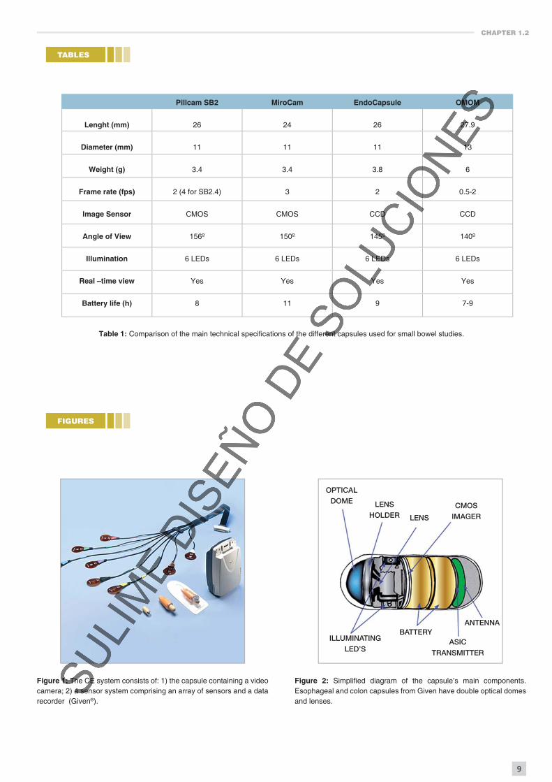
The CE system consists of: 1 – the capsule containing a video camera; 2 – a sensor system comprising an array of sensors and a data recorder (Figure 1) wearable as a belt; 3 – a workstation consisting of a modifi ed commercially available desktop computer.
COMMERCIALLY AVAILABLE CAPSULE ENDOSCOPES
Given Imaging
Given Imaging Ltd (Israel) fi rst delivered wireless capsule endoscopy in 2001. The development of the fi rst CE was dependent on the development of several main components, namely: 1 – an inexpensive, low power, very small image sensor – the CMOS (complementary metal oxide semiconductor); 2 – application-specific integrated circuit (ASIC) chips, which are integrated circuits customized for a particular use (in this case, running a CE), rather than intended for general-purpose use; 3 - miniature white light-emitting diode (LED) light sources (Figure 2)1.
Today, capsule endoscopy devices from Given Imaging include the PillCam SB for the small intestine and the PillCam ESO for esophageal imaging (Figure 3), and Pillcam Colon for the large bowel (Figure 4).
Other CE systems
- Olympus (Japan) has produced the EndoCapsule for the small bowel (Figure 5)2;
- IntroMedic (Korea) developed the MiroCam for small-bowel evaluation using electric-fi eld propagation for data transmission (Figure 6)3;
- Chongqing Jinshan Science and Technology Group (China) created the OMOM small-bowel capsule (Figure 7)4.
TECHNOLOGY BEHIND CAPSULE ENDOSCOPY SYSTEMS
The capsule endoscopes
Each video capsule contains batteries, an ASIC transmitter with antenna and a set of LEDs coupled to a camera, all encapsulated in a biocompatible plastic shell (Figure 8). Images are captured by CCD or CMOS imagers5. These are 2 different technologies for digital acquisition of images.
CMOS technology is most suitable for miniature devices because of its high integration capability and low-power consumption. CCD imagers have usually higher image depth but are bulkier. CMOS use less power than CCDs, making them attractive for miniature devices. Both imagers use pixilated metal oxide semiconductors. They accumulate a signal charge in each pixel, proportional to the local illumination intensity. Each technology has both advantages and disadvantages. CMOS requires less power and provides the capability of adding all of the electronic circuitry into a single microchip1. Using newer ASIC imager chips, and with special power management algorithms, CMOS-based capsules can generate higher frame rates, have a longer duration, and use multiple head capsules. On the other hand, CCD-based capsules produce a higher signal to noise ratio and can give good quality images with a less uniform illumination1. On the downside, they have higher power and space requirements. Ultimately, both technologies have been capable of producing good quality images in the different CE systems already available.
Capsules are provided ready for ingestion in a hermetically sealed case (Figure 8). A magnet in the casing keeps a magnetic switch open that turn the capsule inactive. Once the capsule is removed from the casing, the switch is closed and the capsule becomes active and starts capturing images.
The capsule is then ingested and captures images as it travels along the GI tract. The dome of the capsule is designed to capture images trough the fl uid within the small bowel. Most capsules acquire images at variable fi xed rates: 2 fps for SB2, 4 fps for SB2.4, 2 fps for Endocapsule, 3 fps for MiroCam and 14 fps for Pillcam ESO. Some capsules have variable frame rates: The OMOM capsule can be controlled externally to 0.5 fps, 1 fps or 2 fps and the Pillcam Colon 2 has an automatically adaptive frame rate between 4 and 35 fps1-6.
CHAPTER 1.2
9
Pillcam SB2 MiroCam EndoCapsule OMOM
Lenght (mm) 26 24 26 27.9
Diameter (mm) 11 11 11 13
Weight (g) 3.4 3.4 3.8 6
Frame rate (fps) 2 (4 for SB2.4) 3 2 0.5-2
Image Sensor CMOS CMOS CCD CCD
Angle of View 156º 150º 145º 140º
Illumination 6 LEDs 6 LEDs 6 LEDs 6 LEDs
Real –time view Yes Yes Yes Yes
Battery life (h) 8 11 9 7-9
Table 1: Comparison of the main technical specifi cations of the different capsules used for small bowel studies.
TABLES
FIGURES
Figure 1: The CE system consists of: 1) the capsule containing a video camera; 2) a sensor system comprising an array of sensors and a data recorder (Given®).
Figure 2: Simplifi ed diagram of the capsule’s main components. Esophageal and colon capsules from Given have double optical domes and lenses.
OPTICALDOME LENS
HOLDER LENS
CMOSIMAGER
ILLUMINATINGLED’S
BATTERYASIC
TRANSMITTER
ANTENNA
CHAPTER 1.2
11
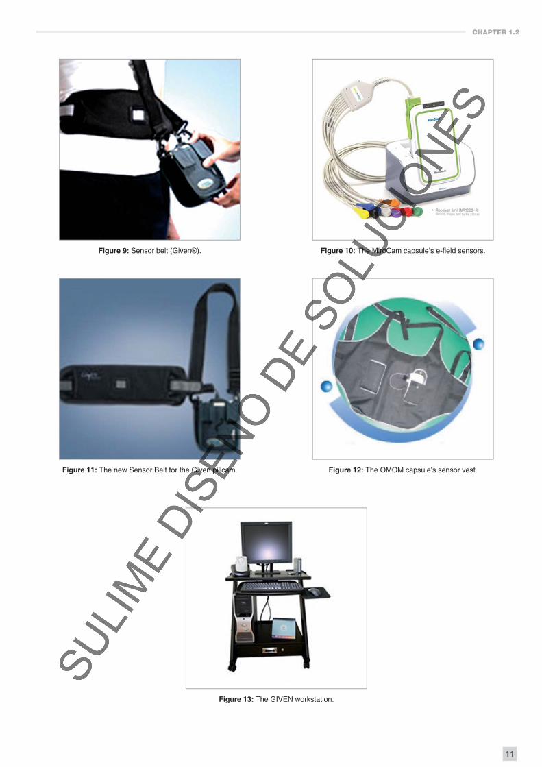
Figure 9: Sensor belt (Given®). Figure 10: The MiroCam capsule’s e-fi eld sensors.
Figure 11: The new Sensor Belt for the Given pillcam. Figure 12: The OMOM capsule’s sensor vest.
Figure 13: The GIVEN workstation.
13
TECHNOLOGY OF CAPSULE ENDOSCOPY - CHAPTER 1.3
Patient’s preparation for capsule endoscopy
AUTHORS
Josefa Mª García-Montes, MD
Federico Argüelles-Arias, MD
Belén Maldonado-Pérez, MD
Francisco Pellicer-Bautista, MD
Juan Manuel Herrerías, MD, PhD, AGAFGastroenterology Service
Hospital Universitario Virgen Macarena. Sevilla. [email protected]
14
CHAPTER 1.3 - TECHNOLOGY OF CAPSULE ENDOSCOPY
Patient’s preparation for capsule endoscopy
AUTHORS Josefa Mª García-Montes, Federico Argüelles-Arias, Belén Maldonado-Pérez, Francisco Pellicer-Bautista, Juan Manuel Herrerías
INTRODUCTION
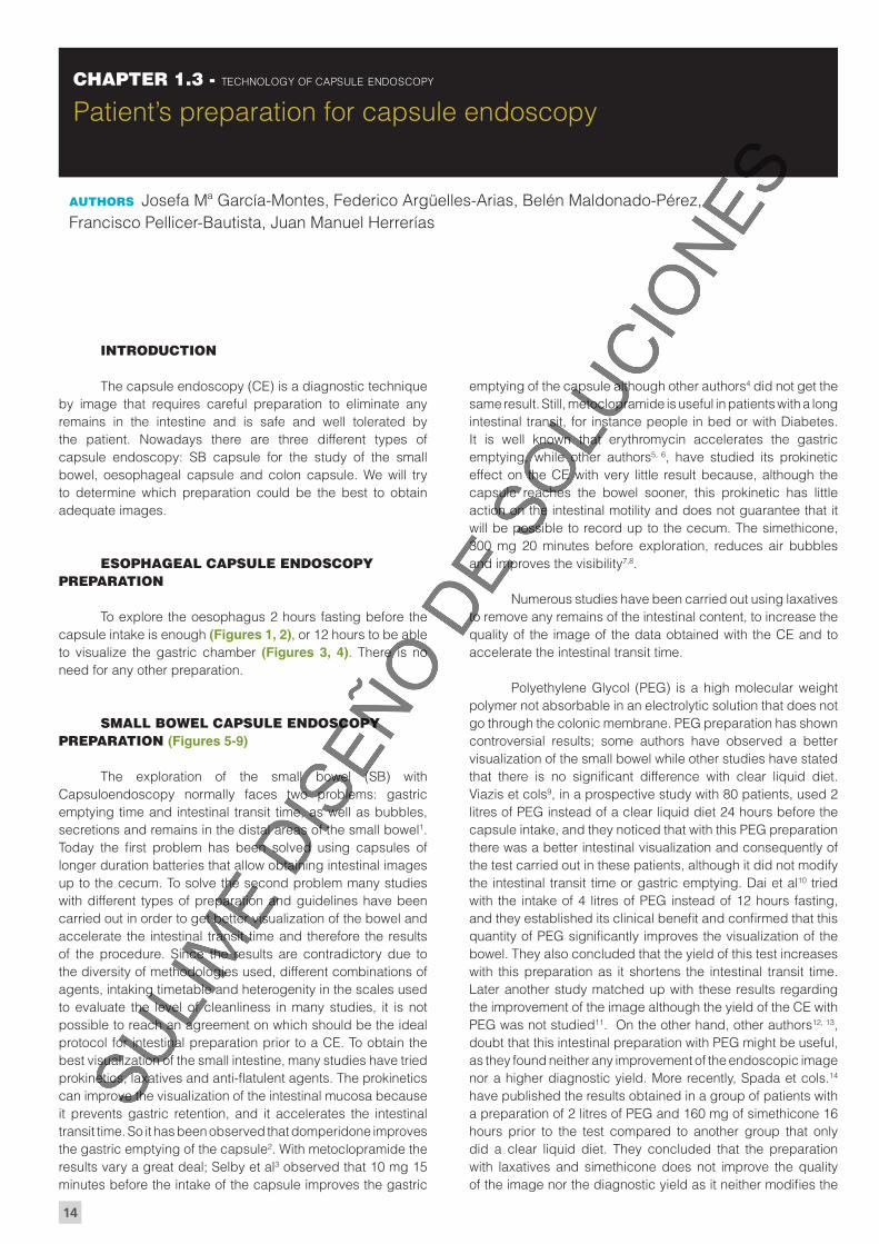
The capsule endoscopy (CE) is a diagnostic technique by image that requires careful preparation to eliminate any remains in the intestine and is safe and well tolerated by the patient. Nowadays there are three different types of capsule endoscopy: SB capsule for the study of the small bowel, oesophageal capsule and colon capsule. We will try to determine which preparation could be the best to obtain adequate images.
ESOPHAGEAL CAPSULE ENDOSCOPY PREPARATION
To explore the oesophagus 2 hours fasting before the capsule intake is enough (Figures 1, 2), or 12 hours to be able to visualize the gastric chamber (Figures 3, 4). There is no need for any other preparation.
SMALL BOWEL CAPSULE ENDOSCOPY PREPARATION (Figures 5-9)
The exploration of the small bowel (SB) with Capsuloendoscopy normally faces two problems: gastric emptying time and intestinal transit time, as well as bubbles, secretions and remains in the distal areas of the small bowel1. Today the fi rst problem has been solved using capsules of longer duration batteries that allow obtaining intestinal images up to the cecum. To solve the second problem many studies with different types of preparation and guidelines have been carried out in order to get better visualization of the bowel and accelerate the intestinal transit time and therefore the results of the procedure. Since the results are contradictory due to the diversity of methodologies used, different combinations of agents, intaking timetable and heterogenity in the scales used to evaluate the level of cleanliness in many studies, it is not possible to reach an agreement on which should be the ideal protocol for intestinal preparation prior to a CE. To obtain the best visualization of the small intestine, many studies have tried prokinetics, laxatives and anti-fl atulent agents. The prokinetics can improve the visualization of the intestinal mucosa because it prevents gastric retention, and it accelerates the intestinal transit time. So it has been observed that domperidone improves the gastric emptying of the capsule2. With metoclopramide the results vary a great deal; Selby et al3 observed that 10 mg 15 minutes before the intake of the capsule improves the gastric
emptying of the capsule although other authors4 did not get the same result. Still, metoclopramide is useful in patients with a long intestinal transit, for instance people in bed or with Diabetes. It is well known that erythromycin accelerates the gastric emptying, while other authors5, 6, have studied its prokinetic effect on the CE with very little result because, although the capsule reaches the bowel sooner, this prokinetic has little action on the intestinal motility and does not guarantee that it will be possible to record up to the cecum. The simethicone, 300 mg 20 minutes before exploration, reduces air bubbles and improves the visibility7,8.
Numerous studies have been carried out using laxatives to remove any remains of the intestinal content, to increase the quality of the image of the data obtained with the CE and to accelerate the intestinal transit time.
Polyethylene Glycol (PEG) is a high molecular weight polymer not absorbable in an electrolytic solution that does not go through the colonic membrane. PEG preparation has shown controversial results; some authors have observed a better visualization of the small bowel while other studies have stated that there is no signifi cant difference with clear liquid diet. Viazis et cols9, in a prospective study with 80 patients, used 2 litres of PEG instead of a clear liquid diet 24 hours before the capsule intake, and they noticed that with this PEG preparation there was a better intestinal visualization and consequently of the test carried out in these patients, although it did not modify the intestinal transit time or gastric emptying. Dai et al10 tried with the intake of 4 litres of PEG instead of 12 hours fasting, and they established its clinical benefi t and confi rmed that this quantity of PEG signifi cantly improves the visualization of the bowel. They also concluded that the yield of this test increases with this preparation as it shortens the intestinal transit time. Later another study matched up with these results regarding the improvement of the image although the yield of the CE with PEG was not studied11. On the other hand, other authors12, 13, doubt that this intestinal preparation with PEG might be useful, as they found neither any improvement of the endoscopic image nor a higher diagnostic yield. More recently, Spada et cols.14 have published the results obtained in a group of patients with a preparation of 2 litres of PEG and 160 mg of simethicone 16 hours prior to the test compared to another group that only did a clear liquid diet. They concluded that the preparation with laxatives and simethicone does not improve the quality of the image nor the diagnostic yield as it neither modifi es the
Technology of capsule endoscopy > Patient’s preparation for capsule endoscopy
Josefa Mª García-Montes, Federico Argüelles-Arias, Belén Maldonado-Pérez, Francisco Pellicer-Bautista, Juan Manuel Herrerías
16
new cleanliness grading scale showed good inter-observer agreement and may be used with the PillCam COLON capsule to assess preparation quality. It also includes the bubbles effect in the preparation: signifi cant (more than 10% of surface area is obscured by bubbles) or insignifi cant (less than 10% of surface area is obscured by bubbles).
STANDARD PREPARATION
The classic preparation is considered nowadays the best one to develop the CCE. This procedure regime is described in table 1. This conventional preparation was fi rst evaluated in two pilot studies. In the Eliakim et al24 study the overall cleanliness of the colon was rated as excellent or good in 84,4% of the cases. In the second pilot study25 the results are better; an excellent or good preparation was achieved in 90% of the cases. In the largest study, the Van Gossum et al study26, the preparation was good or excellent in 72% of the patients. In a recent study published by our group with the same preparation, the grade of cleanliness was good or excellent in 65,6%27.
Also the propulsion of the CCE was evaluated comparing two regimes: a single oral booster dose of sodium phosphate in the fi rst group and in the second one adding a second booster 4 hours after the fi rst dose. In the second group the excretion rate at 10 hours post-ingestion was 78% versus 70% in the fi rst group. Based on these results it has been established that a second booster must be added to the preparation. In a recent study28 the exclusion of NaP booster from CCE preparation resulted in a clinically meaningful reduction of the capsule excretion rate that was only partially compensated by the PEG booster. This second booster should be administered 4 hours after the fi rst booster (and this must be administered when the CCE is out of the stomach). It is administered to reduce capsule delay in the proximal colon and enhance capsule propulsion through the entire colon.
It appears, as it has been mentioned, that colon cleanliness signifi cantly infl uences the sensitivity of capsule endoscopy. In the largest study the sensitivity was signifi cantly higher in patients with good or excellent cleanliness compared to those with poor or fair cleanliness. The sensitivity and specifi city for the detection of polyps (≥6 mm) in patients with good or excellent cleanliness was 75% and 84%, respectively, and for the detection of such polyps in patients with poor or fair cleanliness, the sensitivity and specifi city were 42% and 84%, respectively.
In another paper29 a new regime of preparation consisting of a split regime of PEG administration and a 30 ml dose of sodium phosphate (NaP) was studied. Four senna tablets and a low-residual stools diet were also included. CCE excretion rate, colon cleansing, and accuracy were assessed. At CCE, bowel preparation was rated as good in 78% of patients, fair in 20% and poor in 2%. CCE excretion rate occurred in 83% of patients. They conclude that the combination of a split-dose of PEG solution with a low dose of NaP boosters resulted in high rates of adequate cleansing level and CCE excretion. In a study recently published30 the fi ndings of a single centre study comparing the performance
are reported. For colon cleansing they used their department’s standard preparation procedure for colonoscopy including low-fi bre diet and PEG and added an oral motility agent, Phospho Soda-boosters and a suppository. Level of cleansing on CCE was good in 15 cases (27%), moderate in 30 (54%) and poor in 11 (20%). 34 patients (61%) were reported to have the same cleansing level in both kind of colonoscopies. Nevertheless, they found a lower excretion rate for CCE (64%, n = 36) than in the two previous pilot studies. This might be caused by an additional lapse of time of almost 4 hours between ingestion of the second 2 litres of PEG and initiation of CCE, as motility studies have shown enhanced colonic propulsion of the capsule through PEG.
The development of the new Colon Capsule type 2 has been an important advance because it offers intelligent functionality, superior imaging and a convenient workfl ow. Smart technology enables it to adjust the frame rate in real time to maximize colon tissue coverage, and the imaging devices on either end of the capsule provide a 180˚ view of the colon. The improved study process simplifi es the procedure and patient management, allowing for more effi cient utilization of staff time and resources. To this new Colon Capsule a new preparation has been reported (Table 2).
PREPARATION WITH 4 L PEG VERSUS 2 L PEG
Polyethylene glycol (PEG) solutions are safe and effective, but require consumption of large volumes of fl uid, generally 4 liters. The 2 L PEG solution plus ascorbic acid (PEG + Asc) is also effective, safety and the volume is reduced. Some authors have studied these points. The Ell et al31 study concluded that the PEG + Asc bowel preparation reduces the volume patients have to drink so it was better accepted by patients, and should, therefore, improve effectiveness in routine practice. In another study PEG + Asc provided effective bowel cleansing, which was equivalent to that of sodium picosulphate + magnesium citrate in terms of grading cleansing as overall success or failure32. Nevertheless, it is important to consider the split dose. In this sense the cleansing results are worse if patients receive the full dose PEG + Asc the evening before the procedure compared to the split dose33.
Based on this data, we have developed a study that demonstrated the effi cacy of 2 L PEG. The main aim was to compare the level of cleansing with two different regimens. The secondary aims were to study the presence of bubbles in the colon and also the rate of completed explorations (including observation of haemorrhoidal plexus).
It was a prospective and blinded study. In the fi rst group (A) patients were prepared with 2 liters PEG plus ascorbic acid and in a second group (B) PEG 4 litters. The grade of cleansing was measured using the Leighton scale23 recently published, and they were classifi ed in “excelent-good” and “fair-poor”. In group A 13 patients were included (5 males and 8 females) with an average age of 52 ± 19 and in group B 11 patients (7 males and 4 females) with an average age of 54.44 ± 10. No statistical differences in age and sex were observed between the two groups.
CHAPTER 1.3
19
TABLES
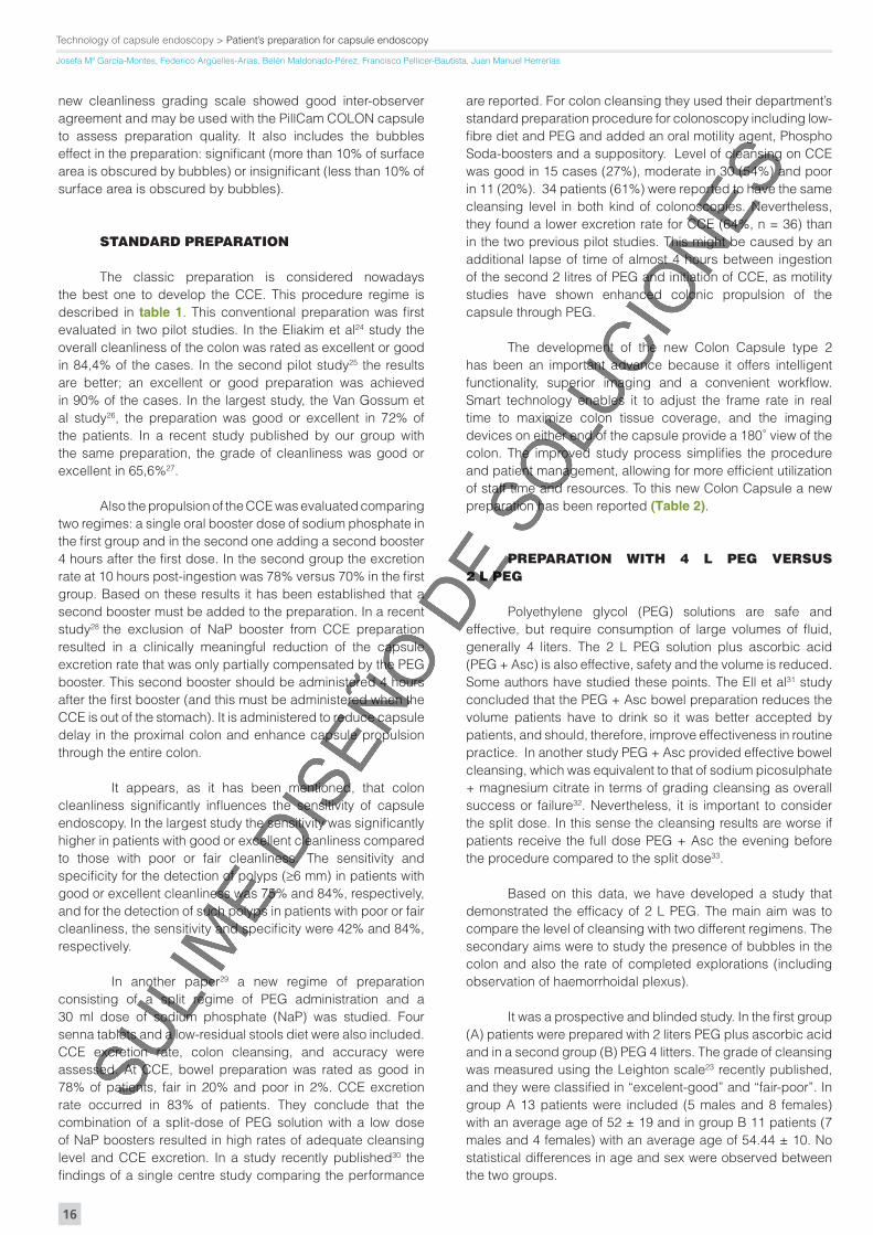
Table 1: Standard preparation.
Day (-1)Clear liquid diet only
18:00-21:00 2 L PEG
Exam Day
6:00-7:00 2 L PEG
7:45 Domperidone (20 mg)
8:00 PillCam Ingestion*
10:00 45 ml NaP + 1 L water
14:00 30 ml NaP + 1 L water
15:00 snack (optional)
16:30 suppository (10 mg Bysacodyl)
Table 2: Colon Capsule 2 preparation.
Schedule Intake
Day -2 Senosides
Day -1 All Day Clear Liquid Diet
Evening 2 L PEG
Exam Day
Morning 2 L PEG
~ 10 am Capsule Ingestion*
1st Boost
small bowel detection 30 ml NaP & 1 L water
2nd Boost
3 hrs after 1st Boost15 ml NaP & 0.5 L water
Suppository
2 hrs after 2nd Boost 10 mg Bisacodyl
* 10 mg Metoclopramide or 20 mg Domperidone tablet if capsule delayed in the
stomach > 1 hour.
Table 3: Results.
PEG 2 L PEG 4 L p
EXCELLENT 15,38% EXCELLENT 16,36% p=ns
GOOD 53,84% GOOD 36,36% p=ns
FAIR 27,68% FAIR 43,63% p=ns
POOR 3,08% POOR 3,64% p=ns
Excellent + good 69,22% Excellent + good 52,72% p=ns
* 10 mg Metoclopramide or 20 mg Domperidone tablet if capsule delayed in the
stomach > 1 hour.
Technology of capsule endoscopy > Patient’s preparation for capsule endoscopy
Josefa Mª García-Montes, Federico Argüelles-Arias, Belén Maldonado-Pérez, Francisco Pellicer-Bautista, Juan Manuel Herrerías
20
FIGURES
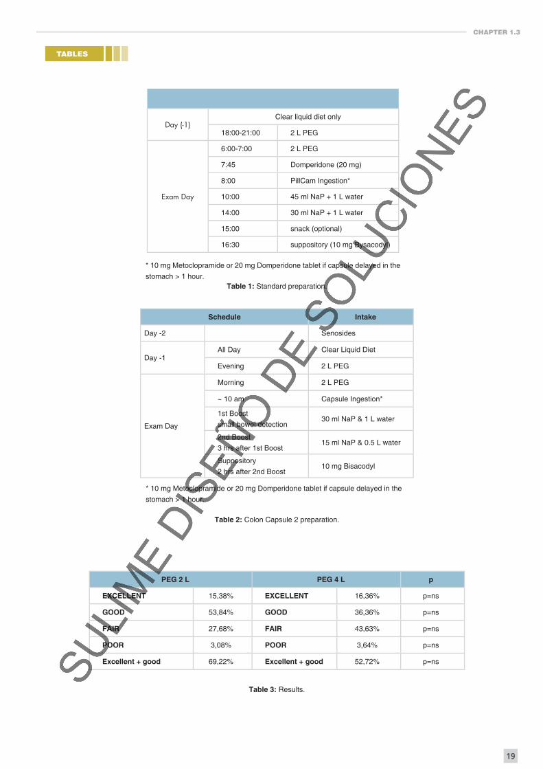
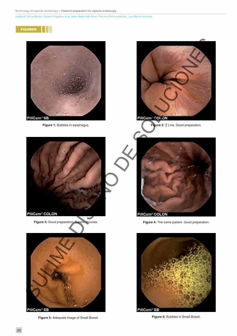
Figure 1: Bubbles in esophagus.
Figure 4: The same patient. Good preparation.
Figure 2: Z Line. Good preparation.
Figure 3: Good preparation. Gastric mucosa.
Figure 5: Adequate image of Small Bowel. Figure 6: Bubbles in Small Bowel.
Technology of capsule endoscopy > Patient’s preparation for capsule endoscopy
Josefa Mª García-Montes, Federico Argüelles-Arias, Belén Maldonado-Pérez, Francisco Pellicer-Bautista, Juan Manuel Herrerías
22
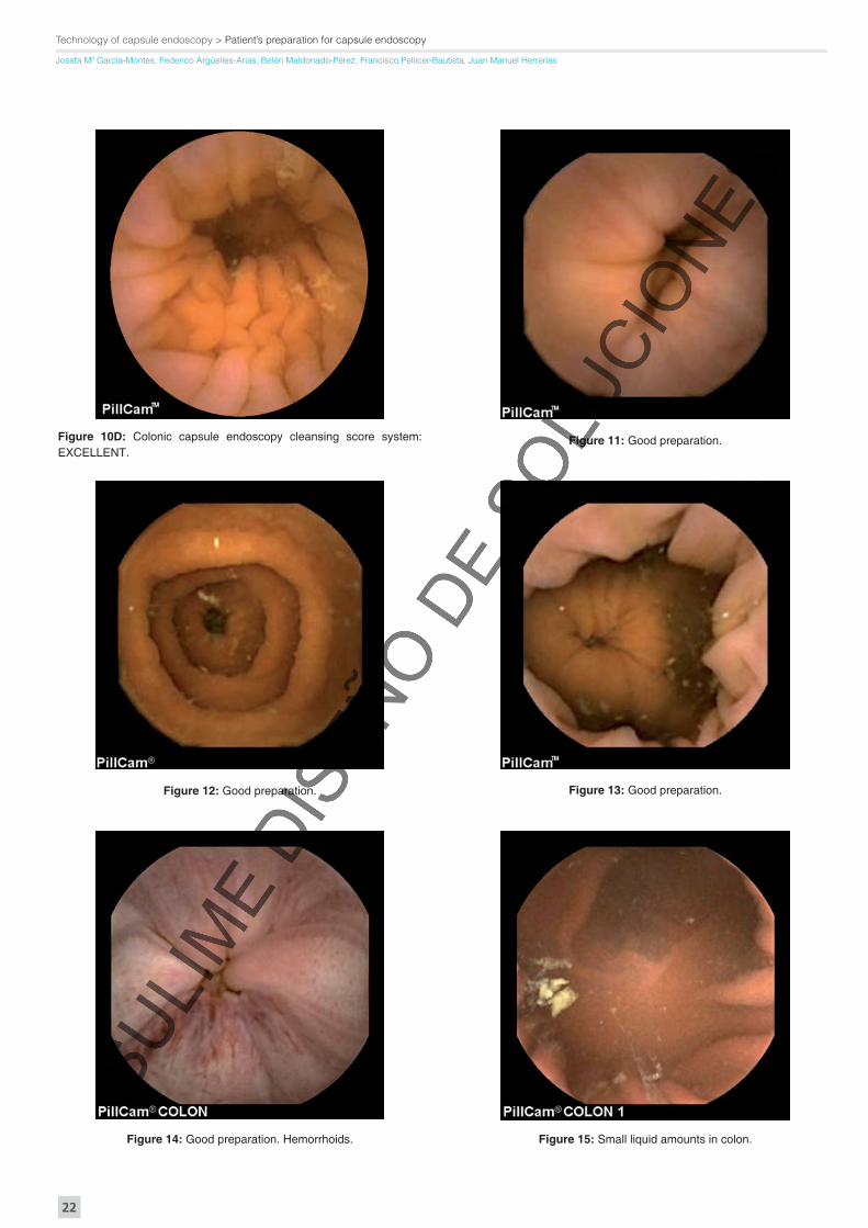
Figure 11: Good preparation.
Figure 13: Good preparation.
Figure 10D: Colonic capsule endoscopy cleansing score system: EXCELLENT.
Figure 12: Good preparation.
Figure 14: Good preparation. Hemorrhoids. Figure 15: Small liquid amounts in colon.
25
TECHNOLOGY OF CAPSULE ENDOSCOPY - CHAPTER 1.4
The procedure of capsule endoscopy
AUTHOR
Miguel Mascarenhas Saraiva, MD, PhD ManopH, Laboratório de Endoscopia e Motilidade Digestiva
Instituto CUF, Porto, PortugalHospital CUF, Porto, Portugal

26
CHAPTER 1.4 - TECHNOLOGY OF CAPSULE ENDOSCOPY
The procedure of capsule endoscopy
AUTHOR Miguel Mascarenhas Saraiva
INTRODUCTION
In order to get a satisfactory examination, the procedure of capsule endoscopy must obey to certain rules, concerning correct preparation, administration, monitoring and downloading.
UNDERGOING A CAPSULE ENTEROSCOPY (CE)
Patient Preparation
The CE is completely different from the standard endoscopy that allows the endoscopist to remove all the residual material to improve the image. In most CE studies, the image quality in the proximal small bowel is much superior to that in the distal ileum, due to residual material.
There are discrepancies about the ideal preparation for capsule endoscopy. In our practice, we keep a patient fasting (NPO) for 12 hours, after a liquid diet for 8 hours. Various studies suggest the usefulness of bowel preparation with PEG solutions2 or sodium phosphate3, 4. Others conclude that, after testing the various preparations, we had to concede that they neither offered any improvement in the time needed to read the study nor in image quality over the 12-h fasting period5. See Chapter 1.3.
At least one day before the examination, the physician should give the patient instructions for undergoing capsule Endoscopy and should verify that the patient understands the instructions. Care must be directed to be sure that the patient is not taking iron medications during the 3 days before the procedure. See Chap 1.5.
Depending on the system that is going to be used, males should be instructed to shave their abdomen 15 cm above and bellow the navel on the day of the test and all patients to wear two piece loose fi tting clothing.
Ingestion of the capsule
When the patient comes to the offi ce, we proceed to system initialization.
An array of sensors is attached to the abdominal wall, and a belt holding a recorder with a battery is fastened
around the waist. Care must be taken for correct placement of sensors, according to the instructions of each system (Figures 1 - 4), because the localization system depends on a correct placement. A new system of sensors has been developed by Given - The SensorBelt - is a comfortable belt worn around the patient’s waist over clothing. It employs easy-fasten straps for quick adjustment and removal. The sensors incorporated within the belt eliminate the need for messy and inconvenient sensor sleeves (Figure 5). For esophageal studies, a different array of sensors is used, that uses three antennas that are attached to specifi c positions. (See Chapter 2.1).
After being sure that an overnight fast of 12 h was respected, patients are asked to ingest the capsule with plenty of water mixed with simeticone to eliminate small bubbles in the gastrointestinal tract (Figure 6). Simeticone administration before or during capsule endoscopy improves the visualization of the mucosa in the proximal small intestine. Some studies assess the effectiveness of simeticone in reducing bowel gas bubbles in patients undergoing capsule endoscopy. The conclusions are that the visibility of the mucosa in the proximal small bowel in patients who received preparation with simeticone was considered to be better, with fewer intraluminal bubbles, than in those without bowel preparation. No adverse effects of simeticone were observed6, 7.
In order to get the best viewing of the esophagus, a different protocol of ingestion is recommended. (See Chapter 2.1).
Battery life of the capsule is 8 +/- 1 hour for Given, more for Olympus or MiroCam (12 h). This time frame is generally suffi cient to image the entire small bowel8. But, in patients with delayed gastric emptying or bowel motility dysfunction related to neuropathic conditions (ex, diabetic), infl ammatory conditions or medication use, may be too short. Because in certain cases progression of the capsule is very slow, several investigators overcome this problem by giving prokinetic drugs to patients, like erythromycin9, or metoclopramide10. However, increasing too much the speed of progression may be the cause of poor visualization and important lesions may be overlooked. When needed (symptoms suggestive of gastroparesia or in some underlying conditions, such as diabetic or undernourished patients), we use, with success, domperidone, a drug that improves antro-duodenal coordination, but does not cause increased small bowel motility11.

CHAPTER 1.4
29
FIGURES
Figure 1: The Given Diagnostic system. a) – Sensor Array for small bowel studies. b) Sensor array for oesophageal studies. c) Data Recorder. d) PillCam SB (for small bowel study). e) PillCam ESO (for oesophageal study).
Figure 2: Placing the abdominal sensors for a capsule enteroscopy with the PillCam SB. Patient prepared for the recording.

CHAPTER 1.4
33
Figure 10: Endoscopic capsule (MiroCam) delivery using the AdvanCE device. The capsule can be delivered to the stomach or to the duodenum, as is exemplifi ed in this case.
Figure 11: The PillCam Express capsule delivering device, developed by Given Imaging.
Figure 12: Real-time monitor for capsule endoscopy (Olympus).

Technology of capsule endoscopy >The procedure of capsule endoscopy
Miguel Mascarenhas-Saraiva
34
Figure 13: The RAPID(R) Access RT, a handheld device that enables real-time viewing during a PillCam endoscopy procedure.
Figure 14: The new data recorder from Given®, has the possibity of direct real time viewing, from the incorporated LCD monitor. It´s use is essencial for studies done with the PillCam Colon2 capsule.
CHAPTER 1.4
35
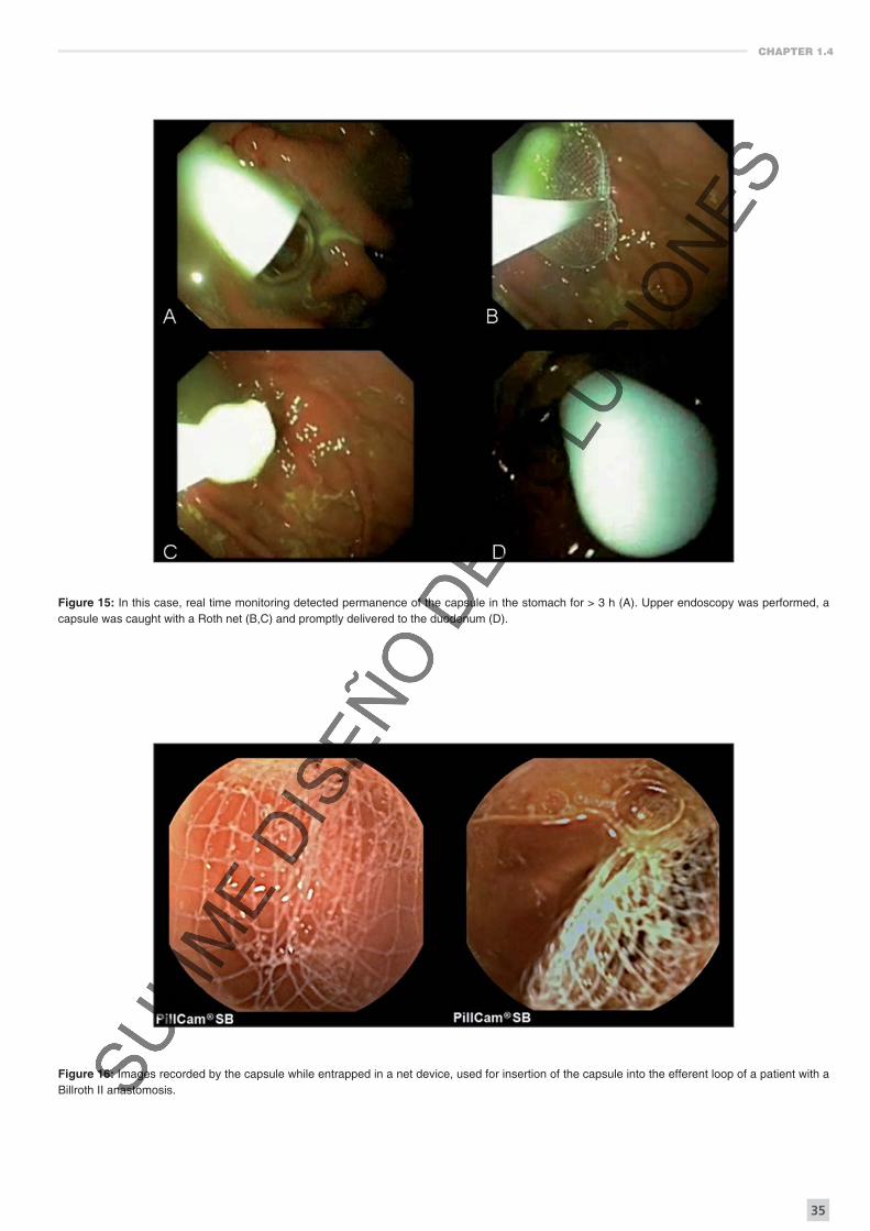
Figure 15: In this case, real time monitoring detected permanence of the capsule in the stomach for > 3 h (A). Upper endoscopy was performed, a capsule was caught with a Roth net (B,C) and promptly delivered to the duodenum (D).
Figure 16: Images recorded by the capsule while entrapped in a net device, used for insertion of the capsule into the efferent loop of a patient with a Billroth II anastomosis.
CHAPTER 1.6
63
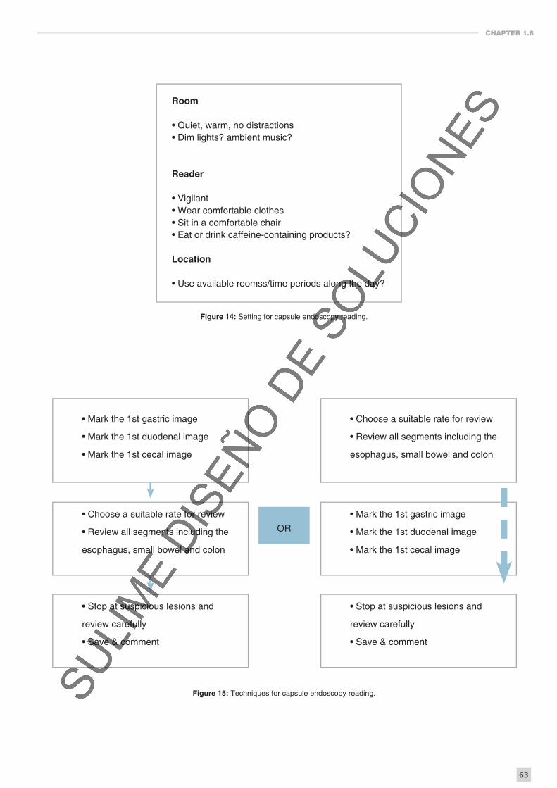
Figure 14: Setting for capsule endoscopy reading.
Figure 15: Techniques for capsule endoscopy reading.
Room
• Quiet, warm, no distractions• Dim lights? ambient music?
Reader
• Vigilant• Wear comfortable clothes• Sit in a comfortable chair• Eat or drink caffeine-containing products?
Location
• Use available roomss/time periods along the day?
• Mark the 1st gastric image
• Mark the 1st duodenal image
• Mark the 1st cecal image
• Choose a suitable rate for review
• Review all segments including the
esophagus, small bowel and colon
• Stop at suspicious lesions and
review carefully
• Save & comment
OR
• Choose a suitable rate for review
• Review all segments including the
esophagus, small bowel and colon
• Mark the 1st gastric image
• Mark the 1st duodenal image
• Mark the 1st cecal image
• Stop at suspicious lesions and
review carefully
• Save & comment
CHAPTER 1.6
65
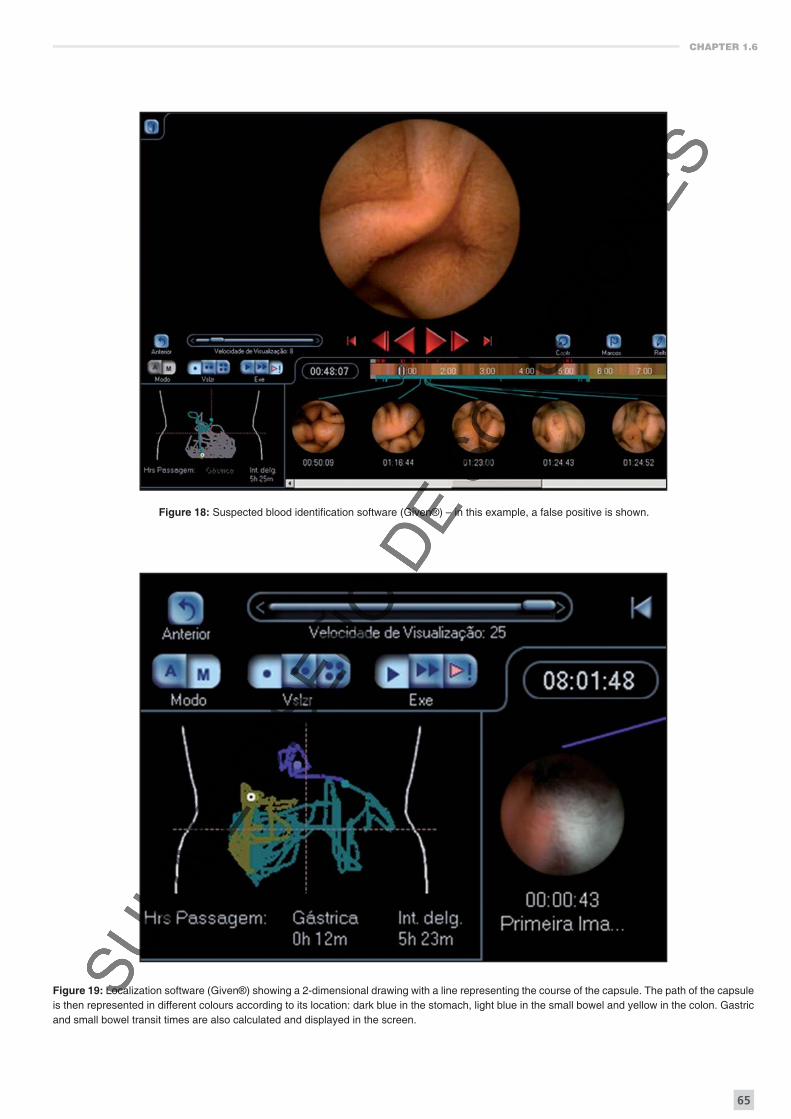
Figure 18: Suspected blood identifi cation software (Given®) – in this example, a false positive is shown.
Figure 19: Localization software (Given®) showing a 2-dimensional drawing with a line representing the course of the capsule. The path of the capsule is then represented in different colours according to its location: dark blue in the stomach, light blue in the small bowel and yellow in the colon. Gastric and small bowel transit times are also calculated and displayed in the screen.

101
TECHNOLOGY OF CAPSULE ENDOSCOPY - CHAPTER 1.10
Patency and Agile capsules
AUTHORS
Ángel Caunedo-Álvarez, MD
Javier Romero-Vázquez, MD
Mileidis San Juan Acosta, MD
Juan Manuel Herrerías, MD, PhD, AGAFGastroenterology Service
Hospital Universitario Virgen Macarena. Sevilla. [email protected]
CHAPTER 1.10
111
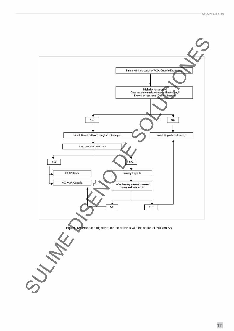
Figure 12: Proposed algorithm for the patients with indication of PillCam SB.
Technology of capsule endoscopy > Patency and Agile capsules
Ángel Caunedo-Álvarez, Javier Romero-Vázquez, Mileidis San Juan Acosta, Juan Manuel Herrerías
112
Intestinal
fl uid
DESINTEGRATION
RESOLUTION
Intestinal
fl uid
NO (OR LATE)
DESINTEGRATION
OBSTRUCTION
Intestinal
fl uid
DESINTEGRATION
RESOLUTION
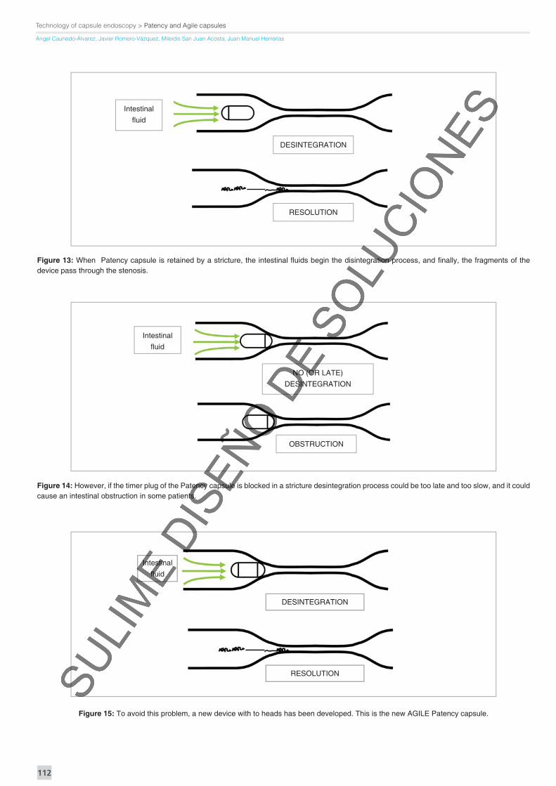
Figure 13: When Patency capsule is retained by a stricture, the intestinal fl uids begin the disintegration process, and fi nally, the fragments of the device pass through the stenosis.
Figure 14: However, if the timer plug of the Patency capsule is blocked in a stricture desintegration process could be too late and too slow, and it could cause an intestinal obstruction in some patients.
Figure 15: To avoid this problem, a new device with to heads has been developed. This is the new AGILE Patency capsule.
CHAPTER 1.10
117
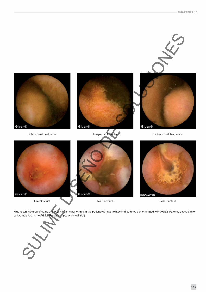
Submucosal ileal tumor Inespecifi c Enteritis Submucosal ileal tumor
Ileal Stricture Ileal Stricture Ileal Stricture
Figure 22: Pictures of some of the of PillCams performed in the patient with gastrointestinal patency demonstrated with AGILE Patency capsule (own series included in the AGILE Patency capsule clinical trial).

119
CAPSULE ENDOSCOPY OF THE ESOPHAGUS - CHAPTER 2.1
Esophageal capsule endoscopy: fi elds of application
AUTHORS
Alba Belda-Cuesta, MD
María Luisa Morales-Barroso, MD
Federico Argüelles-Arias, MD
Ángel Caunedo-Álvarez, MD
Juan Manuel Herrerías, MD, PhD, AGAFGastroenterology Service
Hospital Universitario Virgen Macarena. Sevilla. [email protected]
Capsule endoscopy of the esophagus > Fields of application
Alba Belda-Cuesta, María Luisa Morales-Barroso, Federico Argüelles-Arias, Ángel Caunedo-Álvarez, Juan Manuel Herrerías
122
were 46% and 54%, respectively. So the conclusions are clear and in a cohort at high risk for esophageal SCC, ECE is not sensitive enough to diagnose neoplastic lesions.
In the last months some new indications for ECE have been investigated. The aim of one study recently published21 was to evaluate the ability of ECE to identify high and low risk patients with upper gastrointestinal bleeding (UGIB). Twenty-four patients with a history of UGIB within 48 hours of admission to the Emergency Room (ER) were randomized to CE versus standard clinical assessment. CE was read in real-time at the bedside and later reviewed after download. Positive CE fi ndings included coffee grounds, blood clot, red blood, or a bleeding lesion. CE positive patients underwent gastroscopy within 6 h. Seven of twelve patients were CE positive. All seven had confi rmatory stigmata at gastroscopy. Four of the fi ve CE negative patients had no stigmata at EGD and one was not endoscoped due to comorbidities. The actual lesion was visualized at CE in four of twelve patients during live view and in an additional two patients after download (6/12). Time to endoscopy in the CE positive group was signifi cantly shorter than control patients (2.5 vs. 8.9 h, P = 0.029). So the conclusions were that live view CE identifi es high and low risk ER patients with UGIB and the use of CE to risk stratify these patients signifi cantly reduced time to emergent EGD and therapeutic intervention.
Other indications could be eosinophilic esophagitis, peristatic anormalities in esophagus and also when patients do not want to undergo a conventional gastroscopy.
CONTRAINDICATIONS
ECE contraindications remain roughly the same featured for Small Bowel Capsule Endoscopy.
CE should not be used in patients with swallowing disorders, due to the risk of aspiration. Pregnancy is a contraindication for CE examination because of the microwaves transmitted by the capsule. However, there are two case reports of CE examination during the fi rst trimester of pregnancy22, 23.
CE is not contraindicated in patients with a cardiac pacemaker24 or implantable cardiac defi brillator25 and there is no interference between the two devices.
In case of risk of capsule retention, that has been regarded as low as 0.75% and as high as 6.8%, CCE is not indicated. At the moment there are no cases of capsule retention reported in patients without any known risk factor. Among the risk factors for capsule retention appear history of abdominal surgery, radiotherapy, Crohn’s disease or chronic taking of NSAIDs. A history of prior abdominal surgery in patients with a normal small bowel series is not considered a high risk for retention.
CONCLUSIONS
Endoscopic examination of the oesophagus with video-capsule is a practical reality today, with a diagnostic accuracy that is progressively approaching the conventional endoscopy. In the areas where the CE is currently applicable, which are primarily the detection of Barrett’s in patients with chronic GERD and risk of bleeding varices in patients with liver cirrhosis, its use should be minimized because of the margins of error which may imply inadecuate therapies. It should be kept in a second plan and the oral conventional endoscopy should maintain its leading position today thanks to its wide distribution and good tolerance.
REFERENCES
1. Caunedo A, Rodríguez-Téllez M, García-Montes JM, Gómez-Rodríguez BJ,
Guerrero J, Herrerías JM Jr, Pellicer F, Herrerías JM. Usefulness of capsule
endoscopy in patients with suspected small bowel disease. Rev Esp Enferm
Dig. 2004; 96(1): 10-21.
2. Neu B, Wettschureck E, Rösch T. Is esophageal capsule endoscopy feasible?
Results of a pilot. Endoscopy. 2003; 35(11): 957-61.
3. Ramirez FC, Shaukat MS, Young MA, Johnson DA, Akins R. Feasibility and
safety of string, wireless capsule endoscopy in the diagnosis of Barrett’s
esophagus. Gastrointest Endosc. 2005; 61(6): 741-6.
4. Ramirez FC, Hakim S, Tharalson EM, Shaukat MS, Akins R. Feasibility and
safety of string wireless capsule endoscopy in the diagnosis of esophageal
varices. Am J Gastroenterol. 2005; 100(5): 1065-71.
5. Eliakim R, Yassin K, Shlomi I, Suissa A, Eisen GM. A novel diagnostic tool
for detecting oesophageal pathology: the PillCam oesophageal video capsule.
Aliment Pharmacol Ther. 2004; 20(10): 1083-9.
6. Gralnek IM, Adler SN, Yassin K et al Detecting esophageal disease with
second-generation capsule endoscopy: initial evaluation of the Pill-Cam ESO 2.
Endoscopy 2008; 40: 275–279.
7. Sánchez-Yagüe A, Caunedo-Alvarez A, García-Montes JM, Romero-Vázquez
J, Pellicer-Bautista FJ, Herrerías-Gutiérrez JM. Esophageal capsule endoscopy in
patients refusing conventional endoscopy for the study of suspected esophageal
pathology. Eur J Gastroenterol Hepatol. 2006; 18(9): 977-83.
8. Gralnek IM, Rabinovitz R, Afi k D, et al A simplifi ed ingestion procedure
for esophageal capsule endoscopy: initial evaluation in healthy volunteers.
Endoscopy 2006; 38: 913–918.
9. De Jonge PJF, Van Eijk BC, Geldof H, et al Capsule endoscopy for the
detection of oesophageal mucosal disorders: a comparison of two different
ingestion protocols. Scand J Gastroenterol. 2008; 43: 870–877.
10. Delvaux M, Papanikolaou IS, Fassler I et al Esophageal capsule endoscopy
in patients with suspected esophageal disease: double blinded comparison
with esophagogastroduodenoscopy and assessment of interobserver variability.
Endoscopy 2008; 40: 16–22.
11. Diaz-Rubio M, Moreno-Elola-Olaso C, Rey E, Locke GR 3rd, Rodriguez-
Artalejo F. Symptoms of gastro-oesophageal refl ux: prevalence, severity, duration
and associated factors in a Spanish population. Aliment Pharmacol Ther. 2004;
19(1): 95-105.
Capsule endoscopy of the esophagus > Fields of application
Alba Belda-Cuesta, María Luisa Morales-Barroso, Federico Argüelles-Arias, Ángel Caunedo-Álvarez, Juan Manuel Herrerías
124
TABLES
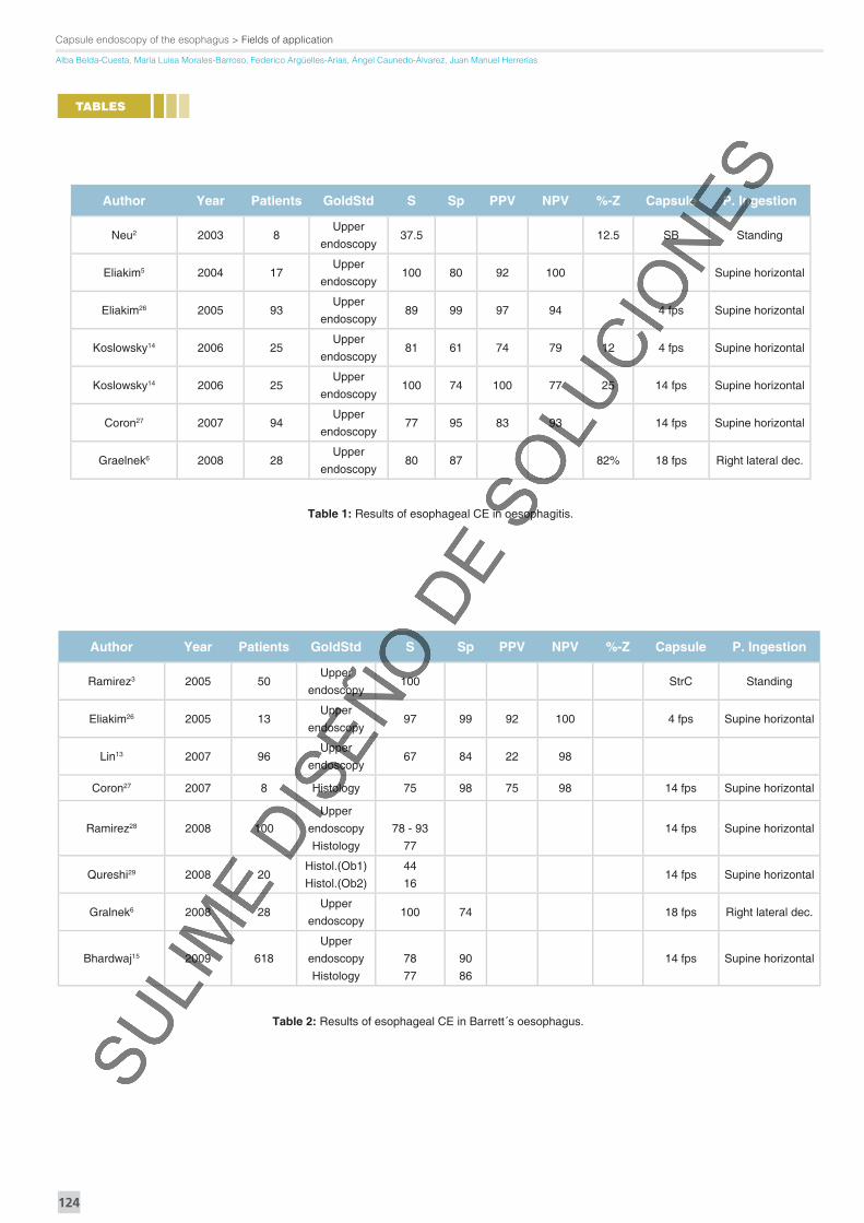
Table 1: Results of esophageal CE in oesophagitis.
Author Year Patients GoldStd S Sp PPV NPV %-Z Capsule P. Ingestion
Neu2 2003 8Upper
endoscopy37.5 12.5 SB Standing
Eliakim5 2004 17Upper
endoscopy100 80 92 100 Supine horizontal
Eliakim26 2005 93Upper
endoscopy89 99 97 94 4 fps Supine horizontal
Koslowsky14 2006 25Upper
endoscopy81 61 74 79 12 4 fps Supine horizontal
Koslowsky14 2006 25Upper
endoscopy100 74 100 77 25 14 fps Supine horizontal
Coron27 2007 94Upper
endoscopy77 95 83 93 14 fps Supine horizontal
Graelnek6 2008 28Upper
endoscopy80 87 82% 18 fps Right lateral dec.
Table 2: Results of esophageal CE in Barrett´s oesophagus.
Author Year Patients GoldStd S Sp PPV NPV %-Z Capsule P. Ingestion
Ramirez3 2005 50Upper
endoscopy100 StrC Standing
Eliakim26 2005 13Upper
endoscopy97 99 92 100 4 fps Supine horizontal
Lin13 2007 96Upper
endoscopy67 84 22 98
Coron27 2007 8 Histology 75 98 75 98 14 fps Supine horizontal
Ramirez28 2008 100
Upper
endoscopy
Histology
78 - 93
77
14 fps Supine horizontal
Qureshi29 2008 20Histol.(Ob1)
Histol.(Ob2)
44
1614 fps Supine horizontal
Gralnek6 2008 28Upper
endoscopy100 74 18 fps Right lateral dec.
Bhardwaj15 2009 618
Upper
endoscopy
Histology
78
77
90
86
14 fps Supine horizontal
339
CAPSULE ENTEROSCOPY - CHAPTER 4.15.1
Complementary procedures to capsule endoscopy:
New ways of enteroscopy
AUTHOR
Enrique Pérez-Cuadrado Martínez, MD Head of Gastroenterology
Hospital General Universitario Morales MeseguerAssociate Professor of UMU Universidad de Murcia, Murcia.
CHAPTER 4.15.1
341
“clean SB”, for preventing invagination of the SB but also for the rare possibility of malignant disease. CE can be used to control metachronic lesions in follow up. In some cases, there are distal polyps and the DBE must reach the cecum (Figure 29).
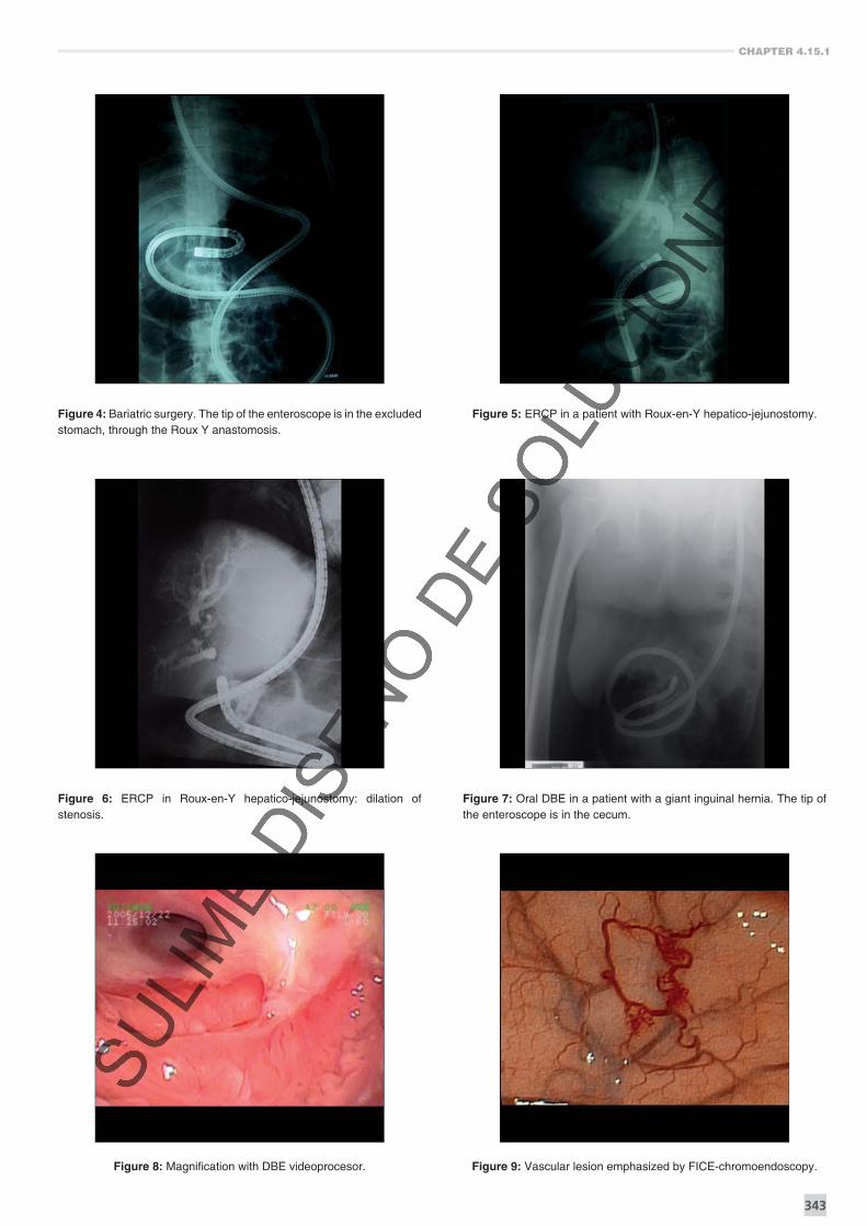
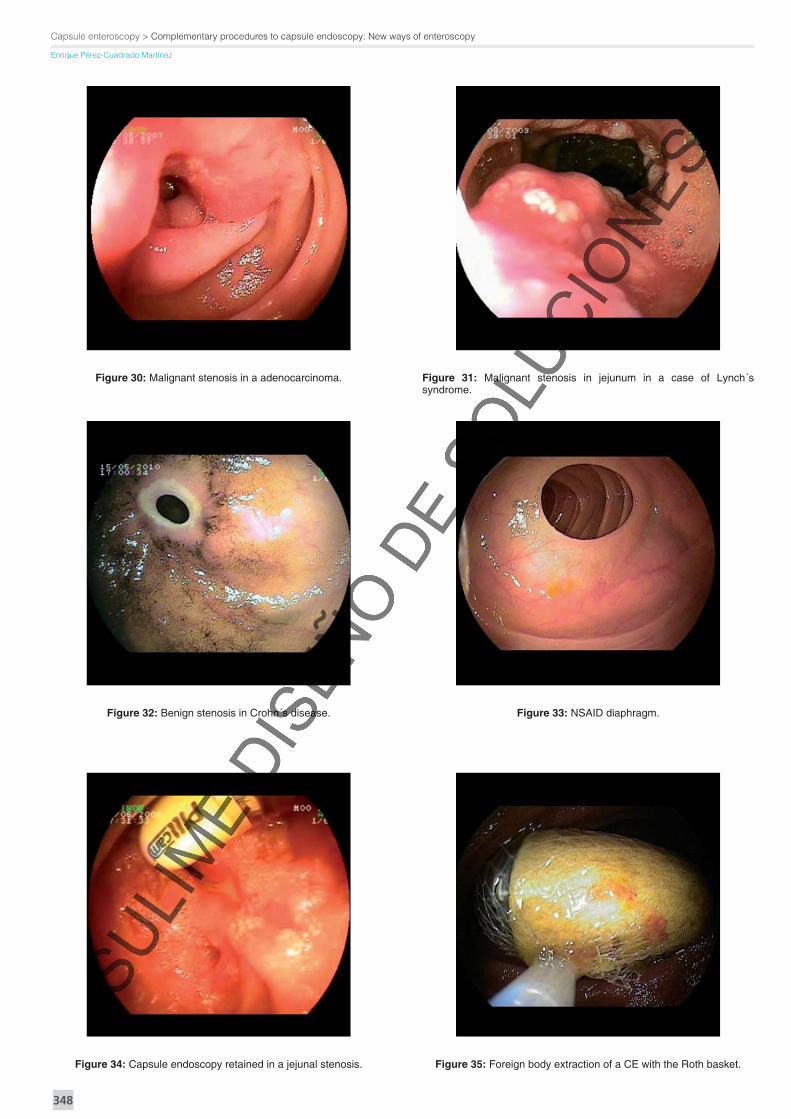
5) INTESTINAL OBSTRUCTION. In case of known stenosis, DBE can be the fi rst investigation line instead of CE. We can detect one or more malignant stenosis (Figures 30, 31) or benign stenosis in Crohn´s disease (Figure 32), NSAIDs diaphragms (Figure 33), with eventual foreign body extraction of the own CE (Figures 34, 35). In Crohn’s disease the fl exible enteroscopy moreover has indications as follows:
1. suspected disease. For a Crohn´s disease diagnosis with biopsies in case of clinical suspicion or indeterminate colitis.
2. established Crohn´s disease. Detectionof complications like fi stula, secondary neoplasia (adenocarcinoma).
3. differential diagnosis. Diseases like cytomegalovirus (Figure 36), amiloidosis (Figure 37) etc.
6) STENT PLACEMENT. Under fl uoroscopic guidance,
with withdrawing the enteroscope and leaving the overtube in place with the guide wire through malignant stenosis, by pushing directly in the overtube an expandable stent on guide wire (Figure 38).
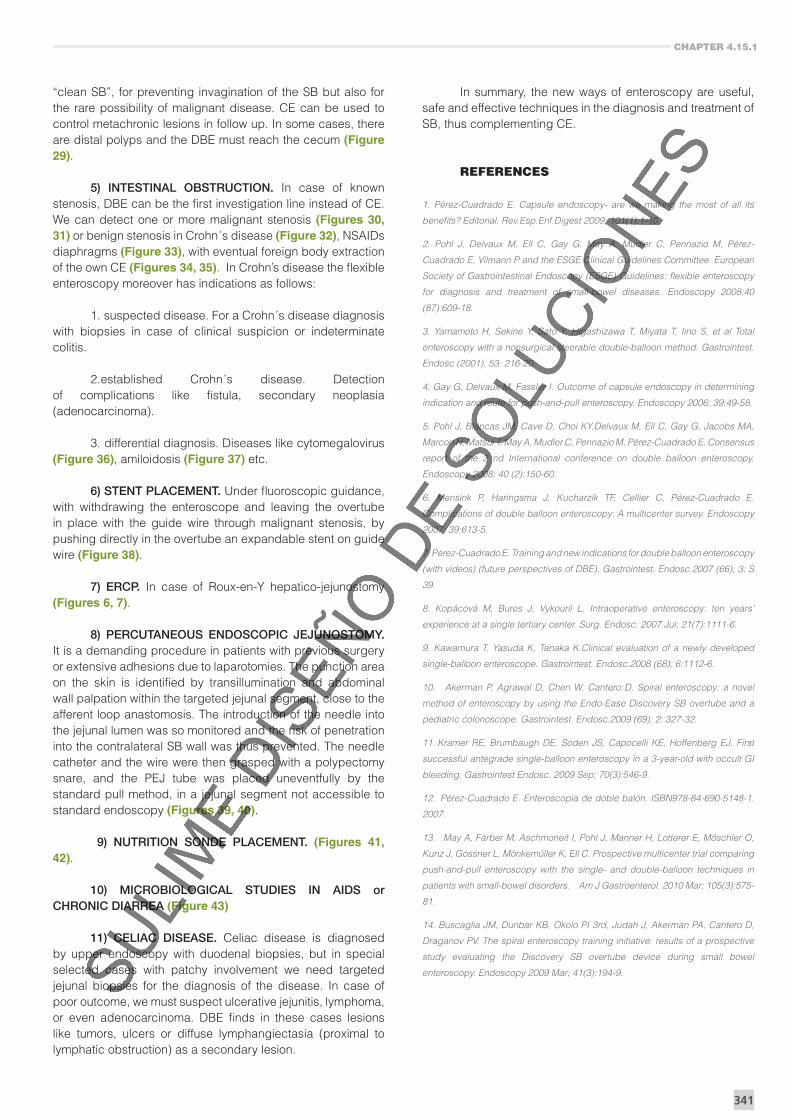
7) ERCP. In case of Roux-en-Y hepatico-jejunostomy (Figures 6, 7).
8) PERCUTANEOUS ENDOSCOPIC JEJUNOSTOMY. It is a demanding procedure in patients with previous surgery or extensive adhesions due to laparotomies. The punction area on the skin is identifi ed by transillumination and abdominal wall palpation within the targeted jejunal segment, close to the afferent loop anastomosis. The introduction of the needle into the jejunal lumen was so monitored and the risk of penetration into the contralateral SB wall was thus prevented. The needle catheter and the wire were then grasped with a polypectomy snare, and the PEJ tube was placed uneventfully by the standard pull method, in a jejunal segment not accessible to standard endoscopy (Figures 39, 40).
9) NUTRITION SONDE PLACEMENT. (Figures 41, 42).
10) MICROBIOLOGICAL STUDIES IN AIDS or CHRONIC DIARREA (Figure 43)
11) CELIAC DISEASE. Celiac disease is diagnosed by upper endoscopy with duodenal biopsies, but in special selected cases with patchy involvement we need targeted jejunal biopsies for the diagnosis of the disease. In case of poor outcome, we must suspect ulcerative jejunitis, lymphoma, or even adenocarcinoma. DBE fi nds in these cases lesions like tumors, ulcers or diffuse lymphangiectasia (proximal to lymphatic obstruction) as a secondary lesion.
In summary, the new ways of enteroscopy are useful, safe and effective techniques in the diagnosis and treatment of SB, thus complementing CE.
REFERENCES
1. Pérez-Cuadrado E. Capsule endoscopy- are we making the most of all its
benefi ts? Editorial. Rev.Esp.Enf.Digest 2009; 101(1):1-10.
2. Pohl J, Delvaux M, Ell C, Gay G, May A, Mudler C, Pennazio M, Pérez-
Cuadrado E, Vilmann P and the ESGE Clinical Guidelines Committee. European
Society of Gastrointestinal Endoscopy (ESGE) Guidelines: fl exible enteroscopy
for diagnosis and treatment of small-bowel diseases. Endoscopy 2008;40
(87):609-18.
3. Yamamoto H, Sekine Y, Sato Y, Higashizawa T, Miyata T, Iino S, et al Total
enteroscopy with a nonsurgical steerable double-balloon method. Gastrointest.
Endosc (2001), 53: 216-20.
4. Gay G, Delvaux M, Fassler I. Outcome of capsule endoscopy in determining
indication and route for push-and-pull enteroscopy. Endoscopy 2006; 39:49-58.
5. Pohl J, Blancas JM, Cave D, Choi KY,Delvaux M, Ell C, Gay G, Jacobs MA,
Marcon N, Matsui T, May A, Mudler C, Pennazio M, Pérez-Cuadrado E. Consensus
report of the 2 nd International conference on double balloon enteroscopy.
Endoscopy 2008; 40 (2):150-60.
6. Mensink P, Haringsma J, Kucharzik TF, Cellier C, Pérez-Cuadrado E.
Complications of double balloon enteroscopy: A multicenter survey. Endoscopy
2007; 39:613-5.
7. Pérez-Cuadrado E. Training and new indications for double balloon enteroscopy
(with videos) (future perspectives of DBE). Gastrointest. Endosc.2007 (66); 3: S
39.
8. Kopácová M, Bures J, Vykouril L. Intraoperative enteroscopy: ten years’
experience at a single tertiary center. Surg. Endosc. 2007 Jul; 21(7):1111-6.
9. Kawamura T, Yasuda K, Tanaka K.Clinical evaluation of a newly developed
single-balloon enteroscope. Gastrointest. Endosc.2008 (68); 6:1112-6.
10. Akerman P, Agrawal D, Chen W, Cantero D. Spiral enteroscopy: a novel
method of enteroscopy by using the Endo-Ease Discovery SB overtube and a
pediatric colonoscope. Gastrointest. Endosc.2009 (69); 2: 327-32.
11. Kramer RE, Brumbaugh DE, Soden JS, Capocelli KE, Hoffenberg EJ. First
successful antegrade single-balloon enteroscopy in a 3-year-old with occult GI
bleeding. Gastrointest Endosc. 2009 Sep; 70(3):546-9.
12. Pérez-Cuadrado E. Enteroscopia de doble balón. ISBN978-84-690-5148-1.
2007.
13. May A, Färber M, Aschmoneit I, Pohl J, Manner H, Lotterer E, Möschler O,
Kunz J, Gossner L, Mönkemüller K, Ell C. Prospective multicenter trial comparing
push-and-pull enteroscopy with the single- and double-balloon techniques in
patients with small-bowel disorders. Am J Gastroenterol. 2010 Mar; 105(3):575-
81.
14. Buscaglia JM, Dunbar KB, Okolo PI 3rd, Judah J, Akerman PA, Cantero D,
Draganov PV. The spiral enteroscopy training initiative: results of a prospective
study evaluating the Discovery SB overtube device during small bowel
enteroscopy. Endoscopy 2009 Mar; 41(3):194-9.
CHAPTER 4.15.1
343
Figure 4: Bariatric surgery. The tip of the enteroscope is in the excluded stomach, through the Roux Y anastomosis.
Figure 5: ERCP in a patient with Roux-en-Y hepatico-jejunostomy.
Figure 6: ERCP in Roux-en-Y hepatico-jejunostomy: dilation of stenosis.
Figure 7: Oral DBE in a patient with a giant inguinal hernia. The tip of the enteroscope is in the cecum.
Figure 8: Magnifi cation with DBE videoprocesor. Figure 9: Vascular lesion emphasized by FICE-chromoendoscopy.
Capsule enteroscopy > Complementary procedures to capsule endoscopy: New ways of enteroscopy
Enrique Pérez-Cuadrado Martínez
348
Figure 30: Malignant stenosis in a adenocarcinoma. Figure 31: Malignant stenosis in jejunum in a case of Lynch´s syndrome.
Figure 32: Benign stenosis in Crohn´s disease. Figure 33: NSAID diaphragm.
Figure 34: Capsule endoscopy retained in a jejunal stenosis. Figure 35: Foreign body extraction of a CE with the Roth basket.