ASSOCIATION BETWEEN NUTRITIONAL STATUS AND...
41
ASSOCIATION BETWEEN NUTRITIONAL STATUS AND SERUM LEPTIN AMONG NEWLY DIAGNOSED BREAST CANCER PATIENTS IN HOSPITAL UNIVERSITI SAINS MALAYSIA (HOSPITAL USM): A CASE CONTROL STUDY by AHMAD KARAMI BIN ISMAIL Thesis submitted in the fulfilment of the requirements for the degree of Master of Science June 2016
Transcript of ASSOCIATION BETWEEN NUTRITIONAL STATUS AND...
ASSOCIATION BETWEEN NUTRITIONAL STATUS
AND SERUM LEPTIN AMONG NEWLY DIAGNOSED
BREAST CANCER PATIENTS IN HOSPITAL
UNIVERSITI SAINS MALAYSIA (HOSPITAL USM):
A CASE CONTROL STUDY
by
AHMAD KARAMI BIN ISMAIL
Thesis submitted in the fulfilment of the requirements
for the degree of
Master of Science
June 2016
ii
ACKNOWLEDGMENT
In the name of Allah, the Most Gracious and the Most Merciful
Alhamdulillah, Thank You ALLAH for being beside me at every time and for giving me
strength and ability to complete this thesis. I am sincerely grateful to both of my
supervisors, Dr. V.M.K. Bhavharaju and Professor Dr. Wan Abdul Manan Wan Muda,
for all the guidance and support throughout the duration of this study and the preparation
of this thesis. May ALLAH showered with His blessings.
I would also like to thank Associate Profesor Dr. Sakinah Harith, the Dietetics
Programme Chairman for her enthusiasm to encourage, motivate and support with my
thesis. A special thanks to all of my colleagues in the Nutrition & Dietetics Programme,
School of Health Sciences especially Associate Profesor Shariza Abdul Razak for their
encouragement and also my acknowledgements are owed to the Mr Arun and Mrs Elina
Husni Tan for his help in my study.
I would like to acknowledge deeply Universiti Sains Malaysia and Ministry of
Science, Technology and Innovation, Malaysia (MOSTI) for funding this research and
provide scholarship of my study. To the Dean, School Of Health Sciences, Profesor
Ahmad Haji Zakaria, thank you for your attention, motivation and support of my study. I
personally want to thanks Kak Na and Kak Ju, staffs at Post-Graduate Section, School of
Medical Sciences (PPSP) for their help along my study.
iii
Finally, I am greatly indebted to my great and beloved wife Maria Elena Razali @
Ghazali and my wonderful children Alya Nazihah, Aisyah Nazirah, Anis Nadhirah &
Ahmad Nazeef for their great help, support and patient encouragement throughout my
study. My deepest gratitude goes to all my family members, special thanks for
motivation, support and encouragement given to me. Love you all.
May ALLAH bless.
iv
TABLE OF CONTENTS
ACKNOWLEDGMENT ............................................................................................. ii
TABLE OF CONTENTS............................................................................................ iv
LIST OF TABLES .................................................................................................... viii
LIST OF FIGURES ………………………………....................................................ix
LIST OF ABBREVIATIONS ..................................................................................... xi
ABSTRAK ................................................................................................................ xiii
ABSTRACT.............................................................................................................. xvi
CHAPTER 1 INTRODUCTION .............................................................................. 1
1.1 Background of the study ........................................................................................ 1
1.2 Problem Statement ................................................................................................. 4
1.3 Objectives .............................................................................................................. 6
1.3.1 General objective ............................................................................................ 6
1.3.2 Specific objectives .......................................................................................... 6
1.4 Study Hypotheses .................................................................................................. 7
CHAPTER 2 LITERATURE REVIEW .................................................................. 9
2.1 Overview of Breast Cancer in Malaysia ................................................................ 9
2.2 Stages of Breast Cancer ......................................................................................... 9
2.2.1 Stage 0 ........................................................................................................... 12
2.2.2 Stage I ........................................................................................................... 12
2.2.3 Stage II .......................................................................................................... 12
v
2.2.4 Stage III…………………………………………………………………… 14
2.2.5 Stage IV…………………………………………………………………......15
2.3 Breast Cancer Risk Factors .................................................................................. 16
2.3.1 Preventable Risk Factors……………………… ……………………….....17
2.3.1.1 Overweight and Obesity……………...…………....…..................... 17
2.3.1.2 Diet……………………………………………………......................22
2.3.1.3 Low Physical Activity Level………..………………….....................25
2.3.2 Non-preventable Risk Factors ..................................................................... 27
2.3.2.1 Family History of Breast Cancer ........................................................ 27
2.3.2.2 Reproductive Factors….......................................................................27
2.3.2.3 Breast Density .................................................................................... 28
2.4 Breast Cancer Prevention......................................................................................29
CHAPTER 3 METHODOLOGY ........................................................................... 31
3.1 Study Design ........................................................................................................ 31
3.2 Sampling .............................................................................................................. 31
3.2.1 Reference population .................................................................................... 31
3.2.2 Source population.......................................................................................... 32
3.2.3 Sampling frame ............................................................................................. 32
3.2.4 Study subjects ............................................................................................... 32
3.2.5 Sample size.................................................................................................... 36
3.2.5.1 Recruitement of subjects ............................................................................ 36
3.3 Research Ethical Approval .................................................................................. 37
3.4 Funding and Research Grant................................................................................ 38
vi
3.5 Research and Measurement Tools........................................................................38
3.6 Data Collection.....................................................................................................38
3.6.1 Patients Profile.............................................................................................38
3.6.2 Questionnaire Forms....................................................................................39
3.6.2.1 Socio-demographic and medical history..........................................39
3.6.2.2 Dietary Intake (1-Day Diet Recall)..................................................40
3.6.3 Anthropometry (Weight, Height, BMI, Waist, Hip) and Body Composition Measurement Using Body Impedance Analysis (BIA)......................................43 3.6.3.1 Weight Measurement.......................................................................43
3.6.3.2 Height Measurement........................................................................45
3.6.3.3 Body Mass Index.............................................................................45
3.6.3.4 Waist and Hip Circumference.........................................................46
3.6.3.5 Body Impedance Analysis (BIA).....................................................48
3.6.4 Biochemical Measurement (Leptin)...........................................................53
3.6.4.1 Serum Leptin Analysis....................................................................53
3.7 Statistical Analysis.............................................................................................. 58
CHAPTER 4 RESULTS …………………………………………………………59
4.1 General characteristics of study subjects ……………………………………....59
4.2 Clinical Profiles and Reproductive Factors among Study Subjects ………….. 63
4.3 Anthropometric Characteristics ………………………………………………..66
4.3.1 Body Composition (WHR and BIA) risk factor of breast cancer...............70
4.4 Biochemical Profile of Study Subjects..................................................... .......... 71
4.5 Dietary Intake of Study Subjects………………………………………... …….71
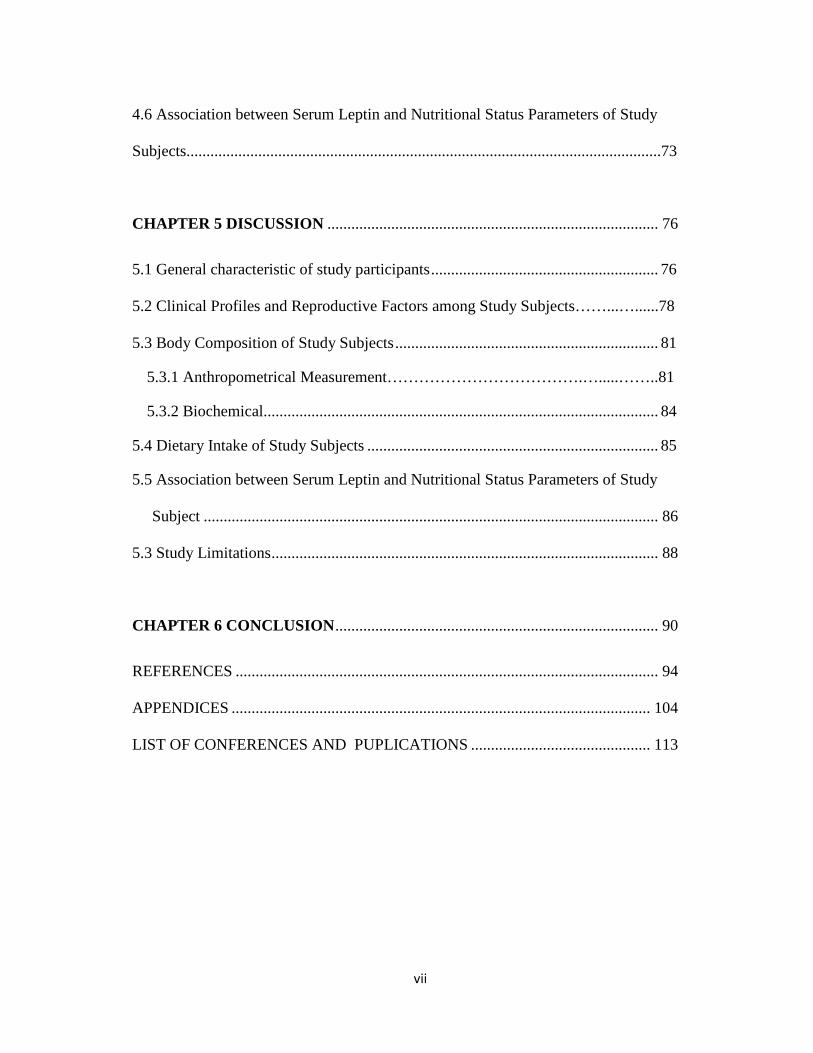
vii
4.6 Association between Serum Leptin and Nutritional Status Parameters of Study
Subjects.......................................................................................................................73
CHAPTER 5 DISCUSSION ................................................................................... 76
5.1 General characteristic of study participants ......................................................... 76
5.2 Clinical Profiles and Reproductive Factors among Study Subjects……...…......78
5.3 Body Composition of Study Subjects .................................................................. 81
5.3.1 Anthropometrical Measurement……………………………….….....……..81
5.3.2 Biochemical................................................................................................... 84
5.4 Dietary Intake of Study Subjects ......................................................................... 85
5.5 Association between Serum Leptin and Nutritional Status Parameters of Study
Subject .................................................................................................................. 86
5.3 Study Limitations ................................................................................................. 88
CHAPTER 6 CONCLUSION ................................................................................. 90
REFERENCES .......................................................................................................... 94
APPENDICES ......................................................................................................... 104
LIST OF CONFERENCES AND PUPLICATIONS ............................................. 113
viii
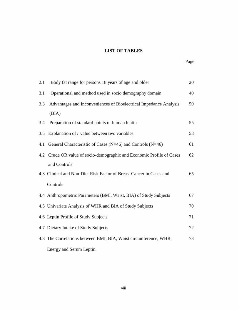
LIST OF TABLES
Page
2.1 Body fat range for persons 18 years of age and older 20
3.1 Operational and method used in socio demography domain 40
3.3 Advantages and Inconveniences of Bioelectrical Impedance Analysis
(BIA)
50
3.4 Preparation of standard points of human leptin 55
3.5 Explanation of r value between two variables 58
4.1 General Characteristic of Cases (N=46) and Controls (N=46) 61
4.2 Crude OR value of socio-demographic and Economic Profile of Cases
and Controls
62
4.3 Clinical and Non-Diet Risk Factor of Breast Cancer in Cases and
Controls
65
4.4 Anthropometric Parameters (BMI, Waist, BIA) of Study Subjects 67
4.5 Univariate Analysis of WHR and BIA of Study Subjects 70
4.6 Leptin Profile of Study Subjects 71
4.7 Dietary Intake of Study Subjects 72
4.8 The Correlations between BMI, BIA, Waist circumference, WHR,
Energy and Serum Leptin.
73
ix
LIST OF FIGURES
1.1 Female Breast; Age Specific Incidence Rate, Kelantan 2004 –
2009
5
1.2 Conceptual Framework of the Study 8
2.1 Diseases and conditions associated with obesity (body mass
index ≥30; calculated as kg/m2)
18
2.2 Body Composition Components 19
2.3 Influence of food, nutrition, obesity, and physical activity on
fundamental processes shown here, which may promote or
inhibit cancer development and progression
23
2.4 The influence of food, nutrition, obesity and physical activity
on the breast cancer process
26
2.5 Breast Cancer Prevention Programs 30
3.1 Study Design Flow Chart 35
3.2
3.3
3.4
WHO Body Mass Index Classification
Rosscraft anthropometry measuring tape
Schematic diagram of fat-free mass (FFM), total body water
(TBW), intracellular water (ICW), extracellular water(ECW)
and body cell mass (BCM)
46
47
49
3.5
3.6
4.1
BIA Measurements
Blood Samples Process
BMI classification profile of study subjects
52
57
69
x
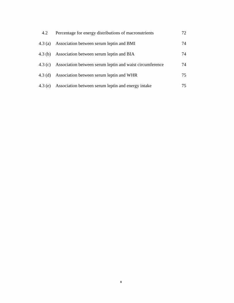
4.2
4.3 (a)
4.3 (b)
4.3 (c)
4.3 (d)
4.3 (e)
Percentage for energy distributions of macronutrients
Association between serum leptin and BMI
Association between serum leptin and BIA
Association between serum leptin and waist circumference
Association between serum leptin and WHR
Association between serum leptin and energy intake
72
74
74
74
75
75
xi
LIST OF ABBREVIATIONS
AICR
AJCC
BC
BIA
BMI
BSE
CBE
American Institute on Cancer Research
American Joint Committee on Cancer
Breast Cancer
Bioelectrical Impedance Analysis
Body Mass Index
Breast Self-Examination
Clinical Breast Examination
CDC Centers for Disease Control and Prevention
CI
cm
CPG
Confidence Interval
Centimetre
Clinical Practice Guidelines
CVD
DCIS
DNA
Cardiovascular Disease
Ductal Carcinoma In Situ
Deoxyribonucleic Acid
FAO
FFM
FM
IKN
kg
Food and Agriculture Organization of the United Nations
Fat Free Mass
Fat Mass
Institut Kanser Negara
Kilogram
xii
LABC
LCIS
MAKNA
Locally Advanced Breast Cancer
Lobular Carcinoma In Situ
Majlis Kanser Kebangsaan
MOH
MOSTI
Ministry Of Health
Ministry of Science, Technology and Innovation
NCHS
NCR
National Centre for Health Statistics
National Cancer Registry
NGOs Non-Governmental Organizations
NHANES National Health and Nutrition Examination Survey
NIH National Institutes of health
OR Odds Ratio
PEM Protein- Energy Malnutrition
RDA
RNI
SPSS
USDA
Recommended Dietary Allowances
Recommended Nutrient Intake
Statistical Package For The Social Sciences
United States Department of Agriculture
WC
WCRF
Waist Circumference
World Cancer Research Fund
WHO
WHR
World Health Organization
Waist Hip Ratio
xiii
PERKAITAN ANTARA STATUS PEMAKANAN DENGAN ARAS HORMON
LEPTIN SERUM DALAM KALANGAN PESAKIT BARU KANSER PAYUDARA
DI HOSPITAL UNIVERSITI SAINS MALAYSIA (HOSPITAL USM): KAJIAN
KES-KAWALAN
ABSTRAK
Kadar insidens penyakit kanser payudara sedang meningkat di seluruh dunia. Di
Malaysia, kanser payudara merupakan kanser yang paling ramai dihidapi oleh wanita
merangkumi 29.9% daripada kes kanser yang baru. Malah, berdasarkan laporan daripada
Daftar Kanser Kebangsaan (NCR), pada tahun 2006, sebanyak 3525 wanita menghidap
kanser payudara di negara ini. Terdapat banyak kajian yang melaporkan bahawa
berlebihan berat badan akan meningkatkan risiko mendapat kanser payudara dan individu
yang gemuk mempunyai aras hormon leptin serum yang tinggi berbanding individu yang
mempunyai berat badan normal. Tujuan kajian ini dijalankan adalah untuk melihat
hubungan antara status pemakanan dan aras hormon leptin dalam kalangan pesakit kanser
payudara baru di Hospital USM. Kajian berbentuk kes kawalan ini melibatkan seramai 46
orang pesakit kanser payudara yang baru didiagnos serta belum menerima sebarang terapi
kanser dan 46 orang kumpulan kawalan berumur dalam lingkungan 20 hingga 70 tahun.
Parameter yang digunakan dalam kajian untuk menilai pengambilan makronutrien ialah
Ingatan Diet 1 Hari (1DDR), antropometri (berat, tinggi, indeks jisim tubuh (IJT) &
ukurlilit pinggang dan pinggul) dan komposisi tubuh dengan menggunakan impedans bio-
elektrikal (BIA). Aras hormon leptin serum diukur dengan kaedah ‘immunoassay’
menggunakan Human Leptin ELISA Kit (AssayMax Human Leptin, Cat.No:EL2001-1;
xiv
AssayPro, USA). Setiap subjek kajian akan diambil keizinan bagi menyertai kajian ini.
Secara keseluruhanya, kajian ini mendapati wanita yang mengalami haid yang awal
(sebelum umur 12 tahun) dan pos menopaus masing-masing berisiko 3 kali ganda untuk
mendapat kanser payudara berbanding wanita yang datang haid selepas umur 12 tahun
[ORKasar=2.9 (95% CI=0.2-1.0)] (p < 0.05) dan pra menopaus [OR Kasar=3.0 (95%
CI=1.2-7.0)] (p < 0.05). Indeks Jisim Tubuh (IJT) antara pesakit kanser payudara (24.5 ±
3.8 kg/m2) dengansubjek kawalan (23.2 ± 4.8 kg/m2) tidak berbeza secara signifikan.
Peratusan lemak tubuh yang diukur dengan kaedah BIA menunjukkan perbezaan
signifikan iaitu pesakit kanser payudara mempunyai aras lemak yang tinggi (36.2 ± 8.7
%) berbanding subjek kawalan (31.2 ± 8.5%)(p<0.05) dan subjek yang mempunyai
peratusan lemak tubuh (BIA) lebih daripada 32% berisiko untuk mendapat kanser
payudara sebanyak 6 kali ganda [OR Kasar =5.9 (95% CI=2.4-14.4)] (p < 0.05)
berbanding subjek yang mempunyai BIA kurang daripada 32%. 72% daripada subjek
kanser payudara mengalami obesiti abdominal iaitu nisbah pinggang-pinggul (WHR)
lebih daripada 0.85 berbanding 22% dalam kalangan subjek kawalan dan hasil kajian
menunjukkan subjek obesiti abdominal kebarangkalian memiliki 8 kali ganda risiko untuk
mendapat kanser payudara [OR Kasar= 8.0 (95% CI=3.2-20.4)](p < 0.05) berbanding
subjek yang mempunyai nisbah pinggang-pinggul kurang daripada 0.85. Pengambilan
tenaga bagi pesakit kanser payudara (1397.0±311.0 kkal/hari) secara signifikan lebih
rendah berbanding subjek kawalan (1693.7±379.5 kkal/hari). Pengambilan tenaga
daripada sumber protein dalam kalangan subjek kanser payudara telah melebihi Saranan
Pengambilan Nutrien Malaysia (RNI) 2005. Aras hormon leptin dalam kalangan subjek
kanser payudara (18.1 ± 4.63 ng/ml) secara signifikan lebih tinggi berbanding subjek
xv
kawalan (14.7 ± 4.43 ng/ml). Hasil kajian menunjukkan wujud hubungan positif antara
leptin dan BIA, ukut lilit pinggang dan nisbah pinggang-pinggul. Kesimpulanya, menarki
awal (sebelum berumur 12 tahun), pos menopaus dan obesiti abdominal meningkatkan
risko kanser payudara. Aras leptin yang tinggi boleh dijadikan sebagai salah satu
indikator bagi risiko kanser payudara.
xvi
ASSOCIATION BETWEEN NUTRITIONAL STATUS AND SERUM LEPTIN
AMONG NEWLY DIAGNOSED BREAST CANCER PATIENTS IN HOSPITAL
UNIVERSITI SAINS MALAYSIA (HOSPITAL USM):
A CASE CONTROL STUDY
ABSTRACT
Breast cancer incidences are increasing worldwide. In Malaysia, the most commonly
diagnosed cancer among Malaysian women is breast cancer about 29.9% of new cancer
cases and The National Cancer Registry (2006) reported that there were 3,525 female
breast cancer cases in Malaysia. Various studies reported that excessive body weight
might increase the risk of breast cancer and obese people have higher serum leptin level.
This study was carried out to investigate the relationship between nutritional status and
serum leptin among newly diagnosed breast cancer patients in Hospital USM. This case
control study involved 46 newly diagnosed breast cancer patients who were not
undergone any treatment a n d 4 6 c o n t r o l s aged between 20-70 years. Informed
consent was obtained from each study subject. Nutritional parameters used were
macronutrient intake as assessed by 1-day dietary records(1DDR), anthropometry
(weight, height, waist & hip circumference, body mass index, BMI) and body
composition data (bio-electrical impedance ,BIA). Serum Leptin were measured by
immunoassay using Human Leptin ELISA Kit (AssayMax Human Leptin,
Cat.No:EL2001-1; AssayPro, USA). Overall, early menarche were at high risk of getting
breast cancer by 3 folds [Crude OR 2.9 (95% CI=0.2-1.0)] (p < 0.05) as compared to
those who were menarche after 12 years old. Being menopause might increase breast
xvii
cancer risk by 3 folds [Crude OR 3.0 (95% CI=1.2-7.0)] (p < 0.05) when compared with
pre-menopause study subjects. The BMI of breast cancer patients (24.5 ± 3.8 kg/m2) and
controls (23.2 ± 4.8kg/m2) were not differed statistically (p>0.05). Percentage of body
fat (measured by BIA) for cases (36.2 ± 8.7 %) were significantly higher compared with
controls (31.2 ± 8.5%) (p<0.05) and subjects with BIA more than 32% were at higher
risk of getting breast cancer by 6 folds [Crude OR =5.9 (95% CI=2.4-14.4)] (p < 0.05)
compared with subjects BIA less 32%. Furthermore, 72% of breast cancer cases had
abdominal obesity (WHR more than 0.85) compared to controls (22%) and the study
result had shown study subjects with abdominal obesity have eight time higher risk of
getting breast cancer as compared to those with waist to hip ratio less 0.85 [Crude OR=
8.0 (95% CI=3.2-20.4)](p < 0.05). Energy intake of cases (1397.0±311.0 kcal/d) were
significantly lower than controls (1693.7±379.5 kcal/d) (p<0.05).The proportion of
energy derived from protein in breast cancer cases was higher as the recommended
nutrient intake for Malaysians (RNI) 2005. Serum leptin level was significantly higher in
breast cancer cases (18.1 ± 4.63 ng/ml) as compared to controls (14.7 ± 4.43 ng/ml)
(p<0.05). Positive relationship had been found between serum leptin level and BIA,
waist and WHR, supporting the evidence that obesity was related to high leptin levels in
breast cancer. In conclusion, early menarche (before 12 years old), menopause and
obesity especially abdominal obesity increased breast cancer risk. Serum leptin could be
an indicator for breast cancer risk.
1
CHAPTER 1
INTRODUCTION
1.1 Background
Breast cancer (ICD10: C50) is a cancer that forms in tissues of the breast, usually the
ducts (tubes that carry milk to the nipple) and lobules (glands that secrete milk). It occurs
when the cells in the ducts or lobules become abnormal and divide uncontrollably. These
abnormal cells begin to invade the surrounding breast tissue and may eventually spread
via blood vessels and lymphatic channels to the lymph nodes, bones, liver, brain and
lungs. It is common in women compared with men. In the United State, estimated new
cases and deaths from breast cancer in 2012 are 226,870 (female) and 2,190 (male). This
cancer cause deaths of 39,510 among female and 410 for male (National Cancer Institute,
2012). A total of 26,089 cancers were diagnosed among all residents in Peninsular
Malaysia in the year 2002 and 1 in 4 Malaysians were at great risk to get cancer. Breast
cancer is the number one cause of cancer deaths in Malaysian women and it accounts for
30.4% of newly diagnosed cancer cases. The incidence continues to rise as confirmed by
the second report of Malaysian National Cancer Registry (Lim, Halimah, & Lim, 2003)
while the actual number of women affected by the disease could be higher than the
official figures as many women fail to seek treatment for various reasons.
2
The aetiology of breast cancer is not fully understood but there are a number of
risk factors that can lead to breast cancer. Age at menarche and menopause, diet,
reproductive history, contraceptive pills, hormonal pills and genetic factors has been
suggested as risk factor for breast cancer (Knoussi, 2006). One of them also is obesity as
the prevalence of obesity increasing worldwide and obese women with adult weight gain
seems to be at increased risk of breast cancer (IARC, 2002). Epidemiologic evidence
suggested that obesity is associated with increased risk of breast cancer in women (Calle
and Thun, 2004). Obese women also are susceptible to metabolic syndrome and this in
turn will put them at higher risk of breast cancer. Women breast cancer patient with high
BMI (overweight or obese) were 2.5 times as likely to die of their disease within 5 years
of diagnosis compared with normal BMI patients (Daling, 2001).
According to World Cancer Research Fund (1997), menarche at early age, having
first pregnancy at late age, late menopause and high body mass index (BMI) and
overweight were associated with increased breast cancer risk. Hormone replacement
theraphy, BMI and high fat and high calorie intake will increase the levels of circulating
free oestrogens (Schairer et al., 2000 &CDC, 2012). Suga et al. (2001) reported that in
postmenopausal women, BMI correlates with elevated expression of cyclin D1 and bcl2
mRNA in the mammary tissue, indicating that a high BMI may increase breast cancer risk
by increasing serum estrogens, modulating cell cycles and inhibiting apoptosis.
3
Leptin is a 167 amino acid protein that is structurally related to members of
cytokine family (Zhang et al., 1997). Leptin is mainly secreted by the white adipose
tissue in direct proportion to the amount of energy stored in fat and acts by binding to
specific protein receptors. Accumulating evidence suggest that the leptin is not mainly an
“anti-obesity hormone” as proposed originally but is primarily a crucial endocrine factor
playing an important role in the regulation of several physiological processes. As a
mitogenic agent, leptin acts to promote cancer growth (Somasundar et al., 2003).
According to Wu et al., (2004), plasma leptin was significantly positively correlated with
BMI, waist hip ratio and waist circumference. Excess exposure of mammary epithelium
to the bioactive substances produced by adipokines at adipose tissue also can contribute
to breast carcinoma – obesity and leptin is the prominent adipokine.
Nutritional and hormonal factors have been proposed to influence total leptin
concentration in humans (Friedman et al., 1998). Macronutrients such as carbohydrate,
fat and protein were found to be associated with obesity and insulin resistance. Few
studies in breast cancer patients showed a direct relation between high fat intake and
onset of the disease where the impact of leptin levels on the initiation and progression of
breast cancer was not addressed (Kabir et al., 2000). The identification of prognostic
factors in cancer is relevant for the clinical management of the disease. Tumour stage
remains the single most important prognostic factor in many advanced cancers. However,
the assessment of nutritional status could be significant in therapeutical strategy
(Andreoly et al., 2011). The relation of obesity as one of the risk factor for breast cancer
4
might be due to characterization of obese person such as an increase in adipocyte size and
number, altered secretion of adipocytokine, and increased angiogenesis. This study was
aimed to assess serum leptin and nutritional status among newly diagnosed breast cancer
patients and the result thereafter could be useful for early detection in order to improve
breast cancer outcome and survival. Prevention and early detection remains the
cornerstone of breast cancer control.
1.2 Problem Statement
The most commonly diagnosed cancer among Malaysian women is breast cancer about
29.9% of new cancer cases and The National Cancer Registry (2006) reported that there
were 3,525 female breast cancer cases in Malaysia. It is also the most common cancer
among female and the most important cancer among population regardless of sex in
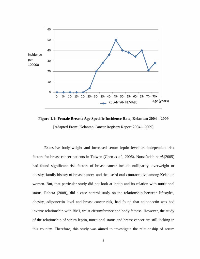
Peninsular Malaysia. According to Kelantan Cancer Registry Report (2004-2008), breast
cancer is the commonest among females with 439 cases with the peak age group was
between 45-49 years old. The youngest age of breast cancer diagnosed in this study was
24 years old.
5
Figure 1.1: Female Breast; Age Specific Incidence Rate, Kelantan 2004 – 2009
[Adapted From: Kelantan Cancer Registry Report 2004 – 2009]
Excessive body weight and increased serum leptin level are independent risk
factors for breast cancer patients in Taiwan (Chen et al., 2006). Norsa’adah et al.(2005)
had found significant risk factors of breast cancer include nulliparity, overweight or
obesity, family history of breast cancer and the use of oral contraceptive among Kelantan
women. But, that particular study did not look at leptin and its relation with nutritional
status. Rabeta (2008), did a case control study on the relationship between lifestyles,
obesity, adiponectin level and breast cancer risk, had found that adiponectin was had
inverse relationship with BMI, waist circumference and body fatness. However, the study
of the relationship of serum leptin, nutritional status and breast cancer are still lacking in
this country. Therefore, this study was aimed to investigate the relationship of serum
0
10
20
30
40
50
60
0- 5- 10- 15- 20- 25- 30- 35- 40- 45- 50- 55- 60- 65- 70- 75+
10 KELANTAN FEMALE
Incidence per 100000
Age (years)
6
leptin and nutritional status among breast cancer patients and controls. In hope that
identifying modifiable risk factors can lead to earlier screening and detection of breast
cancer and help in cancer prevention in this country.
1.3 Objectives
1.3.1 General Objective
To investigate the differences in concentrations of serum leptin and nutritional status in
newly diagnosed breast cancer patients in Hospital Universiti Sains Malaysia (Hospital
USM) and healthy women (control).
1.3.2 Specific Objectives
1. To measure and compare the nutritional status based on the data of
anthropometry, body composition and macronutrient intake among newly
diagnosed breast cancer patients and control.
2. To identify the association between serum leptin and anthropometry (Body
Mass Index, Body Impedance Analysis, and Waist Circumference) among
breast cancer patients and controls.
3. To determine the correlation between serum leptin and energy intake
among breast cancer patients and controls.

7
1.4 Study Hypotheses
1. Ho : No difference of anthropometry, body composition and macronutrient
intake among newly diagnosed breast cancer patients and control.
2. Ho : No association between serum leptin and anthropometry (BMI, BIA,
Waist Circumference) among breast cancer patients.
3. Ho : No association between serum leptin and energy intake among breast
cancer patients.
8
Breast Cancer
Retrospective (Before diagnosed with breast cancer)
Reproductive Factor Early menarche Nullyparity Late menopause Lactation
Breast Cancer Disease
Genetic
Family history Breast density
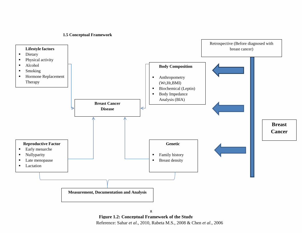
1.5 Conceptual Framework
Lifestyle factors Dietary Physical activity Alcohol Smoking Hormone Replacement
Therapy
Body Composition
Anthropometry (Wt,Ht,BMI)
Biochemical (Leptin) Body Impedance
Analysis (BIA)
Measurement, Documentation and Analysis
Figure 1.2: Conceptual Framework of the Study
Reference: Sahar et al., 2010, Rabeta M.S., 2008 & Chen et al., 2006
9
CHAPTER 2
LITERATURE REVIEW
2.1 Overview Incidence of Breast Cancer in Malaysia
The prevalence of Breast Cancer showed an increasing trend around the both developed
and developing world. In 2012, about 1.7 million people had been diagnosed with breast
cancer cases (World Cancer Research Fund, 2012). In Malaysia, National Cancer
Registry (NCR, 2007) reported that about 3242 female breast cancer cases were
diagnosed compared to 3525 new cases in 2006 (NCR, 2006). Even the number was
decreased; it was still the most common cancer in females and the first most common
cancer among population regardless of sex in Malaysia.
2.2 Stages of Breast Cancer
The early detection of breast cancer is very important in predicting the prognosis of a
woman with this disease. Patient with late stage of breast cancer are found with larger
cancer cells and are more likely to have already spread beyond the breast. In contrast, the
cancer cells that are found during screening mostly are smaller size and still in-situ. There
are three main activities for breast cancer screening in Malaysia which is Breast Self-
10
Awareness (BSA) or previously known as Breast Self-Examination (BSE), Clinical
Breast Examination (CBE) and mammography screening (Maznah et al., 2011).
Undetected breast cancer or not treated early, can spread to breast skin and lymph
nodes. Locally advanced breast cancer (LABC) includes breast cancers with large
primary tumors of more than 5 cm or those with skin and/or chest wall involvement, and
with or without regional lymph node involvement (Stage 3a, 3b and 3c). According to
Taiplin et al. (2004), 52% of advanced stage of breast cancer cases were related to
absence during screening programme, 39% related to absence during examination/check-
up and another 8% did not undergo follow up programme. The main problem with the
implementation of the programme are lack of capital and resources and low educational
level had caused no mammogram breast screening program for the whole high risk
women in Malaysia (Abdullah & Yip, 2004). Therefore, the numbers of breast cancer
patient with late stage are increasing due to late detection. Yip et al. (2006) had found that
almost 30-40% breast cancer patients in Malaysia were on stage 3 and 4.
Cancer stage is based on the size of the cancer, whether the cancer is invasive or
non-invasive, whether lymph nodes are involved, and whether the cancer has spread to
other places beyond the breast. The purpose of the staging system is to help organize the
different factors and some of the personality features of the cancer into categories in order
to best understand the prognosis or outcome of the disease, also to guide treatment
11
decisions of patient and help the patients to understand the results of the treatment
(Breastcancer.org, 2013).
The most common system used to describe the stages of breast cancer is the
American Joint Committee on Cancer (AJCC) TNM system (American Cancer Society,
2009). The TNM staging system classifies cancers based on their T, N, and M stages:
• The letter T followed by a number from 0 to 4 describes the tumor's size
and spread to the skin or to the chest wall under the breast. Higher T
numbers mean a larger tumor and/or wider spread to tissues near the
breast.
• The letter N followed by a number from 0 to 3 indicates whether the
cancer has spread to lymph nodes near the breast and, if so, how many
lymph nodes are affected.
• The letter M followed by a 0 or 1 indicates whether the cancer has spread
to distant organs -- for example, the lungs or bones.
Once the T, N, and M categories have been determined, this information is combined in a
process called stage grouping.
12
2.2.1: Stage 0
This is ductal carcinoma in situ (DCIS), a pre-cancer of the breast & lobular carcinoma in
situ (LCIS) sometimes also is classified as stage 0 breast cancer. In all cases, the cancer
has not spread to lymph nodes or distant sites.
2.2.2: Stage I
i. Stage IA:
The tumor is 2 cm (about 3/4 of an inch) or less across (T1) and has not spread to lymph
nodes (N0) or distant sites (M0).
ii.Stage IB:
The tumor is 2 cm or less across (or is not found) (T0 or T1) with micro metastases in 1 to
3 axillary lymph nodes (the cancer in the lymph nodes is greater than 0.2mm across
and/or more than 200 cells but is not larger than 2 mm)(N1mi). The cancer has not spread
to distant sites (M0).
2.2.3: Stage II
i. Stage IIA:
One of the following applies:
: The tumor is 2 cm or less across (or is not found) (T1 or T0) and either:
: It has spread to 1 to 3 axillary lymph nodes, with the cancer in the lymph nodes
larger than 2 mm across (N1a), OR
13
: Tiny amounts of cancer are found in internal mammary lymph nodes on sentinel
lymph node biopsy (N1b), OR
: It has spread to 1 to 3 lymph nodes under the arm and to internal mammary
lymphnodes (found on sentinel lymph node biopsy) (N1c). OR
: The tumor is larger than 2 cm but less than 5 cm across (T2) but hasn't spread to
the lymph nodes (N0). The cancer hasn't spread to distant sites (M0).
ii. Stage IIB:
One of the following applies:
: The tumor is larger than 2 cm but less than 5 cm across (T2). It has spread to 1 to
3 axillary lymph nodes and/or tiny amounts of cancer are found in internal
mammary lymph nodes on sentinel lymph node biopsy (N1). The cancer hasn't
spread to distant sites (M0). OR
: The tumor is larger than 5 cm across but does not grow into the chest wall or
skin and has not spread to lymph nodes (T3, N0). The cancer hasn't spread to
distant sites (M0).
14
2.2.4: Stage III
i. Stage IIIA:
One of the following applies:
: The tumor is not more than 5 cm across (or cannot be found) (T0 to T2). It has
spread to 4 to 9 axillary lymph nodes, or it has enlarged the internal mammary
lymph nodes (N2). The cancer hasn't spread to distant sites (M0). OR
: The tumor is larger than 5 cm across but does not grow into the chest wall or
skin (T3). It has spread to 1 to 9 axillary nodes, or to internal mammary nodes
(N1 or N2). The cancer hasn't spread to distant sites (M0).
ii. Stage IIIB:
The tumor has grown into the chest wall or skin (T4), and one of the following applies:
: It has not spread to the lymph nodes (N0).
: It has spread to 1 to 3 axillary lymph nodes and/or tiny amounts of cancer are
found in internal mammary lymph nodes on sentinel lymph node biopsy (N1).
: It has spread to 4 to 9 axillary lymph nodes, or it has enlarged the internal
mammary lymph nodes (N2).
15
iii. Stage IIIC:
Any T, N3, M0: The tumor is any size (or can't be found), and one of the following
applies:
: Cancer has spread to 10 or more axillary lymph nodes (N3).
: Cancer has spread to the lymph nodes under the clavicle (collar bone) (N3).
: Cancer has spread to the lymph nodes above the clavicle (N3).
: Cancer involves axillary lymph nodes and has enlarged the internal mammary
lymph nodes (N3).
: Cancer has spread to 4 or more axillary lymph nodes, and tiny amounts of cancer
are found in internal mammary lymph nodes on sentinel lymph node biopsy
(N3).
: The cancer hasn't spread to distant sites (M0).
2.2.5: Stage IV
Any T, any N, M1: The cancer can be of any size (any T) and may or may not have
spread to nearby lymph nodes (any N). It has spread to distant organs or to lymph nodes
far from the breast (M1). The most common sites of spread are the bone, liver, brain, or
lung.
16
2.3 Breast Cancer Risk Factors
The risk factors for breast cancer in Western populations had been extensively
investigated, and it has been suggested that lifestyle-related and reproductive factors were
strongly associated with breast cancer (Norsa’adah et al., 2005). All women should
understand the risk factor and update knowledge regarding breast cancer. Norsa’adah et
al. (2005) had found that similar risk factor identified in Western populations were
responsible for the occurrence of breast cancer in Kelantan. However, the information
regarding risk factor of breast cancer is still low among Asian women.
There were numerous risk factors that can contribute to breast cancer and it can be
divided into preventable and non-preventable. Some examples of non-preventable risk
factors were genetic, breast density, reproductive history, family history, age and prior
breast procedure. While, preventable risk factors were dietary habits, obesity, alcohol use
and physical activity. Some of the breast cancer risks have a different role among pre
menopause and post menopause women. In both premenopausal and postmenopausal
women cancer risk is associated with events that alter hormonal balance, such as age at
menarche, parity, body weight, body fat distribution, and use of exogenous hormones
(Hong et al., 2004).
17
2.3.1 Preventable Risk Factors
Preventable risk factors are the risks that can be controlled. Many epidemiological studies
have looked at the relationship between behavior and lifestyle variables on the incidence
of breast cancer (Lemon et al., 2004).
2.3.1.1 Overweight and obesity
Dietary habit and lifestyle change have resulted in an increased number of obese people.
Obesity is a condition in which a person has an abnormally high and unhealthy proportion
of body fat. World Health Organization (WHO) defines obesity in terms of a scale known
as the body mass index (BMI). BMI provides a more accurate measure of obesity or being
overweight than weight alone (King, 2011). High BMI is generally interpreted as excess
adiposity (overweight or obesity). Obesity is related to many life-threatening diseases
such as cancer diabetes and cardiovascular disease. It also contributes to an acceleration
of the aging process, shortening of the lifespan, depression, and a decrease in quality of
life (Romao et al., 2008).
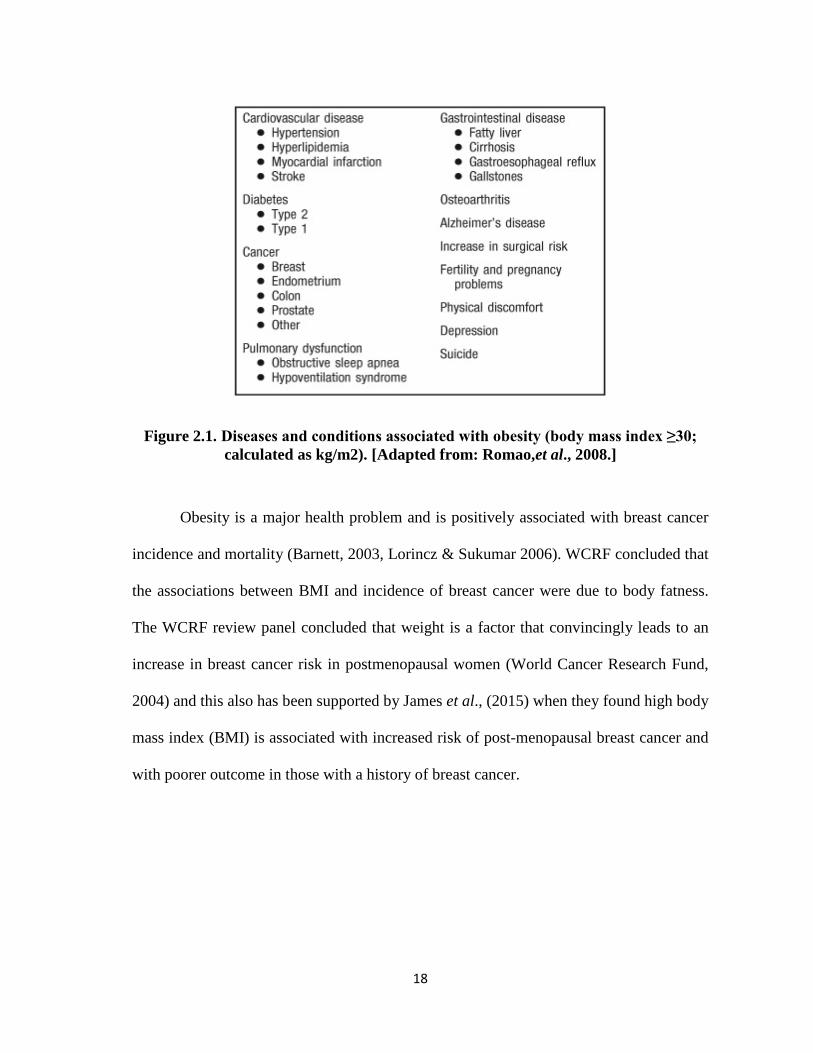
18
Figure 2.1. Diseases and conditions associated with obesity (body mass index ≥30; calculated as kg/m2). [Adapted from: Romao,et al., 2008.]
Obesity is a major health problem and is positively associated with breast cancer
incidence and mortality (Barnett, 2003, Lorincz & Sukumar 2006). WCRF concluded that
the associations between BMI and incidence of breast cancer were due to body fatness.
The WCRF review panel concluded that weight is a factor that convincingly leads to an
increase in breast cancer risk in postmenopausal women (World Cancer Research Fund,
2004) and this also has been supported by James et al., (2015) when they found high body
mass index (BMI) is associated with increased risk of post-menopausal breast cancer and
with poorer outcome in those with a history of breast cancer.
19
Relationship between breast cancer and obesity is not in a direct proportion
(Carmichael & Bates, 2004). However, the mechanism of body weight affected breast
cancer involved complex interactions between obesity, tumor biology and physiological
function of the breast (Rose et al., 2002).
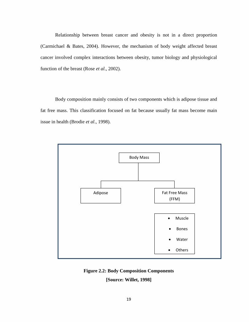
Body composition mainly consists of two components which is adipose tissue and
fat free mass. This classification focused on fat because usually fat mass become main
issue in health (Brodie et al., 1998).
Figure 2.2: Body Composition Components
[Source: Willet, 1998]
• Muscle
• Bones
• Water
• Others
Body Mass
Adipose Fat Free Mass (FFM)
20
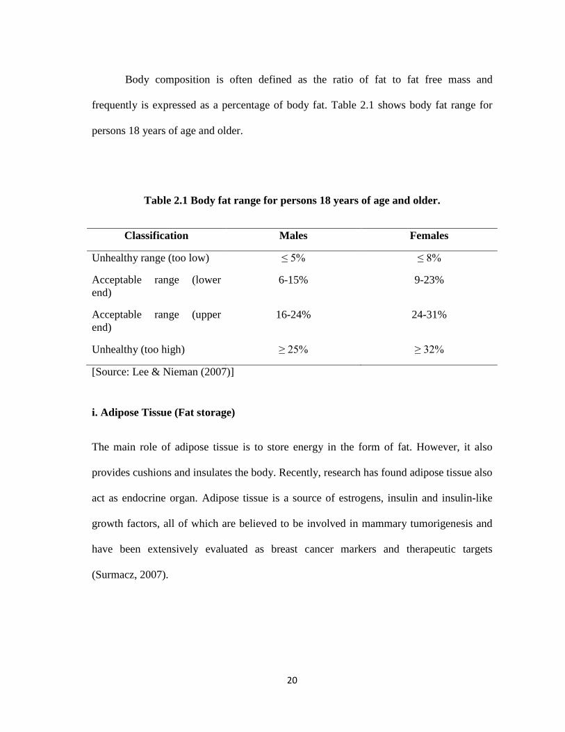
Body composition is often defined as the ratio of fat to fat free mass and
frequently is expressed as a percentage of body fat. Table 2.1 shows body fat range for
persons 18 years of age and older.
Table 2.1 Body fat range for persons 18 years of age and older.
Classification Males Females
Unhealthy range (too low) ≤ 5% ≤ 8%
Acceptable range (lower end)
6-15% 9-23%
Acceptable range (upper end)
16-24% 24-31%
Unhealthy (too high) ≥ 25% ≥ 32%
[Source: Lee & Nieman (2007)]
i. Adipose Tissue (Fat storage)
The main role of adipose tissue is to store energy in the form of fat. However, it also
provides cushions and insulates the body. Recently, research has found adipose tissue also
act as endocrine organ. Adipose tissue is a source of estrogens, insulin and insulin-like
growth factors, all of which are believed to be involved in mammary tumorigenesis and
have been extensively evaluated as breast cancer markers and therapeutic targets
(Surmacz, 2007).
21
Several possible mechanisms have been suggested to explain the association of
obesity with increased risk of breast cancer. One of them is excess amounts of estrogen
production by fat cells. The increased risk of postmenopausal breast cancer is thought to
be due to increased levels of estrogen in obese women. After menopause, when the
ovaries stop producing hormones, fat tissue becomes the most important source of
estrogen. Because obese women have more fat tissue, their estrogen levels are higher,
potentially leading to more rapid growth of estrogen-responsive breast tumors (James,
2015 & American Cancer Society, 2009).
Obese postmenopausal women have increased circulating insulin levels. Insulin
stimulates the growth of breast-cancer cell lines and may increase breast cancer risk
(Kaaks, 1996). Insulin-like growth factors (IGFs) are peptides that help regulate tissue
growth and fat deposition (Stoll, 2000). One of these, IGF-1, stimulates mitosis in breast
cancer cells. IGF-1 levels may be higher among obese women (Lipworth, 1996).
ii. Fat Free Mass
Fat free mass (FFM) is the amount of lean body. It is composed of muscle, water, bone,
and other tissues devoid of fat and lipid (Lee & Nieman, 2007).
22
iii. Leptin
Fat cells produce hormones, called adipokines that may stimulate or inhibit cell growth
(Jarde, 2011). The plasma leptin concentrations increased in direct proportion to the
adipose mass (Zhang et al., 2005). These two hormones were leptin and adiponectin.
Leptin is a multifunctional hormone produced mainly by the adipose tissue and involved
in the regulation of food intake and energy balance. It is more abundant in obese people,
seems to promote cell proliferation, whereas adiponectin, which is less abundant in obese
people, may have antiproliferative effects (Garotalo, 2006). Jarde (2011) had found leptin
also secreted by epithelial tissue of breast tumor. Recent evidence suggests that leptin also
plays an important role in normal mammary development and mammary tumor
formation. Perrera (2008) suggested that leptin promotes mammary tumor growth through
multiple mechanisms, including regulating the cell cycle, apoptosis, and by modulating
the extracellular environment. There is increasing evidence that targeting the
adiponectin:leptin ratio might be a new prognostic and/or therapeutic strategy for
postmenopausal breast cancer (Jarde, 2011). Wu (2009) has shown that leptin was
correlated with breast cancer risk.
2.3.1.2 Diet
There are a few dietary factors related to breast cancer risk. Among them are fiber intake
and dietary fat. Unraveling links between diet and cancer is complex, as thousands of
dietary components are consumed each day; a typical diet may provide more than 25000
bioactive food constituents (FAO, 2003). As shown in Figure 2.3, food can influence
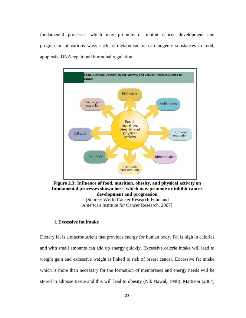
23
fundamental processes which may promote or inhibit cancer development and
progression at various ways such as metabolism of carcinogenic substances in food,
apoptosis, DNA repair and hormonal regulation.
Figure 2.3: Influence of food, nutrition, obesity, and physical activity on
fundamental processes shown here, which may promote or inhibit cancer development and progression
[Source: World Cancer Research Fund and American Institute for Cancer Research, 2007]
i. Excessive fat intake
Dietary fat is a macronutrient that provides energy for human body. Fat is high in calories
and with small amounts can add up energy quickly. Excessive calorie intake will lead to
weight gain and excessive weight is linked to risk of breast cancer. Excessive fat intake
which is more than necessary for the formation of membranes and energy needs will be
stored in adipose tissue and this will lead to obesity (Nik Nawal, 1998). Mattison (2004)
Food, Nutrition,Obesity,Physical Activity and Cellular Processes Linked to cancer
24
had showed statistically significant increased risk of breast cancer with increasing fat
intake.
Dietary fat is relatively well established as a cause of increased endogenous
estrogen production (Wu, 1999). Higher endogenous estrogen levels after menopause are
a known cause of breast cancer (Key, 2002). Low-fat diets are usually associated with
high fiber intake, which may reduce estrogen concentration by decreasing intestinal
reabsorption. Another alternative mechanism by which dietary fat could influence steroid
hormone levels is that increased serum-free fatty acids could displace estradiol from
serum albumin, thus increasing free estradiol concentration (Burning, 1986). Sex
hormone binding globulin decreases with increasing body mass index and insulin
resistance. Other important factor that related with fat is energy-dense lower the age of
menarche and early menarche is an established risk factor for breast cancer (American
Cancer Society, 2007).
ii. Lack of fiber
Dietary fiber is all parts of plant foods that cannot be digested or absorbed by human
body. Fiber can provide many health benefits such as helping to maintain a healthy
weight and lower risk of diabetes, cancer and heart disease. Aune (2011) suggested that
diets rich in fiber are associated with reduced breast cancer risk. Dietary fiber has been
hypothesized to reduce breast cancer risk based on observations that vegetarian women


![Untitled-2 [kakapvc.com] · gold lilan a 137 . kak . kak a 210 Élackgrill grill white star . kak . brown l a 136 white wall . kak . poly plast pvc profile asian beech w.b.s. poly](https://static.fdocuments.us/doc/165x107/613cad764c23507cb63589a7/untitled-2-gold-lilan-a-137-kak-kak-a-210-lackgrill-grill-white-star-.jpg)